Embed Size (px)
Citation preview
Team DMohammed Zuned DesaiAreio Barzan Hashemi
Koji HirotaMichael James Wong
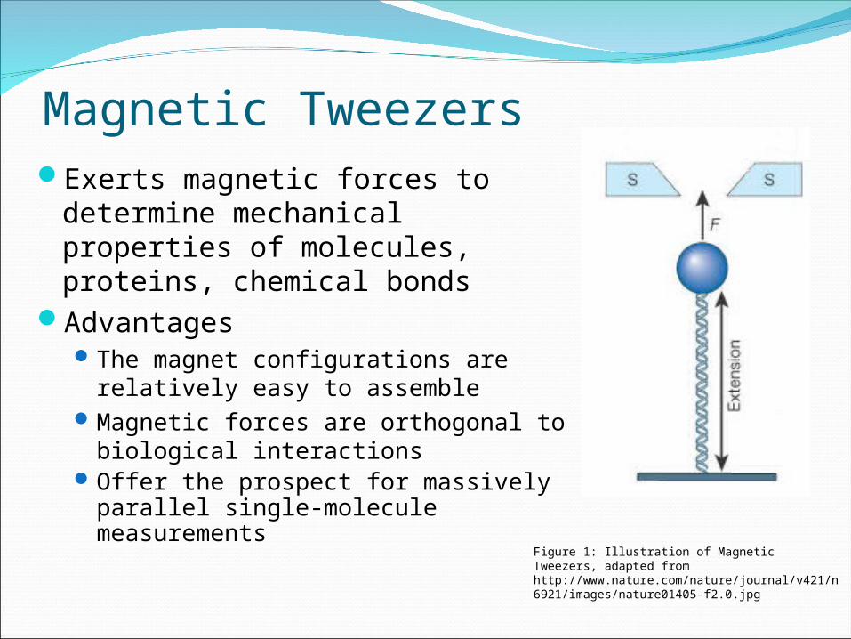
Magnetic TweezersExerts magnetic forces to
determine mechanical properties of molecules, proteins, chemical bonds
AdvantagesThe magnet configurations are
relatively easy to assembleMagnetic forces are orthogonal to
biological interactionsOffer the prospect for massively
parallel single-molecule measurements
Figure 1: Illustration of Magnetic Tweezers, adapted from http://www.nature.com/nature/journal/v421/n6921/images/nature01405-f2.0.jpg
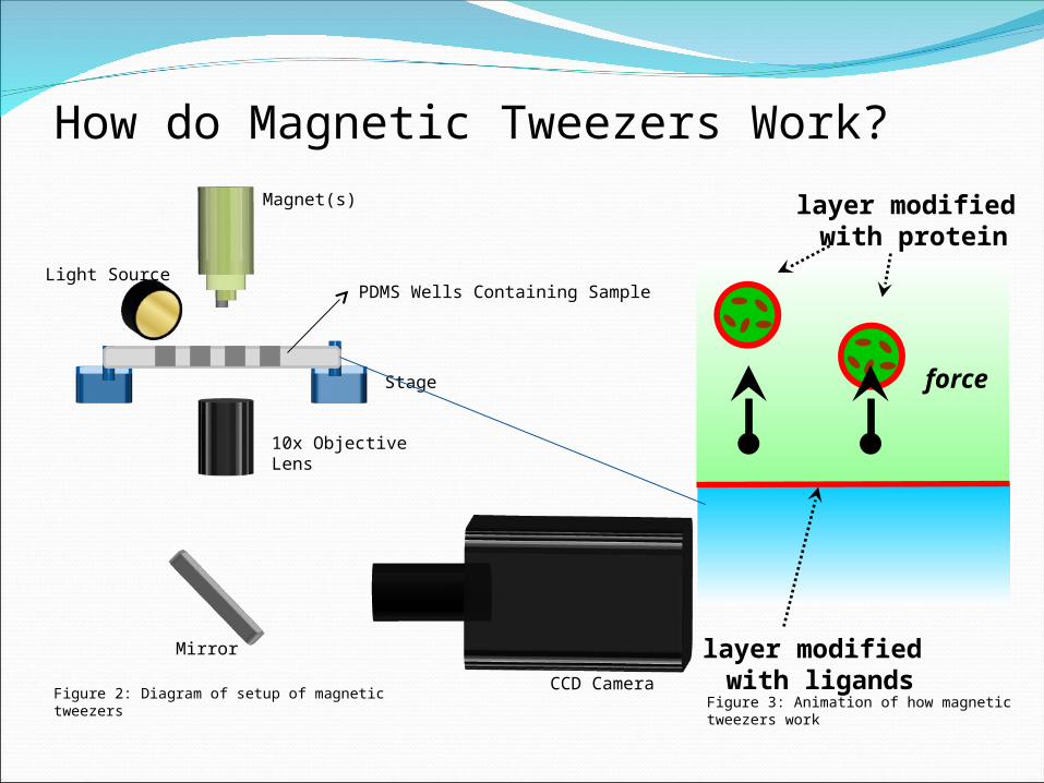
layer modified with protein
force
How do Magnetic Tweezers Work?Magnet(s)
Stage
PDMS Wells Containing Sample
10x Objective Lens
CCD Camera
Light Source
Mirror
Figure 2: Diagram of setup of magnetic tweezersFigure 3: Animation of how magnetic tweezers work
layer modified with ligands
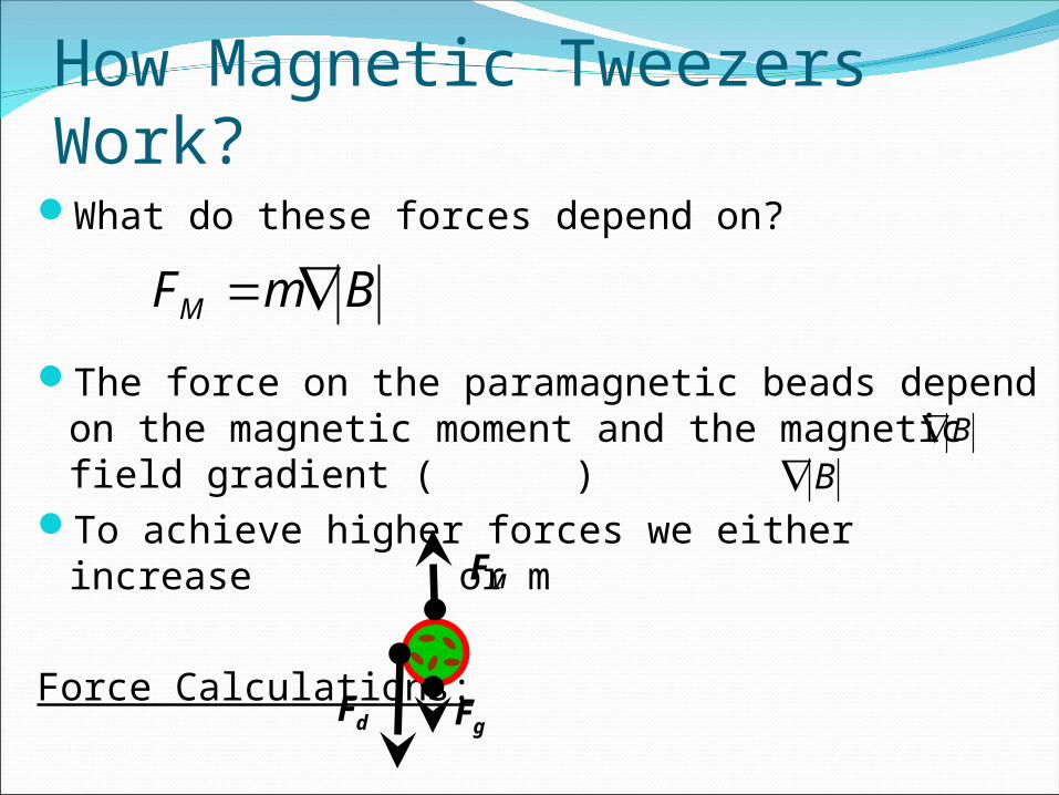
How Magnetic Tweezers Work?What do these forces depend on?
The force on the paramagnetic beads depend on the magnetic moment and the magnetic field gradient ( )
To achieve higher forces we either increase or m
Force Calculations:
BmFM
BB
Fd
Fg
FM
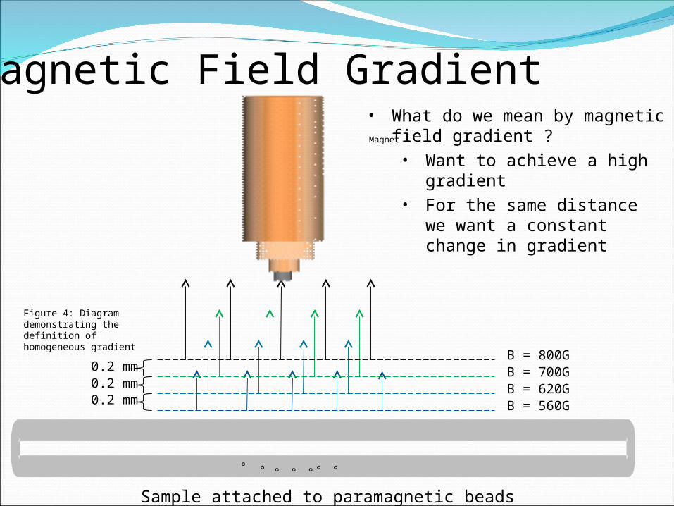
0.2 mm0.2 mm0.2 mm
B = 800GB = 700GB = 620GB = 560G
Magnet
Sample attached to paramagnetic beads
Magnetic Field Gradient• What do we mean by magnetic
field gradient ?• Want to achieve a high
gradient• For the same distance we
want a constant change in gradient
Figure 4: Diagram demonstrating the definition of homogeneous gradient
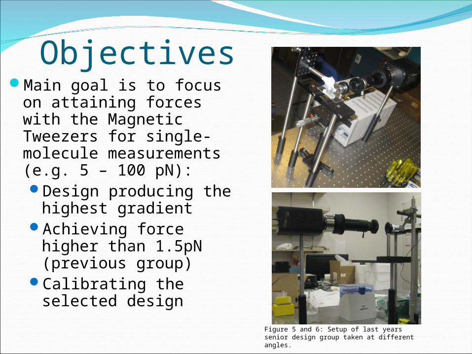
ObjectivesMain goal is to focus on
attaining forces with the Magnetic Tweezers for single-molecule measurements (e.g. 5 – 100 pN):Design producing the
highest gradient Achieving force higher
than 1.5pN (previous group)
Calibrating the selected design
Figure 5 and 6: Setup of last years senior design group taken at different angles.
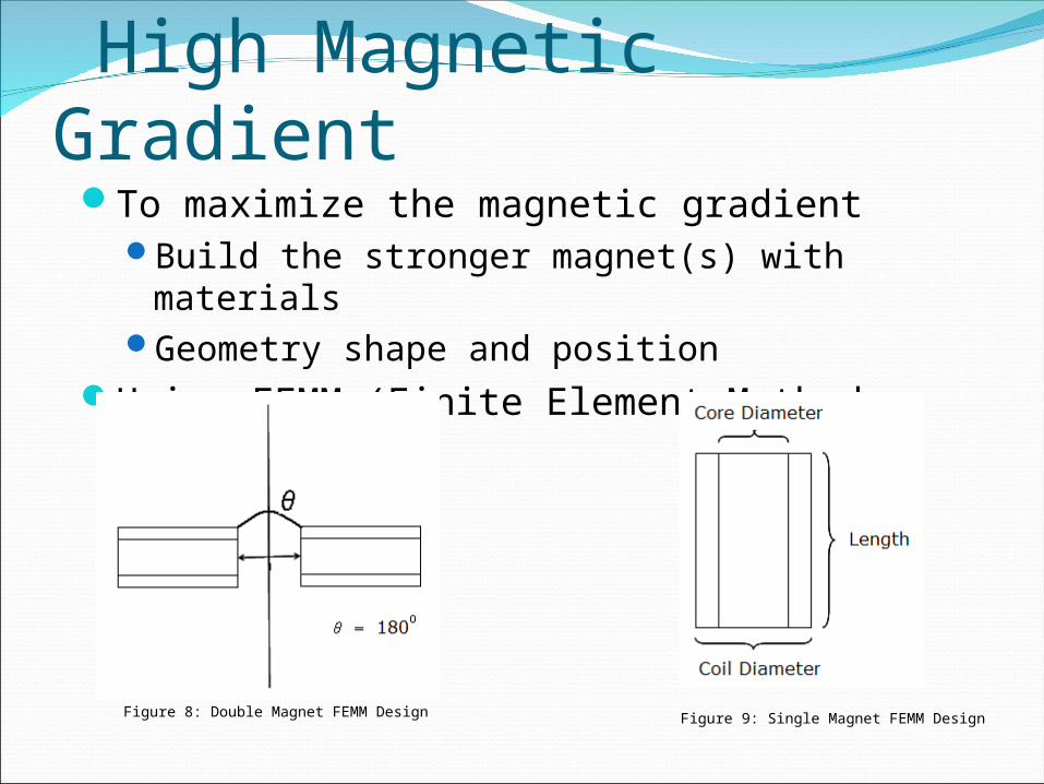
High Magnetic GradientTo maximize the magnetic gradient
Build the stronger magnet(s) with materialsGeometry shape and position
Using FEMM (Finite Element Method Magnetics)
Figure 8: Double Magnet FEMM Design Figure 9: Single Magnet FEMM Design
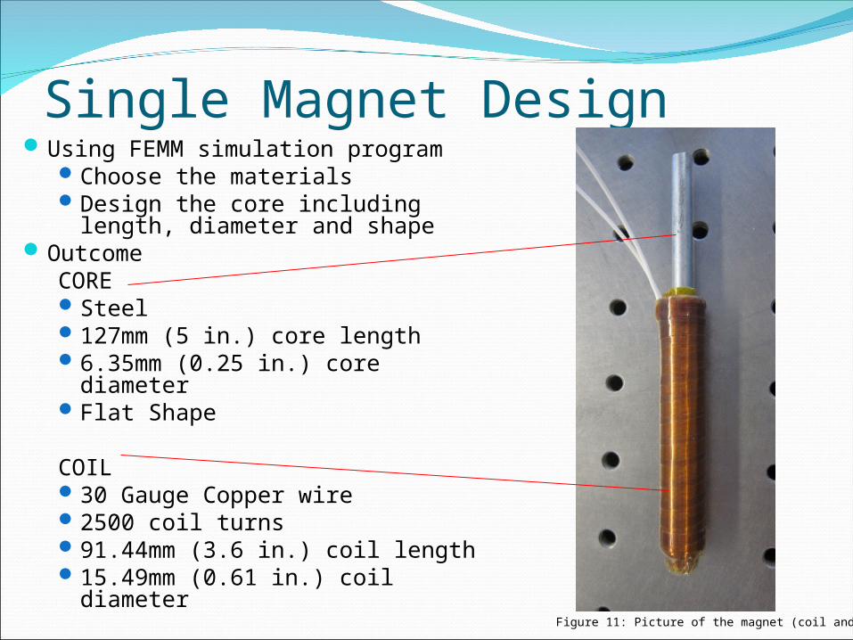
Single Magnet DesignUsing FEMM simulation program
Choose the materialsDesign the core including
length, diameter and shapeOutcome
CORESteel127mm (5 in.) core length 6.35mm (0.25 in.) core diameterFlat Shape
COIL30 Gauge Copper wire2500 coil turns91.44mm (3.6 in.) coil length15.49mm (0.61 in.) coil diameter
Figure 11: Picture of the magnet (coil and core)
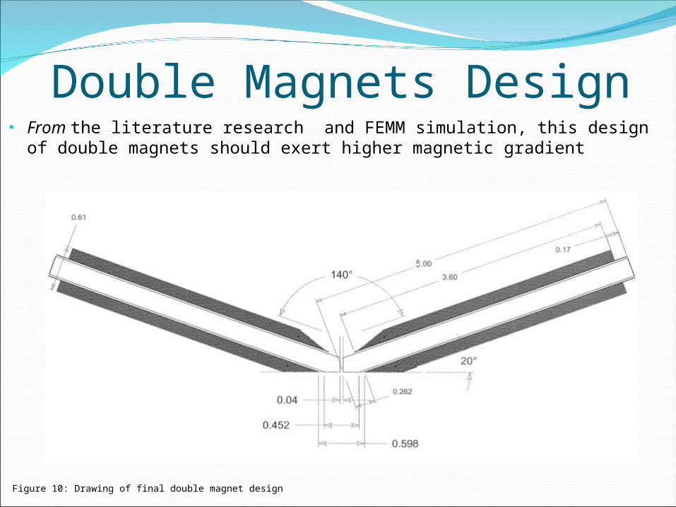
Double Magnets Design
Figure 10: Drawing of final double magnet design
• From the literature research and FEMM simulation, this design of
double magnets should exert higher magnetic gradient
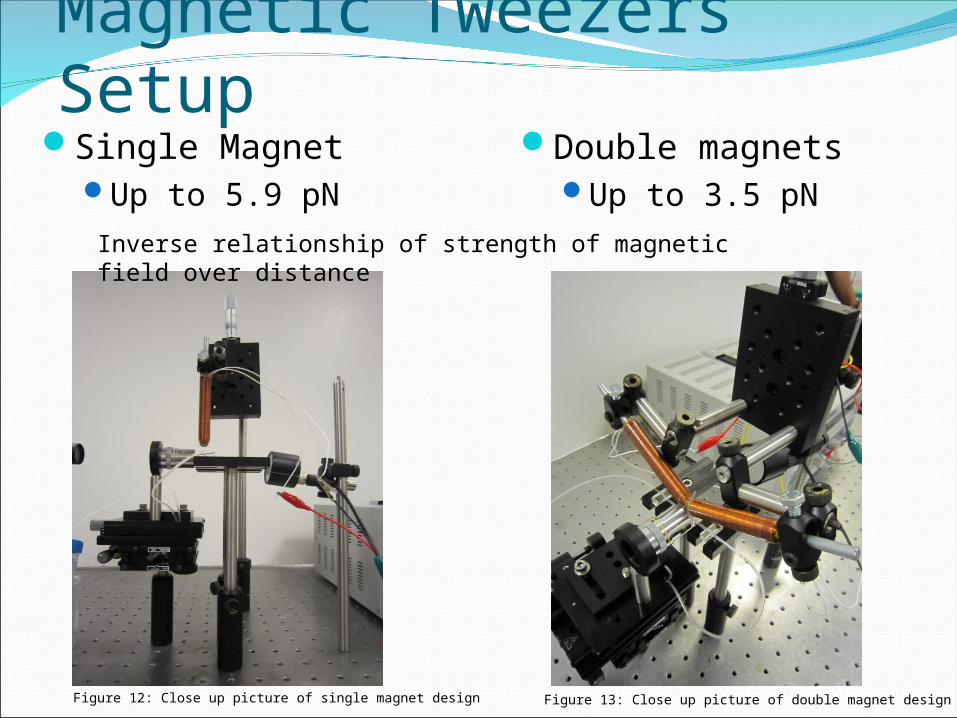
Magnetic Tweezers Setup Single Magnet
Up to 5.9 pN
Double magnetsUp to 3.5 pN
Figure 12: Close up picture of single magnet design Figure 13: Close up picture of double magnet design
Inverse relationship of strength of magnetic field over distance
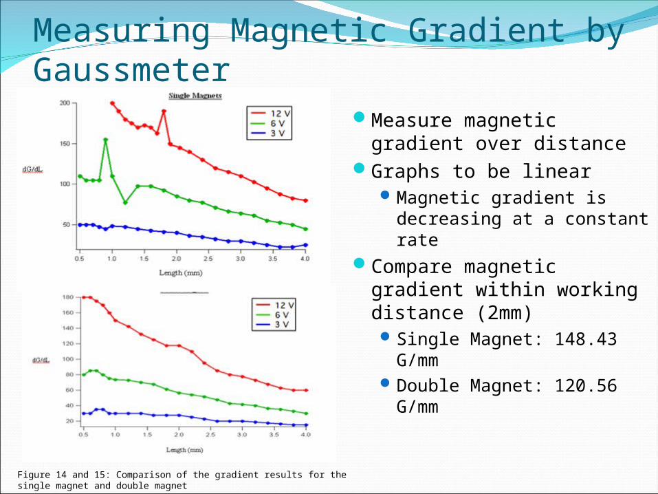
Measuring Magnetic Gradient by Gaussmeter
Measure magnetic gradient over distance
Graphs to be linearMagnetic gradient is
decreasing at a constant rate
Compare magnetic gradient within working distance (2mm) Single Magnet: 148.43
G/mm Double Magnet: 120.56
G/mm
Figure 14 and 15: Comparison of the gradient results for the single magnet and double magnet
Magnet
Stage
1mm Capillary Tube With Paramagnetic Beads
10x Objective Lens
CCD Camera
Light Source
Magnets
Stage
1mm Capillary Tube With Paramagnetic Beads
10x Objective Lens
CCD Camera
Light Source
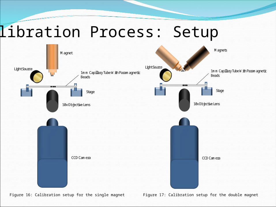
Calibration Process: Setup
Figure 16: Calibration setup for the single magnet Figure 17: Calibration setup for the double magnet
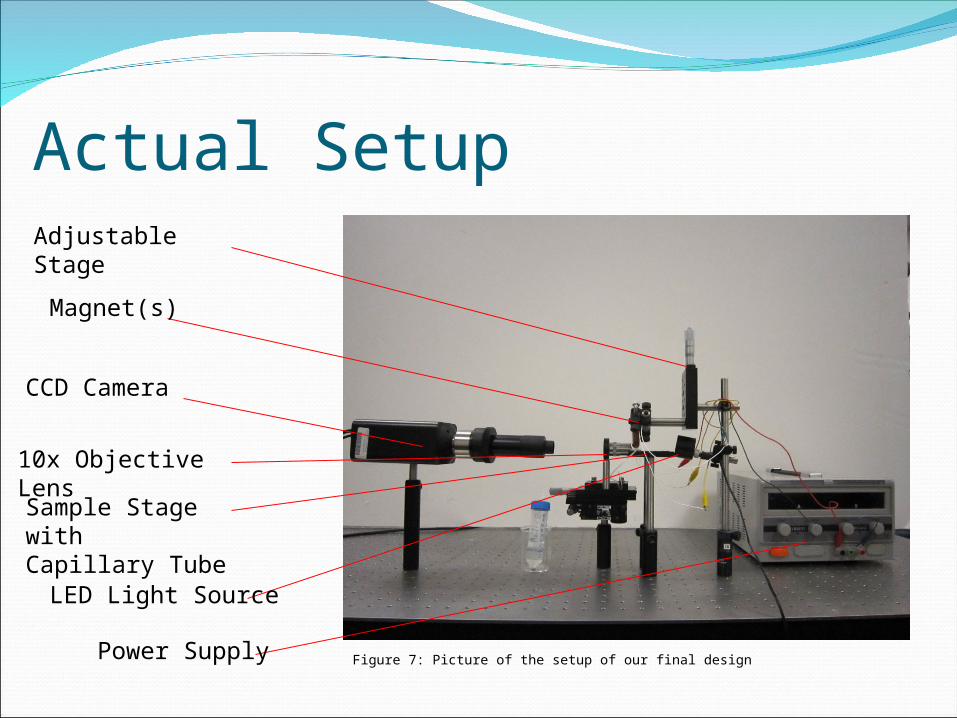
Actual Setup
Power Supply
Adjustable Stage
Magnet(s)
CCD Camera
10x Objective Lens
LED Light Source
Sample Stage withCapillary Tube
Figure 7: Picture of the setup of our final design
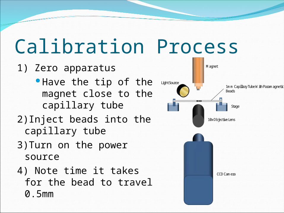
Calibration Process1) Zero apparatus
Have the tip of the magnet close to the capillary tube
2)Inject beads into the capillary tube
3)Turn on the power source
4) Note time it takes for the bead to travel 0.5mm
Magnet
Stage
1mm Capillary Tube With Paramagnetic Beads
10x Objective Lens
CCD Camera
Light Source
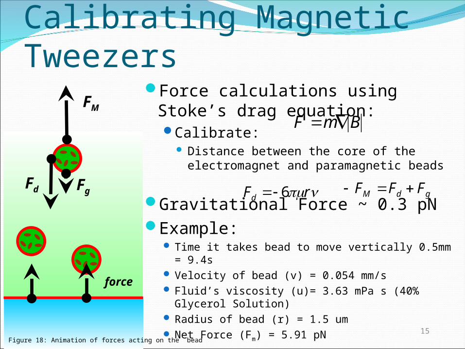
Calibrating Magnetic TweezersForce calculations using Stoke’s
drag equation:Calibrate:
Distance between the core of the electromagnet and paramagnetic beads
Gravitational Force ~ 0.3 pNExample:
Time it takes bead to move vertically 0.5mm = 9.4s
Velocity of bead (v) = 0.054 mm/s Fluid’s viscosity (u)= 3.63 mPa s (40% Glycerol
Solution) Radius of bead (r) = 1.5 um Net Force (Fm) = 5.91 pN
rFd 6
15
gdM FFF
F mB
force
Fd Fg
FM
Figure 18: Animation of forces acting on the bead
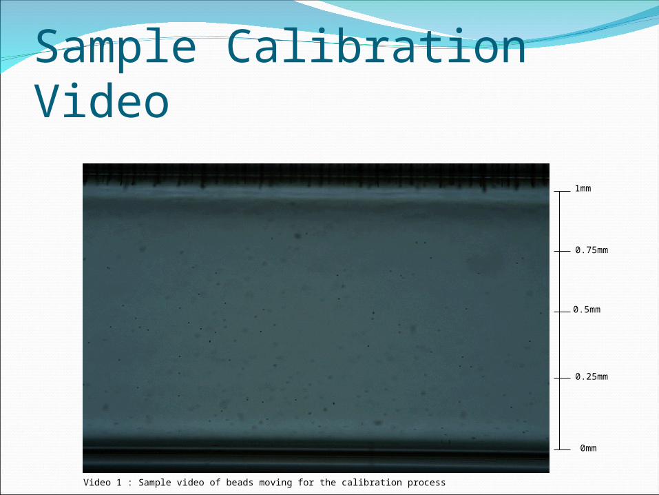
Sample Calibration Video
0.25mm
0.5mm
0.75mm
1mm
0mm
Video 1 : Sample video of beads moving for the calibration process
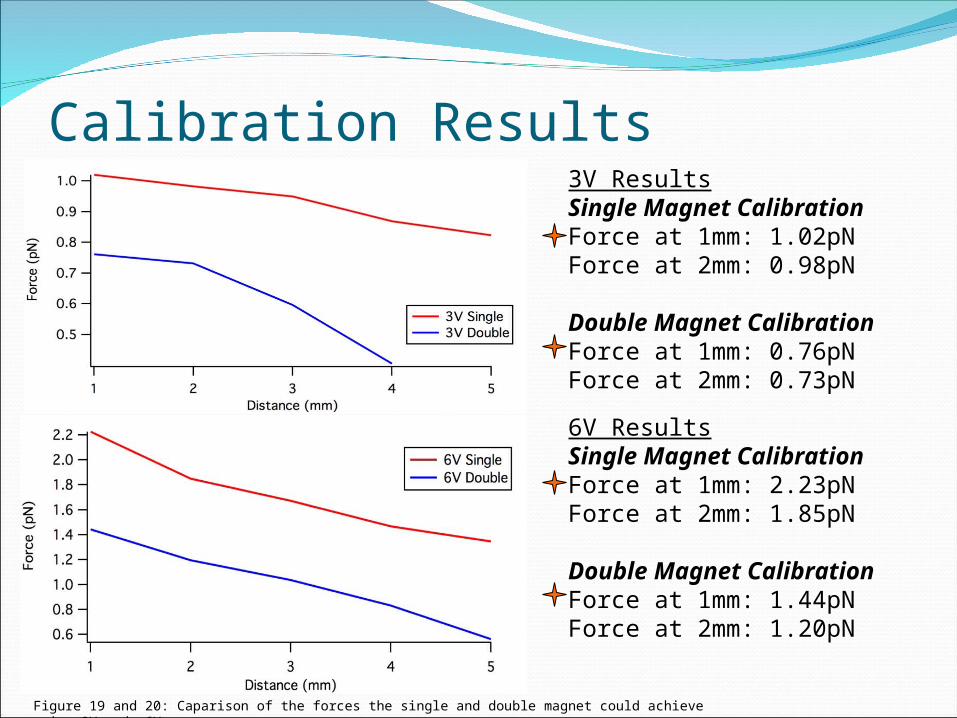
Calibration Results 3V ResultsSingle Magnet CalibrationForce at 1mm: 1.02pNForce at 2mm: 0.98pN
Double Magnet CalibrationForce at 1mm: 0.76pNForce at 2mm: 0.73pN
6V ResultsSingle Magnet CalibrationForce at 1mm: 2.23pNForce at 2mm: 1.85pN
Double Magnet CalibrationForce at 1mm: 1.44pNForce at 2mm: 1.20pN
Figure 19 and 20: Caparison of the forces the single and double magnet could achieve using 3V and 6V
Calibration Results
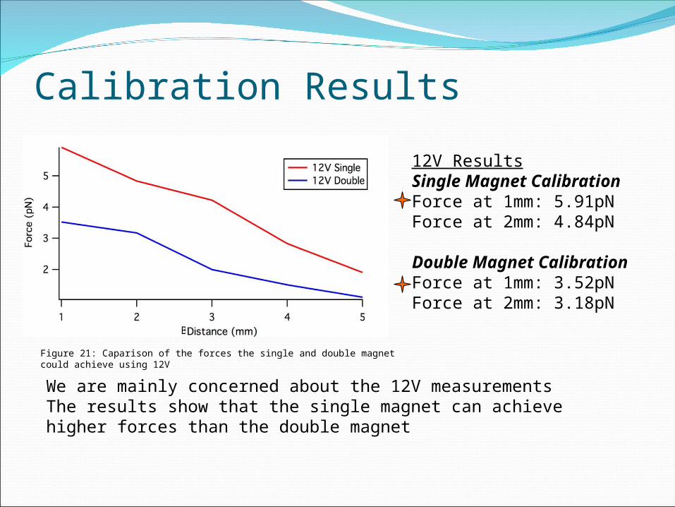
12V ResultsSingle Magnet CalibrationForce at 1mm: 5.91pNForce at 2mm: 4.84pN
Double Magnet CalibrationForce at 1mm: 3.52pNForce at 2mm: 3.18pN
We are mainly concerned about the 12V measurementsThe results show that the single magnet can achieve higher forces than the double magnet
Figure 21: Caparison of the forces the single and double magnet could achieve using 12V
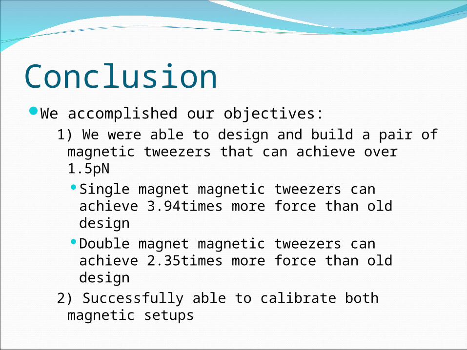
ConclusionWe accomplished our objectives:
1) We were able to design and build a pair of magnetic tweezers that can achieve over 1.5pNSingle magnet magnetic tweezers can achieve
3.94times more force than old designDouble magnet magnetic tweezers can achieve
2.35times more force than old design2) Successfully able to calibrate both magnetic
setups
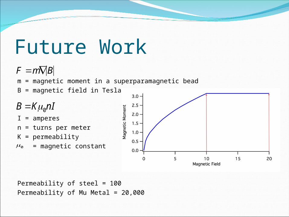
Future Workm = magnetic moment in a superparamagnetic bead B = magnetic field in Tesla
I = amperesn = turns per meterK = permeability = magnetic constant
Permeability of steel = 100Permeability of Mu Metal = 20,000
F mB
0
B K0nI
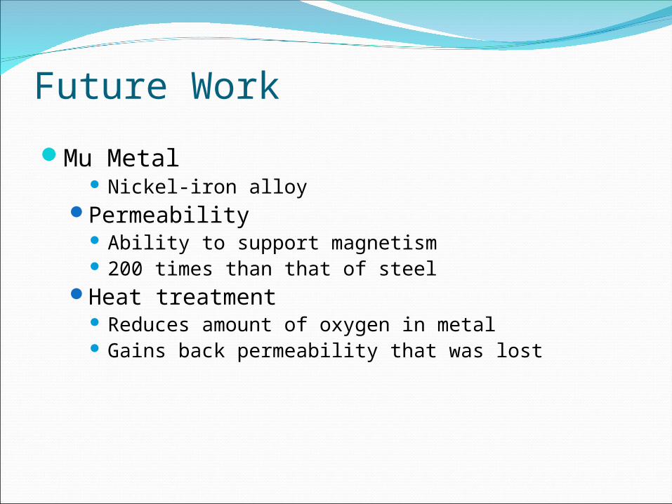
Future Work
Mu Metal Nickel-iron alloy
Permeability Ability to support magnetism 200 times than that of steel
Heat treatment Reduces amount of oxygen in metal Gains back permeability that was lost
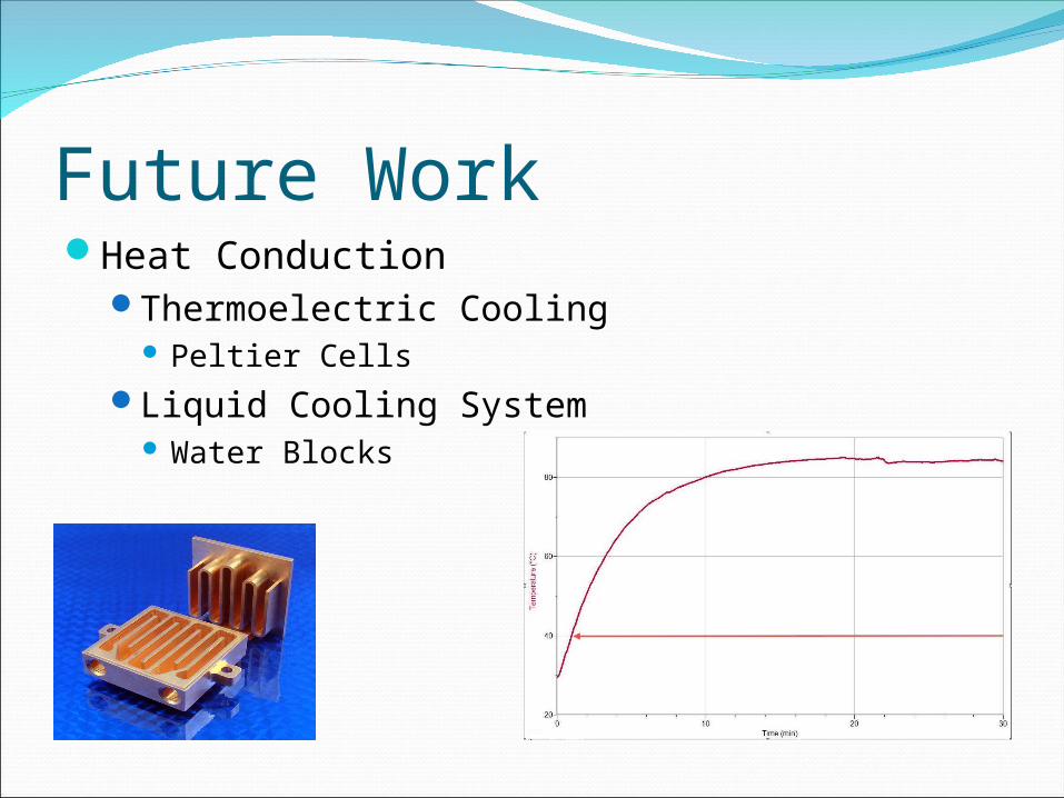
Future WorkHeat Conduction
Thermoelectric Cooling Peltier Cells
Liquid Cooling System Water Blocks
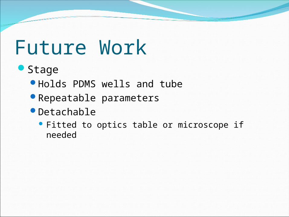
Future WorkStage
Holds PDMS wells and tubeRepeatable parametersDetachable
Fitted to optics table or microscope if needed

AcknowledgmentsDr. Valentine VullevDr. Sharad GuptaDr. Hyle ParkDr. Jerome SchultzGokul UpadhyayulaHong Xu
References 1) Neuman, Keri C, and Nagy, Attila. “Single-molecule force spectroscopy:
optical tweezers, magnetic tweezers and atomic force microscopy.” Nature Publishing Group Vol. 5, NO. 6. June 2008.
2) Danilowicz, Claudia, Greefield, Derek and Prentiss, Mara. “Dissociation of Ligand-Receptor Complexes Using Magnetic Tweezers.” Analytical Chemistry Vol. 77, No. 10. 15 May. 2005.
3) Humphries; David E., Hong; Seok-Cheol, Cozzarelli; Linda A., Pollard; Martin J., Cozzarelli; Nicholas R. “Hybrid magnet devices fro molecule manipulation and small scale high gradient-field applications”. United States Patent and Trademark Office, An Agency of The United States Department of Commerce. <http://patft.uspto.gov>. January 6, 2009.
4) Ibrahim, George; Lu, Jyann-Tyng; Peterson, Katie; Vu, Andrew; Gupta, Dr. Sharad; Vullev, Dr. Valentine. “Magnetic Tweezers for Measuring Forces.” University of California Riverside. Bioengineering Senior Design June 2009.
5) Startracks Medical, “Serves Business, Education, Government and Medical Facilities Worldside.” American Solution. Startracks.org, Inc. Copyright 2003. <http://images.google.com/imgres?imgurl=http://www.startracksmedical.com/supplies/invertedmicroscope.jpg&imgrefurl=http://www.startracksmedical.com/supplies.html&usg=__butCY2zWJa7nAkwkjiPxX_mFy0=&h=450&w=450&sz=24&hl=en&start=2&um=1&tbnid=XH6gnQuJLS7bRM:&tbnh=127&tbnw=127&prev=/images%3Fq%3Dinverted%2Bmicroscope%26hl%3Den%26sa%3DN%26um%3D1>
6) Janshoff A, Neitzert M, Oberdorfer Y, Fuchs H. Force spectroscopy of molecular systems-single molecule spectroscopy of polymers and biomolecules. Angew Chem Int Ed 2000;39:3212-3237.

Questions