Embed Size (px)
Citation preview
1
Rehabilitation of disorders of consciousness after traumatic brain
injuryBiennial Conference of the Brain Injury and
Families/European Confederation (BIF)
Umberto León-DomínguezAutonomous University of Madrid, Spain
Center for Brain Injury Rehabilitation (C.RE.CER.), Seville, Spain
Vienna International Congress
Vienna Medical Academy, Sep. 19th – Sep. 21st, 2013
Departament of Psychiatry, Medicine SchoolLaboratory of consciousness and sleep
Center for Brain Injury Rehabilitation CRECER
Scheme Brief summary of topic
Etiology Diagnosis Treatment
Articles about the treatment of Disorder of consciousness Treatment of DOC: A single case study Cortical connectivity in MCS and SND
Conclusions
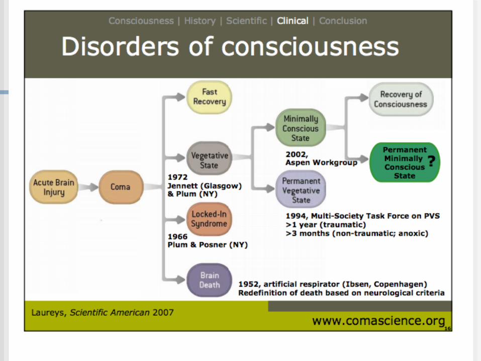
2
3
What is a Disorder Of Consciousness (DOC)?
Disorders of consciousness are amedical or functional condition
that inhibit consciousness.
4
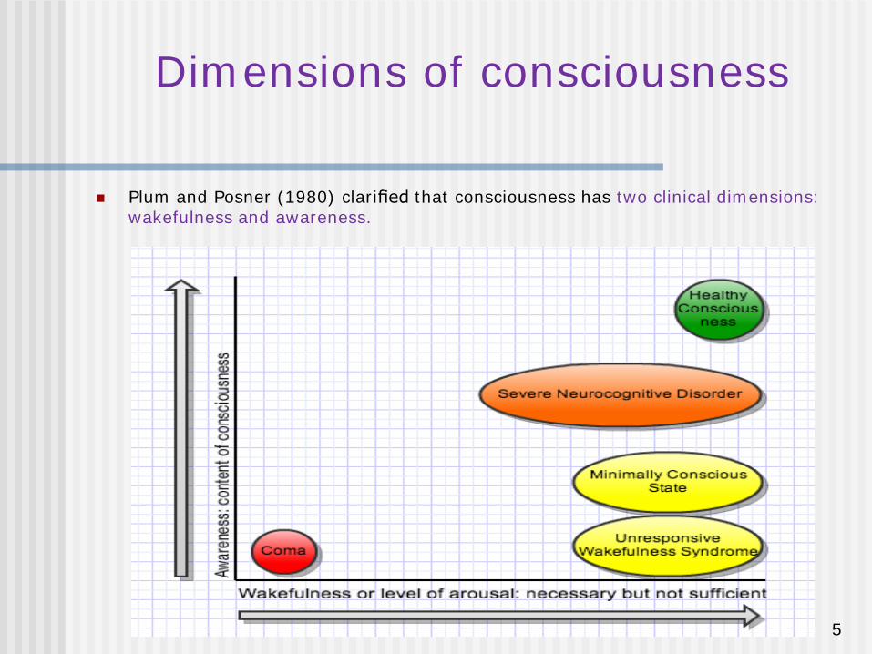
Dimensions of consciousness
Plum and Posner (1980) clarified that consciousness has two clinical dimensions: wakefulness and awareness.
5
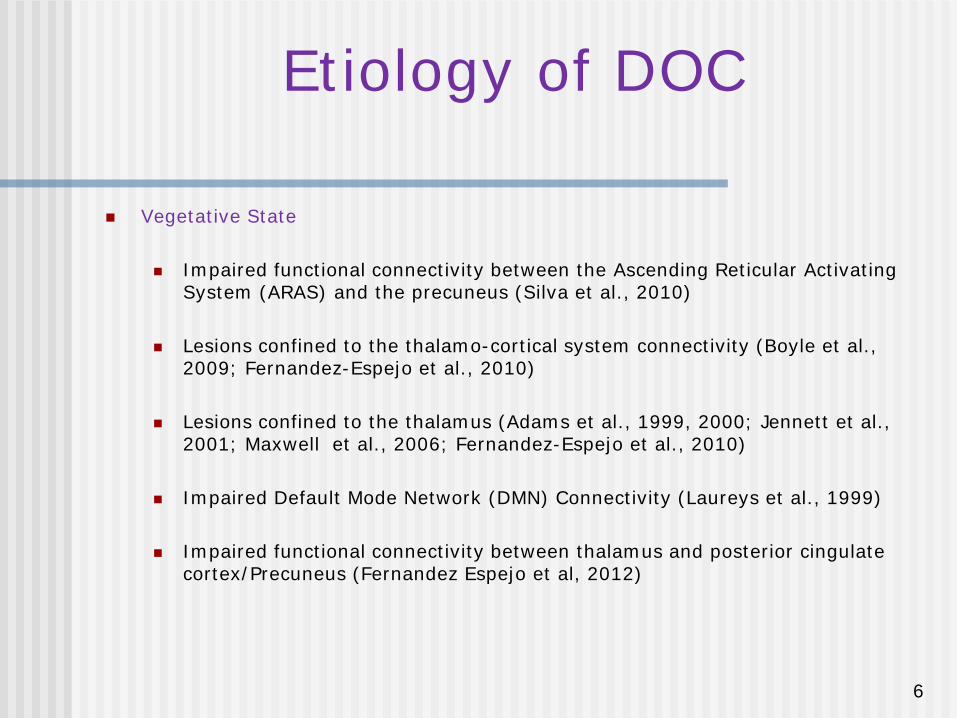
Etiology of DOC
Vegetative State
Impaired functional connectivity between the Ascending Reticular Activating System (ARAS) and the precuneus (Silva et al., 2010)
Lesions confined to the thalamo-cortical system connectivity (Boyle et al., 2009; Fernandez-Espejo et al., 2010)
Lesions confined to the thalamus (Adams et al., 1999, 2000; Jennett et al., 2001; Maxwell et al., 2006; Fernandez-Espejo et al., 2010)
Impaired Default Mode Network (DMN) Connectivity (Laureys et al., 1999)
Impaired functional connectivity between thalamus and posterior cingulate cortex/Precuneus (Fernandez Espejo et al, 2012)
6
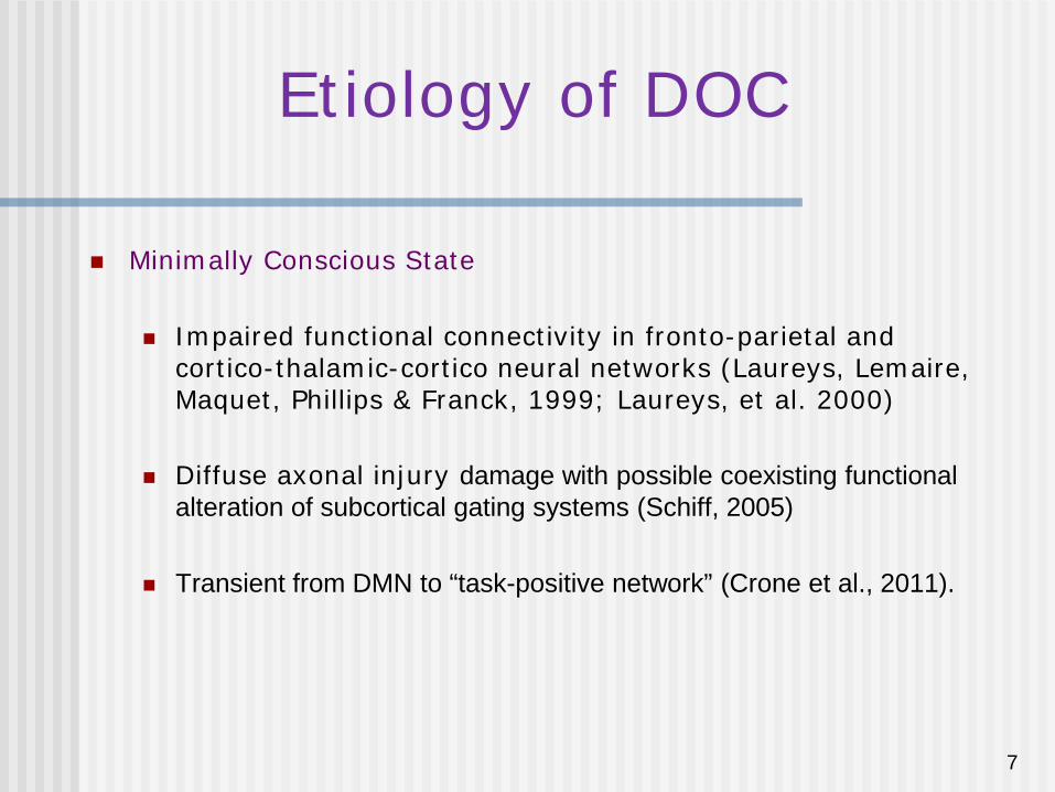
Etiology of DOC
Minimally Conscious State
Impaired functional connectivity in fronto-parietal and cortico-thalamic-cortico neural networks (Laureys, Lemaire, Maquet, Phillips & Franck, 1999; Laureys, et al. 2000)
Diffuse axonal injury damage with possible coexisting functional alteration of subcortical gating systems (Schiff, 2005)
Transient from DMN to “task-positive network” (Crone et al., 2011).
7
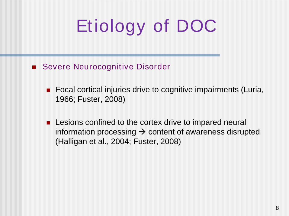
Etiology of DOC
Severe Neurocognitive Disorder
Focal cortical injuries drive to cognitive impairments (Luria, 1966; Fuster, 2008)
Lesions confined to the cortex drive to impared neural information processing content of awareness disrupted (Halligan et al., 2004; Fuster, 2008)
8
Rehabilitation of patients with DOC
9
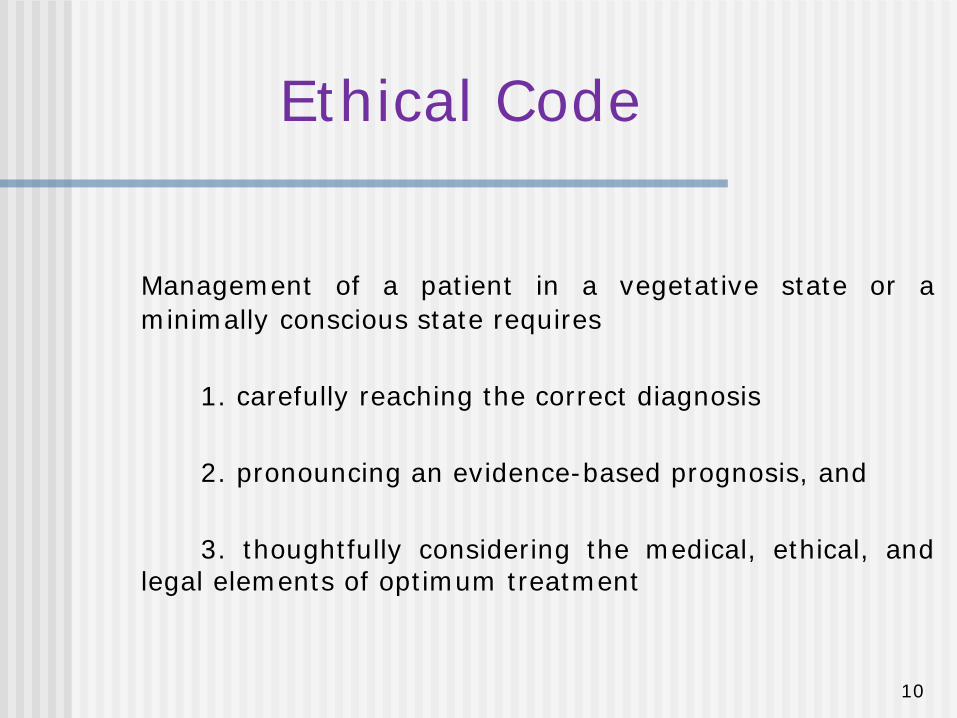
Ethical Code
Management of a patient in a vegetative state or aminimally conscious state requires
1. carefully reaching the correct diagnosis
2. pronouncing an evidence-based prognosis, and
3. thoughtfully considering the medical, ethical, andlegal elements of optimum treatment
10
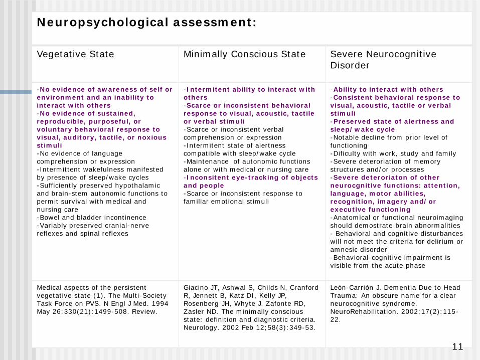
Neuropsychological assessment:
Vegetative State Minimally Conscious State Severe Neurocognitive Disorder
-No evidence of awareness of self or environment and an inability to interact with others-No evidence of sustained, reproducible, purposeful, or voluntary behavioral response to visual, auditory, tactile, or noxious stimuli-No evidence of language comprehension or expression-Intermittent wakefulness manifested by presence of sleep/wake cycles-Sufficiently preserved hypothalamic and brain-stem autonomic functions to permit survival with medical and nursing care-Bowel and bladder incontinence-Variably preserved cranial-nerve reflexes and spinal reflexes
-Intermitent ability to interact with others-Scarce or inconsistent behavioral response to visual, acoustic, tactile or verbal stimuli-Scarce or inconsistent verbal comprehension or expression-Intermitent state of alertness compatible with sleep/wake cycle-Maintenance of autonomic functions alone or with medical or nursing care-Inconsitent eye-tracking of objects and people-Scarce or inconsistent response to familiar emotional stimuli
-Ability to interact with others-Consistent behavioral response to visual, acoustic, tactile or verbal stimuli-Preserved state of alertness and sleep/wake cycle-Notable decline from prior level of functioning-Dificulty with work, study and family-Severe deteroriation of memory structures and/or processes-Severe deteroriaton of other neurocgnitive functions: attention, language, motor abilities, recognition, imagery and/or executive functioning-Anatomical or functional neuroimaging should demostrate brain abnormalities- Behavioral and cognitive disturbances will not meet the criteria for delirium or amnesic disorder-Behavioral-cognitive impairment is visible from the acute phase
Medical aspects of the persistent vegetative state (1). The Multi-Society Task Force on PVS. N Engl J Med. 1994 May 26;330(21):1499-508. Review.
Giacino JT, Ashwal S, Childs N, Cranford R, Jennett B, Katz DI, Kelly JP, Rosenberg JH, Whyte J, Zafonte RD, Zasler ND. The minimally conscious state: definition and diagnostic criteria. Neurology. 2002 Feb 12;58(3):349-53.
León-Carrión J. Dementia Due to Head Trauma: An obscure name for a clear neurocognitive syndrome. NeuroRehabilitation. 2002;17(2):115-22.
11
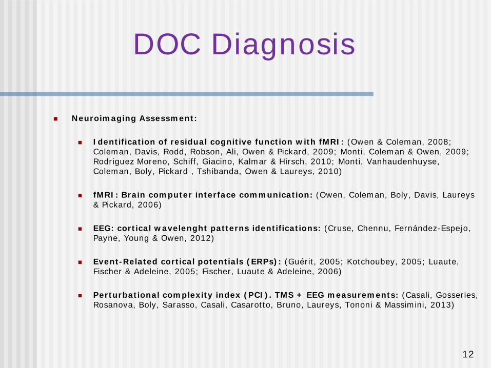
DOC Diagnosis
Neuroimaging Assessment:
Identification of residual cognitive function with fMRI: (Owen & Coleman, 2008; Coleman, Davis, Rodd, Robson, Ali, Owen & Pickard, 2009; Monti, Coleman & Owen, 2009; Rodriguez Moreno, Schiff, Giacino, Kalmar & Hirsch, 2010; Monti, Vanhaudenhuyse, Coleman, Boly, Pickard , Tshibanda, Owen & Laureys, 2010)
fMRI: Brain computer interface communication: (Owen, Coleman, Boly, Davis, Laureys & Pickard, 2006)
EEG: cortical wavelenght patterns identifications: (Cruse, Chennu, Fernández-Espejo, Payne, Young & Owen, 2012)
Event-Related cortical potentials (ERPs): (Guérit, 2005; Kotchoubey, 2005; Luaute, Fischer & Adeleine, 2005; Fischer, Luaute & Adeleine, 2006)
Perturbational complexity index (PCI). TMS + EEG measurements: (Casali, Gosseries, Rosanova, Boly, Sarasso, Casali, Casarotto, Bruno, Laureys, Tononi & Massimini, 2013)
12
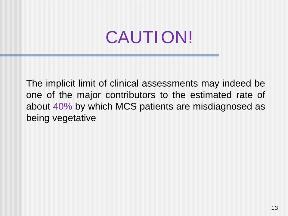
CAUTION!
The implicit limit of clinical assessments may indeed beone of the major contributors to the estimated rate ofabout 40% by which MCS patients are misdiagnosed asbeing vegetative
13
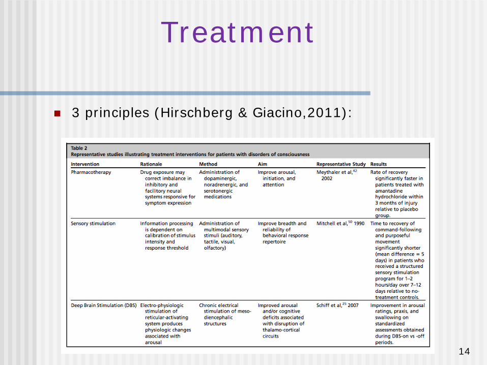
Treatment
3 principles (Hirschberg & Giacino,2011):
14
Treatment
In order to rehabilitate DOC patients we use the Combined Method Therapy (CMT) (Leon-Carrion, 2006):
CMT is a methodological engineering process, integrating knowledge of a patient’s intact and non-intact functional brain circuitry with cognition and neuropharmacology.
The goal is to achieve a new functional cerebral organization which allows the patient to re-establish functional brain connectivity.
15
Treatment of DOC: A single Case
Restoring Cortical Connectivity Directionality and Synchronization is
Essential to Treating Disorder of Consciousness
León-Carrión J, León-Domínguez U, Halper J, Pollonini L, Zouridakis G, Domínguez-Morales MD. 2013. Restoring Cortical Connectivity Directionality and
Synchronization is Essential to Treating Disorder of Consciousness. Current Pharmacological Design, 2013.
16

Treatment of DOC: A single Case
Patient Profile 24-year-old male with TBI resulting
from a traffic accident
MRI showed diffused brain injury with lesions in fronto-temporal areas (predominantly in right and posterior-lateral), as well as inferior-ventral brainstem damage.
The patient scored 8 on the Glasgow Coma Scale (Eyes 4; Motor 3; Verbal 1), 24 on the Disability Rating Scale, and 2.72 on the Rappaport Coma/near Coma Scale (moderate coma).
The patient fulfilled international criteria for the VS
CT Scan
17
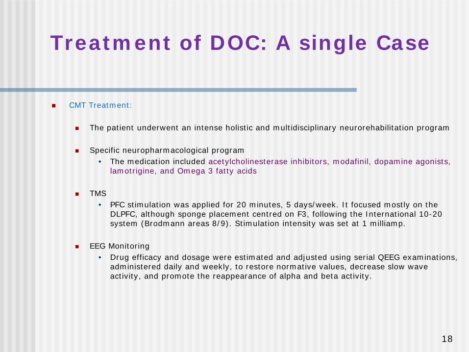
CMT Treatment:
The patient underwent an intense holistic and multidisciplinary neurorehabilitation program
Specific neuropharmacological program• The medication included acetylcholinesterase inhibitors, modafinil, dopamine agonists,
lamotrigine, and Omega 3 fatty acids
TMS• PFC stimulation was applied for 20 minutes, 5 days/week. It focused mostly on the
DLPFC, although sponge placement centred on F3, following the International 10-20 system (Brodmann areas 8/9). Stimulation intensity was set at 1 milliamp.
EEG Monitoring• Drug efficacy and dosage were estimated and adjusted using serial QEEG examinations,
administered daily and weekly, to restore normative values, decrease slow wave activity, and promote the reappearance of alpha and beta activity.
18
Treatment of DOC: A single Case
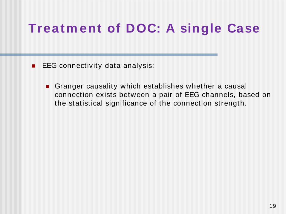
EEG connectivity data analysis:
Granger causality which establishes whether a causal connection exists between a pair of EEG channels, based on the statistical significance of the connection strength.
19
Treatment of DOC: A single Case
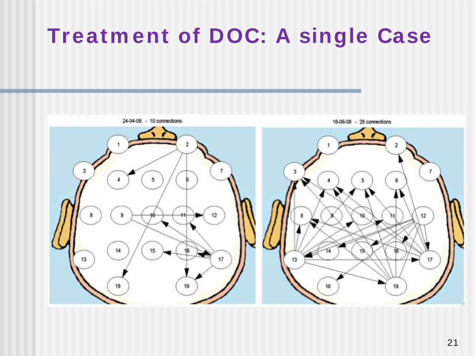
Results
The new pharmacological treatment revealed changes in the patient’s cortical connection (16/06/2008) from that of admission (24/04/2008)
These changes were due to an increase in the number of connections between the anterior and posterior cortex.
Another significant finding was the change in directionality in these connections which took place after beginning the new pharmacological treatment. The initial directionality from anterior to posterior cortex was inverted, and now information was sent from the posterior to the anterior cortex.
20
Treatment of DOC: A single Case
21
Treatment of DOC: A single Case
Synchronization between the anterior and posterior cortex determines
consciousness level in patients with traumatic brain injury (TBI).
Leon-Carrion J, Leon-Dominguez U, Pollonini L, Wu MH, Frye RE, Dominguez-Morales MR, Zouridakis G. (2012). Synchronization between the anterior and posterior
cortex determines consciousness level in patients with traumatic brain injury (TBI). Brain Res, 1476:22-30.
22
Treatment of DOC: Cortical connectivity of MCS and SND
patients
23
The present study aims to explore how synchronized nonrandom neural circuits across cortical regions are integrated to generate consciousness.
In this study we compare two groups:
• patients in the MCS • patients with SND
Treatment of DOC: Cortical connectivity of MCS and SND
patients
24
Hypothesis
Our hypothesis:
1: The emergence of consciousness requires synchronized circuitry to differentially integrate anterior and posterior cortices.
2: Patients with SND should display greater synchrony and connectivity between anterior and posterior regions than patients in the MCS.
25
Experimental Procedures
We used functional connectivity analysis to identify brain connectivity networks in task-free resting state EEG recordings.
Two methods were applied to ascertain the cause and effect relationships among all electrodes:
the first measured synchronization activity between pairs of electrodes
the second determined the strength and direction of functional connectivity
26
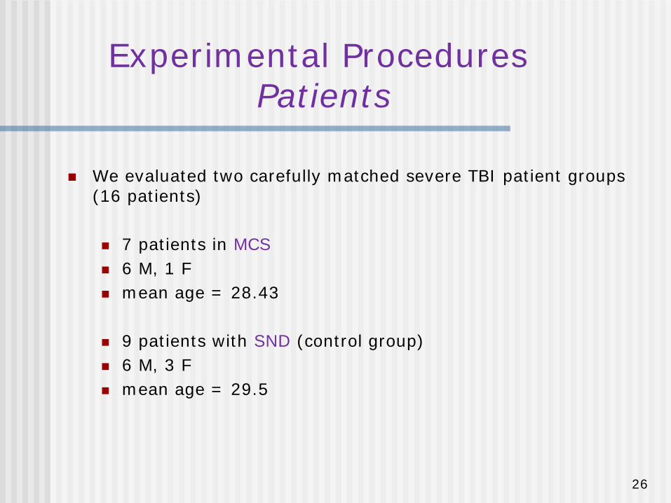
Experimental ProceduresPatients
We evaluated two carefully matched severe TBI patient groups (16 patients)
7 patients in MCS 6 M, 1 F mean age = 28.43
9 patients with SND (control group) 6 M, 3 F mean age = 29.5
27
Results
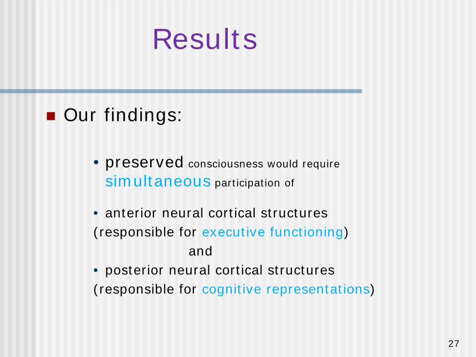
Our findings:
• preserved consciousness would require
simultaneous participation of
• anterior neural cortical structures (responsible for executive functioning)
and • posterior neural cortical structures (responsible for cognitive representations)
28
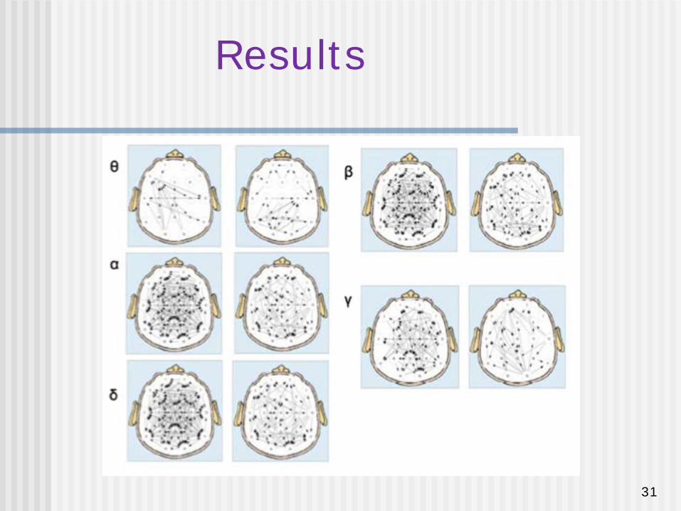
Results
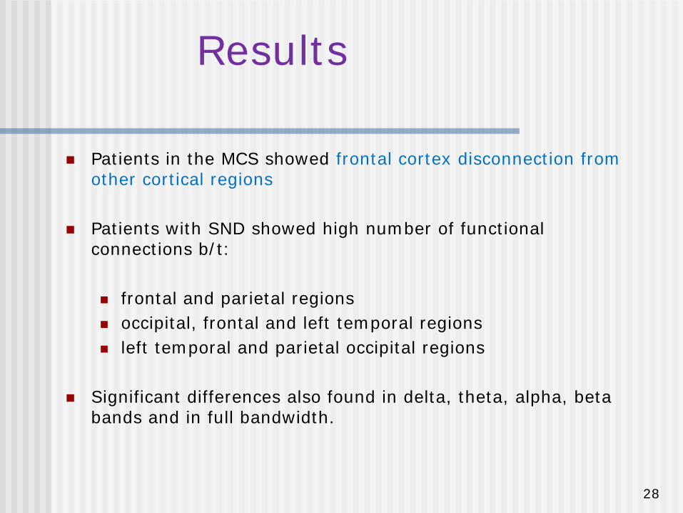
Patients in the MCS showed frontal cortex disconnection from other cortical regions
Patients with SND showed high number of functional connections b/t:
frontal and parietal regions occipital, frontal and left temporal regions left temporal and parietal occipital regions
Significant differences also found in delta, theta, alpha, beta bands and in full bandwidth.
29
Results
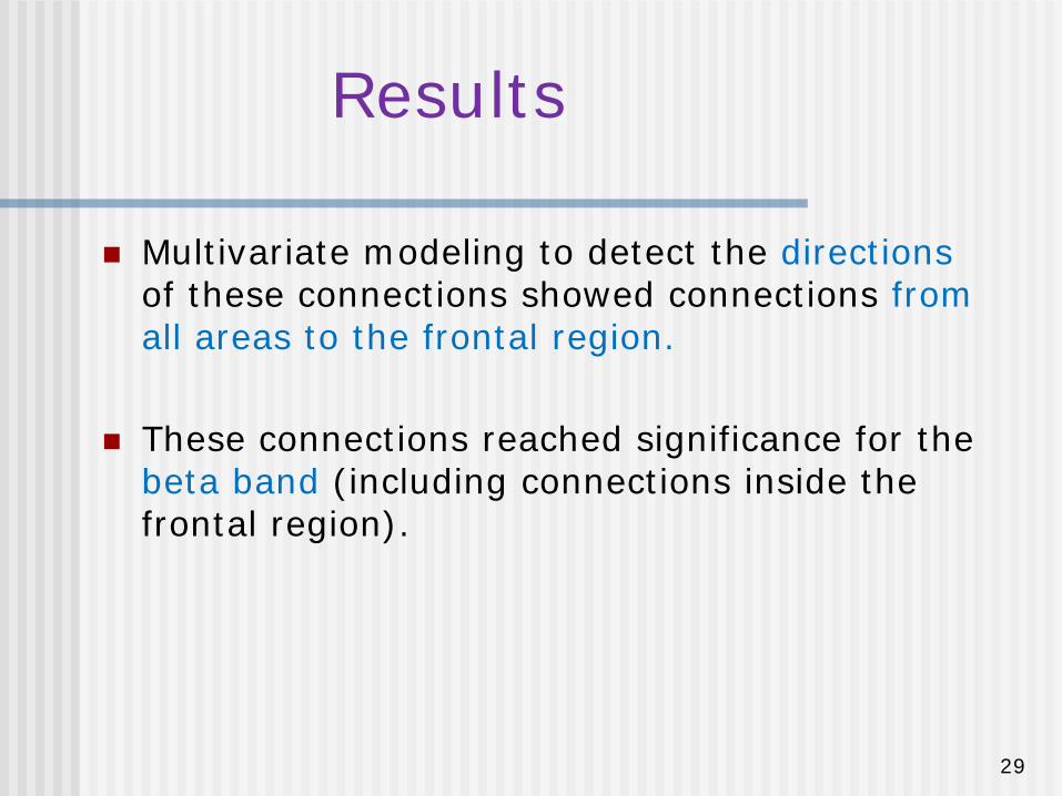
Multivariate modeling to detect the directionsof these connections showed connections from all areas to the frontal region.
These connections reached significance for the beta band (including connections inside the frontal region).
30
Results
Our results illustrate the existence of a large scale network surviving in both MCS and SND
although patients with SND display a higher level of synchronization
Results
31
Results
32
Conclusions
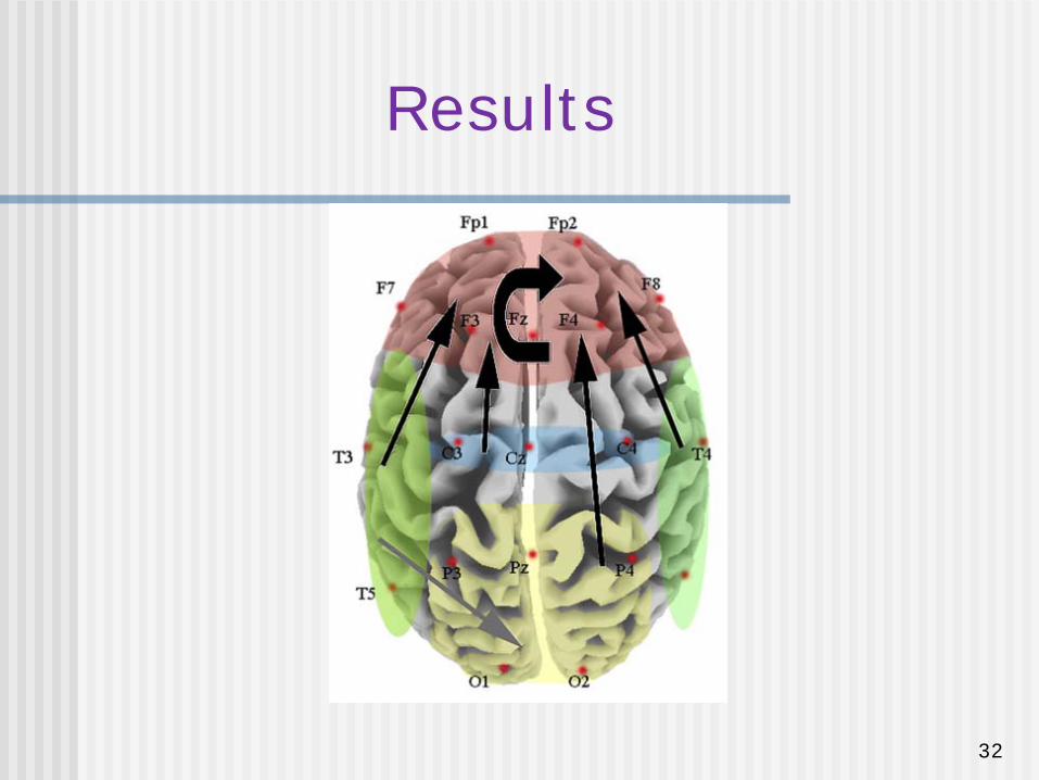
Conclusions:
Awareness level depends on the synchronization between retrorolandic (posterior) and frontal (anterior) cortical areas.
Patients with TBI presented a higher level of awareness when connections between cortical regions followed a posterior-anterior propagation of beta waves.
Clinical application:
These evidence show up the necessity to track the DOC´s treatment with neuroimaging technics to fit the correct pharmacological dose in order to promote posterior-anterior cortical connectivity
33

34
Thank you very much.
•www.neurocrecer.es
•www.discovershadow.com/
35
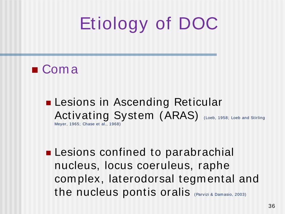
Etiology of DOC
Coma
Lesions in Ascending Reticular Activating System (ARAS) (Loeb, 1958; Loeb and Stirling Meyer, 1965; Chase et al., 1968)
Lesions confined to parabrachial nucleus, locus coeruleus, raphe complex, laterodorsal tegmental and the nucleus pontis oralis (Parvizi & Damasio, 2003)
36
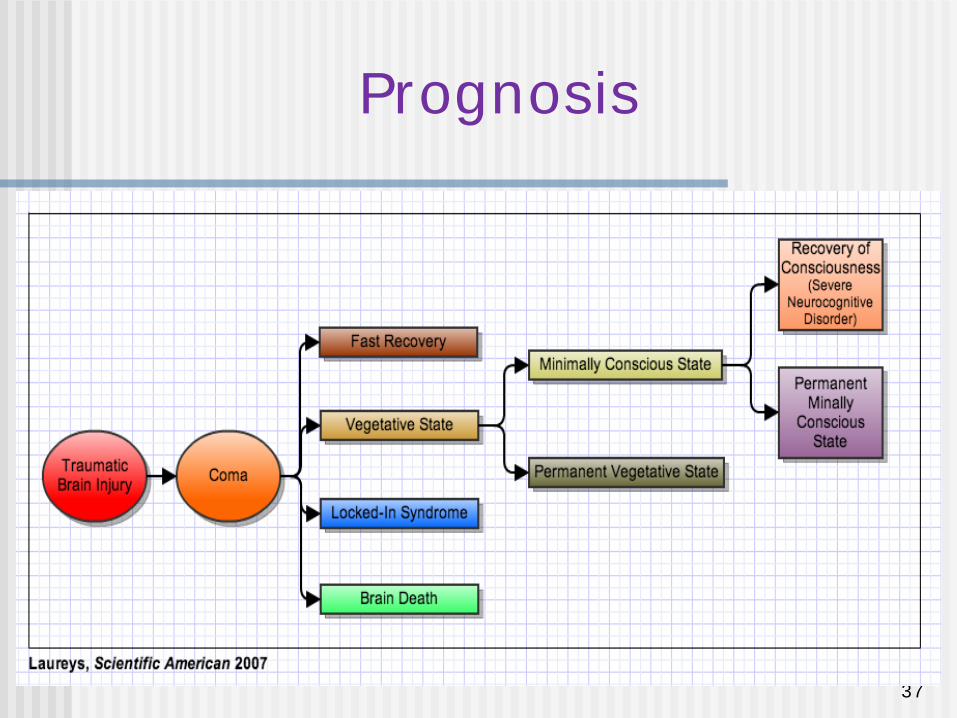
Prognosis
37


![DOSSIER ALBERTO DOMÍNGUEZ GÁLVEZ [ENG]](https://img.pdfslide.us/doc/110x75/568cab691a28ab186da572cc/dossier-alberto-dominguez-galvez-eng.jpg)


![Alberto Domínguez - Perfidia [Piano sheet music]](https://img.pdfslide.us/doc/110x75/55cf9b82550346d033a65aa2/alberto-dominguez-perfidia-piano-sheet-music.jpg)