Embed Size (px)
Citation preview
TAVR – Prospective Engagement from Coordinator to Coding
Elizabeth K. Walsh, RN Administrative Director, Thoracic Aortic Research Program
Relevant Conflicts of Interest
• Edwards Lifesciences, LLC
• Healthcare Leadership Series
• NCDR 14 Planning Committee
• Advisory Board for TAVR Administrators
• Study Operations Steering Committee
Objectives:
• Why does the Heart Team need to understand Coding?
• Documentation, Documentation, Documentation:
Throughout the Process
• Templates: Ensure appropriate Documentation
• The Importance of Tracking and Auditing: Know your
Data
• Compliance with CMS/NCD: Challenges and Solutions
• Where do we go from here?
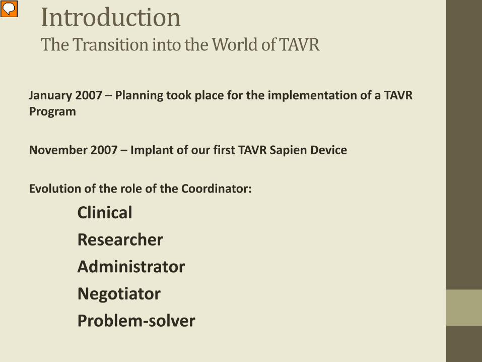
Introduction The Transition into the World of TAVR
January 2007 – Planning took place for the implementation of a TAVR Program November 2007 – Implant of our first TAVR Sapien Device Evolution of the role of the Coordinator:
Clinical Researcher Administrator Negotiator Problem-solver
But most of ALL BY DEFAULT:
“Novice” to “Expert” in TAVR
Coding and Insurance Reimbursement
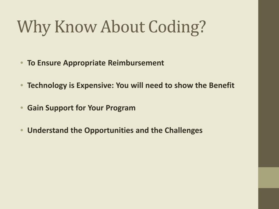
Why Know About Coding?
• To Ensure Appropriate Reimbursement
• Technology is Expensive: You will need to show the Benefit
• Gain Support for Your Program
• Understand the Opportunities and the Challenges
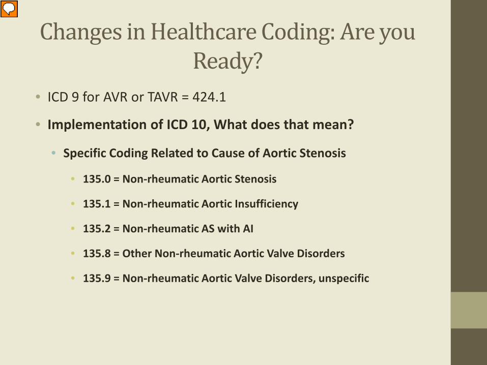
Changes in Healthcare Coding: Are you Ready?
• ICD 9 for AVR or TAVR = 424.1
• Implementation of ICD 10, What does that mean?
• Specific Coding Related to Cause of Aortic Stenosis
• 135.0 = Non-rheumatic Aortic Stenosis
• 135.1 = Non-rheumatic Aortic Insufficiency
• 135.2 = Non-rheumatic AS with AI
• 135.8 = Other Non-rheumatic Aortic Valve Disorders
• 135.9 = Non-rheumatic Aortic Valve Disorders, unspecific
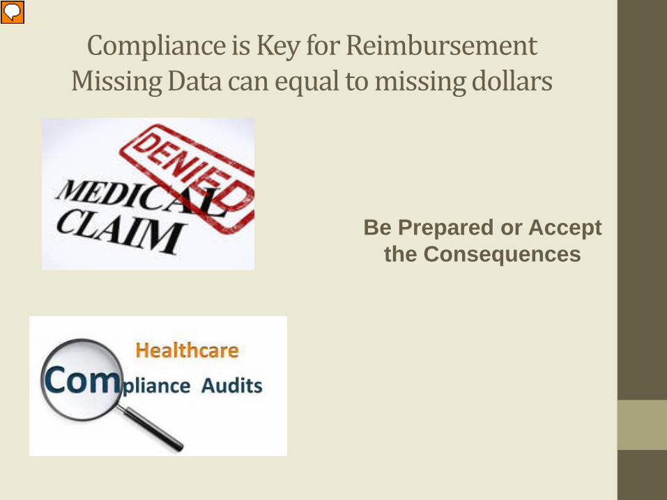
Compliance is Key for Reimbursement Missing Data can equal to missing dollars
Be Prepared or Accept the Consequences
Challenges and Opportunities
Hurdles • Administration’s Support
• Costs
• Capital Investment
• Resources
• Decline of Open AVR and Increase in TAVR
• Heart Team Cultivation
• Learning Curves
• Regulatory
• CMS/NCD Guidelines
• Competition
• Education
Benefits • Providing Care for patients
with AS where there was nothing to offer.
• Collaboration among multidisciplinary providers/teams
• Trailblazing New Technology • Increased Volume • Leader in the Market • Shared Professional Revenue • Concept of a “Transcatheter
Valve Service”
Get Administration on board
• “Show Them The Money” or not: TAVR Benefits
• Understand the Volume: Incremental Volume
• Analyze the Trial Program: Learn from the Past to Predict
the Future
• Keep up with Changes
• Remain Competitive
Benefits of Being of Research Experience: If it is not Documented it did not happen
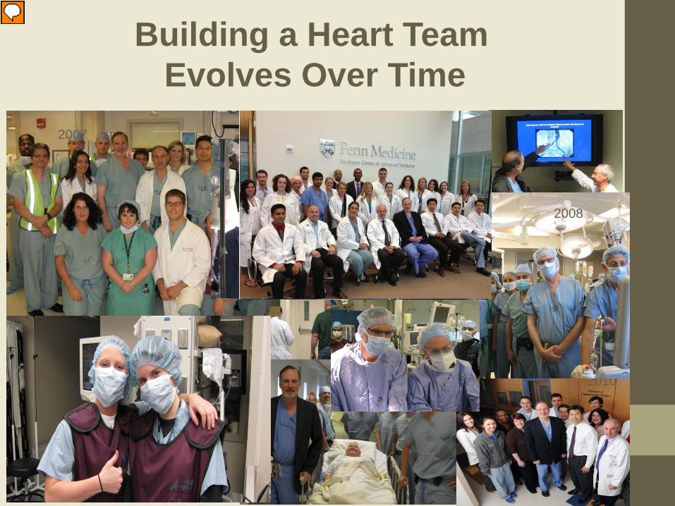
Building a Heart Team Evolves Over Time
2007
2010
2008
TAVR Programs: Commercial and Trial: Both Require Teamwork
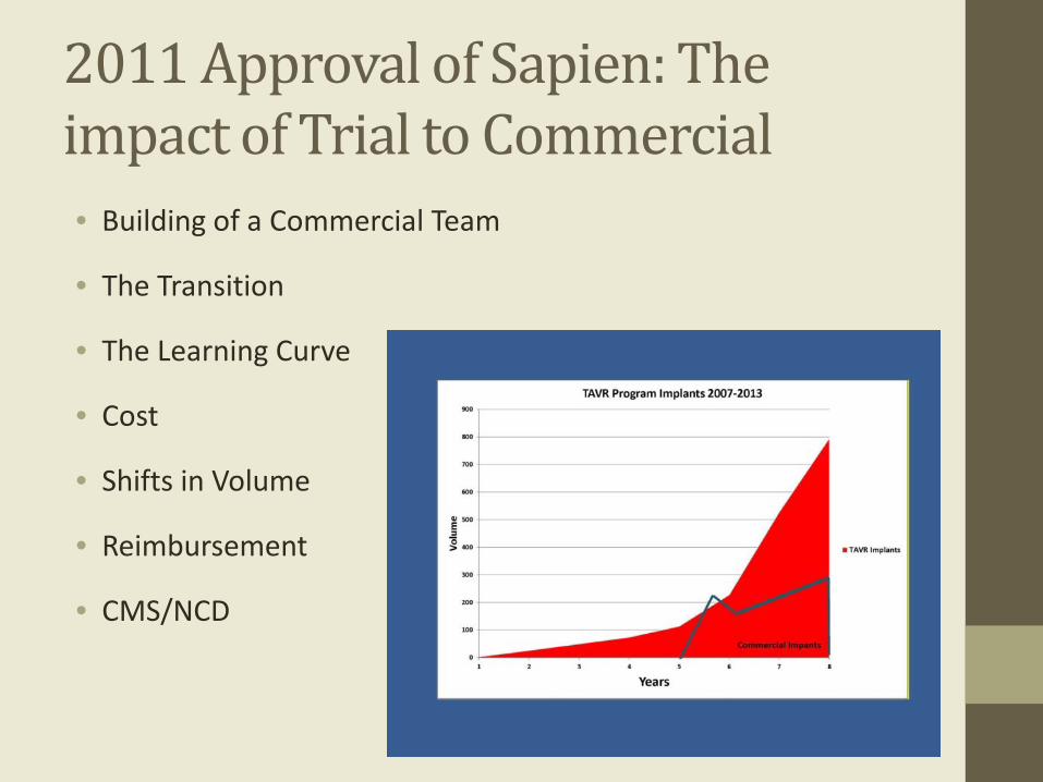
2011 Approval of Sapien: The impact of Trial to Commercial • Building of a Commercial Team
• The Transition
• The Learning Curve
• Cost
• Shifts in Volume
• Reimbursement
• CMS/NCD
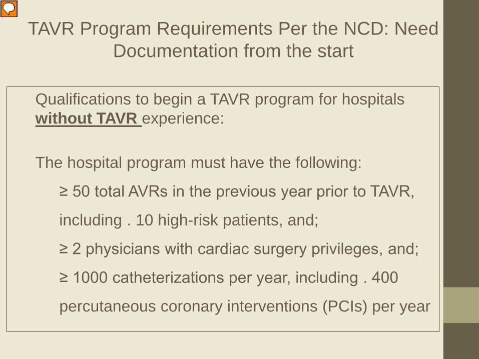
Qualifications to begin a TAVR program for hospitals without TAVR experience: The hospital program must have the following:
≥ 50 total AVRs in the previous year prior to TAVR,
including . 10 high-risk patients, and;
≥ 2 physicians with cardiac surgery privileges, and;
≥ 1000 catheterizations per year, including . 400
percutaneous coronary interventions (PCIs) per year
TAVR Program Requirements Per the NCD: Need Documentation from the start
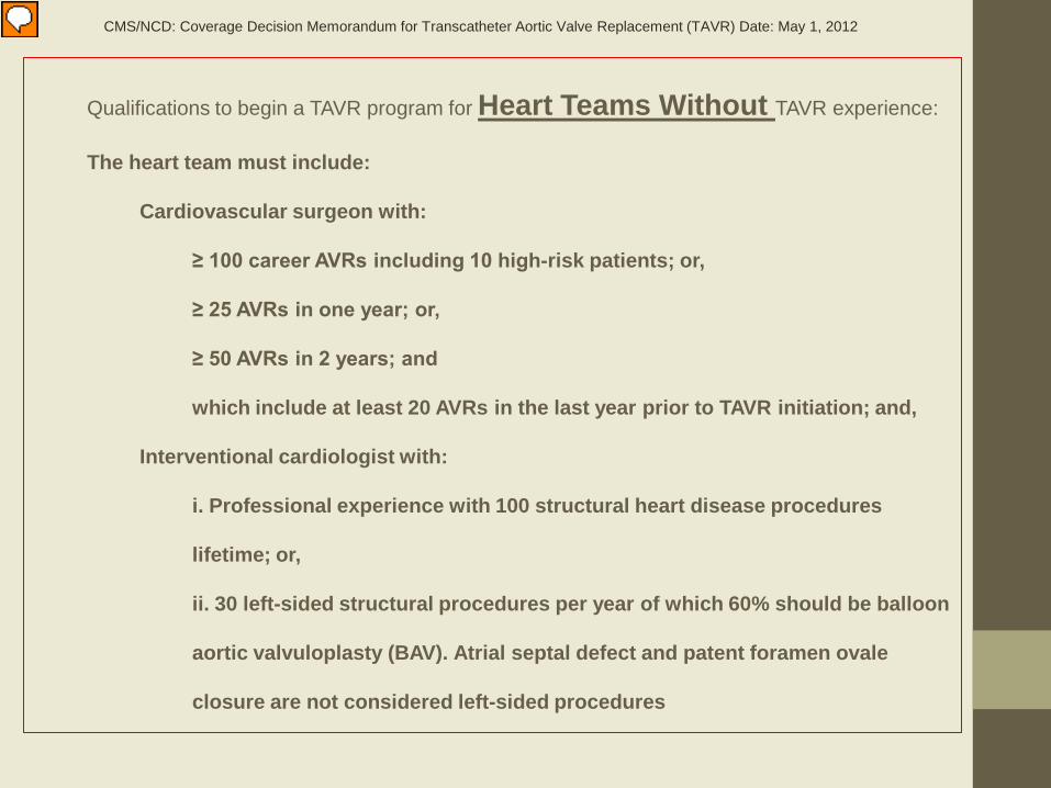
Qualifications to begin a TAVR program for Heart Teams Without TAVR experience:
The heart team must include:
Cardiovascular surgeon with:
≥ 100 career AVRs including 10 high-risk patients; or,
≥ 25 AVRs in one year; or,
≥ 50 AVRs in 2 years; and
which include at least 20 AVRs in the last year prior to TAVR initiation; and,
Interventional cardiologist with:
i. Professional experience with 100 structural heart disease procedures
lifetime; or,
ii. 30 left-sided structural procedures per year of which 60% should be balloon
aortic valvuloplasty (BAV). Atrial septal defect and patent foramen ovale
closure are not considered left-sided procedures
CMS/NCD: Coverage Decision Memorandum for Transcatheter Aortic Valve Replacement (TAVR) Date: May 1, 2012
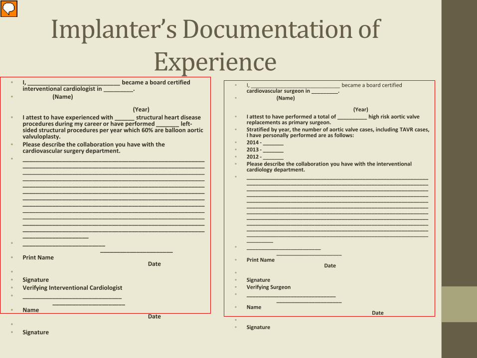
Implanter’s Documentation of Experience
• I, ____________________________ became a board certified interventional cardiologist in _________.
• (Name) (Year)
• I attest to have experienced with ______ structural heart disease procedures during my career or have performed _______ left-sided structural procedures per year which 60% are balloon aortic valvuloplasty.
• Please describe the collaboration you have with the cardiovascular surgery department.
• ________________________________________________________________________________________________________________________________________________________________________________________________________________________________________________________________________________________________________________________________________________________________________________________________________________________________________________________________________________________________________________________________________________________________________________________________________________________________________________________________________________________________________
• _________________________ ______________________
• Print Name Date
• • Signature • Verifying Interventional Cardiologist • ______________________________
______________________ • Name
Date • • Signature
• I, ______________________________ became a board certified cardiovascular surgeon in _________.
• (Name) (Year)
• I attest to have performed a total of __________ high risk aortic valve replacements as primary surgeon.
• Stratified by year, the number of aortic valve cases, including TAVR cases, I have personally performed are as follows:
• 2014 - _______ • 2013 - _______ • 2012 - _______ • Please describe the collaboration you have with the interventional
cardiology department. • _____________________________________________________________
___________________________________________________________________________________________________________________________________________________________________________________________________________________________________________________________________________________________________________________________________________________________________________________________________________________________________________________________________________________________________________________________________________________________________________________________________________________________________________
• _________________________ ______________________
• Print Name Date
• • Signature • Verifying Surgeon • ______________________________
______________________ • Name
Date • • Signature
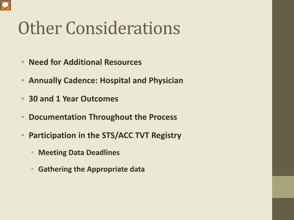
Other Considerations • Need for Additional Resources
• Annually Cadence: Hospital and Physician
• 30 and 1 Year Outcomes
• Documentation Throughout the Process
• Participation in the STS/ACC TVT Registry
• Meeting Data Deadlines
• Gathering the Appropriate data
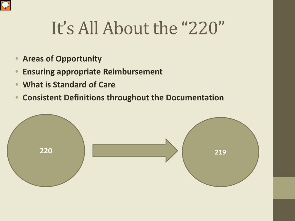
It’s All About the “220” • Areas of Opportunity • Ensuring appropriate Reimbursement • What is Standard of Care • Consistent Definitions throughout the Documentation
220 219
Examination of a Single Site’s Experience Of the Distribution of MS-DRGs and Rates
• 308 patients from November 2007-July 2012
• 235 were implanted with Trial Valve
• 73 were implanted with Commercial Valve
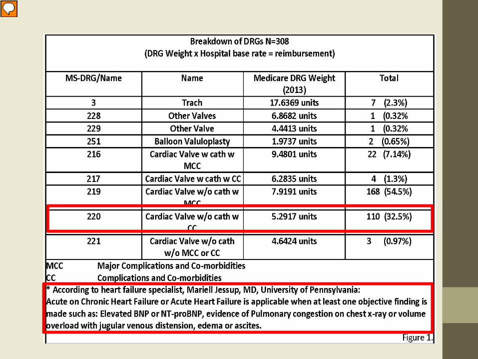
• 110 (35.7%) were coded as a 220
• 59 (53.6%) of the 110 did have evidence of “Acute on Chronic Heart Failure” according to the definition by the AHA President Mariell Jessup
• 4 cases of the 110 were miscoded
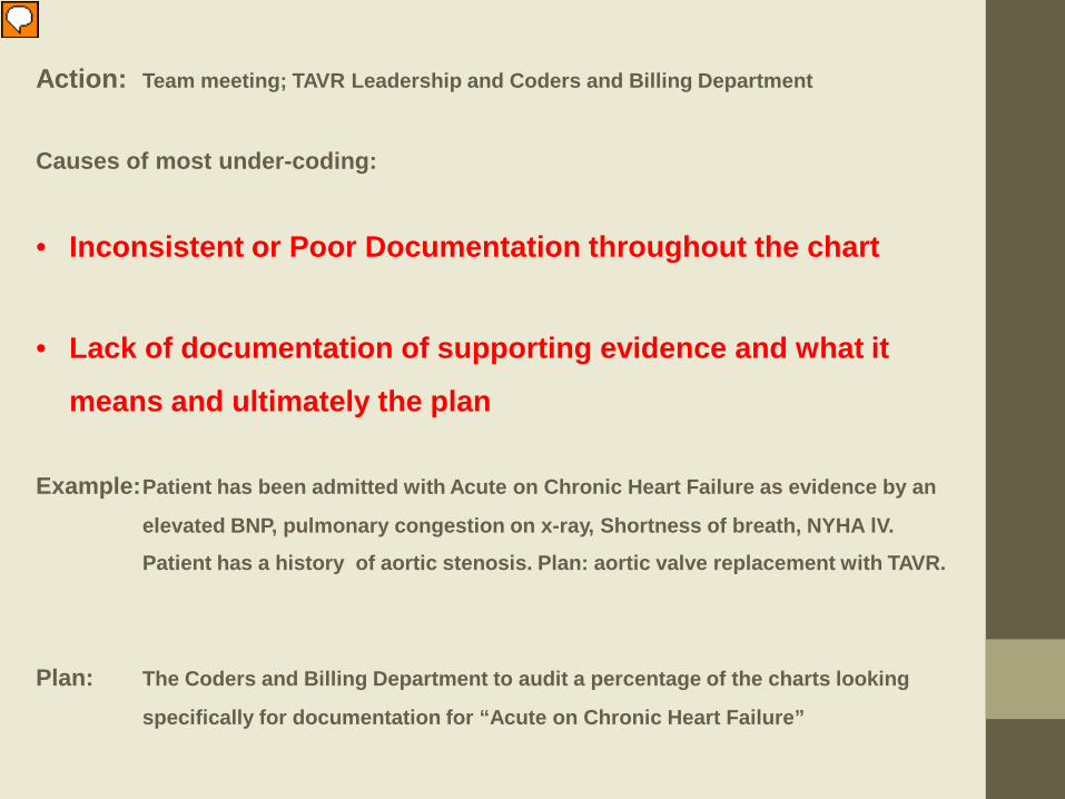
Action: Team meeting; TAVR Leadership and Coders and Billing Department
Causes of most under-coding:
• Inconsistent or Poor Documentation throughout the chart
• Lack of documentation of supporting evidence and what it
means and ultimately the plan
Example: Patient has been admitted with Acute on Chronic Heart Failure as evidence by an
elevated BNP, pulmonary congestion on x-ray, Shortness of breath, NYHA lV.
Patient has a history of aortic stenosis. Plan: aortic valve replacement with TAVR.
Plan: The Coders and Billing Department to audit a percentage of the charts looking
specifically for documentation for “Acute on Chronic Heart Failure”
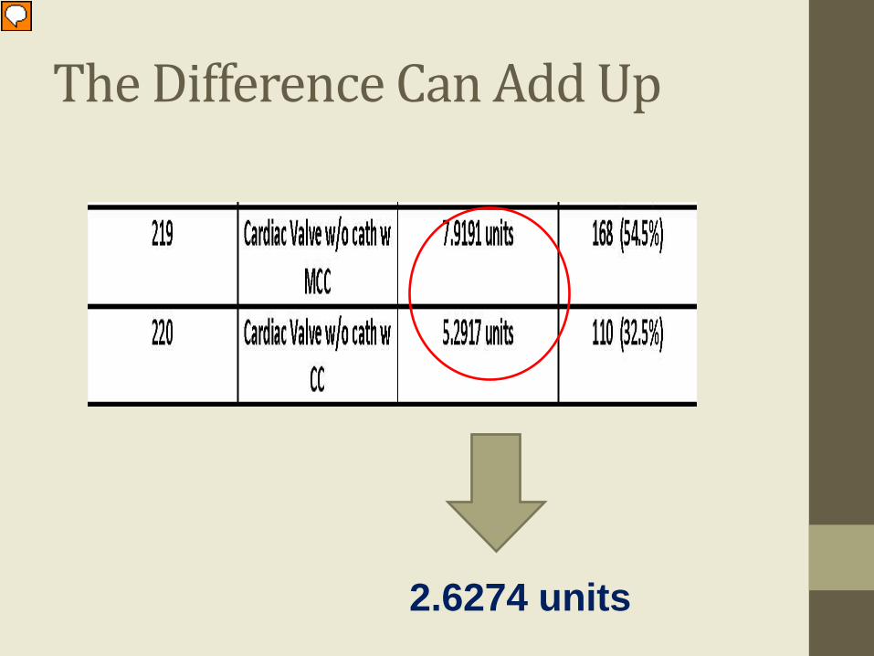
The Difference Can Add Up
2.6274 units
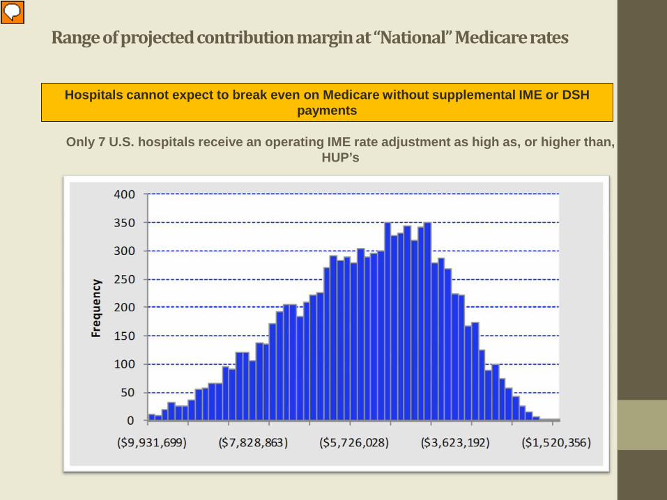
Range of projected contribution margin at “National” Medicare rates
Hospitals cannot expect to break even on Medicare without supplemental IME or DSH payments
Only 7 U.S. hospitals receive an operating IME rate adjustment as high as, or higher than, HUP’s
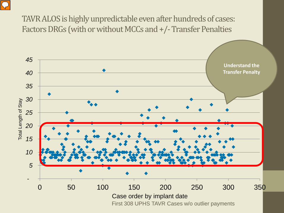
TAVR ALOS is highly unpredictable even after hundreds of cases: Factors DRGs (with or without MCCs and +/- Transfer Penalties
-
5
10
15
20
25
30
35
40
45
0 50 100 150 200 250 300 350
Tota
l Len
gth
of S
tay
Case order by implant date First 308 UPHS TAVR Cases w/o outlier payments
Understand the Transfer Penalty
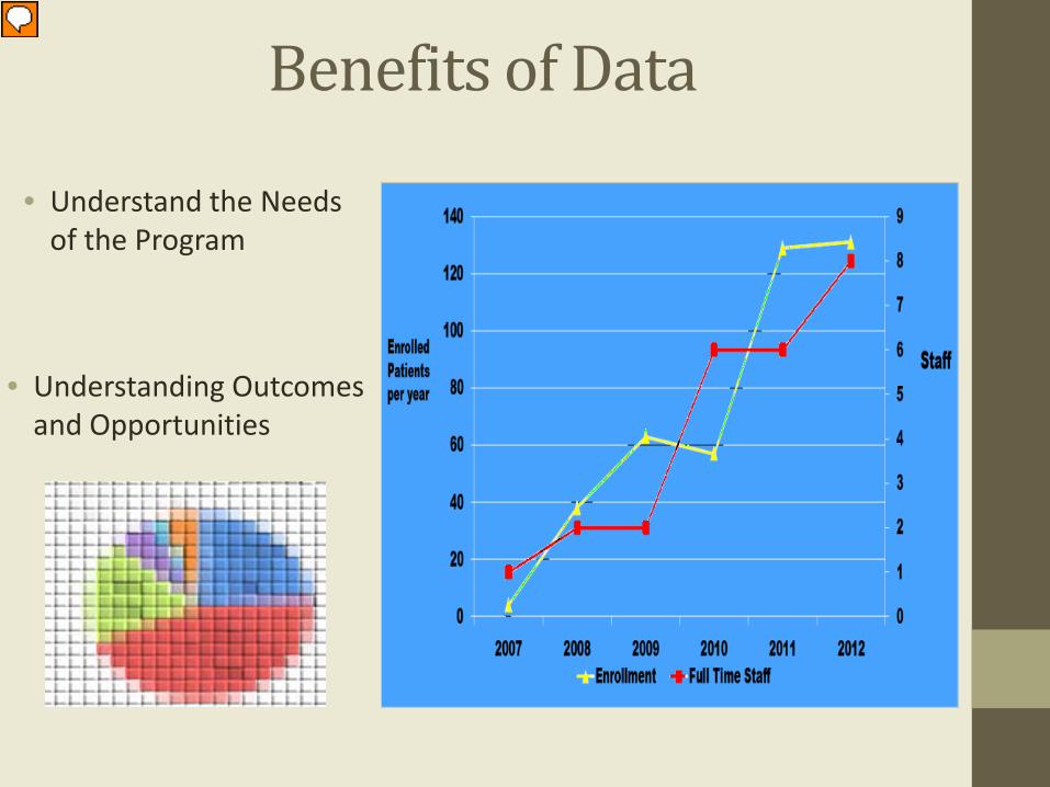
Benefits of Data
• Understand the Needs of the Program
• Understanding Outcomes and Opportunities
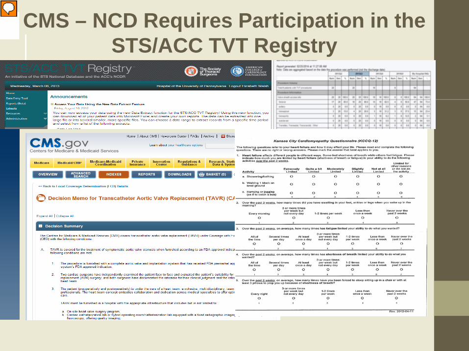
CMS – NCD Requires Participation in the STS/ACC TVT Registry
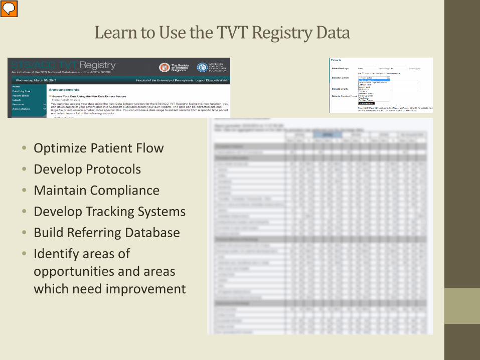
Learn to Use the TVT Registry Data
• Optimize Patient Flow • Develop Protocols • Maintain Compliance • Develop Tracking Systems • Build Referring Database • Identify areas of
opportunities and areas which need improvement
The Value of Tracking Your Patients
• Understanding Referral Patterns
• Volume Analysis
• Resource Justification
• Tracking Outcomes
• Protocol Development
• Understanding Opportunities for Improvement
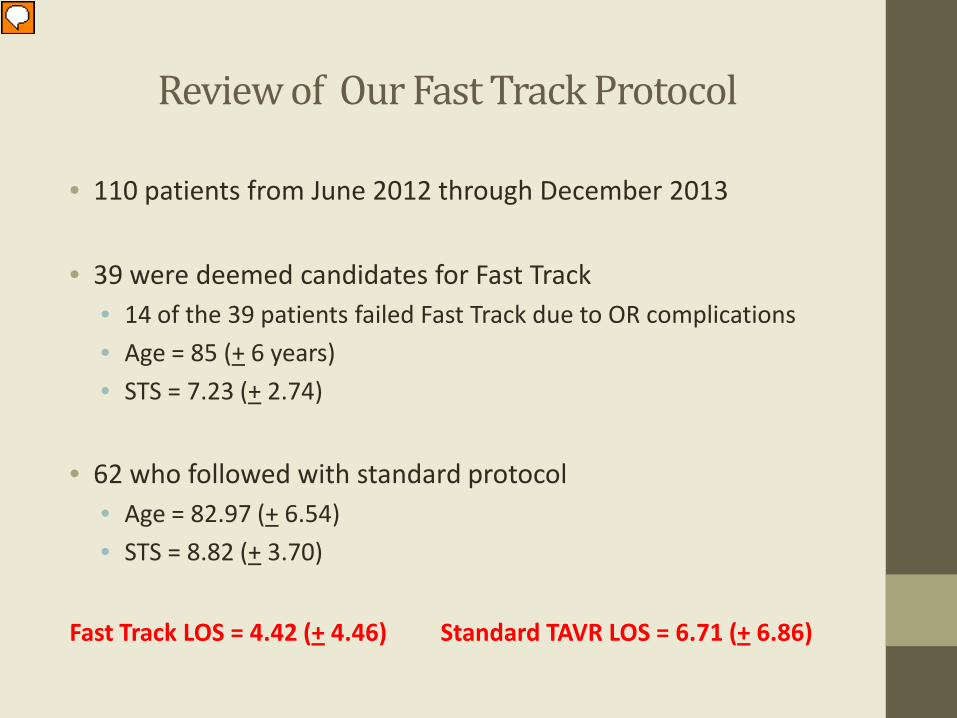
Review of Our Fast Track Protocol
• 110 patients from June 2012 through December 2013
• 39 were deemed candidates for Fast Track • 14 of the 39 patients failed Fast Track due to OR complications • Age = 85 (+ 6 years) • STS = 7.23 (+ 2.74)
• 62 who followed with standard protocol
• Age = 82.97 (+ 6.54) • STS = 8.82 (+ 3.70)
Fast Track LOS = 4.42 (+ 4.46) Standard TAVR LOS = 6.71 (+ 6.86)

Penn Medicine @ Cherry Hill
Penn Med @ Valley Forge
Mercy Fitzgerald Penn Med @ Woodbury
Penn Med @ Radnor
Penn Med @ Bucks
Brandywine
Bayhealth - Kent
Bayhealth - Milford
Phoenixville
Christiana
Geisinger
Lehigh Valley
Lourdes (pending)
Pennsylvania New Jersey
Delaware
Maryland
Doylestown
Relationships are Key
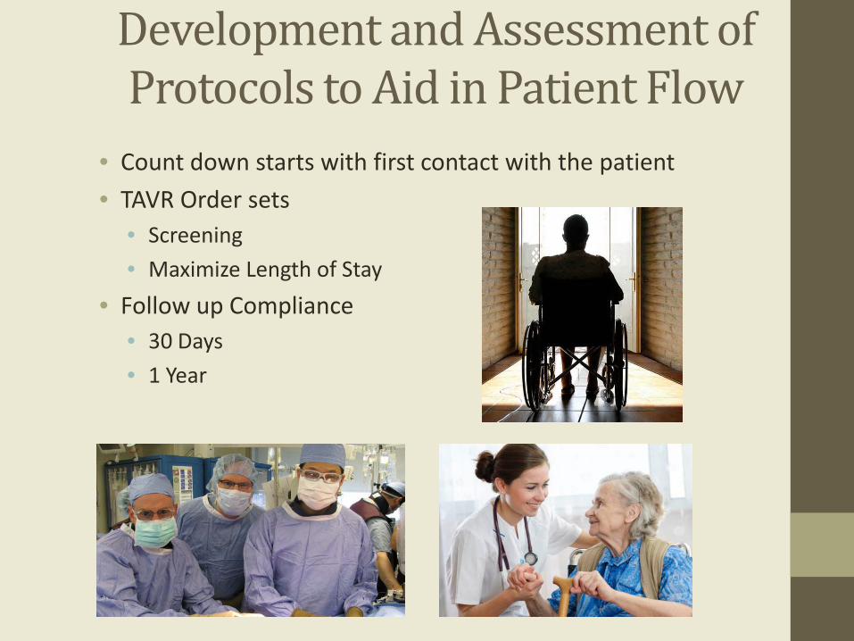
Development and Assessment of Protocols to Aid in Patient Flow
• Count down starts with first contact with the patient • TAVR Order sets
• Screening • Maximize Length of Stay
• Follow up Compliance • 30 Days • 1 Year
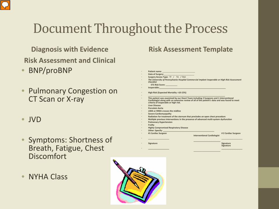
Document Throughout the Process Diagnosis with Evidence
Risk Assessment and Clinical • BNP/proBNP
• Pulmonary Congestion on
CT Scan or X-ray
• JVD
• Symptoms: Shortness of Breath, Fatigue, Chest Discomfort
• NYHA Class
Risk Assessment Template
• Patient name: ____________________________ • Date of Surgery:__________________________ • Surgery Access Type: TF / TA / TAO • The University of Pennsylvania Hospital Commercial Implant Inoperable or High Risk Assessment
Checklist • STS Risk Score: __________ • Inoperable:_________________________________________________________
• High Risk (Expected Mortality >10-15%) • __________________________________________________________________ • This patient was examined by our Heart Team including 2 Surgeons and 1 Interventional
Cardiologist along with an extensive review of all of the patient’s data and was found to meet criteria of inoperable or high risk.
• Liver Disease • Porcelain Aorta • LIMA or RIMA crosses the midline • Severe Cardiomyopathy • Radiation for treatment of the sternum that precludes an open chest procedure • Multiple previous interventions in the presence of advanced multi-system dysfunction • Pulmonary Hypertension • Frailty • Highly Compromised Respiratory Disease • Other: Specify: ____________________________________________ • #1 Cardiac Surgeon # 2 Cardiac Surgeon
Interventional Cardiologist • __________________ __________________
_______________________ • Signature Signature
Signature • __________________ __________________
_______________________
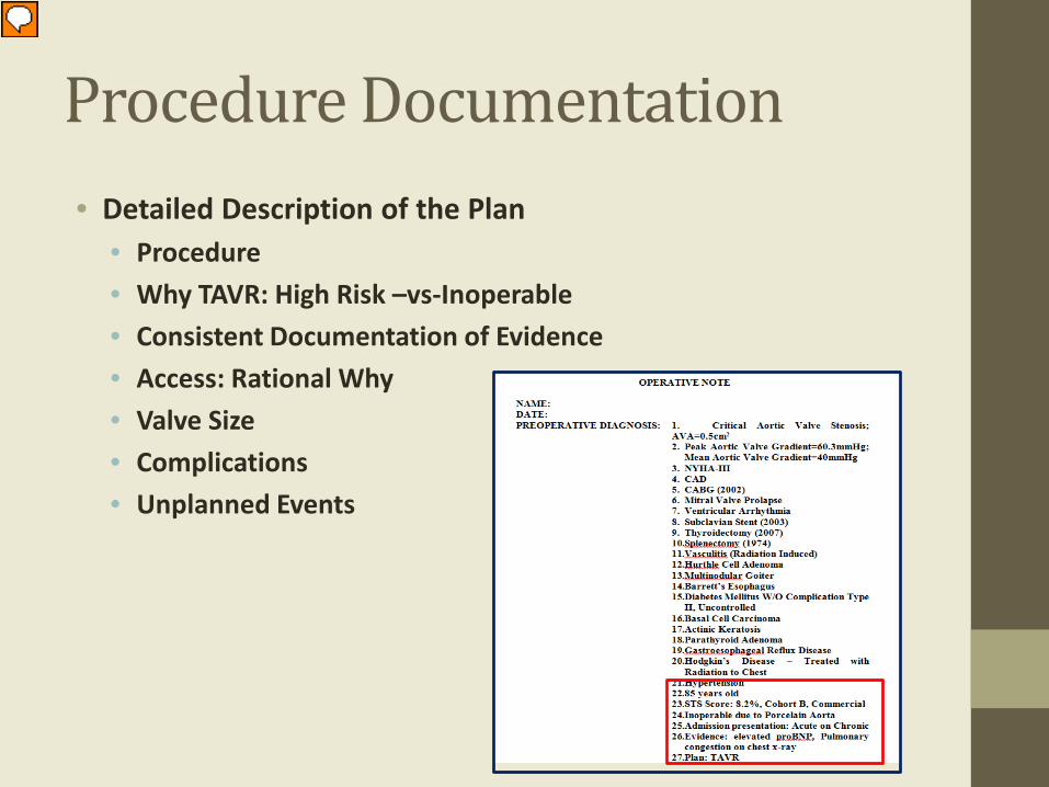
Procedure Documentation • Detailed Description of the Plan
• Procedure • Why TAVR: High Risk –vs-Inoperable • Consistent Documentation of Evidence • Access: Rational Why • Valve Size • Complications • Unplanned Events
Discharge Considerations • Understand the Post-Acute Transfer Rule • Length of Stay • Protocols
• Standard • Fast Track
• Decreasing Readmissions • Nurse Navigators • Schedule Early Post-op visits • Utilization of Outside Referrings and Physician Extenders • Consider supporting cost of local lodging
• Consistent Documentation
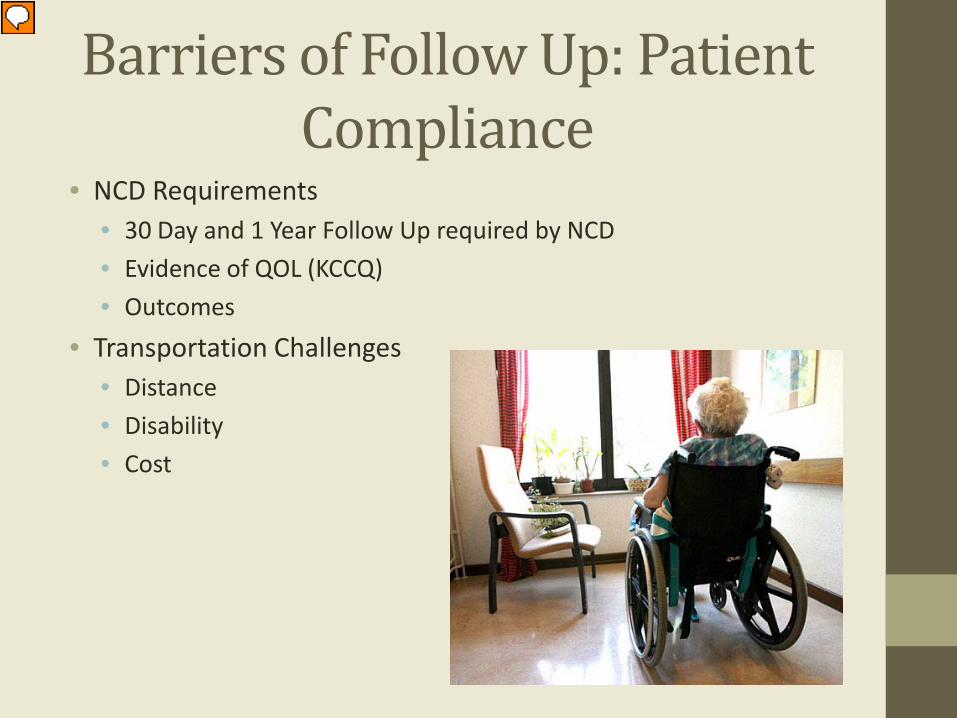
Barriers of Follow Up: Patient Compliance
• NCD Requirements • 30 Day and 1 Year Follow Up required by NCD • Evidence of QOL (KCCQ) • Outcomes
• Transportation Challenges • Distance • Disability • Cost
How to Overcome the Barriers
• Follow up appointments • Understand what is keeping for having the patient return • Can the visit be done locally • Do a phone visit • Does your institution offer vouchers • Work with the families • Involve social workers
• KCCQ
• Mail out to the patient in advance of the their appointment • Handout KCCQ while the patient is in the waiting room • Do the survey over the phone
Understand the Learning Curve • Coding and Documentation
• Develop Templates
• Set up a Tracking System • IT Support • Utilize your EMR System • Referral patterns
• Audit your program
• Where is the missing data • Volume • Understand the trickle down effects • LOS
• Continued Education
• Cross-training • TAVR Protocols
• Fast Track Protocol
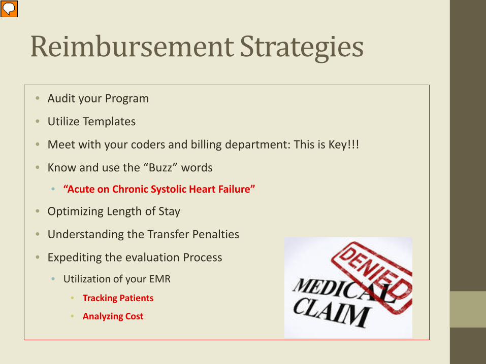
Reimbursement Strategies • Audit your Program
• Utilize Templates
• Meet with your coders and billing department: This is Key!!!
• Know and use the “Buzz” words
• “Acute on Chronic Systolic Heart Failure”
• Optimizing Length of Stay
• Understanding the Transfer Penalties
• Expediting the evaluation Process
• Utilization of your EMR
• Tracking Patients
• Analyzing Cost
Where Do we Go From Here? What are the Needs
• Consistent Definitions Across the Country
• Education of Clinicians on Documentation to meet the needs
of Reimbursement
• Continued Support from Industry for Education and
Awareness of Changes as New Technology Evolves
• Governmental Support to Understand the Importance and
Value of New Technology and the Challenges Associated with
Providing Access

The Ultimate Goal Quality of Life

Build Your TAVR Program: Understand your challenges Then the Sky’s The Limit
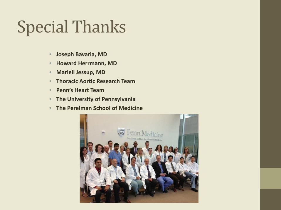
Special Thanks • Joseph Bavaria, MD • Howard Herrmann, MD • Mariell Jessup, MD • Thoracic Aortic Research Team • Penn’s Heart Team • The University of Pennsylvania • The Perelman School of Medicine

Thank You
Questions??
Templates And Protocols Courtesy of Penn Medicine’s Heart Team
Disclosure • The intent of the following information is to provide examples
of documentation and protocols used at the University of
Pennsylvania. Elizabeth K. Walsh and or any of her associates
at the University of Pennsylvania along with Edwards
Lifesciences, LLC are not liable for any damages or dispute of
claim.
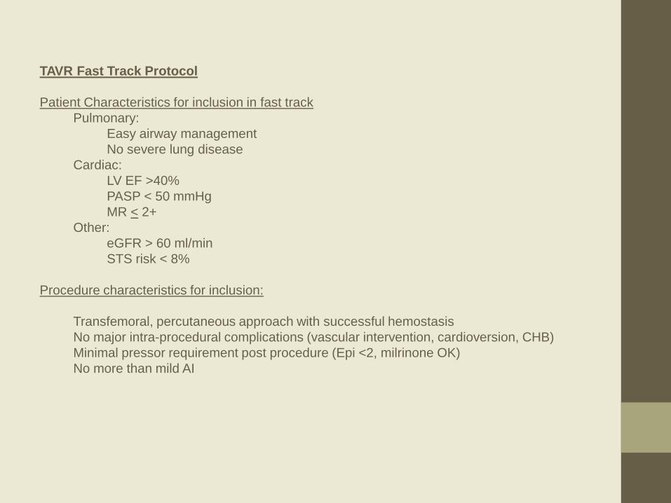
TAVR Fast Track Protocol Patient Characteristics for inclusion in fast track
Pulmonary: Easy airway management No severe lung disease
Cardiac: LV EF >40% PASP < 50 mmHg MR < 2+
Other: eGFR > 60 ml/min STS risk < 8%
Procedure characteristics for inclusion:
Transfemoral, percutaneous approach with successful hemostasis No major intra-procedural complications (vascular intervention, cardioversion, CHB) Minimal pressor requirement post procedure (Epi <2, milrinone OK) No more than mild AI
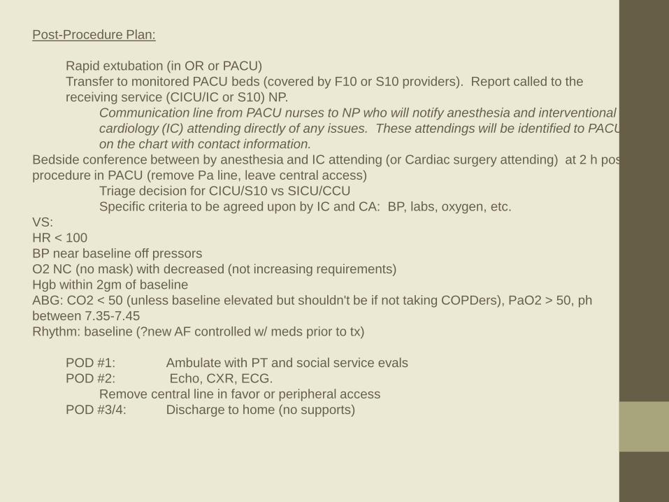
Post-Procedure Plan:
Rapid extubation (in OR or PACU) Transfer to monitored PACU beds (covered by F10 or S10 providers). Report called to the receiving service (CICU/IC or S10) NP.
Communication line from PACU nurses to NP who will notify anesthesia and interventional cardiology (IC) attending directly of any issues. These attendings will be identified to PACU on the chart with contact information.
Bedside conference between by anesthesia and IC attending (or Cardiac surgery attending) at 2 h post procedure in PACU (remove Pa line, leave central access)
Triage decision for CICU/S10 vs SICU/CCU Specific criteria to be agreed upon by IC and CA: BP, labs, oxygen, etc.
VS: HR < 100 BP near baseline off pressors O2 NC (no mask) with decreased (not increasing requirements) Hgb within 2gm of baseline ABG: CO2 < 50 (unless baseline elevated but shouldn't be if not taking COPDers), PaO2 > 50, ph between 7.35-7.45 Rhythm: baseline (?new AF controlled w/ meds prior to tx)
POD #1: Ambulate with PT and social service evals POD #2: Echo, CXR, ECG.
Remove central line in favor or peripheral access POD #3/4: Discharge to home (no supports)
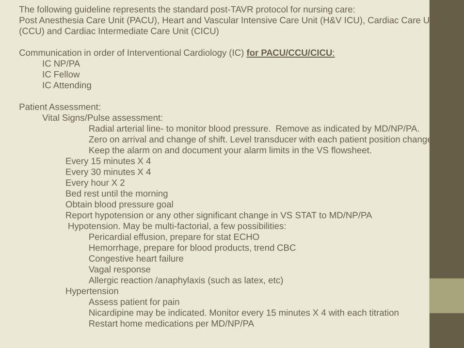
The following guideline represents the standard post-TAVR protocol for nursing care: Post Anesthesia Care Unit (PACU), Heart and Vascular Intensive Care Unit (H&V ICU), Cardiac Care Unit (CCU) and Cardiac Intermediate Care Unit (CICU) Communication in order of Interventional Cardiology (IC) for PACU/CCU/CICU:
IC NP/PA IC Fellow IC Attending
Patient Assessment:
Vital Signs/Pulse assessment: Radial arterial line- to monitor blood pressure. Remove as indicated by MD/NP/PA. Zero on arrival and change of shift. Level transducer with each patient position change. Keep the alarm on and document your alarm limits in the VS flowsheet.
Every 15 minutes X 4 Every 30 minutes X 4 Every hour X 2 Bed rest until the morning Obtain blood pressure goal Report hypotension or any other significant change in VS STAT to MD/NP/PA Hypotension. May be multi-factorial, a few possibilities:
Pericardial effusion, prepare for stat ECHO Hemorrhage, prepare for blood products, trend CBC Congestive heart failure Vagal response Allergic reaction /anaphylaxis (such as latex, etc)
Hypertension Assess patient for pain Nicardipine may be indicated. Monitor every 15 minutes X 4 with each titration Restart home medications per MD/NP/PA
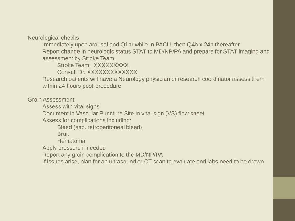
Neurological checks Immediately upon arousal and Q1hr while in PACU, then Q4h x 24h thereafter Report change in neurologic status STAT to MD/NP/PA and prepare for STAT imaging and assessment by Stroke Team.
Stroke Team: XXXXXXXXX Consult Dr. XXXXXXXXXXXXX
Research patients will have a Neurology physician or research coordinator assess them within 24 hours post-procedure
Groin Assessment
Assess with vital signs Document in Vascular Puncture Site in vital sign (VS) flow sheet Assess for complications including:
Bleed (esp. retroperitoneal bleed) Bruit Hematoma
Apply pressure if needed Report any groin complication to the MD/NP/PA If issues arise, plan for an ultrasound or CT scan to evaluate and labs need to be drawn
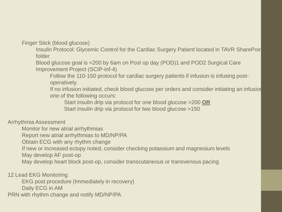
Finger Stick (blood glucose)
Insulin Protocol: Glycemic Control for the Cardiac Surgery Patient located in TAVR SharePoint folder Blood glucose goal is <200 by 6am on Post op day (POD)1 and POD2 Surgical Care Improvement Project (SCIP-inf-4)
Follow the 110-150 protocol for cardiac surgery patients if infusion is infusing post-operatively. If no infusion initiated, check blood glucose per orders and consider initiating an infusion if one of the following occurs:
Start insulin drip via protocol for one blood glucose >200 OR Start insulin drip via protocol for two blood glucose >150
Arrhythmia Assessment
Monitor for new atrial arrhythmias Report new atrial arrhythmias to MD/NP/PA Obtain ECG with any rhythm change If new or increased ectopy noted, consider checking potassium and magnesium levels May develop AF post-op May develop heart block post-op, consider transcutaneous or transvenous pacing
12 Lead EKG Monitoring
EKG post procedure (Immediately in recovery) Daily ECG in AM
PRN with rhythm change and notify MD/NP/PA
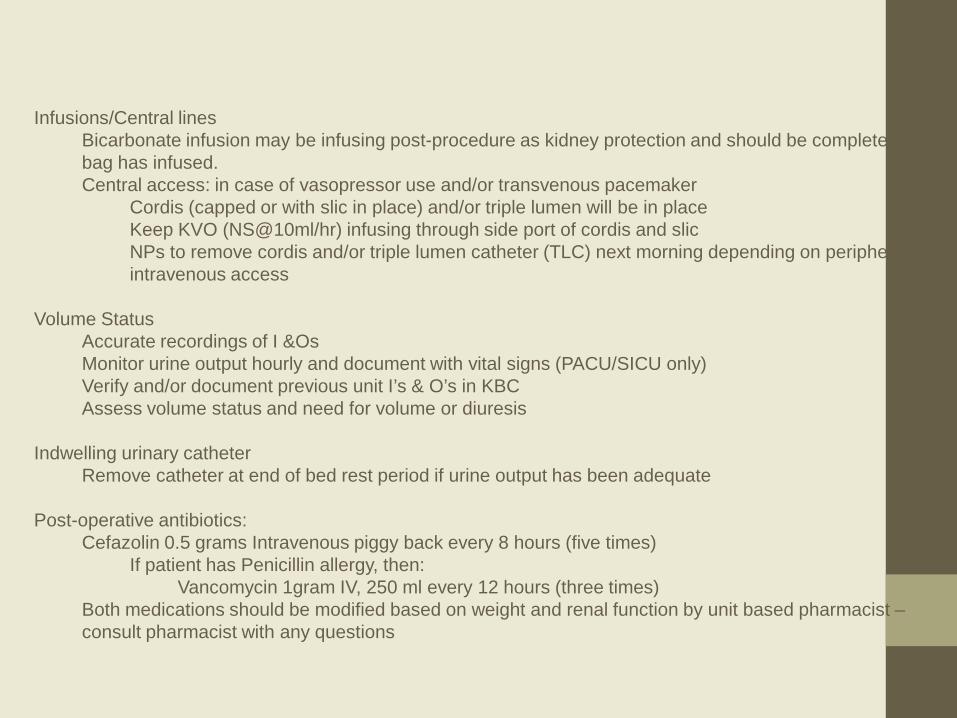
Infusions/Central lines Bicarbonate infusion may be infusing post-procedure as kidney protection and should be complete when bag has infused. Central access: in case of vasopressor use and/or transvenous pacemaker
Cordis (capped or with slic in place) and/or triple lumen will be in place Keep KVO (NS@10ml/hr) infusing through side port of cordis and slic NPs to remove cordis and/or triple lumen catheter (TLC) next morning depending on peripheral intravenous access
Volume Status
Accurate recordings of I &Os Monitor urine output hourly and document with vital signs (PACU/SICU only) Verify and/or document previous unit I’s & O’s in KBC Assess volume status and need for volume or diuresis
Indwelling urinary catheter
Remove catheter at end of bed rest period if urine output has been adequate Post-operative antibiotics:
Cefazolin 0.5 grams Intravenous piggy back every 8 hours (five times) If patient has Penicillin allergy, then:
Vancomycin 1gram IV, 250 ml every 12 hours (three times) Both medications should be modified based on weight and renal function by unit based pharmacist – consult pharmacist with any questions
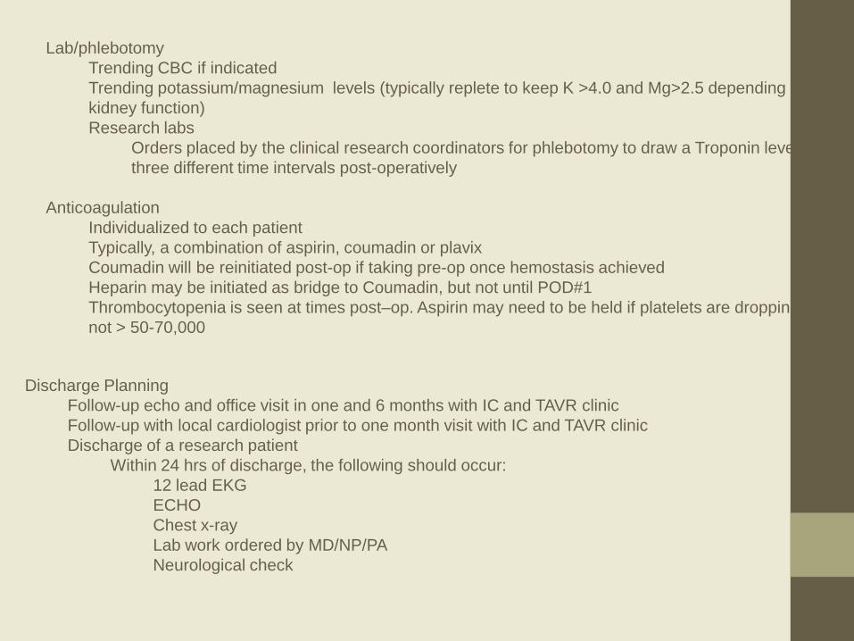
Lab/phlebotomy Trending CBC if indicated Trending potassium/magnesium levels (typically replete to keep K >4.0 and Mg>2.5 depending on kidney function) Research labs
Orders placed by the clinical research coordinators for phlebotomy to draw a Troponin level at three different time intervals post-operatively
Anticoagulation
Individualized to each patient Typically, a combination of aspirin, coumadin or plavix Coumadin will be reinitiated post-op if taking pre-op once hemostasis achieved Heparin may be initiated as bridge to Coumadin, but not until POD#1 Thrombocytopenia is seen at times post–op. Aspirin may need to be held if platelets are dropping and not > 50-70,000
Discharge Planning Follow-up echo and office visit in one and 6 months with IC and TAVR clinic Follow-up with local cardiologist prior to one month visit with IC and TAVR clinic Discharge of a research patient
Within 24 hrs of discharge, the following should occur: 12 lead EKG ECHO Chest x-ray Lab work ordered by MD/NP/PA Neurological check
BASELINE FRAILTY TESTING: 4 evaluations: Albumin, ADLS, Grip Strength, 15 ft walk test Albumin Testing Note: Albumin Level <3.6 g/dL meets cutoff for frailty. Serum Albumin: *** g/dL Date Obtained: *** Time:***
Katz Activities of Daily Living Note: if patient completes 2/6 or less he or she meets cutoff for frailty General Guidelines for Points NO supervision, direction or personal assistance (1 POINT) WITH supervision, direction, personal assistance, or total care (O POINTS) BATHING POINTS:____***_______ Bathes self completely or needs help in bathing only a single part of the body such as the back, genital area or disabled extremity. Needs help with bathing more than one part of the body, getting in or out of the tub or shower. Requires total bathing. (0 POINTS) DRESSING POINTS: _____***______ Gets clothes from closets and drawers and puts on clothes and outer garments complete with fasteners. May have help tying shoes Needs help with dressing self or needs to be completely dressed. (0 POINTS) TOILETING POINTS:_____***______ Goes to toilet, gets on and off, arranges clothes, cleans genital area without help. (1 POINT) Needs help transferring to the toilet, cleaning self or uses bedpan or commode. (0 POINTS) TRANSFERRING POINTS:____***_______ Moves in and out of bed or chair unassisted. Mechanical transferring aides are acceptable. (1 POINT) Needs help in moving from bed to chair or requires a complete transfer. (0 POINTS) CONTINENCE POINTS:_____***______ Exercises complete self control over urination and defecation. (1 POINT) Is partially or totally incontinent of bowel or bladder. (0 POINTS) FEEDING POINTS:_____***______ Gets food from plate into mouth without help. Preparation of food may be done by another person. (1 POINT) Needs partial or total help with feeding or requires parenteral feeding. (0 POINTS) TOTAL POINTS _____***____
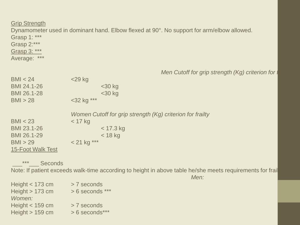
Grip Strength Dynamometer used in dominant hand. Elbow flexed at 90°. No support for arm/elbow allowed. Grasp 1: *** Grasp 2:*** Grasp 3: *** Average: *** Men Cutoff for grip strength (Kg) criterion for frailty BMI < 24 <29 kg BMI 24.1-26 <30 kg BMI 26.1-28 <30 kg BMI > 28 <32 kg *** Women Cutoff for grip strength (Kg) criterion for frailty BMI < 23 < 17 kg BMI 23.1-26 < 17.3 kg BMI 26.1-29 < 18 kg BMI > 29 < 21 kg *** 15-Foot Walk Test ___***___ Seconds Note: If patient exceeds walk-time according to height in above table he/she meets requirements for frailty. Men: Height < 173 cm > 7 seconds Height > 173 cm > 6 seconds *** Women: Height < 159 cm > 7 seconds Height > 159 cm > 6 seconds***
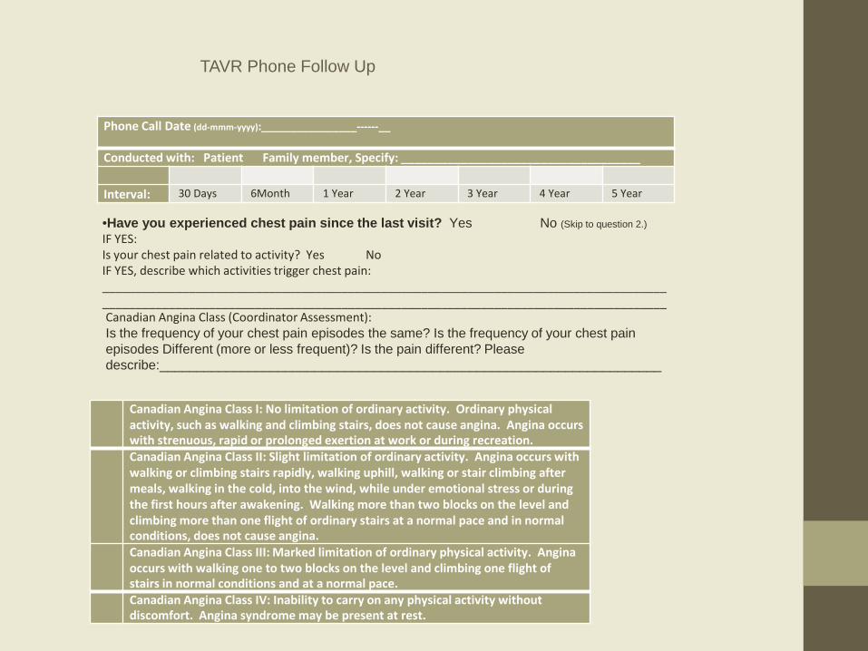
Phone Call Date (dd-mmm-yyyy):________________------__
Conducted with: Patient Family member, Specify: ____________________________________
Interval: 30 Days 6Month 1 Year 2 Year 3 Year 4 Year 5 Year
•Have you experienced chest pain since the last visit? Yes No (Skip to question 2.) IF YES: Is your chest pain related to activity? Yes No IF YES, describe which activities trigger chest pain: _____________________________________________________________________________________ _____________________________________________________________________________________
TAVR Phone Follow Up
Canadian Angina Class I: No limitation of ordinary activity. Ordinary physical activity, such as walking and climbing stairs, does not cause angina. Angina occurs with strenuous, rapid or prolonged exertion at work or during recreation.
Canadian Angina Class II: Slight limitation of ordinary activity. Angina occurs with walking or climbing stairs rapidly, walking uphill, walking or stair climbing after meals, walking in the cold, into the wind, while under emotional stress or during the first hours after awakening. Walking more than two blocks on the level and climbing more than one flight of ordinary stairs at a normal pace and in normal conditions, does not cause angina.
Canadian Angina Class III: Marked limitation of ordinary physical activity. Angina occurs with walking one to two blocks on the level and climbing one flight of stairs in normal conditions and at a normal pace.
Canadian Angina Class IV: Inability to carry on any physical activity without discomfort. Angina syndrome may be present at rest.
Canadian Angina Class (Coordinator Assessment): Is the frequency of your chest pain episodes the same? Is the frequency of your chest pain episodes Different (more or less frequent)? Is the pain different? Please describe:____________________________________________________________________
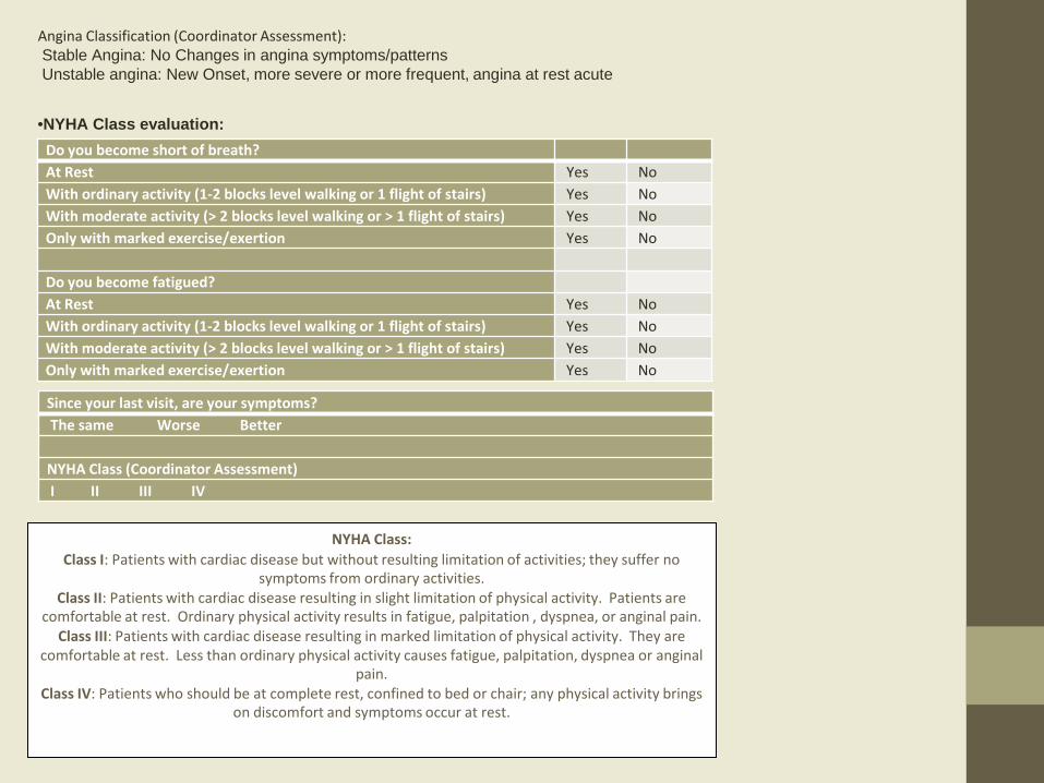
Angina Classification (Coordinator Assessment): Stable Angina: No Changes in angina symptoms/patterns Unstable angina: New Onset, more severe or more frequent, angina at rest acute
Do you become short of breath? At Rest Yes No With ordinary activity (1-2 blocks level walking or 1 flight of stairs) Yes No With moderate activity (> 2 blocks level walking or > 1 flight of stairs) Yes No Only with marked exercise/exertion Yes No Do you become fatigued? At Rest Yes No With ordinary activity (1-2 blocks level walking or 1 flight of stairs) Yes No With moderate activity (> 2 blocks level walking or > 1 flight of stairs) Yes No Only with marked exercise/exertion Yes No
•NYHA Class evaluation:
Since your last visit, are your symptoms? The same Worse Better NYHA Class (Coordinator Assessment) I II III IV
NYHA Class: Class I: Patients with cardiac disease but without resulting limitation of activities; they suffer no
symptoms from ordinary activities. Class II: Patients with cardiac disease resulting in slight limitation of physical activity. Patients are
comfortable at rest. Ordinary physical activity results in fatigue, palpitation , dyspnea, or anginal pain. Class III: Patients with cardiac disease resulting in marked limitation of physical activity. They are
comfortable at rest. Less than ordinary physical activity causes fatigue, palpitation, dyspnea or anginal pain.
Class IV: Patients who should be at complete rest, confined to bed or chair; any physical activity brings on discomfort and symptoms occur at rest.
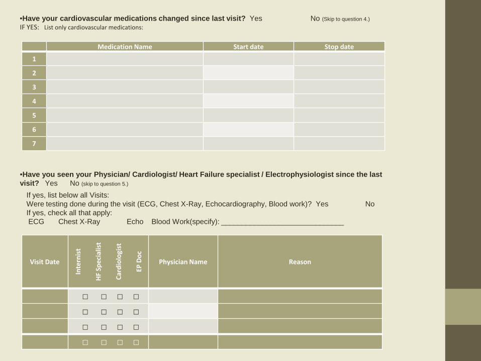
Medication Name Start date Stop date
1
2
3
4
5
6
7
•Have your cardiovascular medications changed since last visit? Yes No (Skip to question 4.) IF YES: List only cardiovascular medications:
•Have you seen your Physician/ Cardiologist/ Heart Failure specialist / Electrophysiologist since the last visit? Yes No (skip to question 5.)
Visit Date
Inte
rnis
t
HF S
peci
alis
t
Card
iolo
gist
EP D
oc
Physician Name Reason
□ □ □ □
□ □ □ □
□ □ □ □
□ □ □ □
If yes, list below all Visits: Were testing done during the visit (ECG, Chest X-Ray, Echocardiography, Blood work)? Yes No If yes, check all that apply: ECG Chest X-Ray Echo Blood Work(specify): ______________________________
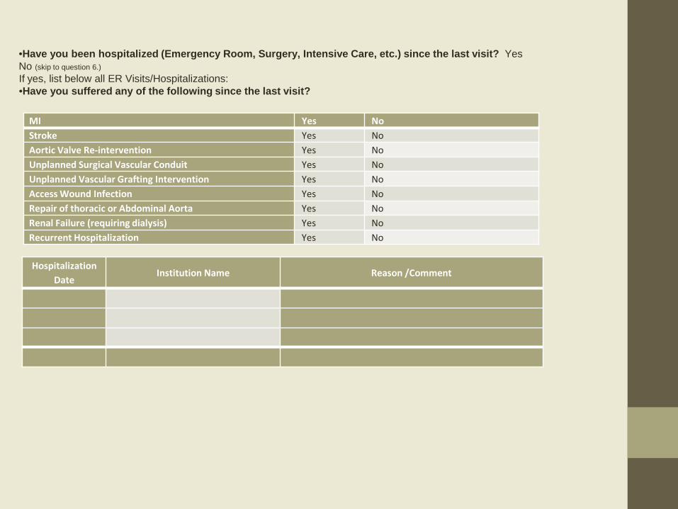
Hospitalization Date
Institution Name Reason /Comment
MI Yes No Stroke Yes No Aortic Valve Re-intervention Yes No Unplanned Surgical Vascular Conduit Yes No Unplanned Vascular Grafting Intervention Yes No Access Wound Infection Yes No Repair of thoracic or Abdominal Aorta Yes No Renal Failure (requiring dialysis) Yes No Recurrent Hospitalization Yes No
•Have you been hospitalized (Emergency Room, Surgery, Intensive Care, etc.) since the last visit? Yes No (skip to question 6.) If yes, list below all ER Visits/Hospitalizations: •Have you suffered any of the following since the last visit?
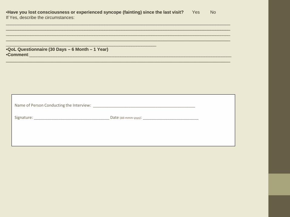
•Have you lost consciousness or experienced syncope (fainting) since the last visit? Yes No If Yes, describe the circumstances: _________________________________________________________________________________________________________________________________________________________________________________________________________________________________________________________________________________________________________________________________________________________________________________________________________________________________________ •QoL Questionnaire (30 Days – 6 Month – 1 Year) •Comment:_____________________________________________________________________________________________________________________________________________________________________________
Name of Person Conducting the Interview: ______________________________________________ Signature: __________________________________ Date (dd-mmm-yyyy): _________________________
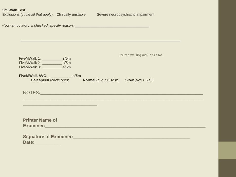
5m Walk Test Exclusions (circle all that apply): Clinically unstable Severe neuropsychiatric impairment
•Non-ambulatory. If checked, specify reason: _____________________________________
Utilized walking aid? Yes / No
FiveMWalk 1: __________ s/5m FiveMWalk 2: __________ s/5m FiveMWalk 3: __________ s/5m FiveMWalk AVG: ___________ s/5m Gait speed (circle one): Normal (avg ≤ 6 s/5m) Slow (avg > 6 s/5
NOTES:____________________________________________________________________________________________________________________________________________________________________ Printer Name of Examiner:_______________________________________________________________ Signature of Examiner:______________________________________________ Date:__________
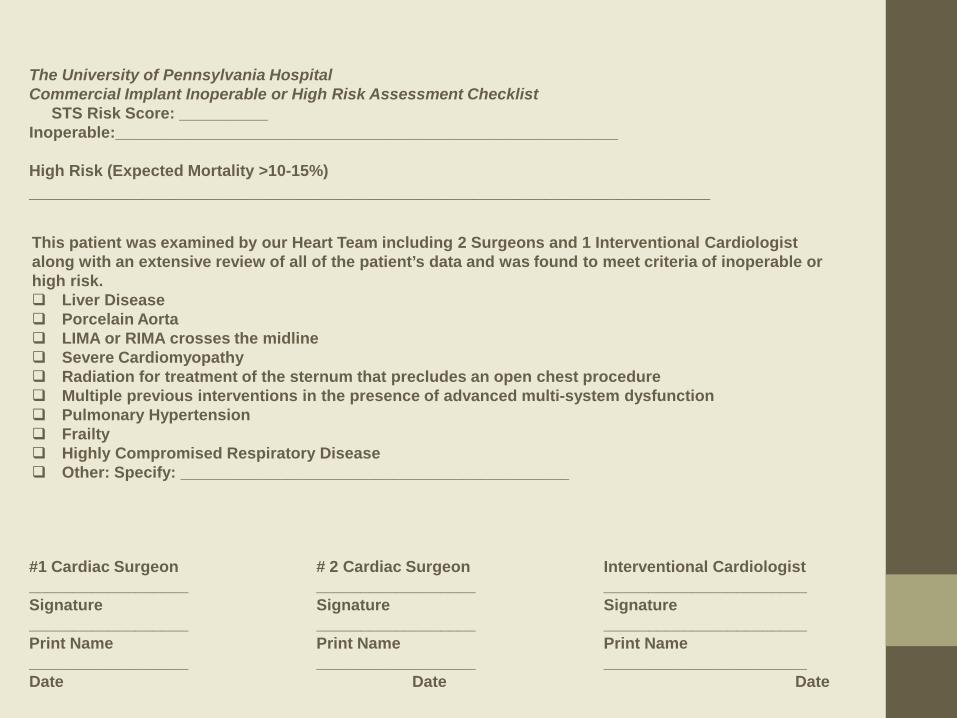
The University of Pennsylvania Hospital Commercial Implant Inoperable or High Risk Assessment Checklist STS Risk Score: __________ Inoperable:_________________________________________________________ High Risk (Expected Mortality >10-15%) __________________________________________________________________
This patient was examined by our Heart Team including 2 Surgeons and 1 Interventional Cardiologist along with an extensive review of all of the patient’s data and was found to meet criteria of inoperable or high risk. Liver Disease Porcelain Aorta LIMA or RIMA crosses the midline Severe Cardiomyopathy Radiation for treatment of the sternum that precludes an open chest procedure Multiple previous interventions in the presence of advanced multi-system dysfunction Pulmonary Hypertension Frailty Highly Compromised Respiratory Disease Other: Specify: ____________________________________________
#1 Cardiac Surgeon # 2 Cardiac Surgeon Interventional Cardiologist __________________ __________________ _______________________ Signature Signature Signature __________________ __________________ _______________________ Print Name Print Name Print Name __________________ __________________ _______________________ Date Date Date
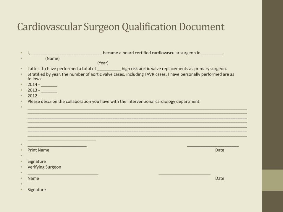
Cardiovascular Surgeon Qualification Document
• I, ______________________________ became a board certified cardiovascular surgeon in _________. • (Name)
(Year) • I attest to have performed a total of __________ high risk aortic valve replacements as primary surgeon. • Stratified by year, the number of aortic valve cases, including TAVR cases, I have personally performed are as
follows: • 2014 - _______ • 2013 - _______ • 2012 - _______ • Please describe the collaboration you have with the interventional cardiology department. • _____________________________________________________________________________________________
___________________________________________________________________________________________________________________________________________________________________________________________________________________________________________________________________________________________________________________________________________________________________________________________________________________________________________________________________________________________________________________________________________________________________________________________________________
• _________________________ ______________________ • Print Name Date • • Signature • Verifying Surgeon • ______________________________ ______________________ • Name Date • • Signature
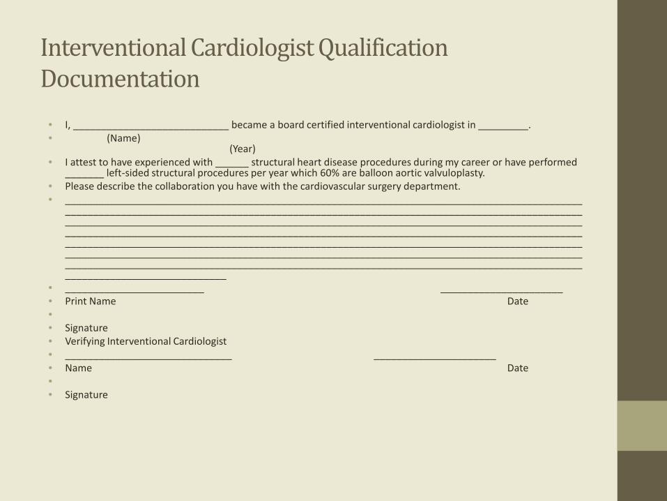
Interventional Cardiologist Qualification Documentation
• I, ____________________________ became a board certified interventional cardiologist in _________. • (Name)
(Year) • I attest to have experienced with ______ structural heart disease procedures during my career or have performed
_______ left-sided structural procedures per year which 60% are balloon aortic valvuloplasty. • Please describe the collaboration you have with the cardiovascular surgery department. • _____________________________________________________________________________________________
___________________________________________________________________________________________________________________________________________________________________________________________________________________________________________________________________________________________________________________________________________________________________________________________________________________________________________________________________________________________________________________________________________________________________________________________________________
• _________________________ ______________________ • Print Name Date • • Signature • Verifying Interventional Cardiologist • ______________________________ ______________________ • Name Date • • Signature
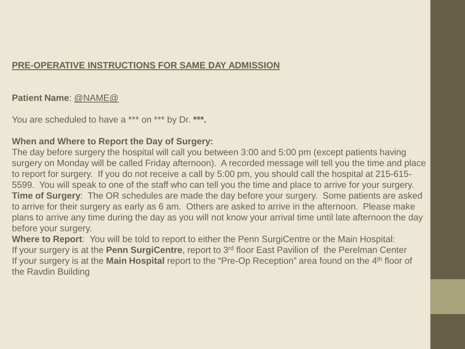
PRE-OPERATIVE INSTRUCTIONS FOR SAME DAY ADMISSION Patient Name: @NAME@ You are scheduled to have a *** on *** by Dr. ***. When and Where to Report the Day of Surgery: The day before surgery the hospital will call you between 3:00 and 5:00 pm (except patients having surgery on Monday will be called Friday afternoon). A recorded message will tell you the time and place to report for surgery. If you do not receive a call by 5:00 pm, you should call the hospital at 215-615-5599. You will speak to one of the staff who can tell you the time and place to arrive for your surgery. Time of Surgery: The OR schedules are made the day before your surgery. Some patients are asked to arrive for their surgery as early as 6 am. Others are asked to arrive in the afternoon. Please make plans to arrive any time during the day as you will not know your arrival time until late afternoon the day before your surgery. Where to Report: You will be told to report to either the Penn SurgiCentre or the Main Hospital: If your surgery is at the Penn SurgiCentre, report to 3rd floor East Pavilion of the Perelman Center If your surgery is at the Main Hospital report to the “Pre-Op Reception” area found on the 4th floor of the Ravdin Building
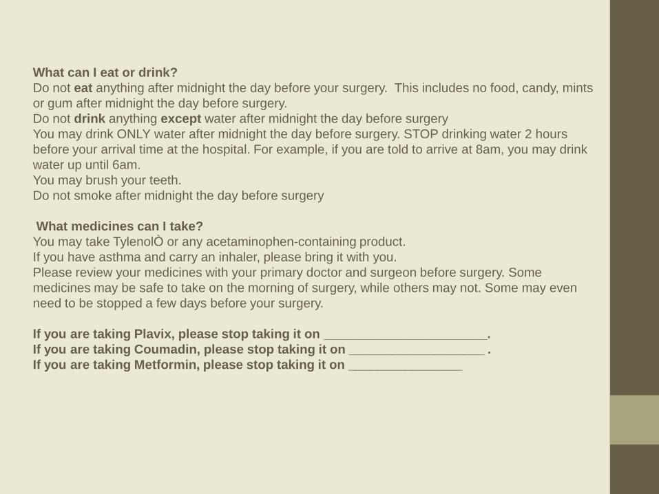
What can I eat or drink? Do not eat anything after midnight the day before your surgery. This includes no food, candy, mints or gum after midnight the day before surgery. Do not drink anything except water after midnight the day before surgery You may drink ONLY water after midnight the day before surgery. STOP drinking water 2 hours before your arrival time at the hospital. For example, if you are told to arrive at 8am, you may drink water up until 6am. You may brush your teeth. Do not smoke after midnight the day before surgery What medicines can I take? You may take TylenolÒ or any acetaminophen-containing product. If you have asthma and carry an inhaler, please bring it with you. Please review your medicines with your primary doctor and surgeon before surgery. Some medicines may be safe to take on the morning of surgery, while others may not. Some may even need to be stopped a few days before your surgery. If you are taking Plavix, please stop taking it on _______________________. If you are taking Coumadin, please stop taking it on ___________________ . If you are taking Metformin, please stop taking it on ________________
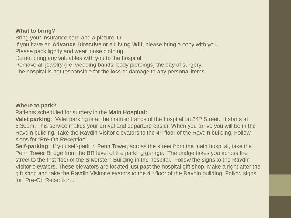
What to bring? Bring your insurance card and a picture ID. If you have an Advance Directive or a Living Will, please bring a copy with you. Please pack lightly and wear loose clothing. Do not bring any valuables with you to the hospital. Remove all jewelry (i.e. wedding bands, body piercings) the day of surgery. The hospital is not responsible for the loss or damage to any personal items. Where to park? Patients scheduled for surgery in the Main Hospital: Valet parking: Valet parking is at the main entrance of the hospital on 34th Street. It starts at 5:30am. This service makes your arrival and departure easier. When you arrive you will be in the Ravdin building. Take the Ravdin Visitor elevators to the 4th floor of the Ravdin building. Follow signs for “Pre-Op Reception”. Self-parking: If you self-park in Penn Tower, across the street from the main hospital, take the Penn Tower Bridge from the BR level of the parking garage. The bridge takes you across the street to the first floor of the Silverstein Building in the hospital. Follow the signs to the Ravdin Visitor elevators. These elevators are located just past the hospital gift shop. Make a right after the gift shop and take the Ravdin Visitor elevators to the 4th floor of the Ravdin building. Follow signs for “Pre-Op Reception”.
Patients scheduled for surgery at the SurgiCentre in the Perelman Center for Advanced Medicine: Valet parking: Valet parking is at the main entrance of the Perelman Center on Civic Center Boulevard. Enter through the revolving door and make a left. You will see a sign for the SurgiCentre. Walk through those doors to the East Pavilion elevators. Take these elevators to the 3rd floor. These will take you to the SurgiCentre reception area. Self-parking: Self-parking is found in the garage under the Perelman Center. You can enter the parking garage in the rear of the building. There are reserved parking spaces on the P1 level for surgery patients and families. An elevator is located in the corner of the garage on the P1 level that takes you directly to the SurgiCentre reception area on the 3rd floor. Surgery Schedule Surgery times vary and delays can occur when caring for patients. We ask for your patience while waiting for surgery. Families can visit other parts of the hospital or the campus while they are waiting. Your family should check-in with the receptionist when they leave and return to the waiting area.
Where will my family wait while I am in surgery? Main Hospital: •If you are staying overnight, your family will wait in the Surgical Family Waiting Lounge on the 2nd floor of the Dulles Building. •If you are going home the same day as your surgery, your family will stay in the reception area on the 4th floor of the Ravdin building. Perelman SurgiCentre: •Your family will wait in the SurgiCentre Reception area (where you reported for surgery). **Wherever your family is waiting, the surgeon will talk to your family in that area. Who can help me if I have questions while I wait?
There is staff in each area who will address any concerns you may have. Staff in these areas will also assist you in getting to any other surgery related appointments you have on this same day, such as radiology. What do I need to know about going home? Patients going home on the same day as your surgery* •You may not drive yourself home if you have had any sedation. •You must have an adult take you home. •If you are taking a bus, train, cab or para-transit, another adult must be with you. *Your surgery will be rescheduled if the above plans cannot be made. Patients being admitted to the hospital after surgery •Please start to make plans on how you will get home in advance. •Plan to leave the hospital around 10 am on the day you are going home.
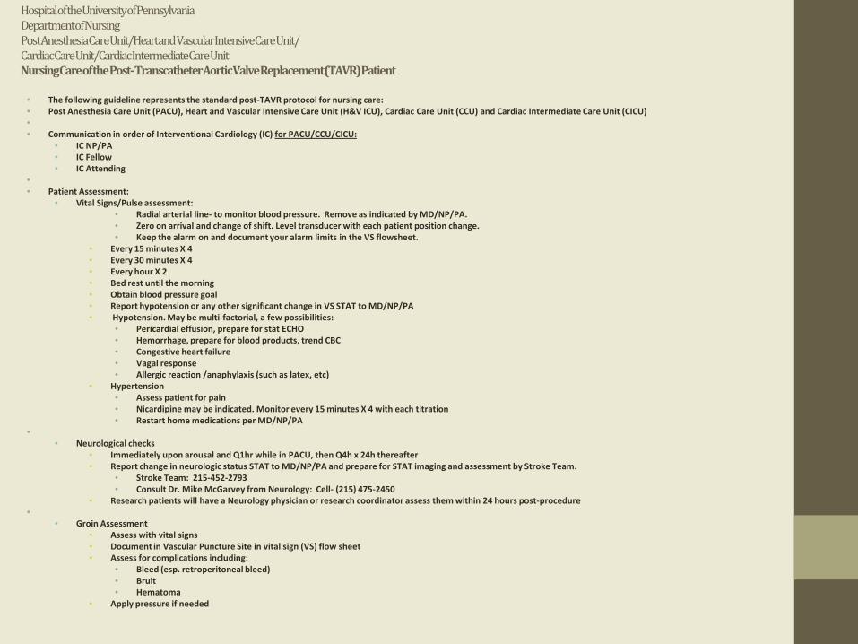
Hospital of the University of Pennsylvania Department of Nursing Post Anesthesia Care Unit/Heart and Vascular Intensive Care Unit/ Cardiac Care Unit/Cardiac Intermediate Care Unit Nursing Care of the Post- Transcatheter Aortic Valve Replacement (TAVR) Patient
• The following guideline represents the standard post-TAVR protocol for nursing care: • Post Anesthesia Care Unit (PACU), Heart and Vascular Intensive Care Unit (H&V ICU), Cardiac Care Unit (CCU) and Cardiac Intermediate Care Unit (CICU) • • Communication in order of Interventional Cardiology (IC) for PACU/CCU/CICU:
• IC NP/PA • IC Fellow • IC Attending
• • Patient Assessment:
• Vital Signs/Pulse assessment: • Radial arterial line- to monitor blood pressure. Remove as indicated by MD/NP/PA. • Zero on arrival and change of shift. Level transducer with each patient position change. • Keep the alarm on and document your alarm limits in the VS flowsheet.
• Every 15 minutes X 4 • Every 30 minutes X 4 • Every hour X 2 • Bed rest until the morning • Obtain blood pressure goal • Report hypotension or any other significant change in VS STAT to MD/NP/PA • Hypotension. May be multi-factorial, a few possibilities:
• Pericardial effusion, prepare for stat ECHO • Hemorrhage, prepare for blood products, trend CBC • Congestive heart failure • Vagal response • Allergic reaction /anaphylaxis (such as latex, etc)
• Hypertension • Assess patient for pain • Nicardipine may be indicated. Monitor every 15 minutes X 4 with each titration • Restart home medications per MD/NP/PA
• • Neurological checks
• Immediately upon arousal and Q1hr while in PACU, then Q4h x 24h thereafter • Report change in neurologic status STAT to MD/NP/PA and prepare for STAT imaging and assessment by Stroke Team.
• Stroke Team: 215-452-2793 • Consult Dr. Mike McGarvey from Neurology: Cell- (215) 475-2450
• Research patients will have a Neurology physician or research coordinator assess them within 24 hours post-procedure •
• Groin Assessment • Assess with vital signs • Document in Vascular Puncture Site in vital sign (VS) flow sheet • Assess for complications including:
• Bleed (esp. retroperitoneal bleed) • Bruit • Hematoma
• Apply pressure if needed
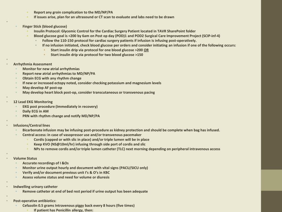
• Report any groin complication to the MD/NP/PA • If issues arise, plan for an ultrasound or CT scan to evaluate and labs need to be drawn
• • Finger Stick (blood glucose)
• Insulin Protocol: Glycemic Control for the Cardiac Surgery Patient located in TAVR SharePoint folder • Blood glucose goal is <200 by 6am on Post op day (POD)1 and POD2 Surgical Care Improvement Project (SCIP-inf-4)
• Follow the 110-150 protocol for cardiac surgery patients if infusion is infusing post-operatively. • If no infusion initiated, check blood glucose per orders and consider initiating an infusion if one of the following occurs:
• Start insulin drip via protocol for one blood glucose >200 OR • Start insulin drip via protocol for two blood glucose >150
• • Arrhythmia Assessment
• Monitor for new atrial arrhythmias • Report new atrial arrhythmias to MD/NP/PA • Obtain ECG with any rhythm change • If new or increased ectopy noted, consider checking potassium and magnesium levels • May develop AF post-op • May develop heart block post-op, consider transcutaneous or transvenous pacing
• • 12 Lead EKG Monitoring
• EKG post procedure (Immediately in recovery) • Daily ECG in AM • PRN with rhythm change and notify MD/NP/PA
• • Infusions/Central lines
• Bicarbonate infusion may be infusing post-procedure as kidney protection and should be complete when bag has infused. • Central access: in case of vasopressor use and/or transvenous pacemaker
• Cordis (capped or with slic in place) and/or triple lumen will be in place • Keep KVO (NS@10ml/hr) infusing through side port of cordis and slic • NPs to remove cordis and/or triple lumen catheter (TLC) next morning depending on peripheral intravenous access
• • Volume Status
• Accurate recordings of I &Os • Monitor urine output hourly and document with vital signs (PACU/SICU only) • Verify and/or document previous unit I’s & O’s in KBC • Assess volume status and need for volume or diuresis
• • Indwelling urinary catheter
• Remove catheter at end of bed rest period if urine output has been adequate • • Post-operative antibiotics:
• Cefazolin 0.5 grams Intravenous piggy back every 8 hours (five times) • If patient has Penicillin allergy, then:
• Vancomycin 1gram IV, 250 ml every 12 hours (three times) • Both medications should be modified based on weight and renal function by unit based pharmacist – consult pharmacist
with any questions • • Lab/phlebotomy
• Trending CBC if indicated • Trending potassium/magnesium levels (typically replete to keep K >4.0 and Mg>2.5 depending on kidney function) • Research labs
• Orders placed by the clinical research coordinators for phlebotomy to draw a Troponin level at three different time intervals post-operatively
• • Anticoagulation
• Individualized to each patient • Typically, a combination of aspirin, coumadin or plavix • Coumadin will be reinitiated post-op if taking pre-op once hemostasis achieved • Heparin may be initiated as bridge to Coumadin, but not until POD#1 • Thrombocytopenia is seen at times post–op. Aspirin may need to be held if platelets are dropping and not > 50-70,000
• • Transthoracic Echocardiogram
• Done prior to discharge • • • Discharge Planning
• Follow-up echo and office visit in one and 6 months with IC and TAVR clinic • Follow-up with local cardiologist prior to one month visit with IC and TAVR clinic • Discharge of a research patient
• Within 24 hrs of discharge, the following should occur: • 12 lead EKG • ECHO • Chest x-ray • Lab work ordered by MD/NP/PA • Neurological check
Hospital of the University of Pennsylvania Department of Nursing
Post Anesthesia Care Unit/Heart and Vascular Intensive Care Unit/ Cardiac Care Unit/Cardiac Intermediate Care Unit
Nursing Care of the Post- Transcatheter Aortic Valve Replacement (TAVR) Patient
Penn Transcatheter Aortic Valve Replacement (TAVR) Anesthesia Guide Fall 2012
• TAVR procedures resemble complex cardiac cases because patients are typically
very elderly with multiple comorbidities. The TAVR patient is typically at excessive risk for conventional aortic valve replacement. In general, the anesthetic plan resembles the clinical approach to a ‘redo sternotomy cardiac case’ with the addition of TIVA (propofol and/or remifentanil) and allowances for concomitant morbidities and frailty. Although most cases go smoothly, the possibility of severe and sudden complications may require immediate cardiopulmonary bypass. So, it is essential to be prepared for these possibilities.
• Case Setup • The day before, you should request a full cardiac setup with 8 Alaris channels,
the so-called “TAVR Setup.” Request from pharmacy norepinephrine and sodium bicarbonate (154 mEq sodium bicarb in 1L D5W) so that pharmacy will have them ready with everything else.
• Order blood early and make sure it is in the pharmacy before starting the procedure: 4 units PRBC for transfemoral cases; 6 units PRBC for transapical cases or patients with prior sternotomy.
• From pharmacy, pick up: 1 Cardiac Pack, 2 sticks Nicardipine, 2 bags Phenylephrine, 1 bottle insulin, 2 large bottles Propofol, the previously requested bicarb infusion, 1 Milrinone infusion, 1 Norepinephrine infusion, 1 Vasopression infusion, 1 Nicardipine infusion, 2 bags Potassium, 2 vials Cisatracurium (if the patient has renal dysfunction, many do)
• From core or antibiotic fridge outside OR31, pick up: 2g Cefazolin and 1g Vancomycin: if the patient has antibiotic allergies, review antibiotic plan with your attending.
• From Pyxis, draw up: 2mg Midazolam, 200mcg Fentanyl, 200mg Propofol and/or 20mg Etomidate, 20mL Vecuronium or Cisatracurium, Vasopressin, and Ephedrine: consider drawing up a syringe of Norepinephrine (64mcg/1mL from bag with 7mL NS yielding 8mcg/mL)
• Setup infusions: formost cases, you will need 8 channels and 6 stopcocks. Add a high pressure extension to the end of the stopcocks. Use the bicarb infusion as your rider and have these infusions ready to go: propofol (@25), remi (@0.1), epinephrine (@2), and phenylephrine (@50). Setup Nicardipine (@4) for transapical cases. Consider milrinone and inhaled flolan if there is significant pulmonary hypertension (PAP > 50) and/or right ventricular dysfunction.
• Arrange the OR: There is limited space. The anesthesia machine goes in the angled red box on the floor. Alaris pumps on one pole to the right of the anesthesia machine. 2 IV warmers on one pole to the left of the anesthesia machine. Triple setup (PA/CVP/ART) attached to OR table using special rail adapter. Add extensions to the IV lines and the A-line.
• Usual setup for a cardiac case: machine check, airway supplies, suction, monitors, Swan setup, calibrate SvO2, central line (regular 9Fr and triple lumen introducer kits) supplies and ultrasound, vanco on microdripper, IV and A-line kits, ABG slips/syringes/bags, ICU signout and blood gas record sheets
• Have additional equipment ready: extra Velcro straps, Arrow quickflash catheters, micropuncture kits, pink foam with arm boards, R2 pads, pacemaker
• Grab circumferential (2-piece) lead for the resident and attending. The lead goes quickly, especially the apron and vest combos which are important for anesthesia staff as we are near the C-arm but turn frequently for the TEE, monitors and other machines.
• It is helpful to see review the anesthetic plan and setup with one of the cardiac anesthesia fellows.
• Anesthetic Plan • • Induction and Line Up • Place external defibrillation pads (R2 pads) as you are transferring the patient to the bed. These will
need to be checked prior to draping. • Routine monitors, reposition all wires from across patients back which might interfere with fluoroscopy,
connect IV, start Vanco • Pre-induction A-line, limit midazolam (0.5 mg per dose). If radial a-line is difficult to place, consider
transducing femoral a-line • Induction: Endotracheal Intubation is typically with an 8.0 mm ETT. • Large bore central IV access: ‘Double venous cannulation’ with an introducer for PA catheter and triple
lumen central venous catheter. • Oximetric CCO PA catheter
• Left Bundle Branch Block: due to risk of complete heart block, only float PA catheter after transvenous pacing wire is deployed. It is imperative to rule out LBBB by careful review of the preoperative electrocardiogram.
• If PA catheter is difficult to float, consider the use of fluoroscopy or having the cardiologist position it from the femoral vein.
• Hook up and check all lines, monitors, IVs, nasal and bladder temp probes • Arrange all monitor wires and lines between the OR table and the anesthesia machine. They need to
have enough slack to allow for table movement during the case. You can also bundle the lines and wires between the OR table and anesthesia machine with blue Velcro straps. This is usually helpful in making sure our lines aren’t disrupted during fluoroscopy.
•
• During the Case • These cases are typically performed via a transfemoral (TF), transapical (TA - left
minithoracotomy) or a transaortic (TAO – ministernotomy) approach. • CPB machine with perfusionist must be available in OR
• Dry setup for TF cases, pump primed for TA/TAO cases • IABP leads are placed and checked after anesthesia induction
• Antibiotics: 1g Vancomycin and 2g Cefazolin: if the patient has a true penicillin allergy, substitute Cefazolin with Levofloxacin. Avoid Gentamicin due to high risk of renal dysfunction in these elderly high-risk patients. If the patient has a true Vancomycin allergy, consider Clindamycin 600mg
• Heparin is administered intravenously prior to vascular access for the TAVI procedure. The heparin dose is much less than required for cardiopulmonary bypass and is typically about 5000 – 10,000 units. The heparin is provided by the perfusionist and the goal ACT is about 250 seconds. Prior to giving the heparin, CONFIRM the dose with the whole team and ASPIRATE on the central venous line to confirm continuity with the bloodstream.
• The patient must not move during the “golden hour” involving balloon aortic valvuloplasty and aortic valve deployment. You can ensure this by redosing of the neuromuscular blocker just prior to this golden hour.
• Risk of bleeding is significant during hardware insertion and/or removal due to vascular or ventricular rupture. Be ready to volume resuscitate with PRBC transfusion during these critical times.
• TIMEOUT: Before beginning the golden hour, the whole team pauses for a TIMEOUT to ensure that all team members are ready, focused and that the hemodynamics are acceptable.
• Rapid ventricular pacing (RVP) episodes precede balloon aortic valvuloplasty, valve positioning and valve deployment. RVP aborts ventricular ejection so that forward flow will not displace the balloon or new valve. Major risks include ventricular dysfunction, cardiac arrest, and severe arrhythmia. To minimize the risk of cardiac arrest, we suggest the following: • Correct hypovolemia. Transfuse for minimum hemoglobin of 10 g/dL. Volume load with at
least 2 liters of crystalloid. Adequate volume expansion decreases vasopressor requirement. • Develop hemodynamic reserve: Begin low-dose epinephrine infusion (1-2 mcg/min) prior to
balloon aortic valvuloplasty to facilitate hemodynamic recovery after RVP. Milrinone infusion and inhaled flolan are typically very helpful in patients with significant pulmonary hypertension and/or right ventricular dysfunction.
• Goal SBP: TA=120 mm Hg, TF= 140mmHg. • Minimizing RVP episodes/duration decreases need for support. • Allow adequate time after RVP for adequate recovery and resuscitation of hemodynamics as
indicated by all monitors. • Consider the supplementary bolus administration of vasoactive agents to maximize the
success of hemodynamic recovery. • Severe AI after valve deployment may require one or more of the following interventions:
reballooning of the prosthetic valve; rapid deployment of a second valve to restore AV competency; and/or temporary hemodynamic support with cardiopulmonary bypass.
• Tracheal extubation is often possible in the OR or shortly after ICU admission. The propofol infusion is useful for transport if tracheal extubation is planned in the ICU. • TF: OR tracheal extubation is frequently feasible. • TA: intercostal blocks/wound infiltration with long-acting local anesthetic by the surgeon are helpful. • Tracheal extubation may be contraindicated due to hypothermia, significant bleeding, hemodynamic instability or
significant pain. • If waiting for an ICU bed, manage hypothermia with a full body warming blanket. Also remember to
check ABGs periodically while waiting. • The femoral vascular lines will typically be removed in the OR at the end of the procedure. If a femoral
vascular line is not removed, please note line location, size and reason for staying in for ICU handover of care e.g. femoral arterial line may be required for close BP monitoring; femoral venous line may be required for transvenous pacing in patient at high risk for complete heart block.
• If a transvenous pacing wire is left in, note whether it is free-floating or embedded in the endocardium. This detail is an important part of the ICU report – free-floating pacing wires will be removed by a nurse practitioner; embedded pacing wires will be removed by an interventional cardiologist.
• A thorough ICU report is essential in the perioperative care of these complex patients. Have the transfer of care form completed. Discuss any uncertainities with your fellow and attending.
• • • •
• Transapical TAVR Considerations • • The major concern is major bleeding at the LV apex during access and closure. Systemic hypertension will exacerbate these
risks; goal SBP is 120 mmHg during this phase. • After TA access has been secured, RVP episodes will take place to facilitate balloon aortic valvuloplasty, valve positioning and
valve deployment. Take special care to minimize the risk of cardiac arrest as described above. • After successful valve deployment and hemodynamic recovery, the risk of LV apical bleeding assumes top priority again. SBP
should be kept in the 120 mmHg range in the OR and into the ICU. Nicardipine infusion is frequently required to meet this goal.
• • Indications for Mechanical Circulatory Support • Indications for Intra-aortic Balloon Counterpulsation (IABP)
• Severe left ventricular dysfunction • Moderate to severe mitral regurgitation • Select cases of severe right ventricular dysfunction
• Hemodynamic decompensation refractory to anesthetic resuscitation and/or IABP may require hemodynamic support with cardiopulmonary bypass.
• Patients at High Risk for Hemodynamic Collapse • Significant LV dysfunction especially with significant mitral regurgitation • Significant RV dysfunction with significant tricuspid regurgitation • High Risk for Aortic Rupture e.g. small calcified aortic root • Severely hypertrophic LV with a small cavity • High Risk for Coronary Obstruction
• f. Significant Pulmonary Hypertension (PA systolic > 50 mm Hg) • High risk patients with extensive ileofemoral arterial disease undergoing transapical procedures may require axillary artery
dissection to facilitate arterial access for possible CPB. •
• TEE Considerations • Full Comprehensive ASE/SCA exam, including relevant 3D imaging • Pre-intervention data required
• AV area • AV gradient • Aortic root internal diameters • Aortic annular area
• Measure the AV annulus diameter as the internal diameter at the most distal part of the LVOT at the point of leaflet insertion (X-plane can be useful)
• Measure the STJ internal diameter and analyze aortic root calcification. • #23 mm valve (the inflated balloon diameter is 22.7mm )
• If STJ <22.7mm with concentric calcification, there is a risk of rupture or device migration. • If STJ ≤22 mm with partial concentric calcification, the STJ will likely stretch, with low rupture risk but device may
migrate. • • • •
• #26 mm valve (the inflated balloon diameter is 25mm) • If STJ ≤25mm with concentric calcification, there is a risk rupture or device migration. • If STJ ≤24mm with partial concentric calcification, the risk of rupture is low, but device may migrate.
• Measure the Sinus of Valsalva internal diameter. If the diameter is less than 4mm greater than the AV annular diameter, deployment may result in sinus obliteration with the risk for coronary ostial occlusion. This scenario is further aggravated in the setting of heavy calcification because it further limits the compliance of the aortic root.
• Passing of wire through AV via TA approach: Monitor for acute MR due to chordal trapping/trauma by intraventricular wires and devices
• Monitor for pericardial fluid/tamponade due to cardiac chamber perforation from intracardiac instrumentation such as right heart perforation from the transvenous pacing wire.
• Monitor for acute aortic dissection/rupture due to intraortic instrumentation. Look for rupture into pericardium or a heart chamber such as the right atrium, left atrium, or right ventricle.
• Balloon Aortic Valvuloplasty (BAV) • Measure AV gradients and assess AR after BAV • Acute AR after BAV can precipitate acute LV failure with acute pulmonary edema and hemodynamic collapse. Bolus
epinephrine is often required till new AVR is deployed. Patient may require CPB for resuscitation. • Aortic Valve Deployment
• Assist with positioning prior to deployment • Post deployment:
• Assess location and severity AR in multiple views. • Measure AV gradients
• Hemodynamic collapse post valve deployment: • Acute AR (valve malposition; large paravalvular leak; large intravalvular leak). The mechanism of significant AR can guide
immediate intervention as follows: • Intravalvular AR: valve re-ballooning and/or deployment of second AV prosthesis within the first valve • Large discrete paravalvular AR can be treated with re-ballooning or insertion of an occlusion device.
• Coronary occlusion (look for SWMA): perfusion can restored with the aid of coronary stents. • Myocardial dysfunction related to ischemia or ventricular failure: requires swift pharmacologic intervention, but may
require ventricular pacing or resuscitation using cardiopulmonary bypass. •
• Tips for New Anesthesia Team Members • The workspace is very limited; therefore, it is imperative the room is setup appropriately to maximize
efficiency. It gets really crowded! • Focus on the room set-up as outlined in this guide. Discuss the issues the day before with a senior
anesthesia resident who is experienced in these cases. • Both IV Warmers must be on the right pole. The left pole is useless and is moved out of the way. • These patients are high risk. Their multiple co-morbidities must be taken into consideration for a
successful anesthetic. • Collect extra empty 5, 10, 20cc syringes and store them in the bottom of the “code cart”, because it is
difficult to access the Herman Miller cart during the case. Similarly, prepare multiple ABG syringe sets before the case, as you will be sending ABGs during this case.
• The echo machine sits right in-front of the Pyxis, therefore, get all of the drugs including uppers/downers/sedatives/narcotics out before the case and store them where they can be accessed easily.
• Have the blood on ice in the room and checked before incision. • Have your drips organized before anesthetic induction. • Typical Anesthetic Meds: Vecuronium/Cisatracurium , Etomidate, Propofol, low-dose Fentanyl (100-
200mcg), Midazolam (1-2mg). NO AMICAR. • The CTICU team targets early tracheal extubation for these patients. • Pearls: Draw up extra remifentanil upfront, and have multiple propofol bottles. If you think of it, get it.
• 10. Be prepared to give a thorough report to the nurse in the CTICU. Have the relevant ICU paperwork completed. If the transvenous pacing wire has been left in situ, be prepared toe explain why. If the transvenous pacing wire has been screwed into the myocardium, make sure to tell the CTICU team, as these special wires will need to be removed by an interventional cardiologist.
•
• Frequently Asked Questions • • Is selective lung ventilation required for transapical TAVI cases? • Response: The incision is a minithoracotomy and the LV apex can be easily accessed during conventional
mechanical ventilation via a single- lumen endotracheal tube. A double-lumen endotracheal tube or a bronchial blocker is not required for surgical exposure.
• • What is the analgesic plan for transapical TAVI cases? Do we need epidural analgesia? • Response: The incision is a minithoracotomy. Effective analgesia is typically provided by titrated narcotics such as
morphine or hydromorphone with/without wound infiltration with local anesthetic (e.g. bupivacaine) during closure.
• • How does review of the preoperative EKG affect the TAVI case? • Response: The EKG often confirms LVH (hypertension; aortic stenosis), RVH (pulmonary hypertension) and AF
which are all common in this high-risk patient cohort. If the patient has left bundle branch block, the PAC should be floated after placement of the temporary transvenous pacing wire. If complete heart block occurs, it can be managed with ventricular pacing. Furthermore, recent studies have demonstrated that preoperative conductive system disease (e.g. bundle branch block; fascicular block) is a significant risk factor for heart block after valve deployment. This may necessitate delayed removal of transvenous pacing wire.
• • 4. Why do patients require double central venous cannulation? • Response: The patients typically require central venous access throughout their hospital stay. The triple lumen
central venous line typically is left in until hospital discharge. • 5. Why do we not use aminocaproic acid in these cases? • Response: The patients typically are not exposed to cardiopulmonary bypass and so have minimal indication for
dampening of fibrinolysis in an effort to minimize bleeding risk.
• 6. What is the main indication for a TA instead of a TF TAVR? • Response: The main indication is severe peripheral vascular disease: the diseased femoral arteries
are too small for the valve delivery system. A second indication is the presence of severe aortic atheroma which would confer an excessive stroke risk for a TF approach.
• 7. Why is the main indication for a TAO instead of a TA TAVR? • Response: The main indications include severe lung disease (to avoid painful incision), and
previous left lung procedures (to avoid adhesions that might complicate surgical entry). Furthermore, a TAO approach also allows direct aortic access for TEVAR, if the patient has a concomitant aortic pathology for endovascular repair.
• References • Fassl J, Augoustides J. Transcatheter aortic valve implantation: Part 1 – development and status of
the procedure. J Cardiothorac Vasc Anesth 24: 498-505, 2010 • Fassl J, Augoustides J. Transcatheter aortic valve implantation: Part 2 – anesthetic considerations. J
Cardiothorac Vasc Anesth 2010 [Epub ahead of print] • Klein AA, Webb ST, Tsui S, et al. Transcatheter aortic valve insertion: anaesthetic implications of
emerging new technology. Br J Anaesth 103: 792 – 799, 2009 • Billings FT 4th, Kodali SK, Shanewise JS. Transcatheter aortic valve implantation: anesthetic
considerations. Anesth Analg 108: 1453 – 1462, 2009 • Patel PA, Fassl J, Thompson A, Augoustides JG. Transcatheter aortic valve replacement – part 3:
the central role of transesophageal echocardiography. J Cardiothorac Vasc Anesth 26: 698-710, 2012
• • The TAVR Penn Anesthesia Group thanks the entire TAVR team as well as our residents and fellows
since 2007 who have all contributed significantly to the development and success of this guide.
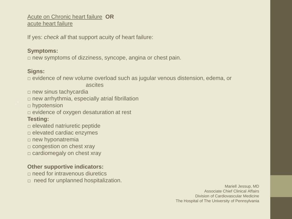
Acute on Chronic heart failure OR acute heart failure If yes: check all that support acuity of heart failure: Symptoms: □ new symptoms of dizziness, syncope, angina or chest pain. Signs: □ evidence of new volume overload such as jugular venous distension, edema, or ascites □ new sinus tachycardia □ new arrhythmia, especially atrial fibrillation □ hypotension □ evidence of oxygen desaturation at rest Testing: □ elevated natriuretic peptide □ elevated cardiac enzymes □ new hyponatremia □ congestion on chest xray □ cardiomegaly on chest xray Other supportive indicators: □ need for intravenous diuretics □ need for unplanned hospitalization. Mariell Jessup, MD
Associate Chief Clinical Affairs Division of Cardiovascular Medicine
The Hospital of The University of Pennsylvania
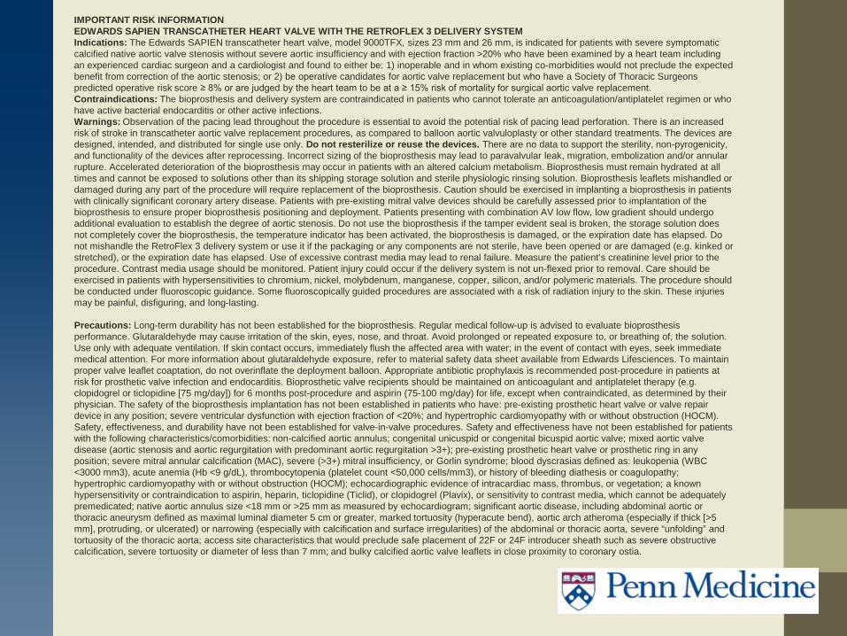
IMPORTANT RISK INFORMATION EDWARDS SAPIEN TRANSCATHETER HEART VALVE WITH THE RETROFLEX 3 DELIVERY SYSTEM Indications: The Edwards SAPIEN transcatheter heart valve, model 9000TFX, sizes 23 mm and 26 mm, is indicated for patients with severe symptomatic calcified native aortic valve stenosis without severe aortic insufficiency and with ejection fraction >20% who have been examined by a heart team including an experienced cardiac surgeon and a cardiologist and found to either be: 1) inoperable and in whom existing co-morbidities would not preclude the expected benefit from correction of the aortic stenosis; or 2) be operative candidates for aortic valve replacement but who have a Society of Thoracic Surgeons predicted operative risk score ≥ 8% or are judged by the heart team to be at a ≥ 15% risk of mortality for surgical aortic valve replacement. Contraindications: The bioprosthesis and delivery system are contraindicated in patients who cannot tolerate an anticoagulation/antiplatelet regimen or who have active bacterial endocarditis or other active infections. Warnings: Observation of the pacing lead throughout the procedure is essential to avoid the potential risk of pacing lead perforation. There is an increased risk of stroke in transcatheter aortic valve replacement procedures, as compared to balloon aortic valvuloplasty or other standard treatments. The devices are designed, intended, and distributed for single use only. Do not resterilize or reuse the devices. There are no data to support the sterility, non-pyrogenicity, and functionality of the devices after reprocessing. Incorrect sizing of the bioprosthesis may lead to paravalvular leak, migration, embolization and/or annular rupture. Accelerated deterioration of the bioprosthesis may occur in patients with an altered calcium metabolism. Bioprosthesis must remain hydrated at all times and cannot be exposed to solutions other than its shipping storage solution and sterile physiologic rinsing solution. Bioprosthesis leaflets mishandled or damaged during any part of the procedure will require replacement of the bioprosthesis. Caution should be exercised in implanting a bioprosthesis in patients with clinically significant coronary artery disease. Patients with pre-existing mitral valve devices should be carefully assessed prior to implantation of the bioprosthesis to ensure proper bioprosthesis positioning and deployment. Patients presenting with combination AV low flow, low gradient should undergo additional evaluation to establish the degree of aortic stenosis. Do not use the bioprosthesis if the tamper evident seal is broken, the storage solution does not completely cover the bioprosthesis, the temperature indicator has been activated, the bioprosthesis is damaged, or the expiration date has elapsed. Do not mishandle the RetroFlex 3 delivery system or use it if the packaging or any components are not sterile, have been opened or are damaged (e.g. kinked or stretched), or the expiration date has elapsed. Use of excessive contrast media may lead to renal failure. Measure the patient’s creatinine level prior to the procedure. Contrast media usage should be monitored. Patient injury could occur if the delivery system is not un-flexed prior to removal. Care should be exercised in patients with hypersensitivities to chromium, nickel, molybdenum, manganese, copper, silicon, and/or polymeric materials. The procedure should be conducted under fluoroscopic guidance. Some fluoroscopically guided procedures are associated with a risk of radiation injury to the skin. These injuries may be painful, disfiguring, and long-lasting. Precautions: Long-term durability has not been established for the bioprosthesis. Regular medical follow-up is advised to evaluate bioprosthesis performance. Glutaraldehyde may cause irritation of the skin, eyes, nose, and throat. Avoid prolonged or repeated exposure to, or breathing of, the solution. Use only with adequate ventilation. If skin contact occurs, immediately flush the affected area with water; in the event of contact with eyes, seek immediate medical attention. For more information about glutaraldehyde exposure, refer to material safety data sheet available from Edwards Lifesciences. To maintain proper valve leaflet coaptation, do not overinflate the deployment balloon. Appropriate antibiotic prophylaxis is recommended post-procedure in patients at risk for prosthetic valve infection and endocarditis. Bioprosthetic valve recipients should be maintained on anticoagulant and antiplatelet therapy (e.g. clopidogrel or ticlopidine [75 mg/day]) for 6 months post-procedure and aspirin (75-100 mg/day) for life, except when contraindicated, as determined by their physician. The safety of the bioprosthesis implantation has not been established in patients who have: pre-existing prosthetic heart valve or valve repair device in any position; severe ventricular dysfunction with ejection fraction of <20%; and hypertrophic cardiomyopathy with or without obstruction (HOCM). Safety, effectiveness, and durability have not been established for valve-in-valve procedures. Safety and effectiveness have not been established for patients with the following characteristics/comorbidities: non-calcified aortic annulus; congenital unicuspid or congenital bicuspid aortic valve; mixed aortic valve disease (aortic stenosis and aortic regurgitation with predominant aortic regurgitation >3+); pre-existing prosthetic heart valve or prosthetic ring in any position; severe mitral annular calcification (MAC), severe (>3+) mitral insufficiency, or Gorlin syndrome; blood dyscrasias defined as: leukopenia (WBC <3000 mm3), acute anemia (Hb <9 g/dL), thrombocytopenia (platelet count <50,000 cells/mm3), or history of bleeding diathesis or coagulopathy; hypertrophic cardiomyopathy with or without obstruction (HOCM); echocardiographic evidence of intracardiac mass, thrombus, or vegetation; a known hypersensitivity or contraindication to aspirin, heparin, ticlopidine (Ticlid), or clopidogrel (Plavix), or sensitivity to contrast media, which cannot be adequately premedicated; native aortic annulus size <18 mm or >25 mm as measured by echocardiogram; significant aortic disease, including abdominal aortic or thoracic aneurysm defined as maximal luminal diameter 5 cm or greater, marked tortuosity (hyperacute bend), aortic arch atheroma (especially if thick [>5 mm], protruding, or ulcerated) or narrowing (especially with calcification and surface irregularities) of the abdominal or thoracic aorta, severe “unfolding” and tortuosity of the thoracic aorta; access site characteristics that would preclude safe placement of 22F or 24F introducer sheath such as severe obstructive calcification, severe tortuosity or diameter of less than 7 mm; and bulky calcified aortic valve leaflets in close proximity to coronary ostia.
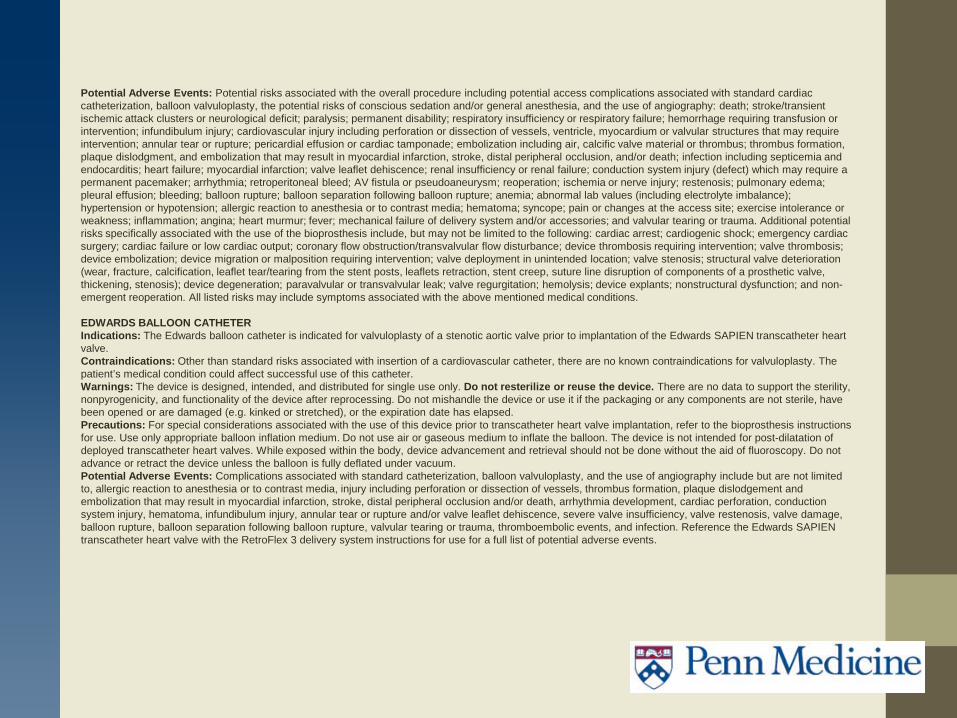
Potential Adverse Events: Potential risks associated with the overall procedure including potential access complications associated with standard cardiac catheterization, balloon valvuloplasty, the potential risks of conscious sedation and/or general anesthesia, and the use of angiography: death; stroke/transient ischemic attack clusters or neurological deficit; paralysis; permanent disability; respiratory insufficiency or respiratory failure; hemorrhage requiring transfusion or intervention; infundibulum injury; cardiovascular injury including perforation or dissection of vessels, ventricle, myocardium or valvular structures that may require intervention; annular tear or rupture; pericardial effusion or cardiac tamponade; embolization including air, calcific valve material or thrombus; thrombus formation, plaque dislodgment, and embolization that may result in myocardial infarction, stroke, distal peripheral occlusion, and/or death; infection including septicemia and endocarditis; heart failure; myocardial infarction; valve leaflet dehiscence; renal insufficiency or renal failure; conduction system injury (defect) which may require a permanent pacemaker; arrhythmia; retroperitoneal bleed; AV fistula or pseudoaneurysm; reoperation; ischemia or nerve injury; restenosis; pulmonary edema; pleural effusion; bleeding; balloon rupture; balloon separation following balloon rupture; anemia; abnormal lab values (including electrolyte imbalance); hypertension or hypotension; allergic reaction to anesthesia or to contrast media; hematoma; syncope; pain or changes at the access site; exercise intolerance or weakness; inflammation; angina; heart murmur; fever; mechanical failure of delivery system and/or accessories; and valvular tearing or trauma. Additional potential risks specifically associated with the use of the bioprosthesis include, but may not be limited to the following: cardiac arrest; cardiogenic shock; emergency cardiac surgery; cardiac failure or low cardiac output; coronary flow obstruction/transvalvular flow disturbance; device thrombosis requiring intervention; valve thrombosis; device embolization; device migration or malposition requiring intervention; valve deployment in unintended location; valve stenosis; structural valve deterioration (wear, fracture, calcification, leaflet tear/tearing from the stent posts, leaflets retraction, stent creep, suture line disruption of components of a prosthetic valve, thickening, stenosis); device degeneration; paravalvular or transvalvular leak; valve regurgitation; hemolysis; device explants; nonstructural dysfunction; and non-emergent reoperation. All listed risks may include symptoms associated with the above mentioned medical conditions. EDWARDS BALLOON CATHETER Indications: The Edwards balloon catheter is indicated for valvuloplasty of a stenotic aortic valve prior to implantation of the Edwards SAPIEN transcatheter heart valve. Contraindications: Other than standard risks associated with insertion of a cardiovascular catheter, there are no known contraindications for valvuloplasty. The patient’s medical condition could affect successful use of this catheter. Warnings: The device is designed, intended, and distributed for single use only. Do not resterilize or reuse the device. There are no data to support the sterility, nonpyrogenicity, and functionality of the device after reprocessing. Do not mishandle the device or use it if the packaging or any components are not sterile, have been opened or are damaged (e.g. kinked or stretched), or the expiration date has elapsed. Precautions: For special considerations associated with the use of this device prior to transcatheter heart valve implantation, refer to the bioprosthesis instructions for use. Use only appropriate balloon inflation medium. Do not use air or gaseous medium to inflate the balloon. The device is not intended for post-dilatation of deployed transcatheter heart valves. While exposed within the body, device advancement and retrieval should not be done without the aid of fluoroscopy. Do not advance or retract the device unless the balloon is fully deflated under vacuum. Potential Adverse Events: Complications associated with standard catheterization, balloon valvuloplasty, and the use of angiography include but are not limited to, allergic reaction to anesthesia or to contrast media, injury including perforation or dissection of vessels, thrombus formation, plaque dislodgement and embolization that may result in myocardial infarction, stroke, distal peripheral occlusion and/or death, arrhythmia development, cardiac perforation, conduction system injury, hematoma, infundibulum injury, annular tear or rupture and/or valve leaflet dehiscence, severe valve insufficiency, valve restenosis, valve damage, balloon rupture, balloon separation following balloon rupture, valvular tearing or trauma, thromboembolic events, and infection. Reference the Edwards SAPIEN transcatheter heart valve with the RetroFlex 3 delivery system instructions for use for a full list of potential adverse events.

EDWARDS SAPIEN TRANSCATHETER HEART VALVE WITH THE ASCENDRA 3 DELIVERY SYSTEM Indications: The Edwards SAPIEN transcatheter heart valve, model 9000TFX, sizes 23 mm and 26 mm, is indicated for patients with severe symptomatic calcified native aortic valve stenosis without severe aortic insufficiency and with ejection fraction > 20% who have been examined by a heart team including an experienced cardiac surgeon and a cardiologist and found to be: 1) inoperable and in whom existing co-morbidities would not preclude the expected benefit from correction of the aortic stenosis; or 2) be operative candidates for aortic valve replacement but who have a Society of Thoracic Surgeons predicted operative risk score ≥ 8% or are judged by the heart team to be at a ≥ 15% risk of mortality for surgical aortic valve replacement. Contraindications: The bioprosthesis and delivery system are contraindicated in patients who cannot tolerate an anticoagulation/antiplatelet regimen or who have active bacterial endocarditis or other active infections. Warnings: Observation of the pacing lead throughout the procedure is essential to avoid the potential risk of pacing lead perforation. There is an increased risk of stroke in transcatheter aortic valve replacement procedures, as compared to balloon aortic valvuloplasty or other standard treatments. The devices are designed, intended, and distributed for single use only. Do not resterilize or reuse the devices. There are no data to support the sterility, non-pyrogenicity, and functionality of the devices after reprocessing. Incorrect sizing of the bioprosthesis may lead to paravalvular leak, migration, embolization and/or annular rupture. Accelerated deterioration of the bioprosthesis may occur in patients with an altered calcium metabolism. Bioprosthesis must remain hydrated at all times and cannot be exposed to solutions other than its shipping storage solution and sterile physiologic rinsing solution. Bioprosthesis leaflets mishandled or damaged during any part of the procedure will require replacement of the bioprosthesis. Caution should be exercised in implanting a bioprosthesis in patients with clinically significant coronary artery disease. Patients with pre-existing mitral valve devices should be carefully assessed prior to implantation of the bioprosthesis to ensure proper bioprosthesis positioning and deployment. Patients presenting with combination AV low flow, low gradient should undergo additional evaluation to establish the degree of aortic stenosis. Do not use the bioprosthesis if the tamper evident seal is broken, the storage solution does not completely cover the bioprosthesis, the temperature indicator has been activated, or the bioprosthesis is damaged, or the expiration date has elapsed. Do not mishandle the Ascendra 3 delivery system or use it if the packaging or any components are not sterile, have been opened or are damaged (e.g. kinked or stretched), or the expiration date has elapsed. Use of excessive contrast media may lead to renal failure. Measure the patient’s creatinine level prior to the procedure. Contrast media usage should be monitored. Care should be exercised in patients with hypersensitivities to chromium, nickel, molybdenum, manganese, copper, silicon, and/or polymeric materials. The procedure should be conducted under fluoroscopic guidance. Some fluoroscopically guided procedures are associated with a risk of radiation injury to the skin. These injuries may be painful, disfiguring, and long-lasting. The safety and efficacy of the transapical procedure has only been evaluated in those patient populations where the transfemoral procedure delivery is not suitable. Precautions: Long-term durability has not been established for the bioprosthesis. Regular medical follow-up is advised to evaluate bioprosthesis performance. Glutaraldehyde may cause irritation of the skin, eyes, nose and throat. Avoid prolonged or repeated exposure to, or breathing of, the solution. Use only with adequate ventilation. If skin contact occurs, immediately flush the affected area with water; in the event of contact with eyes, seek immediate medical attention. For more information about glutaraldehyde exposure, refer to material safety data sheet available from Edwards Lifesciences. To maintain proper valve leaflet coaptation, do not overinflate the deployment balloon. Appropriate antibiotic prophylaxis is recommended post-procedure in patients at risk for prosthetic valve infection and endocarditis. Bioprosthetic valve recipients should be maintained on anticoagulant and antiplatelet therapy (e.g. clopidogrel or ticlopidine [75 mg/day]) for 6 months post procedure and aspirin (75-100 mg/day) for life, except when contraindicated, as determined by their physician. The safety of the bioprosthesis implantation has not been established in patients who have: pre-existing prosthetic heart valve or valve repair device in any position; severe ventricular dysfunction with ejection fraction < 20%; hypertrophic cardiomyopathy with or without obstruction (HOCM). Safety, effectiveness, and durability have not been established for valve-in-valve procedures. Safety and effectiveness have not been established for patients with the following characteristics/comorbidities: non-calcified aortic annulus; congenital unicuspid or congenital bicuspid aortic valve; mixed aortic valve disease (aortic stenosis and aortic regurgitation with predominant aortic regurgitation > 3+); pre-existing prosthetic heart valve or prosthetic ring in any position; severe mitral annular calcification (MAC), severe (> 3+) mitral insufficiency, or Gorlin syndrome; blood dyscrasias defined as: leukopenia (WBC < 3000 mm3), acute anemia (Hb < 9 g/dL), thrombocytopenia (platelet count < 50,000 cells/mm3), or history of bleeding diathesis or coagulopathy; hypertrophic cardiomyopathy with or without obstruction (HOCM); echocardiographic evidence of intracardiac mass, thrombus, or vegetation; a known hypersensitivity or contraindication to aspirin, heparin, ticlopidine (Ticlid), or clopidogrel (Plavix), or sensitivity to contrast media, which cannot be adequately premedicated; native aortic annulus size < 18 mm or > 25 mm as measured by echocardiogram; significant aortic disease, including abdominal aortic or thoracic aneurysm defined as maximal luminal diameter 5 cm or greater; marked tortuosity (hyperacute bend), aortic arch atheroma (especially if thick [> 5 mm], protruding, or ulcerated) or narrowing (especially with calcification and surface irregularities) of the abdominal or thoracic aorta, severe “unfolding” and tortuosity of the thoracic aorta; bulky calcified aortic valve leaflets in close proximity to coronary ostia.
Potential Adverse Events: Potential risks associated with the overall procedure including potential access complications associated with standard cardiac catheterization, balloon valvuloplasty, the potential risks of conscious sedation and/or general anesthesia, and the use of angiography: death; stroke/transient ischemic attack clusters or neurological deficit; paralysis; permanent disability; respiratory insufficiency or respiratory failure; hemorrhage requiring transfusion or intervention; infundibulum injury; cardiovascular injury including perforation or dissection of vessels, ventricle, myocardium or valvular structures that may require intervention; annular tear or rupture; pericardial effusion or cardiac tamponade; embolization including air, calcific valve material or thrombus; thrombus formation, plaque dislodgment, and embolization that may result in myocardial infarction, stroke, distal peripheral occlusion, and/or death; infection including septicemia and endocarditis; heart failure; myocardial infarction; valve leaflet dehiscence; renal insufficiency or renal failure; conduction system injury (defect) which may require a permanent pacemaker; arrhythmia; retroperitoneal bleed; AV fistula or pseudoaneurysm; reoperation; ischemia or nerve injury; restenosis; pulmonary edema; pleural effusion; bleeding; balloon rupture; balloon separation following balloon rupture; anemia; abnormal lab values (including electrolyte imbalance); hypertension or hypotension; allergic reaction to anesthesia or to contrast media; hematoma; syncope; pain or changes at the access site; exercise intolerance or weakness; inflammation; angina; heart murmur; fever; mechanical failure of delivery system and/or accessories; suturing of a peripheral coronary artery; and valvular tearing or trauma. Additional potential risks specifically associated with the use of the bioprosthesis include, but may not be limited to the following: cardiac arrest; cardiogenic shock; emergency cardiac surgery; cardiac failure or low cardiac output; coronary flow obstruction/transvalvular flow disturbance; injury at the site of ventricular access that may require repair; device thrombosis requiring intervention; valve thrombosis; device embolization; device migration or malposition requiring intervention; valve deployment in unintended location; valve stenosis; structural valve deterioration (wear, fracture, calcification, leaflet tear/tearing from the stent posts, leaflet retraction, stent creep, suture line disruption of components of a prosthetic valve, thickening, stenosis); device degeneration; paravalvular or transvalvular leak; injury to the mitral valve; valve regurgitation; hemolysis; device explants; nonstructural dysfunction; non-emergent reoperation. All listed risks may include symptoms associated with the above mentioned medical conditions. ASCENDRA BALLOON AORTIC VALVULOPLASTY CATHETER Indications: The Ascendra balloon aortic valvuloplasty catheter is indicated for valvuloplasty of a stenotic aortic valve prior to implantation of the Edwards SAPIEN transcatheter heart valve. Contraindications: Other than standard risks associated with insertion of a cardiovascular catheter, there are no known contraindications for valvuloplasty. The patient’s medical condition could affect successful use of this catheter. Warnings: The device is designed, intended, and distributed for single use only. Do not resterilize or reuse the device. There are no data to support the sterility, nonpyrogenicity, and functionality of the device after reprocessing. Do not mishandle the device or use it if the packaging or any components are not sterile, have been opened or are damaged (e.g. kinked or stretched), or the expiration date has elapsed. Precautions: For special considerations associated with the use of this device prior to transcatheter heart valve implantation, refer to the bioprosthesis instructions for use (IFU). This catheter has not been tested with any transcatheter valve other than the Edwards SAPIEN transcatheter heart valve. Use only appropriate balloon inflation medium. Do not use air or gaseous medium to inflate the balloon. The device is not intended for post-dilatation of deployed transcatheter heart valves. While exposed within the body, device advancement and retrieval should not be done without the aid of fluoroscopy. Do not advance or retract the device unless the balloon is fully deflated under vacuum. Potential Adverse Events: Complications associated with standard catheterization, balloon valvuloplasty, and the use of angiography include, but are not limited to, allergic reaction to anesthesia or to contrast media, thrombus formation, plaque dislodgement and embolization that may result in myocardial infarction, stroke, distal peripheral occlusion and/or death, arrhythmia development, cardiac perforation, conduction system injury, hematoma, infundibulum injury, annular tear or rupture and/or valve leaflet dehiscence, severe valve insufficiency, valve restenosis, valve damage, balloon rupture, balloon separation following balloon rupture, valvular tearing or trauma, thromboembolic events, and infection. Reference the Edwards SAPIEN transcatheter heart valve with the Ascendra balloon catheter instructions for use for a full list of potential adverse events.
ASCENDRA 3 INTRODUCER SHEATH SET Indications: The Ascendra 3 introducer sheath set is indicated for the introduction and removal of devices used with the Edwards SAPIEN transcatheter heart valve. Contraindications: No known contraindications. Warnings: The devices are designed, intended, and distributed for single use only. Do not resterilize or reuse the devices. There are no data to support the sterility, nonpyrogenicity, and functionality of the devices after reprocessing. Do not mishandle the device or use it if the packaging or any components are not sterile, have been opened or are damaged (e.g. kinked or stretched), or the expiration date has elapsed. Should not be used in patients with left ventricular aneurysm. The Ascendra 3 introducer sheath set must be used with a 0.035" guidewire. Precautions: No known precautions. Potential Adverse Events: Complications associated with cardiac surgical intervention and use of angiography include, but are not limited to, allergic reaction to anesthesia or to contrast media, injury including myocardial injury, thrombus formation, and plaque dislodgement which may result in myocardial infarction, arrhythmia, stroke, and/or death. Reference the Edwards SAPIEN transcatheter heart valve with the Ascendra 3 delivery system instructions for use for a full list of potential adverse events. CRIMPER Indications: The crimper is indicated for use in preparing the Edwards SAPIEN transcatheter heart valve for implantation. Contraindications: No known contraindications. Warnings: The device is designed, intended, and distributed for single use only. Do not resterilize or reuse the device. There are no data to support the sterility, nonpyrogenicity, and functionality of the device after reprocessing. Do not mishandle the device or use it if the packaging or any components are not sterile, have been opened or are damaged, or the expiration date has elapsed. Precautions: For special considerations associated with the use of this device prior to transcatheter heart valve implantation, refer to the bioprosthesis instructions for use. Potential Adverse Events: No known potential adverse events. CAUTION: Federal (United States) law restricts these devices to sale by or on the order of a physician. See instructions for use for full prescribing information, including indications, contraindications, warnings, precautions and adverse events. Edwards, Edwards Lifesciences, Ascendra, Ascendra 3, Edwards SAPIEN, RetroFlex, RetroFlex 3 and SAPIEN are trademarks of Edwards Lifesciences Corporation. All other trademarks are property of their respective owners. LR_11337