Embed Size (px)
Citation preview
1
Targeting lysosomal cysteine protease cathepsin S reveals immunomodulatory
therapeutic strategy for oxaliplatin-induced peripheral neuropathy
Szu-Jung Chen1, Li-Hsien Chen1, Yu-Min Yeh1, Chou-Ching K Lin2, Peng-Chan Lin1,
Han-Wei Huang2, Meng-Ru Shen3,4, Bo-Wen Lin5, Jeng-Chang Lee5, Cheng-Che Lee6,
Yueh-Feng Lee1, Huai-Chueh Chiang1, Jang-Yang Chang1,7,8*
1Department of Internal Medicine, 2Department of Neurology, 3Department of
Obstetrics/Gynecology, 5Department of Surgery, National Cheng Kung University
Hospital, College of Medicine, National Cheng Kung University, Tainan, Taiwan
4Department of Pharmacology, College of Medicine, National Cheng Kung University,
Tainan. Taiwan
6School of Medicine for International students, College of Medicine, I-SHOU
University, Kaohsiung, Taiwan
7National Institute of Cancer Research, 8Institute of Biotechnology and Pharmaceutical
Research, National Health Research Institutes, Zhunan, Taiwan
Corresponding author: Dr. Jang-Yang Chang, Department of Internal Medicine,
National Cheng Kung University Hospital, College of Medicine, National Cheng
Kung University, Tainan, Taiwan. Email: [email protected]
Running title: Cathepsin S regulates oxaliplatin neurotoxicity
Number of figures: 6
2
Abstract
Rationale: Oxaliplatin-induced peripheral neuropathy (OIPN) is a common adverse
effect that causes delayed treatment and poor prognosis among colorectal cancer
(CRC) patients. However, its mechanism remains elusive, and no effective treatment
is available.
Methods: We employed a prospective cohort study of adult patients with
pathologically confirmed stage III CRC receiving adjuvant chemotherapy with an
oxaliplatin-based regimen for investigating OIPN. To further validate the clinical
manifestations and identify a potential therapeutic strategy, animal models, and in
vitro studies on the mechanism of OIPN were applied.
Results: Our work found that (1) consistent with clinical findings, OIPN was
observed in animal models. Targeting the enzymatic activity of cathepsin S (CTSS)
by pharmacological blockade and gene deficiency strategy alleviates the
manifestations of OIPN. (2) Oxaliplatin treatment increases CTSS expression by
enhancing cytosol translocation of interferon response factor 1 (IRF1), which then
facilitates STIM-dependent store-operated Ca2+ entry homeostasis. (3) The cytokine
array demonstrated an increase in anti-inflammatory cytokines and suppression of
proinflammatory cytokines in mice treated with RJW-58. (4) Mechanistically,
inhibiting CTSS facilitated olfactory receptors transcription factor 1 release from
P300/CBP binding, which enhanced binding to the interleukin-10 (IL-10) promoter
region, driving IL-10 downstream signaling pathway. (5) Serum CTSS expression is
increased in CRC patients with oxaliplatin-induced neurotoxicity.
Conclusions: We highlighted the critical role of CTSS in OIPN, which provides a
therapeutic strategy for the common adverse side effects of oxaliplatin.
3
Keywords: Oxaliplatin-induced peripheral neuropathy, Cathepsin S, olfactory
receptor transcription factor 1, IL-10
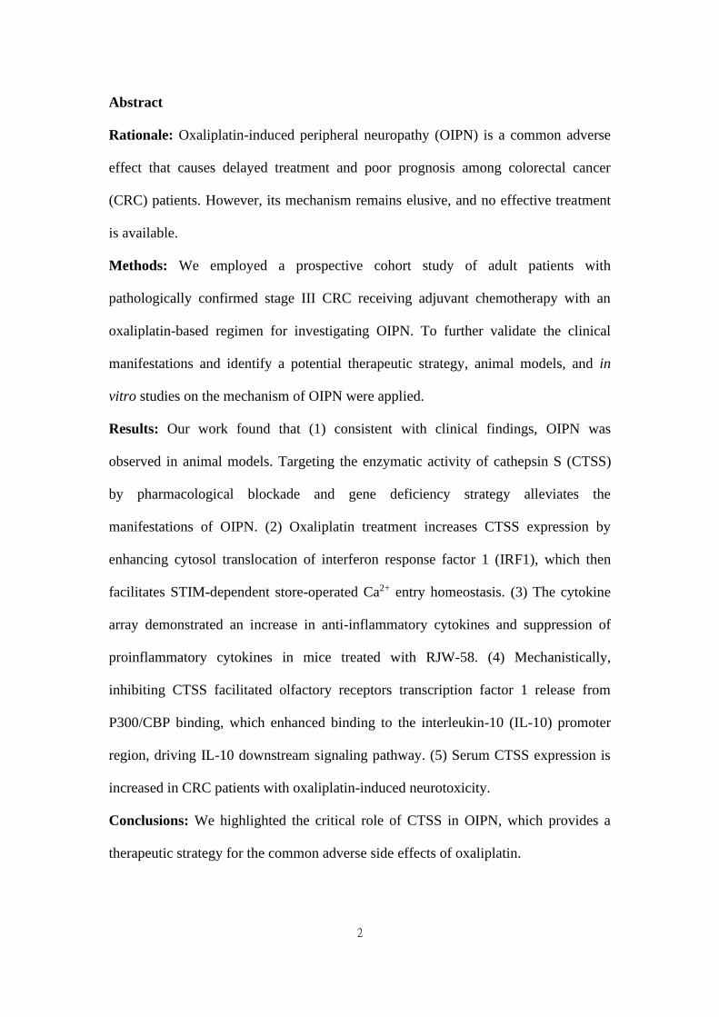
Graphical Abstract:
This study highlights the novel role of CTSS in oxaliplatin-induced neurotoxicity,
as evidenced by in vitro cell-line studies, in vivo OIPN mouse models, and clinical
sample analysis. Blocking the enzymatic activity of lysosomal cathepsin S alleviates
OIPN by regulating the STIM-dependent store-operated Ca2+ entry homeostasis.
Furthermore, RJW-58, CTSS inhibitor, reverses oxaliplatin-induced CTSS production,
activates the anti-inflammatory IL-10/STAT3 cascade, and reduces proinflammatory
cytokines expression, which results in the alleviation of OIPN. RJW-58 might
represent a potential therapeutic strategy for cancer patients with oxaliplatin-induced
neuropathy.
4
Introduction
Oxaliplatin has exhibited significant efficacy in the treatment of human cancers,
especially colorectal cancer (CRC). However, acute or chronic peripheral neuropathy
and concurrent sensory and motor function deficiencies are a common adverse effect
of oxaliplatin [1], resulting in chemotherapy dose reduction and early discontinuation
[2]. Several mechanisms of oxaliplatin-induced peripheral neuropathy have been
proposed, including direct damage, ROS generation, and dysfunction of voltage-gated
ions channels in the dorsal root ganglia and axon [3, 4]. Several clinical trials were
conducted to prevent or treat oxaliplatin-induced peripheral neuropathy based on the
aforementioned mechanisms. However, these studies failed to demonstrate significant
effectiveness in alleviating this side effect [5, 6]. Moreover, increasing evidence
suggests that neuroinflammation also plays a role in the development of neurotoxicity
[7]. In animal studies, inhibiting macrophage infiltration and proinflammatory
cytokines in the dorsal root ganglion (DRG) suppressed the augmentation of
paclitaxel-induced neuropathic nociception [7, 8]. Milligan et al. reported that
intrathecal administration of anti-inflammatory cytokine Interleukin-10 (IL-10) briefly
reversed chronic constriction injury-induced mechanical allodynia and thermal
hyperalgesia [9, 10]. Furthermore, suppression of IL-20, a proinflammatory cytokine,
with IL-20 neutralizing antibody alleviated paclitaxel-induced peripheral neuropathy
in animal models [11]. Therefore, triggering anti-inflammatory cytokines and
suppressing the proinflammatory response may be a therapeutic strategy for managing
chemotherapy-induced peripheral neuropathy (CIPN).
Cathepsin S (CTSS), a lysosomal cysteine protease, is stably present in the
cytoplasm of immune-related cells, such as antigen-presenting cells, B cells, and
phagocytic cells (e.g., macrophages and microglial cells). Several lines of evidence
5
have demonstrated that releasing CTSS from microglial cells causes neuropathic pain
[12] that could be reversed by intraspinal injection of CTSS inhibitor, but the
mechanisms remain unclear. Furthermore, CTSS is reportedly an autocrine factor that
stimulates microglial cell activation and subsequently regulates the release of
proinflammatory cytokines and chemokines [13]. Our studies have indicated that
CTSS plays a vital role in mediating Ca2+ homeostasis by disturbing stromal
interaction molecule 1 (STIM1) trafficking, which alters STIM1-Orai1 interaction and
affects Ca2+ influx [14]. And, studies have reported that STIM1 regulates
stored-operated Ca2+ entry (SOCE) by initiating Ca2+ influx and triggering immune
activation in macrophages, chemotaxis, and neutrophil infiltration, which plays a role
in the development of inflammatory pain [15].
IL-10, a “founding” member of IL-10 family cytokines, was initially described as
a cytokine synthesis inhibitory factor produced by T helper cells with pleiotropic
immunosuppressive functions [16]. Increasing evidence indicates that IL-10 is
expressed by a wide variety of cell types, including macrophages, monocytes,
dendritic cells, natural killer cells, and CD4+ and CD8+ T cells and B cells. The
immunosuppressive activity of IL-10 is mediated by ligand binding with IL-10
receptor (IL-10R1, IL-10R2), which activates Janus tyrosine kinases (JAKs) and
signal transducers and activators of transcription signaling pathways, which inhibit
the expression profile of immunomodulatory genes [17] and release proinflammatory
cytokines, such as IL-6, IL-1β, and TNF-α [18]. However, despite the fact that IL-10
acts as a prime drug target for autoimmune disease, tissue damage, and even cancer,
effective therapies have yet to be developed.
Therefore, we investigated whether targeting CTSS can ameliorate
oxaliplatin-induced peripheral neuropathy (OIPN) by upregulating IL-10 expression
6
and suppressing proinflammatory cytokines. The detailed molecular mechanisms are
described.
Materials and Methods
Clinical cohort
Patients with CRC enrolled in a clinical study investigating the CIPN
(NCT02481336) were used for analysis. The details of this study are described
elsewhere [19]. Briefly, patients with pathologically confirmed stage III or high-risk
stage II CRC have been prospectively recruited from National Cheng Kung
University Hospital (NCKUH) from January 2015 until the present. All patients
received curative surgery followed by 6 months of oxaliplatin and infusional
5-fluorouracil and leucovorin (modified FOLFOX6) as adjuvant chemotherapy.
Patients with poor performance status (ECOG performance score > 1), inadequate
organ functions, prior treatment with neurotoxic chemotherapy (such as oxaliplatin,
cisplatin, carboplatin, taxanes, or vinca alkaloids), preexisting peripheral neuropathy
of any grade, or a family history of genetic or familial neuropathy were excluded. A
complete neurological examination, patients-reported peripheral neuropathy CIPN20
questionnaire (QLQ-CIPN20), and quantitative examination of the peripheral nerve
system (including nerve conduction velocity [NCV], quantitative sensory test [QST],
nerve excitability test [NET], and total neuropathy score [TNS]) were assessed at
baseline, the end of cycles 1 (week 2), 6 (week 12), and 12 (week 24) of
chemotherapy, and 9 (week 36) and 12 months (week 48) after the beginning of
chemotherapy. The CIPN20 questionnaire contained 20 items evaluating the severity
of neurologic symptoms, including nine items for sensory, eight items for motor, and
three items for autonomic symptoms. Scores from 1 to 4 were applied for each item.
7
The scores of the items were added together for all 19 questions, and a final CIPN
score, ranging from 19 to 76, was generated. The score of item 20, which rates men's
sexual function, was not adopted in this study because the information is lacking for
women and is often not provided for male patients either. In addition to collecting
blood from all participants at baseline to identify predictive biomarkers of CIPN,
blood samples at week 48 were also collected in 23 patients to monitor the changes in
biomarkers. This study was approved by the institutional review board of NCKUH
(A-ER-103-395). All participants provided written informed consent before being
enrolled.
Reagents
Oxaliplatin was purchased from Sanofi Pharmaceutical Company (NY, USA).
The procedure for synthesizing the CTSS inhibitor RJW-58 was described previously
[14]. The following primary antibodies were purchased: Cathepsin S (Thermo Fisher
catalog: #PA5-81369), EBF (Santa Cruz, #sc-137065), ATF-3 (Biossua, #BS-0519R),
α-tubulin (Sigma, MO, USA), IRF-1 (Bio-Rad, #VPA00801), CBP (GeneTex,
#GTX101249), Iba1 (Abcam, #ab-5076), NeuN (Novus, #NBP2-10491), IL-10
(Abcam, #ab189392), STIM1 (Cell signaling (D88E10), #5668S), CD11b (Abcam,
#ab52478), CD45 (BD Biosciences, #553079), STAT3 (BD, #50402), P-STAT3 (Cell
signaling, #9145S), NFκB (Cell signaling, #4727S), OR5B3 (Abcam, #ab186624),
OR5M3 (LSBio, #LS-C200406), and β-actin (GeneTex, #GTX109639).
Horseradish-peroxidase–conjugated secondary antibodies were purchased from
GeneTex (#GTX213110, #GTX213111, #GTX224125, and #GTX232040).
8
Cell culture
DRG cell lines (ND 7/23) and mouse microglial cells (SIM-A9) were obtained
from Sigma-Aldrich (#92090903, USA) and Kerafast (END001, USA), respectively.
The ND 7/23 cells were grown in Dulbecco’s Modified Eagle Medium (DMEM,
#12100046; Gibco, USA) and the SIM-A9 cells were maintained in DMEM/F12
(#11320033; Gibco, USA) supplemented with 10% FBS, 100 U/ml of penicillin, and
100 μg/ml of streptomycin-glutamate (Thermo Scientific, Utah, USA). Both cell lines
were maintained in a 5% CO2 incubator (humidified atmosphere) at 37 °C.
Animals
The 6-week-old C57BL/6JNarl mice were maintained in the pathogen-free
facility of the Animal Laboratory of National Cheng Kung University (Institutional
Animal Care and Use Committee [IACUC] number: 107078). These animals were
housed in a temperature-controlled (25 ± 2 °C); humidity of approximately
(60%-80%) and light-controlled environment (12 / 12 h light-dark cycle, lights on
at 6:00 AM) with free access to food and water, following the approved guidelines
by the IACUC of National Cheng Kung University. Before the experiments were
performed, all animals had adapted to the housing environments for at least 7 days.
Our preliminary study revealed that no body weight loss was observed in animals
treated with oxaliplatin 3 mg/kg or RJW-58 25 mg/kg intraperitoneally (i.p.) for 5
days a week over two alternative weeks.
CTSS transgenic mice production
Ctss-/- mice were generated using CRISPR/Cas9-induced knockout in embryos
obtained from the National Laboratory Animal Center. Mouse fertilized eggs were
9
microinjected with CRISPR/Cas9-related point mutation CTSS sequence, TGACTA,
including a stop code and restriction enzyme SpeI recognition cutting site to verify the
mutations at exons 2. The point mutation was designed right after the CTSS
transcription start site to inhibit CTSS RNA and protein expression. Recombinant
embryonic stem cell lines were injected into 161 C57BL/6JNarl blastocysts, 122 of
which were transferred into pseudo‐pregnant females, and 28 live pups were born in
2018. Only one Ctss+/- female strain was detected using polymerase chain reaction
(PCR) genotyping. For recessive phenotypes, intercross between Ctss+/- female mice
with male Ctss+/+ mice in the related C57BL/6JNarl background generated Ctss−/−
mice. The litters were backcrossed three times to generate Ctss−/− mice. Ctss+/+
C57BL/6JNarl mice were used as controls. The following primers were used
for Ctss−/− genotyping: forward 5′-GTCAGGCAGATTGCTACAAG-3′, reverse
5′-ACACTGCTCGGGTGGCAATC-3′, and digestion with restriction SpeI enzymes
(Figure S1).
Mouse behavioral models of oxaliplatin-induced peripheral neuropathy
Following housing adaptions, 7-week-old C57BL/6JNarl mice (weight range:
18-20 g) were used to investigate oxaliplatin-induced neuropathy. Oxaliplatin (3
mg/kg, Sanofi, USA), vehicle (saline), or RJW-58 (25 mg/kg) was administered i.p.
for 5 consecutive days every other week for 2 weeks. A von Frey filament test (Part
#2390, IITC Inc., CA) for mechanical hyperalgesia, tail immersion test (water
temperature: 48-49 °C or ice) for thermal sensitivity, and grip test (BIO-EVF3,
Bioseb Inc., USA) for grip strength were used, following a previously described
method [11, 20]. To quantify locomotor reactivity, we subjected each mouse to an
open field chamber with photocell emitters and receptors equally spaced along the
10
perimeter of the chamber. These photocell emitters and receptors created an x-y grid
of invisible infrared beams and signals, recorded by computer software to calculate
the quantity of mouse motor activity in 10 min. The baseline measurement of each
behavior test was established before drug treatment, and five additional sessions were
measured weekly.
Primary DRG isolation
Primary DRG tissues were removed from adult C57BL/6JNarl mice after the
behavior tests, and the tissues were digested with 0.1% collagenase for 1 h, followed
by 0.25% trypsin for 25 min for cell mixture. The cell mixtures were stained with
NeuN (Novus, #NBP2-10491) antibodies to isolate primary spinal dorsal horn
neurons for flow cytometry analysis to detect ATF3 expression. The cell mixtures
were further stained with both CD11b (Abcam, #ab52478) and CD45 (BD
Biosciences, #553079) antibodies to isolate the microglia for flow cytometry analysis
to detect IL-10 expression.
Flow cytometry analysis
After treatment, the cells were fixed with 3.7% formaldehyde (dissolved in
phosphate buffered saline [PBS]) for 15 min at room temperature and permeabilized
with 0.5% Triton X-100 in 3.7% formaldehyde for another 15 min. The cells were
washed three times with PBS and blocked with CAS-BlockTM solution (#008120;
Life Technologies) for 1 h. The antibodies were prepared in the CAS-BlockTM
solution (#008120; Life Technologies) and stained overnight. The cells were then
washed three times with PBS and stained with secondary antibodies for 2 h. Finally,
the cells were counterstained with DAPI for 2 min and then washed with 1-2 mL of
11
BD FACSFlow buffer (LOT:1833202826) twice. After washing, the cells were
resuspended with BD FACSFlow buffer. The data were acquired on a flow cytometer,
following the manufacturer’s recommendations.
Cytokine array analysis
Ctss+/+ mouse serums were collected after RJW-58 injection (week 4). Cytokine
array analysis was performed following the protocol of the MILLIPLEXMAP mouse
cytokine/chemokine magnetic bead panel (Millipore, MCYTOMAG-70K).
Gene expression profiling and statistical analysis of microarray data
Total RNA samples were extracted using TRIzol (Invitrogen) and purified using
a RNeasy mini-kit (QIAGEN). Gene expression analysis was performed on a
GeneChip platform (Human Genome U133A 2.0 Plus; Affymetrix) following the
National Health Research Institutes Microarray Core facility technique support. Gene
expression changes were calculated as the ratio of 1.2-fold significant alterations of
the experimental groups (CTSS overexpression or si-CTSS treated) compared with
each control group. Compared with the gene expression variance within each
experimental group, most of the olfactory receptor genes detected were differentially
expressed.
Ultrastructure of the sciatic nerve
After neurobehavioral tests at week 6 following oxaliplatin treatments, sciatic
nerve samples were obtained from mice, fixed in 4% glutaraldehyde, and postfixed in
1% osmium tetroxide solution at 4 °C. These samples were then dehydrated in graded
ethanol series and embedded in EMbed 812 (EMS-14120; Hatfield, PA, USA). The
12
sciatic nerve sections (90 nm) were prepared by the Department of Pathology of
NCKUH and observed and imaged using a transmission electron microscope (H7650,
Hitachi, Japan).
Quantification of intraepidermal nerve fiber density (IENF) density
Mouse hind paws were isolated after neurobehavioral tests. Dissected tissues
were fixed with 10% paraformaldehyde overnight and cryoprotected for 72 h with
30% sucrose in PBS at 4° C. The samples were embedded in OCT (#CM 3050S;
Leica) and sliced into 30 μm thick sections. The slides were washed with 0.05%
Triton X-100 in PBS for 4 h and incubated with CAS-BlockTM solution (#008120;
Life Technologies) at room temperature for 1 h. The primary antibodies used were
rabbit anti-PGP9.5 (1:100; #ab108986, Abcam) and goat anti-Collagens VI (1:100,
#AB769; Chemicon). For double staining, two antibodies from different species were
mixed and incubated overnight with the sample sections at 4 °C. After washing away
the primary antibodies with PBS, the tissues were incubated with secondary
antibodies (Alexa-488, 594, or 647, 1:200; Invitrogen) and Hoechst 33342 (10 mg/mL,
#H1399; Invitrogen) for 2 h at room temperature. Immunofluorescence images were
acquired using a multiphoton laser scanning microscope with a 40× objective
(FV1000MPE, Olympus).
Intracellular [Ca2+]i measurement
ND7/23 or SIM-A9 cells (passage numbers: 10-15) were prepared and cultured
for 3 days to measure intracellular Ca2+ concentration ([Ca2+]i). The cells were
incubated with 2 μM Fluo-4/acetoxymethyl ester (Fluo-4/AM, F14201, Molecular
Probes, USA) in DMEM medium at 37 °C for 30 min and then washed three times
13
with Ca2+ free HEPES solution (145 mM NaCl, 5 mM KCl, 0 mM CaCl2, 10 mM
glucose, 1 mM MgCl2, and 5 mM HEPES, with an osmolarity of 297-300, and pH
7.4). Fluo-4 was excited alternatively at 488 nm and the fluorescence intensity was
analyzed at 520 nm. The Image XpressMicro automated wide-field fluorescent
microscope (Molecular Devices, Sunnyvale, CA, USA) was then used to calculate
[Ca2+]i. For [Ca2+]i measurement, the cells were stimulated using 2 μM thapsigargin
(TG, Cayman Chemical, USA), recorded for 7 min.
Western blot
Cells were lysed with CelLytic MT Cell Lysis Reagent (Sigma) containing a
protease inhibitor cocktail (Roche). The total cell lysates were separated with
SDS-PAGE gel, transferred to polyvinylidene fluoride membranes (Amersham Life
Science), blocked with 5% nonfat milk at room temperature for 1 h, washed three
times with TBST (0.1% Tween-20), and then hybridized overnight with the primary
antibody at 4 °C. The membranes were then washed three times with TBST (0.1%
Tween-20) and hybridized with the appropriate peroxidase-conjugated secondary
antibody (Santa Cruz) at room temperature for 1 h. Finally, the membranes were
washed three times with TBST (0.1% Tween-20) and immunoreactivity was detected
using Chemiluminescence (Millipore).
Nuclear extraction assay
Samples were lysed using 500 μL of buffer A (10 mM HEPES, 1.5 mM MgCl2,
10 mM KCl, 0.5 mM DTT, and 0.05% NP-40; pH 7.9). After 10 min of centrifugation
at 3,000 rpm at 4 °C, the supernatant was collected as a cytosolic fraction. The pellet
was washed three times with a wash buffer (10 mM HEPES, 1.5 mM MgCl2, and 10
14
mM KCl; pH 7.9) and lysed using 450 μL of buffer B (5 mM HEPES, 1.5 mM MgCl2,
0.2 mM EDTA, 0.5 mM DTT, 26% glycerol, and 300 mM NaCl; pH 7.9). The
supernatant was collected after 20 min of centrifugation at 13,200 rpm at 4 °C as a
nuclear fraction. Finally, 100 μg of the cytosolic and nuclear fractions were subjected
to gel electrophoresis and analyzed using immunoblotting.
Coimmunoprecipitation and immunoblotting
Samples were lysed using an immunoprecipitation (IP) lysis buffer (0.5% NP-40
and 10% glycerol in PBS). A mixture of 2 mg of cell protein lysates, 5 μg of
anti-STIM1 or anti-OLF1 antibodies, and 100 μL of protein-A/G sepharose beads
were incubated overnight at 4 °C. The mixture was washed twice with wash buffer
(0.5% NP-40, 0.1% Triton X-100 in PBS). The precipitated protein samples were
processed for SDS-PAGE and detected using immunoblotting with the appropriate
primary antibodies.
Gene reporter assay
The reporter gene plasmids of CTSS-, IL10-pGL4.17 were purchased from
GENEWIZ (product numbers: BB9283-1/N332028). The plasmids were introduced
into cells using the oligofectamine transfection reagent for 24 h and then processed
using the Dual-Luciferase 1000 Assay system (Promega, Madison, USA, E1910),
following the manufacturer protocol.
Chromatin immunoprecipitation PCR (ChIP)
The samples were crosslinked with 37% formaldehyde for 10 min at 37 °C. The
cells were then harvested and processed for DNA fragmentation using sonication on
15
ice. Following the manufacturer instructions, the resultant chromatin-DNA fractions
were purified using a ChIP assay kit (Millipore, 2487694). The chromatin was
immunoprecipitated at 4 °C using anti-IRF1, anti-OLF1, and normal IgG (negative
control). The primer sequences used were as follows:
OLF1 on IL-10 promoter: forward 5′-CTGCCTTCATTATCAGTGTG-3′,
reverse 5′-CAGCACAAAGCAAGATGTTG-3′; IRF1 on CTSS promoter: forward
5′-ATGCTCTATCTTTTCATCAGTCAG-3′, reverse
5′-GCCAACAAGAATCTCTCAAACAG-3′.
Quantitative reverse transcription PCR
The total RNA was extracted from samples using TRIzol reagent (Thermo Fisher,
#74108). First-strand cDNA was synthesized using the SuperScript III first-strand
synthesis system following the manufacturer instructions. The transcription level of
CTSS, IL-10, TNF-α, IL-1β, IL-6, and MCP-1 were quantified using the Applied
Biosystems 7500 Real-Time PCR System, with RPL13A as an internal control. The
primer sequences used were as follows: CTSS:
forward 5′-CCATTGGGATCTCTGGAAGAAAA-3′, reverse
5′-TCATGCCCACTTGGTAGGTAT-3′; IL-10: forward
5′-GCTCTTACTGACTGGCATGAG-3′, reverse
5′-CGCAGCTCTAGGAGCATGTG-3′; MCP-1 Forward:
5′-AGGTCCCTGTCATGCTTCTG-3′, Reverse:
5′-GCTGCTGGTGATCCTCTTGT-3′; TNF-α Forward:
5’-AGTGACAAGCCCGTAGCCC-3’, Reverse:
5’-AGCCTTGTCCCTTGAAGAG-3’; IL-1β Forward:
5’-GCTGAAAGCTCTCCACCTC-3’, Reverse:
16
5’-GAGGTGCTGATGTACCAGTT-3’; IL-6 Forward:
5’-CAGGAAATTTGCCTATTGAAAAT-3’, Reverse:
5’-TTGGATGGTCTTGGTCCTTAG-3’.
Statistical analysis
At least seven mice were randomized to each group for all animal behavioral
tests. The results are expressed as the mean ± SEM. Differences between groups were
compared using a two-tailed Student’s t-test or two-way analysis of variance,
performed using Prism. Statistical significance was set as P < 0.05. Experimenters
were blinded to all drug treatments.
Results
Evaluation of oxaliplatin-induced peripheral neuropathy in patients with
colorectal cancer
From January 2015 to July 2019, 87 patients were recruited into a clinical study
investigating chemotherapy-induced peripheral neuropathies: 45 (51.7%) men and 42
(48.3%) women. By July 2019, 76 of 87 patients had completed the 6-month adjuvant
chemotherapy with modified FOLFOX6, and 75 patients (86.2%) had completed the
neurologic study scheduled at week 12, 67 (77%) at week 36, and 63 (71.3%) at week
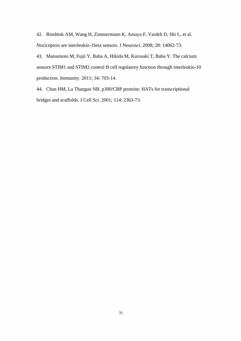
48. A significant reduction in both amplitude and conduction velocity was observed in
sensory nerves, including the median sensory nerve (Figure 1A), sural sensory nerve
(Figure 1B), and ulnar sensory nerve (Figure 1C), after 6 cycles of chemotherapy
(week 12) compared with the baseline results of nerve conduction (P < 0.0001). The
slowing in conduction velocities and the reduced amplitude deteriorated further at
week 36 and reached a plateau at week 48, which was 6 months after completion of
17
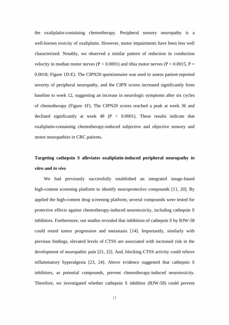
the oxaliplatin-containing chemotherapy. Peripheral sensory neuropathy is a
well-known toxicity of oxaliplatin. However, motor impairments have been less well
characterized. Notably, we observed a similar pattern of reduction in conduction
velocity in median motor nerves (P < 0.0001) and tibia motor nerves (P = 0.0015, P =
0.0018; Figure 1D-E). The CIPN20 questionnaire was used to assess patient-reported
severity of peripheral neuropathy, and the CIPN scores increased significantly from
baseline to week 12, suggesting an increase in neurologic symptoms after six cycles
of chemotherapy (Figure 1F). The CIPN20 scores reached a peak at week 36 and
declined significantly at week 48 (P < 0.0001). These results indicate that
oxaliplatin-containing chemotherapy-induced subjective and objective sensory and
motor neuropathies in CRC patients.
Targeting cathepsin S alleviates oxaliplatin-induced peripheral neuropathy in
vitro and in vivo
We had previously successfully established an integrated image-based
high-content screening platform to identify neuroprotective compounds [11, 20]. By
applied the high-content drug screening platform, several compounds were tested for
protective effects against chemotherapy-induced neurotoxicity, including cathepsin S
inhibitors. Furthermore, our studies revealed that inhibition of cathepsin S by RJW-58
could retard tumor progression and metastasis [14]. Importantly, similarly with
previous findings, elevated levels of CTSS are associated with increased risk in the
development of neuropathic pain [21, 22]. And, blocking CTSS activity could relieve
inflammatory hyperalgesia [23, 24]. Above evidence suggested that cathepsin S
inhibitors, as potential compounds, prevent chemotherapy-induced neurotoxicity.
Therefore, we investigated whether cathepsin S inhibitor (RJW-58) could prevent
18
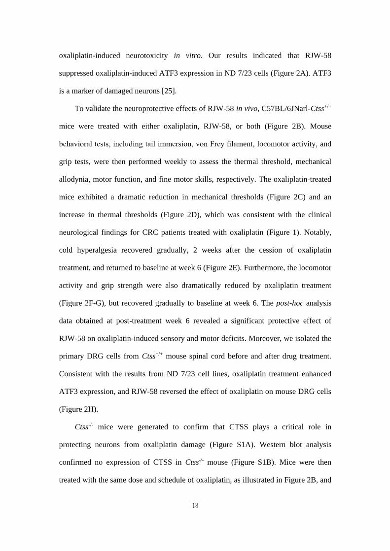
oxaliplatin-induced neurotoxicity in vitro. Our results indicated that RJW-58
suppressed oxaliplatin-induced ATF3 expression in ND 7/23 cells (Figure 2A). ATF3
is a marker of damaged neurons [25].
To validate the neuroprotective effects of RJW-58 in vivo, C57BL/6JNarl-Ctss+/+
mice were treated with either oxaliplatin, RJW-58, or both (Figure 2B). Mouse
behavioral tests, including tail immersion, von Frey filament, locomotor activity, and
grip tests, were then performed weekly to assess the thermal threshold, mechanical
allodynia, motor function, and fine motor skills, respectively. The oxaliplatin-treated
mice exhibited a dramatic reduction in mechanical thresholds (Figure 2C) and an
increase in thermal thresholds (Figure 2D), which was consistent with the clinical
neurological findings for CRC patients treated with oxaliplatin (Figure 1). Notably,
cold hyperalgesia recovered gradually, 2 weeks after the cession of oxaliplatin
treatment, and returned to baseline at week 6 (Figure 2E). Furthermore, the locomotor
activity and grip strength were also dramatically reduced by oxaliplatin treatment
(Figure 2F-G), but recovered gradually to baseline at week 6. The post-hoc analysis
data obtained at post-treatment week 6 revealed a significant protective effect of
RJW-58 on oxaliplatin-induced sensory and motor deficits. Moreover, we isolated the
primary DRG cells from Ctss+/+ mouse spinal cord before and after drug treatment.
Consistent with the results from ND 7/23 cell lines, oxaliplatin treatment enhanced
ATF3 expression, and RJW-58 reversed the effect of oxaliplatin on mouse DRG cells
(Figure 2H).
Ctss-/- mice were generated to confirm that CTSS plays a critical role in
protecting neurons from oxaliplatin damage (Figure S1A). Western blot analysis
confirmed no expression of CTSS in Ctss-/- mouse (Figure S1B). Mice were then
treated with the same dose and schedule of oxaliplatin, as illustrated in Figure 2B, and
19
neurobehavioral tests were then performed. No differences were observed in sensory
and motor function test results between Ctss-/- mice treated with or without oxaliplatin
(Figure S2A-E). Together, these data indicate that CTSS plays a pivotal role in the
development of OIPN.
Histology of sciatic nerve and quantification of intraepidermal nerve fiber
density
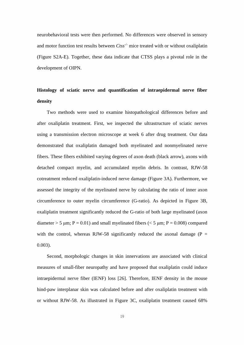
Two methods were used to examine histopathological differences before and
after oxaliplatin treatment. First, we inspected the ultrastructure of sciatic nerves
using a transmission electron microscope at week 6 after drug treatment. Our data
demonstrated that oxaliplatin damaged both myelinated and nonmyelinated nerve
fibers. These fibers exhibited varying degrees of axon death (black arrow), axons with
detached compact myelin, and accumulated myelin debris. In contrast, RJW-58
cotreatment reduced oxaliplatin-induced nerve damage (Figure 3A). Furthermore, we
assessed the integrity of the myelinated nerve by calculating the ratio of inner axon
circumference to outer myelin circumference (G-ratio). As depicted in Figure 3B,
oxaliplatin treatment significantly reduced the G-ratio of both large myelinated (axon
diameter > 5 μm; P = 0.01) and small myelinated fibers (< 5 μm; P = 0.008) compared
with the control, whereas RJW-58 significantly reduced the axonal damage (P =
0.003).
Second, morphologic changes in skin innervations are associated with clinical
measures of small-fiber neuropathy and have proposed that oxaliplatin could induce
intraepidermal nerve fiber (IENF) loss [26]. Therefore, IENF density in the mouse
hind-paw interplanar skin was calculated before and after oxaliplatin treatment with
or without RJW-58. As illustrated in Figure 3C, oxaliplatin treatment caused 68%
20
IENF loss in the interplanar epidermis (Figure 3D; P = 0.003). However, no
differences in IENF density were observed between oxaliplatin-treated mice
co-treated with RJW-58 and control mice.
Furthermore, Ctss-/- mice were also used to examine the neuroprotective role of
CTSS in oxaliplatin-induced nerve damage. Our results indicated that no differences
in ultrastructure changes in the sciatic nerve or the number of IENF between
oxaliplatin-treated and control mice (Figure S2F-I). Furthermore, no differences were
revealed in the expression level of ATF3 in microglial cells isolated from DRG in
mice treated with or without oxaliplatin (Figure S2J). These findings further confirm
the neuroprotective effect of CTSS inhibition in OIPN.
Oxaliplatin treatment increases CTSS expression and binding to STIM1
DNA damage caused by drugs, such as cisplatin and doxorubicin, results in the
upregulation of interferon regulatory factor 1 (IRF1) [27]. A study revealed that CTSS
expression is regulated by transcriptional factor IRF1 [28]. Therefore, we determined
whether oxaliplatin could enhance CTSS expression by regulating IRF1 in SIM-A9
cells. Our data demonstrated that oxaliplatin treatment increased cytosol translocation
of IRF1 to the nucleus (Figure 4A) and enhanced IRF1 binding to the CTSS promoter
region, which augmented the transcriptional level of CTSS in vitro and in vivo (Figure
4B-E).
We further investigated whether serum CTSS level could serve as a predictive
biomarker of oxaliplatin-induced peripheral neuropathy. Our data revealed that the
serum level of CTSS in mice increased at week 2 (P = 0.049), peaked at week 4 (P <
0.0001), and returned to baseline level at week 6 after oxaliplatin treatment (Figure
4F). These changes were consistent with findings that sensory and motor function
21
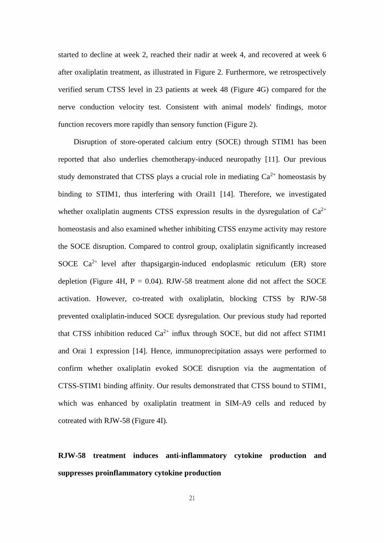
started to decline at week 2, reached their nadir at week 4, and recovered at week 6
after oxaliplatin treatment, as illustrated in Figure 2. Furthermore, we retrospectively
verified serum CTSS level in 23 patients at week 48 (Figure 4G) compared for the
nerve conduction velocity test. Consistent with animal models' findings, motor
function recovers more rapidly than sensory function (Figure 2).
Disruption of store-operated calcium entry (SOCE) through STIM1 has been
reported that also underlies chemotherapy-induced neuropathy [11]. Our previous
study demonstrated that CTSS plays a crucial role in mediating Ca2+ homeostasis by
binding to STIM1, thus interfering with Orail1 [14]. Therefore, we investigated
whether oxaliplatin augments CTSS expression results in the dysregulation of Ca2+
homeostasis and also examined whether inhibiting CTSS enzyme activity may restore
the SOCE disruption. Compared to control group, oxaliplatin significantly increased
SOCE Ca2+ level after thapsigargin-induced endoplasmic reticulum (ER) store
depletion (Figure 4H, P = 0.04). RJW-58 treatment alone did not affect the SOCE
activation. However, co-treated with oxaliplatin, blocking CTSS by RJW-58
prevented oxaliplatin-induced SOCE dysregulation. Our previous study had reported
that CTSS inhibition reduced Ca2+ influx through SOCE, but did not affect STIM1
and Orai 1 expression [14]. Hence, immunoprecipitation assays were performed to
confirm whether oxaliplatin evoked SOCE disruption via the augmentation of
CTSS-STIM1 binding affinity. Our results demonstrated that CTSS bound to STIM1,
which was enhanced by oxaliplatin treatment in SIM-A9 cells and reduced by
cotreated with RJW-58 (Figure 4I).
RJW-58 treatment induces anti-inflammatory cytokine production and
suppresses proinflammatory cytokine production
22
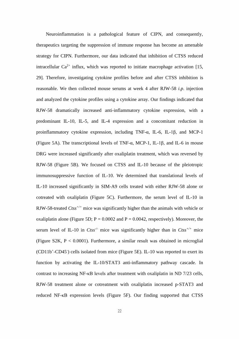
Neuroinflammation is a pathological feature of CIPN, and consequently,
therapeutics targeting the suppression of immune response has become an amenable
strategy for CIPN. Furthermore, our data indicated that inhibition of CTSS reduced
intracellular Ca2+ influx, which was reported to initiate macrophage activation [15,
29]. Therefore, investigating cytokine profiles before and after CTSS inhibition is
reasonable. We then collected mouse serums at week 4 after RJW-58 i.p. injection
and analyzed the cytokine profiles using a cytokine array. Our findings indicated that
RJW-58 dramatically increased anti-inflammatory cytokine expression, with a
predominant IL-10, IL-5, and IL-4 expression and a concomitant reduction in
proinflammatory cytokine expression, including TNF-α, IL-6, IL-1β, and MCP-1
(Figure 5A). The transcriptional levels of TNF-α, MCP-1, IL-1β, and IL-6 in mouse
DRG were increased significantly after oxaliplatin treatment, which was reversed by
RJW-58 (Figure 5B). We focused on CTSS and IL-10 because of the pleiotropic
immunosuppressive function of IL-10. We determined that translational levels of
IL-10 increased significantly in SIM-A9 cells treated with either RJW-58 alone or
cotreated with oxaliplatin (Figure 5C). Furthermore, the serum level of IL-10 in
RJW-58-treated Ctss+/+ mice was significantly higher than the animals with vehicle or
oxaliplatin alone (Figure 5D; P = 0.0002 and P = 0.0042, respectively). Moreover, the
serum level of IL-10 in Ctss-/- mice was significantly higher than in Ctss+/+ mice
(Figure S2K, P < 0.0001). Furthermore, a similar result was obtained in microglial
(CD11b+-CD45-) cells isolated from mice (Figure 5E). IL-10 was reported to exert its
function by activating the IL-10/STAT3 anti-inflammatory pathway cascade. In
contrast to increasing NF-κB levels after treatment with oxaliplatin in ND 7/23 cells,
RJW-58 treatment alone or cotreatment with oxaliplatin increased p-STAT3 and
reduced NF-κB expression levels (Figure 5F). Our finding supported that CTSS
23
inhibition results in an anti-inflammatory response in oxaliplatin-induced neuropathy,
through the upregulation of IL-10.
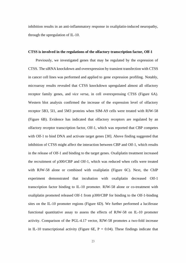
CTSS is involved in the regulations of the olfactory transcription factor, Olf-1
Previously, we investigated genes that may be regulated by the expression of
CTSS. The siRNA knockdown and overexpression by transient transfection with CTSS
in cancer cell lines was performed and applied to gene expression profiling. Notably,
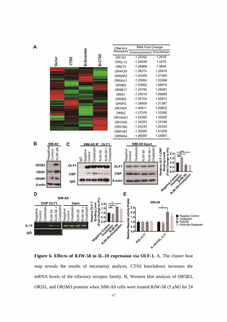
microarray results revealed that CTSS knockdown upregulated almost all olfactory
receptor family genes, and vice versa, in cell overexpressing CTSS (Figure 6A).
Western blot analysis confirmed the increase of the expression level of olfactory
receptor 5B3, 5I1, and 5M3 proteins when SIM-A9 cells were treated with RJW-58
(Figure 6B). Evidence has indicated that olfactory receptors are regulated by an
olfactory receptor transcription factor, Olf-1, which was reported that CBP competes
with Olf-1 to bind DNA and activate target genes [30]. Above finding suggested that
inhibition of CTSS might affect the interaction between CBP and Olf-1, which results
in the release of Olf-1 and binding to the target genes. Oxaliplatin treatment increased
the recruitment of p300/CBP and Olf-1, which was reduced when cells were treated
with RJW-58 alone or combined with oxaliplatin (Figure 6C). Next, the ChIP
experiment demonstrated that incubation with oxaliplatin decreased Olf-1
transcription factor binding to IL-10 promoter. RJW-58 alone or co-treatment with
oxaliplatin promoted released Olf-1 from p300/CBP for binding to the Olf-1-binding
sites on the IL-10 promoter regions (Figure 6D). We further performed a luciferase
functional quantitative assay to assess the effects of RJW-58 on IL-10 promoter
activity. Comparison of the PGL-4.17 vector, RJW-58 promotes a two-fold increase
in IL-10 transcriptional activity (Figure 6E, P = 0.04). These findings indicate that
24
oxaliplatin treatment facilitated the assembling of p300/CBP and Olf-1. By contrast,
inhibiting CTSS by RJW-58 decreased the recruitment of CBP and Olf-1. These
released Olf-1 transcription factors further bind to Olf-1-binding sites on the IL-10
promoter regions (Figure 6D), which results in an increased IL-10 transcriptional
activity (Figure 6E).
Discussion
Oxaliplatin, a platinum derivative, is widely used as first-line adjuvant therapy in
advanced CRC [31]. Oxaliplatin can cause acute pain syndromes, which usually
subside within days after cessation of treatment, and chronic distal neuropathy, which
occurs after several cycles of treatment [32]. However, less is known regarding
oxaliplatin-induced motor impairments from clinical studies or animal models. Our
clinical study demonstrated that oxaliplatin could cause both sensory and motor
impairments in patients receiving a standard dose and course of adjuvant
chemotherapy with an oxaliplatin-containing regimen after the definitive operation
(Figure 1). Furthermore, despite numerous animal models having been established to
understand the mechanisms and explore the therapeutic strategies to treat or prevent
oxaliplatin-induced peripheral neuropathy, most of these models were not fully
relevant to clinical findings. Therefore, establishing an appropriate animal model is
warranted. By administering oxaliplatin according to the treatment protocol illustrated
in Figure 2B, we demonstrated that oxaliplatin treatment caused both sensory and
motor neuropathies without reducing body weight in the mice model. Furthermore,
oxaliplatin treatment significantly reduced the G-ratio of both large myelinated and
small myelinated fibers, compared with the control, assessed using electron
microscopy (Figure 3). These animal data further support that oxaliplatin can cause
25
both sensory and motor impairments, which is consistent with clinical findings
(Figures 1 and 2). Discrepancies between the findings in this study and other studies
are mainly caused by different dosing schedules and cumulative doses of oxaliplatin
[33, 34]. We demonstrated, using this animal model, that CTSS inhibition could
protect animals from oxaliplatin-induced neuropathy, which was supported by the
results from mouse behavioral tests, EM studies of sciatic nerves, and quantification
of IENF density (Figures 2 and 3). The pivotal role of CTSS in oxaliplatin-induced
neuropathy was further validated in the Ctss-/- knockout mouse model (Figure S1).
Lines of evidence have demonstrated that the ubiquitous second messenger Ca2+
plays a crucial role in fundamental physiological functions [35], including cell
motility, apoptosis, cell cycle control, and gene expression. Under inflammations,
dysregulation of intracellular Ca2+ homeostasis is a vital mechanism for regulating
neurite outgrowth and subsequent neurite damage [36]. Our previous studies have
revealed that SOCE components participate in modulating calcium homeostasis in
response to inflammation and partly contribute to paclitaxel-induced neuropathies,
which were also determined in the present study [11, 20]. A study demonstrated that
oxaliplatin was degraded to oxalate and platinum inside the cell. Oxalate by chelating
intracellular Ca2+ disrupts neuronal membrane potentials and further dysregulates the
activity of voltage-gated ion channels. However, these disruptions only caused cold
allodynia in mice [37, 38]. Furthermore, oxaliplatin treatment was reported to induce
intracellular Ca2+ influx, but the mechanism remains unclear [39]. Our previous study
has shown CTSS inhibition reduced Ca2+ influx through affecting SOCE activation
and interfering STIM1 trafficking, which leads to the suppression of tumor cell
progression. Furthermore, immunofluorescence data further demonstrated that CTSS
could bind to STIM1, which was reversed by CTSS inhibitor. This effect was caused
26
by accumulating STIM1 puncta in the ER, thereby reducing the interaction between
active STIM1 and EB1 [14]. In the present study, we demonstrated that oxaliplatin
treatment significantly increased the SOCE Ca2+ level after thapsigargin-induced ER
store depletion, which was reversed by CTSS inhibition with RJW-58 (Figure 4H).
Notably, we determined that oxaliplatin treatment increased CTSS expression by
promoting IRF1 translocation from the cytosol to the nucleus, thereby enhancing
IRF1 binding to the CTSS promoter region, which amplified the transcriptional level
of CTSS (Figure 4A-E). Furthermore, chemotherapeutics stimuli may cause
alterations of lysosomal membrane permeability and leakage of cathepsins into the
cytoplasm [28]. These data explain why oxaliplatin could increase CTSS expression,
causing an increase in binding to STIM1 (Figure 4I), indicating that the regulation of
CTSS expression contributes to the effect of oxaliplatin on intracellular Ca2+
homeostasis.
Neuroinflammation is a hallmark of the pathogenesis of CIPN. Increasing
evidence demonstrates that excessive inflammation in the damaged nervous tissue
after chemotherapy contributes to the initiation and maintenance of peripheral
neuropathies [36]. Oxaliplatin treatment increased the production and release of TNF
and IL-1β through the upregulation of P2X purinoreceptor 7 in human neuroblastoma
cell lines [40]. Furthermore, in vivo study revealed that oxaliplatin treatment causes an
increased number of activated satellite glial cells in the spinal cord [41]. CTSS is
reportedly an autocrine factor that stimulates microglial cell activation and
subsequently regulates the release of proinflammatory cytokines and chemokines [13].
In the present study, we demonstrated that CTSS inhibition with RJW-58 increases
the serum levels of anti-inflammatory cytokines (predominantly IL-10, IL-4, and IL-7)
and suppresses proinflammatory cytokines (Figure 5A). This effect was further
27
validated in an animal model, which demonstrates that oxaliplatin treatment results in
the upregulation of TNF-α, IL-6, IL-1β, and MCP-1 in mice DRG cells, which is
suppressed by cotreated with RJW-58 (Figure 5B). Studies have reported that
exogenous administration of the anti-inflammatory cytokine IL-10 suppresses
allodynia in surgery [9, 10] and chemotherapy-induced neuropathy [42]. Furthermore,
increasing evidence suggests that STIM-dependent Ca2+ influx is required for IL-10
production because dysregulation of Ca2+ homeostasis impairs IL-10 secretion [43].
In this study, we determined that RJW-58 reversed oxaliplatin-induced
dysregulation of Ca2+ homeostasis and increased endogenous IL-10 by activating the
transcription factor Olf-1. We discovered that Olf-1 and transcriptional coactivators
CBP coregulated the induction of IL-10 genes. Studies have indicated that the HAT
activity of p300/CBP is involved in regulating transcription by remodeling chromatin,
which is inhibited by binding with Olf-1 [30, 44]. In the ChIP experiment, oxaliplatin
treatment facilitated the assembling of p300/CBP and Olf-1, whereas RJW-58
reversed this assembling and further drove IL-10 gene transcription by activating
Olf-1. Furthermore, the elevation of endogenous IL-10 after RJW-58 treatment
sequentially triggers the IL-10/STAT3 cascade. IL-10 is also involved in raising
feedback inhibition, which limits the inflammation cascade. Therefore, IL-10
decreases NF-κB expression, which causes a reduction in the induction of
inflammation in mouse DRG neurons. These findings suggest that RJW-58 reverses
oxaliplatin-induces CTSS production, activates the anti-inflammatory IL-10/STAT3
cascade, and reduces proinflammatory cytokines expression, which results in the
alleviation of CIPN.
28
Conclusions
We demonstrated that CTSS plays a critical role in the development of
oxaliplatin-induced neuropathy. Targeting CTSS may reveal a therapeutic strategy to
protect neuron cells from oxaliplatin-induced damage. Furthermore, the serum level
of CTSS may serve as a biomarker for the risk of oxaliplatin-induced peripheral
neuropathies. A proof-of-concept large cohort prospective clinical study is ongoing.
Conflict of Interest Disclosure Statement
The authors declared that they have no conflicts of interest in this work.
Abbreviations
ANOVA: analysis of variance; APCs: antigen-presenting cells; ATF3: activating
transcription factor 3; BSA: bovine serum albumin; CTSS: cathepsin S; cDNA:
complementary DNA; CRISPR/Cas9; clustered regularly interspaced short;
palindromic repeats/CRISPR-associated protein 9; CRC: colorectal cancer; CIPN:
chemotherapy-induced peripheral neuropathy; ChIP: chromatin immunoprecipitation
PCR; DMEM: Dulbecco's modified Eagle's medium; DNA: deoxyribonucleic acid;
DRG: dorsal root ganglion; EDTA: ethylenediaminetetraacetic acid; ELISA:
enzyme-linked immunosorbent assay; ER: endoplasmic reticulum; FACS: BD
fluorescence-activated cell sorter; FBS: fetal bovine serum; FITC: fluorescein
isothiocyanate; GAPDH: glyceraldehyde 3-phosphate dehydrogenase; GFP: green
fluorescent protein; h: hour; HBSS: Hanks' balanced salt solution; HRP: horseradish
peroxidase; HEPES: 4-(2-hydroxyethyl)-1-piperazine ethanesulfonic acid; IRF1:
interferon response factor 1; IL-10: interleukin-10; IL-1β: interleukin-1β; IL-6:
interleukin-6; IL-7: interleukin-7; i.p.: intraperitoneal(ly); IENF: intraepidermal nerve
29
fiber; JAKs: Janus tyrosine kinases; m: meter; M: molar; mRNA: messenger RNA;
MCP1: monocyte chemoattractant protein 1; NF-κB: nuclear factor-κB; NP-40:
Nonidet P-40; NCV: nerve conduction velocity; NET: nerve excitability test; OIPN:
oxaliplatin-induced peripheral neuropathy; Olf-1: olfactory neuronal transcription
factor 1; PAGE: polyacrylamide gel electrophoresis; PBS: phosphate-buffered saline;
PCR: polymerase chain reaction; P300/CBP: p300 and CREB-binding protein; QST:
quantitative sensory test; R: receptor; RNA: ribonucleic acid; RT: reverse
transcription; ROS: reactive oxygen species; s: second; SDS: sodium dodecyl sulfate;
SEM: standard error of the mean; siRNA: small interfering RNA; STAT3: signal
transducer and activator of transcription 3; STIM1: stromal interaction molecule 1;
SOCE: stored-operated Ca2+ entry; TNF-α: tumor necrosis factor-α; TNS: total
neuropathy score
Acknowledgments
The authors would like to thank the Bio-image Core Facility of the National Core
Facility Program for Biotechnology, Ministry of Science and Technology, Taiwan, for
technical services. We also thank the NHRI Microarray Core facility for their help with
technique. This study was supported by the following grants: the National Health
Research Institutes (CA-107-PP-22), the Ministry of Science and Technology (MOST
106-2314-B-006-076-MY3), the Ministry of Health and Welfare (MOHW
109-TDU-B-211-134018, MOHW110-TDU-B-211-144018), Higher Education
Sprout Project, Ministry of Education to the Headquarters of University Advancement
at National Cheng Kung University (NCKU).
Author contributions

30
J.Y. Chang conceived and supervised this study. S.J. Chen, L.H. Chen, M.R. Shen,
and J.Y. Chang designed the research. S.J. Chen, L.H. Chen, H.C. Chiang, C.C. Lee,
Y.F. Lee developed methodology and performed the experiments. Y.M. Yeh, C.C. K
Lin, P.C. Lin, H.W. Huang, M.R. Shen, B.W. Lin, J.C. Lee provided administrative or
technical support. S.J. Chen, L.H. Chen, Y.M. Yeh and J.Y. Chang wrote the
manuscript.
31
References
1. Hill JM, Speer RJ. Organo-platinum complexes as antitumor agents (review).
Anticancer Res. 1982; 2: 173-86.
2. Izycki D, Niezgoda AA, Kazmierczak M, Piorunek T, Izycka N, Karaszewska B,
et al. Chemotherapy-induced peripheral neuropathy - diagnosis, evolution and
treatment. Ginekol Pol. 2016; 87: 516-21.
3. Saif MW, Reardon J. Management of oxaliplatin-induced peripheral neuropathy.
Ther Clin Risk Manag. 2005; 1: 249-58.
4. Aromolaran KA, Goldstein PA. Ion channels and neuronal hyperexcitability in
chemotherapy-induced peripheral neuropathy; cause and effect? Mol Pain. 2017; 13:
1744806917714693.
5. Alberti P, Canta A, Chiorazzi A, Fumagalli G, Meregalli C, Monza L, et al.
Topiramate prevents oxaliplatin-related axonal hyperexcitability and oxaliplatin
induced peripheral neurotoxicity. Neuropharmacology. 2020; 164: 107905.
6. Zareba P, Gryzlo B, Malawska K, Salat K, Hofner GC, Nowaczyk A, et al.
Novel mouse GABA uptake inhibitors with enhanced inhibitory activity toward
mGAT3/4 and their effect on pain threshold in mice. Eur J Med Chem. 2020; 188:
111920.
7. Lees JG, Makker PG, Tonkin RS, Abdulla M, Park SB, Goldstein D, et al.
Immune-mediated processes implicated in chemotherapy-induced peripheral
neuropathy. Eur J Cancer. 2017; 73: 22-9.
8. Zhang H, Li Y, de Carvalho-Barbosa M, Kavelaars A, Heijnen CJ, Albrecht PJ,
et al. Dorsal Root Ganglion Infiltration by Macrophages Contributes to Paclitaxel
Chemotherapy-Induced Peripheral Neuropathy. J Pain. 2016; 17: 775-86.
32
9. Milligan ED, Langer SJ, Sloane EM, He L, Wieseler-Frank J, O'Connor K, et al.
Controlling pathological pain by adenovirally driven spinal production of the
anti-inflammatory cytokine, interleukin-10. Eur J Neurosci. 2005; 21: 2136-48.
10. Shen KF, Zhu HQ, Wei XH, Wang J, Li YY, Pang RP, et al. Interleukin-10
down-regulates voltage gated sodium channels in rat dorsal root ganglion neurons.
Exp Neurol. 2013; 247: 466-75.
11. Chen LH, Yeh YM, Chen YF, Hsu YH, Wang HH, Lin PC, et al. Targeting
interleukin-20 alleviates paclitaxel-induced peripheral neuropathy. Pain. 2020; 161:
1237-1254.
12. Clark AK, Malcangio M. Microglial signalling mechanisms: Cathepsin S and
Fractalkine. Exp Neurol. 2012; 234: 283-92.
13. Barclay J, Clark AK, Ganju P, Gentry C, Patel S, Wotherspoon G, et al. Role of
the cysteine protease cathepsin S in neuropathic hyperalgesia. Pain. 2007; 130:
225-34.
14. Lin HH, Chen SJ, Shen MR, Huang YT, Hsieh HP, Lin SY, et al. Lysosomal
cysteine protease cathepsin S is involved in cancer cell motility by regulating
store-operated Ca(2+) entry. Biochim Biophys Acta Mol Cell Res. 2019; 1866:
118517.
15. Nascimento Da Conceicao V, Sun Y, Zboril EK, De la Chapa JJ, Singh BB. Loss
of Ca(2+) entry via Orai-TRPC1 induces ER stress, initiating immune activation in
macrophages. J Cell Sci. 2019; 133; jcs237610.
16. Ip WKE, Hoshi N, Shouval DS, Snapper S, Medzhitov R. Anti-inflammatory
effect of IL-10 mediated by metabolic reprogramming of macrophages. Science. 2017;
356: 513-9.
33
17. Hutchins AP, Diez D, Miranda-Saavedra D. The IL-10/STAT3-mediated
anti-inflammatory response: recent developments and future challenges. Brief Funct
Genomics. 2013; 12: 489-98.
18. Titos E, Rius B, Lopez-Vicario C, Alcaraz-Quiles J, Garcia-Alonso V, Lopategi
A, et al. Signaling and Immunoresolving Actions of Resolvin D1 in Inflamed Human
Visceral Adipose Tissue. J Immunol. 2016; 197: 3360-70.
19. Huang HW, Wu PY, Su PF, Li CI, Yeh YM, Lin PC, et al. A Simplified
Diagnostic Classification Scheme of Chemotherapy-Induced Peripheral Neuropathy.
Dis Markers. 2020; 2020: 3402108.
20. Chen YF, Chen LH, Yeh YM, Wu PY, Chen YF, Chang LY, et al. Minoxidil is a
potential neuroprotective drug for paclitaxel-induced peripheral neuropathy. Sci Rep.
2017; 7: 45366.
21. Inoue K, Tsuda M. Microglia in neuropathic pain: cellular and molecular
mechanisms and therapeutic potential. Nat Rev Neurosci. 2018; 19: 138-52.
22. Nieto FR, Clark AK, Grist J, Hathway GJ, Chapman V, Malcangio M.
Neuron-immune mechanisms contribute to pain in early stages of arthritis. J
Neuroinflammation. 2016; 13: 96.
23. Clark AK, Yip PK, Grist J, Gentry C, Staniland AA, Marchand F, et al.
Inhibition of spinal microglial cathepsin S for the reversal of neuropathic pain. Proc
Natl Acad Sci U S A. 2007; 104: 10655-60.
24. Zhao P, Lieu T, Barlow N, Metcalf M, Veldhuis NA, Jensen DD, et al. Cathepsin
S causes inflammatory pain via biased agonism of PAR2 and TRPV4. J Biol Chem.
2014; 289: 27215-34.
34
25. Makker PG, Duffy SS, Lees JG, Perera CJ, Tonkin RS, Butovsky O, et al.
Characterisation of Immune and Neuroinflammatory Changes Associated with
Chemotherapy-Induced Peripheral Neuropathy. PLoS One. 2017; 12: e0170814.
26. Lauria G, Dacci P, Lombardi R, Cazzato D, Porretta-Serapiglia C, Taiana M, et
al. Side and time variability of intraepidermal nerve fiber density. Neurology. 2015;
84: 2368-71.
27. Pavan S, Olivero M, Cora D, Di Renzo MF. IRF-1 expression is induced by
cisplatin in ovarian cancer cells and limits drug effectiveness. Eur J Cancer. 2013; 49:
964-73.
28. Kramer L, Turk D, Turk B. The Future of Cysteine Cathepsins in Disease
Management. Trends Pharmacol Sci. 2017; 38: 873-98.
29. Zumerle S, Cali B, Munari F, Angioni R, Di Virgilio F, Molon B, et al.
Intercellular Calcium Signaling Induced by ATP Potentiates Macrophage
Phagocytosis. Cell Rep. 2019; 27: 1-10 e4.
30. Zhao F, McCarrick-Walmsley R, Akerblad P, Sigvardsson M, Kadesch T.
Inhibition of p300/CBP by early B-cell factor. Mol Cell Biol. 2003; 23: 3837-46.
31. Alcindor T, Beauger N. Oxaliplatin: a review in the era of molecularly targeted
therapy. Curr Oncol. 2011; 18: 18-25.
32. Cassidy J, Misset JL. Oxaliplatin-related side effects: characteristics and
management. Semin Oncol. 2002; 29: 11-20.
33. Xiao WH, Zheng H, Bennett GJ. Characterization of oxaliplatin-induced chronic
painful peripheral neuropathy in the rat and comparison with the neuropathy induced
by paclitaxel. Neuroscience. 2012; 203: 194-206.
34. Gadgil S, Ergun M, van den Heuvel SA, van der Wal SE, Scheffer GJ,
Hooijmans CR. A systematic summary and comparison of animal models for
35
chemotherapy induced (peripheral) neuropathy (CIPN). PLoS One. 2019; 14:
e0221787.
35. Chen YF, Chen YT, Chiu WT, Shen MR. Remodeling of calcium signaling in
tumor progression. J Biomed Sci. 2013; 20: 23.
36. Sisignano M, Baron R, Scholich K, Geisslinger G. Mechanism-based treatment
for chemotherapy-induced peripheral neuropathic pain. Nat Rev Neurol. 2014; 10:
694-707.
37. Gamelin L, Capitain O, Morel A, Dumont A, Traore S, Anne le B, et al.
Predictive factors of oxaliplatin neurotoxicity: the involvement of the oxalate outcome
pathway. Clin Cancer Res. 2007; 13: 6359-68.
38. Sakurai M, Egashira N, Kawashiri T, Yano T, Ikesue H, Oishi R.
Oxaliplatin-induced neuropathy in the rat: involvement of oxalate in cold hyperalgesia
but not mechanical allodynia. Pain. 2009; 147: 165-74.
39. Schulze C, McGowan M, Jordt SE, Ehrlich BE. Prolonged oxaliplatin exposure
alters intracellular calcium signaling: a new mechanism to explain
oxaliplatin-associated peripheral neuropathy. Clin Colorectal Cancer. 2011; 10:
126-33.
40. Massicot F, Hache G, David L, Chen D, Leuxe C, Garnier-Legrand L, et al.
P2X7 Cell Death Receptor Activation and Mitochondrial Impairment in
Oxaliplatin-Induced Apoptosis and Neuronal Injury: Cellular Mechanisms and In
Vivo Approach. PLoS One. 2013; 8: e66830.
41. LaPointe NE, Morfini G, Brady ST, Feinstein SC, Wilson L, Jordan MA. Effects
of eribulin, vincristine, paclitaxel and ixabepilone on fast axonal transport and
kinesin-1 driven microtubule gliding: implications for chemotherapy-induced
peripheral neuropathy. Neurotoxicology. 2013; 37: 231-9.
36
42. Binshtok AM, Wang H, Zimmermann K, Amaya F, Vardeh D, Shi L, et al.
Nociceptors are interleukin-1beta sensors. J Neurosci. 2008; 28: 14062-73.
43. Matsumoto M, Fujii Y, Baba A, Hikida M, Kurosaki T, Baba Y. The calcium
sensors STIM1 and STIM2 control B cell regulatory function through interleukin-10
production. Immunity. 2011; 34: 703-14.
44. Chan HM, La Thangue NB. p300/CBP proteins: HATs for transcriptional
bridges and scaffolds. J Cell Sci. 2001; 114: 2363-73.
37
Figure legends
38
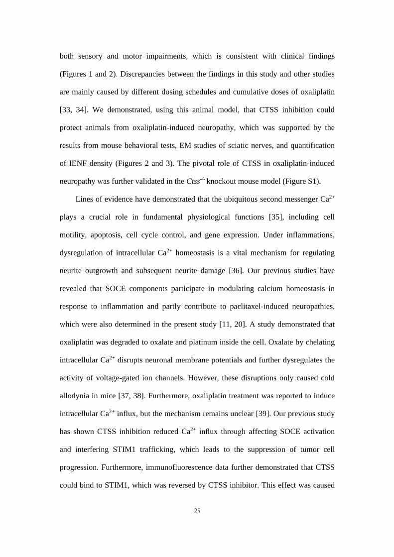
Figure 1. Sensory and motor nerve deficit in patients with colorectal cancer
(CRC) after oxaliplatin treatment. A-C, Sequential measurement of the median,
sural, and ulnar sensory nerve by NCV test before and after oxaliplatin treatment at
baseline and weeks 12, 36, and 48 in CRC patients. D-E, Sequential measurement of
median and tibial motor nerve before and after oxaliplatin treatment at weeks 12, 36,
and 48 in CRC patients. F, The CIPN score at baseline and weeks 12, 36, and 48 in
patients with colorectal cancer. **** P < 0.0001, *** P < 0.001, ** P < 0.01, * P <
0.05 compared with baseline using a two-tailed Student’s t-test. Each value represents
the mean ± SEM from CRC patients.
39
40
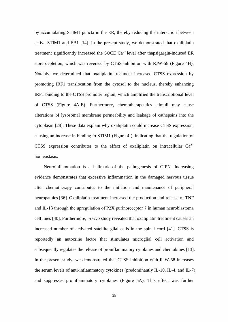
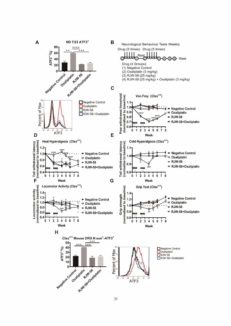
Figure 2. The neuropathic effects of oxaliplatin in the Ctss+/+ mouse model. A,
Flow analysis of ATF3 expression in ND 7/23 cell treated with control, oxaliplatin (1
μM), RJW-58 (5 μM), or a combination of oxaliplatin (1 μM) and RJW-58 (5 μM),
respectively. B, The protocol of drug administration and behavioral tests in the mouse
model. The basal levels of each behavioral assay were obtained before treatment. In
the first week, PBS control (100 μL), oxaliplatin (3 mg/kg, 100 μL), and RJW-58 (25
mg/kg, 100 μL) were injected intraperitoneally five times per week for 2 weeks.
Behavioral tests were performed weekly. C, Von Frey filament test to detect the paw
withdrawal threshold to mechanical stimulus. The Y-axis is the normalized pressure
from touch to paw withdrawal. D-E, Tail immersion test to assess thermal sensation.
The Y-axis is the normalized latency from tail immersion to tail withdrawal. F,
Locomotor activity test to detect allodynia. The Y-axis is the normalized moved
distance baseline. G, Grip test to detect neuromuscular function. The Y-axis is the
normalized moved distance baseline. The black arrow indicates drug infusion. H,
Flow analysis of ATF3 expression of primary DRG cells at week 4 for NeuN+-ATF3+
double staining. **** P < 0.0001, *** P < 0.001, ** P < 0.01, * P < 0.05 compared
with baseline using a two-tailed Student’s t-test. Each value is represented as mean ±
SEM from at least seven mice in each group.
41
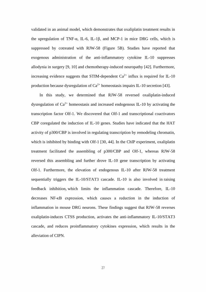
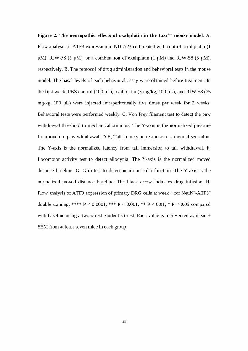
Figure 3. RJW-58 protects peripheral nerves from oxaliplatin-induced damage.
A, Transmission electron micrographs of the sciatic nerve from Ctss+/+ mice treated
with control, oxaliplatin, RJW-58, or the combination of oxaliplatin and RJW-58.
42
Bottom: G-ratio's measurement represents the ratio of axon diameter (yellow line) to
myelin (red line). N: nucleus of Schwann cell. Scale bar: 10 μm (top), 500 nm (middle
and bottom). B, The myelinated fiber density of sciatic nerves from mice in different
treatment groups was measured at week 6 (n = 5 mice/group). The G-ratio was
calculated and compared between different treatment groups. C-D, Representative
immunofluorescence images and quantitative analysis of intraepidermal nerve fibers
of mice treated with control, oxaliplatin, RJW-58, and a combination of oxaliplatin
and RJW-58. Scale bar: 25 μm. **** P < 0.0001, *** P < 0.001, ** P < 0.01, * P <
0.05 compared with baseline using a two-tailed Student’s t-test. Each value represents
the mean ± SEM from at least 7 mice in each group.
43
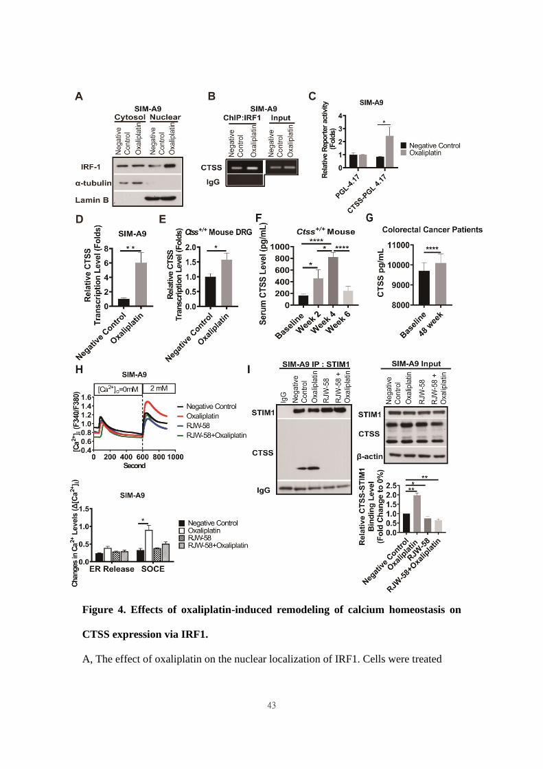
Figure 4. Effects of oxaliplatin-induced remodeling of calcium homeostasis on
CTSS expression via IRF1.
A, The effect of oxaliplatin on the nuclear localization of IRF1. Cells were treated
44
with or without oxaliplatin (1 μM) for 24 h, and the cytosolic and nuclear fractions
were then separated, as described in the Materials and Methods section. B, The effect
of oxaliplatin treatment for 24 h on IRF1 binding to the CTSS promoter regions in
SIM-A9 cells was analyzed using ChIP. C, The effect of oxaliplatin treatment for 24 h
on the transcriptional activity of CTSS promoters was determined using the promoter
assay. D, The effect of oxaliplatin treatment for 24 h on the transcription levels of
CTSS genes in SIM-A9 was determined using RT-qPCR. E, The effect of oxaliplatin
on the transcription levels analysis of CTSS genes in mouse primary DRG at week 4
was determined using RT-qPCR. F, The time-dependent mouse serum CTSS level (n
= 10). G, Patients with colorectal cancer’ serum CTSS level at week 48 compared to
baseline (n = 23). **** P < 0.0001, Two-way ANOVA analysis. H, For the drug
treatment, the cells were pretreated with DMSO or RJW-58 (5 μM). The treatment
time for ND 7/23 and SIM-A9 was 24 h. The calcium influx from the ER and SOCE
in CTSS overexpression (n = 50 for each condition) cell was assessed using
single-cell intracellular Ca2+ measurement. I, CTSS was disassociated from STIM1
following RJW-58 treatment. **** P < 0.0001, *** P < 0.001, ** P < 0.01, * P < 0.05.
The statistical significance was determined using a two-tailed Student’s t-test.

45
46
Figure 5. Effects of RJW-58 in IL-10 expression in vitro and in vivo. A, Heat map
of cytokine array clustering analysis. RJW-58 increased IL-10 secretion in B6 mouse
serum at week 4. B, The effect of RJW-58 (5 μM) in primary DRG cells on the
transcription levels of TNF-α, IL-6, IL-1β, and MCP-1 genes at week 4 was
determined using RT-qPCR. C, Western blotting of IL-10 expression level in SIM-A9
after RJW-58 for 24 h treatment. D, The mouse serum IL-10 level after RJW-58
treatment at week 4. E, Flow analysis of CD11b+-CD45- B6 primary microglial cells
at week 4 for IL-10 expression. F, Western blotting of the NF-κB/STAT3 signaling
pathway under treatment with RJW-58 for 24 h. **** P < 0.0001, *** P < 0.001, ** P
< 0.01, * P < 0.05. The statistical significance was determined using a two-tailed
Student’s t-test. Data are presented as the mean ± SEM.
47
Figure 6. Effects of RJW-58 in IL-10 expression via OLF-1. A, The cluster heat
map reveals the results of microarray analysis. CTSS knockdown increases the
mRNA levels of the olfactory receptor family. B, Western blot analysis of OR5B3,
OR5I1, and OR5M3 proteins when SIM-A9 cells were treated RJW-58 (5 μM) for 24
48
h. C, OLF-1 disassociated with CBP by RJW-58 for 24 h treatment in SIM-A9 cells.
D, The effect of RJW-58 for 24 h on OLF-1 binding to the IL-10 promoter region in
SIM-A9 cells was analyzed using ChIP. E, RJW-58 treatment facilitated IL-10
promoter activity in SIM-A9. IL-10 promoters were identified using the promoter
assay. **** P < 0.0001, *** P < 0.001, ** P < 0.01, * P < 0.05. The statistical
significance was determined using a two-tailed Student’s t-test. Data are presented as
the mean ± SEM.


![TITRE IN AATD AND COPD · 2010-06-04 · proteases, such as cathepsin-B, and the matrix metalloproteases (MMP™s) [4]. In general the serine and cysteine proteases are capable of](https://img.pdfslide.us/doc/110x75/5e7dd6a4e473de6de66ce452/titre-in-aatd-and-copd-2010-06-04-proteases-such-as-cathepsin-b-and-the-matrix.jpg)