Embed Size (px)
Citation preview
International Journal of Pharmaceutics 166 (1998) 27–35
Targeted drug delivery to the brain via phosphonate derivativesII. Anionic chemical delivery system for zidovudine (AZT)
Gabor Somogyi 1, Peter Buchwald, Daishuke Nomi 2, Laszlo Prokai, Nicholas Bodor *
Center for Drug Disco6ery, Uni6ersity of Florida, Health Science Center, PO Box 100497, Gaines6ille, FL 32610-0497, USA
Received 8 August 1997; received in revised form 31 October 1997; accepted 16 December 1997
Abstract
The previously described anionic chemical delivery system (aCDS) approach (Somogyi, G., Nishitani, S., Nomi, D.,Buchwald, P., Prokai, L., Bodor, N., 1997. Targeted drug delivery to the brain via phosphonate derivatives. I. Design,synthesis and evaluation of an anionic chemical delivery system for testosterone. Int. J. Pharm. (166, 15–26) wasapplied for brain-targeted delivery of AZT. For this system, the whole designed metabolic sequence, ending withrelease of the active drug at the targeted organ, was completed. As a less hindered ester function was built into thisaCDS, cleavage by esterases, the first metabolic step in the decomposition process, was fairly rapid. The negativelycharged decomposition product (AZT-P−) could be detected for about 48 h in different organs. In vitro experimentsproved that AZT is released from this phosphonate derivative after phosphorolytic attack, the second metabolic stepof the designed sequence, but only at a slow rate. While the phosphonate derivative of a secondary hydroxyl groupproved completely resistant to such phosphorolytic attacks, alkaline phosphatase, but not phosphodiesterase, was ableto cleave the P–O bond of the phosphonate derivative at the primary hydroxyl group in this system. After i.v.administration of AZT-aCDS in rabbits, AZT could be detected in the brain, albeit only at very low concentrations.Even if sufficiently high drug levels could not be delivered with the present aCDSs, the sequence was completed, therate of metabolism was controllable, and the approach is flexible enough; therefore, further, adequate manipulationsmight render such anionic chemical delivery systems into a useful addition of the drug targeting arsenal. © 1998Elsevier Science B.V. All rights reserved.
Keywords: Brain-targeted delivery; Anionic chemical delivery system (aCDS); Azidothymidine (AZT); Alkyl phos-phatase and phosphodiesterase
* Corresponding author. Tel.: +1 352 3928186; fax: +1 352 3928589.1 On leave of absence from the Department of Forensic Medicine of the Medical University of Debrecen, Hungary.2 On leave of absence from the Otsuka Pharmaceutical Company, Ltd., Tokushima, Japan.
0378-5173/98/$19.00 © 1998 Elsevier Science B.V. All rights reserved.
PII S0378-5173(98)00012-X
G. Somogyi et al. / International Journal of Pharmaceutics 166 (1998) 27–3528
1. Introduction
Within the general principles of retrometabolicdrug design (Bodor, 1994; Bodor and Buchwald,1997), a novel anionic chemical delivery system(aCDS) approach, involving an (acyloxy)alkylphosphonate targetor moiety (Bodor, 1995), hasbeen developed and applied recently for brain-targeted delivery of testosterone (Somogyi et al.,1997). Such enzymatic physical-chemical basedCDSs rely on a designed metabolic sequence thatfirst transforms the systemically administered,lipophilic CDS into a hydrophilic intermediatethat will be locked inside the brain by the blood–brain barrier (BBB) and then provides a sustainedrelease of the active drug at its site of action(Bodor, 1987, 1994; Bodor and Buchwald, 1997).For the testosterone-aCDS that uses a (pivaloy-loxy)methyl (POM) phosphonate ester targetormoiety, we have found that the first ester iscleaved through a hydrolytic reaction yielding thenegatively charged, ‘locked in’ intermediate, butdephosphorylation of the phosphonate derivativeat the secondary hydroxyl group, the enzymaticstep that should release the active drug, does notoccur in a detectable manner.
Here we report work on a similar aCDS forAZT (3%-azido-3%-deoxythymidine, zidovudine),where attachment of the (acyloxy)alkyl phospho-nate targetor could be carried out at a primaryhydroxyl group. There are several other reasonsto investigate a brain-targeted delivery system forAZT. Since in the brain of patients with AIDSdementia, HIV-1 virus, proviral DNA, viral nu-cleic acid, viral antigens, and HIV-1 virions arepresent (McArthur, 1987), an ideal chemothera-peutic agent for HIV-1 should penetrate the cen-tral nervous system (Yarchoan and Broder, 1987),even if it is not certain whether or not the pathol-ogy of AIDS dementia complex is caused by adirect infection of the brain or by an indirectmechanism. Despite entry into the human cere-brospinal fluid (CSF) (Klecker et al., 1987), AZT,like most polar nucleosides, does not readily crossthe blood–brain barrier, e.g. radioactive AZT wasnot shown to penetrate in significant amountsinto the brain of rats (Ellison et al., 1988). Thismodified riboside has been shown to be useful in
improving the neuropsychiatric course of AIDSencephalopathy in a few patients, but the dosesrequired to elicit this improvement precipitatesevere anemia, bone marrow suppression, andother toxic effects (Yarchoan et al., 1987; Mitsuyaet al., 1990). In an effort to ameliorate the prog-nosis of AIDS encephalopathy, the dihydropy-ridine–pyridinium salt redox-type CDS approach(Bodor, 1987) was already applied to AZT withpromising results (Brewster et al., 1988; Torrenceet al., 1988; Gallo et al., 1989; Gogu et al., 1989;Little et al., 1990). Since the phosphonate deriva-tive of a secondary hydroxyl group proved resis-tant to phosphorolytic attacks in the anionicchemical delivery system for testosterone, we in-vestigated the possibilities of this delivery systemfor AZT, where attachment of the (acyloxy)alkylphosphonate targetor was carried out at a pri-mary alcohol, and cleavage of this less hinderedP–O bond is more likely.
2. Materials and methods
2.1. Chemistry
Melting points (MP) were obtained using aFisher–Johns melting points apparatus and areuncorrected. Mass spectra (MS) were recorded bya Kratos Analytical MS80RFA instrument usingfast atom bombardment (FAB). Proton nuclearmagnetic resonance spectra (1H NMR) wererecorded in a Varian EM390 (90 MHz) spectrom-eter. Samples were dissolved in an appropriatedeuterated solvent and chemical shifts (d) re-ported in ppm relative to an internal standard(tetramethylsilane, TMS). Elemental analyseswere performed by Atlantic Microlabs (Atlanta,GA). All starting materials were of reagent gradeand obtained from Aldrich Chemical Co. (Mil-waukee, WI). 3%-Azido-3%-deoxythymidine (AZT)was obtained from Pharmatec Inc. (Alachua, FL).Merck Kieselgel 60 (70–230 Mesh ASTM) andAldrich Florisil (100–200 Mesh) were used forcolumn chromatography. Phosphodiesterase I(E.C. 3.1.4.1) type IV (0.028 units/mg solid), andalkaline phosphatase (E.C. 3.1.3.1, 2000 units)were used for enzymatic assay, and were pur-chased from Sigma Chem. Co. (St. Louis, MO).
G. Somogyi et al. / International Journal of Pharmaceutics 166 (1998) 27–35 29
2.2. Synthesis
2.2.1. Chloromethyl hexanoate (1)To a mixture of hexanoyl chloride (25 g, 0.186
mol) and paraformaldehyde (5.58 g, 0.186 mol), acatalytic amount (550 mg) of zinc chloride wasadded in an ice bath. After the resulting exother-mic reaction subsided, the mixture was heated at90–100°C for 4.5 h. Purification by distillation atreduced pressure gave 22.79 g of a colorless liquidin 75% yield; boiling point 37–40°C/0.55 mmHg.NMR (CDCl3): 0.90 (t, 3H, J=6 Hz), 1.20–1.80(m, 6H), 2.37 (t, 2H, J=7 Hz), 5.67 (s, 2H).
2.2.2. Iodomethyl hexanoate (2)Chloromethyl hexanoate (1) (205 mg, 1.25
mmol) was stirred with sodium iodide (900 mg,6.0 mmol) in 3 ml of dry acetone for 4 h at roomtemperature. The insoluble (sodium chloride) ma-terial was removed by filtration and washed withfresh acetone. The filtrate was evaporated; hexaneand 5% aqueous sodium thiosulfate solution wereadded to the residue. After the mixture was thor-oughly shaken, the organic layer was separatedand washed with 5% aqueous sodium thiosulfatesolution, then dried over sodium sulfate. The sol-vent was evaporated. Iodomethyl hexanoate,CH3(CH2)4COOCH2I, was obtained as a yellowoil in 78% yield (250 mg). NMR (CDCl3): 0.90 (t,3H, J=6 Hz), 1.20–1.80 (m, 6H), 2.30 (t, 2H,J=7 Hz), 5.89 (s, 2H). The compound was usedfor the next reaction without further purification.
2.2.3. 5 %-(3 %-Azido-3 %-deoxythymidyl)methylphosphonate (AZT-P−, 3)
AZT (zidovudine, 3%-azido-3%-deoxythymidine)(10.0 g, 37.4 mmol), sodium carbonate (11.9 g,112 mmol), methylphosphonic dichloride (14.9 g,112 mmol), and dry acetone (50 ml) were com-bined in a round-bottom flask under a stream ofnitrogen; the mixture was stirred at room temper-ature for 17 h and then cooled. The acetonesolvent was dried with anhydrous K2CO3, andthen distilled (b.p. 56.2°C) before using. To theice-cooled, stirred residual mixture, water (4.0 ml,225 mmol) was added dropwise, followed by 60ml of methanol. To the resultant suspension,Florisil® (100 g) was added, and the mixture was
evaporated to dryness. The crude material waspurified by column chromatography on Florisil®
(200 g) with dichloromethane–methanol (20:1 to1:1 gradient) as eluent to give 4.33 g of a crudeamorphous solid. The crude solid was dissolved inmethanol (21.6 ml), then 216 ml of ether wereadded. The formed precipitate was collected byfiltration and washed with ether. The resultingpale yellow amorphous solid was dried in vacuumand used for the next reaction without furtherpurification. Yield: 4.53 g (42%). NMR (MeOH-d4): 1.32 (d, 3H, J=17 Hz), 1.91 (s, 3H), 2.25–2.50 (m, 2H), 3.85–4.15 (m, 3H), 4.25–4.50 (m,1H), 6.17 (t, 1H, J=6 Hz), 7.64 (s, 1H). NMR(DMSO-d6): 1.5 (d, 3H, J=17 Hz), 1.80 (s, 3H),2.20–2.45 (m, 2H), 3.75–4.05 (m, 3H), 4.25–4.60(m, 1H), 6.10 (t, 1H, J=7 Hz), 7.72 (s, 1H).
2.2.4. 5 %-(3 %-Azido-3 %-deoxythymidyl)hexanoyloxymethyl methylphosphonate(AZT-aCDS, 4)
AZT-P− (3) (4.5 g, 13 mmol), cesium fluoride(4.41 g, 29 mmol), freshly prepared iodomethylhexanoate (6.68 g, 26 mmol), and dimethylfor-mamide (45 ml) were mixed under a stream ofnitrogen and stirred at room temperature for 20h. The reaction mixture was then poured into 300ml of ether and washed successively with water(100 ml), 5% aqueous sodium thiosulfate solution(100 ml), and again with water (100 ml). Eachaqueous layer was extracted with one 100-ml por-tion of ether. The organic layers were combined,dried over magnesium sulfate, filtered, and con-centrated to give 3.95 g of brown oil. The crudematerial was purified by column chromatographyon silica gel (40 g) using hexane–ethyl acetate (1:1to 0:1 gradient) as eluent to give 1.03 g of aslightly yellow viscous oil in 16.7% yield. MS: m/z474 (MH+). NMR (CDCl3): 0.89 (t, 3H, J=6Hz), 1.10–1.85 (m, 6H), 1.62 (d, 3H, J=18 Hz),1.92 (s, 3H), 2.20–2.50 (m, 4H), 5.66 (d, 2H,J=13 Hz), 6.14 (t, 1H, J=6 Hz), 7.26 (s, 1H),9.40 (bs, 1H). Elemental analysis forC18H28N5O8P×1.5H2O. Theory: C, 43.20; H,6.24; N, 13.99. Found: C, 42.94; H, 5.95; N,14.12.
G. Somogyi et al. / International Journal of Pharmaceutics 166 (1998) 27–3530
2.3. Analytical method
The HPLC (UV/VIS detection) method of Lit-tle et al. (1990) was adapted and modified for thepresent work. The chromatographic analysis wasperformed in a system consisting of Spectra-Physics (Palo Alto, CA) SP 8810 solvent deliverysystem, SP 8780 auto sampler, SP 8456 UV/VISvariable wavelength detector operated at 266 nm,and SP 4290 integrator. The column was a 3mSupelco LC-8-DB (7.5 cm, 4.6 mm i.d.) with a15-mm long (3m) LC-8-DB guard column. Themobile phase consisted of 10% acetonitrile, 90%0.01 M phosphate buffer (pH 7.0) and 5 mMtetramethyl ammonium perchlorate. The flow ratewas 0.8 ml/min with a column pressure of 2210p.s.i. at ambient temperature. In this system, re-tention times for AZT and AZT-P− were 8.86and 3.19 min, respectively. For detection of AZT-aCDS the same system was used but the mobilephase consisted of 50% acetonitrile and 50%buffer. The retention time was 4.7 min. The limitof detection was 0.05 mg/g tissue.
2.4. In 6itro stability studies
2.4.1. In 6itro stability of AZT-P− (3) tophosphodiesterase IV and alkaline phosphatase I
Control samples: 3 ml of buffer (pH 7.4 phos-phate buffer, pH 8.8 Tris buffer, pH 9.8 glycinebuffer) and 60 ml of stock solution of AZT-P−
(1.0 mg/ml) were measured into a vial. The vialwas closed by a cap and put into a waterbath at atemperature of 37°C. At appropriate time inter-vals aliquots (400 ml) were removed and added to800 ml of acetonitrile containing 5% DMSO and1% acetic acid. Twenty ml of this solution wereanalyzed by HPLC. Enzyme-treated samples: Theabove buffer solutions were used with addition of3 mg type IV phosphodiesterase I or 60 ml alka-line phosphatase.
2.4.2. In 6itro stability of AZT-aCDS (4)Adult, male, New Zealand, white rabbits weigh-
ing 2500–3000 g were used. The animals werekept in individual cages with free access to foodand water. A group of five rabbits was used, andanother group of three animals served as control.
In vitro investigations were performed in blood,brain, liver, lung, and testes, respectively. Freshlycollected whole blood was used. Tissue ho-mogenates were prepared by homogenizing (usinga Tekmar-tissuemizer) freshly collected organ tis-sues with isotonic phosphate buffer (pH 7.4) togive 20% (w/w) homogenate. Four hundred ml ofstock solution of AZT-aCDS (4) (concentration,1.0 mg/ml in DMSO) were added to 5 g of 20%(w/w) biological medium at 37°C and mixed usinga Fisher-brand Touch-Mixer for 10 s. At appro-priate time intervals, 400 ml of samples were takenand mixed to 800 ml of acetonitrile containing 5%DMSO and 1% acetic acid. The mixture wasshaken using the Touch-Mixer for 1 min andcentrifuged for 12 min at 12000 r.p.m. The super-natant was removed with an insulin syringe andfiltered through a Millipore (Type H, pore size0.45 mm) filter. The solution was analyzed byHPLC, injecting 20 ml of sample. Quantitationwas done by a calibration curve.
2.5. In 6i6o distribution/metabolism studies
Groups of at least three rabbits were used.AZT-aCDS (4) was dissolved in DMSO and ad-ministered in the ear vein of conscious animals ata dose of 54.4 mg/kg. In the control group onlythe solvent was administered. Animals were sac-rificed by injection of a pentobarbital overdose atappropriate time intervals (0.1, 3, 8, 24, 30 and 48h). Trunk blood was collected into heparinizedtubes. Organs (brain, liver, lung, and testes) wereremoved and immediately frozen. Samples forHPLC analysis were prepared as described previ-ously for the in vitro studies.
3. Results and discussion
3.1. Synthesis
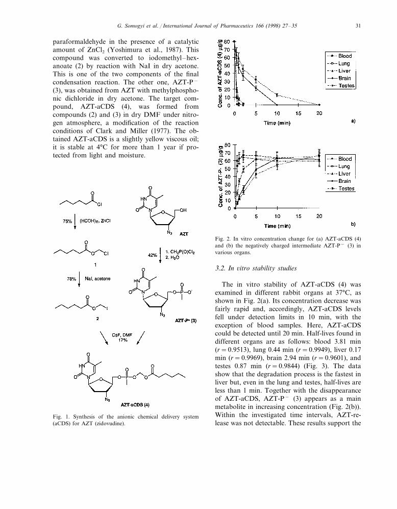
The synthetic route for the synthesis of theAZT anionic chemical delivery system AZT-aCDS (5%-(3%-azido-3%-deoxythymidyl)-hexanoy-loxymethyl methylphosphonate), is summarized inFig. 1. Chloromethyl hexanoate (1) was preparedby reaction of hexanoyl chloride with
G. Somogyi et al. / International Journal of Pharmaceutics 166 (1998) 27–35 31
paraformaldehyde in the presence of a catalyticamount of ZnCl2 (Yoshimura et al., 1987). Thiscompound was converted to iodomethyl–hex-anoate (2) by reaction with NaI in dry acetone.This is one of the two components of the finalcondensation reaction. The other one, AZT-P−
(3), was obtained from AZT with methylphospho-nic dichloride in dry acetone. The target com-pound, AZT-aCDS (4), was formed fromcompounds (2) and (3) in dry DMF under nitro-gen atmosphere, a modification of the reactionconditions of Clark and Miller (1977). The ob-tained AZT-aCDS is a slightly yellow viscous oil;it is stable at 4°C for more than 1 year if pro-tected from light and moisture.
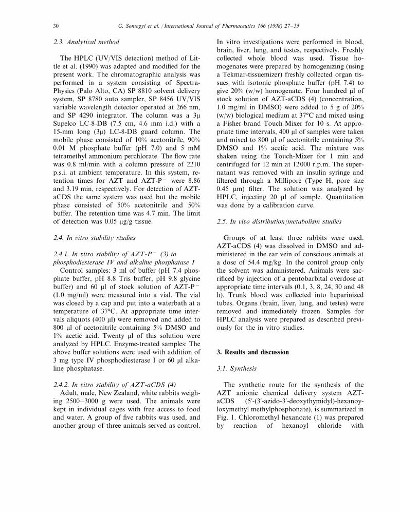
Fig. 2. In vitro concentration change for (a) AZT-aCDS (4)and (b) the negatively charged intermediate AZT-P− (3) invarious organs.
Fig. 1. Synthesis of the anionic chemical delivery system(aCDS) for AZT (zidovudine).
3.2. In 6itro stability studies
The in vitro stability of AZT-aCDS (4) wasexamined in different rabbit organs at 37°C, asshown in Fig. 2(a). Its concentration decrease wasfairly rapid and, accordingly, AZT-aCDS levelsfell under detection limits in 10 min, with theexception of blood samples. Here, AZT-aCDScould be detected until 20 min. Half-lives found indifferent organs are as follows: blood 3.81 min(r=0.9513), lung 0.44 min (r=0.9949), liver 0.17min (r=0.9969), brain 2.94 min (r=0.9601), andtestes 0.87 min (r=0.9844) (Fig. 3). The datashow that the degradation process is the fastest inliver but, even in the lung and testes, half-lives areless than 1 min. Together with the disappearanceof AZT-aCDS, AZT-P− (3) appears as a mainmetabolite in increasing concentration (Fig. 2(b)).Within the investigated time intervals, AZT-re-lease was not detectable. These results support the
G. Somogyi et al. / International Journal of Pharmaceutics 166 (1998) 27–3532
hypothesis (Somogyi et al., 1997) that the maindecomposition step of AZT-aCDS is the hy-drolytic cleavage by esterases yielding AZT-P−
(3) as a main metabolite. Cleavage of this less-hin-dered hexanoate ester function is faster than thatof the previously employed pivaloate ester intestosterone-aCDS, proving that the metabolismof such aCDSs is controllable as required by thegeneral principles of retrometabolic drug design(Bodor and Buchwald, 1997).
3.3. In 6i6o studies
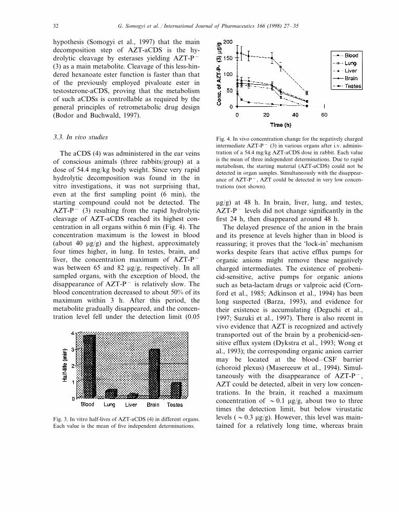
The aCDS (4) was administered in the ear veinsof conscious animals (three rabbits/group) at adose of 54.4 mg/kg body weight. Since very rapidhydrolytic decomposition was found in the invitro investigations, it was not surprising that,even at the first sampling point (6 min), thestarting compound could not be detected. TheAZT-P− (3) resulting from the rapid hydrolyticcleavage of AZT-aCDS reached its highest con-centration in all organs within 6 min (Fig. 4). Theconcentration maximum is the lowest in blood(about 40 mg/g) and the highest, approximatelyfour times higher, in lung. In testes, brain, andliver, the concentration maximum of AZT-P−
was between 65 and 82 mg/g, respectively. In allsampled organs, with the exception of blood, thedisappearance of AZT-P− is relatively slow. Theblood concentration decreased to about 50% of itsmaximum within 3 h. After this period, themetabolite gradually disappeared, and the concen-tration level fell under the detection limit (0.05
Fig. 4. In vivo concentration change for the negatively chargedintermediate AZT-P− (3) in various organs after i.v. adminis-tration of a 54.4 mg/kg AZT-aCDS dose in rabbit. Each valueis the mean of three independent determinations. Due to rapidmetabolism, the starting material (AZT-aCDS) could not bedetected in organ samples. Simultaneously with the disappear-ance of AZT-P−, AZT could be detected in very low concen-trations (not shown).
mg/g) at 48 h. In brain, liver, lung, and testes,AZT-P− levels did not change significantly in thefirst 24 h, then disappeared around 48 h.
The delayed presence of the anion in the brainand its presence at levels higher than in blood isreassuring; it proves that the ‘lock-in’ mechanismworks despite fears that active efflux pumps fororganic anions might remove these negativelycharged intermediates. The existence of probeni-cid-sensitive, active pumps for organic anionssuch as beta-lactam drugs or valproic acid (Corn-ford et al., 1985; Adkinson et al., 1994) has beenlong suspected (Barza, 1993), and evidence fortheir existence is accumulating (Deguchi et al.,1997; Suzuki et al., 1997). There is also recent invivo evidence that AZT is recognized and activelytransported out of the brain by a probenicid-sen-sitive efflux system (Dykstra et al., 1993; Wong etal., 1993); the corresponding organic anion carriermay be located at the blood–CSF barrier(choroid plexus) (Masereeuw et al., 1994). Simul-taneously with the disappearance of AZT-P−,AZT could be detected, albeit in very low concen-trations. In the brain, it reached a maximumconcentration of �0.1 mg/g, about two to threetimes the detection limit, but below virustaticlevels (�0.3 mg/g). However, this level was main-tained for a relatively long time, whereas brain
Fig. 3. In vitro half-lives of AZT-aCDS (4) in different organs.Each value is the mean of five independent determinations.
G. Somogyi et al. / International Journal of Pharmaceutics 166 (1998) 27–35 33
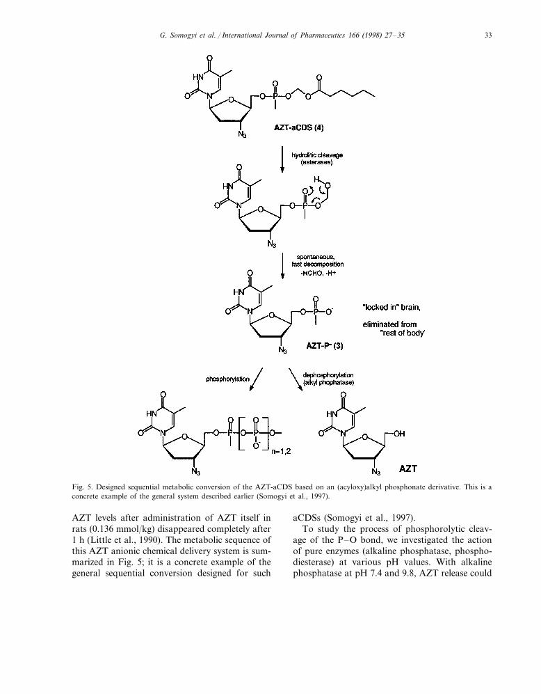
Fig. 5. Designed sequential metabolic conversion of the AZT-aCDS based on an (acyloxy)alkyl phosphonate derivative. This is aconcrete example of the general system described earlier (Somogyi et al., 1997).
AZT levels after administration of AZT itself inrats (0.136 mmol/kg) disappeared completely after1 h (Little et al., 1990). The metabolic sequence ofthis AZT anionic chemical delivery system is sum-marized in Fig. 5; it is a concrete example of thegeneral sequential conversion designed for such
aCDSs (Somogyi et al., 1997).To study the process of phosphorolytic cleav-
age of the P–O bond, we investigated the actionof pure enzymes (alkaline phosphatase, phospho-diesterase) at various pH values. With alkalinephosphatase at pH 7.4 and 9.8, AZT release could
G. Somogyi et al. / International Journal of Pharmaceutics 166 (1998) 27–3534
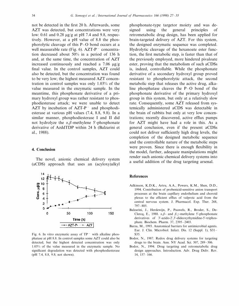
not be detected in the first 20 h. Afterwards, someAZT was detected, but concentrations were verylow: 0.61 and 0.28 mg/g at pH 7.4 and 9.8, respec-tively. However, at a pH value of 8.8 the phos-phorolytic cleavage of this P–O bond occurs at awell measurable rate (Fig. 6). AZT-P− concentra-tion decreased about 50% in a period of 136 hand, at the same time, the concentration of AZTincreased continuously and reached a 7.06 mg/gfinal value. In the control samples, AZT couldalso be detected, but the concentration was foundto be very low; the highest measured AZT concen-tration in control samples was only 1.03% of thevalue measured in the enzymatic sample. In themeantime, this phosphonate derivative of a pri-mary hydroxyl group was rather resistant to phos-phodiesterase attack; we were unable to detectAZT by incubation of AZT-P− and phosphodi-esterase at various pH values (7.4, 8.8, 9.8). In asimilar manner, phosphodiesterase I and II didnot hydrolyze the a,b-methylene 5%-phosphonatederivative of AzddTDP within 24 h (Balzarini etal., 1988).
4. Conclusion
The novel, anionic chemical delivery system(aCDS) approach that uses an (acyloxy)alkyl
phosphonate-type targetor moiety and was de-signed using the general principles ofretrometabolic drug design, has been applied forbrain-targeted delivery of AZT. For this system,the designed enzymatic sequence was completed.Hydrolytic cleavage of the hexanoate ester func-tion, the first metabolic step, is faster than that ofthe previously employed, more hindered pivaloateester, proving that the metabolism of such aCDSsis, indeed, controllable. While the phosphonatederivative of a secondary hydroxyl group provedresistant to phosphorolytic attack, the secondmetabolic step that releases the active drug, alka-line phosphatase cleaves the P–O bond of thephosphonate derivative of the primary hydroxylgroup in this system, but only at a relatively slowrate. Consequently, some AZT released from sys-temically administered aCDS was detectable inthe brain of rabbits but only at very low concen-trations; recently discovered, active efflux pumpsfor AZT might have had a role in this. As ageneral conclusion, even if the present aCDSscould not deliver sufficiently high drug levels, thecompletion of the designed metabolic sequenceand the controllable nature of the metabolic stepswere proven. Since there is enough flexibility inthe model, further, adequate manipulations mightrender such anionic chemical delivery systems intoa useful addition of the drug targeting arsenal.
References
Adkinson, K.D.K., Artru, A.A., Powers, K.M., Shen, D.D.,1994. Contribution of probenicid-sensitive anion transportprocesses at the brain capillary endothelium and choroidplexus to the efficient efflux of valproic acid from thecentral nervous system. J. Pharmacol. Exp. Ther. 268,797–805.
Balzarini, J., Herdewijn, P., Pauwels, R., Broder, S., De-Clercq, E., 1988. a,b- and b,g-methylene 5%-phosphonatederivatives of 3%-azido-2%,3%-dideoxythymidine-5%-triphos-phate. Biochem. Pharm. 37, 2395–2403.
Barza, M., 1993. Anatomical barriers for antimicrobial agents.Eur. J. Clin. Microbiol. Infect. Dis. 12 (Suppl. 1), S31–S35.
Bodor, N., 1987. Redox drug delivery systems for targetingdrugs to the brain. Ann. NY Acad. Sci. 507, 289–306.
Bodor, N., 1994. Drug targeting and retrometabolic drugdesign approaches. Introduction. Adv. Drug Deliv. Rev.14, 157–166.
Fig. 6. In vitro enzymatic assay of TP− with alkaline phos-phatase at pH 8.8. In control samples some AZT could also bedetected, but the highest detected concentration was only1.03% of the value measured in the enzymatic sample. Nosignificant degradation was detected with phosphodiesterase(pH 7.4, 8.8, 9.8; not shown).
G. Somogyi et al. / International Journal of Pharmaceutics 166 (1998) 27–35 35
Bodor, N., 1995. Targeted drug delivery via phosphonatederivatives. US Patent 5,413,996, May 9.
Bodor, N., Buchwald, P., 1997. Drug targeting viaretrometabolic approaches. Pharmacol. Ther. 76, 1–27.
Brewster, M., Little, R., Venkatraghavan, V., Bodor, N., 1988.Brain-enhanced delivery of antiviral agents (Abstract). An-tiviral Res. 9, 127.
Clark, J.H., Miller, J.M., 1977. Hydrogen bonding in organicsyntheses V. Potassium fluoride in carboxylic acids as analternative to crown ether with acid salts in the preparationof phenacyl esters. Tetrahedron Lett. 7, 599–602.
Cornford, E.M., Diep, C.P., Pardridge, W.M., 1985. Blood–brain barrier transport of valproic acid. J. Neurochem. 44,1541–1550.
Deguchi, Y., Nozawa, K., Yamada, S., Yokoyama, Y., Kimura,R., 1997. Quantitative evaluation of brain distribution andblood–brain barrier efflux transport of probenicid in rats bymicrodialysis: Possible involvement of the monocarboxylicacid transport system. J. Pharmacol. Exp. Ther. 280, 551–560.
Dykstra, K.H., Arya, A., Arriola, D.M., Bungay, P.M., Mor-rison, P.F., Dedrick, R.L., 1993. Microdialysis study ofzidovudine (AZT) transport in rat brain. J. Pharmacol. Exp.Ther. 267, 1227–1236.
Ellison, S., Terasaki, T., Pardridge, W.M., 1988. AZT anddideoxy-nucleosides do not cross the blood–brain barrier(Abstract). Clin. Res. 36, 117A.
Gallo, J., Boubinot, F., Doshi, D., Etse, J., Bhandti, V.,Schinazi, R., Chu, C.K., 1989. Evaluation of brain targetingof anti-HIV nucleosides delivered via dihydropyridine pro-drugs (Abstract). Pharm. Res. 6, S161.
Gogu, S.R., Aggarwal, S.K., Rangan, S.R.S., Agrawal, K.C.,1989. A prodrug of zidovudine with enhanced efficacyagainst human immunodeficiency virus. Biochem. Biophys.Res. Commun. 160, 656–661.
Klecker, R.W. Jr., Collins, J.M., Yarchoan, R., Thomas, R.,Jenkins, J.F., Broder, S., Myers, C.E., 1987. Plasma andcerebrospinal fluid pharmacokinetics of 3%-azido-3%-de-oxythymidine: A novel pyrimidine analog with potentialapplication for the treatment of patients with AIDS andrelated diseases. Clin. Pharmacol. Ther. 41, 407–412.
Little, R., Bailey, D., Brewster, M.E., Estes, K.S., Clemmons,R.M., Saab, A., Bodor, N., 1990. Improved delivery throughbiological membranes XXXIII. Brain enhanced delivery of
azidothymidine (AZT). J. Biopharm. Sci. 1, 1–18.Masereeuw, R., Jaehde, U., Langemeijer, M.W.E., de Boer,
A.G., Breimer, D.D., 1994. In vitro and in vivo transportof zidovudine (AZT) across the blood–brain barrier and theeffect of transport inhibitors. Pharm. Res. 11, 324–330.
Mitsuya, H., Yarchoan, R., Broder, S., 1990. Molecular targetsfor AIDS therapy. Science 249, 1533–1544.
McArthur, J.C., 1987. Neurologic manifestations of AIDS.Medicine 66, 407–437.
Somogyi, G., Nishitani, S., Nomi, D., Buchwald, P., Prokai, L.,Bodor, N., 1997. Targeted drug delivery to the brain viaphosphonate derivatives. I. Design, synthesis and evaluationof an anionic chemical delivery system for testosterone. Int.J. Pharm. 166, 15–26.
Suzuki, H., Terasaki, T., Sugiyama, Y., 1997. Role of effluxtransport across the blood–brain barrier and blood-cere-brospinal fluid barrier on the disposition of xenobiotics inthe central nervous system. Adv. Drug Deliv. Rev. 25,257–285.
Torrence, P.T., Kinjo, J., Lesiak, K., Balzarini, J., DeClerq, E.,1988. AIDS dementia: Synthesis and properties of a deriva-tive of 3%-azido-3%-deoxythimidine (AZT) that may become‘locked’ in the central nervous system. FEBS Lett. 234,135–140.
Wong, S.L., Belle, K., Sawchuk, R.J., 1993. Distributionaltransport kinetics of zidovudine between plasma and brainextracellular fluid/cerebrospinal fluid in the rabbit: Investi-gation of the inhibitory effect of probenicid utilizing micro-dialysis. J. Pharmacol. Exp. Ther. 264, 899–909.
Yarchoan, R., Broder, S., 1987. Development of antiretroviraltherapy for the acquired immunodeficiency syndrome andrelated disorders. A progress report. New Engl. J. Med. 316,557–564.
Yarchoan, R., Berg, G., Brouwers, P., Fischl, M.A., Spitzer,A.R., Wichman, A., Grafman, J., Thomas, R.V., Safai, B.,Brunetti, A., Perno, C.F., Schmidt, P.J., Larson, S.M.,Myers, C.E., Broder, S., 1987. Response of human im-munodeficiency-virus associated neurological disease to 3%-azido-3%-deoxythymidine. Lancet i, 132–135.
Yoshimura, Y., Hamaguchi, N., Yashiki, T., 1987. Synthesisand oral absorption of acyloxymethyl esters of 7b-(2-(-aminothiazol-4-yl)acetamido)-3-(((1-(2-dimethylamino-ethyl)-1H-tetrazol-5-yl)thio)-methyl)ceph-3-em-4-carboxy-lic acid (cefotiam). Int. J. Pharm. 38, 179–190.
.