Embed Size (px)
Citation preview
Article
Targeted Delivery of Immu
nomodulators to LymphNodesGraphical Abstract
Highlights
d The MECA79 antibody binds peripheral lymph node
addressin (PNAd)
d Tacrolimus-loaded microparticles coated with MECA79
target lymph nodes
d Microparticles accumulate in draining lymph nodes after
intravenous delivery
d Targeted delivery of tacrolimus prolonged allograft survival
Azzi et al., 2016, Cell Reports 15, 1202–1213May 10, 2016 ª 2016 The Authorshttp://dx.doi.org/10.1016/j.celrep.2016.04.007
Authors
Jamil Azzi, Qian Yin, Mayuko Uehara, ...,
Ulrich H. von Andrian, Jianjun Cheng,
Reza Abdi
[email protected] (J.C.),[email protected] (R.A.)
In Brief
Azzi et al. show the targeted delivery of
microparticles containing tacrolimus to
the draining lymph nodes of recipients
following organ transplantation via
intravenous administration.
Microparticles containing tacrolimus
coated with an antibody against
peripheral lymph node addressin are
trafficked to the draining lymph nodes,
resulting in a significant prolongation of
murine heart allograft survival.
Cell Reports
Article
Targeted Delivery of Immunomodulatorsto Lymph NodesJamil Azzi,1,7 Qian Yin,4,7 Mayuko Uehara,1,7 Shunsuke Ohori,1 Li Tang,4 Kaimin Cai,4 Takaharu Ichimura,1
Martina McGrath,1 Omar Maarouf,1 Eirini Kefaloyianni,1 Scott Loughhead,3 Jarolim Petr,5 Qidi Sun,4 Mincheol Kwon,4
Stefan Tullius,2 Ulrich H. von Andrian,3,6 Jianjun Cheng,4,* and Reza Abdi1,*1Transplantation Research Center, Renal Division, Brigham and Women’s Hospital, Harvard Medical School, Boston, MA 02115, USA2Division of Transplant Surgery and Transplant Surgery Research Laboratory, Brigham and Women’s Hospital, Harvard Medical School,
Boston, MA 02115, USA3Department of Microbiology and Immunobiology, Harvard Medical School, Boston, MA 02115, USA4Department of Materials Science and Engineering, University of Illinois at Urbana�Champaign, Urbana, IL 61820, USA5Department of Pathology, Clinical Laboratories Division, Brigham andWomen’s Hospital, Harvard Medical School, Boston, MA 02115, USA6The Ragon Institute of MGH, MIT, and Harvard, Cambridge, MA 02139, USA7Co-first author
*Correspondence: [email protected] (J.C.), [email protected] (R.A.)
http://dx.doi.org/10.1016/j.celrep.2016.04.007
SUMMARY
Active-targeted delivery to lymph nodes represents amajor advance toward more effective treatment ofimmune-mediated disease. The MECA79 antibodyrecognizes peripheral node addressin molecules ex-pressed by high endothelial venules of lymph nodes.Bymimicking lymphocyte trafficking to the lymph no-des, we have engineered MECA79-coated micropar-ticles containing an immunosuppressive medication,tacrolimus. Following intravenous administration,MECA79-bearing particles showed marked accumu-lation in the draining lymph nodes of transplanted an-imals. Using an allograft heart transplant model, weshow that targeted lymph node delivery of micropar-ticles containing tacrolimus can prolong heart allo-graft survival with negligible changes in tacrolimusserum level. Using MECA79 conjugation, we havedemonstrated targeted delivery of tacrolimus to thelymph nodes following systemic administration,with the capacity for immune modulation in vivo.
INTRODUCTION
Lymph nodes (LNs) function as a primary site for the priming and
activation of immune cells in a wide variety of immune-mediated
diseases (Goldstein et al., 2003; von Andrian and Mempel,
2003). Recognition of non-self alloantigens occurs in the draining
lymph nodes (DLNs), resulting in the formation of alloreactive
T cells and subsequent transplant rejection (Lakkis et al., 2000).
Similarly, pancreatic lymph nodes play a key role in the formation
of autoreactive T cells and in the pathogenesis of type 1 diabetes
(Park and Kupper, 2015). LNs are also common sites for primary
lymphoproliferative malignancies and metastatic niches (Stacker
et al., 2014). Therefore, developing targeted delivery of drugs to
the LNs represents a significantmilestone and holds the potential
1202 Cell Reports 15, 1202–1213, May 10, 2016 ª 2016 The AuthorsThis is an open access article under the CC BY-NC-ND license (http://
to increase the efficacy of immune and cancer therapies. Tar-
geteddrugdelivery alsoallows reducedsystemicdosingwithcor-
responding reductions in off-target toxicities.
The optimal, most clinically applicable drug delivery system
would involve intravenous administration of an agent that would
then localize toandactat a chosensite. In recent years, innovative
approaches have been developed to target drugs specifically to
the lymphoid system (Dane et al., 2011; Hunter et al., 2014; Jewell
et al., 2011; Liu et al., 2014; Reddy et al., 2007; Yeste et al., 2012).
Despite these advances, a LN-targeted drug delivery system us-
ing the intravenous route remains to be fully developed. Success-
ful targeted delivery of immunoregulatory molecules or chemo-
therapy drugs to the LNs following intravenous administration
holds immense potential for application in the treatment of a
wide variety of immune-mediated diseases and cancers.
The primary concept behind our microparticle (MP) delivery
system essentially shadows the footsteps of naive T cells and
central memory cells, which circulate between the blood and
LNs for antigen surveillance (von Andrian and Mackay, 2000).
The trafficking of lymphocytes from the circulation to LNs is initi-
ated by a highly regulated process referred to as tethering, which
is controlled by selectinmolecules (Somers et al., 2000). L-selec-
tin, expressed on leukocytes, recognizes sulfated sialyl-LewisX-
like sugars, called peripheral node addressins (PNAds), which
are expressed by high endothelial venules (HEVs) in the LNs
(Carlow et al., 2009; Sperandio et al., 2009; von Andrian and
Mackay, 2000). Through this interaction, L-selectin plays a key
role in the continuous homing of naive T cells to the LNs, where
they encounter antigens presented by antigen-presenting cells.
The monoclonal antibody (Ab) MECA79 binds PNAd, and, utiliz-
ing this interaction, we have successfully designed an innovative
LN-targeted drug delivery strategy. Our platform consists of
MECA79-bearing particles with the capacity to load and release
tacrolimus (TAC). TAC is the most commonly clinically used
immunosuppressant post-transplantation, and by directing this
agent to the site of T cell activation in the lymph node, we have
developed a functional, clinically relevant approach. Using a car-
diac transplant model, we show proof of concept of the targeted
creativecommons.org/licenses/by-nc-nd/4.0/).
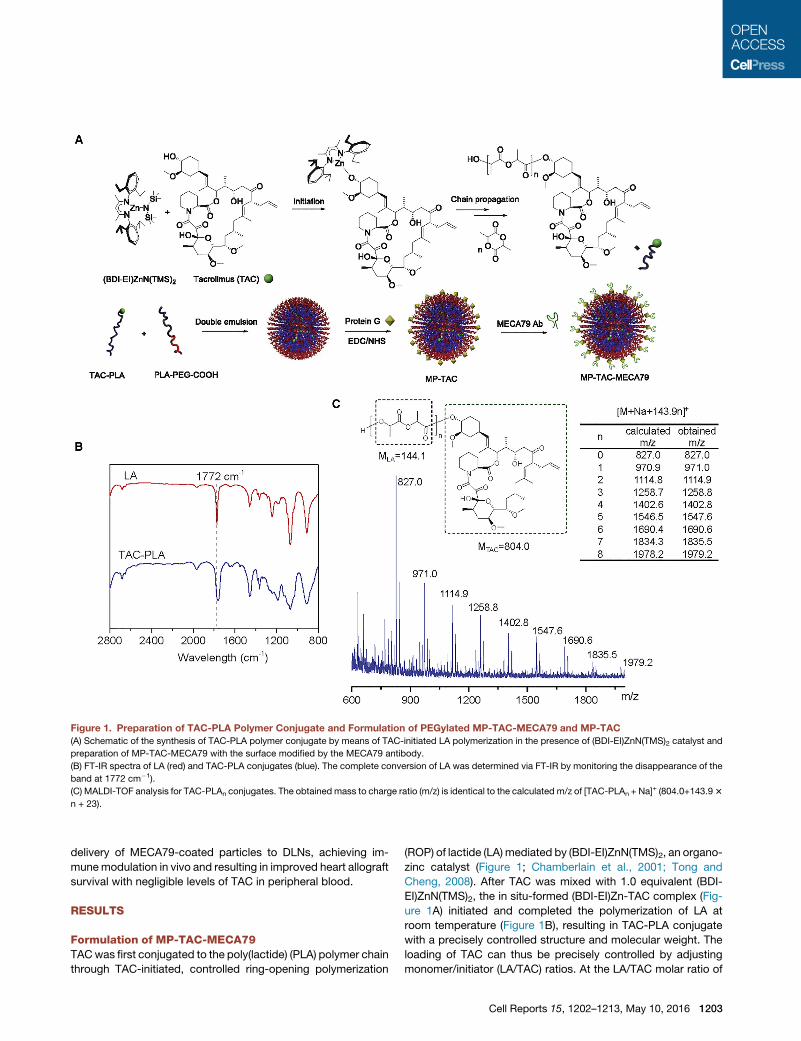
Figure 1. Preparation of TAC-PLA Polymer Conjugate and Formulation of PEGylated MP-TAC-MECA79 and MP-TAC
(A) Schematic of the synthesis of TAC-PLA polymer conjugate by means of TAC-initiated LA polymerization in the presence of (BDI-EI)ZnN(TMS)2 catalyst and
preparation of MP-TAC-MECA79 with the surface modified by the MECA79 antibody.
(B) FT-IR spectra of LA (red) and TAC-PLA conjugates (blue). The complete conversion of LA was determined via FT-IR by monitoring the disappearance of the
band at 1772 cm�1).
(C) MALDI-TOF analysis for TAC-PLAn conjugates. The obtained mass to charge ratio (m/z) is identical to the calculated m/z of [TAC-PLAn + Na]+ (804.0+143.93
n + 23).
delivery of MECA79-coated particles to DLNs, achieving im-
munemodulation in vivo and resulting in improved heart allograft
survival with negligible levels of TAC in peripheral blood.
RESULTS
Formulation of MP-TAC-MECA79TACwas first conjugated to the poly(lactide) (PLA) polymer chain
through TAC-initiated, controlled ring-opening polymerization
(ROP) of lactide (LA) mediated by (BDI-EI)ZnN(TMS)2, an organo-
zinc catalyst (Figure 1; Chamberlain et al., 2001; Tong and
Cheng, 2008). After TAC was mixed with 1.0 equivalent (BDI-
EI)ZnN(TMS)2, the in situ-formed (BDI-EI)Zn-TAC complex (Fig-
ure 1A) initiated and completed the polymerization of LA at
room temperature (Figure 1B), resulting in TAC-PLA conjugate
with a precisely controlled structure and molecular weight. The
loading of TAC can thus be precisely controlled by adjusting
monomer/initiator (LA/TAC) ratios. At the LA/TAC molar ratio of
Cell Reports 15, 1202–1213, May 10, 2016 1203
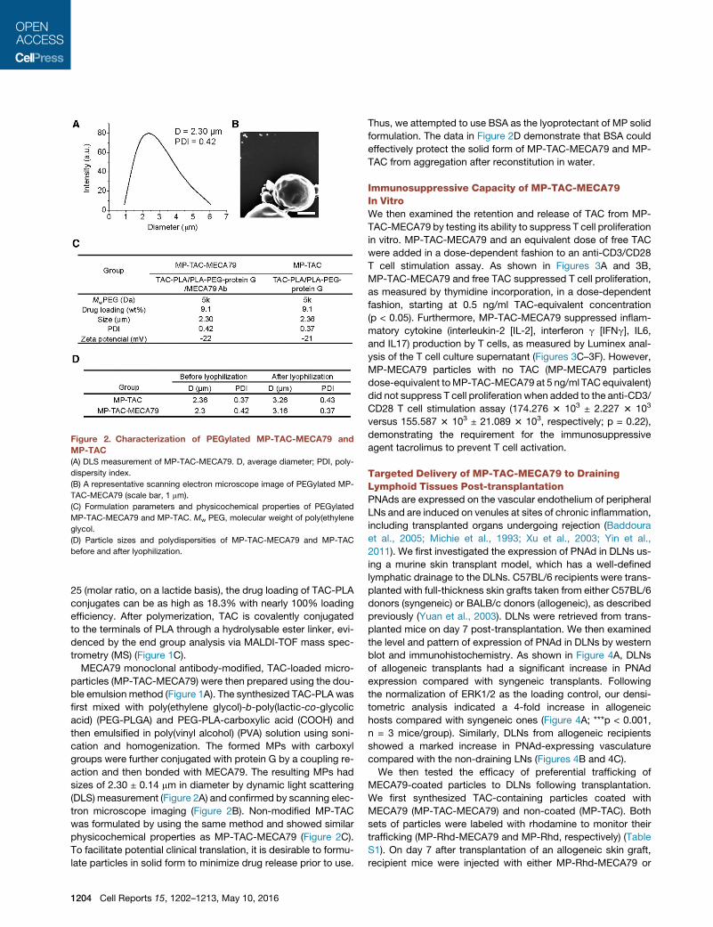
Figure 2. Characterization of PEGylated MP-TAC-MECA79 and
MP-TAC
(A) DLS measurement of MP-TAC-MECA79. D, average diameter; PDI, poly-
dispersity index.
(B) A representative scanning electron microscope image of PEGylated MP-
TAC-MECA79 (scale bar, 1 mm).
(C) Formulation parameters and physicochemical properties of PEGylated
MP-TAC-MECA79 and MP-TAC. Mw PEG, molecular weight of poly(ethylene
glycol.
(D) Particle sizes and polydispersities of MP-TAC-MECA79 and MP-TAC
before and after lyophilization.
25 (molar ratio, on a lactide basis), the drug loading of TAC-PLA
conjugates can be as high as 18.3% with nearly 100% loading
efficiency. After polymerization, TAC is covalently conjugated
to the terminals of PLA through a hydrolysable ester linker, evi-
denced by the end group analysis via MALDI-TOF mass spec-
trometry (MS) (Figure 1C).
MECA79 monoclonal antibody-modified, TAC-loaded micro-
particles (MP-TAC-MECA79) were then prepared using the dou-
ble emulsion method (Figure 1A). The synthesized TAC-PLA was
first mixed with poly(ethylene glycol)-b-poly(lactic-co-glycolic
acid) (PEG-PLGA) and PEG-PLA-carboxylic acid (COOH) and
then emulsified in poly(vinyl alcohol) (PVA) solution using soni-
cation and homogenization. The formed MPs with carboxyl
groups were further conjugated with protein G by a coupling re-
action and then bonded with MECA79. The resulting MPs had
sizes of 2.30 ± 0.14 mm in diameter by dynamic light scattering
(DLS) measurement (Figure 2A) and confirmed by scanning elec-
tron microscope imaging (Figure 2B). Non-modified MP-TAC
was formulated by using the same method and showed similar
physicochemical properties as MP-TAC-MECA79 (Figure 2C).
To facilitate potential clinical translation, it is desirable to formu-
late particles in solid form to minimize drug release prior to use.
1204 Cell Reports 15, 1202–1213, May 10, 2016
Thus, we attempted to use BSA as the lyoprotectant of MP solid
formulation. The data in Figure 2D demonstrate that BSA could
effectively protect the solid form of MP-TAC-MECA79 and MP-
TAC from aggregation after reconstitution in water.
Immunosuppressive Capacity of MP-TAC-MECA79In VitroWe then examined the retention and release of TAC from MP-
TAC-MECA79 by testing its ability to suppress T cell proliferation
in vitro. MP-TAC-MECA79 and an equivalent dose of free TAC
were added in a dose-dependent fashion to an anti-CD3/CD28
T cell stimulation assay. As shown in Figures 3A and 3B,
MP-TAC-MECA79 and free TAC suppressed T cell proliferation,
as measured by thymidine incorporation, in a dose-dependent
fashion, starting at 0.5 ng/ml TAC-equivalent concentration
(p < 0.05). Furthermore, MP-TAC-MECA79 suppressed inflam-
matory cytokine (interleukin-2 [IL-2], interferon g [IFNg], IL6,
and IL17) production by T cells, as measured by Luminex anal-
ysis of the T cell culture supernatant (Figures 3C–3F). However,
MP-MECA79 particles with no TAC (MP-MECA79 particles
dose-equivalent toMP-TAC-MECA79 at 5 ng/ml TAC equivalent)
did not suppress T cell proliferation when added to the anti-CD3/
CD28 T cell stimulation assay (174.276 3 103 ± 2.227 3 103
versus 155.587 3 103 ± 21.089 3 103, respectively; p = 0.22),
demonstrating the requirement for the immunosuppressive
agent tacrolimus to prevent T cell activation.
Targeted Delivery of MP-TAC-MECA79 to DrainingLymphoid Tissues Post-transplantationPNAds are expressed on the vascular endothelium of peripheral
LNs and are induced on venules at sites of chronic inflammation,
including transplanted organs undergoing rejection (Baddoura
et al., 2005; Michie et al., 1993; Xu et al., 2003; Yin et al.,
2011). We first investigated the expression of PNAd in DLNs us-
ing a murine skin transplant model, which has a well-defined
lymphatic drainage to the DLNs. C57BL/6 recipients were trans-
planted with full-thickness skin grafts taken from either C57BL/6
donors (syngeneic) or BALB/c donors (allogeneic), as described
previously (Yuan et al., 2003). DLNs were retrieved from trans-
planted mice on day 7 post-transplantation. We then examined
the level and pattern of expression of PNAd in DLNs by western
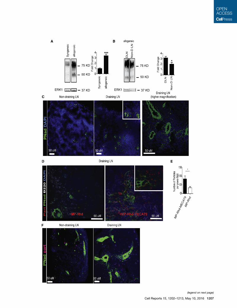
blot and immunohistochemistry. As shown in Figure 4A, DLNs
of allogeneic transplants had a significant increase in PNAd
expression compared with syngeneic transplants. Following
the normalization of ERK1/2 as the loading control, our densi-
tometric analysis indicated a 4-fold increase in allogeneic
hosts compared with syngeneic ones (Figure 4A; ***p < 0.001,
n = 3 mice/group). Similarly, DLNs from allogeneic recipients
showed a marked increase in PNAd-expressing vasculature
compared with the non-draining LNs (Figures 4B and 4C).
We then tested the efficacy of preferential trafficking of
MECA79-coated particles to DLNs following transplantation.
We first synthesized TAC-containing particles coated with
MECA79 (MP-TAC-MECA79) and non-coated (MP-TAC). Both
sets of particles were labeled with rhodamine to monitor their
trafficking (MP-Rhd-MECA79 and MP-Rhd, respectively) (Table
S1). On day 7 after transplantation of an allogeneic skin graft,
recipient mice were injected with either MP-Rhd-MECA79 or
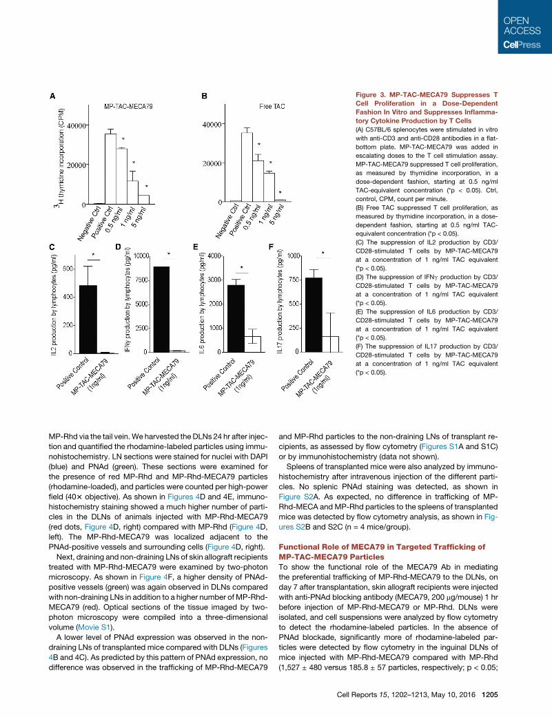
Figure 3. MP-TAC-MECA79 Suppresses T
Cell Proliferation in a Dose-Dependent
Fashion In Vitro and Suppresses Inflamma-
tory Cytokine Production by T Cells
(A) C57BL/6 splenocytes were stimulated in vitro
with anti-CD3 and anti-CD28 antibodies in a flat-
bottom plate. MP-TAC-MECA79 was added in
escalating doses to the T cell stimulation assay.
MP-TAC-MECA79 suppressed T cell proliferation,
as measured by thymidine incorporation, in a
dose-dependent fashion, starting at 0.5 ng/ml
TAC-equivalent concentration (*p < 0.05). Ctrl,
control, CPM, count per minute.
(B) Free TAC suppressed T cell proliferation, as
measured by thymidine incorporation, in a dose-
dependent fashion, starting at 0.5 ng/ml TAC-
equivalent concentration (*p < 0.05).
(C) The suppression of IL2 production by CD3/
CD28-stimulated T cells by MP-TAC-MECA79
at a concentration of 1 ng/ml TAC equivalent
(*p < 0.05).
(D) The suppression of IFNg production by CD3/
CD28-stimulated T cells by MP-TAC-MECA79
at a concentration of 1 ng/ml TAC equivalent
(*p < 0.05).
(E) The suppression of IL6 production by CD3/
CD28-stimulated T cells by MP-TAC-MECA79
at a concentration of 1 ng/ml TAC equivalent
(*p < 0.05).
(F) The suppression of IL17 production by CD3/
CD28-stimulated T cells by MP-TAC-MECA79
at a concentration of 1 ng/ml TAC equivalent
(*p < 0.05).
MP-Rhd via the tail vein.We harvested the DLNs 24 hr after injec-
tion and quantified the rhodamine-labeled particles using immu-
nohistochemistry. LN sections were stained for nuclei with DAPI
(blue) and PNAd (green). These sections were examined for
the presence of red MP-Rhd and MP-Rhd-MECA79 particles
(rhodamine-loaded), and particles were counted per high-power
field (403 objective). As shown in Figures 4D and 4E, immuno-
histochemistry staining showed a much higher number of parti-
cles in the DLNs of animals injected with MP-Rhd-MECA79
(red dots, Figure 4D, right) compared with MP-Rhd (Figure 4D,
left). The MP-Rhd-MECA79 was localized adjacent to the
PNAd-positive vessels and surrounding cells (Figure 4D, right).
Next, draining and non-draining LNs of skin allograft recipients
treated with MP-Rhd-MECA79 were examined by two-photon
microscopy. As shown in Figure 4F, a higher density of PNAd-
positive vessels (green) was again observed in DLNs compared
with non-draining LNs in addition to a higher number of MP-Rhd-
MECA79 (red). Optical sections of the tissue imaged by two-
photon microscopy were compiled into a three-dimensional
volume (Movie S1).
A lower level of PNAd expression was observed in the non-
draining LNs of transplanted mice compared with DLNs (Figures
4B and 4C). As predicted by this pattern of PNAd expression, no
difference was observed in the trafficking of MP-Rhd-MECA79
and MP-Rhd particles to the non-draining LNs of transplant re-
cipients, as assessed by flow cytometry (Figures S1A and S1C)
or by immunohistochemistry (data not shown).
Spleens of transplanted mice were also analyzed by immuno-
histochemistry after intravenous injection of the different parti-
cles. No splenic PNAd staining was detected, as shown in
Figure S2A. As expected, no difference in trafficking of MP-
Rhd-MECA and MP-Rhd particles to the spleens of transplanted
mice was detected by flow cytometry analysis, as shown in Fig-
ures S2B and S2C (n = 4 mice/group).
Functional Role of MECA79 in Targeted Trafficking ofMP-TAC-MECA79 ParticlesTo show the functional role of the MECA79 Ab in mediating
the preferential trafficking of MP-Rhd-MECA79 to the DLNs, on
day 7 after transplantation, skin allograft recipients were injected
with anti-PNAd blocking antibody (MECA79, 200 mg/mouse) 1 hr
before injection of MP-Rhd-MECA79 or MP-Rhd. DLNs were
isolated, and cell suspensions were analyzed by flow cytometry
to detect the rhodamine-labeled particles. In the absence of
PNAd blockade, significantly more of rhodamine-labeled par-
ticles were detected by flow cytometry in the inguinal DLNs of
mice injected with MP-Rhd-MECA79 compared with MP-Rhd
(1,527 ± 480 versus 185.8 ± 57 particles, respectively; p < 0.05;
Cell Reports 15, 1202–1213, May 10, 2016 1205
n = 3–4mice/group) (Figures 5A and 5C). Blocking PNAd reduced
the trafficking of MP-Rhd-MECA79 to the DLNs (1,527 ± 480
versus 98 ± 56 particles, respectively; p < 0.05; n = 3–4 mice/
group). However, PNAd blockade did not affect the observed
low levels of MP-Rhd trafficking to the DLNs (185.8 ± 57 versus
193 ± 71 particles, respectively; p = not significant [ns]; n =
3–4 mice/group) (Figures 5B and 5C). This shows that the prefer-
ential trafficking of MP-Rhd-MECA79 to DLNs after transplanta-
tion is mediated by the binding of MECA79 to LN-expressed
PNAd.
To assess the accumulation of particles in other lymphoid tis-
sues, non-draining LNs (pancreatic LNs) were isolated, and cell
suspensions were analyzed by flow cytometry to detect rhoda-
mine-labeled particles under PNAd blockade. The presence or
absence of PNAd blockade had no effect on the accumulation
of intravenously injected MP-Rhd-MECA79 or MP-Rhd in non-
draining lymph nodes (Figures S1A–S1C). After allogeneic trans-
plantation, draining lymph nodes enlarge markedly because of
allostimulated lymphocyte proliferation. As anticipated, more
than 10-fold more cells were isolated from DLNs than non-drain-
ing lymph nodes, as measured by flow cytometry (Figure S1D).
This effect leads to the observed substantial differences in abso-
lute cell count despite a similar overall percentage, as seen by
flow cytometry analysis.
Therapeutic Efficacy of MP-TAC-MECA79 in ProlongingHeart Allograft SurvivalTo examine the therapeutic efficacy of MP-TAC-MECA79 in a
more clinically applicable solid organ transplant model, we next
analyzed the expression of PNAd in the draining and non-draining
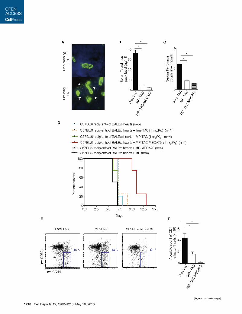
LNs of heart transplant recipients. As shown in Figure 6A, immu-
nohistochemistry analysis of DLNs of mice transplanted with
heart allografts showed a significant increase in PNAd expression
compared with non-draining LNs on day 7 post-transplantation.
Furthermore, a newly formed network of PNAd-positive cells
from much larger vessels in the DLNs was also observed (ar-
rows). To study allograft survival, C57BL/6 recipients of BALB/c
hearts were injected daily with MP-TAC-MECA79, MP-TAC, or
free TAC (each dosed at 1 mg/kg TAC equivalent) for 6 consecu-
tive days. Given that high serum levels of TAC cause allograft
vascular injury and toxicity, we were interested in the effect of
our treatment particles on systemic TAC exposure. To assess
this, we measured the blood levels of TAC in transplanted mice
on day 3 after transplantation (Azzi et al., 2010; Chapman and
Nankivell, 2006; Vercauteren et al., 1998; Wong et al., 2005).
As shown in Figure 6B, the TAC peak level 2 hr after injection
of the compounds was significantly lower in the MP-TAC-
MECA79 compared with the free TAC group (2.63 ± 0.56 versus
37.00 ± 2.62, respectively; p < 0,05; n = 3 mice/group). Further-
more, as shown in Figure 6C, the TAC trough level 24 hr after in-
jection was also significantly lower for MP-TAC-MECA79
compared with free TAC (2.45 ± 0.05 versus 0.60 ± 0.05, respec-
tively; p < 0.05; n = 3 mice/group). No significant difference was
seen in the peak and trough levels of TAC in MP-TAC-MECA79
and MP-TAC (Figures 6B and 6C).
Interestingly, despite amore than 15-fold decrease in the peak
level of TAC, transplanted mice treated with MP-TAC-MECA79
showed a significant prolongation of heart allograft survival
1206 Cell Reports 15, 1202–1213, May 10, 2016
compared with control groups (median survival 11 days versus
7 days, respectively; p < 0.05; n = 4–5/group). Furthermore, heart
allograft recipients treated with MP-MECA79 (without TAC) or
MP (without TAC) showed no increase in allograft survival
compared with untreated mice or mice treated with MP-TAC or
free TAC (median survival 7 days, p = ns, n = 4–5/group) (Fig-
ure 6D). These data confirm that the allograft-prolonging effect
is dependent on the presence of TACwithin microparticles deliv-
ered to DLNs.
Finally, we measured the suppressive effects of MP-TAC-
MECA79 on activated T cells in the DLNs of treated animals.
Activated alloreactive T cells are identified by their upregulated
expression of CD44 and downregulation of CD62L. These
CD4+CD62LlowCD44high T cells only require low levels of anti-
genic stimulation to undergo substantial proliferation and are
the effector population in allograft rejection (Azzi et al., 2015;
Jones et al., 1999; Krupnick et al., 2014; Vergani et al., 2013).
On day 7, mice were harvested, and the DLNs of heart allograft
recipients were analyzed by flow cytometry. CD4 effector
T cells (CD4+CD62LlowCD44high) were significantly reduced
in mice treated with MP-TAC-MECA79 compared with the
MP-TAC- and free TAC-treated groups (271.0 ± 65.00 versus
1,686 ± 361.9 versus 4,508 ± 735.5, respectively; p < 0.05;
n = 3 mice/group) (Figures 6E and 6F).
DISCUSSION
The potential for targeted drug delivery to improve the efficacy/
safety ratio of existing drugs has fueled considerable research
and prompted clinicians to apply this technology across a range
of clinical settings (Wagner et al., 2006). However, targeted de-
livery to specific tissue sites following systemic administration
remains challenging (Mitragotri et al., 2014). The primary focus
of our work has been to achieve active-targeted delivery to the
HEVs of LNs via an intravenous route. The DLN is a primary
site of alloreactive and autoreactive T cell activation and there-
fore plays a central role in the pathogenesis of transplant rejec-
tion and autoimmune diseases, rendering this site an attractive
target for drug delivery (Lakkis and Sayegh, 2003; Park and Kup-
per, 2015).
Solid organ transplantation is a life-saving procedure for end
organ failure, but its success is blunted by the limitations of
currently available immunosuppressive agents. Calcineurin in-
hibitors such as TAC are the cornerstone of immunosuppressive
therapy after transplantation. However, TAC-induced organ
toxicity, cardiovascular events, and malignancies are major
causes of graft loss and mortality (Halloran, 2004). There have
been numerous sizable clinical trials targeted at sparing, mini-
mizing, or withdrawing calcineurin inhibitors in an attempt to
reduce their toxicity. However, these trials have faced significant
challenges, including decreased efficacy, persistent toxicities,
and new toxicities caused by the replacement drug (Flechner
et al., 2008; Haller and Oberbauer, 2009). Therefore, developing
innovative strategies to improve the ratio of efficacy and toxicity
of immunosuppressive therapies remains one of the greatest
challenges in the treatment of immune-mediated diseases,
including transplant rejection (Azzi et al., 2013). Given that nano-
therapeutics are already in clinical use, this platform has the
(legend on next page)
Cell Reports 15, 1202–1213, May 10, 2016 1207
potential to tackle one of themost immediate clinical problems in
transplantation.
Migration of naive lymphocytes to the LNs is initiated by the
interaction of L-selectin expressed on lymphocytes with a series
of glycoprotein ligands expressed by HEVs in the lymph nodes,
referred to as PNAd molecules (Berg et al., 1992; Butcher and
Picker, 1996; Campbell et al., 2003; Hanninen et al., 1996;Michie
et al., 1993; Streeter et al., 1988; van Zante et al., 2003).
Following transplantation, we observed increased PNAd expres-
sion in DLNs. PNAds are recognized by theMECA79monoclonal
antibody, and, by engineering MECA79-coated microparticles,
we sought to exploit the role of PNAd in lymphocyte trafficking
in a model of transplant rejection. Our data indicate that
MECA79 surface-bearing particles showed significantly greater
trafficking to DLNs compared with control particles. We de-
monstrated significantly less accumulation of microparticles in
non-draining LNs where PNAd expression was unchanged. To
show the functionality of MECA79, we blocked the interaction
of MECA79 with PNAd prior to injection of particles, which
abrogated targeted delivery to DLNs. Furthermore, MECA79-
conjugated and -non-conjugated particles traffic equally to the
spleen, which does not express PNAd. These data indicate
that the marked increase in PNAd expression in the DLNs
following transplantation drives the increased trafficking of
MECA79-bearing particles to that site. This suggests that our tar-
geted delivery system should be applicable to other disease
states where PNAd is upregulated, highlighting its potential to
treat a broad spectrum of diseases.
Although a skin transplant model with clear lymphatic
drainage to DLNswas used to show proof of concept of targeted
delivery, we then used a heart transplant model as a clinically
applicable model to show proof of concept of efficacy. We
used TAC-loaded particles because of TAC’s importance as a
clinical agent in the prevention of transplant rejection. Within
the lymph node, MECA79-conjugated MPs with TAC reduced
T cell proliferation, as evidenced by the marked reduction in
CD44highCD62Llow activated alloreactive T cells in the DLNs of
treated animals. At the molecular level, TAC inhibits calcineurin,
which is responsible for the dephosphorylation of nuclear factor
of activated T cells (NFAT). NFAT increases the activity of IL-2
and other cytokine-encoding genes (Azzi et al., 2013; Kolata,
1983; Zhang et al., 1996). Blockade of this pathway therefore
Figure 4. Increased Expression of PNAd in DLNs Facilitates Targeted
(A) Western blot analysis of cell suspensions from the DLNs of mice transplante
expression in allogeneic compared with syngeneic transplants. Total ERK1 was
calculated after densitometric analysis and correction for ERK1 (***p < 0.001).
(B) Western blot analysis of cell suspensions from the DLNs and non-DLNs of mice
expression in DLNs compared with non-DLNs. The fold change over the non-DL
(*p < 0.05).
(C) DLNs of mice transplanted with allogeneic skin grafts were analyzed by imm
MECA79 (MP-Rhd-MECA79) or MP-TAC (MP-Rhd). DLN showed greatly increa
objective was used. Further extension of PNAd-positive cells from much larger v
(D) MECA79 particles but not control particles are localized to adjacent spaces of
up by PNAd. The cell nuclei were visualized by DAPI. A 403 objective was used
(E) A higher number of particles was counted per high-power field (2003) in DLN
(*p < 0.05, n = 4 mice/group).
(F) Two-photon imaging shows greatly increased PNAd expression (green) in DL
observed in the DLNs compared with the non-draining LNs.
1208 Cell Reports 15, 1202–1213, May 10, 2016
prevents T cell activation and proliferation. Furthermore, peak
TAC levels are closely correlated with the development of toxic-
ities, and serum drug levels are readily quantifiable, allowing us
to demonstrate both the efficacy of this directed therapy but
also its considerable potential to decrease unwanted effects
by minimizing total drug exposure. Indeed, treatment of allograft
recipients with MP-TAC-MECA79 induced higher allograft sur-
vival compared with the other groups with markedly lower levels
of TAC (>15-fold less) in peripheral blood. It is important to note
that the extent of prolongation of graft survival reported here is
based on using a single immunosuppressive agent, TAC, which
does not induce tolerance on its own in any transplant setting.
Given that we used a stringent model of alloreactivity with a com-
plete major histocompatibility complex (MHC) mismatch and
achieved negligible TAC levels in peripheral blood, the observed
prolongation of heart allograft survival represents a major immu-
nomodulatory effect in vivo. These findings show the ability of
this platform to achieve a greater degree of therapeutic efficacy
while holding the potential to improve the side effect profile
through reduced systemic drug exposure.
L-selectin itself is a rolling receptor because of its very short
bonding time and inefficient accumulation. We chose a
MECA79-based platform because MECA79 is likely to have
broader coverage and also carries the advantage of blocking
lymphocyte trafficking. MP-MECA79 without TAC showed no
prolongation of graft survival, indicating that impaired lympho-
cyte homing because of PNAd blockade is unlikely to be a signif-
icant contributor to the observed graft survival. Furthermore, of
all experimental groups, only MP-TAC-MECA79 showed prolon-
gation of allograft survival, indicating dependence upon the local
activity of TAC within the LNs to mediate this effect. With stan-
dard tools, it is impractical to compartmentalize TAC in the
DLNs. In future studies, we foresee using evolving cutting edge
matrix laser dissectionmethods, whichmay allow better analysis
of the kinetics of targeted drugs in a very small environment such
as the LNs.
There have been major efforts to build lymphatic delivery plat-
forms for a wide variety of therapeutic and diagnostic applica-
tions. Many of these strategies use lymphatic absorption with
promising results (Cho and Lee, 2014; Dane et al., 2011; Hunter
et al., 2014; Jewell et al., 2011; Liu et al., 2014; Reddy et al., 2007;
Yeste et al., 2012; Zhang and Lu, 2014). Our strategy, which
Delivery of MP-MECA79 in a Transplant Mouse Model
d with syngeneic or allogeneic skin grafts showed a 4-fold increase in PNAd
used as a loading control. The fold change over the syngeneic sample was
transplanted with allogeneic skin grafts showed significant increases in PNAd
N sample was calculated after densitometric analysis and correction for ERK1
unohistochemistry after intravenous injection of rhodamine-labeled MP-TAC-
sed PNAd (arrows) compared with non-draining LNs (pancreatic LNs). A 203
essels in the DLNs was also observed (left). A 403 objective was used.
PNAd-positive endothelial cells in the DLNs. Many MECA79 particles are lined
.
s isolated from mice treated with MP-Rhd-MECA79 compared with MP-Rhd
Ns compared with non-draining LNs. A higher number of MP-Rhd-MECA79 is
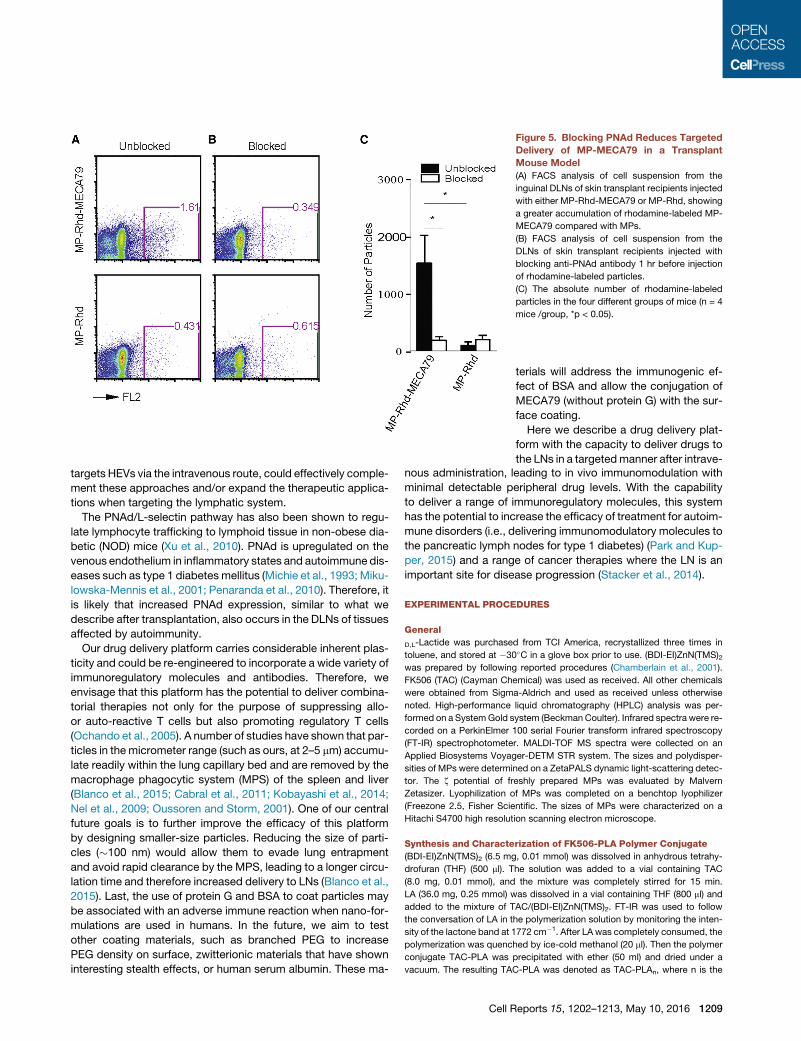
Figure 5. Blocking PNAd Reduces Targeted
Delivery of MP-MECA79 in a Transplant
Mouse Model
(A) FACS analysis of cell suspension from the
inguinal DLNs of skin transplant recipients injected
with either MP-Rhd-MECA79 or MP-Rhd, showing
a greater accumulation of rhodamine-labeled MP-
MECA79 compared with MPs.
(B) FACS analysis of cell suspension from the
DLNs of skin transplant recipients injected with
blocking anti-PNAd antibody 1 hr before injection
of rhodamine-labeled particles.
(C) The absolute number of rhodamine-labeled
particles in the four different groups of mice (n = 4
mice /group, *p < 0.05).
targets HEVs via the intravenous route, could effectively comple-
ment these approaches and/or expand the therapeutic applica-
tions when targeting the lymphatic system.
The PNAd/L-selectin pathway has also been shown to regu-
late lymphocyte trafficking to lymphoid tissue in non-obese dia-
betic (NOD) mice (Xu et al., 2010). PNAd is upregulated on the
venous endothelium in inflammatory states and autoimmune dis-
eases such as type 1 diabetes mellitus (Michie et al., 1993; Miku-
lowska-Mennis et al., 2001; Penaranda et al., 2010). Therefore, it
is likely that increased PNAd expression, similar to what we
describe after transplantation, also occurs in the DLNs of tissues
affected by autoimmunity.
Our drug delivery platform carries considerable inherent plas-
ticity and could be re-engineered to incorporate a wide variety of
immunoregulatory molecules and antibodies. Therefore, we
envisage that this platform has the potential to deliver combina-
torial therapies not only for the purpose of suppressing allo-
or auto-reactive T cells but also promoting regulatory T cells
(Ochando et al., 2005). A number of studies have shown that par-
ticles in the micrometer range (such as ours, at 2–5 mm) accumu-
late readily within the lung capillary bed and are removed by the
macrophage phagocytic system (MPS) of the spleen and liver
(Blanco et al., 2015; Cabral et al., 2011; Kobayashi et al., 2014;
Nel et al., 2009; Oussoren and Storm, 2001). One of our central
future goals is to further improve the efficacy of this platform
by designing smaller-size particles. Reducing the size of parti-
cles (�100 nm) would allow them to evade lung entrapment
and avoid rapid clearance by the MPS, leading to a longer circu-
lation time and therefore increased delivery to LNs (Blanco et al.,
2015). Last, the use of protein G and BSA to coat particles may
be associated with an adverse immune reaction when nano-for-
mulations are used in humans. In the future, we aim to test
other coating materials, such as branched PEG to increase
PEG density on surface, zwitterionic materials that have shown
interesting stealth effects, or human serum albumin. These ma-
Cell R
terials will address the immunogenic ef-
fect of BSA and allow the conjugation of
MECA79 (without protein G) with the sur-
face coating.
Here we describe a drug delivery plat-
form with the capacity to deliver drugs to
the LNs in a targetedmanner after intrave-
nous administration, leading to in vivo immunomodulation withminimal detectable peripheral drug levels. With the capability
to deliver a range of immunoregulatory molecules, this system
has the potential to increase the efficacy of treatment for autoim-
mune disorders (i.e., delivering immunomodulatory molecules to
the pancreatic lymph nodes for type 1 diabetes) (Park and Kup-
per, 2015) and a range of cancer therapies where the LN is an
important site for disease progression (Stacker et al., 2014).
EXPERIMENTAL PROCEDURES
General
D,L-Lactide was purchased from TCI America, recrystallized three times in
toluene, and stored at �30�C in a glove box prior to use. (BDI-EI)ZnN(TMS)2was prepared by following reported procedures (Chamberlain et al., 2001).
FK506 (TAC) (Cayman Chemical) was used as received. All other chemicals
were obtained from Sigma-Aldrich and used as received unless otherwise
noted. High-performance liquid chromatography (HPLC) analysis was per-
formed on a SystemGold system (Beckman Coulter). Infrared spectra were re-
corded on a PerkinElmer 100 serial Fourier transform infrared spectroscopy
(FT-IR) spectrophotometer. MALDI-TOF MS spectra were collected on an
Applied Biosystems Voyager-DETM STR system. The sizes and polydisper-
sities of MPs were determined on a ZetaPALS dynamic light-scattering detec-
tor. The z potential of freshly prepared MPs was evaluated by Malvern
Zetasizer. Lyophilization of MPs was completed on a benchtop lyophilizer
(Freezone 2.5, Fisher Scientific. The sizes of MPs were characterized on a
Hitachi S4700 high resolution scanning electron microscope.
Synthesis and Characterization of FK506-PLA Polymer Conjugate
(BDI-EI)ZnN(TMS)2 (6.5 mg, 0.01 mmol) was dissolved in anhydrous tetrahy-
drofuran (THF) (500 ml). The solution was added to a vial containing TAC
(8.0 mg, 0.01 mmol), and the mixture was completely stirred for 15 min.
LA (36.0 mg, 0.25 mmol) was dissolved in a vial containing THF (800 ml) and
added to the mixture of TAC/(BDI-EI)ZnN(TMS)2. FT-IR was used to follow
the conversation of LA in the polymerization solution by monitoring the inten-
sity of the lactone band at 1772 cm�1. After LA was completely consumed, the
polymerization was quenched by ice-cold methanol (20 ml). Then the polymer
conjugate TAC-PLA was precipitated with ether (50 ml) and dried under a
vacuum. The resulting TAC-PLA was denoted as TAC-PLAn, where n is the
eports 15, 1202–1213, May 10, 2016 1209
(legend on next page)
1210 Cell Reports 15, 1202–1213, May 10, 2016
monomer/initiator (LA/TAC) molar ratio. The polymer was then analyzed by
MALDI-TOF mass spectrometry.
Preparation of MP-TAC-MECA79
TAC-MPs were prepared using the water-in-oil-in-water (W/O/W) solvent
evaporation procedure (also known as the double emulsion method). In brief,
50 ml aqueous solution was emulsified in a mix of 1 ml solution of PEG-PLGA
(30mg), PLA-PEG-COOH (6mg), and TAC-PLA (30mg containing 5.5mg TAC)
in dichloromethane (DCM) using a probe sonicator (Sonic & Materials) at 10 W
(1%) for 15–30 s. The emulsion was then poured into 50 ml of aqueous PVA
(1%) aqueous solution, and the mixture was homogenized for 1 min at
8,000 rpm. The resulting emulsion was poured into 300 ml of aqueous PVA
(0.3%) with gentle stirring, after which the organic solvent was evaporated
by stirring at room temperature (RT) for 3 hr in the hood. TheMPswere isolated
by centrifugation at 10,000 rpm for 20–30 min, washed with distilled (DI) water
(20 ml), and dispersed in DI water (20 ml). To modify the MP surface with
MECA79 Ab, 1-ethyl-3-(3-dimethylaminopropyl)carbodiimide (10 mg) and
N-hydroxysuccinimide (6 mg) were added to 20 ml MP solution (66 mg), fol-
lowed by addition of protein G (0.5 mg in 40 ml DI water) after 10 min (Tong
et al., 2010). Themixture reacted overnight at RT. TheMPwas then centrifuged
at 10,000 rpm for 20 min, washed with DI water (20 ml), and dispersed in 20 ml
DI water. The concentration of MECA79 Ab is 200 mg/ml. 50 ml of Ab solution
was added into MP solution.
The MP was then washed as described above and dispersed in DI water for
use. TheMPswithout added IgM during the formulation were used as a control
(MP-TAC). The MP size was determined by DLS and scanning electron
microscopy.
For the preparation of Rhd-labeled MP-Rhd or MP-Rhd-MECA79, Rhd-PLA
(30 mg) was mixed with the DCM solution of the PEG-PLGA and PLA-PEG-
COOH polymer for subsequent double emulsion. The resulting fluorescent
MPs were collected and washed similarly.
x Potential Measurements
The x potential of the MPs was determined with a Malvern Zetasizer. The
freshly prepared MPs were dispersed in DI water to a concentration of
0.5 mg/ml.
Lyophilization of MPs in the Presence of BSA as a Lyoprotectant
BSA as a lyoprotectant was added to the concentrated MP suspension at a
mass ratio of BSA/MP = 10/1. The solution was then lyophilized to dry powder,
which was stored at�20�C prior to use. The MPs were reconstituted with wa-
ter and stirred for 5 min. The sizes of the resulting NPs were analyzed by DLS.
Mice
C57BL/6 andBALB/c(H-2d) micewere obtained from The Jackson Laboratory.
All animals were used at 6–10 weeks of age (20–25 g) and housed in accor-
Figure 6. MP-TAC-MECA79 Prolongs Heart Allograft Survival in Mice
Free TAC Group
(A) Immunohistochemistry analysis of the DLN of mice transplanted with heart al
LNs (axillary LNs) on day 7 post-transplantation. A 203 objective was used. Furth
also observed, giving a mesh-like appearance (arrows).
(B) C57BL/6mouse recipients of fully mismatched BALB/c hearts were injected da
TAC at 1mg/kg TAC equivalent. The TAC level wasmeasured in the peripheral bloo
the peak TAC level 2 hr after injection of the compounds was significantly lower
(C) The TAC trough level 24 hr after injection was significantly lower for MP-TAC
(D) Kaplan-Meier survival curve of heart allograft recipients treated with MP-TAC-
injected daily via the tail vein with eitherMP-TAC-MECA79, MP-TAC,MP,MP-ME
treating recipients with MP-TAC-MECA79 significantly increased heart allogra
respectively; n = 4/group; p < 0.05).
(E) FACS analysis of DLNs from heart transplant recipients treated with either M
percentage of effector CD4 T cells (CD4+CD62LlowCD44high) in DLNs of mice tre
groups.
(F) The absolute count of CD4 effector T cells in the DLNs of the three different gr
T cells in the DLNs of mice treated with MP-TAC-MECA79 compared with mice
dance with institutional and NIH guidelines. The Harvard Medical School Ani-
mal Management Committee approved all animal experiments.
Murine Skin Transplantation
Full-thickness trunk skin grafts (1 cm2) harvested from BALB/c donors were
transplanted onto the flanks of C57BL/6 recipient mice, sutured with 6.0 silk,
and secured with dry gauze and a bandage for 7 days. The draining LN har-
vested was the inguinal LN. To count the absolute number of Rd-labeled par-
ticles in LNs, LN samples were prepared into single-cell suspension, and the
cell number was enumerated by flow cytometry. The percentage of Rd-labeled
events was measured by flow cytometry. This percentage was then applied to
the count measured from the LN single-cell suspension, and the absolute
count for Rd-labeled events was calculated.
Murine Cardiac Transplantation
Vascularized intra-abdominal heterotopic transplantation of cardiac allografts
was performed using microsurgical techniques (Corry, 1973). Graft survival
was assessed by daily palpation. Rejection was defined as complete cessa-
tion of cardiac contractility as determined by direct visualization and was
confirmed by histological examination. Para-aortic lymph nodes from the
host were harvested as draining LNs of the heart allografts.
Immunohistochemistry
Lymph node, spleen, lung, and liver tissue samples were snap-frozen with op-
tical cutting temperature compound (OCT) in liquid nitrogen and stored at
�70�C until use. Frozen sections (8 mm) were prepared using a cryostat and
immediately used or stored at �70�C until use. The sections were fixed with
ice-cold acetone for 1 min, washed with PBS for 10 min, and blocked for
30 min with 3% BSA-PBS as a blocking solution. The anti-HEV marker
PNAd-Alexa Fluor 488 and anti-B cell B220-APC antibodies (eBioscience)
were diluted (1:200) in the blocking solution. Sections were incubated with
these antibodies for 1 hr at room temperature or overnight at 4�C. Sectionswere washed with PBS. After washing, a coverslip was placed with Vector-
shield (Vector Laboratories) mounting medium containing DAPI, and sections
were analyzed using a Nikon C1 laser confocal microscope. For negative con-
trols, the sections were stained by omitting the primary antibody.
Two-Photon Microscopy
Draining and non-draining LNs from recipients of an allogeneic skin graft were
frozen in OCT, and 200-mm sections were generated. Optical sections of the
tissue were imaged by two-photon microscopy and compiled into a three-
dimensional volume.
Western Blot
Lymph nodes were lysed in ice-cold lysis buffer, and the protein concentration
of cell lysates was measured using the Bradford (Bio-Rad) assay. Equal
amounts of protein were separated by 10% SDS-PAGE and transferred to a
with Lower Serum Peak and Trough TAC Levels Compared with the
lografts showed an increase in PNAd expression compared with non-draining
er extension of PNAd-positive cells from much larger vessels in the DLNs was
ily via the tail vein with either MP-TAC-MECA79,MP-TAC,MP-MECA79, or free
d of the transplantedmice on day 3 after transplantation. The graph shows that
for MP-TAC-MECA79 compared with free TAC (*p < 0.05, n = 4 mice/group).
-MECA79 compared with free TAC (*p < 0.05).
MECA79. C57BL/6 mouse recipients of fully mismatched BALB/c hearts were
CA79, or free TAC at 1mg/kg TAC equivalent. Compared with the other groups,
ft survival (median survival 7 versus 7 versus 7 versus 7 versus 11 days,
P-TAC-MECA79, MP-TAC, or free TAC showing a significant reduction in the
ated with MP-TAC-MECA79 compared with the MP-TAC- or free TAC-treated
oups of transplanted mice shows a significantly lower number of CD4 effector
treated with MP-TAC or free TAC (*p < 0.05, n = 3–4 mice/group).
Cell Reports 15, 1202–1213, May 10, 2016 1211
nitrocellulose membrane. The membranes were blocked with 5% non-fat milk
in Tris-buffered saline (TBS)-0.05%Tween (TBST) and incubated overnight at
4�C with the following primary antibodies: MECA79 (anti-PNAd, BD Biosci-
ences) and ERK1 (Santa Cruz Biotechnology). The blots were washed and
developed with SuperSignal West Pico or West Dura chemiluminescent sub-
strates (Thermo Scientific) using a Bio-Rad ChemiDoc imaging system, and
the bands were quantified using Image Lab (Bio-Rad) software. MECA79 is
a carbohydrate epitope found on a group of sulfation-decorated sialomucins,
including sulfated ligands for CD62L (CD34, GlyCAM-1, Sgp200, and a subset
of MAdCAM-1). This set of antigens has been referred to as PNAd with a mo-
lecular mass of 50–250 kD. Therefore, in the western blot, bands of multiple
molecular weights were detected.
Flow Cytometric Analysis
Cells recovered from spleens and peripheral lymphoid tissues were analyzed
by flow cytometry with a FACS Canto-II flow cytometer (BD Biosciences) and
analyzed using FlowJo software version 9.3.2 (Tree Star).
Anti-CD3/CD28 Stimulation Assay
100 ml of anti-CD3 Ab diluted in PBS (1 mg/ml, BD Biosciences) was added to
each well of a 96-well flat-bottom plate, placed at 37�C for 4 hr, and then
washed twice with PBS. Soluble anti-CD28 Ab (1 mg/ml, BD Biosciences)
was added to each well in the presence of 5 3 105 splenocytes and an
increasing concentration of MP-TAC-MECA79. Cultures were pulsed with trit-
urated thymidine 72 hr after stimulation to assess cell proliferation.
Luminex Assay
A 21-plex cytokine kit (Millipore) was used according to the manufacturer’s
instructions.
Statistics
Kaplan-Meier survival graphs were constructed, and a log-rank comparison of
the groups was used to calculate p values. Unpaired t test was used for com-
parison of experimental groups. Differences were considered to be significant
for p % 0.05. Prism software was used for data analysis and drawing graphs
(GraphPad). Data represent mean ± SEM.
SUPPLEMENTAL INFORMATION
Supplemental Information includes two figures, one table, and one movie and
can be found with this article online at http://dx.doi.org/10.1016/j.celrep.2016.
04.007.
AUTHOR CONTRIBUTIONS
J.A. designed and performed experiments, analyzed and interpreted data, and
drafted the manuscript. M.U. and S.O. performed microsurgery and analyzed
and interpreted data. Q.Y., L.T., K.C., and J.C. contributed significantly to the
chemical design of the polylactide-drug conjugates. Q.Y., L.T., K.C., O.S., and
M.K. contributed significantly to the synthesis of the polylactide-drug conju-
gate and to the characterization of the polylactide-drug conjugates and the
related nanoconjugates. Q.Y., L.T., K.C., and M.K. contributed significantly
to the scaling up of the polylactide-drug conjugates for in vitro and in vivo
studies. T.I. and J.P. performed experiments and analyzed data. S.T. and
U.H.v.A. helped with the study design. J.C. and R.A. helped with the study
design, interpreted data, and critically revised and finalized the manuscript.
ACKNOWLEDGMENTS
This work was supported by an American Heart Association grant (to J.A.), the
National Institute of Allergy and Infectious Diseases of the NIH under Award
RO1AI091930 (to R.A.), and the NIH Director’s New Innovator Award
1DP2OD007246 (to J.C.). The synthesis of the polylactide-drug conjugates
and nanoconjugates was partially supported by NSF-CHE-1308485. This
research was also supported in part by a gift from Mr. Matthew S. Forelli.
U.H.v.A. was supported by funds from the HMS Center for Immune Imaging
and the Ragon Institute and by NIH grants AI112521, AI111595, and
AR068383. Q.Y. was funded at the University of Illinois at Urbana�Champaign
1212 Cell Reports 15, 1202–1213, May 10, 2016
by NIH National Cancer Institute Alliance for Nanotechnology in Cancer
(Midwest Cancer Nanotechnology Training Center) (R25 CA154015A). The
synthesis of the polylactide-drug conjugates and nanoconjugates was partially
supported by NSF-CHE-1308485.
Received: August 19, 2015
Revised: February 21, 2016
Accepted: March 28, 2016
Published: April 28, 2016
REFERENCES
Azzi, J., Tang, L., Moore, R., Tong, R., El Haddad, N., Akiyoshi, T., Mfarrej, B.,
Yang, S., Jurewicz, M., Ichimura, T., et al. (2010). Polylactide-cyclosporin A
nanoparticles for targeted immunosuppression. FASEB J. 24, 3927–3938.
Azzi, J.R., Sayegh, M.H., and Mallat, S.G. (2013). Calcineurin inhibitors: 40
years later, can’t live without .. J. Immunol. 191, 5785–5791.
Azzi, J., Ohori, S., Ting, C., Uehara, M., Abdoli, R., Smith, B.D., Safa, K., Sol-
hjou, Z., Lukyanchykov, P., Patel, J., et al. (2015). Serine protease inhibitor-6
differentially affects the survival of effector and memory alloreactive CD8-T
cells. Am. J. Transplant. 15, 234–241.
Baddoura, F.K., Nasr, I.W., Wrobel, B., Li, Q., Ruddle, N.H., and Lakkis, F.G.
(2005). Lymphoid neogenesis in murine cardiac allografts undergoing chronic
rejection. Am. J. Transplant. 5, 510–516.
Berg, E.L., Magnani, J., Warnock, R.A., Robinson, M.K., and Butcher, E.C.
(1992). Comparison of L-selectin and E-selectin ligand specificities: the L-se-
lectin can bind the E-selectin ligands sialyl Le(x) and sialyl Le(a). Biochem. Bio-
phys. Res. Commun. 184, 1048–1055.
Blanco, E., Shen, H., and Ferrari, M. (2015). Principles of nanoparticle design
for overcoming biological barriers to drug delivery. Nat. Biotechnol. 33,
941–951.
Butcher, E.C., and Picker, L.J. (1996). Lymphocyte homing and homeostasis.
Science 272, 60–66.
Cabral, H., Matsumoto, Y., Mizuno, K., Chen, Q., Murakami, M., Kimura, M.,
Terada, Y., Kano, M.R., Miyazono, K., Uesaka, M., et al. (2011). Accumulation
of sub-100 nm polymeric micelles in poorly permeable tumours depends on
size. Nat. Nanotechnol. 6, 815–823.
Campbell, D.J., Kim, C.H., and Butcher, E.C. (2003). Chemokines in the sys-
temic organization of immunity. Immunol. Rev. 195, 58–71.
Carlow, D.A., Gossens, K., Naus, S., Veerman, K.M., Seo, W., and Ziltener,
H.J. (2009). PSGL-1 function in immunity and steady state homeostasis. Im-
munol. Rev. 230, 75–96.
Chamberlain, B.M., Cheng, M., Moore, D.R., Ovitt, T.M., Lobkovsky, E.B., and
Coates, G.W. (2001). Polymerization of lactide with zinc and magnesium beta-
diiminate complexes: stereocontrol and mechanism. J. Am. Chem. Soc. 123,
3229–3238.
Chapman, J.R., and Nankivell, B.J. (2006). Nephrotoxicity of cyclosporin A:
short-term gain, long-term pain? Nephrol. Dial. Transplant. 21, 2060–2063.
Cho, H.Y., and Lee, Y.B. (2014). Nano-sized drug delivery systems for
lymphatic delivery. J. Nanosci. Nanotechnol. 14, 868–880.
Corry, A.M. (1973). 1973 meeting in Kansas City. Bull. Med. Libr. Assoc. 61,
39–43.
Dane, K.Y., Nembrini, C., Tomei, A.A., Eby, J.K., O’Neil, C.P., Velluto, D.,
Swartz, M.A., Inverardi, L., and Hubbell, J.A. (2011). Nano-sized drug-loaded
micelles deliver payload to lymph node immune cells and prolong allograft sur-
vival. J. Control. Release 156, 154–160.
Flechner, S.M., Kobashigawa, J., and Klintmalm, G. (2008). Calcineurin inhib-
itor-sparing regimens in solid organ transplantation: focus on improving renal
function and nephrotoxicity. Clin. Transplant. 22, 1–15.
Goldstein, D.R., Tesar, B.M., Akira, S., and Lakkis, F.G. (2003). Critical role of
the Toll-like receptor signal adaptor protein MyD88 in acute allograft rejection.
J. Clin. Invest. 111, 1571–1578.
Haller, M., and Oberbauer, R. (2009). Calcineurin inhibitor minimization, with-
drawal and avoidance protocols after kidney transplantation. Transpl. Int.
22, 69–77.
Halloran, P.F. (2004). Immunosuppressive drugs for kidney transplantation.
N. Engl. J. Med. 351, 2715–2729.
Hanninen, A., Salmi, M., Simell, O., Andrew, D., and Jalkanen, S. (1996). Recir-
culation and homing of lymphocyte subsets: dual homing specificity of beta
7-integrin(high)-lymphocytes in nonobese diabetic mice. Blood 88, 934–944.
Hunter, Z., McCarthy, D.P., Yap, W.T., Harp, C.T., Getts, D.R., Shea, L.D., and
Miller, S.D. (2014). A biodegradable nanoparticle platform for the induction of
antigen-specific immune tolerance for treatment of autoimmune disease. ACS
Nano 8, 2148–2160.
Jewell, C.M., Lopez, S.C., and Irvine, D.J. (2011). In situ engineering of the
lymph node microenvironment via intranodal injection of adjuvant-releasing
polymer particles. Proc. Natl. Acad. Sci. USA 108, 15745–15750.
Jones, N.D., Van Maurik, A., Hara, M., Gilot, B.J., Morris, P.J., and Wood, K.J.
(1999). T-cell activation, proliferation, and memory after cardiac transplanta-
tion in vivo. Ann. Surg. 229, 570–578.
Kobayashi, H., Turkbey, B., Watanabe, R., and Choyke, P.L. (2014). Cancer
drug delivery: considerations in the rational design of nanosized bio-
conjugates. Bioconjug. Chem. 25, 2093–2100.
Kolata, G. (1983). Drug transforms transplant medicine. Science 221, 40–42.
Krupnick, A.S., Lin, X., Li, W., Higashikubo, R., Zinselmeyer, B.H., Hartzler, H.,
Toth, K., Ritter, J.H., Berezin, M.Y., Wang, S.T., et al. (2014). Central memory
CD8+ T lymphocytes mediate lung allograft acceptance. J. Clin. Invest. 124,
1130–1143.
Lakkis, F.G., and Sayegh, M.H. (2003). Memory T cells: a hurdle to immuno-
logic tolerance. J. Am. Soc. Nephrol. 14, 2402–2410.
Lakkis, F.G., Arakelov, A., Konieczny, B.T., and Inoue, Y. (2000). Immunologic
‘ignorance’ of vascularized organ transplants in the absence of secondary
lymphoid tissue. Nat. Med. 6, 686–688.
Liu, H., Moynihan, K.D., Zheng, Y., Szeto, G.L., Li, A.V., Huang, B., Van Egeren,
D.S., Park, C., and Irvine, D.J. (2014). Structure-based programming of lymph-
node targeting in molecular vaccines. Nature 507, 519–522.
Michie, S.A., Streeter, P.R., Bolt, P.A., Butcher, E.C., and Picker, L.J. (1993).
The human peripheral lymph node vascular addressin. An inducible endothe-
lial antigen involved in lymphocyte homing. Am. J. Pathol. 143, 1688–1698.
Mikulowska-Mennis, A., Xu, B., Berberian, J.M., and Michie, S.A. (2001).
Lymphocyte migration to inflamed lacrimal glands is mediated by vascular
cell adhesion molecule-1/alpha(4)beta(1) integrin, peripheral node addressin/
l-selectin, and lymphocyte function-associated antigen-1 adhesion pathways.
Am. J. Pathol. 159, 671–681.
Mitragotri, S., Burke, P.A., and Langer, R. (2014). Overcoming the challenges
in administering biopharmaceuticals: formulation and delivery strategies. Nat.
Rev. Drug Discov. 13, 655–672.
Nel, A.E., Madler, L., Velegol, D., Xia, T., Hoek, E.M., Somasundaran, P., Klaes-
sig, F., Castranova, V., and Thompson, M. (2009). Understanding bio-
physicochemical interactions at the nano-bio interface. Nat.Mater. 8, 543–557.
Ochando, J.C., Yopp, A.C., Yang, Y., Garin, A., Li, Y., Boros, P., Llodra, J.,
Ding, Y., Lira, S.A., Krieger, N.R., and Bromberg, J.S. (2005). Lymph node oc-
cupancy is required for the peripheral development of alloantigen-specific
Foxp3+ regulatory T cells. J. Immunol. 174, 6993–7005.
Oussoren, C., and Storm, G. (2001). Liposomes to target the lymphatics by
subcutaneous administration. Adv. Drug Deliv. Rev. 50, 143–156.
Park, C.O., and Kupper, T.S. (2015). The emerging role of resident memory
T cells in protective immunity and inflammatory disease. Nat. Med. 21,
688–697.
Penaranda, C., Tang, Q., Ruddle, N.H., and Bluestone, J.A. (2010). Prevention
of diabetes by FTY720-mediated stabilization of peri-islet tertiary lymphoid or-
gans. Diabetes 59, 1461–1468.
Reddy, S.T., van der Vlies, A.J., Simeoni, E., Angeli, V., Randolph, G.J., O’Neil,
C.P., Lee, L.K., Swartz, M.A., and Hubbell, J.A. (2007). Exploiting lymphatic
transport and complement activation in nanoparticle vaccines. Nat. Bio-
technol. 25, 1159–1164.
Somers, W.S., Tang, J., Shaw, G.D., and Camphausen, R.T. (2000). Insights
into the molecular basis of leukocyte tethering and rolling revealed by struc-
tures of P- and E-selectin bound to SLe(X) and PSGL-1. Cell 103, 467–479.
Sperandio, M., Gleissner, C.A., and Ley, K. (2009). Glycosylation in immune
cell trafficking. Immunol. Rev. 230, 97–113.
Stacker, S.A., Williams, S.P., Karnezis, T., Shayan, R., Fox, S.B., and Achen,
M.G. (2014). Lymphangiogenesis and lymphatic vessel remodelling in cancer.
Nat. Rev. Cancer 14, 159–172.
Streeter, P.R., Rouse, B.T., and Butcher, E.C. (1988). Immunohistologic and
functional characterization of a vascular addressin involved in lymphocyte
homing into peripheral lymph nodes. J. Cell Biol. 107, 1853–1862.
Tong, R., and Cheng, J. (2008). Paclitaxel-initiated, controlled polymerization
of lactide for the formulation of polymeric nanoparticulate delivery vehicles.
Angew. Chem. Int. Ed. Engl. 47, 4830–4834.
Tong, R., Yala, L., Fan, T.M., and Cheng, J. (2010). The formulation of aptamer-
coated paclitaxel-polylactide nanoconjugates and their targeting to cancer
cells. Biomaterials 31, 3043–3053.
van Zante, A., Gauguet, J.M., Bistrup, A., Tsay, D., von Andrian, U.H., and
Rosen, S.D. (2003). Lymphocyte-HEV interactions in lymph nodes of a sulfo-
transferase-deficient mouse. J. Exp. Med. 198, 1289–1300.
Vercauteren, S.B., Bosmans, J.L., Elseviers, M.M., Verpooten, G.A., and De
Broe, M.E. (1998). A meta-analysis and morphological review of cyclo-
sporine-induced nephrotoxicity in auto-immune diseases. Kidney Int. 54,
536–545.
Vergani, A., Tezza, S., D’Addio, F., Fotino, C., Liu, K., Niewczas, M., Bassi, R.,
Molano, R.D., Kleffel, S., Petrelli, A., et al. (2013). Long-term heart transplant
survival by targeting the ionotropic purinergic receptor P2X7. Circulation
127, 463–475.
von Andrian, U.H., andMackay, C.R. (2000). T-cell function andmigration. Two
sides of the same coin. N. Engl. J. Med. 343, 1020–1034.
von Andrian, U.H., and Mempel, T.R. (2003). Homing and cellular traffic in
lymph nodes. Nat. Rev. Immunol. 3, 867–878.
Wagner, V., Dullaart, A., Bock, A.K., and Zweck, A. (2006). The emerging nano-
medicine landscape. Nat. Biotechnol. 24, 1211–1217.
Wong,W., Venetz, J.P., Tolkoff-Rubin, N., and Pascual, M. (2005). 2005 immu-
nosuppressive strategies in kidney transplantation: which role for the calci-
neurin inhibitors? Transplantation 80, 289–296.
Xu, B., Wagner, N., Pham, L.N., Magno, V., Shan, Z., Butcher, E.C., and
Michie, S.A. (2003). Lymphocyte homing to bronchus-associated lymphoid tis-
sue (BALT) is mediated by L-selectin/PNAd, alpha4beta1 integrin/VCAM-1,
and LFA-1 adhesion pathways. J. Exp. Med. 197, 1255–1267.
Xu, B., Cook, R.E., and Michie, S.A. (2010). Alpha4beta7 integrin/MAdCAM-1
adhesion pathway is crucial for B cell migration into pancreatic lymph nodes in
nonobese diabetic mice. J. Autoimmun. 35, 124–129.
Yeste, A., Nadeau, M., Burns, E.J., Weiner, H.L., and Quintana, F.J. (2012).
Nanoparticle-mediated codelivery of myelin antigen and a tolerogenic small
molecule suppresses experimental autoimmune encephalomyelitis. Proc.
Natl. Acad. Sci. USA 109, 11270–11275.
Yin, N., Zhang, N., Xu, J., Shi, Q., Ding, Y., and Bromberg, J.S. (2011). Target-
ing lymphangiogenesis after islet transplantation prolongs islet allograft sur-
vival. Transplantation 92, 25–30.
Yuan, X., Salama, A.D., Dong, V., Schmitt, I., Najafian, N., Chandraker, A.,
Akiba, H., Yagita, H., and Sayegh, M.H. (2003). The role of the CD134-
CD134 ligand costimulatory pathway in alloimmune responses in vivo.
J. Immunol. 170, 2949–2955.
Zhang, X.Y., and Lu, W.Y. (2014). Recent advances in lymphatic targeted drug
delivery system for tumor metastasis. Cancer Biol. Med. 11, 247–254.
Zhang, B.W., Zimmer, G., Chen, J., Ladd, D., Li, E., Alt, F.W., Wiederrecht, G.,
Cryan, J., O’Neill, E.A., Seidman, C.E., et al. (1996). T cell responses in calci-
neurin A alpha-deficient mice. J. Exp. Med. 183, 413–420.
Cell Reports 15, 1202–1213, May 10, 2016 1213