Embed Size (px)
Citation preview
Novel Immunomodulators from Hard Ticks SelectivelyReprogramme Human Dendritic Cell ResponsesStephen G. Preston1,2, Juraj Majtan3, Chrisoula Kouremenou1, Oliwia Rysnik4, Lena F. Burger1,
Alejandro Cabezas Cruz5, Maylin Chiong Guzman6, Miles A. Nunn7, Guido C. Paesen8,
Patricia A. Nuttall2,7, Jonathan M. Austyn1*
1 Nuffield Department of Surgical Sciences, University of Oxford, John Radcliffe Hospital, Oxford, United Kingdom, 2 Department of Zoology, University of Oxford, Oxford,
United Kingdom, 3 Institute of Zoology, Slovak Academy of Sciences, Bratislava, Slovakia, 4 Nuffield Department of Clinical Medicine, University of Oxford, John Radcliffe
Hospital, Oxford, United Kingdom, 5 University of South Bohemia, Faculty of Science and Biology Centre of the ASCR, Institute of Parisitology, Ceske Budejovice, Czech
Republic, 6 Center for Genetic Engineering and Biotechnology, Animal Biotechnology Division, Havana, Cuba, 7 NERC Centre for Ecology & Hydrology, Crowmarsh Gifford,
Wallingford, Oxfordshire, United Kingdom, 8 Division of Structural Biology, Henry Wellcome Building for Genomic Medicine, Oxford, United Kingdom
Abstract
Hard ticks subvert the immune responses of their vertebrate hosts in order to feed for much longer periods than otherblood-feeding ectoparasites; this may be one reason why they transmit perhaps the greatest diversity of pathogens of anyarthropod vector. Tick-induced immunomodulation is mediated by salivary components, some of which neutralise elementsof innate immunity or inhibit the development of adaptive immunity. As dendritic cells (DC) trigger and help to regulateadaptive immunity, they are an ideal target for immunomodulation. However, previously described immunoactivecomponents of tick saliva are either highly promiscuous in their cellular and molecular targets or have limited effects on DC.Here we address the question of whether the largest and globally most important group of ticks (the ixodid metastriates)produce salivary molecules that specifically modulate DC activity. We used chromatography to isolate a salivary glandprotein (Japanin) from Rhipicephalus appendiculatus ticks. Japanin was cloned, and recombinant protein was produced in abaculoviral expression system. We found that Japanin specifically reprogrammes DC responses to a wide variety of stimuli invitro, radically altering their expression of co-stimulatory and co-inhibitory transmembrane molecules (measured by flowcytometry) and their secretion of pro-inflammatory, anti-inflammatory and T cell polarising cytokines (assessed by Luminexmultiplex assays); it also inhibits the differentiation of DC from monocytes. Sequence alignments and enzymaticdeglycosylation revealed Japanin to be a 17.7 kDa, N-glycosylated lipocalin. Using molecular cloning and database searches,we have identified a group of homologous proteins in R. appendiculatus and related species, three of which we haveexpressed and shown to possess DC-modulatory activity. All data were obtained using DC generated from at least fourhuman blood donors, with rigorous statistical analysis. Our results suggest a previously unknown mechanism for parasite-induced subversion of adaptive immunity, one which may also facilitate pathogen transmission.
Citation: Preston SG, Majtan J, Kouremenou C, Rysnik O, Burger LF, et al. (2013) Novel Immunomodulators from Hard Ticks Selectively Reprogramme HumanDendritic Cell Responses. PLoS Pathog 9(6): e1003450. doi:10.1371/journal.ppat.1003450
Editor: Jenifer Coburn, Medical College of Wisconsin, United States of America
Received January 2, 2013; Accepted May 7, 2013; Published June 27, 2013
Copyright: � 2013 Preston et al. This is an open-access article distributed under the terms of the Creative Commons Attribution License, which permitsunrestricted use, distribution, and reproduction in any medium, provided the original author and source are credited.
Funding: This work was funded by the Natural Environment Research Council (http://www.nerc.ac.uk/); the European Union Sixth Framework Programme(http://cordis.europa.eu/lifescihealth/home.html via the DC-THERA Network of Excellence, Project No. LSHB-CT-2004-512074); and, from Sept 2010 to Sept 2011,by IXO Therapeutics Ltd. (http://www.ixo-ltd.com/). The funders had no role in study design, data collection and analysis, decision to publish, or preparation of themanuscript.
Competing Interests: I have read the journal’s policy and have the following conflicts. Some of the data in this publication have been used in the patentapplications WO2010/032008 and WO2011/117582 which are owned by the Natural Environment Research Council (NERC) in agreement with the University ofOxford, and were licenced to IXO Therapeutics Ltd. until 31st Dec 2012; the licence has now expired and there are currently no commercial activities relating tothese patents. SGP, CK, PAN and JMA were formerly shareholders of, and SGP, GCP and JMA were consultants to, IXO Therapeutics Ltd.; this spin-out company isno longer operational. This does not alter our adherence to all PLoS Pathogens policies on sharing data and materials.
* E-mail: [email protected]
Introduction
Hard ticks (Ixodidae) adopt a feeding strategy in which they cut
into the skin of their hosts to insert their mouthparts, then remain
attached for extended periods (in the case of adult females, a week
or more) taking a single, large blood meal. This makes them
unique amongst blood-feeding arthropods (such as mosquitoes and
sand flies) which otherwise feed little and often, with each meal
lasting just minutes [1,2]. In order to feed successfully, hard ticks
must somehow overcome not only haemostasis and the rapidly-
responding components of innate immunity, but also the slower-
developing adaptive immune response of their vertebrate hosts.
Their apparent ability to overcome these challenges and to subvert
host immunity may help explain why they transmit possibly the
greatest diversity of pathogens of any arthropod vector. For
example, Rhipicephalus appendiculatus (the brown ear tick) transmits
the protozoan Theileria parva, the causative agent of the devastating
cattle disease East Coast fever, and Nairobi sheep disease virus
which causes severe disease in sheep and goats; R. sanguineus (the
brown dog tick) transmits the bacterium Rickettsia conori, causing
Mediterranean spotted fever in humans; R. (Boophilus) microplus (the
cattle tick) is globally the most important tick parasite of livestock,
transmitting babesiosis and anaplasmosis infections; and Dermacen-
tor andersonii (the Rocky Mountain wood tick) transmits the
PLOS Pathogens | www.plospathogens.org 1 June 2013 | Volume 9 | Issue 6 | e1003450
bacterium causing Rocky Mountain spotted fever, the most lethal
form of rickettsial illness in humans.
Innate immunity is triggered primarily by evolutionary-
conserved features of pathogen-derived molecules, or by the
molecular signatures of tissue damage or stress [3]. These are
typically detected by pattern recognition receptors (PRRs) on
tissue-resident cells, such as mast cells and macrophages, as well as
soluble PRRs in the tissue fluids. The former include Toll-like
receptors (TLRs), while the latter include components that activate
the complement cascade. A major outcome of both TLR and
complement activation is the initiation of an inflammatory
response. Locally, this results both in increased vascular perme-
ability, with the movement of soluble effector molecules to the site
of insult and, importantly, in further recruitment of innate effector
cells such as neutrophils and monocytes into the tissue. Hard ticks
appear to protect themselves from the effector molecules of innate
immunity, in part by producing a diversity of proteins that bind to
and neutralise soluble mediators, such as mast cell-derived
histamine and complement components [4–7]. They also possess
mechanisms to limit the development of the inflammatory
response in the shape of ‘‘evasins’’ which bind to and neutralise
chemokines, the primary mediators of leucocyte recruitment [8],
as well as proteins that appear to inhibit neutrophil function [9]. In
contrast, adaptive immunity is mediated by two types of
lymphocyte, T cells and B cells, with the former being broadly
divided into CD4+ and CD8+ T cells. CD4+ T cells orchestrate the
immune response by recruiting, activating, and regulating other
effector cells (including those of innate immunity), whereas CD8+
T cells develop into cytotoxic T cells which eliminate cells with
intracellular infections, and B cells differentiate into antibody-
secreting plasma cells. To counter these responses, components of
tick saliva and salivary gland extracts (SGEs) from hard ticks can
inhibit adaptive immunity by inhibiting lymphocyte function or by
binding and neutralising antibodies [10–13].
Dendritic cells (DC) bridge innate and adaptive immunity. DC
are the key initiators and modulators of T cell responses, and so
play pivotal roles in the initiation and regulation of adaptive
immunity as a whole [14–16]. They are resident within most
peripheral tissues, including the skin, and act as immune
‘‘sentinels’’ which sample antigens from their surroundings for
subsequent recognition by T cells (antigen presentation), whilst
also detecting ‘‘danger’’, in the shape of pathogens or tissue
damage, through their expression of PRRs [14,15]. In response to
such stimuli, DC undergo a programme of phenotypic and
functional changes termed maturation, during which they also
migrate from the periphery into secondary lymphoid tissues. Here,
they activate naıve antigen-specific T cells [16]. DC are uniquely
effective in doing so through their capacities both to degrade
protein antigens to peptides for loading onto MHC Class I or II
molecules (for recognition by CD8+ and CD4+ T cells, respec-
tively) and to express the specialised ‘‘co-stimulatory’’ molecules
which are required for T cell activation. Their influence on the T
cell response is not however simply limited to its initiation.
Following activation, CD4+ T cells may differentiate into different
subclasses of effector cells (notably Th17, Th1 and Th2 cells, each
of which drives the appropriate immune response for the
elimination of a different class of threat), or into regulatory T
cells (Treg), which can contribute to a state of antigen-specific
immunological non-responsiveness or tolerance. DC direct this
differentiation, through factors which include their profile of co-
stimulatory molecule expression and their secretion of T cell-
polarising cytokines [15,17].
It is clear that manipulation of DC provides a mechanism by
which a parasite or pathogen could profoundly affect the adaptive
immune response, either by inhibiting the response completely (for
example, by preventing DC activation of T cells entirely, or by
driving Treg differentiation) or misdirecting it, thus resulting in the
generation of a type of adaptive response that is ineffectual at
repelling the invader. Given this, it is no surprise that many
parasites (including viruses, bacteria, protozoa, and metazoa) have
evolved strategies to modulate DC function [18–22]. The same
also seems true of hard ticks, which are classified into two main
groups, the metastriates and prostriates, with the former repre-
senting the majority of known species [23]. Both groups elaborate
broad-spectrum immune modulators which also have effects on
DC: prostriate ticks produce prostaglandin E2 (PGE2), while
metastriate ticks produce PGE2, adenosine, and Sialostatin L [24–
26]. However, only in prostriates has a modulatory protein been
identified which acts on DC with any degree of specificity. This
protein, Salp15, inhibits pro-inflammatory cytokine secretion by
DC whilst additionally modulating CD4+ T cell function [27,28].
To our knowledge, no such molecule has been reported in any
metastriate species. We therefore hypothesised that metastriate
ticks may have evolved distinctive DC-modulatory proteins. Here
we report the identification and characterisation of a unique class
of proteins specific to metastriate ticks. These molecules selectively
and profoundly modulate the maturation of DC in response to
diverse stimuli, and prevent their development from precursors.
Results
Rhipicephalus appendiculatus salivary glands produce aDC-modulatory protein, Japanin
To search for DC modulators produced by metastriate ticks we
designed a simple screen, based on the capacity of tick salivary
gland extracts (SGE) to modulate DC maturation in response to
bacterial lipopolysaccharide (LPS); LPS was employed as it is by
far the best studied DC maturation stimulus. We initially focused
our efforts on studying SGE from Rhipicephalus appendiculatus, the
vector of Theileria parva. Cattle are the preferred hosts of R.
appendiculatus at all life stages, but collections have also been taken
from other large mammals, including humans. Rodents, however,
do not appear to be good hosts for any life stage [29]. We
employed human DC throughout this study.
To screen for the presence of DC modulators, we first treated
monocyte-derived DC with SGE from adult R. appendiculatus, then
added LPS as a model stimulus. LPS acts as an agonist for TLR4
on DC and triggers their maturation, the extent of which can be
Author Summary
Dendritic cells (DC) are specialised cells of the vertebrateimmune system. DC can sense different types of infectiousagents and parasites, and both trigger and help regulatethe specific types of immunity needed to eliminate them.We have discovered that the largest and globally mostimportant group of hard ticks produce a unique family ofproteins in their saliva that selectively targets DC, radicallyaltering functions that would otherwise induce robustimmune responses; these proteins also prevent DCdeveloping from precursor cells. The production of thesesalivary molecules may help to explain two highly unusualfeatures of these hard ticks compared with other blood-feeding parasites: their ability to feed continuously on theirvertebrate hosts for considerable lengths of time (7 days ormore) without eliciting potentially damaging immuneresponses, and their capacity to transmit possibly thegreatest variety of pathogens of any type of invertebrate.
Novel Tick-Derived Dendritic Cell Modulators
PLOS Pathogens | www.plospathogens.org 2 June 2013 | Volume 9 | Issue 6 | e1003450
assessed by determining surface levels of the co-stimulatory
molecule CD86, which is reliably increased (‘‘upregulated’’)
during normal DC maturation. DC-modulatory activity in SGE
was measured as a reduction in CD86 upregulation in response to
LPS. Such an activity was indeed observed following incubation
with SGE from 3 day-fed females, but not after incubation with
SGE from unfed or 6-day fed females, nor with SGE from any
males (figure S1). SGE from 3 day-fed females (SGE-3F) was
therefore selected for further study.
Treatment of SGE-3F with Proteinase K abrogated its DC-
modulatory activity, while mock-treatment had no effect, showing
that a proteinaceous SGE component was sufficient for DC
modulation in this assay (figure S2). We cannot entirely exclude
contributions by PGE2 or adenosine, both previously described as
non-protein immunomodulatory components of tick saliva (see
above), but the failure of mock-treatment (using the same buffers
and temperatures) to substantially reduce activity shows that any
such contribution must relatively small compared with the effects
of the active protein component(s). Furthermore, the complete
abrogation of activity in Proteinase K samples suggests that any
non-protein active components are labile under the treatment
conditions (i.e. due to thermal instability). Prostaglandin E2 is
unstable in aqueous solution, particularly at high temperatures
[30] and may have been destroyed by treatment. Adenosine,
however, would be expected to survive heat treatment, so our
results suggest that it is not present in significant quantities in SGE-
3F. Previous studies have described adenosine in saliva from 5–7
day fed R. sanguineus [26], but we are not aware of any reports of its
presence in saliva, or in SGE, after 3 days of feeding. Its presumed
absence from our samples may, therefore, be attributable to the
length of feeding, or to the use of SGE rather than saliva.
Multiple rounds of chromatography were then used to isolate
the active protein. SGE-3F was first passed through a Q column at
pH7.0, removing many SGE components but leaving the DC-
modulatory activity intact. The Q column flow-through was then
fractionated using size exclusion chromatography, and a fraction
with DC-modulatory activity was further fractionated using
HPLC. Activity was detected in a group of consecutive fractions,
centred around a single peak on the HPLC trace, from which
Edman sequencing generated a 16 residue N-terminal sequence:
TPSMPAINTQTLYLAR.
We used the above N-terminal sequence to design degenerate
primers for polymerase chain reaction (PCR) cloning of a 460 bp
sequence from R. appendiculatus salivary gland cDNA (data not
shown). This sequence (representing the 39 region of the candidate
mRNA) was, in turn, employed to design primers for amplification
of the 59 region using 59RACE. Finally, we performed PCR
cloning of the complete coding sequence of the putative DC-
modulatory protein, which we named ‘‘Japanin’’ (Genbank
accession KC412662). Its 531 bp coding sequence encodes a
176 amino acid peptide. Analysis with SignalP 4.0 [31] suggests
that it is a secretory protein, lacking a transmembrane domain,
and comprising a 24 residue cleavable signal peptide followed by a
152 residue mature peptide of predicted 17.7 kDa molecular
weight, the N-terminal sequence of which is consistent with that
obtained by Edman degradation. Inspection of the primary amino
acid sequence of Japanin indicated that it is a member of the
lipocalin family (see below).
To facilitate detection and purification of recombinant Japanin,
we constructed a polyhistidine-tagged version using PCR. This
‘‘Japanin-his’’ DNA sequence (comprising a Kozak consensus
sequence for initiation of translation [32], the full-length Japanin
coding sequence, and a tag sequence encoding a diglycine linker
and six histidine residues) was subcloned into bacterial and
mammalian expression vectors (pET52b and pCDNA3.1), and
into a transfer vector (pBacPAK8) for the generation of
recombinant baculovirus (see materials and methods). The
polyhistidine tag enabled the detection of recombinant Japanin
in Western blots with an anti-polyhistidine antibody. We used this
to show that Sf9 cells infected with Japanin-His-baculovirus
secreted recombinant Japanin, as did pcDNA3.1-Japanin-His
transfected HEK293T and CHO cells. We were not, however,
able to recover intact recombinant Japanin from bacterial
expression cultures (data not shown).
Since it seemed more appropriate to use an arthropod, rather
than a mammalian, system for expression of a tick protein, Sf9-
derived Japanin was used in subsequent experiments. It was
isolated from the supernatant of Sf9 expression cultures by binding
to Talon resin, and further purified with a gel filtration polishing
step (see Materials and Methods).
In order to confirm that we had indeed identified a protein with
DC-modulatory properties, we employed the same assay previ-
ously used to screen SGE, assessing the ability of Japanin to inhibit
DC upregulation of CD86 in response to LPS. To establish
whether any effect was dose-dependent, we tested the effect of
Japanin at a range of concentrations. As can be seen from the
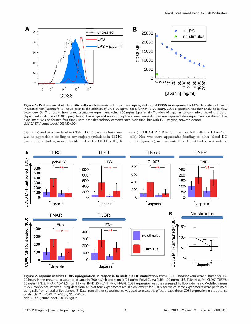
results of a representative experiment in figure 1, Japanin had a
potent and dose-dependent effect on DC maturation, although
responses to any given dose differed between donors (not shown);
figure 2a (see TLR4) shows analysis of the data from 20
independent experiments in which CD86 upregulation was
reduced by an average of around 50% by 500 ng/ml Japanin.
That Japanin was apparently produced only by three day-fed
female ticks amongst the cohorts examined is not entirely
surprising, as temporal regulation and gender differences in tick
saliva proteins are well-described phenomena [33]. Hard tick
feeding occurs in two stages: slow and rapid [34]. In adult females,
slow feeding lasts 6 to 7 days or more with a 10-fold weight gain;
rapid feeding lasts only a further 12–24 hours during which body
weight increases a further 10-fold [35]. These distinct feeding
stages may explain why Japanin is present in 3 day-fed female
SGE (from the slow-feeding stage) but apparently not in 6 day-fed
SGE (from the rapid-feeding stage). Likewise, the initiation of
feeding is required for de novo production of many factors in tick
saliva, so the absence of DC modulators in SGE from unfed ticks
was unsurprising. The absence of DC modulatory activity in male
tick SGE at all time-points may be related to the fact that they take
a smaller blood meal than females [1] and so may have less need to
modulate DC function and, potentially, the host’s adaptive
immune response. In fact, there are numerous reports of
differences between conspecific male and female ticks in SGE
activities, for example in immunoglobulin-binding proteins [36],
histamine binding proteins [4], and chemokine binding proteins
[37]. One possible reason is that females focus on imbibing an
enormous blood meal to produce thousands of eggs, increasing in
size a hundred-fold, whereas male R. appendiculatus demonstrate
‘mate guarding’ by secreting male-specific immunomodulatory
proteins that help their mate to feed [36].
Japanin specifically binds to dendritic cellsHaving shown that Japanin has DC-modulatory properties, we
next assessed whether or not it binds, and potentially acts,
specifically on DC. We labelled recombinant Japanin with a
fluorochrome, and measured its binding to monocyte-derived DC
and to peripheral blood mononuclear cell subsets (PBMC) by flow
cytometry. Fluorochrome-labelled OmCI (a tick-derived lipocalin
with no known effect on DC [38]) was used as a control for non-
specific binding. Japanin bound strongly to monocyte-derived DC
Novel Tick-Derived Dendritic Cell Modulators
PLOS Pathogens | www.plospathogens.org 3 June 2013 | Volume 9 | Issue 6 | e1003450
(figure 3a) and at a low level to CD1c+ DC (figure 3c) but there
was no appreciable binding to any major populations in PBMC
(figure 3b), including monocytes (defined as lin2CD14+ cells), B
cells (lin+HLA-DR+CD142), T cells or NK cells (lin+HLA-DR2
cells). Nor was there appreciable binding to other blood DC
subsets (figure 3c), or to activated T cells that had been stimulated
Figure 2. Japanin inhibits CD86 upregulation in response to multiple DC maturation stimuli. (A) Dendritic cells were cultured for 18–20 hours in the presence or absence of Japanin (500 ng/ml) and stimuli: (25 mg/ml Poly(I:C), via TLR3; 100 ng/ml LPS, TLR4; 4 mg/ml CL097, TLR7/8;20 ng/ml IFNa2, IFNAR; 10–12.5 ng/ml TNFa, TNFR; 20 ng/ml IFNc, IFNGR). CD86 expression was then assessed by flow cytometry. Modelled means695% confidence intervals using data from at least four experiments are shown, except for CL097 for which three experiments were performed,using cells from a total of five donors. (B) Data from all these experiments was used to assess the effect of Japanin on CD86 expression in the absenceof stimuli. ** p,0.01, * p,0.05, NS p.0.05.doi:10.1371/journal.ppat.1003450.g002
Figure 1. Pretreatment of dendritic cells with Japanin inhibits their upregulation of CD86 in response to LPS. Dendritic cells wereincubated with japanin for 24 hours prior to the addition of LPS (100 ng/ml) for a further 18–20 hours. CD86 expression was then analysed by flowcytometry. (A) The results from a representative experiment using 500 ng/ml japanin. (B) Titration of Japanin concentration, showing a dose-dependent inhibition of CD86 upregulation. The range and mean of duplicate measurements from one representative experiment are shown. Thisexperiment was performed four times, with dose-dependency demonstrated each time, but with EC50 varying between donors.doi:10.1371/journal.ppat.1003450.g001
Novel Tick-Derived Dendritic Cell Modulators
PLOS Pathogens | www.plospathogens.org 4 June 2013 | Volume 9 | Issue 6 | e1003450
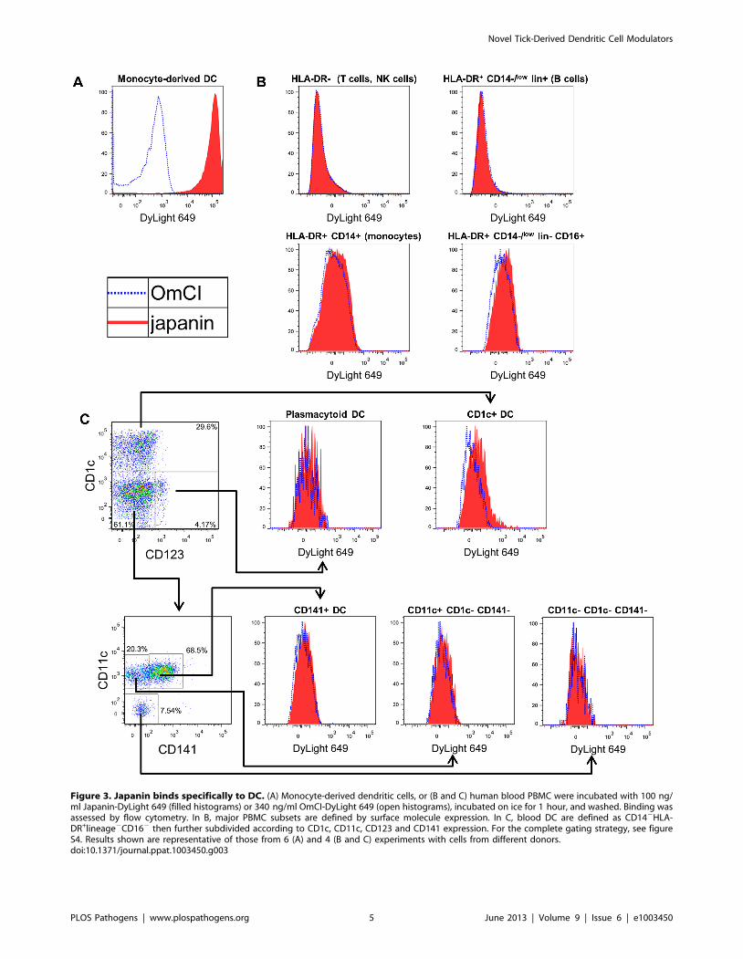
Figure 3. Japanin binds specifically to DC. (A) Monocyte-derived dendritic cells, or (B and C) human blood PBMC were incubated with 100 ng/ml Japanin-DyLight 649 (filled histograms) or 340 ng/ml OmCI-DyLight 649 (open histograms), incubated on ice for 1 hour, and washed. Binding wasassessed by flow cytometry. In B, major PBMC subsets are defined by surface molecule expression. In C, blood DC are defined as CD142HLA-DR+lineage2CD162 then further subdivided according to CD1c, CD11c, CD123 and CD141 expression. For the complete gating strategy, see figureS4. Results shown are representative of those from 6 (A) and 4 (B and C) experiments with cells from different donors.doi:10.1371/journal.ppat.1003450.g003
Novel Tick-Derived Dendritic Cell Modulators
PLOS Pathogens | www.plospathogens.org 5 June 2013 | Volume 9 | Issue 6 | e1003450

for 4 days with anti-CD3/CD28 beads (figure S3). Gating
strategies are shown in figure S4. These data clearly show Japanin
to be a highly-specific DC modulator, the first described from a
tick. Notably, it does not bind to T cells, even after their activation,
suggesting that it could potentially influence adaptive immunity
solely by acting on DC. The results distinguish Japanin from the
prostriate tick-derived Salp15, which binds to both DC through
DC-SIGN, and T cells through CD4, directly modulating the
activity of both cell types [28].
Japanin reprogrammes dendritic cell maturationDC maturation is a complex process which can endow the cells
with both stimulatory and inhibitory functions. Classically, it
involves upregulation of co-stimulatory molecules, such as CD86,
which help to initiate adaptive responses, along with the secretion
of pro-inflammatory cytokines and T cell-polarising cytokines. DC
may also upregulate co-inhibitory molecules, such as CD274
(B7H1; PD-L1), which help to suppress adaptive responses, and
secrete anti-inflammatory cytokines or express alternate T cell-
polarising molecules. The balance between these various responses
is determined in part by the nature and duration of the maturation
stimulus, including any intrinsic host-derived DC-modulating
factors, and these in turn help to determine the strength and
nature of the subsequent T cell response and the overall type of
immunity that results.
Being a prokaryotic product, LPS is not produced by ticks. We
reasoned that Japanin was therefore not likely to have evolved as a
specific regulator of LPS-induced maturation, and hypothesised
that it may also modulate DC maturation in response to other
stimuli. We found that Japanin was capable of inhibiting CD86
upregulation in response to a wide range of stimuli including the
TLR3 agonist poly(I:C) and the TLR7/8 agonist CL097, as well
as the cytokines IFN-a2 and IFN-c which signal through entirely
distinct intracellular pathways (figure 2a). Preliminary studies
further suggested that Japanin inhibits CD86 upregulation in
response to the TNF-family member CD154 (CD40L ligand)
which is crucial for cross-talk between DC and activated T cells
(data not shown). In fact, the only stimulus tested for which
Japanin did not induce a clear inhibition of CD86 upregulation
was TNF-a (figure 2a). Furthermore, the modulatory activity of
Japanin is not based on interruption of receptor-proximal
signalling components, as these are not shared between TLR
and IFN-receptor signalling pathways [39].
Given that the role of DC in adaptive immunity is not limited
to activation, but extends to educating and directing the T cell
response, we speculated that the tick might benefit more from
subverting DC maturation than from simply inhibiting it; for
example this might result in the development of a type of
immunity that is harmless to the tick, or even in the induction of
tolerance. To investigate whether Japanin had effects more
sophisticated than a simple inhibition of CD86 expression, we
extended our studies to the expression of other membrane
molecules associated with DC maturation (using flow cytometry),
and to the secretion of a wide variety of cytokines (using
multiplex analysis of culture supernatants). In these experiments,
we added Japanin at the same time as LPS, rather than as a pre-
treatment, as trial experiments showed that this reduced intra-
experimental variance in cytokine concentration readings (data
not shown).
We found that the effects of Japanin extend to a marked
reduction in the upregulation of not just CD86 but also the
maturation marker CD83 (figure 4a) and to a dramatic reduction
in the secretion of a range of cytokines. The latter included pro-
inflammatory [IL-1-b, IL-6, TNF-a] and/or Th17-polarising
[IL-1-b, IL-6] and Th1-polarising [IL-12p70, IFN-c] molecules
(figure 4b). However, this was not due to a complete inhibition of
DC maturation, as Japanin enhanced expression both of the co-
inhibitory molecule CD274 (figure 4a) and of the anti-inflamma-
tory cytokine IL-10 (figure 4b). Moreover, Japanin had no
significant effect on expression of MHC Class II molecules
(HLA-DR) or the co-stimulatory molecule CD40 (figure 4a), which
are respectively required for antigen presentation to, and cross-talk
with, T cells. Nor did it have a significant effect on LPS-induced
secretion of IL-7, IL-8 or CCL11 (figure S5). Interestingly, Japanin
was also active in the absence of LPS, reducing CD86 expression,
increasing CD274 (as well as CD40) expression, and enhancing
IL-10 secretion (figures 2b, 4a, 4b) during the ‘spontaneous’ DC
maturation that occurred slowly while in culture. Collectively, the
above results show that Japanin acts through a sophisticated
reprogramming of the DC maturation process, rather than by
simply blocking it. Such a complex spectrum of effects has never
previously been reported for a DC modulator (see discussion)
suggesting that Japanin affects a distinct and novel transcriptional
programme in DC.
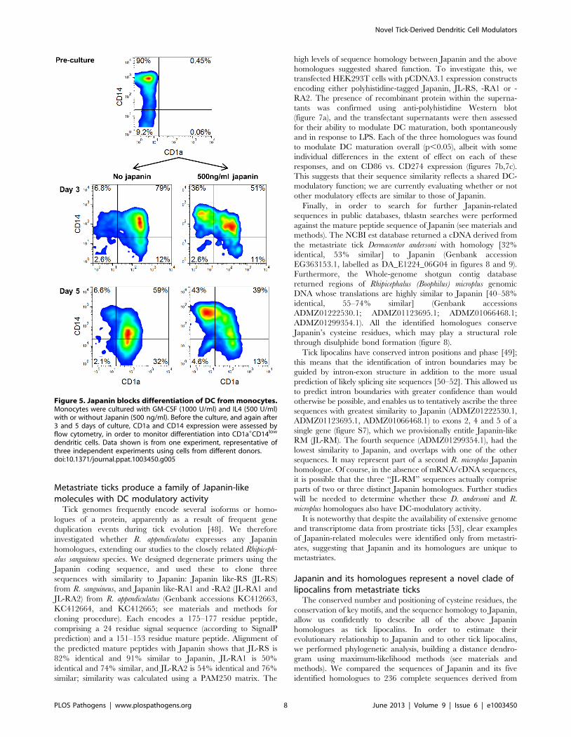
Japanin arrests dendritic cell development frommonocytes
Following maturation in response to injury or other stimuli,
skin-resident DC typically migrate out of the skin into the
lymphatics and move to the draining lymph nodes. This can
happen extremely rapidly, and involve the large majority of skin
DC, potentially resulting in a situation where an exodus of pre-
existing DC could leave the skin effectively DC-free and
‘‘unguarded’’ [40,41]. In order to avert this, monocytes are
recruited from the blood into sites of inflammation where they
are capable of differentiating into DC, thus replenishing the DC
population and restoring immune monitoring [42,43]. It would
seem to be advantageous to the tick to be able to prevent such
replenishment, and so we looked at the ability of Japanin to affect
the differentiation of DC from monocytes in vitro. To do this, we
employed the same system which we used previously for the
generation of DC from monocytes in the presence of GM-CSF
and IL-4, but this time added Japanin to the differentiation
cultures from the start. In the absence of Japanin, these
conditions typically promoted the development of CD14high
CD1a2 monocytes into CD14low CD1a+ DC. However we found
that when Japanin was added ,50% of cells retained the
CD14highCD1a2 monocyte-like phenotype, even after 5 days in
culture (figure 5).
The above results appear to conflict with the finding that
Japanin does not bind monocytes. However, we found that
binding of Japanin to monocytes increases during culture with
GM-CSF and IL-4 (figure S6), suggesting that it could act during
the early stages of differentiation of monocytes into DC. Japanin
therefore seems to impose a powerful block on this differentiation
process. Given the influx of monocytes into the bite site in response
to local tissue damage [44], the effect of Japanin on differentiating
monocytes may be as important to ticks as its effects on DC,
preventing the re-establishment of immune surveillance following
initial DC exodus. Further study of the Japanin-treated cells will be
required to elucidate whether they truly resemble ‘‘arrested’’
monocytes, or whether they have differentiated along an
alternative pathway, but what is clear is that DC differentiation
is blocked or greatly altered.
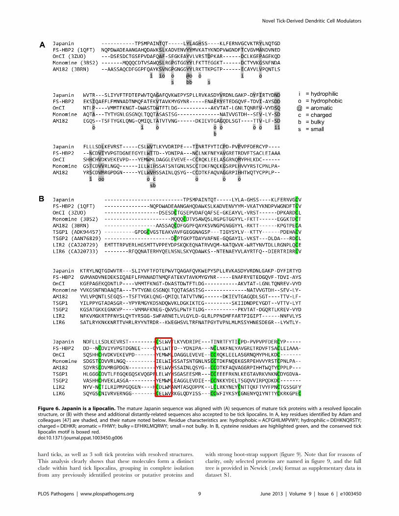
Japanin is a lipocalinLipocalins are a family of small (,20 kDa) proteins char-
acterised by an eight-stranded antiparallel b-barrel fold with a
Novel Tick-Derived Dendritic Cell Modulators
PLOS Pathogens | www.plospathogens.org 6 June 2013 | Volume 9 | Issue 6 | e1003450
repeated +1 topology, typically preceded by a short N-terminal
310-helix and followed by a C-terminal a-helix. They frequently
have one or more binding pocket(s) for small molecule ligand(s)
[45,46]. Inspection of the primary amino acid sequence of Japanin
indicated that it is a lipocalin, a conclusion supported by
comparisons with other tick-derived lipocalins (figure 6). Japanin
conserves residue properties at the key positions described by
Adam and colleagues [47] to a similar extent as tick proteins with
resolved lipocalin structures (figure 6a), and shares the position of
cysteine residues and the presence of a conserved motif ((Y/C)-
hydrophilic-(L/M)-W-hydrophobic) with these and with other tick
proteins thought to be lipocalins (figure 6b). This provisional
description of Japanin as a lipocalin has recently been confirmed
by the resolution of its crystal structure, details of which are
currently being prepared for publication (personal communica-
tion, Susan Lea).
Figure 4. Japanin modulates dendritic cell maturation, rather than simply inhibiting it. Dendritic cells were cultured in the presence orabsence of Japanin (500 ng/ml) and LPS (100 ng/ml) for 18–20 hours. (A) CD40, CD83, CD86, CD274 and HLA-DR expression were then assessed byflow cytometry, and (B)the concentration of pro-inflammatory cytokines in the culture supernatant was measured by Luminex1. Modelled means695% confidence intervals using data from at least four experiments are shown, except where marked ` where above-scale readings in the LPS-onlymade it impossible to calculate meaningful confidence intervals; the graphs show the lowest possible mean value (taking an above-scale value to beequal to the maximum possible on-scale value). ** p,0.01, * p,0.05, N p,0.1, NS p.0.05.doi:10.1371/journal.ppat.1003450.g004
Novel Tick-Derived Dendritic Cell Modulators
PLOS Pathogens | www.plospathogens.org 7 June 2013 | Volume 9 | Issue 6 | e1003450
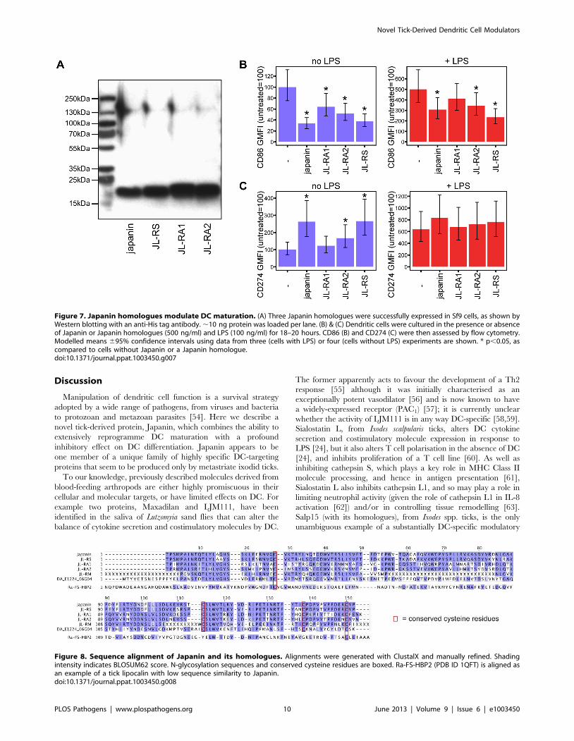
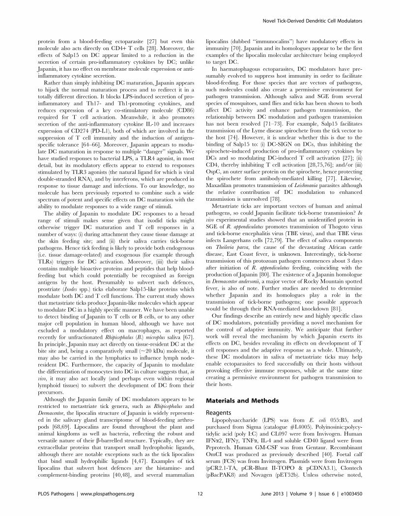
Metastriate ticks produce a family of Japanin-likemolecules with DC modulatory activity
Tick genomes frequently encode several isoforms or homo-
logues of a protein, apparently as a result of frequent gene
duplication events during tick evolution [48]. We therefore
investigated whether R. appendiculatus expresses any Japanin
homologues, extending our studies to the closely related Rhipiceph-
alus sanguineus species. We designed degenerate primers using the
Japanin coding sequence, and used these to clone three
sequences with similarity to Japanin: Japanin like-RS (JL-RS)
from R. sanguineus, and Japanin like-RA1 and -RA2 (JL-RA1 and
JL-RA2) from R. appendiculatus (Genbank accessions KC412663,
KC412664, and KC412665; see materials and methods for
cloning procedure). Each encodes a 175–177 residue peptide,
comprising a 24 residue signal sequence (according to SignalP
prediction) and a 151–153 residue mature peptide. Alignment of
the predicted mature peptides with Japanin shows that JL-RS is
82% identical and 91% similar to Japanin, JL-RA1 is 50%
identical and 74% similar, and JL-RA2 is 54% identical and 76%
similar; similarity was calculated using a PAM250 matrix. The
high levels of sequence homology between Japanin and the above
homologues suggested shared function. To investigate this, we
transfected HEK293T cells with pCDNA3.1 expression constructs
encoding either polyhistidine-tagged Japanin, JL-RS, -RA1 or -
RA2. The presence of recombinant protein within the superna-
tants was confirmed using anti-polyhistidine Western blot
(figure 7a), and the transfectant supernatants were then assessed
for their ability to modulate DC maturation, both spontaneously
and in response to LPS. Each of the three homologues was found
to modulate DC maturation overall (p,0.05), albeit with some
individual differences in the extent of effect on each of these
responses, and on CD86 vs. CD274 expression (figures 7b,7c).
This suggests that their sequence similarity reflects a shared DC-
modulatory function; we are currently evaluating whether or not
other modulatory effects are similar to those of Japanin.
Finally, in order to search for further Japanin-related
sequences in public databases, tblastn searches were performed
against the mature peptide sequence of Japanin (see materials and
methods). The NCBI est database returned a cDNA derived from
the metastriate tick Dermacentor andersoni with homology [32%
identical, 53% similar] to Japanin (Genbank accession
EG363153.1, labelled as DA_E1224_06G04 in figures 8 and 9).
Furthermore, the Whole-genome shotgun contig database
returned regions of Rhipicephalus (Boophilus) microplus genomic
DNA whose translations are highly similar to Japanin [40–58%
identical, 55–74% similar] (Genbank accessions
ADMZ01222530.1; ADMZ01123695.1; ADMZ01066468.1;
ADMZ01299354.1). All the identified homologues conserve
Japanin’s cysteine residues, which may play a structural role
through disulphide bond formation (figure 8).
Tick lipocalins have conserved intron positions and phase [49];
this means that the identification of intron boundaries may be
guided by intron-exon structure in addition to the more usual
prediction of likely splicing site sequences [50–52]. This allowed us
to predict intron boundaries with greater confidence than would
otherwise be possible, and enables us to tentatively ascribe the three
sequences with greatest similarity to Japanin (ADMZ01222530.1,
ADMZ01123695.1, ADMZ01066468.1) to exons 2, 4 and 5 of a
single gene (figure S7), which we provisionally entitle Japanin-like
RM (JL-RM). The fourth sequence (ADMZ01299354.1), had the
lowest similarity to Japanin, and overlaps with one of the other
sequences. It may represent part of a second R. microplus Japanin
homologue. Of course, in the absence of mRNA/cDNA sequences,
it is possible that the three ‘‘JL-RM’’ sequences actually comprise
parts of two or three distinct Japanin homologues. Further studies
will be needed to determine whether these D. andersoni and R.
microplus homologues also have DC-modulatory activity.
It is noteworthy that despite the availability of extensive genome
and transcriptome data from prostriate ticks [53], clear examples
of Japanin-related molecules were identified only from metastri-
ates, suggesting that Japanin and its homologues are unique to
metastriates.
Japanin and its homologues represent a novel clade oflipocalins from metastriate ticks
The conserved number and positioning of cysteine residues, the
conservation of key motifs, and the sequence homology to Japanin,
allow us confidently to describe all of the above Japanin
homologues as tick lipocalins. In order to estimate their
evolutionary relationship to Japanin and to other tick lipocalins,
we performed phylogenetic analysis, building a distance dendro-
gram using maximum-likelihood methods (see materials and
methods). We compared the sequences of Japanin and its five
identified homologues to 236 complete sequences derived from
Figure 5. Japanin blocks differentiation of DC from monocytes.Monocytes were cultured with GM-CSF (1000 U/ml) and IL4 (500 U/ml)with or without Japanin (500 ng/ml). Before the culture, and again after3 and 5 days of culture, CD1a and CD14 expression were assessed byflow cytometry, in order to monitor differentiation into CD1a+CD14low
dendritic cells. Data shown is from one experiment, representative ofthree independent experiments using cells from different donors.doi:10.1371/journal.ppat.1003450.g005
Novel Tick-Derived Dendritic Cell Modulators
PLOS Pathogens | www.plospathogens.org 8 June 2013 | Volume 9 | Issue 6 | e1003450
hard ticks, as well as 3 soft tick proteins with resolved structures.
This analysis clearly shows that these molecules form a distinct
clade within hard tick lipocalins, grouping in complete isolation
from any previously identified proteins or putative proteins and
with strong boot-strap support (figure 9). Note that for reasons of
clarity, only selected proteins are named in figure 9, and the full
tree is provided in Newick (.nwk) format as supplementary data in
dataset S1.
Figure 6. Japanin is a lipocalin. The mature Japanin sequence was aligned with (A) sequences of mature tick proteins with a resolved lipocalinstructure, or (B) with these and additional distantly-related sequences also accepted to be tick lipocalins. In A, key residues identified by Adam andcolleagues [47] are shaded, and their nature noted below. Residue characteristics are: hydrophobic = ACFGHILMPVWY; hydrophilic = DEHKNQRSTY;charged = DEHKR; aromatic = FHWY; bulky = EFHIKLMQRWY; small = not bulky. In B, cysteine residues are highlighted green, and the conserved ticklipocalin motif is boxed red.doi:10.1371/journal.ppat.1003450.g006
Novel Tick-Derived Dendritic Cell Modulators
PLOS Pathogens | www.plospathogens.org 9 June 2013 | Volume 9 | Issue 6 | e1003450
Discussion
Manipulation of dendritic cell function is a survival strategy
adopted by a wide range of pathogens, from viruses and bacteria
to protozoan and metazoan parasites [54]. Here we describe a
novel tick-derived protein, Japanin, which combines the ability to
extensively reprogramme DC maturation with a profound
inhibitory effect on DC differentiation. Japanin appears to be
one member of a unique family of highly specific DC-targeting
proteins that seem to be produced only by metastriate ixodid ticks.
To our knowledge, previously described molecules derived from
blood-feeding arthropods are either highly promiscuous in their
cellular and molecular targets, or have limited effects on DC. For
example two proteins, Maxadilan and LJM111, have been
identified in the saliva of Lutzomyia sand flies that can alter the
balance of cytokine secretion and costimulatory molecules by DC.
The former apparently acts to favour the development of a Th2
response [55] although it was initially characterised as an
exceptionally potent vasodilator [56] and is now known to have
a widely-expressed receptor (PAC1) [57]; it is currently unclear
whether the activity of LJM111 is in any way DC-specific [58,59].
Sialostatin L, from Ixodes scalpularis ticks, alters DC cytokine
secretion and costimulatory molecule expression in response to
LPS [24], but it also alters T cell polarisation in the absence of DC
[24], and inhibits proliferation of a T cell line [60]. As well as
inhibiting cathepsin S, which plays a key role in MHC Class II
molecule processing, and hence in antigen presentation [61],
Sialostatin L also inhibits cathepsin L1, and so may play a role in
limiting neutrophil activity (given the role of cathepsin L1 in IL-8
activation [62]) and/or in controlling tissue remodelling [63].
Salp15 (with its homologues), from Ixodes spp. ticks, is the only
unambiguous example of a substantially DC-specific modulatory
Figure 7. Japanin homologues modulate DC maturation. (A) Three Japanin homologues were successfully expressed in Sf9 cells, as shown byWestern blotting with an anti-His tag antibody. ,10 ng protein was loaded per lane. (B) & (C) Dendritic cells were cultured in the presence or absenceof Japanin or Japanin homologues (500 ng/ml) and LPS (100 ng/ml) for 18–20 hours. CD86 (B) and CD274 (C) were then assessed by flow cytometry.Modelled means 695% confidence intervals using data from three (cells with LPS) or four (cells without LPS) experiments are shown. * p,0.05, ascompared to cells without Japanin or a Japanin homologue.doi:10.1371/journal.ppat.1003450.g007
Figure 8. Sequence alignment of Japanin and its homologues. Alignments were generated with ClustalX and manually refined. Shadingintensity indicates BLOSUM62 score. N-glycosylation sequences and conserved cysteine residues are boxed. Ra-FS-HBP2 (PDB ID 1QFT) is aligned asan example of a tick lipocalin with low sequence similarity to Japanin.doi:10.1371/journal.ppat.1003450.g008
Novel Tick-Derived Dendritic Cell Modulators
PLOS Pathogens | www.plospathogens.org 10 June 2013 | Volume 9 | Issue 6 | e1003450
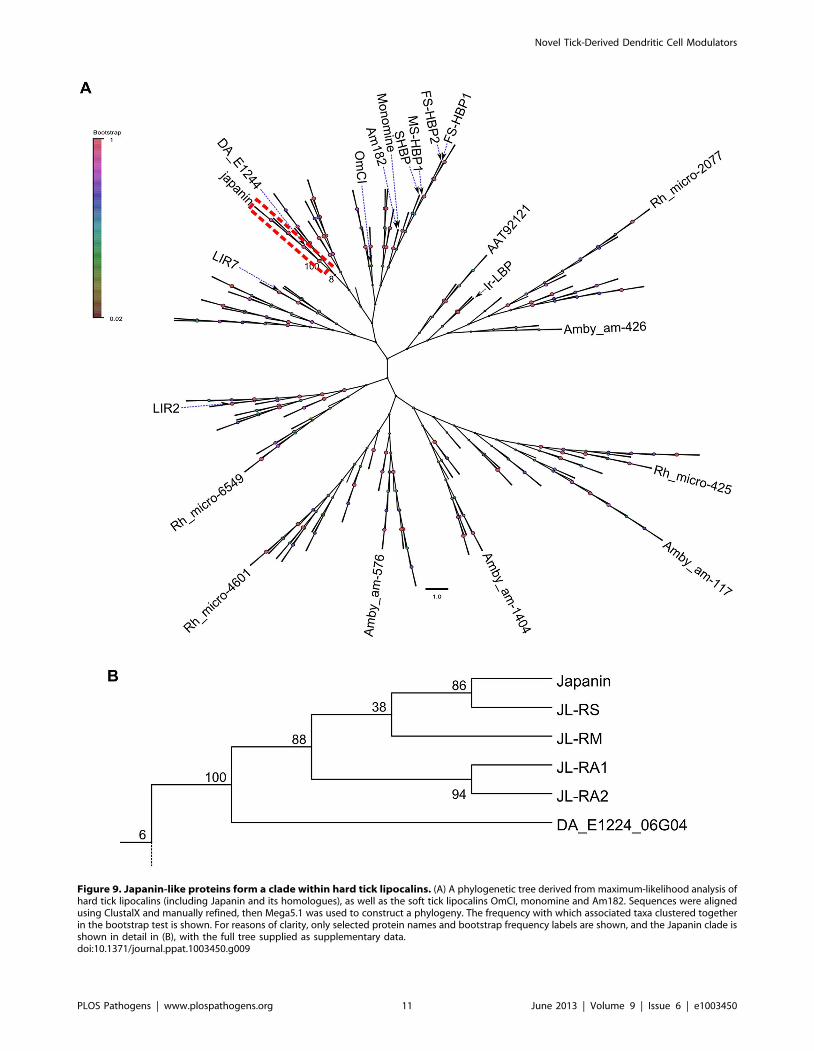
Figure 9. Japanin-like proteins form a clade within hard tick lipocalins. (A) A phylogenetic tree derived from maximum-likelihood analysis ofhard tick lipocalins (including Japanin and its homologues), as well as the soft tick lipocalins OmCI, monomine and Am182. Sequences were alignedusing ClustalX and manually refined, then Mega5.1 was used to construct a phylogeny. The frequency with which associated taxa clustered togetherin the bootstrap test is shown. For reasons of clarity, only selected protein names and bootstrap frequency labels are shown, and the Japanin clade isshown in detail in (B), with the full tree supplied as supplementary data.doi:10.1371/journal.ppat.1003450.g009
Novel Tick-Derived Dendritic Cell Modulators
PLOS Pathogens | www.plospathogens.org 11 June 2013 | Volume 9 | Issue 6 | e1003450
protein from a blood-feeding ectoparasite [27] but even this
molecule also acts directly on CD4+ T cells [28]. Moreover, the
effects of Salp15 on DC appear limited to a reduction in the
secretion of certain pro-inflammatory cytokines by DC; unlike
Japanin, it has no effect on membrane molecule expression or anti-
inflammatory cytokine secretion.
Rather than simply inhibiting DC maturation, Japanin appears
to hijack the normal maturation process and to redirect it in a
totally different direction. It blocks LPS-induced secretion of pro-
inflammatory and Th17- and Th1-promoting cytokines, and
reduces expression of a key co-stimulatory molecule (CD86)
required for T cell activation. Meanwhile, it also promotes
secretion of the anti-inflammatory cytokine IL-10 and increases
expression of CD274 (PD-L1), both of which are involved in the
suppression of T cell immunity and the induction of antigen-
specific tolerance [64–66]. Moreover, Japanin appears to modu-
late DC maturation in response to multiple ‘‘danger’’ signals. We
have studied responses to bacterial LPS, a TLR4 agonist, in most
detail, but its modulatory effects appear to extend to responses
stimulated by TLR3 agonists (the natural ligand for which is viral
double-stranded RNA), and by interferons, which are produced in
response to tissue damage and infections. To our knowledge, no
molecule has been previously reported to combine such a wide
spectrum of potent and specific effects on DC maturation with the
ability to modulate responses to a wide range of stimuli.
The ability of Japanin to modulate DC responses to a broad
range of stimuli makes sense given that ixodid ticks might
otherwise trigger DC maturation and T cell responses in a
number of ways: (i) during attachment they cause tissue damage at
the skin feeding site; and (ii) their saliva carries tick-borne
pathogens. Hence tick feeding is likely to provide both endogenous
(i.e. tissue damage-related) and exogenous (for example through
TLRs) triggers for DC activation. Moreover, (iii) their saliva
contains multiple bioactive proteins and peptides that help blood-
feeding but which could potentially be recognised as foreign
antigens by the host. Presumably to subvert such defences,
prostriate (Ixodes spp.) ticks elaborate Salp15-like proteins which
modulate both DC and T cell functions. The current study shows
that metastriate ticks produce Japanin-like molecules which appear
to modulate DC in a highly specific manner. We have been unable
to detect binding of Japanin to T cells or B cells, or to any other
major cell population in human blood, although we have not
excluded a modulatory effect on macrophages, as reported
recently for unfractionated Rhipicephalus (B.) microplus saliva [67].
In principle, Japanin may act directly on tissue-resident DC at the
bite site and, being a comparatively small (,20 kDa) molecule, it
may also be carried in the lymphatics to influence lymph node-
resident DC. Furthermore, the capacity of Japanin to modulate
the differentiation of monocytes into DC in culture suggests that, in
vivo, it may also act locally (and perhaps even within regional
lymphoid tissues) to subvert the development of DC from their
precursors.
Although the Japanin family of DC modulators appears to be
restricted to metastriate tick genera, such as Rhipicephalus and
Dermacentor, the lipocalin structure of Japanin is widely represent-
ed in the salivary gland transcriptome of blood-feeding arthro-
pods [68,69]. Lipocalins are found throughout the plant and
animal kingdoms as well as bacteria, reflecting the robust and
versatile nature of their b-barrelled structure. Typically, they are
extracellular proteins that transport small hydrophobic ligands,
although there are notable exceptions such as the tick lipocalins
that bind small hydrophilic ligands [4,47]. Examples of tick
lipocalins that subvert host defences are the histamine- and
complement-binding proteins [40,48], and several mammalian
lipocalins (dubbed ‘‘immunocalins’’) have modulatory effects in
immunity [70]. Japanin and its homologues appear to be the first
examples of the lipocalin molecular architecture being employed
to target DC.
In haematophagous ectoparasites, DC modulators have pre-
sumably evolved to suppress host immunity in order to facilitate
blood-feeding. For those species that are vectors of pathogens,
such molecules could also create a permissive environment for
pathogen transmission. Although saliva and SGE from several
species of mosquitoes, sand flies and ticks has been shown to both
affect DC activity and enhance pathogen transmission, the
relationship between DC modulation and pathogen transmission
has not been resolved [71–73]. For example, Salp15 facilitates
transmission of the Lyme disease spirochete from the tick vector to
the host [74]. However, it is unclear whether this is due to the
binding of Salp15 to: (i) DC-SIGN on DCs, thus inhibiting the
spirochete-induced production of pro-inflammatory cytokines by
DCs and so modulating DC-induced T cell activation [27]; (ii)
CD4, thereby inhibiting T cell activation [28,75,76]; and/or (iii)
OspC, an outer surface protein on the spirochete, hence protecting
the spirochete from antibody-mediated killing [77]. Likewise,
Maxadilan promotes transmission of Leishmania parasites although
the relative contribution of DC modulation to enhanced
transmission is unresolved [78].
Metastriate ticks are important vectors of human and animal
pathogens, so could Japanin facilitate tick-borne transmission? In
vivo experimental studies showed that an unidentified protein in
SGE of R. appendiculatus promotes transmission of Thogoto virus
and tick-borne encephalitis virus (TBE virus), and that TBE virus
infects Langerhans cells [72,79]. The effect of saliva components
on Theileria parva, the cause of the devastating African cattle
disease, East Coast fever, is unknown. Interestingly, tick-borne
transmission of this protozoan pathogen commences about 3 days
after initiation of R. appendiculatus feeding, coinciding with the
production of Japanin [80]. The existence of a Japanin homologue
in Dermacentor andersonii, a major vector of Rocky Mountain spotted
fever, is also of note. Further studies are needed to determine
whether Japanin and its homologues play a role in the
transmission of tick-borne pathogens; one possible approach
would be through their RNA-mediated knockdown [81].
Our findings describe an entirely new and highly specific class
of DC modulators, potentially providing a novel mechanism for
the control of adaptive immunity. We anticipate that further
work will reveal the mechanism by which Japanin exerts its
effects on DC, besides revealing its effects on development of T
cell responses and the adaptive response as a whole. Ultimately,
these DC modulators in saliva of metastriate ticks may help
enable ectoparasites to feed successfully on their hosts without
provoking effective immune responses, while at the same time
creating a permissive environment for pathogen transmission to
their hosts.
Materials and Methods
ReagentsLipopolysaccharide (LPS) was from E. coli 055:B5, and
purchased from Sigma (catalogue #L4005). Polyinosinic:polycy-
tidylic acid (poly I:C) and CL097 were from Invivogen. Human
IFNa2, IFNc, TNFa, IL-4 and soluble CD40 ligand were from
Peprotech. Human GM-CSF was from Gentaur. Recombinant
OmCI was produced as previously described [40]. Foetal calf
serum (FCS) was from Invitrogen. Plasmids were from Invitrogen
(pCR2.1-TA, pCR-Blunt II-TOPO & pCDNA3.1), Clontech
(pBacPAK8) and Novagen (pET52b). Unless otherwise noted,
Novel Tick-Derived Dendritic Cell Modulators
PLOS Pathogens | www.plospathogens.org 12 June 2013 | Volume 9 | Issue 6 | e1003450
PCR was carried out using Phusion Hot Start DNA polymerase
(NEB). Phosphate buffered saline (PBS) and Hanks Balanced Salt
Solution were from PAA.
Cell cultureMammalian cells were cultured at 37uC/5% CO2 in complete
RPMI (C-RPMI), consisting of RPMI 1640 (PAA) supplemented
with 10% FCS, 100 U/ml penicillin (PAA), 100 mg/ml strepto-
mycin (PAA). Tissue culture plastics were from Corning. Sf9 insect
cells were cultured at 28uC in Sf900III serum-free medium
(Invitrogen). Sf9 liquid culture was in Erlenmeyer flasks with
shaking at 110 rpm. Standard E. coli strains and techniques were
used to produce plasmids during molecular cloning. Human blood
products were from anonymous healthy donors, and supplied by
the National Blood Service (England & Wales).
Generation of human dendritic cellsHuman monocyte-derived DC were generated using a
protocol derived from the method of Sallusto and Lanzavecchia
[82]. Peripheral blood mononuclear cells (PBMC) were isolated
from Buffy coats and leucocyte cones using gradient centrifuga-
tion with Lymphoprep (Axis Shield). Monocytes were isolated
from PBMC by negative selection using the EasySep Human
Monocyte Enrichment Kit (Stemcell) as per manufacturer’s
instructions, then cultured at 56105/ml in C-RPMI supplement-
ed with 1000 U/ml human GM-CSF and 100 ng/ml human IL-
4. Cultures were fed after three days by replacing one third of the
medium with fresh C-RPMI supplemented with 3000 U/ml
GM-CSF and 300 ng/ml IL-4, and cells were harvested for use
in assays after 5 or 6 days of culture. Prior to some assays, DC
were frozen in Voluven (Fresenius Kabi) supplemented with
DMSO (Hybrimax grade, Sigma) and FCS to give final
concentrations of 5.5% hydroxyethyl starch 130/0.4, 4.8%
DMSO and 3.8% FCS in isotonic saline. Freezing was carried
out at 1uC/minute.
DC activity assayDC were cultured at 16106 cells/ml in flat-bottomed 96-well
tissue culture plates in C-RPMI supplemented with 1000 U/ml
GM-CSF and 100 ng/ml IL-4. Japanin was added to 500 ng/ml
(unless otherwise stated), and a maturation stimuli was either
added immediately or after 24 hours in culture. Cells were then
cultured for 18–22 hours, and analysed by flow cytometry. In
some experiments, multiplex measurement of supernatant cyto-
kine concentrations was also performed. In the experiments shown
in figures S1 and S2, sufficient SGE was added to give a final SGE-
derived protein concentration of 50 mg/ml
T cell isolation and stimulationPBMC were obtained from leucocyte cones as described above,
and T cells were isolated by negative selection using the Easysep
human T cell enrichment kit (Stemcell) as per manufacturer’s
instructions. They were then stimulated by culture with Human T-
activator CD3/CD28 Dynabeads (Life Technologies) for four
days, as per manufacturer’s instructions.
Tick rearing and salivary gland extract preparationR. appendiculatus ticks were reared according to Jones et al. [83]
Salivary glands were dissected under a microscope and rinsed
briefly in cold PBS. Salivary gland extract (SGE) was prepared by
disruption of freshly-prepared salivary glands in PBS with a 1 ml
Dounce homogenizer. The SGE was clarified by centrifugation
(.10000 g for 3 min) and stored at 220uC.
SGE fractionationSGE from 350 salivary glands was diluted in 50 mM
Na2HPO4/50 mM NaCl (pH7.0) and passed through a 1 ml
Hi-Trap Q sepharose anion exchange column (GE). Unbound
material (Q column flowthrough) was concentrated to a final
volume of 500 ml using a 5000MWCO Vivaspin 6 centrifugal
concentrator (GE Healthcare) which had been pre-treated with c-
globulin to prevent non-specific absorbance of proteins. The Q
column flowthrough was then fractionated by gel filtration over a
Superdex 75 HR10/30 column (GE Healthcare) using 50 mM
Hepes (pH7.6), 150 mM NaCl as running buffer, and each
fraction assayed for DC modulatory activity. Consecutive active
fractions were pooled, dialysed against 50 mM HEPES (pH 8.3),
and fractionated by High Performance Liquid Chromatography
(HPLC) on a C4 column, with elution using a 0–100% gradient of
acetonitrile. HPLC fractions were freeze-dried under vacuum,
redissolved in PBS, and assayed for DC modulatory activity. The
fraction with maximal activity was used for Edman degradation
sequencing.
Japanin cloningThe template for Japanin cloning was cDNA generated from 1
day-fed female R. appendiculatus salivary glands. RNA was isolated
from 30 salivary glands using Trizol reagent (Invitrogen), and
cDNA generated using ImPromII reverse transcriptase (Promega).
Initial cloning of Japanin sequence was performed using Taq DNA
polymerase (NEB) in nested PCR with degenerate primers
designed against the N-terminal peptide sequence. A ,600 bp
product was gel purified using the QIAquick gel extraction kit
(Qiagen) and ligated into the pCR2.1-TA cloning vector.)
Sequencing of this construct revealed the 39 region of the Japanin
coding sequence; this was used to design primers for the
amplification of the 59 region using 59 RACE System for Rapid
Amplification of cDNA Ends (Invitrogen) in conjunction with
Japanin-specific primers. Amplified DNA was gel purified and
sequenced, providing the 59 region of the coding sequence. The 59
and 39 sequences obtained thus far were then used to design
primers for the amplification of full-length Japanin coding
sequence using two rounds nested PCR, with the second round
using primers which added a 59 BamHI restriction site and a 39
NotI restriction site. The second round product was digested with
BamHI and NotI (NEB), and ligated into similarly digested
pBacPAK8 to generate pBacPAK8-Japanin. In order to obtain a
polyhistidine-tagged version of Japanin, nested PCR was per-
formed with Phusion DNA polymerase using pBacPAK8-Japanin
as a template, employing reverse primers designed so as to replace
the 39 stop sequence and NotI-site with DNA encoding two glycine
residues (to serve as a flexible linker) and six histidine residues (the
‘‘polyhistidine tag’’), followed by a stop sequence, and then finally
a NotI site. The product from this PCR was digested with BamHI
and NotI, and ligated into similarly digested pBacPAK8 (to
produce pBacPAK8-Japanin-his), pCDNA3.1 (pCDNA3.1-Japa-
nin-his) and pET52b (pET52b-Japanin-his).
Homologue cloningPartial sequences of JL-RA1, JL-RA2 and JL-RS were obtained
from Rhipicephalus appendiculatus (JL-RA1/2) or R. sanguineus
(JL-RS) cDNA expression libraries which had been previously
generated in the Lambda Zap II vector (Stratagene). PCR was
performed using a degenerate, Japanin-derived forward primer
(ACMSAKACYCTYTACCTYGYG) in combination with either
a vector specific reverse primer (TTATGCTGAGTGATACCC),
in the case of JL-RA2, or a Japanin-specific reverse primer
(ATATGCGGCCGCTTATGGATAGCACCTCTCGT), in the
Novel Tick-Derived Dendritic Cell Modulators
PLOS Pathogens | www.plospathogens.org 13 June 2013 | Volume 9 | Issue 6 | e1003450

case of JL-RA1 and -RS. PCR products were cloned into pCR-
Blunt II-TOPO and sequenced, providing sequences for the 39
region of each DNA. Sequences of the 59 region of each were then
obtained from the same libraries by PCR using a vector-specific
forward primer (CGCAATTAACCCTCACTAAAGGGAAC)
with gene-specific reverse primers (CGTTAGTTTCAGT-
GAACGTGAGTGTCC for JL-RA1; CGTTTGGTATCTT-
CATTTTAGATGAGTATCC for JL-RA2; CATGAGAA-
CAGCTTCGATGAATATGC for JL-RS), and products cloned
into pCR-Blunt II-TOPO and sequenced. Full-length cDNAs,
each with the addition of sequence encoding a C-terminal
diglycine linker and a polyhistidine tag (GGHHHHHH) were
obtained as synthetic genes (from DNA2.0) and subcloned into
pBacPAK8 using standard techniques. Recombinant JL-RA1, -
RA2 and -RS was produced and purified as described below.
Proteinase K treatmentProteinase K treatment of SGE-3F was performed by incubation
with 150 mg/ml Proteinase K (Sigma) for 2 hours at 50uC, followed
by heating to 98uC for 10 minutes to inactivate the enzyme.
Protein expressionRecombinant baculovirus was obtained using the approach of
Possee et al. [84] Briefly, Sf9 cell monolayer was co-transfected
with flashBac Gold baculovirus (Oxford Expression) and pBac-
PAK8 transfer vector (described above), using Cellfectin (Invitro-
gen) as per manufacturer’s instructions. Recombinant virus was
amplified by infection of Sf9 cells in liquid culture at a low
multiplicity-of-infection (moi), and the amplified virus used to
infect Sf9 liquid cultures at moi = 2 for protein expression. Viral
titre was assessed by plaque assay.
Protein purificationThe medium was cleared by centrifugation (2000 g, 10 min)
72 hours after infection, and proteins were precipitated by adding
polyethylene glycol (PEG4000, Sigma; 18 g/100 ml). The precip-
itate was dissolved in HBSS (pH 7.4), loaded on to a 1 ml Talon
column (Clontech), and eluted using 150 mM imidazole. The
protein-containing eluate fractions were pooled, concentrated using
a 9K MWCO Pierce Protein Concentrator (Thermo Scientific),
then further purified by size exclusion chromatography with a
Superdex 75 HR10/30 column (GE Healthcare) using PBS (pH7.4)
as running buffer. Concentration of purified protein was measured
by its absorbance at 280 nm using extinction coefficients reported
by the ProtParam tool (http://web.expasy.org/protparam/). Purity
was confirmed by silver stain of SDS-PAGE gels.
Western blottingSDS-PAGE was performed using precast Precise Tris-HEPES
gels (Thermo Scientific) as per manufacturer’s instructions.
Proteins were wet transferred to PVDF membrane (Thermo
Scientific) using 30 V for 1 hour in Towbin buffer (25 mM Tris,
192 mM glycine) with 20% methanol. Membranes were blocked
with StartingBlock T20-PBS (Thermo Scientific), then stained first
with biotinylated anti-His tag antibody (Penta-His, Qiagen), and
then with streptavidin-HRP (Jackson ImmunoResearch). All
staining steps, and extensive washing, was in PBS/0.05% Tween
20. Bands were visualised by luminescent substrate (ECL, Thermo
Scientific) with X-ray film (CL-XPosure, Thermo Scientific).
Flow cytometryCells were stained in PBS/2% FCS and analysed with a
FACSCanto flow cytometer (Becton Dickinson). The following
antibodies were used: 5C3 (anti-CD40, APC-conjugated); HB15e
(anti-CD83, FITC-conjugated); GL1 (anti-CD86, PE-conjugated);
MIH1 (anti-CD274, PE-Cy7-conjugated); LN3 (anti-HLA-DR,
APC-conjugated). All were from eBioscience. Isotype control
antibodies showed negligible binding to DC. Cells were gated
according to FSC/SSC, and, in some experiments, according to
exclusion of 7AAD (Sigma). Japanin did not increase the
frequency of 7AAD-staining cells.
Multiplex analysis of culture supernatantsClarified tissue culture supernatants were diluted with 1 volume
of PBS and stored at 220uC. They were analysed using Milliplex
MAP Luminex beads (Millipore) as per manufacturer’s instruc-
tions.
Binding assaysRecombinant Japanin and OmCI were labelled with DyLight
649 using DyLight 649 Amine-Reactive Dye (Thermo Scientific)
as per manufacturer’s instructions. In the experiment shown in
figure S6, examining the binding of Japanin at different points
during the differentiation of DC from monocytes, proteins were
instead labelled with Alexa Fluor 488, using the Alexa Fluor 488
Microscale Protein Labelling Kit (Life Technologies) as per
manufacturer’s instructions. Cells were incubated with 100 ng/
ml labelled Japanin or 340 ng/ml labelled OmCI for 1 hour on
ice in HBSS (containing 1.3 mM Ca2+ 0.8 mM Mg2+)/2% FCS,
washed extensively, and analysed by flow cytometry.
For the determination of Japanin binding to PBMC subsets,
PBMC were incubated with Fc block (Miltenyi Biotec) for
15 minutes on ice, washed, then incubated with DyLight 649-
labelled Japanin or OmCI as described above, but with the
addition of the following antibodies: anti-CD1c-Brilliant Violet
421 (clone L161, Biolegend); CD3-biotin (clone OKT3, Biole-
gend); CD7-biotin (clone 124-1D1, eBioscience); CD14-Brilliant
Violet 650 (clone M5E2, Biolegend); CD11c-PE-Texas Red (clone
B-ly6, BD Biosciences); CD19-biotin (clone HIB19, eBioscience);
CD20-biotin (clone2H7, eBioscience); CD45-eFluor 605NC (clone
HI30, eBioscience); CD56-biotin (clone HCD56, Biolegend);
CD123-PerCP-Cy5.5 (clone6H6, Biolegend); CD141-PE (clone
AD5-14H12, Miltenyi Biotec); HLA-DR-V500 (clone G46-6, BD
Biosciences). The cells were washed, then incubated with
streptavidin-Alexa Fluor 700 (Life Technologies), and washed
again prior to analysis. Dead cells were excluded by using Fixable
Viability Dye eFluor 780 (eBioscience) in the first staining step.
The biotinylated antibody panel visualised with streptavidin-Alexa
Fluor 700 (CD3/CD7/CD19/CD20/CD56) is referred to in the
text and figures as ‘‘lineage’’ (or ‘‘lin’’).
Database searchesTranslated BLAST (Basic Local Alignment Search Tool [85]
searches were performed with the mature Japanin peptide
sequence as the query, using the NCBI online interface (http://
blast.ncbi.nlm.nih.gov/). Similarity scores were obtained with
blastp or tblastn, as appropriate, using the same interface, and with
a PAM250 matrix.
Phylogenetic analysisAn initial group of 4 tick lipocalins with published structures
(FS-HBP2 [4], Am182 [86], Monomine [86] and OmCI [87])
were aligned using ClustalX [88], and this seed alignment used to
construct a gap penalty mask. This mask was then employed in the
alignment of an additional 242 hard tick lipocalins using ClustalX.
Sequences for alignment were taken from: (i) the table provided as
Novel Tick-Derived Dendritic Cell Modulators
PLOS Pathogens | www.plospathogens.org 14 June 2013 | Volume 9 | Issue 6 | e1003450
supplementary data by Francischetti and colleagues [89], from
which all complete sequences identified as hard tick lipocalins,
were used, with the exception of those described as group VIII,
which we do not believe to be lipocalins (based on the absence of
conserved sequence features); (ii) LIR2 and LIR7 [90]; (iii) Ir-LBP
[91]; (iv) the sequences described in this paper. Sequences are
named according to their published abbreviation, Genbank
accession number, or as referred to by Francischetti and
colleagues. This alignment was manually refined to align key
conserved sequence features, and MUSCLE [92] used to realign
subsections of the alignment between conserved features. The
edges were trimmed manually to leave a conserved core.
Evolutionary history was then inferred using the maximum-
likelihood method. After model selection according to AICc and
BIC criteria, the Whelan and Goldman + Freq. model [93] was
used, with initial tree(s) for the heuristic search generated by
applying the Neighbour-Joining method to a matrix of pairwise
distances estimated using a JTT model. A discrete Gamma
distribution was used to model evolutionary rate differences
among sites (5 categories (+G, parameter = 7.3058)). The boot-
strap consensus tree inferred from 50 replicates is taken to
represent the evolutionary history of the taxa analysed. All
positions with less than 90% site coverage were eliminated. That
is, no more than 10% alignment gaps, missing data, or ambiguous
bases were allowed at any position. There were a total of 112
positions in the final dataset. An alternative analysis where all
positions with less than 95% site coverage were supported the
conclusion that Japanin-like proteins form a clade, as did an
analysis using the neighbour-joining method (with evolutionary
distances computed using the JTT matrix-based method).
Statistical analysisFor analysis of cytokine secretion and flow cytometry data, it
was necessary to fit a two-level model in order to take into account
within-donor correlations. Accordingly, a linear mixed effects
model with donor as a random effect was employed, with p values
estimated using Markov chain Monte Carlo sampling (MCMC).
Normality and stability of variance were also required; this was
achieved by means of a log transformation. The inverse
(exponential) transformation to arrive at the model values involves
Jensen’s inequality bias: this is a 2nd order effect which varies
according to the reciprocal of the sample size, and in this case was
negligible for practical purposes. In some cases, above-scale values
necessitated putting data into ordered categories, after which a
two-level ordinal regression model, with donor as a random effect,
was fitted successfully.
SoftwareStatistical analyses were performed with R [94], using the lme4
[95] and ordinal [96] packages for modelling, and the languageR
package [97] for MCMC sampling. Line and bar charts were
produced with R, using the ggplot2 [98], plotrix [99] and Cairo
[100] packages. Flow cytometry data was collected using
FACSDiva (BD Biosciences), and analysed and plotted with
FlowJo (Tree Star). Sequence alignments were viewed and edited
using UGENE [101], and formatted for publication with Jalview.
Phylogenetic analyses were conducted using MEGA version 5.1
[102], and trees were formatted for publication with FigTree 1.4
(http://tree.bio.ed.ac.uk/software/figtree/).
Accession numbersUniProt accession numbers for proteins mentioned in the text
can be found in Table S1.
Supporting Information
Dataset S1 A phylogenetic tree of hard tick lipocalins indetail. Detail of the data presented in simplified form in figure 9.
This file is in .nwk format and can be accessed through Mega5
software, which is provided free for research and education at
http://www.megasoftware.net/mega.php (for Windows) or
http://www.megasoftware.net/megamac.php (for Mac).
(NWK)
Figure S1 Salivary gland extract from 3 day-fed femaleRhipicephalus appendiculatus ticks modulates DC mat-uration. Dendritic cells were incubated with 50 mg/ml salivary
gland extract for 24 hours prior to the addition of LPS (100 ng/
ml) for a further 18–20 hours. CD86 expression was then analysed
by flow cytometry. Salivary gland extracts were generated from
male (M) or female (F) ticks, either unfed (D0), or fed for three (D3)
or six (D6) days.
(TIF)
Figure S2 The DC-modulatory activity of salivary glandextract is abolished by treatment with Proteinase K.Salivary gland extracts from three-day fed female R. appendiculatus
ticks were treated with Proteinase K to digest salivary gland
proteins. Dendritic cells were incubated with these, or mock-
treated, extracts for 24 hours prior to the addition of LPS (100 ng/
ml) for a further 18–20 hours. CD86 expression was then analysed
by flow cytometry, and expressed as the product of the percentage
of cells expressing CD86 cells and their geometric mean
fluorescence intensity.
(TIF)
Figure S3 Japanin does not bind to activated T cells.Human T cells were stimulated for four days with (B) CD3/CD28
beads, or left untreated (A), then incubated on ice for 1 hour with
100 ng/ml Japanin-DyLight 649 (filled histograms) or 340 ng/ml
OmCI (open histograms), and washed. Binding was assessed by
flow cytometry.
(TIF)
Figure S4 Gating strategies for differentiation of PBMCcell-types. PBMC were (A) initially gated by forward-scatter &
side-scatter, then (B) live leucocytes were selected by gating on
CD45 & Viability stain. (C) Live leucocytes were subdivided
according to CD14 & HLA-DR expression, and (D) the HLA-
DR+CD142 subset (antigen-presenting cells other than mono-
cytes) was gated according to lineage & CD16. The CD162lin2
subset was further subdivided into DC subsets as shown in figure 2.
(TIF)
Figure S5 Additional data showing cytokine secretion inresponse to LPS with and without the presence ofJapanin. Dendritic cells were cultured in the presence or absence
of Japanin (500 ng/ml) and LPS (100 ng/ml) for 18–20 hours.
The concentration of the indicated cytokines and chemokine in
the culture supernatant was then measured by Luminex. Modelled
means 695% confidence intervals using data from at least four
experiments are shown. ** p,0.01, N p,0.1, NS p.0.05.
(TIF)
Figure S6 The ability to bind Japanin is upregulatedduring the differentiation of monocytes into dendriticcells. Freshly isolated monocytes, or those cultured with GM-CSF
and IL-4 for 1–5 days, were incubated with 100 ng/ml Japanin-
Alexa 488 (filled histograms) or 100 ng/ml OmCI-Alexa 488
(open histograms), incubated on ice for 1 hour, and washed.
Binding was assessed by flow cytometry.
(TIF)
Novel Tick-Derived Dendritic Cell Modulators
PLOS Pathogens | www.plospathogens.org 15 June 2013 | Volume 9 | Issue 6 | e1003450
Figure S7 Splicing site predictions suggest that threeshort Rhipicephalus (Boophilus) microplus genomicsequences may be three exons of a Japanin homologue.(A) Translation of three R. microplus sequences obtained from the
NCBI whole genome shotgun database. Putative splicing sites,
with the same intron phase as the conserved lipocalin pattern, are
in bold and underlined, with splice junctions marked by green
stars. (B) Alignment of the three R. microplus sequences with
Japanin, assuming splicing follows the suggested pattern. (C)
Alignment of the three R. microplus sequences (‘‘JL-RM’’) with R.
appendiculatus female-specific histamine binding protein 2 (Ra-FS-
HBP2), a tick lipocalin with a known intron structure. Blue stars
indicate the position of Ra-FS-HBP2 introns, while green stars
show the position of introns according to the splicing sites
indicated in A. Note that the phase of each putative JL-RM intron
is the same as the closest Ra-FS-HBP2 intron.
(TIF)
Table S1 Accession numbers of proteins. UniProt acces-
sion numbers of proteins mentioned in the text, and not given
elsewhere, are listed here.
(DOCX)
Acknowledgments
We thank Daniel Lunn for statistical analysis; Jan Digby and Gareth
Williams for Luminex data acquisition and analysis; Philipp Becker for trial
experiments; Wilson Tang for homologue protein expression; Julie
Schulthess and Carolina Arancibia for assistance with PBMC subset
staining and for supplying antibodies; and Susan Lea for comments on the
manuscript.
Author Contributions
Conceived and designed the experiments: SGP MAN GCP PAN JMA.
Performed the experiments: SGP JM CK OR LFB ACC MCG GCP.
Analyzed the data: SGP JM CK OR LFB ACC MCG GCP PAN JMA.
Contributed reagents/materials/analysis tools: SGP CK JM LFB ACC
MCG GCP. Wrote the paper: SGP PAN JMA.
References
1. Kaufman WR (2007) Gluttony and sex in female ixodid ticks: how do they
compare to other blood-sucking arthropods? Journal of insect physiology 53:
264–273. doi:10.1016/j.jinsphys.2006.10.004.
2. Marquardt WH, editor (2004) Biology of Disease Vectors. 2nd ed. Burlington:
Academic Press. 786 p.
3. Bianchi ME (2007) DAMPs, PAMPs and alarmins: all we need to know about
danger. Journal of leukocyte biology 81: 1–5. doi:10.1189/jlb.0306164.
4. Paesen GC, Adams PL, Harlos K, Nuttall PA, Stuart DI (1999) Tick histamine-
binding proteins: isolation, cloning, and three-dimensional structure. Molecular
cell 3: 661–671.
5. Valenzuela JG, Charlab R, Mather TN, Ribeiro JM (2000) Purification,
cloning, and expression of a novel salivary anticomplement protein from the
tick, Ixodes scapularis. The Journal of biological chemistry 275: 18717–18723.
doi:10.1074/jbc.M001486200.
6. Tyson K, Elkins C, Patterson H, Fikrig E, de Silva A (2007) Biochemical and
functional characterization of Salp20, an Ixodes scapularis tick salivary protein
that inhibits the complement pathway. Insect molecular biology 16: 469–479.
7. Paesen GC, Siebold C, Harlos K, Peacey MF, Nuttall PA, et al. (2007) A tick
protein with a modified Kunitz fold inhibits human tryptase. Journal of
molecular biology 368: 1172–1186. doi:10.1016/j.jmb.2007.03.011.
8. Deruaz M, Frauenschuh A, Alessandri AL, Dias JM, Coelho FM, et al. (2008)
Ticks produce highly selective chemokine binding proteins with antiinflamma-
tory activity. The Journal of experimental medicine 205: 2019–2031.
doi:10.1084/jem.20072689.
9. Guo X, Booth CJ, Paley MA, Wang X, DePonte K, et al. (2009) Inhibition of
neutrophil function by two tick salivary proteins. Infection and immunity 77:
2320–2329. doi:10.1128/IAI.01507-08.
10. Hannier S, Liversidge J, Sternberg JM, Bowman AS (2004) Characterization of
the B-cell inhibitory protein factor in Ixodes ricinus tick saliva: a potential role
in enhanced Borrelia burgdoferi transmission. Immunology 113: 401–408.
11. Yu D, Liang J, Yu H, Wu H, Xu C, et al. (2006) A tick B-cell inhibitory protein
from salivary glands of the hard tick, Hyalomma asiaticum asiaticum.
Biochemical and biophysical research communications 343: 585–590.
doi:10.1016/j.bbrc.2006.02.188.
12. Mejri N, Rutti B, Brossard M (2002) Immunosuppressive effects of ixodes
ricinus tick saliva or salivary gland extracts on innate and acquired immune
response of BALB/c mice. Parasitology research 88: 192–197.
13. Wang H, Nuttall PA (1995) Immunoglobulin-G binding proteins in the ixodid
ticks, Rhipicephalus appendiculatus, Amblyomma variegatum and Ixodes
hexagonus. Parasitology 111(2): 161–165.
14. Harman AN, Bye CR, Nasr N, Sandgren KJ, Kim M, et al. (2012)
Identification of Lineage Relationships and Novel Markers of Blood and Skin
Human Dendritic Cells. Journal of immunology 190:66–79. doi:10.4049/
jimmunol.1200779.
15. Reis e Sousa C (2006) Dendritic cells in a mature age. Nature reviews
Immunology 6: 476–483. doi:10.1038/nri1845.
16. Helft J, Ginhoux F, Bogunovic M, Merad M (2010) Origin and functional
heterogeneity of non-lymphoid tissue dendritic cells in mice. Immunological
reviews 234: 55–75.
17. Odobasic D, Leech MT, Xue JR, Holdsworth SR (2008) Distinct in vivo roles
of CD80 and CD86 in the effector T-cell responses inducing antigen-induced
arthritis. Immunology 124: 503–513.
18. Shan M, Klasse PJ, Banerjee K, Dey AK, Iyer SPN, et al. (2007) HIV-1 gp120
mannoses induce immunosuppressive responses from dendritic cells. PLoS
Pathogens 3: e169. doi:10.1371/journal.ppat.0030169.
19. Agrawal A, Lingappa J, Leppla SH, Agrawal S, Jabbar A, et al. (2003)
Impairment of dendritic cells and adaptive immunity by anthrax lethal toxin.Nature 424: 329–334. doi:10.1038/nature01794.
20. Geijtenbeek TBH, Van Vliet SJ, Koppel EA, Sanchez-Hernandez M,
Vandenbroucke-Grauls CMJE, et al. (2003) Mycobacteria target DC-SIGNto suppress dendritic cell function. The Journal of experimental medicine 197:
7–17.
21. Figueiredo AB, Serafim TD, Marques-da-Silva EA, Meyer-Fernandes JR,
Afonso LCC (2012) Leishmania amazonensis impairs DC function byinhibiting CD40 expression via A2B adenosine receptor activation. European
journal of immunology 42: 1203–1215. doi:10.1002/eji.201141926.
22. Whelan M, Harnett MM, Houston KM, Patel V, Harnett W, et al. (2000) A
filarial nematode-secreted product signals dendritic cells to acquire a phenotypethat drives development of Th2 cells. Journal of immunology 164: 6453–6460.
23. Barker SC, Murrell A (2004) Systematics and evolution of ticks with a list of
valid genus and species names. Parasitology 129 Suppl: S15–36.
24. Sa-Nunes A, Bafica A, Antonelli LR, Choi EY, Francischetti IMB, et al. (2009)
The immunomodulatory action of sialostatin L on dendritic cells reveals itspotential to interfere with autoimmunity. Journal of immunology 182: 7422–
7429. doi:10.4049/jimmunol.0900075.
25. Sa-Nunes A, Bafica A, Lucas DA, Conrads TP, Veenstra TD, et al. (2007)
Prostaglandin E2 is a major inhibitor of dendritic cell maturation and functionin Ixodes scapularis saliva. Journal of immunology 179: 1497–1505.
26. Oliveira CJF, Sa-Nunes A, Francischetti IMB, Carregaro V, Anatriello E, et al.
(2011) Deconstructing tick saliva: non-protein molecules with potent immuno-modulatory properties. The Journal of biological chemistry 286: 10960–10969.
doi:10.1074/jbc.M110.205047.
27. Hovius JWR, de Jong MAWP, den Dunnen J, Litjens M, Fikrig E, et al. (2008)
Salp15 binding to DC-SIGN inhibits cytokine expression by impairing bothnucleosome remodeling and mRNA stabilization. PLoS Pathogens 4: e31.
doi:10.1371/journal.ppat.0040031.
28. Garg R, Juncadella IJ, Ramamoorthi N, Ashish, Ananthanarayanan SK, et al.
(2006) Cutting edge: CD4 is the receptor for the tick saliva immunosuppressor,Salp15. Journal of immunology 177: 6579–6583.
29. Walker JB, Keirans JE, Horak IG (2000) The Genus Rhipicephalus (Acari,
Ixodidae): a guide to the brown ticks of the world. Cambridge: Cambridge
University Press.
30. Stehle RG (1982) Physical Chemistry, Stability, and Handling of Prostaglan-dins E2, F2a, D2, and I2: A Critical Summary. Methods in Enzymology 86:
436–458.
31. Petersen TN, Brunak S, von Heijne G, Nielsen H (2011) SignalP 4.0:
discriminating signal peptides from transmembrane regions. Nature methods 8:785–786. doi:10.1038/nmeth.1701.
32. Kozak M (1987) At least six nucleotides preceding the AUG initiator codon
enhance translation in mammalian cells. Journal of molecular biology 196:947–950.
33. Aljamali MN, Ramakrishnan VG, Weng H, Tucker JS, Sauer JR, et al. (2009)Microarray analysis of gene expression changes in feeding female and male
lone star ticks, Amblyomma americanum (L). Archives of insect biochemistryand physiology 71: 236–253. doi:10.1002/arch.20318.
34. Kemp DH, Stone BF, Binnington KC (1982) In:Obenchain FD and Galun R,
editors. Physiology of Ticks. Oxford: Pergamon Press. pp119–168.
35. Kaufman WR (1989) Tick-host interaction: a synthesis of current concepts.
Parasitology today 5: 47–56.
36. Wang H, Paesen GC, Nuttall PA, Barbour AG (1998) Male ticks help their
mates to feed. Nature 391: 753–754.
Novel Tick-Derived Dendritic Cell Modulators
PLOS Pathogens | www.plospathogens.org 16 June 2013 | Volume 9 | Issue 6 | e1003450
37. Vancova I, Hajnicka V, Slovak M, Nuttall PA (2009) Anti-Chemokine
Activities Of Ixodid Ticks Depend On Tick Species, Developmental Stage,
And Duration Of Feeding. Veterinary Parasitology 167: 274–278.
38. Nunn MA, Sharma A, Paesen GC, Adamson S, Lissina O, et al. (2005)
Complement inhibitor of C5 activation from the soft tick Ornithodoros
moubata. Journal of immunology 174: 2084–2091.
39. Akira S, Uematsu S, Takeuchi O (2006) Pathogen Recognition and Innate
Immunity. Cell 124: 783–801.
40. Roake JA, Rao AS, Morris PJ, Larsen CP, Hankins DF, et al. (1995) Dendritic
cell loss from nonlymphoid tissues after systemic administration of lipopoly-
saccharide, tumor necrosis factor, and interleukin 1. The Journal of
experimental medicine 181: 2237–2247.
41. Larsen CP, Steinman RM, Witmer-Pack M, Hankins DF, Morris PJ, et al.
(1990) Migration and maturation of Langerhans cells in skin transplants and
explants. The Journal of experimental medicine 172: 1483–1493.
42. Cheong C, Matos I, Choi JH, Dandamudi DB, Shrestha E, et al. (2010)
Microbial Stimulation Fully Differentiates Monocytes to DC-SIGN/CD209+Dendritic Cells for Immune T Cell Areas. Cell 143: 416–429.
43. Randolph GJ, Beaulieu S, Lebecque S, Steinman RM, Muller WA (1998)
Differentiation of monocytes into dendritic cells in a model of transendothelial
trafficking. Science 282: 480–483.
44. Castelli E, Caputo V, Morello V, Tomasino RM (2008) Local reactions to tick
bites. The American Journal of dermatopathology 30: 241–248. doi:10.1097/
DAD.0b013e3181676b60.
45. Flower DR (1996) The lipocalin protein family: structure and function. The
Biochemical journal 318(Pt 1): 1–14.
46. Paesen GC, Adams PL, Nuttall PA, Stuart DL (2000) Tick histamine-binding
proteins: lipocalins with a second binding cavity. Biochimica et biophysica acta
1482: 92–101.
47. Adam B, Charloteaux B, Beaufays J, Vanhamme L, Godfroid E, et al. (2008)
Distantly related lipocalins share two conserved clusters of hydrophobic
residues: use in homology modeling. BMC structural biology 8: 1. doi:10.1186/
1472-6807-8-1.
48. Mans BJ, Neitz AWH (2004) Adaptation of ticks to a blood-feeding
environment: evolution from a functional perspective. Insect biochemistry
and molecular biology 34: 1–17.
49. Mans BJ, Neitz AWH (2004) Exon-intron structure of outlier tick lipocalins
indicate a monophyletic origin within the larger lipocalin family. Insect
biochemistry and molecular biology 34: 585–594. doi:10.1016/
j.ibmb.2004.03.006.
50. Sharp PA, Burge CB (1997) Classification of introns: U2-type or U12-type. Cell
91: 875–879.
51. Irimia M, Penny D, Roy SW (2007) Coevolution of genomic intron number
and splice sites. Trends in Genetics 23: 321–325.
52. Carmel I, Tal S, Vig I, Ast G (2004) Comparative analysis detects dependencies
among the 59 splice-site positions. Rna 10: 828–840.
53. Pagel Van Zee J, Geraci NS, Guerrero FD, Wikel SK, Stuart JJ, et al. (2007)
Tick genomics: the Ixodes genome project and beyond. International journal
for parasitology 37: 1297–1305. doi:10.1016/j.ijpara.2007.05.011.
54. Rescigno M, Borrow P (2001) The host-pathogen interaction: new themes from
dendritic cell biology. Cell 106: 267–270.
55. Wheat WH, Pauken KE, Morris RV, Titus RG (2008) Lutzomyia longipalpis
salivary peptide maxadilan alters murine dendritic cell expression of CD80/86,
CCR7, and cytokine secretion and reprograms dendritic cell-mediated cytokine
release from cultures containing allogeneic T cells. Journal of immunology 180:
8286–8298.
56. Lerner EA, Ribeiro JM, Nelson RJ, Lerner MR (1991) Isolation of maxadilan,
a potent vasodilatory peptide from the salivary glands of the sand fly Lutzomyia
longipalpis. The Journal of biological chemistry 266: 11234–11236.
57. Vaudry D, Falluel-Morel A, Bourgault S, Basille M, Burel D, et al. (2009)
Pituitary adenylate cyclase-activating polypeptide and its receptors: 20 years
after the discovery. Pharmacological reviews 61: 283–357. doi:10.1124/
pr.109.001370.
58. Grespan R, Lemos HP, Carregaro V, Verri WA, Souto FO, et al. (2012) The
protein LJM 111 from Lutzomyia longipalpis salivary gland extract (SGE)
accounts for the SGE-inhibitory effects upon inflammatory parameters in
experimental arthritis model. International immunopharmacology 12: 603–
610. doi:10.1016/j.intimp.2012.02.004.
59. Xu X, Oliveira F, Chang BW, Collin N, Gomes R, et al. (2011) Structure and
function of a ‘‘yellow’’ protein from saliva of the sand fly Lutzomyia longipalpis
that confers protective immunity against Leishmania major infection. The
Journal of biological chemistry 286: 32383–32393. doi:10.1074/
jbc.M111.268904.
60. Kotsyfakis M, Sa-Nunes A, Francischetti IMB, Mather TN, Andersen JF, et al.
(2006) Antiinflammatory and immunosuppressive activity of sialostatin L, a
salivary cystatin from the tick Ixodes scapularis. The Journal of biological
chemistry 281: 26298–26307. doi:10.1074/jbc.M513010200.
61. Hsing LC, Rudensky AY (2005) The lysosomal cysteine proteases in MHC
class II antigen presentation. Immunological reviews 207: 229–241.
62. Ohashi K, Naruto M, Nakaki T, Sano E (2003) Identification of interleukin-8
converting enzyme as cathepsin L. Biochimica et Biophysica Acta (BBA)-
Proteins & Proteomics 1649: 30–39.
63. Lecaille F, Kaleta J, Bromme D (2002) Human and parasitic papain-like
cysteine proteases: their role in physiology and pathology and recent
developments in inhibitor design. Chemical reviews 102: 4459.
64. Butte MJ, Keir ME, Phamduy TB, Sharpe AH, Freeman GJ (2007)
Programmed death-1 ligand 1 interacts specifically with the B7-1 costimulatory
molecule to inhibit T cell responses. Immunity 27: 111–122. doi:10.1016/
j.immuni.2007.05.016.
65. Francisco LM, Salinas VH, Brown KE, Vanguri VK, Freeman GJ, et al. (2009)
PD-L1 regulates the development, maintenance, and function of induced
regulatory T cells. The Journal of experimental medicine 206: 3015–3029.
doi:10.1084/jem.20090847.
66. Levings MK, Gregori S, Tresoldi E, Cazzaniga S, Bonini C, et al. (2005)
Differentiation of Tr1 cells by immature dendritic cells requires IL-10 but not
CD25+ CD4+ Tr cells. Blood 105: 1162–1169.
67. Brake DK, Wikel SK, Tidwell JP, Perez de Leon AA (2010) Rhipicephalus
microplus salivary gland molecules induce differential CD86 expression in
murine macrophages. Parasites & vectors 3: 103. doi:10.1186/1756-3305-3-
103.
68. Francischetti IMB, Mans BJ, Meng Z, Gudderra N, Veenstra TD, et al. (2008)
An insight into the sialome of the soft tick, Ornithodorus parkeri. Insect
biochemistry and molecular bio logy 38: 1–21. doi :10.1016/
j.ibmb.2007.09.009.
69. Ribeiro JMC, Assumpcao TCF, Pham VM, Francischetti IMB, Reisenman CE
(2012) An insight into the sialotranscriptome of Triatoma rubida (Hemiptera:
Heteroptera). Journal of medical entomology 49: 563–572.
70. Logdberg L, Wester L (2000) Immunocalins: a lipocalin subfamily that
modulates immune and inflammatory responses. Biochimica et biophysica acta
1482: 284–297.
71. Ader DB, Celluzzi C, Bisbing J, Gilmore L, Gunther V, et al. (2004)
Modulation of dengue virus infection of dendritic cells by Aedes aegypti saliva.
Viral immunology 17: 252–265. doi:10.1089/0882824041310496.
72. Nuttall PA, Labuda M (2008) Saliva-assisted transmission of tick-borne
pathogens. In: Bowman AS, Nuttall PA, editors. Ticks: Biology, Disease and
Control. Cambridge: Cambridge University Press. pp205–219.
73. da Silva HB, Caetano SS, Monteiro I, Gomez-Conde I, Hanson K, et al. (n.d.)
Early skin immunological disturbance after Plasmodium-infected mosquito
bites. Cellular immunology 277: 22–32. doi:10.1016/j.cellimm.2012.06.003.
74. Ramamoorthi N, Narasimhan S, Pal U, Bao F, Yang XF, et al. (2005) The
Lyme disease agent exploits a tick protein to infect the mammalian host. Nature
436: 573–577. doi:10.1038/nature03812.
75. Anguita J, Ramamoorthi N, Hovius JWR, Das S, Thomas V, et al. (2002)
Salp15, an ixodes scapularis salivary protein, inhibits CD4(+) T cell activation.
Immunity 16: 849–859.
76. Juncadella IJ, Garg R, Bates TC, Olivera ER, Anguita J (2008) The Ixodes
scapularis salivary protein, salp15, prevents the association of HIV-1 gp120 and
CD4. Biochemical and biophysical research communications 367: 41–46.
doi:10.1016/j.bbrc.2007.12.104.
77. Schuijt TJ, Hovius JWR, van Burgel ND, Ramamoorthi N, Fikrig E, et al.
(2008) The tick salivary protein Salp15 inhibits the killing of serum-sensitive
Borrelia burgdorferi sensu lato isolates. Infection and immunity 76: 2888–2894.
doi:10.1128/IAI.00232-08.
78. Morris RV, Shoemaker CB, David JR, Lanzaro GC, Titus RG (2001) Sandfly
maxadilan exacerbates infection with Leishmania major and vaccinating
against it protects against L. major infection. Journal of immunology 167:
5226–5230.
79. Labuda M, Austyn JM, Zuffova E, Kozuch O, Fuchsberger N, et al. (1996)
Importance of localized skin infection in tick-borne encephalitis virus
transmission. Virology 219: 357–366. doi:10.1006/viro.1996.0261.
80. Konnai S, Yamada S, Imamura S, Simuunza M, Chembensof M, et al. (2007)
Attachment duration required for Rhipicephalus appendiculatus to transmit
Theileria parva to the host. Vector borne and zoonotic diseases (Larchmont,
NY) 7: 241–248. doi:10.1089/vbz.2006.0616.
81. de la Fuente J, Kocan KM, Almazan C, Blouin EF (2007) RNA interference for
the study and genetic manipulation of ticks. Trends in parasitology 23: 427–
433.
82. Sallusto F, Lanzavecchia A (1994) Efficient presentation of soluble antigen by
cultured human dendritic cells is maintained by granulocyte/macrophage
colony-stimulating factor plus interleukin 4 and downregulated by tumor
necrosis factor alpha. The Journal of experimental medicine 179: 1109–1118.
83. Jones LDDC, Steele G, Nuttall P (1988) The rearing and maintenance of
ixodid and argasid ticks in the laboratory. Anim Technol 39: 99–106.
84. Possee RD, Hitchman RB, Richards KS, Mann SG, Siaterli E, et al. (2008)
Generation of baculovirus vectors for the high-throughput production of
proteins in insect cells. Biotechnology and bioengineering 101: 1115–1122.
doi:10.1002/bit.22002.
85. Altschul SF, Gish W, Miller W, Myers EW, Lipman DJ, et al. (1990) Basic local
alignment search tool. Journal of molecular biology 215: 403–410.
86. Mans BJ, Ribeiro JMC, Andersen JF (2008) Structure, function, and evolution
of biogenic amine-binding proteins in soft ticks. The Journal of biological
chemistry 283: 18721–18733. doi:10.1074/jbc.M800188200.
87. Roversi P, Lissina O, Johnson S, Ahmat N, Paesen GC, et al. (2007) The
structure of OMCI, a novel lipocalin inhibitor of the complement system.
Journal of molecular biology 369: 784–793. doi:10.1016/j.jmb.2007.03.064.
Novel Tick-Derived Dendritic Cell Modulators
PLOS Pathogens | www.plospathogens.org 17 June 2013 | Volume 9 | Issue 6 | e1003450
88. Larkin MA, Blackshields G, Brown NP, Chenna R, McGettigan PA, et al.
(2007) Clustal W and Clustal X version 2.0. Bioinformatics (Oxford, England)23: 2947–2948. doi:10.1093/bioinformatics/btm404.
89. Francischetti IMB, Sa-Nunes A, Mans BJ, Santos IM, Ribeiro JMC (2009) The
role of saliva in tick feeding. Frontiers in Bioscience 14: 2051–2088.90. Beaufays J, Adam B, Decrem Y, Prevot P-P, Santini S, et al. (2008) Ixodes ricinus
tick lipocalins: identification, cloning, phylogenetic analysis and biochemicalcharacterization. PLoS One 3: e3941. doi:10.1371/journal.pone.0003941.
91. Beaufays J, Adam B, Menten-Dedoyart C, Fievez L, Grosjean A, et al. (2008)
Ir-LBP, an ixodes ricinus tick salivary LTB4-binding lipocalin, interferes withhost neutrophil function. PLoS One 3: e3987. doi:10.1371/journal.-
pone.0003987.92. Edgar RC (2004) MUSCLE: multiple sequence alignment with high accuracy
and high throughput. Nucleic Acids Research 32: 1792–1797. Availa-ble:http://nar.oxfordjournals.org/content/32/5/1792.abstract.
93. Whelan S, Goldman N (2001) A general empirical model of protein evolution
derived from multiple protein families using a maximum-likelihood approach.Molecular biology and evolution 18: 691–699.
94. Team RC (2012) R: A Language and Environment for Statistical Computing.Available:http://www.R-project.org/.
95. Bates D, Maechler M, Bolker B (2012) lme4: Linear mixed-effects models using
S4 classes. Available:http://CRAN.R-project.org/package = lme4.96. Christensen RHB (2012) ordinal–Regression Models for Ordinal Data.
97. Baayen RH (2011) languageR: Data sets and functions with ‘‘Analyzing
Linguistic Data: A practical introduction to statistics’’. Available:http://CRAN.R-project.org/package = languageR.
98. Wickham H (2009) ggplot2: elegant graphics for data analysis. Availa-ble:http://had.co.nz/ggplot2/book.
99. J L (2006) Plotrix: a package in the red light district of R. R-News 6: 8–12.
100. Urbanek S, Horner J (2012) Cairo: R graphics device using cairo graphicslibrary for creating high-quality bitmap (PNG, JPEG, TIFF), vector (PDF,
SVG, PostScript) and display (X11 and Win32) output. Available:http://CRAN.R-project.org/package = Cairo.
101. Okonechnikov K, Golosova O, Fursov M (2012) Unipro UGENE: a unifiedbioinformatics toolkit. Bioinformatics (Oxford, England) 28: 1166–1167.
doi:10.1093/bioinformatics/bts091.
102. Tamura K, Peterson D, Peterson N, Stecher G, Nei M, et al. (2011) MEGA5:molecular evolutionary genetics analysis using maximum likelihood, evolution-
ary distance, and maximum parsimony methods. Molecular biology andevolution 28: 2731–2739. doi:10.1093/molbev/msr121.
Novel Tick-Derived Dendritic Cell Modulators
PLOS Pathogens | www.plospathogens.org 18 June 2013 | Volume 9 | Issue 6 | e1003450