Embed Size (px)
Citation preview

Talib jawad
Connective tissue
CARTILAGE and
BONE
True necessities
for life as we
know it.
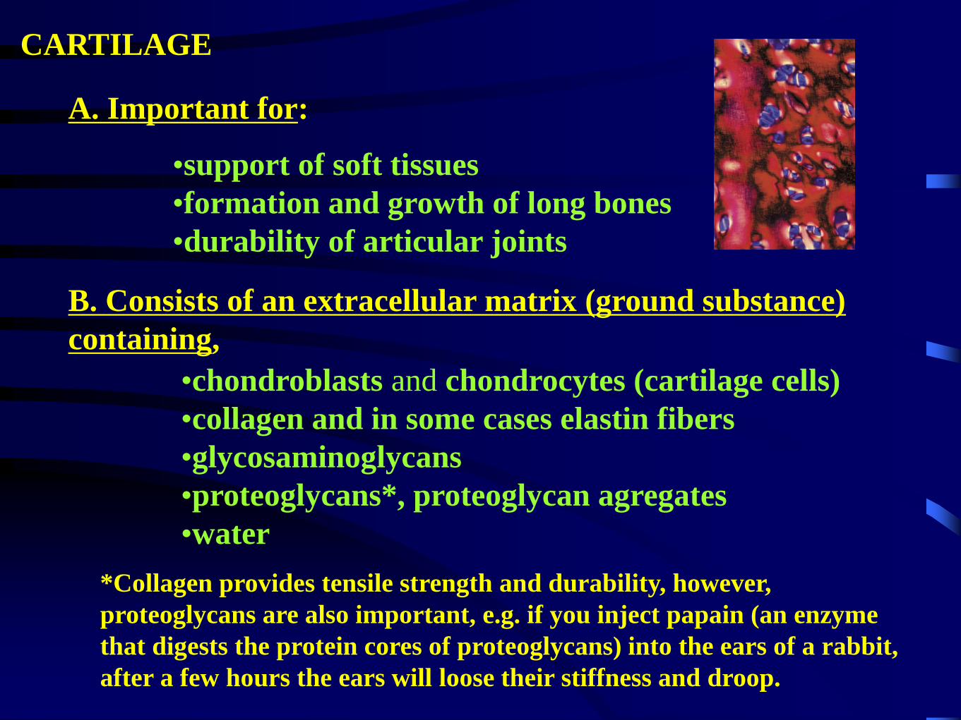
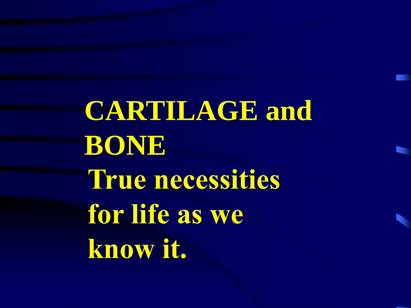
CARTILAGE
A. Important for:
•support of soft tissues
•formation and growth of long bones
•durability of articular joints
B. Consists of an extracellular matrix (ground substance)
containing,
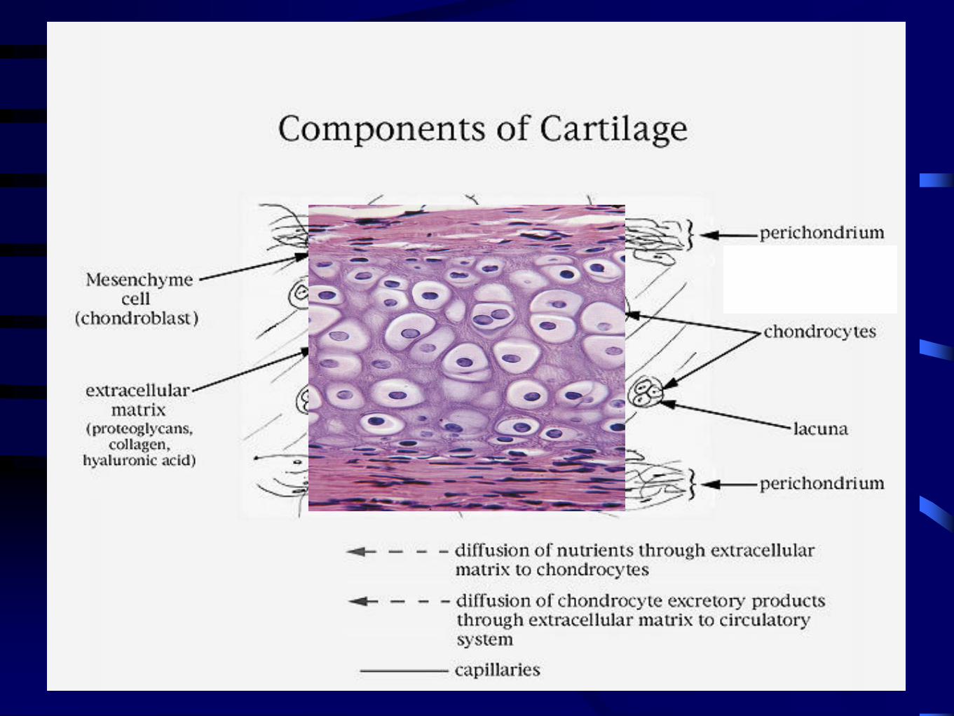
•chondroblasts and chondrocytes (cartilage cells)
•collagen and in some cases elastin fibers
•glycosaminoglycans
•proteoglycans*, proteoglycan agregates
•water
*Collagen provides tensile strength and durability, however,
proteoglycans are also important, e.g. if you inject papain (an enzyme
that digests the protein cores of proteoglycans) into the ears of a rabbit,
after a few hours the ears will loose their stiffness and droop.

C. The qualities of the different types of cartilage depend on,
1. Differences in the type of collagen and concentration of collagen and
elastin fibers in the extracellular matrix
2. The types of proteoglycan molecules that these fibers are associated
with.
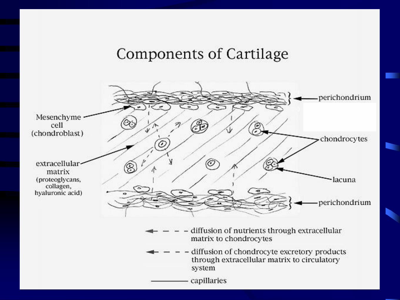
E. The cartilage itself is devoid of blood vessels.
1. Nutrition of cells within the cartilage matrix
is dependent on the diffusion of nutrients
from blood capillaries in perichondrium
and/or adjacent tissues.
D. Hyaline and elastic cartilage are surrounded by a connective tissue capsule
called the perichondrium that is composed of fibroblasts and associated fibers and
ground substance.
Copyright 2000 R. Nims & S.C. Kempf
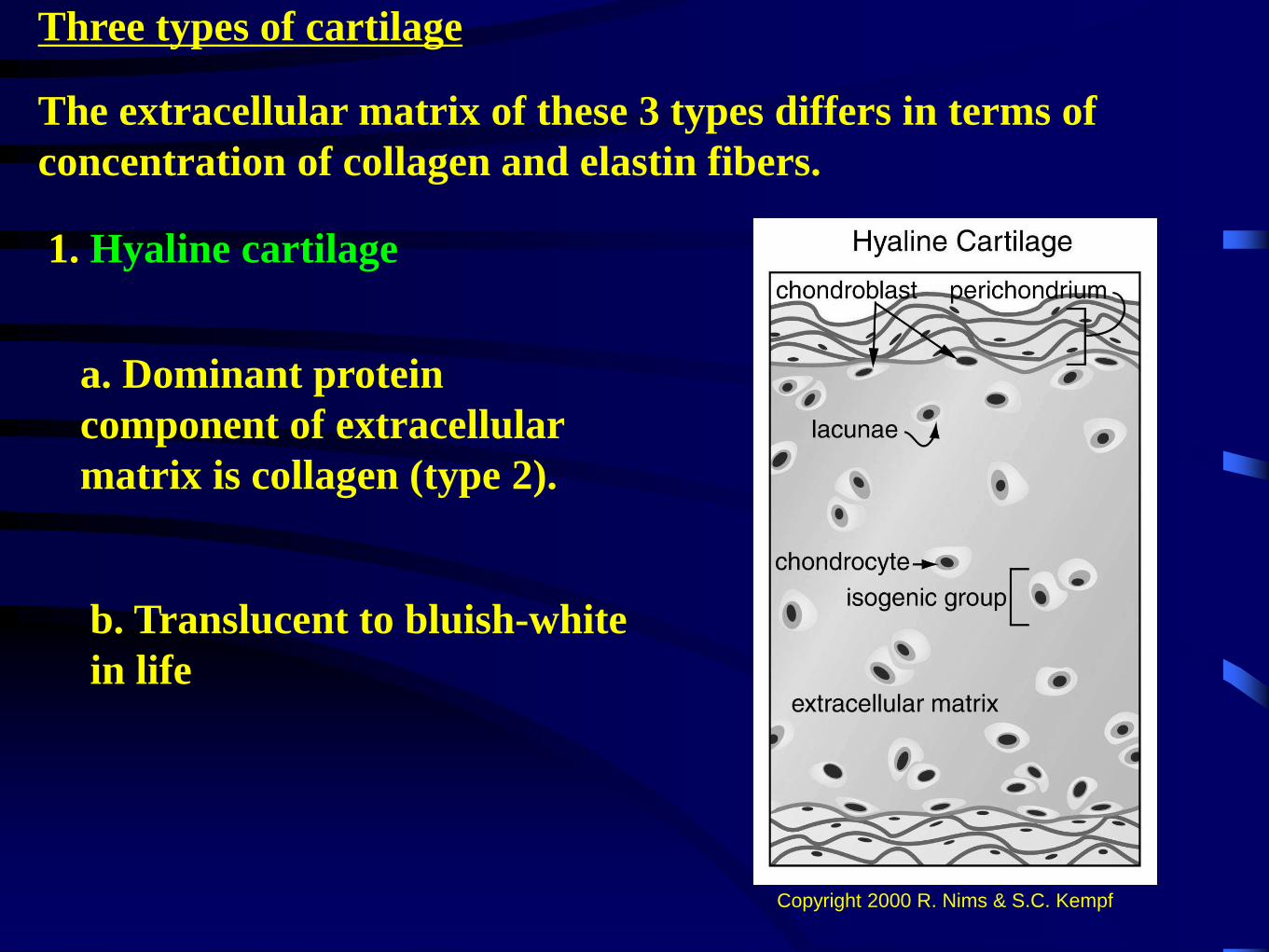
Three types of cartilage
The extracellular matrix of these 3 types differs in terms of
concentration of collagen and elastin fibers.
1. Hyaline cartilage
a. Dominant protein
component of extracellular
matrix is collagen (type 2).
b. Translucent to bluish-white
in life
Copyright 2000 R. Nims & S.C. Kempf
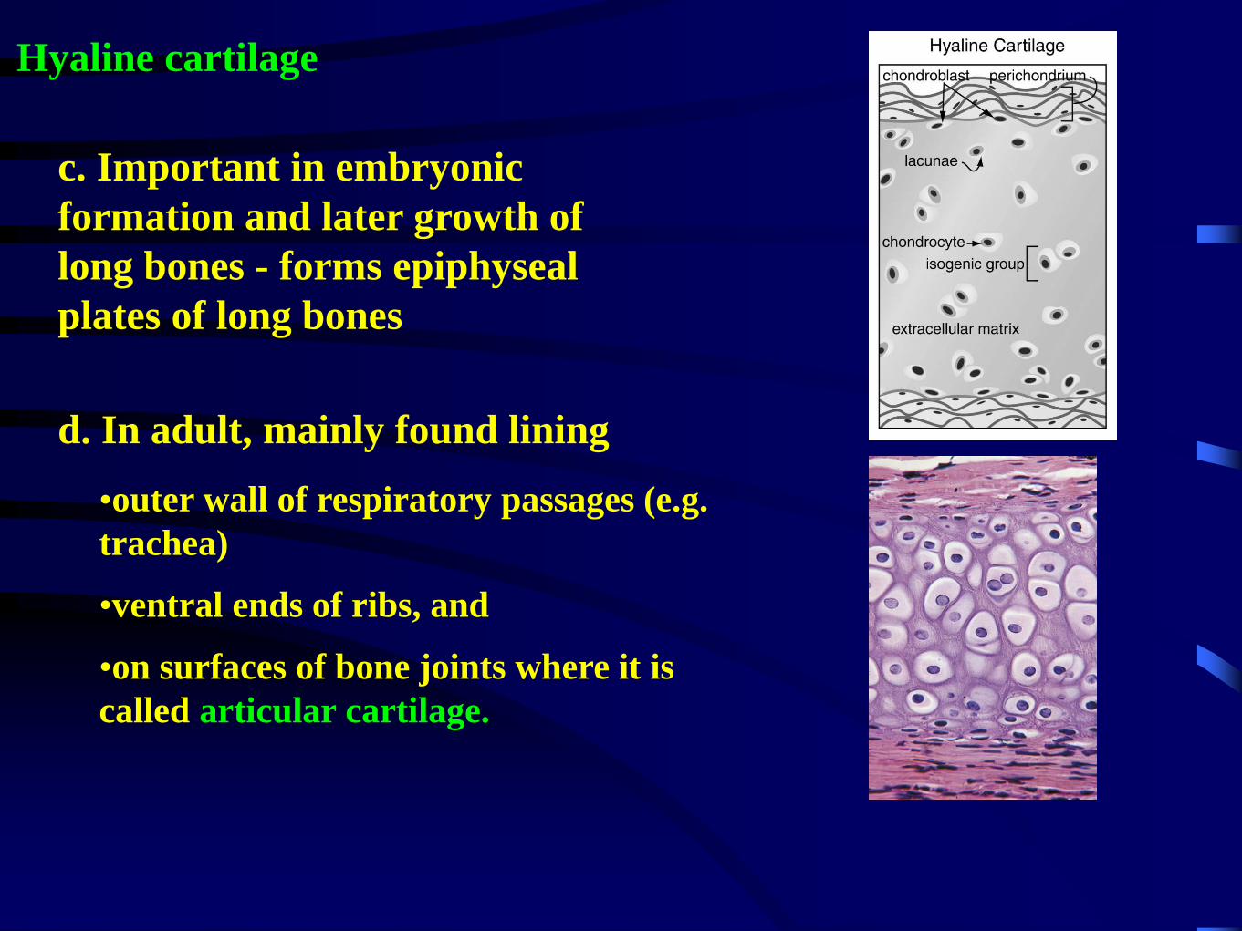
Hyaline cartilage
c. Important in embryonic
formation and later growth of
long bones - forms epiphyseal
plates of long bones
d. In adult, mainly found lining
•outer wall of respiratory passages (e.g.
trachea)
•ventral ends of ribs, and
•on surfaces of bone joints where it is
called articular cartilage.
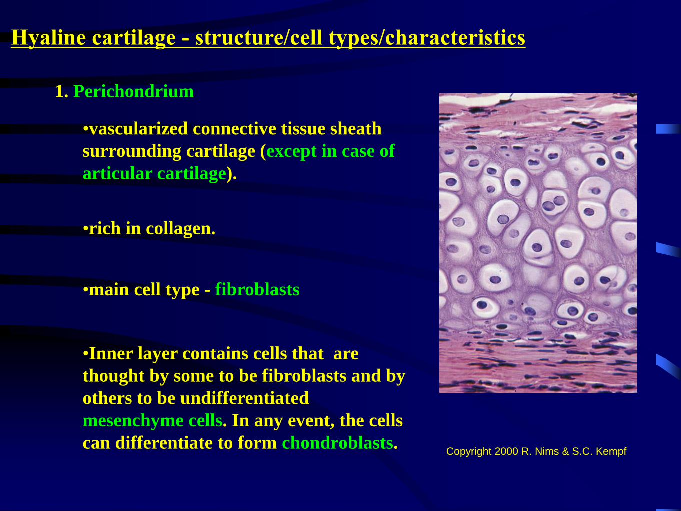
Hyaline cartilage - structure/cell types/characteristics
1. Perichondrium
•vascularized connective tissue sheath
surrounding cartilage (except in case of
articular cartilage).
•rich in collagen.
•main cell type - fibroblasts
•Inner layer contains cells that are
thought by some to be fibroblasts and by
others to be undifferentiated
mesenchyme cells. In any event, the cells
can differentiate to form chondroblasts.Copyright 2000 R. Nims & S.C. Kempf

2. Chondroblasts - immature cartilage
cells. Secrete extracellular matrix, but
are not yet imprisoned in a lacuna.
3. Chondrocytes
•Mature cartilage cells that are embedded
in the extracellular matrix.
•Reside in small spaces within the matrix
that are called lacunae.
•Sometimes form groups of 2 or 3 - isogenic
group
•Chondrocytes have an eliptic shape.
•Organelle systems in cytoplasm are typical
of cells that secrete.
Hyaline cartilage
4. Can undergo calcification (arthritis) and can act as the template for bone
formation and growth.
Copyright 2000 R. Nims & S.C. Kempf
11
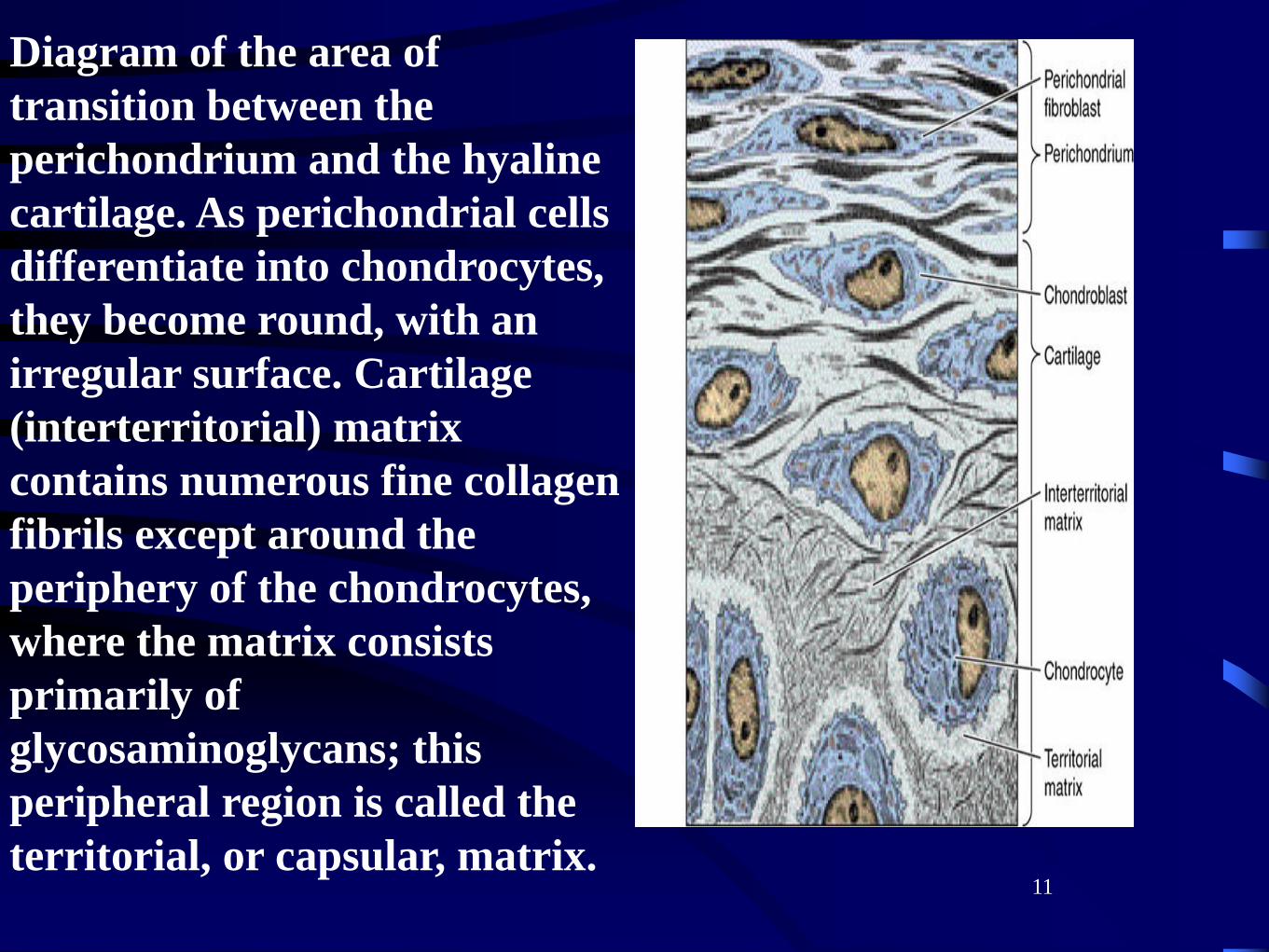
Diagram of the area of
transition between the
perichondrium and the hyaline
cartilage. As perichondrial cells
differentiate into chondrocytes,
they become round, with an
irregular surface. Cartilage
(interterritorial) matrix
contains numerous fine collagen
fibrils except around the
periphery of the chondrocytes,
where the matrix consists
primarily of
glycosaminoglycans; this
peripheral region is called the
territorial, or capsular, matrix.

Three types of cartilage
2. Elastic cartilage
a. High concentration of elastin
fibers in extracellular matrix.
(e.g. external ears-pinna)
e. Does not calcify
b. Ground substance - yellow
in color (due to elastin content)
c. Chondrocytes are more
closely packed, no isogenic
groups.
d. Chondrocytes exhibit less
accumulation of glycogen and
lipids than in hyaline cartilage.
Copyright 2000 R. Nims & S.C. Kempf
fibroblasts
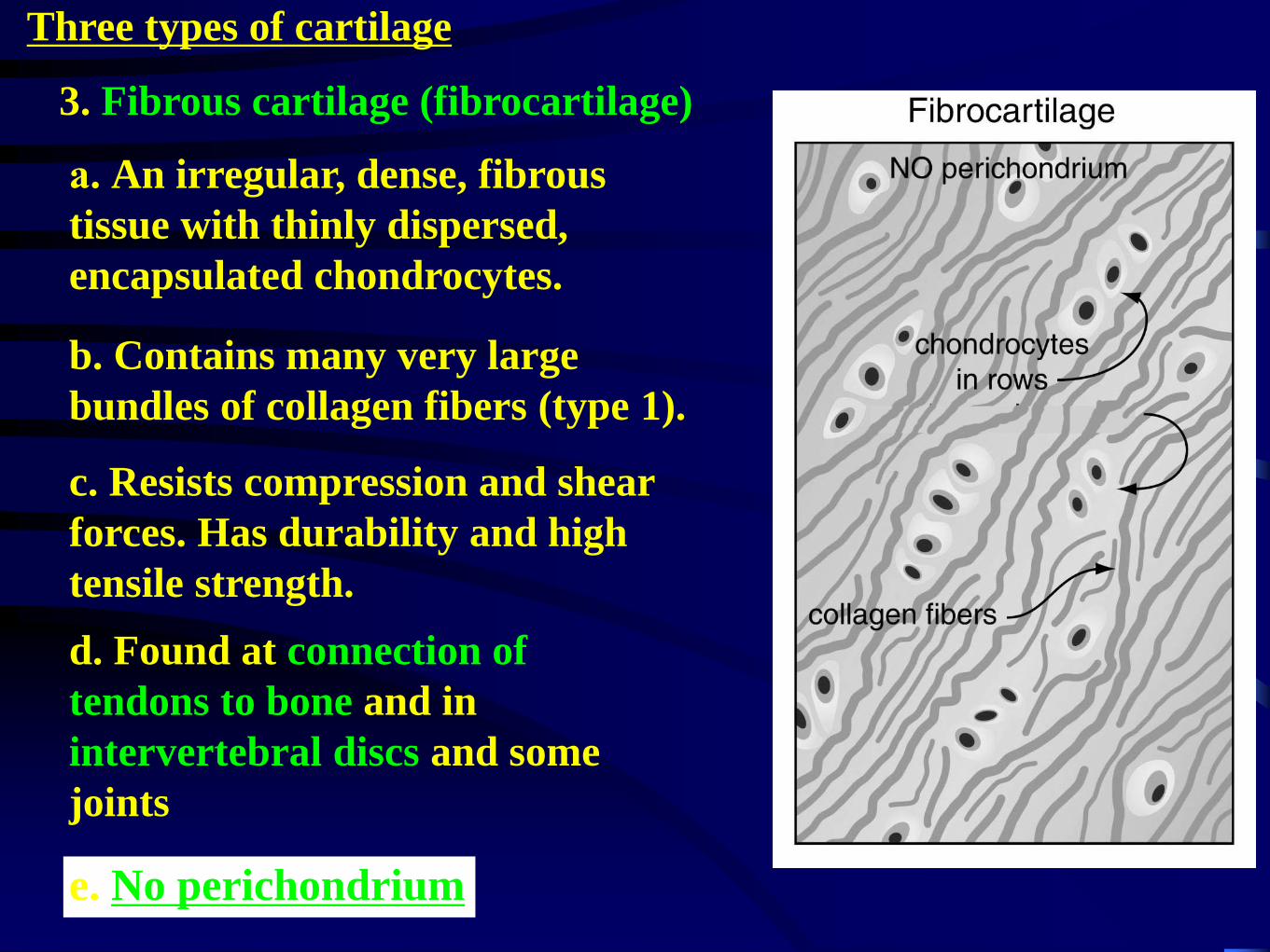
3. Fibrous cartilage (fibrocartilage)
Three types of cartilage
d. Found at connection of
tendons to bone and in
intervertebral discs and some
joints
b. Contains many very large
bundles of collagen fibers (type 1).
c. Resists compression and shear
forces. Has durability and high
tensile strength.
e. No perichondrium
a. An irregular, dense, fibrous
tissue with thinly dispersed,
encapsulated chondrocytes.
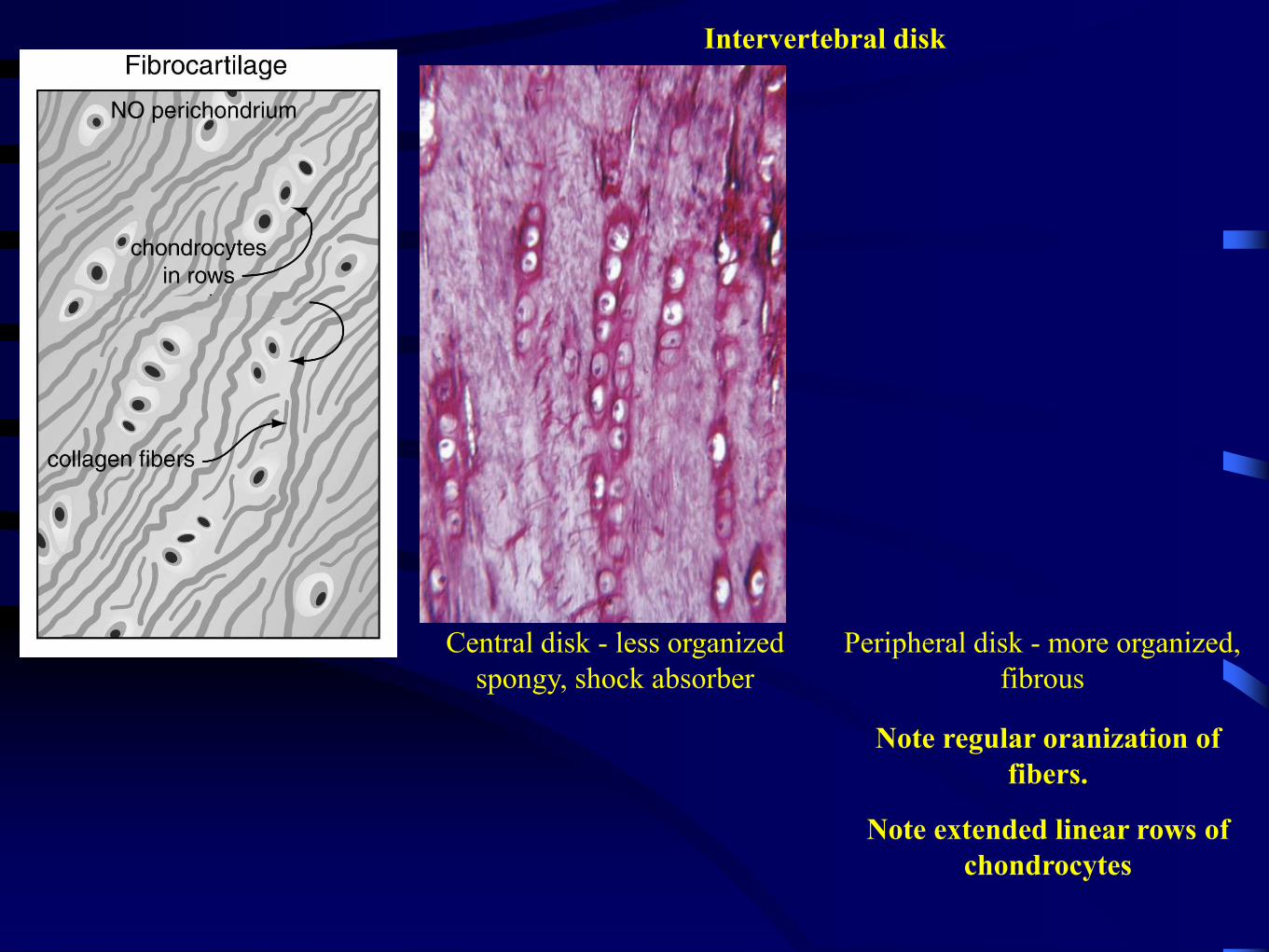
Note regular oranization of
fibers.
Note extended linear rows of
chondrocytes
Central disk - less organized
spongy, shock absorber
Peripheral disk - more organized,
fibrous
Intervertebral disk
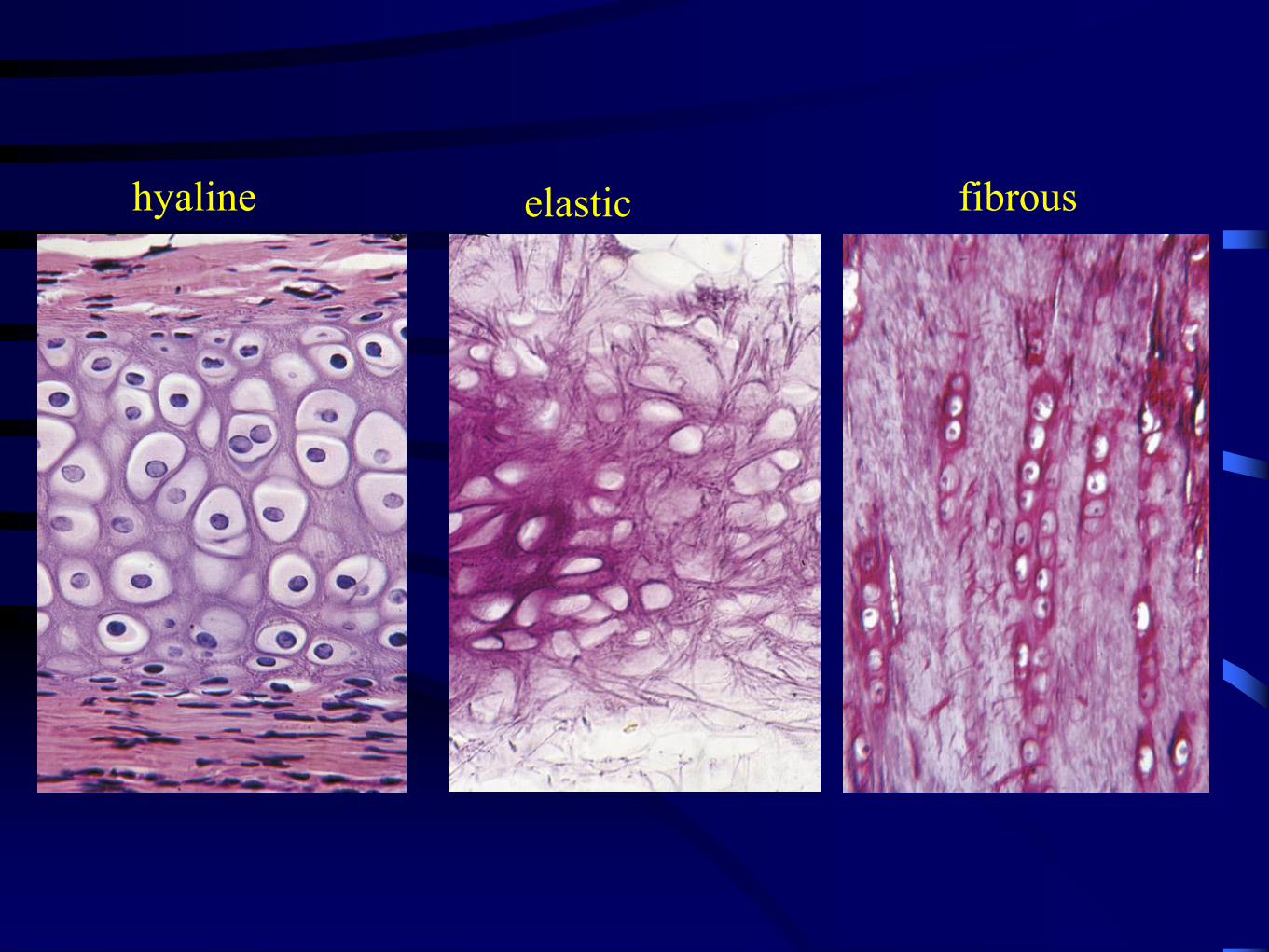
hyaline elastic fibrous
16
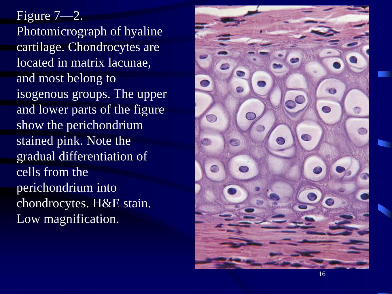
Figure 7—2.
Photomicrograph of hyaline
cartilage. Chondrocytes are
located in matrix lacunae,
and most belong to
isogenous groups. The upper
and lower parts of the figure
show the perichondrium
stained pink. Note the
gradual differentiation of
cells from the
perichondrium into
chondrocytes. H&E stain.
Low magnification.
17
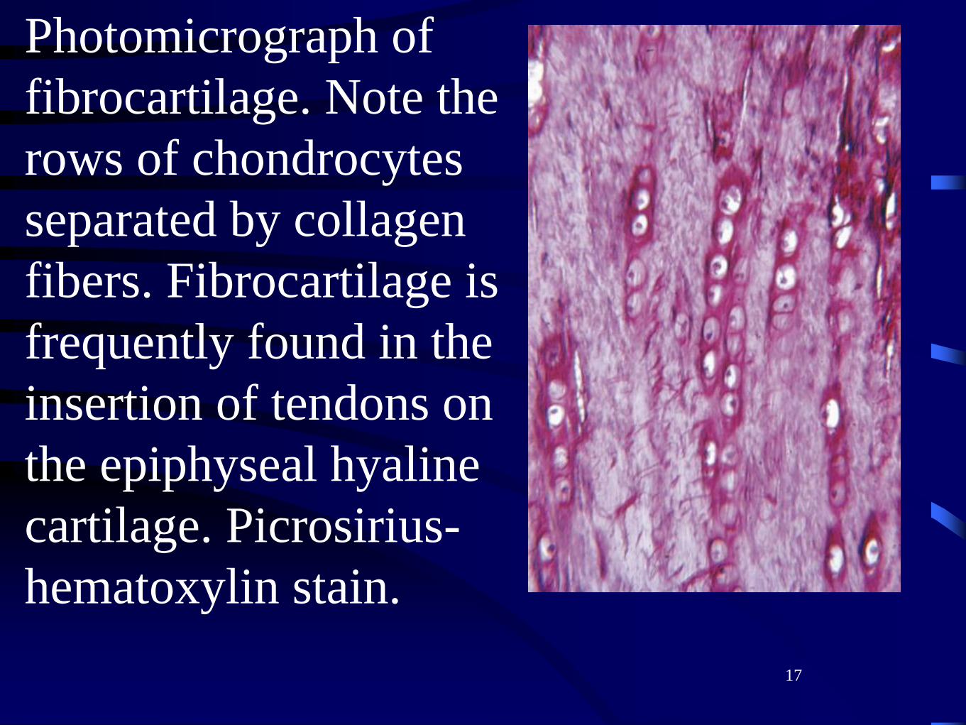
Photomicrograph of
fibrocartilage. Note the
rows of chondrocytes
separated by collagen
fibers. Fibrocartilage is
frequently found in the
insertion of tendons on
the epiphyseal hyaline
cartilage. Picrosirius-
hematoxylin stain.
18
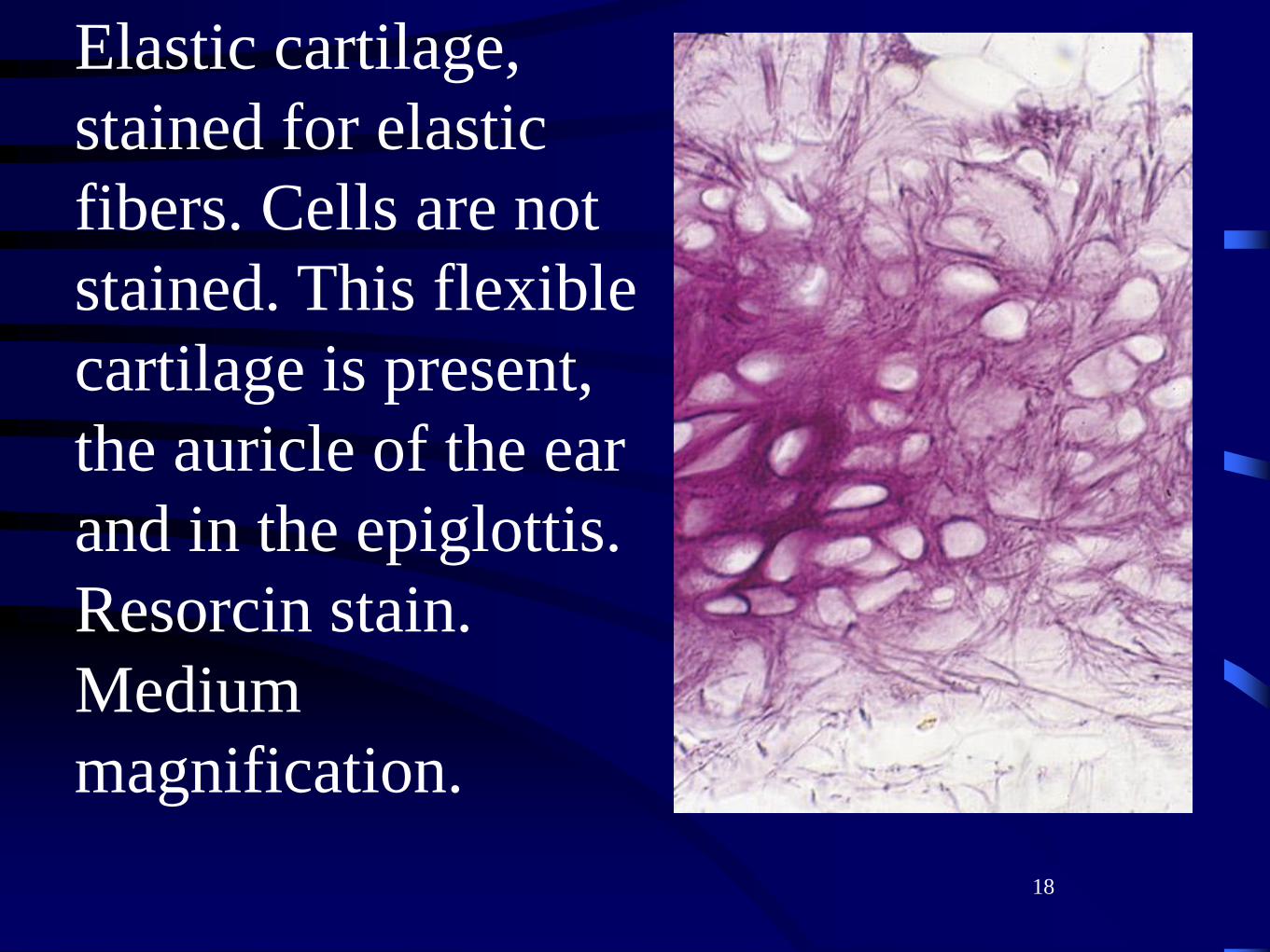
Elastic cartilage,
stained for elastic
fibers. Cells are not
stained. This flexible
cartilage is present,
the auricle of the ear
and in the epiglottis.
Resorcin stain.
Medium
magnification.
In both interstitial and
appositional cartilage
growth,
19
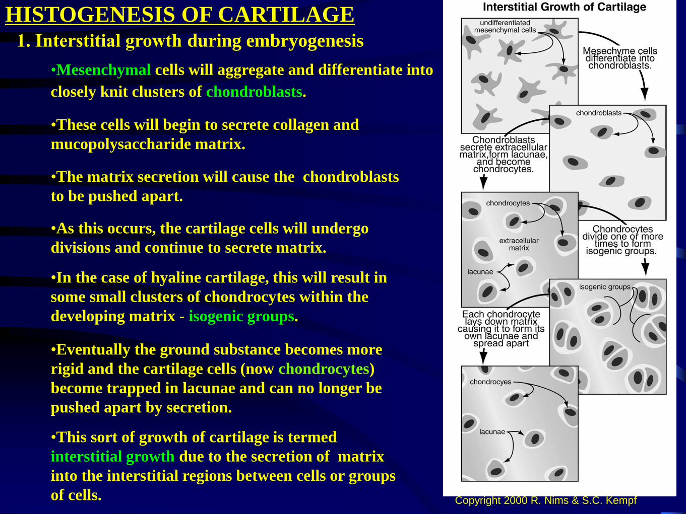
HISTOGENESIS OF CARTILAGE1. Interstitial growth during embryogenesis
Copyright 2000 R. Nims & S.C. Kempf
•As this occurs, the cartilage cells will undergo
divisions and continue to secrete matrix.
•Mesenchymal cells will aggregate and differentiate into
closely knit clusters of chondroblasts.
•These cells will begin to secrete collagen and
mucopolysaccharide matrix.
•The matrix secretion will cause the chondroblasts
to be pushed apart.
•In the case of hyaline cartilage, this will result in
some small clusters of chondrocytes within the
developing matrix - isogenic groups.
•This sort of growth of cartilage is termed
interstitial growth due to the secretion of matrix
into the interstitial regions between cells or groups
of cells.
•Eventually the ground substance becomes more
rigid and the cartilage cells (now chondrocytes)
become trapped in lacunae and can no longer be
pushed apart by secretion.
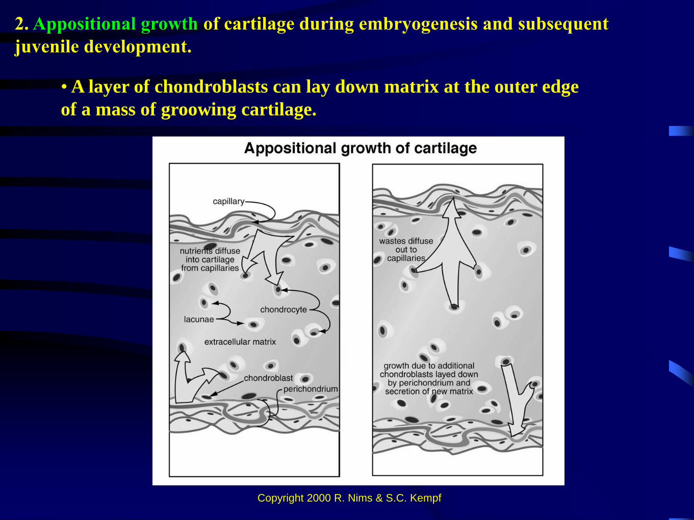
• A layer of chondroblasts can lay down matrix at the outer edge
of a mass of groowing cartilage.
2. Appositional growth of cartilage during embryogenesis and subsequent
juvenile development.
Copyright 2000 R. Nims & S.C. Kempf
•
3. In both interstitial and appositional
cartilage growth,
A: As the cartilage continues to
grow, the central regions become
more rigid due to secretory
products
and the cells in this region take
on the characteristics of mature
chondrocytes.
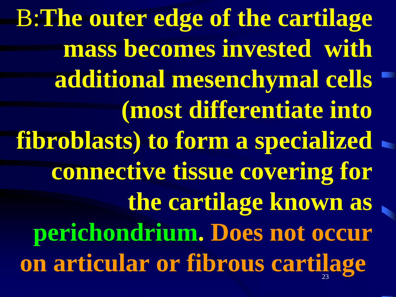
B:The outer edge of the cartilage
mass becomes invested with
additional mesenchymal cells
(most differentiate into
fibroblasts) to form a specialized
connective tissue covering for
the cartilage known as
perichondrium. Does not occur
on articular or fibrous cartilage23
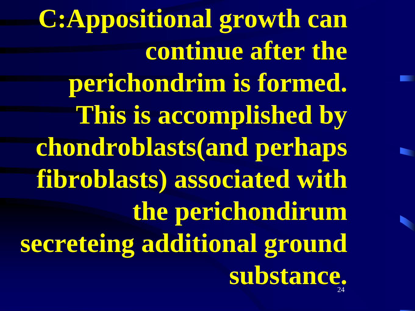
C:Appositional growth can
continue after the
perichondrim is formed.
This is accomplished by
chondroblasts(and perhaps
fibroblasts) associated with
the perichondirum
secreteing additional ground
substance.24
25
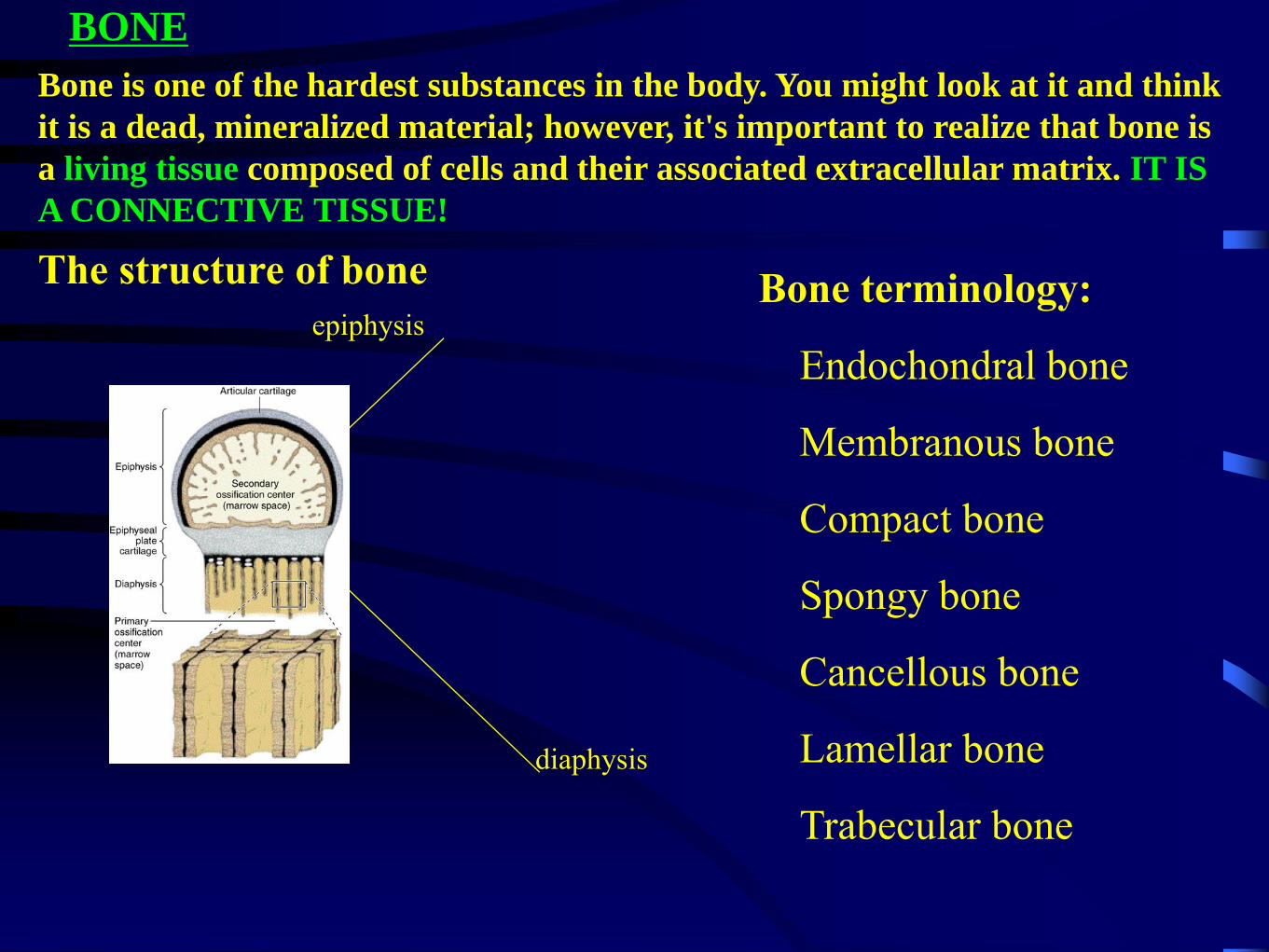
BONE
Bone is one of the hardest substances in the body. You might look at it and think
it is a dead, mineralized material; however, it's important to realize that bone is
a living tissue composed of cells and their associated extracellular matrix. IT IS
A CONNECTIVE TISSUE!
The structure of bone
epiphysis
diaphysis
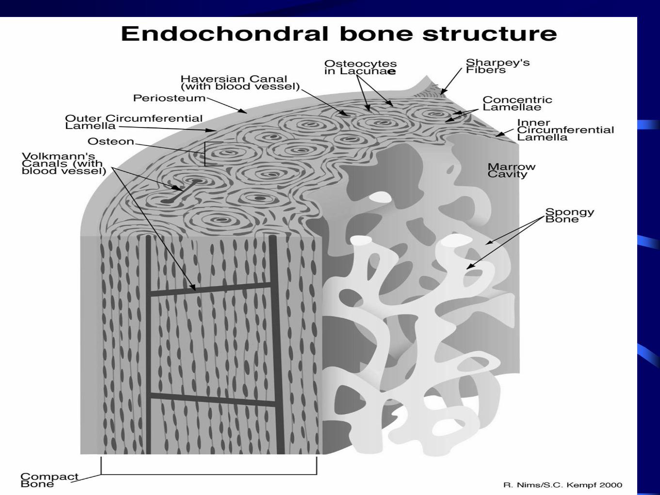
Endochondral bone
Membranous bone
Compact bone
Spongy bone
Cancellous bone
Lamellar bone
Trabecular bone
Bone terminology:
27
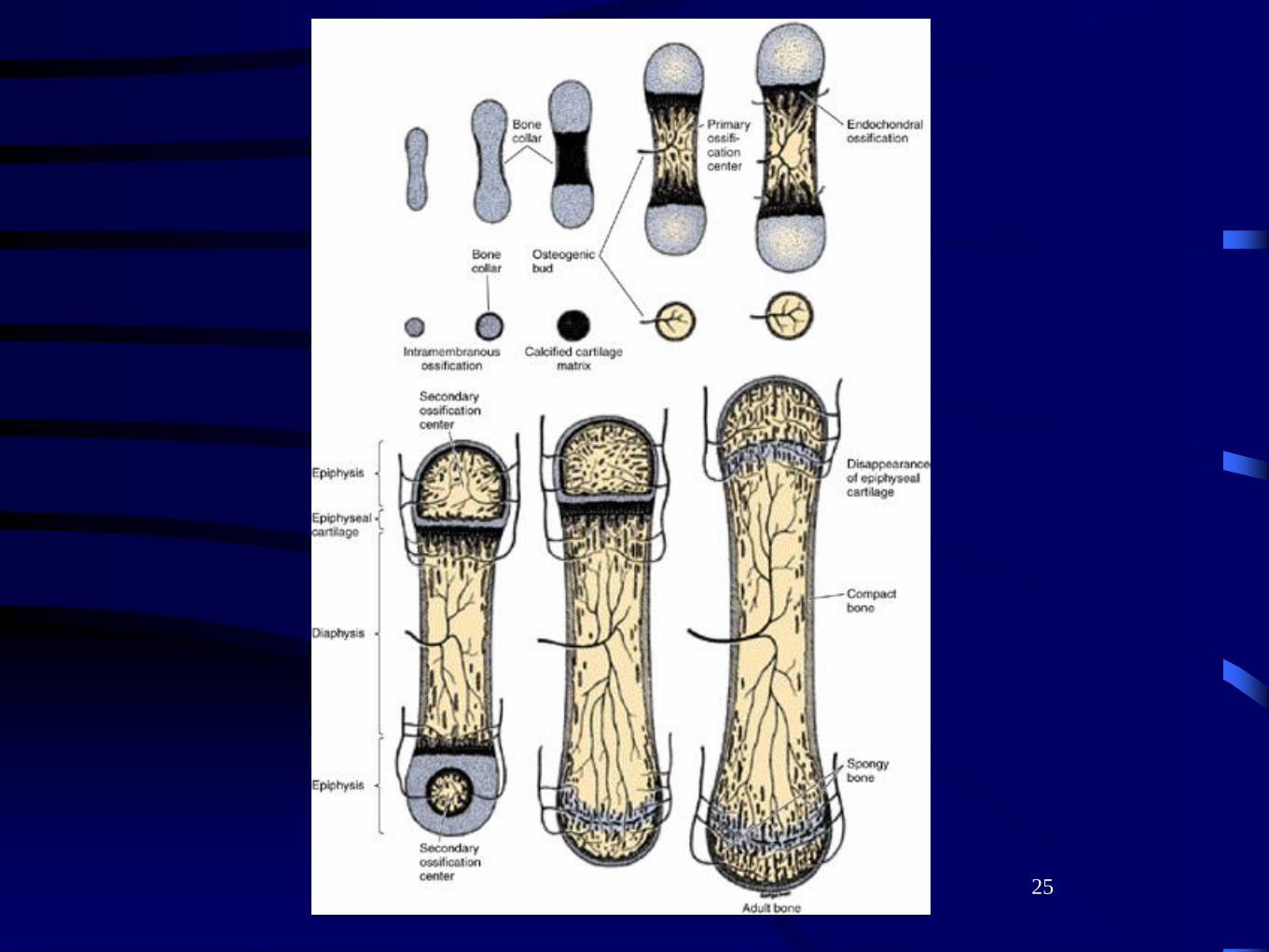
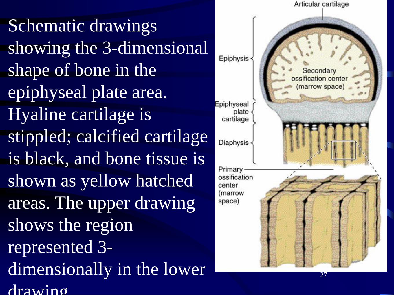
Schematic drawings
showing the 3-dimensional
shape of bone in the
epiphyseal plate area.
Hyaline cartilage is
stippled; calcified cartilage
is black, and bone tissue is
shown as yellow hatched
areas. The upper drawing
shows the region
represented 3-
dimensionally in the lower
drawing.
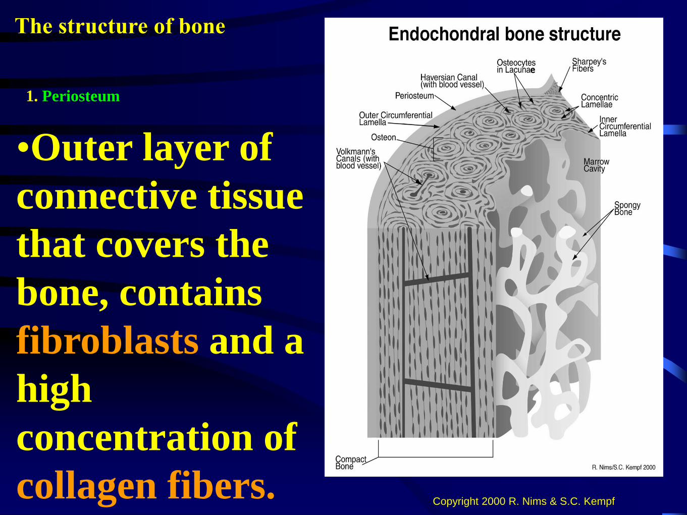
The structure of bone
1. Periosteum
Copyright 2000 R. Nims & S.C. Kempf
•Outer layer of
connective tissue
that covers the
bone, contains
fibroblasts and a
high
concentration of
collagen fibers.
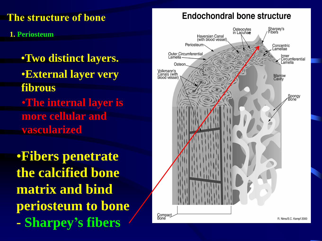
•Two distinct layers.
•External layer very
fibrous
•The internal layer is
more cellular and
vascularized
•Fibers penetrate
the calcified bone
matrix and bind
periosteum to bone
- Sharpey’s fibers
The structure of bone
1. Periosteum
30
•The inner layer
provides cells for
bone histogenesis
during development
and in the healing of
fractures.
31
32
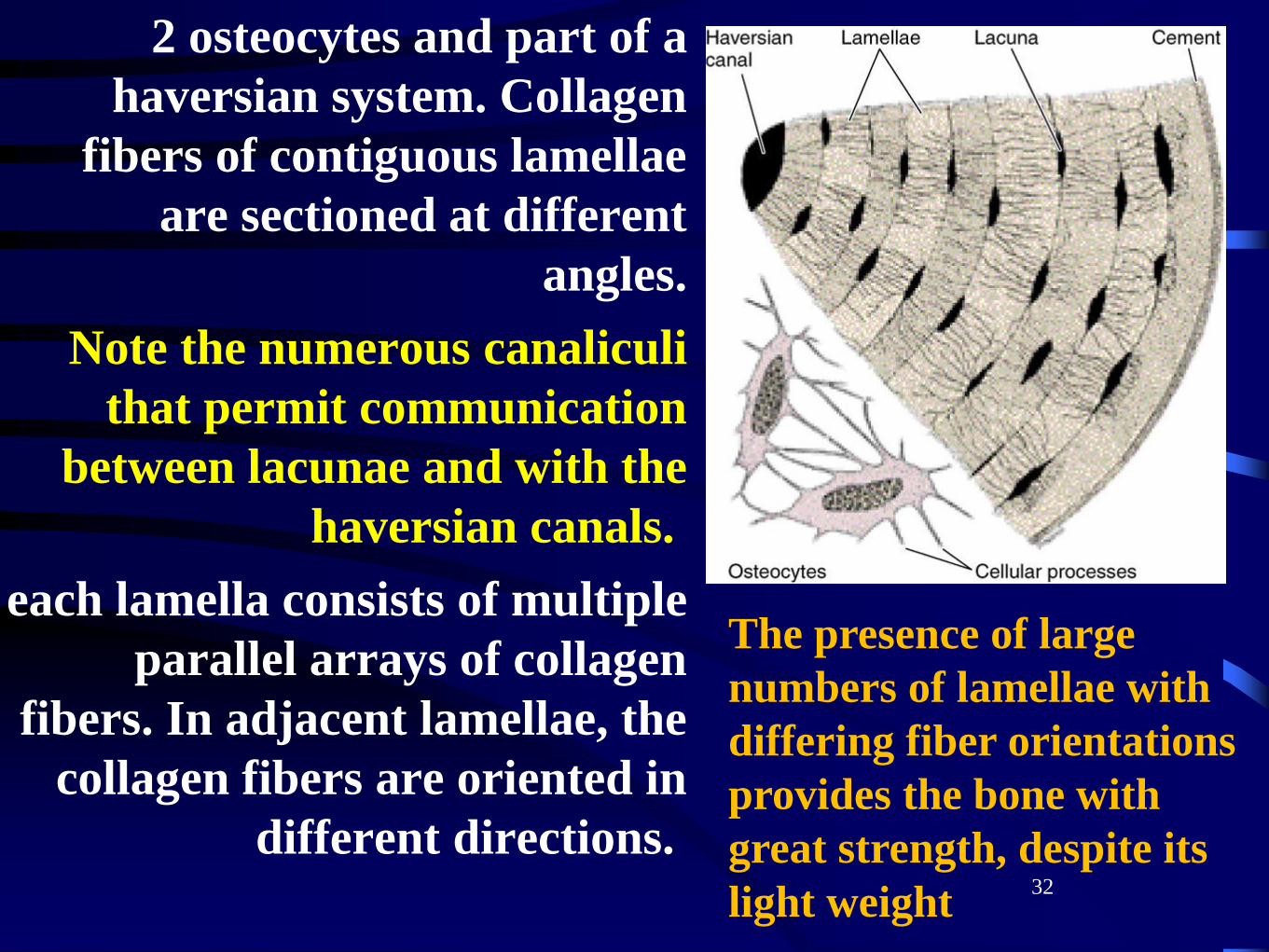
2 osteocytes and part of a
haversian system. Collagen
fibers of contiguous lamellae
are sectioned at different
angles.
Note the numerous canaliculi
that permit communication
between lacunae and with the
haversian canals.
each lamella consists of multiple
parallel arrays of collagen
fibers. In adjacent lamellae, the
collagen fibers are oriented in
different directions.
The presence of large
numbers of lamellae with
differing fiber orientations
provides the bone with
great strength, despite its
light weight
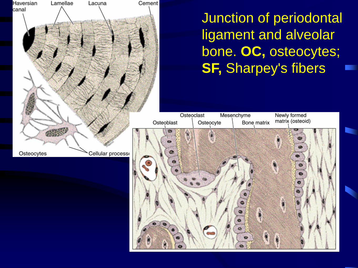
Junction of periodontal
ligament and alveolar
bone. OC, osteocytes;
SF, Sharpey's fibers
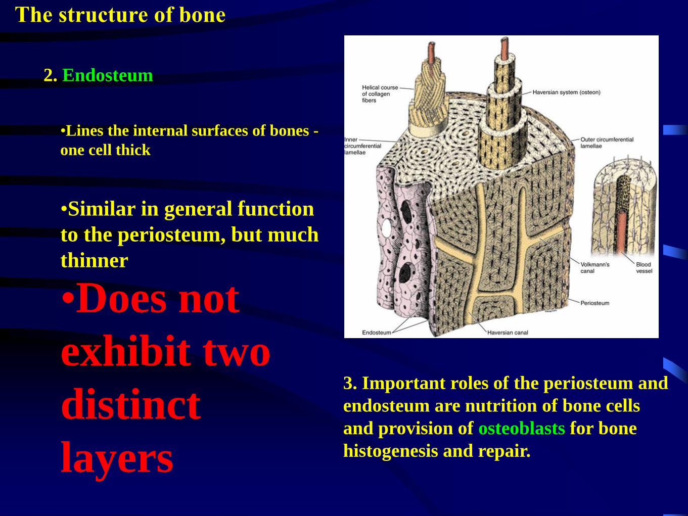
2. Endosteum
The structure of bone
•Lines the internal surfaces of bones -
one cell thick
•Similar in general function
to the periosteum, but much
thinner
•Does not
exhibit two
distinct
layers
http://www.medsch.wisc.edu/anatomy/histo/res/l/ct/ct44.jpg
3. Important roles of the periosteum and
endosteum are nutrition of bone cells
and provision of osteoblasts for bone
histogenesis and repair.
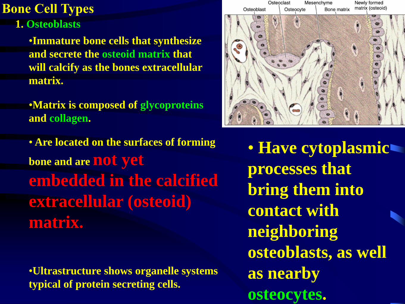
Bone Cell Types1. Osteoblasts
•Immature bone cells that synthesize
and secrete the osteoid matrix that
will calcify as the bones extracellular
matrix.
•Matrix is composed of glycoproteins
and collagen.
• Are located on the surfaces of forming
bone and are not yet
embedded in the calcified
extracellular (osteoid)
matrix.
•Ultrastructure shows organelle systems
typical of protein secreting cells.
• Have cytoplasmic
processes that
bring them into
contact with
neighboring
osteoblasts, as well
as nearby
osteocytes.

36
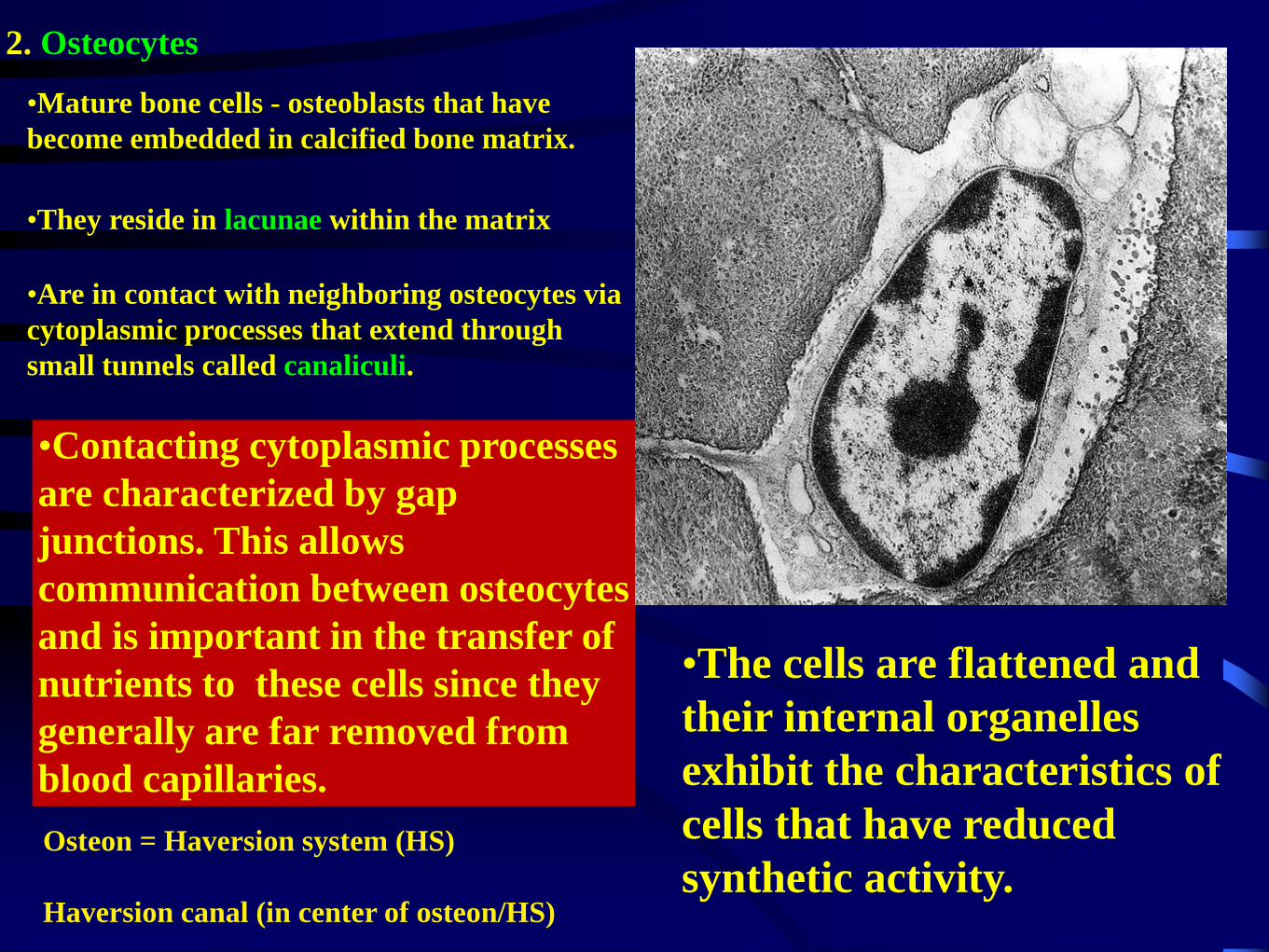
2. Osteocytes
•Mature bone cells - osteoblasts that have
become embedded in calcified bone matrix.
•They reside in lacunae within the matrix
•Are in contact with neighboring osteocytes via
cytoplasmic processes that extend through
small tunnels called canaliculi.
•Contacting cytoplasmic processes
are characterized by gap
junctions. This allows
communication between osteocytes
and is important in the transfer of
nutrients to these cells since they
generally are far removed from
blood capillaries.
•The cells are flattened and
their internal organelles
exhibit the characteristics of
cells that have reduced
synthetic activity.Osteon = Haversion system (HS)
Haversion canal (in center of osteon/HS)
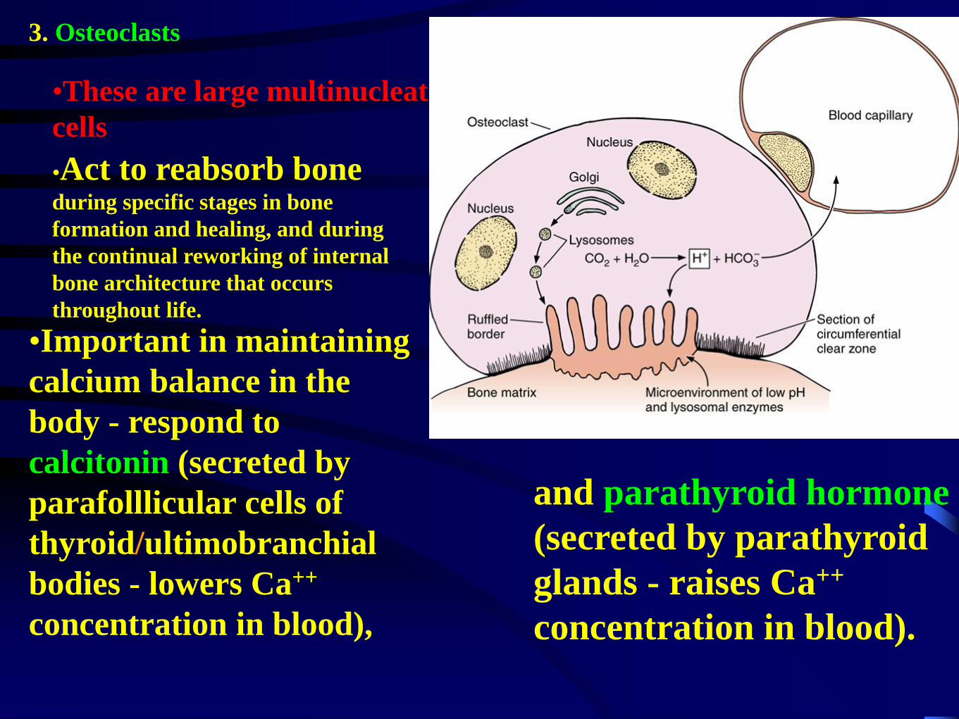
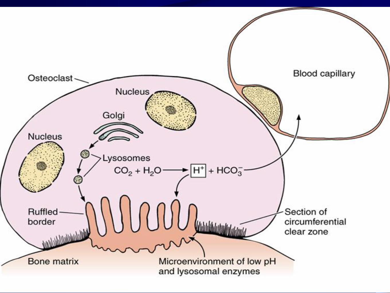
3. Osteoclasts
•These are large multinucleate
cells
•Act to reabsorb bone during specific stages in bone
formation and healing, and during
the continual reworking of internal
bone architecture that occurs
throughout life.
•Important in maintaining
calcium balance in the
body - respond to
calcitonin (secreted by
parafolllicular cells of
thyroid/ultimobranchial
bodies - lowers Ca++
concentration in blood),
and parathyroid hormone
(secreted by parathyroid
glands - raises Ca++
concentration in blood).
39
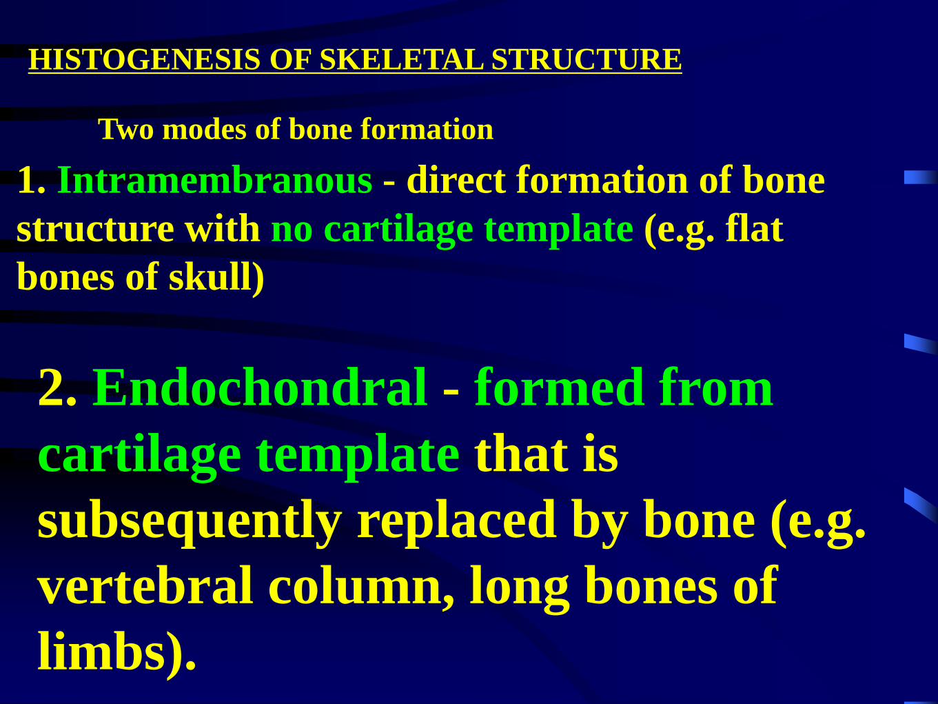
HISTOGENESIS OF SKELETAL STRUCTURE
Two modes of bone formation
2. Endochondral - formed from
cartilage template that is
subsequently replaced by bone (e.g.
vertebral column, long bones of
limbs).
1. Intramembranous - direct formation of bone
structure with no cartilage template (e.g. flat
bones of skull)
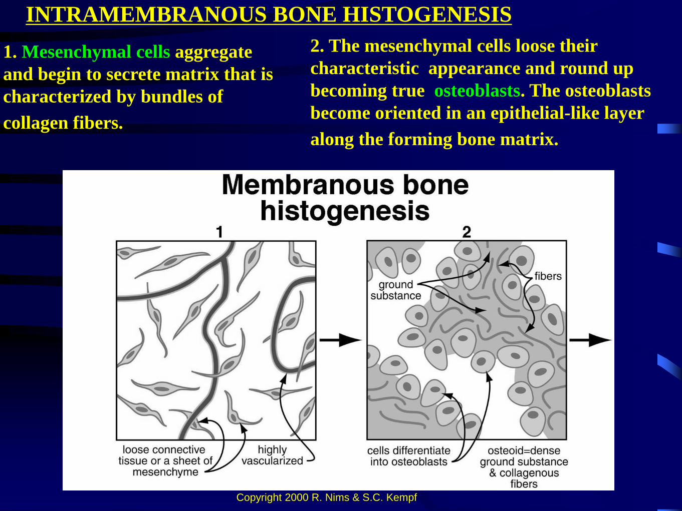
INTRAMEMBRANOUS BONE HISTOGENESIS
1. Mesenchymal cells aggregate
and begin to secrete matrix that is
characterized by bundles of
collagen fibers.
2. The mesenchymal cells loose their
characteristic appearance and round up
becoming true osteoblasts. The osteoblasts
become oriented in an epithelial-like layer
along the forming bone matrix.
Copyright 2000 R. Nims & S.C. Kempf
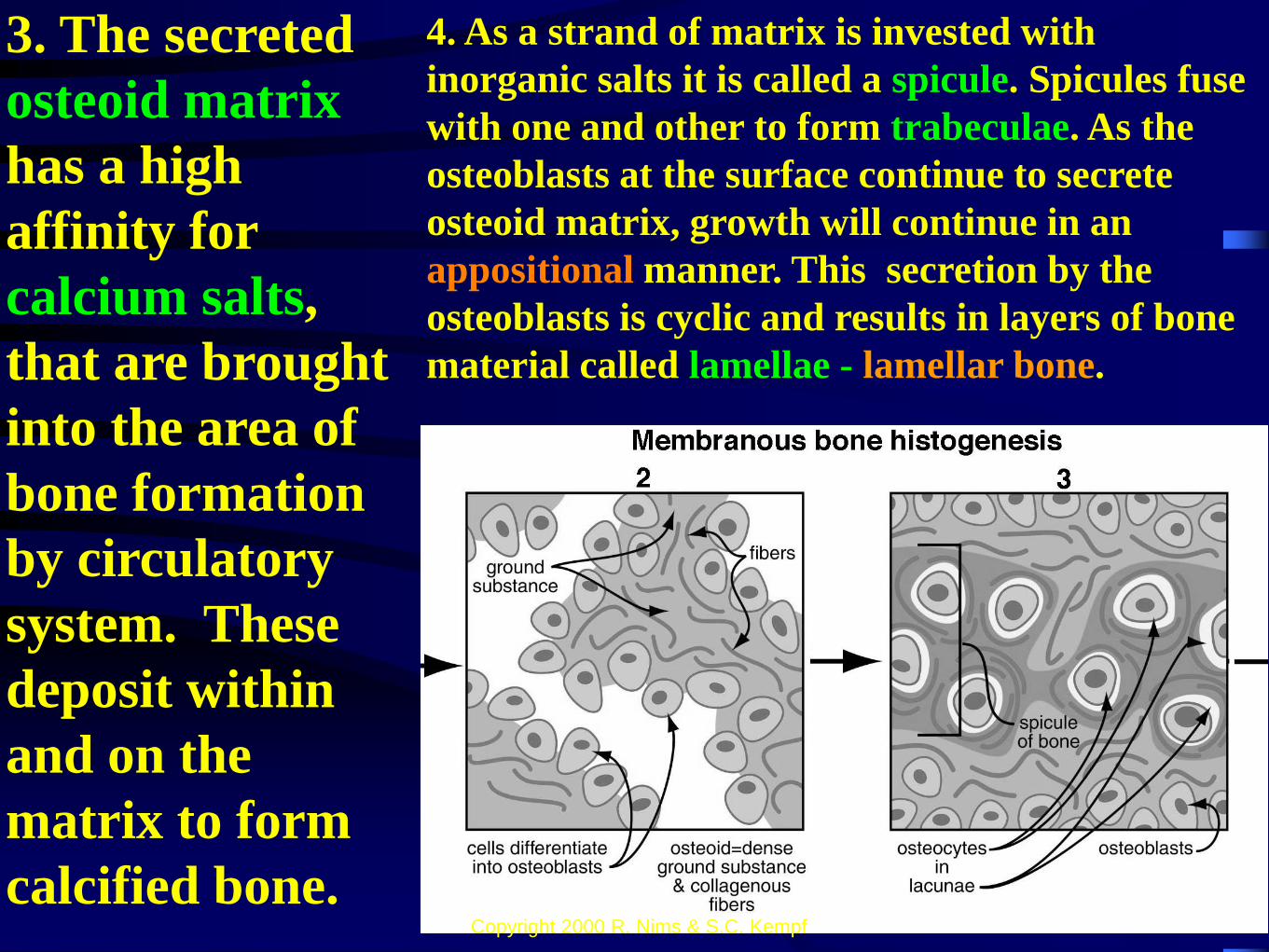
3. The secreted
osteoid matrix
has a high
affinity for
calcium salts,
that are brought
into the area of
bone formation
by circulatory
system. These
deposit within
and on the
matrix to form
calcified bone.
4. As a strand of matrix is invested with
inorganic salts it is called a spicule. Spicules fuse
with one and other to form trabeculae. As the
osteoblasts at the surface continue to secrete
osteoid matrix, growth will continue in an
appositional manner. This secretion by the
osteoblasts is cyclic and results in layers of bone
material called lamellae - lamellar bone.
Copyright 2000 R. Nims & S.C. Kempf
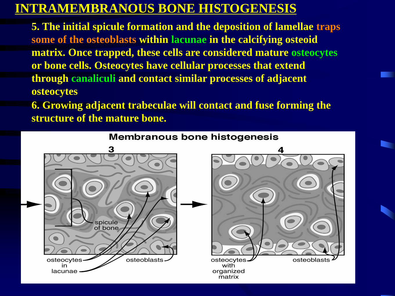
5. The initial spicule formation and the deposition of lamellae traps
some of the osteoblasts within lacunae in the calcifying osteoid
matrix. Once trapped, these cells are considered mature osteocytes
or bone cells. Osteocytes have cellular processes that extend
through canaliculi and contact similar processes of adjacent
osteocytes
INTRAMEMBRANOUS BONE HISTOGENESIS
6. Growing adjacent trabeculae will contact and fuse forming the
structure of the mature bone.
ENDOCHONDRAL BONE - what
is it?
2. Most of the bones in the mammalian body
are initially formed by endochondral means.
1. A template of hyaline cartilage is
layed down prior to calcification and
the establishment of true bone. During
fetal development, this template is
shaped like a miniature copy of the
bone that will form from it.
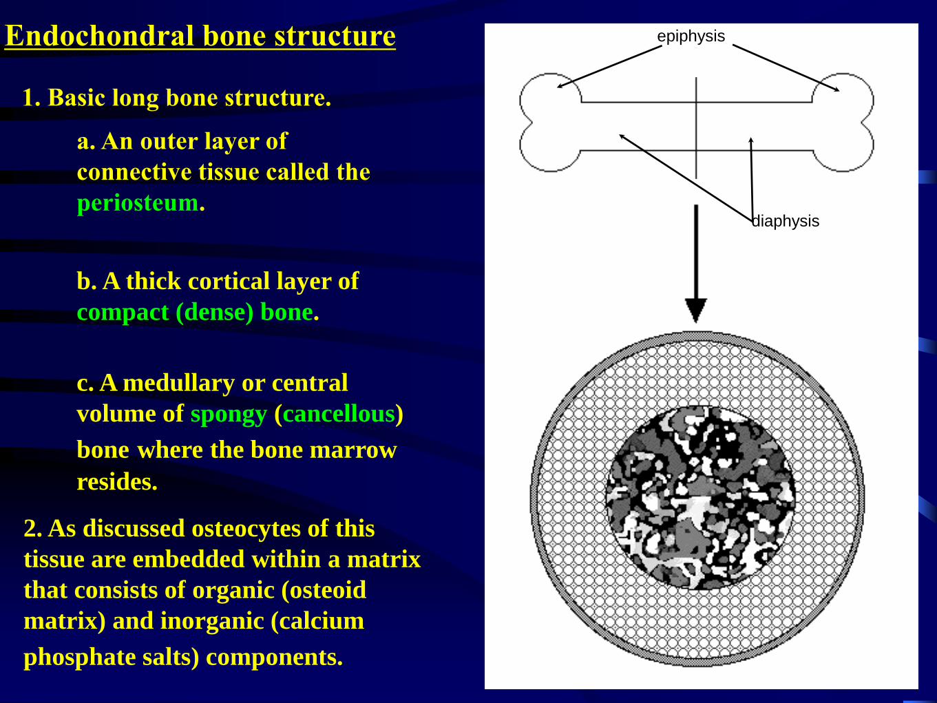
Endochondral bone structure
1. Basic long bone structure.
c. A medullary or central
volume of spongy (cancellous)
bone where the bone marrow
resides.
b. A thick cortical layer of
compact (dense) bone.
a. An outer layer of
connective tissue called the
periosteum.
2. As discussed osteocytes of this
tissue are embedded within a matrix
that consists of organic (osteoid
matrix) and inorganic (calcium
phosphate salts) components.
epiphysis
diaphysis
Endochondral bone structure
1. Periosteal lamellae - layers secreted, as the bone
grew, by osteoblasts associated with the inner side of
the periosteum - appositional growth - circumferential
lamellae - lamellar bone. and
2.Inner component consisting of multiple
osteons and interstitial bone
Compact bone - 2 regions
a. concentric sub-layers (lamellar bone)
surrounding longitudinal tunnels for blood
vessels and nerves that are called the
haversian canals
Osteons:
Gross long bone
structure.
ENDOCHONDRAL BONE HISTOGENESIS
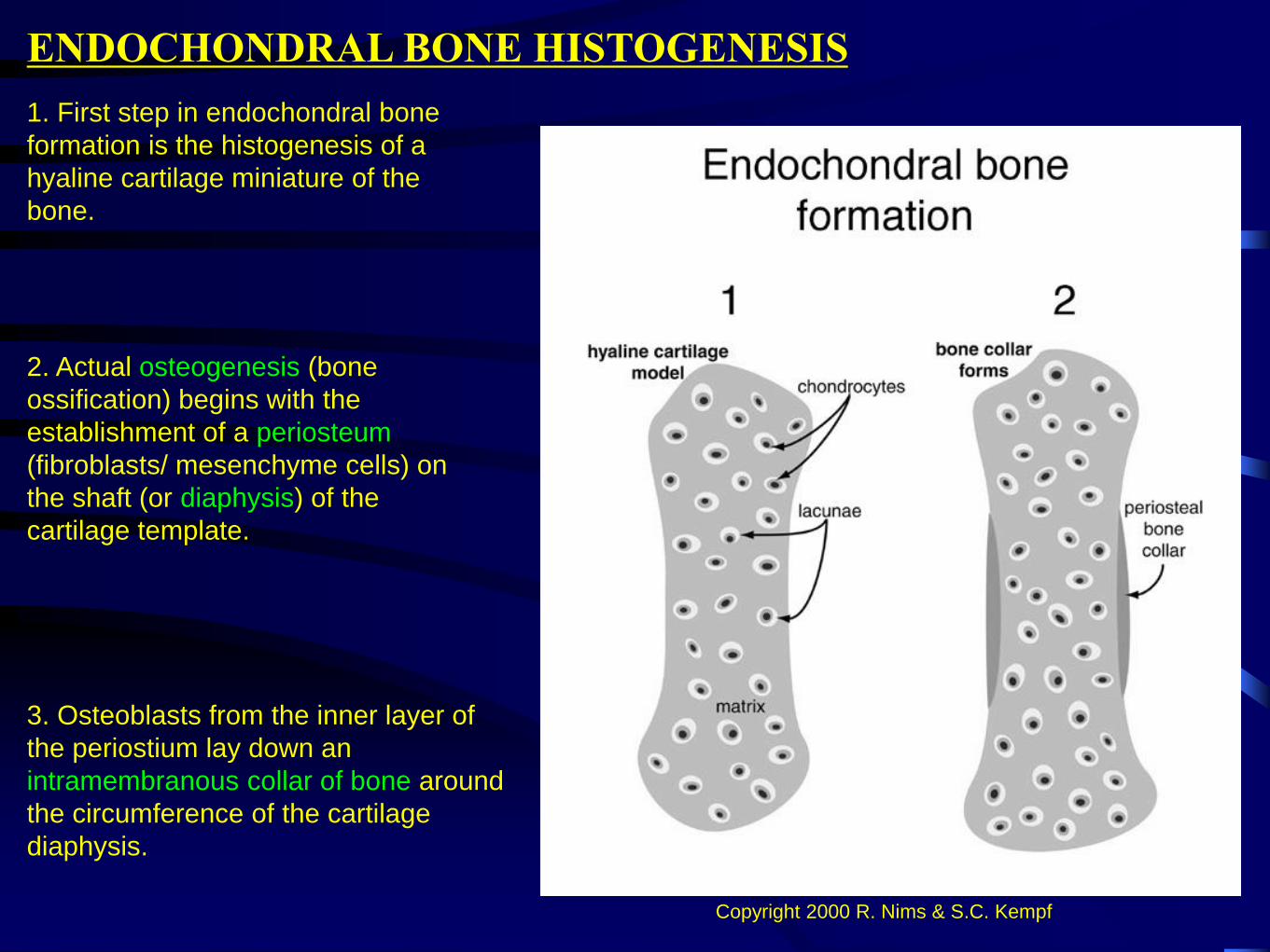
1. First step in endochondral bone
formation is the histogenesis of a
hyaline cartilage miniature of the
bone.
2. Actual osteogenesis (bone
ossification) begins with the
establishment of a periosteum
(fibroblasts/ mesenchyme cells) on
the shaft (or diaphysis) of the
cartilage template.
3. Osteoblasts from the inner layer of
the periostium lay down an
intramembranous collar of bone around
the circumference of the cartilage
diaphysis.
Copyright 2000 R. Nims & S.C. Kempf
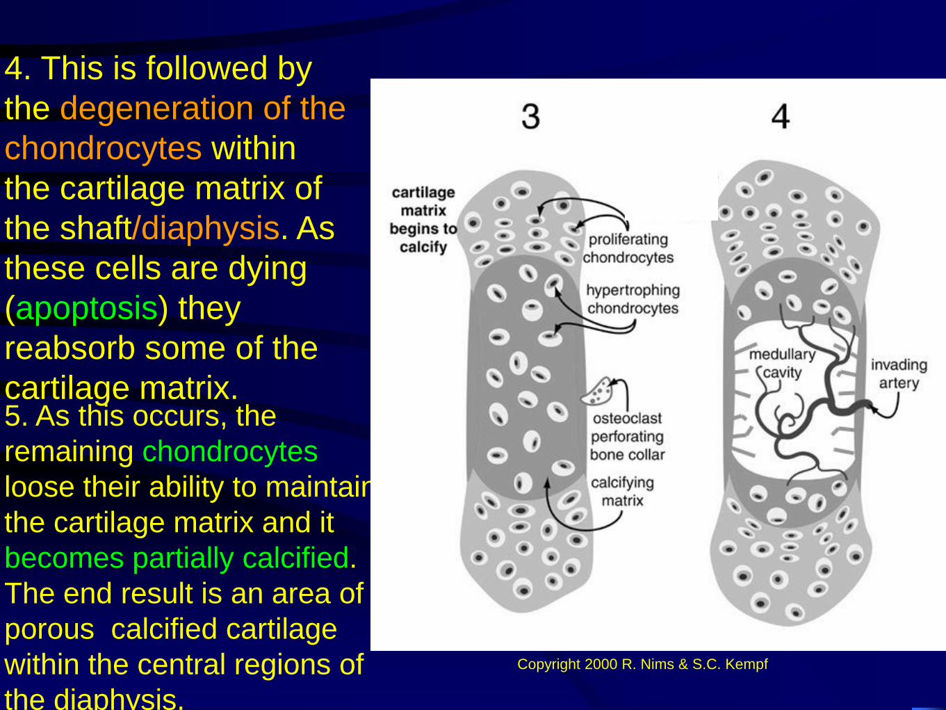
4. This is followed by
the degeneration of the
chondrocytes within
the cartilage matrix of
the shaft/diaphysis. As
these cells are dying
(apoptosis) they
reabsorb some of the
cartilage matrix.5. As this occurs, the
remaining chondrocytes
loose their ability to maintain
the cartilage matrix and it
becomes partially calcified.
The end result is an area of
porous calcified cartilage
within the central regions of
the diaphysis.
Copyright 2000 R. Nims & S.C. Kempf
ENDOCHONDRAL BONE HISTOGENESIS
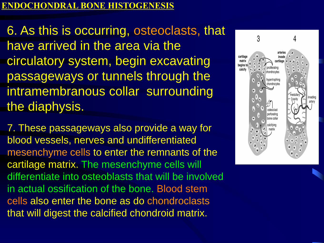
6. As this is occurring, osteoclasts, that
have arrived in the area via the
circulatory system, begin excavating
passageways or tunnels through the
intramembranous collar surrounding
the diaphysis.
7. These passageways also provide a way for
blood vessels, nerves and undifferentiated
mesenchyme cells to enter the remnants of the
cartilage matrix. The mesenchyme cells will
differentiate into osteoblasts that will be involved
in actual ossification of the bone. Blood stem
cells also enter the bone as do chondroclasts
that will digest the calcified chondroid matrix.
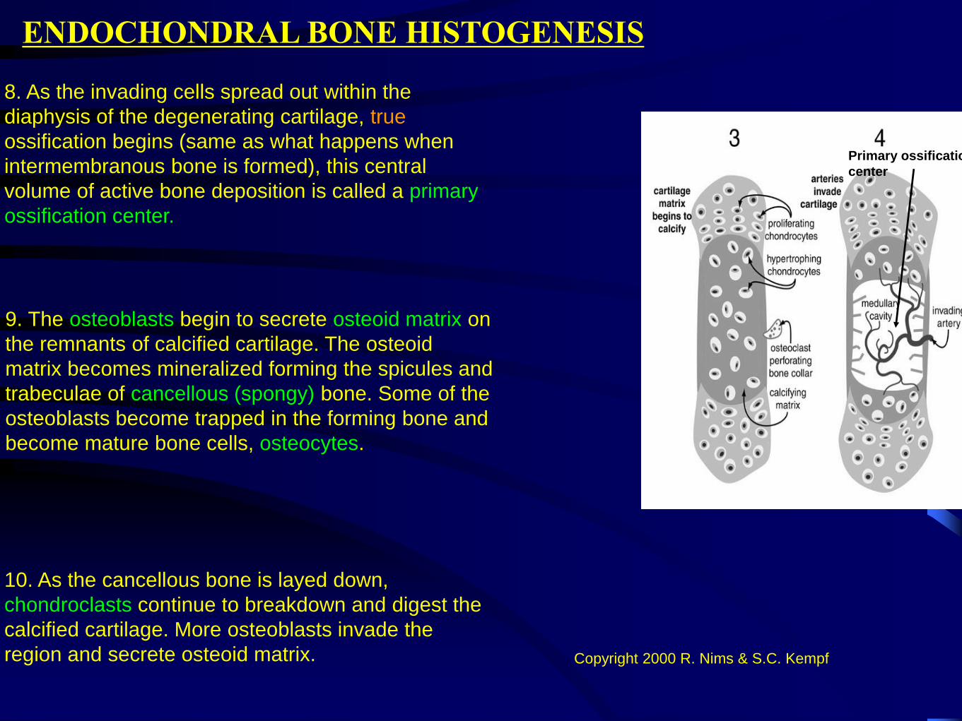
8. As the invading cells spread out within the
diaphysis of the degenerating cartilage, true
ossification begins (same as what happens when
intermembranous bone is formed), this central
volume of active bone deposition is called a primary
ossification center.
ENDOCHONDRAL BONE HISTOGENESIS
9. The osteoblasts begin to secrete osteoid matrix on
the remnants of calcified cartilage. The osteoid
matrix becomes mineralized forming the spicules and
trabeculae of cancellous (spongy) bone. Some of the
osteoblasts become trapped in the forming bone and
become mature bone cells, osteocytes.
Primary ossification
center
Copyright 2000 R. Nims & S.C. Kempf
10. As the cancellous bone is layed down,
chondroclasts continue to breakdown and digest the
calcified cartilage. More osteoblasts invade the
region and secrete osteoid matrix.
ENDOCHONDRAL BONE HISTOGENESIS
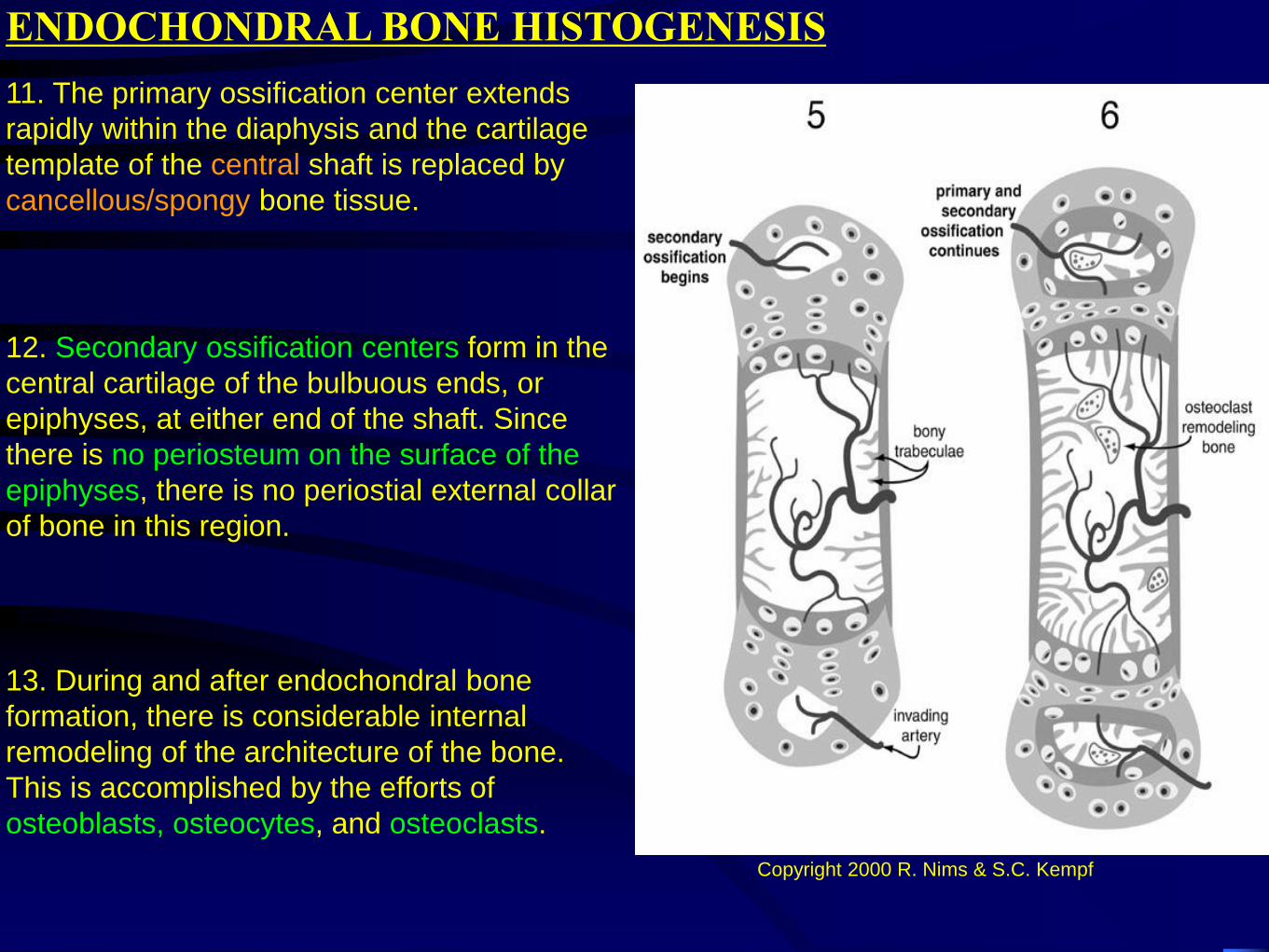
11. The primary ossification center extends
rapidly within the diaphysis and the cartilage
template of the central shaft is replaced by
cancellous/spongy bone tissue.
12. Secondary ossification centers form in the
central cartilage of the bulbuous ends, or
epiphyses, at either end of the shaft. Since
there is no periosteum on the surface of the
epiphyses, there is no periostial external collar
of bone in this region.
13. During and after endochondral bone
formation, there is considerable internal
remodeling of the architecture of the bone.
This is accomplished by the efforts of
osteoblasts, osteocytes, and osteoclasts.
Copyright 2000 R. Nims & S.C. Kempf
ENDOCHONDRAL BONE HISTOGENESIS
14. The osteoclasts continue to
breakdown cancellous bone even while
more bone is being laid down by the
osteoblasts.
15. The central region of cancellous
(spongy) bone persists and contains
the marrow cavities. More peripherally,
small longitudinal channels are
excavated through the spongy bone
that will become the haversian
systems.
16. As these peripheral channels are hollowed
out, osteoblasts from the marrow invade the
channels and form an epithelium on the inner
wall (endostium).
These osteoblasts lay down cyclical layers of
osteoid matrix which becomes mineralized
and decreases the diameter of the channels -
formation of osteons or Haversian systems
(appositional bone formation).
As this occurs, some of the osteoblasts are
trapped within the matrix forming concentric
circles of osteocytes.
http://socrates.berkeley.edu/~jmp/slippert.htm
ENDOCHONDRAL BONE HISTOGENESIS
17. As this ossification takes place, cavities like those present in spongy bone are not
retained, thus, this region becomes a solid compact mass of ossified matrix except for
the Haversian and Volkman’s canals. This is the compact or dense bone. Since there is
no cartilage precursor to the compact bone, it may be considered intramembranous as
far as its mode of formation is concerned.
19. The Haversian and Volkmann’s canals are tubes through which blood vessels
and nerves can pass within the compact bone.
18. Osteoclasts excavate radial tunnels between haversian canals and also connecting
to the marrow cavities and the periosteum. These will become the canals of Volkmann.
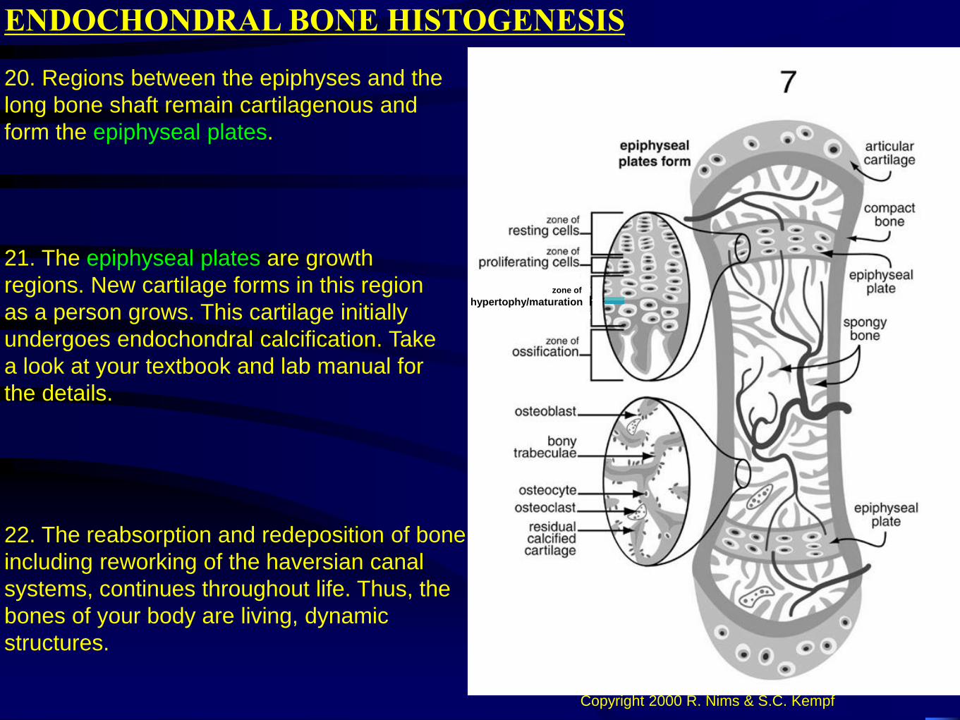
22. The reabsorption and redeposition of bone,
including reworking of the haversian canal
systems, continues throughout life. Thus, the
bones of your body are living, dynamic
structures.
ENDOCHONDRAL BONE HISTOGENESIS
21. The epiphyseal plates are growth
regions. New cartilage forms in this region
as a person grows. This cartilage initially
undergoes endochondral calcification. Take
a look at your textbook and lab manual for
the details.
20. Regions between the epiphyses and the
long bone shaft remain cartilagenous and
form the epiphyseal plates.
Copyright 2000 R. Nims & S.C. Kempf
zone of
hypertophy/maturation
http://137.222.110.150/calnet/bones/page6.htm
http://www.cytochemistry.net/microanatomy/bone/endochondral_bone_development.htm
(maturation)
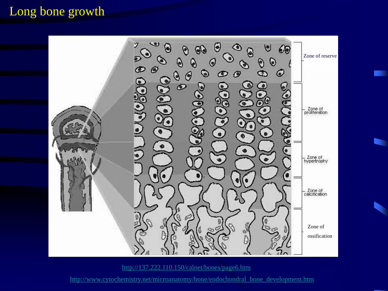
Long bone growth
Zone of
ossification
Zone of reserve

http://137.222.110.150/calnet/bones/page6.htm
http://www.cytochemistry.net/microanatomy/bone/endochondral_bone_development.htm
(maturation)
Long bone growth
Zone of
ossification
Zone of reserve