Embed Size (px)
Citation preview
ENGINEERING OF TRANSCRIPTION ACTIVATOR-LIKE EFFECTOR
NUCLEASES (TALENS) FOR TARGETED GENOME EDITING
BY
NING SUN
DISSERTATION
Submitted in partial fulfillment of the requirements for the degree of Doctor of Philosophy in Biochemistry
in the Graduate College of the University of Illinois at Urbana-Champaign, 2013
Urbana, Illinois Doctoral Committee:
Professor Huimin Zhao, Chair Professor Susan A. Martinis Associate Professor Fei Wang Professor Yi Lu Professor James H. Morrissey
ii
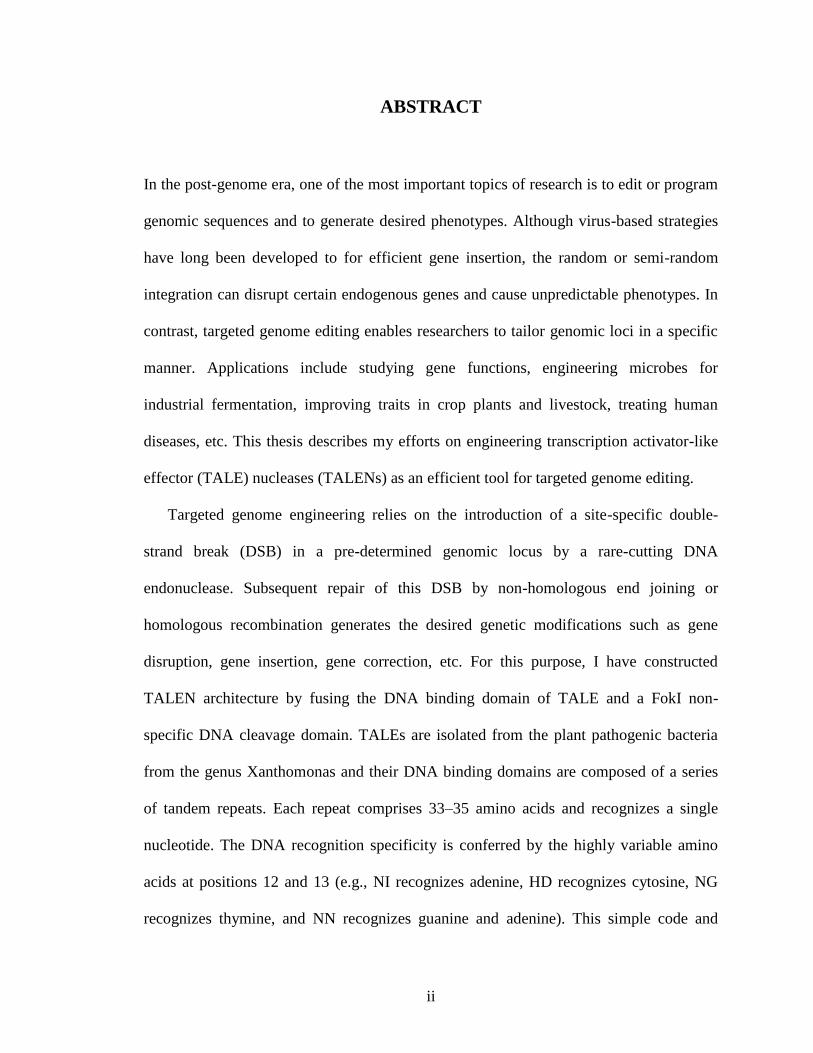
ABSTRACT
In the post-genome era, one of the most important topics of research is to edit or program
genomic sequences and to generate desired phenotypes. Although virus-based strategies
have long been developed to for efficient gene insertion, the random or semi-random
integration can disrupt certain endogenous genes and cause unpredictable phenotypes. In
contrast, targeted genome editing enables researchers to tailor genomic loci in a specific
manner. Applications include studying gene functions, engineering microbes for
industrial fermentation, improving traits in crop plants and livestock, treating human
diseases, etc. This thesis describes my efforts on engineering transcription activator-like
effector (TALE) nucleases (TALENs) as an efficient tool for targeted genome editing.
Targeted genome engineering relies on the introduction of a site-specific double-
strand break (DSB) in a pre-determined genomic locus by a rare-cutting DNA
endonuclease. Subsequent repair of this DSB by non-homologous end joining or
homologous recombination generates the desired genetic modifications such as gene
disruption, gene insertion, gene correction, etc. For this purpose, I have constructed
TALEN architecture by fusing the DNA binding domain of TALE and a FokI non-
specific DNA cleavage domain. TALEs are isolated from the plant pathogenic bacteria
from the genus Xanthomonas and their DNA binding domains are composed of a series
of tandem repeats. Each repeat comprises 33–35 amino acids and recognizes a single
nucleotide. The DNA recognition specificity is conferred by the highly variable amino
acids at positions 12 and 13 (e.g., NI recognizes adenine, HD recognizes cytosine, NG
recognizes thymine, and NN recognizes guanine and adenine). This simple code and
iii
independent DNA binding of the repeat units enable TALEs to bind to any custom-
designed DNA sequence. The fusion of a FokI cleavage domain makes TALENs a new
class of artificial DNA endonucleases which serve as a powerful tool for targeted genome
editing.
To monitor in vivo activities of TALEN, I have constructed various reporter systems
constructed in yeast and human cells. To maximize the genome editing efficiency of
TALENs, I have optimized the TALEN scaffold by the truncation of N- and C-termini of
TALEs. Two TALEN scaffolds were identified with efficient activity in modifying both
yeast and human genomes. To further improve TALEN platform, I have constructed a
high-throughput screening system and identified the SunnyTALEN architecture through
directed evolution. Compared with the existing TALEN platform, SunnyTALEN shows
significantly increased genome editing efficacy in both yeast and human cells. To
demonstrate the application of TALEN technology in human therapeutics, I have
corrected the sickle cell disease mutation in patient-derived induced pluripotent stem
cells. The corrected stem cells can serve as a regenerative medicine for the treatment of
human genetic disorders. Lastly, I have created a novel single-chain TALEN architecture,
which can be used to decrease the payload for efficient TALEN delivery.

iv
ACKNOWLEDGMENTS
First of all, I would like to thank my advisor, Professor Huimin Zhao, for his continuous
support and encouragement to my research projects and career development. His
intelligence, diligence and love in research demonstrate what a real “USTCer” should be
like and he has been and will always be my role model.
I would like to thank Professor Fei Wang for kindly sharing the expertise for handling
human stem cells. I am very grateful for the encouragement and the great suggestions that
I received from my thesis committee: Professor Susan Martinis, Professor Yi Lu,
Professor Fei Wang and Professor James Morrissey. I would also like to thank Dr. Ben
Montez and Dr. Barbara Pilas at the Roy J. Carver Biotechnology Center for their
assistance with cell sorting.
I would like to thank all of the members of the Zhao lab. Their good cheer and humor
made the Zhao group a big happy family. I am grateful to Dr. Fei Wen for mentoring me
when I did rotation in the lab. Thank her and Dr. Zengyi Shao for their valuable
suggestions on my career development. I would like to thank Jing Liang, Zehua Bao and
Xiong Xiong for their assistance with my research projects. Special thanks to Emmanuel
“Luigi” Chanco, Tong “Tony” Si and Sujit Jagtap for their friendship. The lunch parties
we had were full of joy and a lot of excellent ideas came from the random discussions.
Last but not least, I would like to thank my parents for their love and support over the
years. Special thanks to my wife, Dr. Mianzhi Gu, for being with me through the ups and
downs of graduate school. I could not have enjoyed this journey without her.
v
TABLE OF CONTENTS
CHAPTER 1. INTRODUCTION .................................................................................... 1
1.1. Targeted genome editing ....................................................................................... 1
1.2. Engineered homing endonucleases ....................................................................... 2
1.3. Zinc finger nucleases.............................................................................................. 6
1.3.1 Gene disruption .................................................................................................. 7
1.3.2 Gene insertion .................................................................................................... 8
1.3.3 Gene correction .................................................................................................. 9
1.3.4 Chromosomal rearrangements ......................................................................... 11
1.4. TALENs ................................................................................................................ 13
1.4.1. Scaffold optimization ...................................................................................... 15
1.4.2 DNA recognition specificity ............................................................................ 20
1.4.3. Assembly of TALE repeat arrays ................................................................... 24
1.4.4 Future perspectives .......................................................................................... 29
1.5 Project overview .................................................................................................... 31
1.6 References .............................................................................................................. 33
CHAPTER 2. SCAFFOLD OPTIMIZATION OF TALENS FOR USE IN
TREATMENT OF SICKLE CELL DISEASE ............................................................ 55
2.1. Introduction .......................................................................................................... 55
2.2. Results ................................................................................................................... 57
2.2.1. Construction and optimization of TALENs .................................................... 57
2.2.2. Custom-designed TALENs stimulated gene targeting in human cells ........... 63
2.2.4. Targeting “unnatural” TALE sites .................................................................. 70
2.3. Discussion.............................................................................................................. 71
2.4. Materials and methods ........................................................................................ 75
2.4.1. Materials ......................................................................................................... 75
2.4.2. Yeast reporter system ...................................................................................... 75
2.4.3. Construction of TALEN expression vectors ................................................... 76
2.4.4. Human gene targeting system ......................................................................... 78
vi
2.4.5. Immunoblotting............................................................................................... 79
2.4.6. H2AX phosphorylation assay ......................................................................... 79
2.5. References ............................................................................................................. 80
CHAPTER 3. SEAMLESS GENE CORRECTION OF SICKLE CELL DISEASE
MUTATION IN HUMAN INDUCED PLURIPOTENT STEM CELLS USING
TALENS........................................................................................................................... 85
3.1. Introduction .......................................................................................................... 85
3.2. Results ................................................................................................................... 87
3.2.1. Construction of a donor plasmid for HBBS gene correction ........................... 87
3.2.2. Seamless correction of the HBBS gene in the SCD patient-derived hiPSCs ... 89
3.2.3. Characterization of the gene-corrected hiPSCs .............................................. 91
3.3. Discussion.............................................................................................................. 94
3.4. Materials and methods ........................................................................................ 97
3.4.1. Cell culture ...................................................................................................... 97
3.4.2. Plasmid construction ....................................................................................... 98
3.4.3. TALEN-mediated genome editing of hiPSCs................................................. 99
3.4.4. Nested PCR of targeted integration .............................................................. 100
3.4.5. Transposon excision in targeted hiPSCs ....................................................... 101
3.4.6. Bsu36I analysis and sequencing of genomic HBB locus .............................. 101
3.4.7. Off-target cleavage analysis .......................................................................... 102
3.4.8. Immunostaining ............................................................................................ 103
3.4.9. Karyotyping .................................................................................................. 103
3.4.10. Teratoma formation and analysis ................................................................ 103
3.5. References ........................................................................................................... 104
CHAPTER 4. DIRECTED EVOLUTION OF TALENS WITH IMPROVED
GENOME EDITING EFFICACY .............................................................................. 111
4.1. Introduction ........................................................................................................ 111
4.2. Results ................................................................................................................. 113
4.2.1. Development of a high-throughput screening system .................................. 113
vii
4.2.2. Directed evolution of TALENs with improved activity ............................... 115
4.2.3. Isolation and characterization of the SunnyTALEN scaffold ....................... 119
4.2.4. Application of the SunnyTALEN scaffold for human genome editing ........ 121
4.3. Discussion............................................................................................................ 124
4.4. Materials and Methods ...................................................................................... 128
4.4.1. Materials ....................................................................................................... 128
4.4.2. Yeast reporter strain ...................................................................................... 129
4.4.3. Library creation ............................................................................................. 129
4.4.4. High-throughput screening ........................................................................... 130
4.4.5. A modified surrogate reporter system........................................................... 131
4.4.6. Western blot analysis .................................................................................... 132
4.4.7. TALEN construction ..................................................................................... 132
4.4.8. SURVEYOR nuclease assay......................................................................... 132
4.4.9. H2AX phosphorylation assay ....................................................................... 133
4.4.10. Sequencing analysis of endogenous gene mutations .................................. 134
4.4.11. An eGFP gene conversion assay ................................................................ 134
4.4.12. Computational modeling ............................................................................. 135
4.5. References ........................................................................................................... 136
CHAPTER 5. DEVELOPMENT OF A SINGLE-CHAIN TALEN
ARCHITECTURE ........................................................................................................ 142
5.1. Introduction ........................................................................................................ 142
5.2. Results ................................................................................................................. 145
5.2.1. Construction of a scTALEN library .............................................................. 145
5.2.2. High-throughput screening of active scTALENs ......................................... 147
5.2.3. In vivo activity in human cells ...................................................................... 148
5.3. Discussion............................................................................................................ 150
5.4. Materials and Methods ...................................................................................... 151
5.4.1. Materials ....................................................................................................... 151
5.4.2. Yeast eGFP reporter strain ........................................................................... 152
5.4.3. Creation of a scTALEN library..................................................................... 152

viii
5.4.4. High-throughput screening ........................................................................... 153
5.4.5. LacZ reporter assay in yeast .......................................................................... 154
5.4.6. In vivo activity in human cells ...................................................................... 155
5.4.7. Western blot analysis .................................................................................... 156
5.5. References ........................................................................................................... 156
1
CHAPTER 1. INTRODUCTION
1.1. Targeted genome editing
Targeted genome engineering or editing enables researchers to modify genomic loci of
interest in a precise manner, which has various applications in industry, agriculture and
human therapeutics. Virus-based strategies have been developed to efficiently integrate
an exogenous gene into a mammalian genome (1). However, the integration process is
often random, which may disrupt certain endogenous genes and cause unpredictable
phenotypes. For example, in the case of human gene therapy, insertional mutagenesis
caused by random integration of viral vectors is a major safety concern, which limits the
wide adoption of this technology (2,3). Chimeric zinc finger recombinases (4) and zinc
finger transposases (5) have been developed to integrate foreign DNA into the genome in
a more specific manner, but they are still in the preliminary stage and have met limited
success. A more established strategy for targeted genome engineering relies on the
introduction of a site-specific double-strand break (DSB) in a pre-determined locus by an
engineered DNA endonuclease. Subsequent repair of this DSB by non-homologous end
joining (NHEJ) or homologous recombination (HR) will generate desired genetic
modifications (6). The NHEJ mechanism can disrupt a gene by introducing frame-shift
mutations while the HR mechanism will result in gene deletion, gene insertion or gene
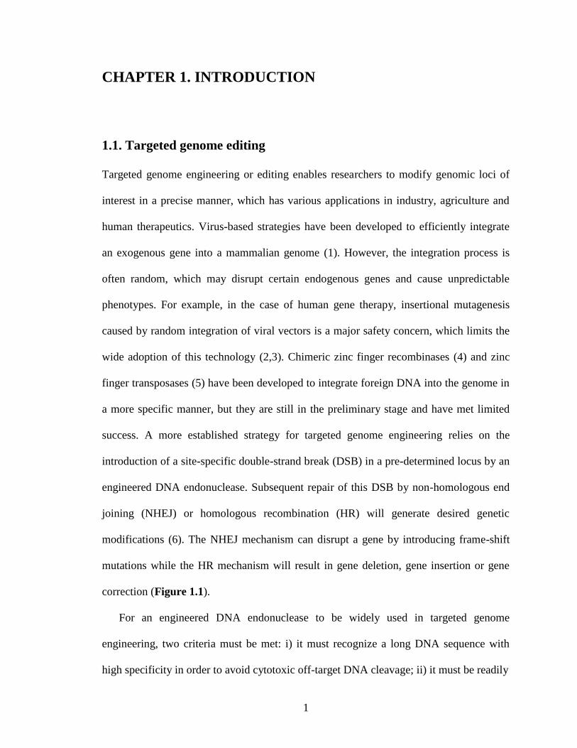
correction (Figure 1.1).
For an engineered DNA endonuclease to be widely used in targeted genome
engineering, two criteria must be met: i) it must recognize a long DNA sequence with
high specificity in order to avoid cytotoxic off-target DNA cleavage; ii) it must be readily
2
Figure 1.1. The DNA DSB generated by a site-specific endonuclease will be repaired by either NHEJ
or HR, resulting in gene disruption, gene deletion, gene addition or gene correction.
designed to recognize and cleave a defined sequence in the genome. Three major classes
of DNA endonucleases have been developed as genome engineering tools, including
engineered homing endonucleases (also called meganucleases), zinc finger nucleases, and
transcription activator-like effector nucleases (TALENs).
1.2. Engineered homing endonucleases
Homing endonucleases (HEs), also known as meganucleases, represent a family of
naturally occurring rare-cutting endonucleases, which can be found in all kingdoms of
life. HEs specifically recognize and cleave long DNA sequences (14-40 bp), which
represent a promising tool to promote chromosomal DSBs for targeted genome
engineering (reviewed by (7,8)). Among all the identified HE families, members of the
eukaryal and archael LAGLIDADG family exhibit the highest overall DNA recognition
specificity. LAGLIDADG homing endonucleases (LHEs) use antiparallel β-sheets as a
3
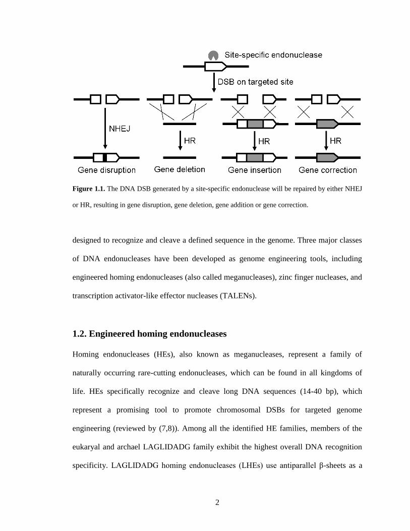
DNA recognition module by forming a saddle on the DNA helix major groove, and the
DNA cleavage is catalyzed by the divalent metal cations in the active site (Figure 1.2A)
(9). Some HE proteins such as I-CreI and I-MsoI form homodimers and cleave
palindromic or pseudo-palindromic target sites, while other members such as I-SceI and
I-AniI are monomeric, cleaving non-palindromic DNA sequences.
Several hundreds of HEs have been identified. However, the repertoire of DNA
recognition sequences is very limited compared to the size of the human genome
(>20,000 genes), making it nearly impossible to find a natural HE recognition site in a
pre-determined genomic region. Therefore, HEs must be engineered to recognize and
cleave target DNA sequences, which is a very challenging task due to the complex,
highly cooperative network of protein-DNA contacts and the tight coupling of cognate
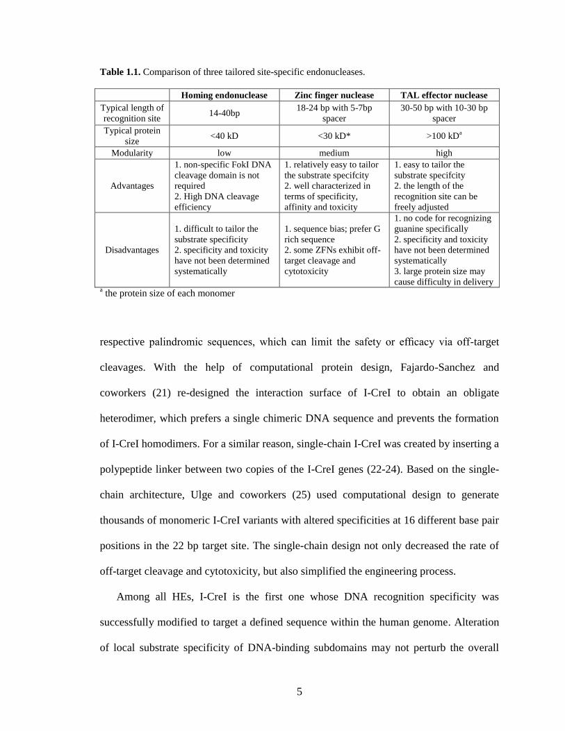
site binding to subsequent catalysis of DNA bond cleavage (Table 1.1). Directed
evolution strategies have been developed to modify the sequence specificity of known
HEs (10,11). For example, Doyon and coworkers (12) and Chen and coworkers (13)
successfully isolated I-SceI variants capable of cleaving novel DNA sequences by the
help of a bacterial based in vivo selection system. Instead of creating or screening large
protein libraries, the Baker group used computational protein design to reprogram HEs
and successfully altered the specificity of I-MsoI and I-AniI (14-16). To engineer
homodimeric HEs such as I-CreI to recognize novel non-palindromic targets, each
monomer has to be engineered separately to target its palindromic site as the first step.
Next, the two different I-CreI monomers can be co-expressed and heterodimerized upon
binding the chimeric DNA sequence, resulting in cleavage (17). Using this strategy,
however, cleavage-competent I-CreI homodimers may also form and cleave their
4
Figure 1.2. Schematic and structures of engineered site-specific endonucleases. Two perpendicular
views are presented, with the DNA duplex shown in grey. (A) Homing endonuclease I-CreI (blue)
binds to the target site as a dimer. Antiparallel β-sheets are used for DNA recognition and DNA
cleavage is catalyzed by the magnesium ions (purple spheres) within the central catalytic core
(adapted from (18), PDB no. 2VBL). (B) Zinc finger nucleases function as heterodimers and bind to
DNA targets by zinc finger domains (orange). DNA cleavage is executed by the dimerization of FokI
DNA cleavage domain (green). Zinc fingers coordinate zinc ions (cyan spheres) with a combination of
cysteine and histidine residues. The α-helix of each zinc-finger interacts with three nucleotides in the
DNA major groove (adapted from (19), PDB no. 1MEY). (C) TAL effector nucleases use central
tandem repeats (pink) for DNA sequence recognition and the FokI DNA cleavage domain (green) for
DNA cleavage. The TAL tandem repeats form a right-handed super-helical structure, binding to the
major groove of DNA (adapted from (20), PDB no. 3V6T).
5
Table 1.1. Comparison of three tailored site-specific endonucleases.
Homing endonuclease Zinc finger nuclease TAL effector nuclease
Typical length of
recognition site 14-40bp
18-24 bp with 5-7bp
spacer
30-50 bp with 10-30 bp
spacer
Typical protein
size <40 kD <30 kD* >100 kD
a
Modularity low medium high
Advantages
1. non-specific FokI DNA
cleavage domain is not
required
2. High DNA cleavage
efficiency
1. relatively easy to tailor
the substrate specifcity
2. well characterized in
terms of specificity,
affinity and toxicity
1. easy to tailor the
substrate specifcity
2. the length of the
recognition site can be
freely adjusted
Disadvantages
1. difficult to tailor the
substrate specificity
2. specificity and toxicity
have not been determined
systematically
1. sequence bias; prefer G
rich sequence
2. some ZFNs exhibit off-
target cleavage and
cytotoxicity
1. no code for recognizing
guanine specifically
2. specificity and toxicity
have not been determined
systematically
3. large protein size may
cause difficulty in delivery a the protein size of each monomer
respective palindromic sequences, which can limit the safety or efficacy via off-target
cleavages. With the help of computational protein design, Fajardo-Sanchez and
coworkers (21) re-designed the interaction surface of I-CreI to obtain an obligate
heterodimer, which prefers a single chimeric DNA sequence and prevents the formation
of I-CreI homodimers. For a similar reason, single-chain I-CreI was created by inserting a
polypeptide linker between two copies of the I-CreI genes (22-24). Based on the single-
chain architecture, Ulge and coworkers (25) used computational design to generate
thousands of monomeric I-CreI variants with altered specificities at 16 different base pair
positions in the 22 bp target site. The single-chain design not only decreased the rate of
off-target cleavage and cytotoxicity, but also simplified the engineering process.
Among all HEs, I-CreI is the first one whose DNA recognition specificity was
successfully modified to target a defined sequence within the human genome. Alteration
of local substrate specificity of DNA-binding subdomains may not perturb the overall
6
protein structure and function, making I-CreI certain degree of modularity. By combining
rational design and high throughput screening, I-CreI variants were engineered to target
the human RAG1 gene associated with severe combined immunodeficiency (26,27) and
the human XPC gene associated with xeroderma pigmentosum (18,28). Similarly, several
I-CreI variants were generated to cleave Herpes simplex virus type 1 (HSV1) genome
sequences (29). Moreover, single-chain I-CreI was used to induce high levels of gene
targeting at the endogenous human RAG1 gene locus (27,30). In addition, Takeuchi and
coworkers (31) used I-OnuI as a template and created a variant to cleave a sequence
within the monoamine oxidase B (MAO-B) gene in human cells. Since this gene is a
potential therapeutic target for a variety of neurodegenerative disorders, targeted
disruption or modifications of the MAO-B gene might have great value for future clinical
research.
1.3. Zinc finger nucleases
Zinc finger nucleases (ZFNs) are artificial DNA nucleases constructed by fusing several
zinc finger domains to the sequence-independent cleavage domain of the type IIS
restriction endonuclease FokI (32). Zinc finger domains serve as DNA recognition
modules, and modifying the zinc finger domains can target the ZFNs to novel DNA
sequences. A typical zinc finger domain contains an α-helix recognizing a specific DNA
triplet. Therefore, a typical ZFN monomer containing 3 or 4 zinc finger domains
recognizes a 9 or 12 bp DNA target site. Since the non-specific DNA cleavage domain of
FokI functions as a dimer and has the highest cleavage efficiency in the inverse
orientation (33), the typical custom-designed ZFN functions as a heterodimer recognizing
7
an 18-24 bp DNA sequence with a 4-6 bp spacer between each half site (Figure 1.2B).
Compared with the engineered homing endonucleases, custom-designed ZFNs are easier
to construct due to their moderate degree of modularity (Table 1.1). Multiple platforms
such as the CompoZrTM
service, modular assembly, Oligomerized Pool Engineering
(OPEN), and Context Dependent Assembly (CoDA) have been used to generate site-
specific ZFNs (reviewed by (34-36)). Because several zinc finger domains can tolerate
mismatched DNA bases, off-target ZFN cleavage has been reported (37,38). The
recognition of secondary degenerate sites of ZFNs will generate undesired genomic
DSBs and result in unexpected side effects. Therefore, the sequence fidelity of ZFNs has
to be improved for future ZFN design because it is critical to their safe and successful
application in targeted genome engineering (Table 1.1). Recent advances in the ZFN
technology involve a large number of applications such as gene disruption, gene insertion,
gene correction, and chromosomal rearrangement (Figure 1.1), as will be highlighted
below.
1.3.1 Gene disruption
The errors introduced during NHEJ-mediated chromosomal DSB repair can be used to
achieve gene disruption (Figure 1.1). This approach has been applied in various
mammalian cells to efficiently knock out targeted genes in a single step (recently
reviewed by (36)). There are ongoing clinical trials for the treatment of human
immunodeficiency virus (HIV) infection, which is based on ZFN-mediated CCR5 gene
disruption (39,40). Recently, Cradick and coworkers (41) described a novel therapeutic
strategy for treatment of hepatitis B by using ZFNs to target the hepatitis B virus genome.
8
Besides human cells, ZFNs were successfully applied to other mammals such as rat
(42,43), rabbit (44) and pig (45). Moreover, in addition to NHEJ-mediated gene
disruptions, Zou and coworkers inactivated the endogenous PIG-A locus associated with
paroxysmal nocturnal hemoglobinuria in human embryonic stem cells (ESCs) and
induced pluripotent stem cells (iPSCs) by DSB-induced HR (46). Successful HR-
mediated targeting resulted in the insertion of an antibiotic selection cassette into the
PIG-A gene exon, thereby inactivating the PIG-A gene.
1.3.2 Gene insertion
Precise gene insertion can be achieved by ZFN-induced DNA repair (Figure 1.1).
Hockemeyer and coworkers (47) reported the highly efficient targeting of three genes in
human iPSCs via ZFN-based HR-mediated genome insertion. Specifically, they
successfully integrated the enhanced green fluorescent protein (eGFP) reporter gene into
the OCT4, AAVS1 and PITX3 loci. Note that the AAVS1 locus within the PPP1R12C gene
on chromosome 19 is considered as a ‘safe harbor’ for addition of a transgene into the
human genome (48). DeKelver and coworkers (49) described the use of ZFNs to insert
various expression cassettes into the AAVS1 locus for isogenic transgenesis application.
ZFN-mediated gene insertion turned out to be efficient (at a frequency of up to 15%) in
both transformed cell lines (K562, HeLa, HEK293, U2OS, and others) and primary
human cells (fibroblasts and ESCs). It was demonstrated that ZFN-driven gene addition
into the AAVS1 locus did not introduce any adverse effect on the cell resulting from its
disruption. Moreover, both promoterless (driven by the native PPP1R12C gene promoter)
and promoter-containing inserts placed into the AAVS1 locus exhibited consistent gene
9
expression levels over 50 cell generations. Later, Zou and coworkers (50) integrated a
single-copy gp91(phox) gene into the AAVS1 locus for treatment of the X-linked chronic
granulomatous disease (X-CGD). X-CGD patients have a defect in neutrophil
microbicidal reactive oxygen species (ROS) generation because of the gp91(phox)
deficiency. With the help of a ZFN-induced DSB, a therapeutic gp91(phox) minigene was
inserted into one allele of the AAVS1 locus in the X-CGD iPSCs without off-target
insertions, which complemented the gp91(phox) gene deficiency and substantially
restored neutrophil ROS production.
1.3.3 Gene correction
Gene correction is mediated by DSB-induced HR, which can be exploited for gene
replacement, especially for the treatment of monogenic diseases. Compared with gene
insertion, in situ gene correction is more challenging because the designed ZFN cleavage
site must be close to or directly at the site of the mutation (Figure 1.1). Through site-
specific chromosomal DSBs induced by ZFNs, Sebastiano and coworkers (51)
demonstrated HR-mediated efficient correction of the sickle cell anemia mutation in
patient-derived human iPSCs. Two pairs of ZFNs were constructed by the publicly
available OPEN method (52) to target the disease-causing β-globin gene. They first used
those ZFNs to simultaneously correct the E6V mutation and insert a drug-resistance
cassette into a neighboring intron 58 or 82 bp downstream of the ZFN cleavage sites.
Isolation of drug-resistant clones and PCR screening followed by DNA sequencing
enabled them to identify several iPSC clones containing the successfully corrected allele.
Next they used the Cre recombinase to excise the selection gene cassette flanked by the
10
loxP sites, leaving a residual 34 bp loxP site “scar” in the intron of the corrected β-globin
gene. Around the same time, Zou and coworkers (53) achieved site-specific gene
correction of the β-globin gene in patient-derived human iPSCs. The ZFNs they used
were custom designed by CompoZrTM
from Sigma-Aldrich (St. Louis, MO, USA). A
similar two-step strategy was used to correct the sickle cell disease causing mutation: (i)
Correction of the E6V mutation by ZFN-induced HR and integration of a loxP-flanked
drug-resistant cassette into a neighboring intron; (ii) Cre recombinase-mediated excision
of the drug-resistant cassette. To evaluate the gene expression of the corrected β-globin
gene, they differentiated the corrected iPSCs into erythroid cells and surprisingly
discovered that the expression level of the corrected β-globin gene was partially repressed.
Since Cre-recombinase mediated gene excision in step (ii) left a residual loxP sequence
in the intron, it is possible that the remaining loxP “scar” interfered with the
transcriptional regulatory elements and thus altered the gene expression from the targeted
allele. In addition to the unpredicted effect from the residual loxP site, such a strategy
may not be applicable to all genes, especially when the location of the desired editing
events is away from the intron region.
Instead of using the Cre/loxP recombination system that leaves small ectopic
sequences in the targeted genome, Yusa and coworkers combined ZFN with the piggyBac
technology and achieved a “scarless” gene correction in patient-derived iPSCs (54).
piggyBac is a moth-derived DNA transposon, which enables the removal of transgenes
flanked by its inverted repeats without leaving any residual sequences (55). To correct the
single mutation (E342K) in the A1AT gene causing α1-antitrypsin deficiency, ZFN pairs
were designed to specifically cleave the mutation site. Through ZFN-induced HR, they
11
corrected the A1AT gene mutation and simultaneously inserted a PGK-puroΔtk cassette
flanked by piggyBac repeats into the TTAA site. Targeted clones were isolated by
puromycin selection. Next, the selection cassette was excised from the iPSC genome by
transient expression of the piggyBac transposase and subsequent counter-selection,
leaving no residual ectopic sequences in the targeted allele. Not only did their approach
achieve seamless gene correction of diseased human iPSCs, but also enabled biallelic
correction with high efficiency. Without using a selectable marker, Soldner and
coworkers employed a selection-free targeting strategy (56). Briefly, given the high gene
editing activity of the ZFNs, they constructed a donor vector lacking a selection cassette,
consisting of only a homology arm flanking the ZFN cleavage site carrying a wild-type
α-synuclein gene. An eGFP-expressing plasmid was coelectroporated into Parkinson’s
disease patient-derived iPSCs together with the donor construct and ZFN-expression
vectors to enrich transfected cells by fluorescence-activated cell sorting. One correctly
targeted clone with the A53T (G209) mutation repaired was isolated after screening 240
single-cell-derived clones by Southern blot analysis. The high efficiency suggests that
one could isolate corrected clones by dilution cloning without going through drug
selection.
1.3.4 Chromosomal rearrangements
Chromosomal rearrangements include large-scale gene deletions, insertions,
duplications and inversions, which are associated with many genetic diseases and cancer
(57). ZFNs have been utilized to generate targeted chromosomal rearrangements, which
enables researchers to study gene functions at the genomic level (Figure 1.3). Brunet and
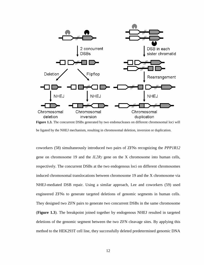
12
Figure 1.3. The concurrent DSBs generated by two endonucleases on different chromosomal loci will
be ligated by the NHEJ mechanism, resulting in chromosomal deletion, inversion or duplication.
coworkers (58) simultaneously introduced two pairs of ZFNs recognizing the PPP1R12
gene on chromosome 19 and the IL2Rγ gene on the X chromosome into human cells,
respectively. The concurrent DSBs at the two endogenous loci on different chromosomes
induced chromosomal translocations between chromosome 19 and the X chromosome via
NHEJ-mediated DSB repair. Using a similar approach, Lee and coworkers (59) used
engineered ZFNs to generate targeted deletions of genomic segments in human cells.
They designed two ZFN pairs to generate two concurrent DSBs in the same chromosome
(Figure 1.3). The breakpoint joined together by endogenous NHEJ resulted in targeted
deletions of the genomic segment between the two ZFN cleavage sites. By applying this
method to the HEK293T cell line, they successfully deleted predetermined genomic DNA
13
segments in the range of ~729 bp to 15 Mb with frequencies of 0.1% to 10%. In addition
to genomic deletions, they demonstrated that two concurrent DSBs introduced by ZFN
pairs were sufficient to promote frequent genomic inversions and duplications in human
cells with frequencies ranging from 0.01% to 5% (60). Harnessing various combinations
of two ZFNs, they achieved duplications of genomic DNA between the two cleavage
sites whose length ranged from 230 kb to 835 kb (Figure 1.3). They also demonstrated
that the two concurrent genomic DSBs from ZFN cleavages induced inversions of 15 kb
to 15 Mb DNA segments in the human genome (Figure 1.3). As proof of concept for
therapeutic application of this technique, they constructed a ZFN pair to target the intron
1 homolog in the human F8 gene, whose inversion causes severe hemophilia A (61). The
genomic DSB generated by the ZFN pair induced the inversion of the 140 kb DNA
segment bearing the promoter and exon 1 of the F8 gene with a frequency of 0.2%-0.4%.
Their strategy demonstrated the promise of restoring genomic integrity in severe
hemophilia A patients by reverting the inverted DNA segment back to the wild-type
orientation.
1.4. TALENs
Even though ZFNs have been used for targeted genome editing in various organisms, two
major limitations prevent their wider applications. ZF domains have limited modularity
due to the context-dependent DNA-binding effects, making it difficult for ZFNs to target
any desired DNA sequence (62). Moreover, lack of specificity of some ZF domains can
generate off-target cleavage, leading to undesired mutations and chromosomal
aberrations (38,63). Recently, TALENs have rapidly emerged as an alternative genome
14
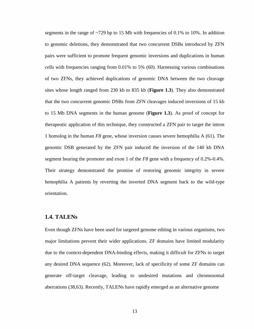
Figure 1.4. Schematic of TALEN architecture. A TALEN is composed of a NTS (pink box), a central
repeat domain, a CTS (cyan box) and a FokI catalytic domain (orange oval). The central repeat
domain comprises a series of repeat units that are responsible for specific recognition of thymine (red
boxes), adenine (green boxes), cytosine (blue boxes) and guanine (yellow boxes). The formation of a
heterodimer by two TALENs in a tail-to-tail orientation at the target site executes a site-specific DNA
DSB. The TALE binding sites on the target DNA are shown in black and the spacer is shown in grey.
editing tool to ZFNs (recently reviewed by (64)). Similar to ZFNs, TALENs use the non-
specific FokI domain as the DNA cleavage module and function as dimmers (Figure
1.2C and Figure 1.4). However, the DNA binding domains of TALENs are composed of
a series of tandem repeats as in TALEs of the plant pathogenic bacteria from the genus
Xanthomonas (as reviewed in (65,66)). Each repeat comprises 33-35 aa and recognizes a
single nucleotide. The last repeat typically has only 20 aa, and is therefore called a ‘half-
repeat’. The DNA recognition specificity is conferred by the highly variable amino acids
at positions 12 and 13 (e.g. NI recognizes adenine, HD recognizes cytosine, NG
recognizes thymine, and NN recognizes guanine and adenine) (67,68). Unlike the
context-dependent DNA binding of ZFNs, TALENs can be easily and rapidly constructed
to target essentially any DNA sequence due to the simple protein-DNA code and the
modular nature. In addition, TALENs exhibit significantly reduced off-target effects and
cytotoxicities compared with ZFNs, making them an efficient genome editing tool
15
(69,70). Within the last three years, TALENs have been widely applied to modify
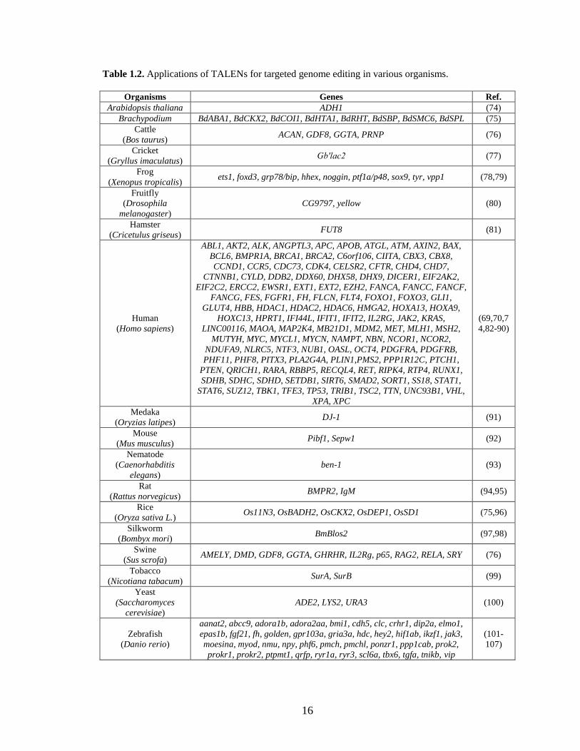
endogenous genes in a variety of organisms (Table 1.2). Applications include studying
gene functions in model organisms, improving traits in crop plants and livestock,
generating disease models, and treating genetic disorders in humans. In this article, we
provide a comprehensive review of TALEN technology including the optimization of the
scaffold, improvement of the DNA recognition specificity, and assembly of TALE repeat
arrays. Due to the ease of design and high efficiency of genome editing, TALENs have
opened up many new avenues for basic and applied biological research.
1.4.1. Scaffold optimization
The original TALEN construction was reported by two independent groups. Li and
coworkers fused the full-length natural TALE (AvrXa7 and PthXo1) with the FokI
catalytic domain, creating the TALENs bearing a 288 aa N-terminal segment (NTS) and a
295 aa C-terminal segment (CTS) (71). Based on a yeast reporter assay, the optimal
spacer length between the two TALEN binding sites was determined to be 16-31 bp
(Table 1.3). Alternatively, the TALENs created by Christian and coworkers encompasses
a 287 aa NTS and a 231 aa CTS (72). This scaffold allows efficient DNA cleavage
against target sites with 13-30 bp spacers (Table 1.3). Since naturally-occurring TALEs
are transcription activators from a plant bacterial pathogen, their NTSs harbor protein
secretion signal peptides while their CTSs contain nuclear localization signal peptides
and a transcription activator domain (73). These sequences can impair the catalytic
activity when fused with the FokI cleavage domain. To identify the optimum TALEN
architecture with highest cleavage efficiency and minimal peptide portion, scaffold
16
Table 1.2. Applications of TALENs for targeted genome editing in various organisms.
Organisms Genes Ref.
Arabidopsis thaliana ADH1 (74)
Brachypodium BdABA1, BdCKX2, BdCOI1, BdHTA1, BdRHT, BdSBP, BdSMC6, BdSPL (75)
Cattle
(Bos taurus) ACAN, GDF8, GGTA, PRNP (76)
Cricket
(Gryllus imaculatus) Gb′lac2 (77)
Frog
(Xenopus tropicalis) ets1, foxd3, grp78/bip, hhex, noggin, ptf1a/p48, sox9, tyr, vpp1 (78,79)
Fruitfly
(Drosophila
melanogaster)
CG9797, yellow (80)
Hamster
(Cricetulus griseus) FUT8 (81)
Human
(Homo sapiens)
ABL1, AKT2, ALK, ANGPTL3, APC, APOB, ATGL, ATM, AXIN2, BAX,
BCL6, BMPR1A, BRCA1, BRCA2, C6orf106, CIITA, CBX3, CBX8,
CCND1, CCR5, CDC73, CDK4, CELSR2, CFTR, CHD4, CHD7,
CTNNB1, CYLD, DDB2, DDX60, DHX58, DHX9, DICER1, EIF2AK2,
EIF2C2, ERCC2, EWSR1, EXT1, EXT2, EZH2, FANCA, FANCC, FANCF,
FANCG, FES, FGFR1, FH, FLCN, FLT4, FOXO1, FOXO3, GLI1,
GLUT4, HBB, HDAC1, HDAC2, HDAC6, HMGA2, HOXA13, HOXA9,
HOXC13, HPRT1, IFI44L, IFIT1, IFIT2, IL2RG, JAK2, KRAS,
LINC00116, MAOA, MAP2K4, MB21D1, MDM2, MET, MLH1, MSH2,
MUTYH, MYC, MYCL1, MYCN, NAMPT, NBN, NCOR1, NCOR2,
NDUFA9, NLRC5, NTF3, NUB1, OASL, OCT4, PDGFRA, PDGFRB,
PHF11, PHF8, PITX3, PLA2G4A, PLIN1,PMS2, PPP1R12C, PTCH1,
PTEN, QRICH1, RARA, RBBP5, RECQL4, RET, RIPK4, RTP4, RUNX1,
SDHB, SDHC, SDHD, SETDB1, SIRT6, SMAD2, SORT1, SS18, STAT1,
STAT6, SUZ12, TBK1, TFE3, TP53, TRIB1, TSC2, TTN, UNC93B1, VHL,
XPA, XPC
(69,70,7
4,82-90)
Medaka
(Oryzias latipes) DJ-1 (91)
Mouse
(Mus musculus) Pibf1, Sepw1 (92)
Nematode
(Caenorhabditis
elegans)
ben-1 (93)
Rat
(Rattus norvegicus) BMPR2, IgM (94,95)
Rice
(Oryza sativa L.) Os11N3, OsBADH2, OsCKX2, OsDEP1, OsSD1 (75,96)
Silkworm
(Bombyx mori) BmBlos2 (97,98)
Swine
(Sus scrofa) AMELY, DMD, GDF8, GGTA, GHRHR, IL2Rg, p65, RAG2, RELA, SRY (76)
Tobacco
(Nicotiana tabacum) SurA, SurB (99)
Yeast
(Saccharomyces
cerevisiae)
ADE2, LYS2, URA3 (100)
Zebrafish
(Danio rerio)
aanat2, abcc9, adora1b, adora2aa, bmi1, cdh5, clc, crhr1, dip2a, elmo1,
epas1b, fgf21, fh, golden, gpr103a, gria3a, hdc, hey2, hif1ab, ikzf1, jak3,
moesina, myod, nmu, npy, phf6, pmch, pmchl, ponzr1, ppp1cab, prok2,
prokr1, prokr2, ptpmt1, qrfp, ryr1a, ryr3, scl6a, tbx6, tgfa, tnikb, vip
(101-
107)
17
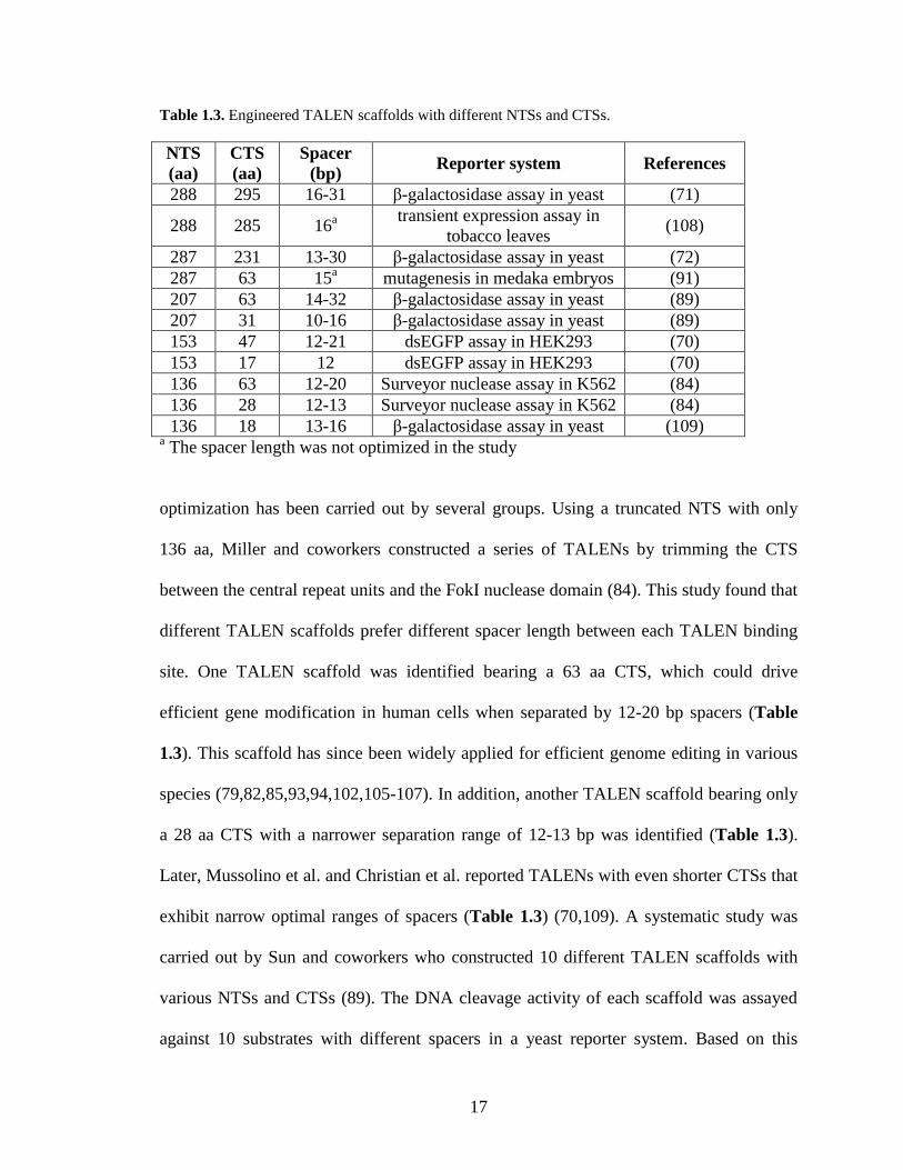
Table 1.3. Engineered TALEN scaffolds with different NTSs and CTSs.
NTS
(aa)
CTS
(aa)
Spacer
(bp) Reporter system References
288 295 16-31 β-galactosidase assay in yeast (71)
288 285 16a
transient expression assay in
tobacco leaves (108)
287 231 13-30 β-galactosidase assay in yeast (72)
287 63 15a mutagenesis in medaka embryos (91)
207 63 14-32 β-galactosidase assay in yeast (89)
207 31 10-16 β-galactosidase assay in yeast (89)
153 47 12-21 dsEGFP assay in HEK293 (70)
153 17 12 dsEGFP assay in HEK293 (70)
136 63 12-20 Surveyor nuclease assay in K562 (84)
136 28 12-13 Surveyor nuclease assay in K562 (84)
136 18 13-16 β-galactosidase assay in yeast (109) a The spacer length was not optimized in the study
optimization has been carried out by several groups. Using a truncated NTS with only
136 aa, Miller and coworkers constructed a series of TALENs by trimming the CTS
between the central repeat units and the FokI nuclease domain (84). This study found that
different TALEN scaffolds prefer different spacer length between each TALEN binding
site. One TALEN scaffold was identified bearing a 63 aa CTS, which could drive
efficient gene modification in human cells when separated by 12-20 bp spacers (Table
1.3). This scaffold has since been widely applied for efficient genome editing in various
species (79,82,85,93,94,102,105-107). In addition, another TALEN scaffold bearing only
a 28 aa CTS with a narrower separation range of 12-13 bp was identified (Table 1.3).
Later, Mussolino et al. and Christian et al. reported TALENs with even shorter CTSs that
exhibit narrow optimal ranges of spacers (Table 1.3) (70,109). A systematic study was
carried out by Sun and coworkers who constructed 10 different TALEN scaffolds with
various NTSs and CTSs (89). The DNA cleavage activity of each scaffold was assayed
against 10 substrates with different spacers in a yeast reporter system. Based on this
18
10×10 matrix, two TALEN scaffolds with high DNA cleavage efficiency in both yeast
and human cells were identified. One bearing a 207 aa NTS and a 31 aa CTS prefers
target sites with 10-16 bp spacers while another bearing a 207 aa NTS and a 63 aa CTS
has highest efficiency when separated by 14-32 bp spacers (Table 1.3). It is noteworthy
that the TALEN with a 50 aa NTS has no catalytic activity against any target sites. Later
on, Gao and coworkers solved the crystal structure of the TALE NTS and discovered an
extended N-terminal DNA binding region composed of the 127 aa immediately preceding
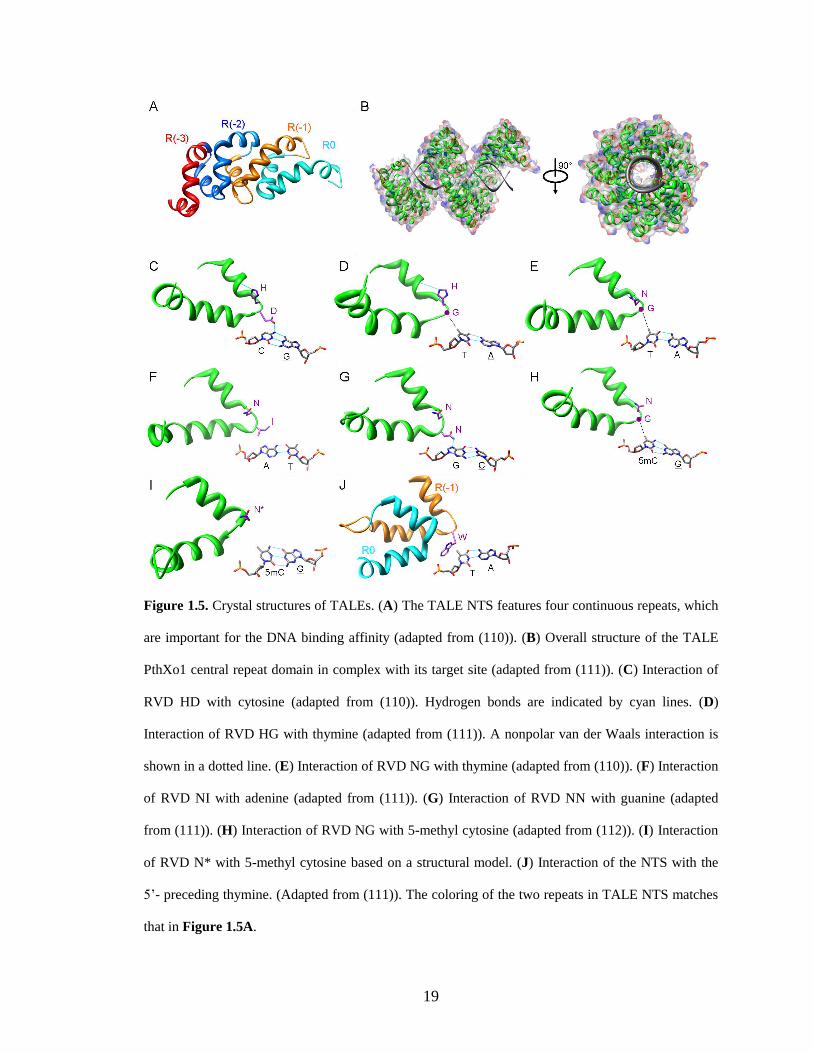
the central repeat units (110). The 127 aa NTS features four continuous repeats. Each
repeat contains two α-helices and an intervening loop (Figure 1.5A), a structural feature
highly similar to that of the central repeat unit. Although the 127 aa NTS does not confer
sequence specificity, it is crucial for DNA binding. This feature explains why all the
effective TALEN scaffolds have at least 127 aa preceding the central repeat units (Table
1.3).
A second-generation GoldyTALEN scaffold has been demonstrated to improve the
genome editing efficiency in zebrafish (101). GoldyTALEN is based on a scaffold
previously reported by Miller and coworkers (84) which consists of a 136 aa NTS and a
63 aa CTS. However, it has 9 different aa substitutions at the NTS and 5 different aa
substitutions at the CTS. Using the GoldyTALEN scaffold and zebrafish delivery system,
certain loci were modified with 100% efficiency. Moreover, they provided the first
example of HR-based genome editing in zebrafish using single-stranded DNA as a donor.
The GoldyTALEN scaffold was also applied for efficient gene knockout in livestock (76).
Because the FokI catalytic domain must dimerize to become active, two TALEN
subunits are assembled as heterodimers at the cleavage site. However, cleavage-
19
Figure 1.5. Crystal structures of TALEs. (A) The TALE NTS features four continuous repeats, which
are important for the DNA binding affinity (adapted from (110)). (B) Overall structure of the TALE
PthXo1 central repeat domain in complex with its target site (adapted from (111)). (C) Interaction of
RVD HD with cytosine (adapted from (110)). Hydrogen bonds are indicated by cyan lines. (D)
Interaction of RVD HG with thymine (adapted from (111)). A nonpolar van der Waals interaction is
shown in a dotted line. (E) Interaction of RVD NG with thymine (adapted from (110)). (F) Interaction
of RVD NI with adenine (adapted from (111)). (G) Interaction of RVD NN with guanine (adapted
from (111)). (H) Interaction of RVD NG with 5-methyl cytosine (adapted from (112)). (I) Interaction
of RVD N* with 5-methyl cytosine based on a structural model. (J) Interaction of the NTS with the
5’- preceding thymine. (Adapted from (111)). The coloring of the two repeats in TALE NTS matches
that in Figure 1.5A.
20
competent homodimers composed of each subunit may also form and generate off-target
cleavage, which can limit safety or efficiency. To address this limitation, obligate
heterodimer mutations were introduced at the dimer interface of the FokI cleavage
domain which prevented homodimerization based on electrostatic and hydrophobic
interactions. The creation of FokI variants that preferentially heterodimerize successfully
reduced off-target cleavage of ZFNs and relieved toxicity (113-115). A similar principle
was applied to TALENs by Cade and coworkers (102) for the generation of zebrafish
knockout lines. The heterodimeric TALENs show similar or even greater activities than
their homodimeric counterparts. Moreover, the TALENs constructed with heterodimeric
FokI domains induced smaller numbers of abnormal or dead embryos, indicating reduced
toxicity. This obligate heterodimeric TALEN configuration has also been reported by
other groups for gene knockout studies (79,104,105).
1.4.2 DNA recognition specificity
The DNA recognition specificity of TALENs is conferred by the repeat-variable
diresidues (RVDs) at positions 12 and 13 of each repeat. More than 20 different RVDs
have been identified in TALEs, among which NI, NG, HD, NN, and HG are the most
common ones recognizing the nucleotides A, T, C, G/A, and T, respectively (67,68).
Based on crystal structures, TALE binds to target DNA as a right-handed superhelix
(Figure 1.5B). Each repeat unit forms a left-handed, two-helix bundle that presents an
RVD-containing loop to the DNA major groove (20,111). The first residue of each RVD
(residue 12 of each repeat), either His or Asn, does not contact DNA directly. Instead, the
side chain forms a hydrogen bond to the backbone carbonyl oxygen of Ala at position 8
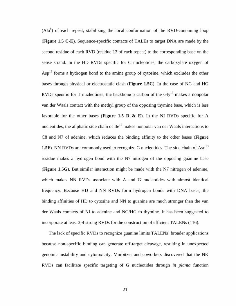
21
(Ala8) of each repeat, stabilizing the local conformation of the RVD-containing loop
(Figure 1.5 C-E). Sequence-specific contacts of TALEs to target DNA are made by the
second residue of each RVD (residue 13 of each repeat) to the corresponding base on the
sense strand. In the HD RVDs specific for C nucleotides, the carboxylate oxygen of
Asp13
forms a hydrogen bond to the amine group of cytosine, which excludes the other
bases through physical or electrostatic clash (Figure 1.5C). In the case of NG and HG
RVDs specific for T nucleotides, the backbone α carbon of the Gly13
makes a nonpolar
van der Waals contact with the methyl group of the opposing thymine base, which is less
favorable for the other bases (Figure 1.5 D & E). In the NI RVDs specific for A
nucleotides, the aliphatic side chain of Ile13
makes nonpolar van der Waals interactions to
C8 and N7 of adenine, which reduces the binding affinity to the other bases (Figure
1.5F). NN RVDs are commonly used to recognize G nucleotides. The side chain of Asn13
residue makes a hydrogen bond with the N7 nitrogen of the opposing guanine base
(Figure 1.5G). But similar interaction might be made with the N7 nitrogen of adenine,
which makes NN RVDs associate with A and G nucleotides with almost identical
frequency. Because HD and NN RVDs form hydrogen bonds with DNA bases, the
binding affinities of HD to cytosine and NN to guanine are much stronger than the van
der Waals contacts of NI to adenine and NG/HG to thymine. It has been suggested to
incorporate at least 3-4 strong RVDs for the construction of efficient TALENs (116).
The lack of specific RVDs to recognize guanine limits TALENs’ broader applications
because non-specific binding can generate off-target cleavage, resulting in unexpected
genomic instability and cytotoxicity. Morbitzer and coworkers discovered that the NK
RVDs can facilitate specific targeting of G nucleotides through in planta function
22
analysis (117). Based on the SELEX assay, Miller and coworkers provided in vitro
evidence that RVD NK has a much stronger preference for guanine over adenine, which
represents a promising code for the specific recognition of G nucleotides (84). However,
substitution of RVD NN with NK significantly reduced TALEN activity in zebrafish
embryo (105). Substantially lower activities in NK containing TALEs have also been
observed in plants and in mammalian cells (116,118). Therefore, RVD NK is not ideal
for guanine recognition because the improvement in specificity sacrifices efficiency.
Alternatively, NH has been reported as a competent guanine-specific RVD, which has
much higher efficiency than RVD NK (116,118). Computational modeling analysis
showed that the imidazole ring on the His13
of the NH RVD has a compact base-stacking
interaction with the guanine base, suggesting a possible mechanism for its increased
specificity for G nucleotide while maintaining the binding affinity (118).
Although successfully used in various cellular contexts, TALE DNA binding domains
have been reported to be incapable of targeting methylated DNA (119). Often considered
as the fifth base, 5-methyl cytosine (5mC) is a major epigenetic mark and widely
distributed in fungi, plant and mammalian genomes (120). In addition, 5mC has been
identified in CpG islands of many promoters, which are important regulatory regions for
genome modification (121). Recently, two groups discovered that RVD NG and N* (an
asterisk indicates a deletion at residue 13 in the repeat unit) can accommodate 5mC
efficiently in vitro and in vivo (112,122). Thymine is structurally similar to 5mC, with the
only difference at position 4, which is not involved in binding to TALE repeats. This
observation indicates that the NG RVD specific for thymine might be used to recognize
5mC. The protein crystal structure solved by Deng and coworkers shows that lack of side
23
chain of Gly13
in NG RVDs provides sufficient space to accommodate the 5-methyl
group of 5mC and allows the formation of van der Waals contacts (Figure 1.5H) (112).
Because RVDs are followed immediately by two conserved Gly residues, N* is roughly
equivalent to NG except for a shortened RVD loop (Figure 1.5I). Using N* to code for
5mC, Valton and coworkers demonstrated the first example of TALEN-mediated
modification at a methylated locus in human cells (122). Accommodation of 5mC by
TALE repeats through the RVD NG or N* extends the DNA recognition code and
enables researchers to design TALENs to target hypermethylated DNA regions, which
has great potential in epigenetics studies and human therapeutic applications.
All naturally-occurring TALE target sites are preceded by a 5’-thymine at position 0,
which was previously believed to be essential for TALE function (67,68). The TALE
crystal structure reveals that two degenerate repeats prior to the central repeat domain
appear to cooperate to specify the conserved 5’-thymine (111). The indole ring of a Trp
residue in the repeat R(-1) forms a van der Waals contact with the methyl group of the
thymine base, suggesting a possible mechanism for the conserved specificity at position 0
(Figure 1.5J). Sun and coworkers reported that TALENs with shorter CTSs (31 aa) show
higher efficiency against natural TALE recognition sites preceded by a 5’-T than that
against unnatural TALE sites preceded by A, C or G. However, TALEN variants with
longer CTSs (63-117 aa) are capable of cleaving unnatural DNA substrates with similar
efficiency compared with that of natural TALE sites (89). Other studies also provided
evidence that a thymine at position 0 is not strictly required for TALEN activity
(84,123,124). Notably, there are nine leucine zipper-like heptad repeats closely linked to
the C terminus of the TALE central repeat domain (125). These leucine-rich repeats may

24
mediate TALE/DNA interactions and increase DNA binding affinity, making the 5’-T
less of a requirement. Detailed structural studies could help solve this uncertainty. The
requirement of a preceding 5’-T can be mitigated using certain TALEN scaffolds,
allowing greater flexibility in choosing target sites in genome editing endeavors.
1.4.3. Assembly of TALE repeat arrays
Because of the high similarity between each TALE repeat unit, it is challenging to
construct plasmids encoding long arrays of TALE repeats. To address this limitation,
numerous methods have been developed to assemble the highly repetitive TALE central
repeat domains rapidly and cost-effectively (64). Based on a standard cloning strategy,
Sander and coworkers described a restriction enzyme and ligation (REAL) method, in
which single TALE repeats are joined together using routine restriction digestion and
ligation techniques (107). Initially, they constructed a library of plasmids encoding
various individual TALE repeats by DNA synthesis. In the assembly step, two TALE
repeats are first joined together by ligating compatible overhangs generated by digestion
with restriction endonucleases. Next, the ligation product encoding two TALE repeats is
joined with another TALE repeat dimer in the same manner, resulting in a DNA fragment
encoding four TALE repeats. This process continues in an iterative fashion until a TALE
repeat array of the desired length is assembled (Figure 1.6A). Using a large plasmid
library of pre-assembled multiple TALE repeats, REAL can be performed in a more rapid
and less labor-intensive fashion, which is referred to as REAL-Fast (126). With the help
of isocaudamer restriction enzymes (e.g. NheI and SpeI), a unit assembly method has
25
been described for building long TALE repeat arrays in the similar hierarchical fashion
(105).
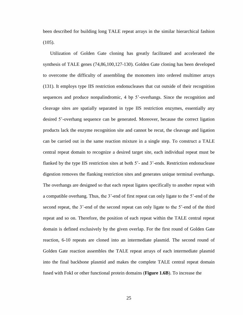
Utilization of Golden Gate cloning has greatly facilitated and accelerated the
synthesis of TALE genes (74,86,100,127-130). Golden Gate cloning has been developed
to overcome the difficulty of assembling the monomers into ordered multimer arrays
(131). It employs type IIS restriction endonucleases that cut outside of their recognition
sequences and produce nonpalindromic, 4 bp 5’-overhangs. Since the recognition and
cleavage sites are spatially separated in type IIS restriction enzymes, essentially any
desired 5’-overhang sequence can be generated. Moreover, because the correct ligation
products lack the enzyme recognition site and cannot be recut, the cleavage and ligation
can be carried out in the same reaction mixture in a single step. To construct a TALE
central repeat domain to recognize a desired target site, each individual repeat must be
flanked by the type IIS restriction sites at both 5’- and 3’-ends. Restriction endonuclease
digestion removes the flanking restriction sites and generates unique terminal overhangs.
The overhangs are designed so that each repeat ligates specifically to another repeat with
a compatible overhang. Thus, the 3’-end of first repeat can only ligate to the 5’-end of the
second repeat, the 3’-end of the second repeat can only ligate to the 5’-end of the third
repeat and so on. Therefore, the position of each repeat within the TALE central repeat
domain is defined exclusively by the given overlap. For the first round of Golden Gate
reaction, 6-10 repeats are cloned into an intermediate plasmid. The second round of
Golden Gate reaction assembles the TALE repeat arrays of each intermediate plasmid
into the final backbone plasmid and makes the complete TALE central repeat domain
fused with FokI or other functional protein domains (Figure 1.6B). To increase the
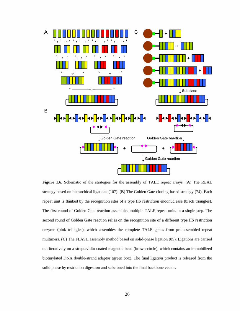
26
Figure 1.6. Schematic of the strategies for the assembly of TALE repeat arrays. (A) The REAL
strategy based on hierarchical ligations (107). (B) The Golden Gate cloning-based strategy (74). Each
repeat unit is flanked by the recognition sites of a type IIS restriction endonuclease (black triangles).
The first round of Golden Gate reaction assembles multiple TALE repeat units in a single step. The
second round of Golden Gate reaction relies on the recognition site of a different type IIS restriction
enzyme (pink triangles), which assembles the complete TALE genes from pre-assembled repeat
multimers. (C) The FLASH assembly method based on solid-phase ligation (85). Ligations are carried
out iteratively on a streptavidin-coated magnetic bead (brown circle), which contains an immobilized
biotinylated DNA double-strand adaptor (green box). The final ligation product is released from the
solid phase by restriction digestion and subcloned into the final backbone vector.
27
assembly efficiency, the lacZ gene and the toxic ccdB gene are introduced for blue/white
screening and selection, respectively. With the help of preassembled TALE repeat
tetramers and trimers, a single-step Golden Gate strategy has been developed to generate
TALENs that recognize 15 bp target sites within two days (69). Due to its ease of use and
public availability, Golden Gate assembly provides a convenient means to construct
TALENs for academic laboratories.
For industrial scale synthesis, development of a solid-phase ligation strategy has
facilitated the cloning of TALE genes in a high-throughput and cost-effective manner
(85,90,123). The solid-phase strategy assembles TALE repeat units on a streptavidin-
coated magnetic bead that contains an immobilized biotinylated DNA double-strand
adaptor with a restriction endonuclease site on one end. Ligation of TALE repeat units is
unidirectional and iterative. In each cycle, newly added TALE repeat units are ligated to
the immobilized DNA fragments and subsequent washing steps remove undesired
products. The cycle is continued until an array of the desired length is assembled. The
final ligation product is then released from the solid phase by restriction digestion and
subcloned into the final backbone vector (Figure 1.6C). With this strategy, TALE repeat
units are assembled on solid-phase rather than in solution, thereby avoiding the need for
gel isolation, purification or analysis of intermediate plasmids. With 376 archived
plasmids encoding TALE repeats as tetramers, trimers, dimers and monomers, the fast
ligation-based automatable solid-phase high-throughput (FLASH) system enables
assembly of 96 TALE genes in less than one day. With automation, FLASH can make
sequence-verified TALE expression plasmids for <$100 each, including the cost of labor
(85). Instead of using a pre-assembled plasmid library, iterative capped assembly (ICA)
28
builds full-length arrays from individual TALE repeat monomers (123). Introduction of
capping oligonucleotides eliminates incomplete ligation and monomer self-ligation,
which are essential for the production of pure full-length TALE repeat arrays. With
automation, ICA enables efficient assembly of TALE genes bearing up to 21 repeats
followed by ligation into an expression plasmid within three hours. Wang and coworkers
performed solid-phase ligation on a chip, which allows the synthesis of >100 TALE
genes bearing 16 or 20 repeats in three days (90).
Recently, a ligation-independent cloning (LIC) technique has been developed for
high-throughput assembly of TALE genes (87). Compared with Golden Gate cloning,
LIC relies on much longer (10-30 bp) nonpalindromic overhangs to anneal with the
overhangs of other fragments in a highly specific manner. The long overhangs are
generated by the controllable 3’-exonuclease activity of T4 DNA polymerase. Because
the fragments’ long overlaps do not dissociate during transformation, the annealed
products can be directly transformed into Escherichia coli without prior ligation step and
ligated through bacterial ligases. Because of its high fidelity, LIC circumvents agar-based
single-colony picking step, which allows growth of cells directly in polyclonal cultures
after transformation. Using 64 repeat dimer-containing plasmids, LIC allows generation
of correctly assembled TALE genes bearing 18.5 repeat units in three days through a
hierarchical, two step assembly process. In addition, a comprehensive 5-mer TALE
repeat unit fragment library composed of 3072 plasmids was created, which enables
automated assembly of >600 TALE genes bearing 15.5 repeat units in one day.
29
1.4.4 Future perspectives
The last three years witnessed the tremendous progress of the TALEN technology. The
scaffold optimization isolated TALEN variants with high DNA cleavage efficiency,
which is essential for targeted genome editing. The characterization of novel RVDs
extended the DNA recognition code and helped to minimize off-target cleavage activity
of TALENs by increasing guanine recognition specificity. Development of novel
strategies for convenient and quick assembly of TALE repeat arrays enabled high-
throughput synthesis of TALENs and made TALEN technology accessible and affordable
for any academic or industrial lab.
Besides TALENs, there are other tools available for editing genomes. Meganucleases
are natural DNA endnucleases with high activity and specificity, but it is difficult to tailor
their DNA recognition specificities. It is relatively easier to engineer ZFNs to target
custom-designed DNA sequences, but some of them suffer from requirement of intensive
labor for construction and off-target effects (as reviewed in (132)). Recently, Clustered
regularly interspaced short palindromic repeats (CRISPR)-mediated DNA cleavage has
been applied for genome editing (133-138). This system can be reprogrammed readily
using customized RNAs and enable multiplex genome engineering. However, the limited
target specificity (14 bp) can cause off-target cleavage and the requirement for a
protospacer adjacent motif (PAM) restricts its targeting range. Compared with these tools,
TALENs have the advantage of high specificity and modularity, but there are limitations
that remain to be addressed for their further improvement. The bulky size of TALENs
might limit their broader applications, especially in the cases when efficient gene delivery
cannot be achieved. Development of strategies for efficient delivery of TALEN genes
30
into cells would enable TALEN-mediated genome editing in more different organisms
and cell types. In eukaryotic cells, DNA is packaged into chromatin. Therefore,
chromosomal context and epigenetic modifications play a major role in the DNA
accessibility of TALENs. The combination of epigenetic modification tools and TALEN
technology could expand the range of target for TALEN-mediated genome modifications,
which might be a potential area for future exploration. Unlike ZFNs, the off-target effects
of TALENs have not been comprehensively characterized. Mussolino and coworkers
carried out a side-by-side comparison between ZFNs and TALENs and found
significantly reduced nuclease-associated cytotoxicities of TALENs (70). Ding and
coworkers also reported minimal off-target-effects of TALENs using exome sequencing
whole-genome sequencing at low coverage, but they still could not completely rule out
TALEN off-target effects (69). Therefore, careful screening of the complete genome of
TALEN-modified cells using deep sequencing analysis would be instructive for safe use
of TALENs, especially for human clinical applications.
Other than fusing with FokI to make DNA endonucleases, TALEs have been used to
create novel chimeric proteins by fusing with other functional protein domains. TALE-
based transcription activators have been constructed to induce transcription of
endogenous genes in plants (117) and human cells (74,119,127,139-141). By fusing with
transcription repressor domains, TALEs have been used to generate artificial repressors
for sequence-specific gene repression in bacteria (142), yeast (143), plants (144) and
human cells (118,141). In addition, chimeric TALE recombinases (TALERs) have been
constructed by fusing a hyperactivated catalytic domain from the DNA invertase Gin
with a TALE central repeat domain (145). TALERs with optimized architecture
31
recombine DNA efficiently in bacterial and mammalian cells, providing an alternative
approach for targeted genome editing. It would be interesting to combine TALE central
repeat arrays with other different functional domains for many different applications in
the future. For example, a TALE combined with a ligand-binding domain can be applied
for high-throughput drug screening; a TALE combined with a DNA methyltransferase
can be used for targeted DNA modification; a TALE combined with a histone
deacetylase can be used for specific chromatin modification; a TALE combined with a
cytosine deaminase can be applied for endogenous targeted mutagenesis, etc.
Thanks to simplicity in design, convenience in construction and high success rates
across species, TALEN technology has received much attention since its invention.
Although challenges and obstacles remain, TALEN technology will continue to be an
important topic for future research and development and benefit both basic and applied
biological sciences.
1.5 Project overview
My thesis research focuses on the design, construction, optimization and application of
TALENs as an efficient tool for genome engineering. Because the wild-type TALEs are
transcription activators for bacterial infection of host plants, their sequences other than
the DNA binding domain can be detrimental to the nuclease activity once fused with the
FokI DNA cleavage domain. To maximize TALEN efficiency, protein engineering
strategies including scaffold optimization and directed evolution have been utilized in
yeast cells. A high-throughput screening of a linker library identified a novel TALEN
architecture that can cleave the DNA target as a monomer. To demonstrate their
32
applications in human therapy, I applied the optimized TALENs for use in the treatment
of sickle cell disease. TALEN-mediated genome editing enabled the correction of the
disease-causing gene in induced pluripotent stem cells isolated from a patient.
Chapter 2 describes scaffold optimization of TALENs for use in treatment of sickle
cell disease. By using a yeast reporter system, a systematic study was carried out to
optimize TALEN architecture for maximal in vivo cleavage efficiency. In contrast to the
previous reports, the engineered TALENs were capable of recognizing and cleaving
target binding sites preceded by A, C or G. More importantly, the optimized TALENs
efficiently cleaved a target sequence within the human β-globin (HBB) gene associated
with sickle cell disease and increased the efficiency of targeted gene repair by >1000-fold
in human cells. In addition, these TALENs showed no detectable cytotoxicity. These
results demonstrate the potential of optimized TALENs as a powerful genome editing
tool for therapeutic applications.
Chapter 3 reports the seamless gene correction of the sickle cell disease mutation in
human induced pluripotent stem cells using TALENs. The TALENs I have engineered
are highly specific and generate minimal off-target effects. In combination with piggyBac
transposon, TALEN-mediated gene targeting leaves no residual ectopic sequences at the
site of correction and the corrected stem cells retain full pluripotency and a normal
karyotype. This study demonstrates an important first step of using TALENs for the
treatment of genetic diseases, which represents a significant advance toward cell and
gene therapies.
Chapter 4 reports the development of a TALEN variant, SunnyTALEN, with >2.5-
fold improved genome editing efficacy in human cells. A high-throughput screening
33
system has been constructed in yeast cells in order to improve TALEN efficiency through
directed evolution. After multiple rounds of mutagenesis and screening, 14 TALEN
mutants were isolated with improved activity either in yeast or human cells, among which
SunnyTALEN shows highest activity for human genome editing. The corresponding
scaffold increases the rate of genetic modification at all the 13 tested loci of human
genome and is compatible with heterodimer TALEN architectures. This enhanced and
high-efficiency TALEN variant represents a novel second-generation TALEN system and
has great potential for biological and therapeutic applications.
Finally, Chapter 5 provides preliminary results of the development and
characterization of a novel TALEN architecture, single-chain TALEN (scTALEN).
Compared with the conventional TALEN scaffold, scTALEN has two FokI DNA
cleavage domains which are linked by a polypeptide linker. The appropriate linker was
isolated from a polypeptide linker library from high-throughput screening. Instead of
forming a heterodimer, the scTALEN we identified can cleave the DNA substrate as a
monomer, with the formation of a catalytic active intra-molecular FokI dimer. The
corresponding scTALEN has moderate in vivo activity in both yeast and human cells.
Compared with the conventional TALEN architecture, scTALEN has only half the
protein size, which significantly decreases the gene delivery payload, especially for
certain cell types such as primary human cells with low DNA or RNA delivery efficiency.
1.6 References
1. Levasseur, D.N., Ryan, T.M., Pawlik, K.M. and Townes, T.M. (2003) Correction
of a mouse model of sickle cell disease: lentiviral/antisickling beta-globin gene
34
transduction of unmobilized, purified hematopoietic stem cells. Blood, 102, 4312-
4319.
2. Check, E. (2002) A tragic setback. Nature, 420, 116-118.
3. Marshall, E. (1999) Gene therapy death prompts review of adenovirus vector.
Science, 286, 2244-2245.
4. Gersbach, C.A., Gaj, T., Gordley, R.M., Mercer, A.C. and Barbas, C.F., 3rd.
(2011) Targeted plasmid integration into the human genome by an engineered
zinc-finger recombinase. Nucleic Acids Res, 39, 7868-7878.
5. Feng, X., Bednarz, A.L. and Colloms, S.D. (2010) Precise targeted integration by
a chimaeric transposase zinc-finger fusion protein. Nucleic Acids Res, 38, 1204-
1216.
6. Jensen, N.M., Dalsgaard, T., Jakobsen, M., Nielsen, R.R., Sorensen, C.B., Bolund,
L. and Jensen, T.G. (2011) An update on targeted gene repair in mammalian cells:
methods and mechanisms. J Biomed Sci, 18, 10.
7. Arnould, S., Delenda, C., Grizot, S., Desseaux, C., Paques, F., Silva, G.H. and
Smith, J. (2010) The I-CreI meganuclease and its engineered derivatives:
applications from cell modification to gene therapy. Protein Eng Des Sel, 24, 27-
31.
8. Marcaida, M.J., Munoz, I.G., Blanco, F.J., Prieto, J. and Montoya, G. (2010)
Homing endonucleases: from basics to therapeutic applications. Cell Mol Life Sci,
67, 727-748.
35
9. Chevalier, B.S. and Stoddard, B.L. (2001) Homing endonucleases: structural and
functional insight into the catalysts of intron/intein mobility. Nucleic Acids Res,
29, 3757-3774.
10. Chames, P., Epinat, J.C., Guillier, S., Patin, A., Lacroix, E. and Paques, F. (2005)
In vivo selection of engineered homing endonucleases using double-strand break
induced homologous recombination. Nucleic Acids Res, 33, e178.
11. Chen, Z. and Zhao, H. (2005) A highly sensitive selection method for directed
evolution of homing endonucleases. Nucleic Acids Res, 33, e154.
12. Doyon, J.B., Pattanayak, V., Meyer, C.B. and Liu, D.R. (2006) Directed evolution
and substrate specificity profile of homing endonuclease I-SceI. J Am Chem Soc,
128, 2477-2484.
13. Chen, Z., Wen, F., Sun, N. and Zhao, H. (2009) Directed evolution of homing
endonuclease I-SceI with altered sequence specificity. Protein Eng Des Sel, 22,
249-256.
14. Ashworth, J., Havranek, J.J., Duarte, C.M., Sussman, D., Monnat, R.J., Jr.,
Stoddard, B.L. and Baker, D. (2006) Computational redesign of endonuclease
DNA binding and cleavage specificity. Nature, 441, 656-659.
15. Ashworth, J., Taylor, G.K., Havranek, J.J., Quadri, S.A., Stoddard, B.L. and
Baker, D. (2010) Computational reprogramming of homing endonuclease
specificity at multiple adjacent base pairs. Nucleic Acids Res, 38, 5601-5608.
16. Thyme, S.B., Jarjour, J., Takeuchi, R., Havranek, J.J., Ashworth, J., Scharenberg,
A.M., Stoddard, B.L. and Baker, D. (2009) Exploitation of binding energy for
catalysis and design. Nature, 461, 1300-1304.
36
17. Arnould, S., Chames, P., Perez, C., Lacroix, E., Duclert, A., Epinat, J.C., Stricher,
F., Petit, A.S., Patin, A., Guillier, S. et al. (2006) Engineering of large numbers of
highly specific homing endonucleases that induce recombination on novel DNA
targets. J Mol Biol, 355, 443-458.
18. Redondo, P., Prieto, J., Munoz, I.G., Alibes, A., Stricher, F., Serrano, L.,
Cabaniols, J.P., Daboussi, F., Arnould, S., Perez, C. et al. (2008) Molecular basis
of xeroderma pigmentosum group C DNA recognition by engineered
meganucleases. Nature, 456, 107-111.
19. Kim, C.A. and Berg, J.M. (1996) A 2.2 A resolution crystal structure of a
designed zinc finger protein bound to DNA. Nat Struct Biol, 3, 940-945.
20. Deng, D., Yan, C., Pan, X., Mahfouz, M., Wang, J., Zhu, J.K., Shi, Y. and Yan, N.
(2012) Structural basis for sequence-specific recognition of DNA by TAL
effectors. Science, 335, 720-723.
21. Fajardo-Sanchez, E., Stricher, F., Paques, F., Isalan, M. and Serrano, L. (2008)
Computer design of obligate heterodimer meganucleases allows efficient cutting
of custom DNA sequences. Nucleic Acids Res, 36, 2163-2173.
22. Epinat, J.C., Arnould, S., Chames, P., Rochaix, P., Desfontaines, D., Puzin, C.,
Patin, A., Zanghellini, A., Paques, F. and Lacroix, E. (2003) A novel engineered
meganuclease induces homologous recombination in yeast and mammalian cells.
Nucleic Acids Res, 31, 2952-2962.
23. Gao, H., Smith, J., Yang, M., Jones, S., Djukanovic, V., Nicholson, M.G., West,
A., Bidney, D., Falco, S.C., Jantz, D. et al. (2010) Heritable targeted mutagenesis
in maize using a designed endonuclease. Plant J, 61, 176-187.
37
24. Li, H., Pellenz, S., Ulge, U., Stoddard, B.L. and Monnat, R.J., Jr. (2009)
Generation of single-chain LAGLIDADG homing endonucleases from native
homodimeric precursor proteins. Nucleic Acids Res, 37, 1650-1662.
25. Ulge, U.Y., Baker, D.A. and Monnat, R.J., Jr. (2011) Comprehensive
computational design of mCreI homing endonuclease cleavage specificity for
genome engineering. Nucleic Acids Res, 39, 4330-4339.
26. Smith, J., Grizot, S., Arnould, S., Duclert, A., Epinat, J.C., Chames, P., Prieto, J.,
Redondo, P., Blanco, F.J., Bravo, J. et al. (2006) A combinatorial approach to
create artificial homing endonucleases cleaving chosen sequences. Nucleic Acids
Res, 34, e149.
27. Munoz, I.G., Prieto, J., Subramanian, S., Coloma, J., Redondo, P., Villate, M.,
Merino, N., Marenchino, M., D'Abramo, M., Gervasio, F.L. et al. (2011)
Molecular basis of engineered meganuclease targeting of the endogenous human
RAG1 locus. Nucleic Acids Res, 39, 729-743.
28. Arnould, S., Perez, C., Cabaniols, J.P., Smith, J., Gouble, A., Grizot, S., Epinat,
J.C., Duclert, A., Duchateau, P. and Paques, F. (2007) Engineered I-CreI
derivatives cleaving sequences from the human XPC gene can induce highly
efficient gene correction in mammalian cells. J Mol Biol, 371, 49-65.
29. Grosse, S., Huot, N., Mahiet, C., Arnould, S., Barradeau, S., Clerre, D.L., Chion-
Sotinel, I., Jacqmarcq, C., Chapellier, B., Ergani, A. et al. (2011) Meganuclease-
mediated inhibition of HSV1 infection in cultured cells. Mol Ther, 19, 694-702.
30. Grizot, S., Smith, J., Daboussi, F., Prieto, J., Redondo, P., Merino, N., Villate, M.,
Thomas, S., Lemaire, L., Montoya, G. et al. (2009) Efficient targeting of a SCID
38
gene by an engineered single-chain homing endonuclease. Nucleic Acids Res, 37,
5405-5419.
31. Takeuchi, R., Lambert, A.R., Mak, A.N., Jacoby, K., Dickson, R.J., Gloor, G.B.,
Scharenberg, A.M., Edgell, D.R. and Stoddard, B.L. (2011) Tapping natural
reservoirs of homing endonucleases for targeted gene modification. Proc Natl
Acad Sci U S A, 108, 13077-13082.
32. Porteus, M.H. and Carroll, D. (2005) Gene targeting using zinc finger nucleases.
Nat Biotechnol, 23, 967-973.
33. Bitinaite, J., Wah, D.A., Aggarwal, A.K. and Schildkraut, I. (1998) FokI
dimerization is required for DNA cleavage. Proc Natl Acad Sci U S A, 95, 10570-
10575.
34. Carroll, D. (2011) Genome engineering with zinc-finger nucleases. Genetics, 188,
773-782.
35. Cathomen, T. and Joung, J.K. (2008) Zinc-finger nucleases: the next generation
emerges. Mol Ther, 16, 1200-1207.
36. Urnov, F.D., Rebar, E.J., Holmes, M.C., Zhang, H.S. and Gregory, P.D. (2010)
Genome editing with engineered zinc finger nucleases. Nat Rev Genet, 11, 636-
646.
37. Gabriel, R., Lombardo, A., Arens, A., Miller, J.C., Genovese, P., Kaeppel, C.,
Nowrouzi, A., Bartholomae, C.C., Wang, J., Friedman, G. et al. (2011) An
unbiased genome-wide analysis of zinc-finger nuclease specificity. Nat
Biotechnol, 29, 816-823.
39
38. Pattanayak, V., Ramirez, C.L., Joung, J.K. and Liu, D.R. (2011) Revealing off-
target cleavage specificities of zinc-finger nucleases by in vitro selection. Nat
Methods, 8, 765-770.
39. Perez, E.E., Wang, J., Miller, J.C., Jouvenot, Y., Kim, K.A., Liu, O., Wang, N.,
Lee, G., Bartsevich, V.V., Lee, Y.L. et al. (2008) Establishment of HIV-1
resistance in CD4+ T cells by genome editing using zinc-finger nucleases. Nat
Biotechnol, 26, 808-816.
40. Holt, N., Wang, J., Kim, K., Friedman, G., Wang, X., Taupin, V., Crooks, G.M.,
Kohn, D.B., Gregory, P.D., Holmes, M.C. et al. (2010) Human hematopoietic
stem/progenitor cells modified by zinc-finger nucleases targeted to CCR5 control
HIV-1 in vivo. Nat Biotechnol, 28, 839-847.
41. Cradick, T.J., Keck, K., Bradshaw, S., Jamieson, A.C. and McCaffrey, A.P. (2010)
Zinc-finger nucleases as a novel therapeutic strategy for targeting hepatitis B
virus DNAs. Mol Ther, 18, 947-954.
42. Geurts, A.M., Cost, G.J., Freyvert, Y., Zeitler, B., Miller, J.C., Choi, V.M.,
Jenkins, S.S., Wood, A., Cui, X., Meng, X. et al. (2009) Knockout rats via
embryo microinjection of zinc-finger nucleases. Science, 325, 433.
43. Mashimo, T., Takizawa, A., Voigt, B., Yoshimi, K., Hiai, H., Kuramoto, T. and
Serikawa, T. (2010) Generation of knockout rats with X-linked severe combined
immunodeficiency (X-SCID) using zinc-finger nucleases. PLoS One, 5, e8870.
44. Flisikowska, T., Thorey, I.S., Offner, S., Ros, F., Lifke, V., Zeitler, B., Rottmann,
O., Vincent, A., Zhang, L., Jenkins, S. et al. (2011) Efficient immunoglobulin
40
gene disruption and targeted replacement in rabbit using zinc finger nucleases.
PLoS One, 6, e21045.
45. Hauschild, J., Petersen, B., Santiago, Y., Queisser, A.L., Carnwath, J.W., Lucas-
Hahn, A., Zhang, L., Meng, X., Gregory, P.D., Schwinzer, R. et al. (2011)
Efficient generation of a biallelic knockout in pigs using zinc-finger nucleases.
Proc Natl Acad Sci U S A, 108, 12013-12017.
46. Zou, J., Maeder, M.L., Mali, P., Pruett-Miller, S.M., Thibodeau-Beganny, S.,
Chou, B.K., Chen, G., Ye, Z., Park, I.H., Daley, G.Q. et al. (2009) Gene targeting
of a disease-related gene in human induced pluripotent stem and embryonic stem
cells. Cell Stem Cell, 5, 97-110.
47. Hockemeyer, D., Soldner, F., Beard, C., Gao, Q., Mitalipova, M., DeKelver, R.C.,
Katibah, G.E., Amora, R., Boydston, E.A., Zeitler, B. et al. (2009) Efficient
targeting of expressed and silent genes in human ESCs and iPSCs using zinc-
finger nucleases. Nat Biotechnol, 27, 851-857.
48. Kotin, R.M., Linden, R.M. and Berns, K.I. (1992) Characterization of a preferred
site on human chromosome 19q for integration of adeno-associated virus DNA by
non-homologous recombination. Embo J, 11, 5071-5078.
49. DeKelver, R.C., Choi, V.M., Moehle, E.A., Paschon, D.E., Hockemeyer, D.,
Meijsing, S.H., Sancak, Y., Cui, X., Steine, E.J., Miller, J.C. et al. (2010)
Functional genomics, proteomics, and regulatory DNA analysis in isogenic
settings using zinc finger nuclease-driven transgenesis into a safe harbor locus in
the human genome. Genome Res, 20, 1133-1142.
41
50. Zou, J., Sweeney, C.L., Chou, B.K., Choi, U., Pan, J., Wang, H., Dowey, S.N.,
Cheng, L. and Malech, H.L. (2011) Oxidase-deficient neutrophils from X-linked
chronic granulomatous disease iPS cells: functional correction by zinc finger
nuclease-mediated safe harbor targeting. Blood, 117, 5561-5572.
51. Sebastiano, V., Maeder, M.L., Angstman, J.F., Haddad, B., Khayter, C., Yeo,
D.T., Goodwin, M.J., Hawkins, J.S., Ramirez, C.L., Batista, L.F. et al. (2011) In
situ genetic correction of the sickle cell anemia mutation in human induced
pluripotent stem cells using engineered zinc finger nucleases. Stem Cells, 29,
1717-1726.
52. Maeder, M.L., Thibodeau-Beganny, S., Osiak, A., Wright, D.A., Anthony, R.M.,
Eichtinger, M., Jiang, T., Foley, J.E., Winfrey, R.J., Townsend, J.A. et al. (2008)
Rapid "open-source" engineering of customized zinc-finger nucleases for highly
efficient gene modification. Mol Cell, 31, 294-301.
53. Zou, J., Mali, P., Huang, X., Dowey, S.N. and Cheng, L. (2011) Site-specific gene
correction of a point mutation in human iPS cells derived from an adult patient
with sickle cell disease. Blood, 118, 4599-4608.
54. Yusa, K., Rashid, S.T., Strick-Marchand, H., Varela, I., Liu, P.Q., Paschon, D.E.,
Miranda, E., Ordonez, A., Hannan, N.R., Rouhani, F.J. et al. (2011) Targeted
gene correction of alpha1-antitrypsin deficiency in induced pluripotent stem cells.
Nature, 478, 391-394.
55. Wang, W., Lin, C., Lu, D., Ning, Z., Cox, T., Melvin, D., Wang, X., Bradley, A.
and Liu, P. (2008) Chromosomal transposition of PiggyBac in mouse embryonic
stem cells. Proc Natl Acad Sci U S A, 105, 9290-9295.
42
56. Soldner, F., Laganiere, J., Cheng, A.W., Hockemeyer, D., Gao, Q., Alagappan, R.,
Khurana, V., Golbe, L.I., Myers, R.H., Lindquist, S. et al. (2011) Generation of
isogenic pluripotent stem cells differing exclusively at two early onset Parkinson
point mutations. Cell, 146, 318-331.
57. Feuk, L. (2010) Inversion variants in the human genome: role in disease and
genome architecture. Genome medicine, 2, 11.
58. Brunet, E., Simsek, D., Tomishima, M., DeKelver, R., Choi, V.M., Gregory, P.,
Urnov, F., Weinstock, D.M. and Jasin, M. (2009) Chromosomal translocations
induced at specified loci in human stem cells. Proc Natl Acad Sci U S A, 106,
10620-10625.
59. Lee, H.J., Kim, E. and Kim, J.S. (2010) Targeted chromosomal deletions in
human cells using zinc finger nucleases. Genome Res, 20, 81-89.
60. Lee, H.J., Kweon, J., Kim, E., Kim, S. and Kim, J.S. (2012) Targeted
chromosomal duplications and inversions in the human genome using zinc finger
nucleases. Genome Res, 22, 539-548.
61. Bagnall, R.D., Waseem, N., Green, P.M. and Giannelli, F. (2002) Recurrent
inversion breaking intron 1 of the factor VIII gene is a frequent cause of severe
hemophilia A. Blood, 99, 168-174.
62. Ramirez, C.L., Foley, J.E., Wright, D.A., Muller-Lerch, F., Rahman, S.H., Cornu,
T.I., Winfrey, R.J., Sander, J.D., Fu, F., Townsend, J.A. et al. (2008) Unexpected
failure rates for modular assembly of engineered zinc fingers. Nat Methods, 5,
374-375.
43
63. Radecke, S., Radecke, F., Cathomen, T. and Schwarz, K. (2010) Zinc-finger
nuclease-induced gene repair with oligodeoxynucleotides: wanted and unwanted
target locus modifications. Mol Ther, 18, 743-753.
64. Joung, J.K. and Sander, J.D. (2013) TALENs: a widely applicable technology for
targeted genome editing. Nat Rev Mol Cell Biol, 14, 49-55.
65. Bogdanove, A.J. and Voytas, D.F. (2011) TAL effectors: customizable proteins
for DNA targeting. Science, 333, 1843-1846.
66. Munoz Bodnar, A., Bernal, A., Szurek, B. and Lopez, C.E. (2013) Tell me a tale
of TALEs. Mol Biotechnol, 53, 228-235.
67. Boch, J., Scholze, H., Schornack, S., Landgraf, A., Hahn, S., Kay, S., Lahaye, T.,
Nickstadt, A. and Bonas, U. (2009) Breaking the code of DNA binding specificity
of TAL-type III effectors. Science, 326, 1509-1512.
68. Moscou, M.J. and Bogdanove, A.J. (2009) A simple cipher governs DNA
recognition by TAL effectors. Science, 326, 1501.
69. Ding, Q., Lee, Y.K., Schaefer, E.A., Peters, D.T., Veres, A., Kim, K.,
Kuperwasser, N., Motola, D.L., Meissner, T.B., Hendriks, W.T. et al. (2013) A
TALEN Genome-Editing System for Generating Human Stem Cell-Based
Disease Models. Cell Stem Cell, 12, 238-251.
70. Mussolino, C., Morbitzer, R., Lutge, F., Dannemann, N., Lahaye, T. and
Cathomen, T. (2011) A novel TALE nuclease scaffold enables high genome
editing activity in combination with low toxicity. Nucleic Acids Res, 39, 9283-
9293.
44
71. Li, T., Huang, S., Jiang, W.Z., Wright, D., Spalding, M.H., Weeks, D.P. and Yang,
B. (2011) TAL nucleases (TALNs): hybrid proteins composed of TAL effectors
and FokI DNA-cleavage domain. Nucleic Acids Res, 39, 359-372.
72. Christian, M., Cermak, T., Doyle, E.L., Schmidt, C., Zhang, F., Hummel, A.,
Bogdanove, A.J. and Voytas, D.F. (2010) Targeting DNA double-strand breaks
with TAL effector nucleases. Genetics, 186, 757-761.
73. White, F.F., Potnis, N., Jones, J.B. and Koebnik, R. (2009) The type III effectors
of Xanthomonas. Mol Plant Pathol, 10, 749-766.
74. Cermak, T., Doyle, E.L., Christian, M., Wang, L., Zhang, Y., Schmidt, C., Baller,
J.A., Somia, N.V., Bogdanove, A.J. and Voytas, D.F. (2011) Efficient design and
assembly of custom TALEN and other TAL effector-based constructs for DNA
targeting. Nucleic Acids Res, 39, e82.
75. Shan, Q., Wang, Y., Chen, K., Liang, Z., Li, J., Zhang, Y., Zhang, K., Liu, J.,
Voytas, D.F., Zheng, X. et al. (2013) Rapid and efficient gene modification in rice
and Brachypodium using TALENs. Mol Plant, doi: 10.1093/mp/sss1162.
76. Carlson, D.F., Tan, W., Lillico, S.G., Stverakova, D., Proudfoot, C., Christian, M.,
Voytas, D.F., Long, C.R., Whitelaw, C.B. and Fahrenkrug, S.C. (2012) Efficient
TALEN-mediated gene knockout in livestock. Proc Natl Acad Sci U S A, 109,
17382-17387.
77. Watanabe, T., Ochiai, H., Sakuma, T., Horch, H.W., Hamaguchi, N., Nakamura,
T., Bando, T., Ohuchi, H., Yamamoto, T., Noji, S. et al. (2012) Non-transgenic
genome modifications in a hemimetabolous insect using zinc-finger and TAL
effector nucleases. Nat Commun, 3, 1017.
45
78. Ishibashi, S., Cliffe, R. and Amaya, E. (2012) Highly efficient bi-allelic mutation
rates using TALENs in Xenopus tropicalis. Biol Open, 1, 1273-1276.
79. Lei, Y., Guo, X., Liu, Y., Cao, Y., Deng, Y., Chen, X., Cheng, C.H., Dawid, I.B.,
Chen, Y. and Zhao, H. (2012) Efficient targeted gene disruption in Xenopus
embryos using engineered transcription activator-like effector nucleases
(TALENs). Proc Natl Acad Sci U S A, 109, 17484-17489.
80. Liu, J., Li, C., Yu, Z., Huang, P., Wu, H., Wei, C., Zhu, N., Shen, Y., Chen, Y.,
Zhang, B. et al. (2012) Efficient and specific modifications of the Drosophila
genome by means of an easy TALEN strategy. J Genet Genomics, 39, 209-215.
81. Cristea, S., Freyvert, Y., Santiago, Y., Holmes, M.C., Urnov, F.D., Gregory, P.D.
and Cost, G.J. (2013) In vivo cleavage of transgene donors promotes nuclease-
mediated targeted integration. Biotechnol Bioeng, 110, 871-880.
82. Hockemeyer, D., Wang, H., Kiani, S., Lai, C.S., Gao, Q., Cassady, J.P., Cost, G.J.,
Zhang, L., Santiago, Y., Miller, J.C. et al. (2011) Genetic engineering of human
pluripotent cells using TALE nucleases. Nat Biotechnol, 29, 731-734.
83. Kim, H., Um, E., Cho, S.R., Jung, C., Kim, H. and Kim, J.S. (2011) Surrogate
reporters for enrichment of cells with nuclease-induced mutations. Nat Methods, 8,
941-943.
84. Miller, J.C., Tan, S., Qiao, G., Barlow, K.A., Wang, J., Xia, D.F., Meng, X.,
Paschon, D.E., Leung, E., Hinkley, S.J. et al. (2011) A TALE nuclease
architecture for efficient genome editing. Nat Biotechnol, 29, 143-148.
46
85. Reyon, D., Tsai, S.Q., Khayter, C., Foden, J.A., Sander, J.D. and Joung, J.K.
(2012) FLASH assembly of TALENs for high-throughput genome editing. Nat
Biotechnol, 30, 460-465.
86. Sanjana, N.E., Cong, L., Zhou, Y., Cunniff, M.M., Feng, G. and Zhang, F. (2012)
A transcription activator-like effector toolbox for genome engineering. Nat Protoc,
7, 171-192.
87. Schmid-Burgk, J.L., Schmidt, T., Kaiser, V., Honing, K. and Hornung, V. (2013)
A ligation-independent cloning technique for high-throughput assembly of
transcription activator-like effector genes. Nat Biotechnol, 31, 76-81.
88. Stroud, D.A., Formosa, L.E., Wijeyeratne, X.W., Nguyen, T.N. and Ryan, M.T.
(2013) Gene knockout using transcription activator-like effector nucleases
(TALENs) reveals that human NDUFA9 protein is essential for stabilizing the
junction between membrane and matrix arms of complex I. J Biol Chem, 288,
1685-1690.
89. Sun, N., Liang, J., Abil, Z. and Zhao, H. (2012) Optimized TAL effector
nucleases (TALENs) for use in treatment of sickle cell disease. Mol Biosyst, 8,
1255-1263.
90. Wang, Z., Li, J., Huang, H., Wang, G., Jiang, M., Yin, S., Sun, C., Zhang, H.,
Zhuang, F. and Xi, J.J. (2012) An integrated chip for the high-throughput
synthesis of transcription activator-like effectors. Angew Chem Int Ed Engl, 51,
8505-8508.
47
91. Ansai, S., Sakuma, T., Yamamoto, T., Ariga, H., Uemura, N., Takahashi, R. and
Kinoshita, M. (2013) Efficient targeted mutagenesis in medaka using custom-
designed transcription activator-like effector nucleases. Genetics, 193, 739-749.
92. Sung, Y.H., Baek, I.J., Kim, D.H., Jeon, J., Lee, J., Lee, K., Jeong, D., Kim, J.S.
and Lee, H.W. (2013) Knockout mice created by TALEN-mediated gene
targeting. Nat Biotechnol, 31, 23-24.
93. Wood, A.J., Lo, T.W., Zeitler, B., Pickle, C.S., Ralston, E.J., Lee, A.H., Amora,
R., Miller, J.C., Leung, E., Meng, X. et al. (2011) Targeted genome editing across
species using ZFNs and TALENs. Science, 333, 307.
94. Tesson, L., Usal, C., Menoret, S., Leung, E., Niles, B.J., Remy, S., Santiago, Y.,
Vincent, A.I., Meng, X., Zhang, L. et al. (2011) Knockout rats generated by
embryo microinjection of TALENs. Nat Biotechnol, 29, 695-696.
95. Tong, C., Huang, G., Ashton, C., Wu, H., Yan, H. and Ying, Q.L. (2012) Rapid
and cost-effective gene targeting in rat embryonic stem cells by TALENs. J Genet
Genomics, 39, 275-280.
96. Li, T., Liu, B., Spalding, M.H., Weeks, D.P. and Yang, B. (2012) High-efficiency
TALEN-based gene editing produces disease-resistant rice. Nat Biotechnol, 30,
390-392.
97. Ma, S., Zhang, S., Wang, F., Liu, Y., Liu, Y., Xu, H., Liu, C., Lin, Y., Zhao, P.
and Xia, Q. (2012) Highly efficient and specific genome editing in silkworm
using custom TALENs. PLoS One, 7, e45035.
48
98. Sajwan, S., Takasu, Y., Tamura, T., Uchino, K., Sezutsu, H. and Zurovec, M.
(2013) Efficient disruption of endogenous Bombyx gene by TAL effector
nucleases. Insect Biochem Mol Biol, 43, 17-23.
99. Zhang, Y., Zhang, F., Li, X., Baller, J.A., Qi, Y., Starker, C.G., Bogdanove, A.J.
and Voytas, D.F. (2013) Transcription activator-like effector nucleases enable
efficient plant genome engineering. Plant Physiol, 161, 20-27.
100. Li, T., Huang, S., Zhao, X., Wright, D.A., Carpenter, S., Spalding, M.H., Weeks,
D.P. and Yang, B. (2011) Modularly assembled designer TAL effector nucleases
for targeted gene knockout and gene replacement in eukaryotes. Nucleic Acids
Res, 39, 6315-6325.
101. Bedell, V.M., Wang, Y., Campbell, J.M., Poshusta, T.L., Starker, C.G., Krug,
R.G., 2nd, Tan, W., Penheiter, S.G., Ma, A.C., Leung, A.Y. et al. (2012) In vivo
genome editing using a high-efficiency TALEN system. Nature, 491, 114-118.
102. Cade, L., Reyon, D., Hwang, W.Y., Tsai, S.Q., Patel, S., Khayter, C., Joung, J.K.,
Sander, J.D., Peterson, R.T. and Yeh, J.R. (2012) Highly efficient generation of
heritable zebrafish gene mutations using homo- and heterodimeric TALENs.
Nucleic Acids Res, 40, 8001-8010.
103. Chen, S., Oikonomou, G., Chiu, C.N., Niles, B.J., Liu, J., Lee, D.A.,
Antoshechkin, I. and Prober, D.A. (2013) A large-scale in vivo analysis reveals
that TALENs are significantly more mutagenic than ZFNs generated using
context-dependent assembly. Nucleic Acids Res, 41, 2769-2778.
104. Dahlem, T.J., Hoshijima, K., Jurynec, M.J., Gunther, D., Starker, C.G., Locke,
A.S., Weis, A.M., Voytas, D.F. and Grunwald, D.J. (2012) Simple methods for
49
generating and detecting locus-specific mutations induced with TALENs in the
zebrafish genome. PLoS Genet, 8, e1002861.
105. Huang, P., Xiao, A., Zhou, M., Zhu, Z., Lin, S. and Zhang, B. (2011) Heritable
gene targeting in zebrafish using customized TALENs. Nat Biotechnol, 29, 699-
700.
106. Moore, F.E., Reyon, D., Sander, J.D., Martinez, S.A., Blackburn, J.S., Khayter, C.,
Ramirez, C.L., Joung, J.K. and Langenau, D.M. (2012) Improved somatic
mutagenesis in zebrafish using transcription activator-like effector nucleases
(TALENs). PLoS One, 7, e37877.
107. Sander, J.D., Cade, L., Khayter, C., Reyon, D., Peterson, R.T., Joung, J.K. and
Yeh, J.R. (2011) Targeted gene disruption in somatic zebrafish cells using
engineered TALENs. Nat Biotechnol, 29, 697-698.
108. Mahfouz, M.M., Li, L., Shamimuzzaman, M., Wibowo, A., Fang, X. and Zhu, J.K.
(2011) De novo-engineered transcription activator-like effector (TALE) hybrid
nuclease with novel DNA binding specificity creates double-strand breaks. Proc
Natl Acad Sci U S A, 108, 2623-2628.
109. Christian, M.L., Demorest, Z.L., Starker, C.G., Osborn, M.J., Nyquist, M.D.,
Zhang, Y., Carlson, D.F., Bradley, P., Bogdanove, A.J. and Voytas, D.F. (2012)
Targeting G with TAL effectors: a comparison of activities of TALENs
constructed with NN and NK repeat variable di-residues. PLoS One, 7, e45383.
110. Gao, H., Wu, X., Chai, J. and Han, Z. (2012) Crystal structure of a TALE protein
reveals an extended N-terminal DNA binding region. Cell Res, 22, 1716-1720.
50
111. Mak, A.N., Bradley, P., Cernadas, R.A., Bogdanove, A.J. and Stoddard, B.L.
(2012) The crystal structure of TAL effector PthXo1 bound to its DNA target.
Science, 335, 716-719.
112. Deng, D., Yin, P., Yan, C., Pan, X., Gong, X., Qi, S., Xie, T., Mahfouz, M., Zhu,
J.K., Yan, N. et al. (2012) Recognition of methylated DNA by TAL effectors.
Cell Res, 22, 1502-1504.
113. Szczepek, M., Brondani, V., Buchel, J., Serrano, L., Segal, D.J. and Cathomen, T.
(2007) Structure-based redesign of the dimerization interface reduces the toxicity
of zinc-finger nucleases. Nat Biotechnol, 25, 786-793.
114. Miller, J.C., Holmes, M.C., Wang, J., Guschin, D.Y., Lee, Y.L., Rupniewski, I.,
Beausejour, C.M., Waite, A.J., Wang, N.S., Kim, K.A. et al. (2007) An improved
zinc-finger nuclease architecture for highly specific genome editing. Nat
Biotechnol, 25, 778-785.
115. Doyon, Y., Vo, T.D., Mendel, M.C., Greenberg, S.G., Wang, J., Xia, D.F., Miller,
J.C., Urnov, F.D., Gregory, P.D. and Holmes, M.C. (2011) Enhancing zinc-
finger-nuclease activity with improved obligate heterodimeric architectures. Nat
Methods, 8, 74-79.
116. Streubel, J., Blucher, C., Landgraf, A. and Boch, J. (2012) TAL effector RVD
specificities and efficiencies. Nat Biotechnol, 30, 593-595.
117. Morbitzer, R., Romer, P., Boch, J. and Lahaye, T. (2010) Regulation of selected
genome loci using de novo-engineered transcription activator-like effector
(TALE)-type transcription factors. Proc Natl Acad Sci U S A, 107, 21617-21622.
51
118. Cong, L., Zhou, R., Kuo, Y.C., Cunniff, M. and Zhang, F. (2012) Comprehensive
interrogation of natural TALE DNA-binding modules and transcriptional
repressor domains. Nat Commun, 3, 968.
119. Bultmann, S., Morbitzer, R., Schmidt, C.S., Thanisch, K., Spada, F., Elsaesser, J.,
Lahaye, T. and Leonhardt, H. (2012) Targeted transcriptional activation of silent
oct4 pluripotency gene by combining designer TALEs and inhibition of
epigenetic modifiers. Nucleic Acids Res, 40, 5368-5377.
120. Su, Z., Han, L. and Zhao, Z. (2011) Conservation and divergence of DNA
methylation in eukaryotes: new insights from single base-resolution DNA
methylomes. Epigenetics, 6, 134-140.
121. Maunakea, A.K., Nagarajan, R.P., Bilenky, M., Ballinger, T.J., D'Souza, C.,
Fouse, S.D., Johnson, B.E., Hong, C., Nielsen, C., Zhao, Y. et al. (2010)
Conserved role of intragenic DNA methylation in regulating alternative promoters.
Nature, 466, 253-257.
122. Valton, J., Dupuy, A., Daboussi, F., Thomas, S., Marechal, A., Macmaster, R.,
Melliand, K., Juillerat, A. and Duchateau, P. (2012) Overcoming transcription
activator-like effector (TALE) DNA binding domain sensitivity to cytosine
methylation. J Biol Chem, 287, 38427-38432.
123. Briggs, A.W., Rios, X., Chari, R., Yang, L., Zhang, F., Mali, P. and Church, G.M.
(2012) Iterative capped assembly: rapid and scalable synthesis of repeat-module
DNA such as TAL effectors from individual monomers. Nucleic Acids Res, 40,
e117.
52
124. Yu, Y., Streubel, J., Balzergue, S., Champion, A., Boch, J., Koebnik, R., Feng, J.,
Verdier, V. and Szurek, B. (2011) Colonization of rice leaf blades by an African
strain of Xanthomonas oryzae pv. oryzae depends on a new TAL effector that
induces the rice nodulin-3 Os11N3 gene. Mol Plant Microbe Interact, 24, 1102-
1113.
125. Yang, Y. and Gabriel, D.W. (1995) Xanthomonas avirulence/pathogenicity gene
family encodes functional plant nuclear targeting signals. Mol Plant Microbe
Interact, 8, 627-631.
126. Reyon, D., Khayter, C., Regan, M.R., Joung, J.K. and Sander, J.D. (2012)
Engineering designer transcription activator-like effector nucleases (TALENs) by
REAL or REAL-Fast assembly. Curr Protoc Mol Biol, Chapter 12, Unit 12 15.
127. Geissler, R., Scholze, H., Hahn, S., Streubel, J., Bonas, U., Behrens, S.E. and
Boch, J. (2011) Transcriptional activators of human genes with programmable
DNA-specificity. PLoS One, 6, e19509.
128. Li, L., Piatek, M.J., Atef, A., Piatek, A., Wibowo, A., Fang, X., Sabir, J.S., Zhu,
J.K. and Mahfouz, M.M. (2012) Rapid and highly efficient construction of TALE-
based transcriptional regulators and nucleases for genome modification. Plant
Mol Biol, 78, 407-416.
129. Morbitzer, R., Elsaesser, J., Hausner, J. and Lahaye, T. (2011) Assembly of
custom TALE-type DNA binding domains by modular cloning. Nucleic Acids Res,
39, 5790-5799.
53
130. Weber, E., Gruetzner, R., Werner, S., Engler, C. and Marillonnet, S. (2011)
Assembly of designer TAL effectors by Golden Gate cloning. PLoS One, 6,
e19722.
131. Engler, C., Kandzia, R. and Marillonnet, S. (2008) A one pot, one step, precision
cloning method with high throughput capability. PLoS One, 3, e3647.
132. Sun, N., Abil, Z. and Zhao, H. (2012) Recent advances in targeted genome
engineering in mammalian systems. Biotechnol J, 7, 1074-1087.
133. Cho, S.W., Kim, S., Kim, J.M. and Kim, J.S. (2013) Targeted genome
engineering in human cells with the Cas9 RNA-guided endonuclease. Nat
Biotechnol, 31, 230-232.
134. Cong, L., Ran, F.A., Cox, D., Lin, S., Barretto, R., Habib, N., Hsu, P.D., Wu, X.,
Jiang, W., Marraffini, L.A. et al. (2013) Multiplex genome engineering using
CRISPR/Cas systems. Science, 339, 819-823.
135. Hwang, W.Y., Fu, Y., Reyon, D., Maeder, M.L., Tsai, S.Q., Sander, J.D.,
Peterson, R.T., Yeh, J.R. and Joung, J.K. (2013) Efficient genome editing in
zebrafish using a CRISPR-Cas system. Nat Biotechnol, 31, 227-229.
136. Jiang, W., Bikard, D., Cox, D., Zhang, F. and Marraffini, L.A. (2013) RNA-
guided editing of bacterial genomes using CRISPR-Cas systems. Nat Biotechnol,
31, 233-239.
137. Jinek, M., East, A., Cheng, A., Lin, S., Ma, E. and Doudna, J. (2013) RNA-
programmed genome editing in human cells. Elife, 2, e00471.
54
138. Mali, P., Yang, L., Esvelt, K.M., Aach, J., Guell, M., DiCarlo, J.E., Norville, J.E.
and Church, G.M. (2013) RNA-guided human genome engineering via Cas9.
Science, 339, 823-826.
139. Garg, A., Lohmueller, J.J., Silver, P.A. and Armel, T.Z. (2012) Engineering
synthetic TAL effectors with orthogonal target sites. Nucleic Acids Res, 40, 7584-
7595.
140. Tremblay, J.P., Chapdelaine, P., Coulombe, Z. and Rousseau, J. (2012)
Transcription activator-like effector proteins induce the expression of the frataxin
gene. Hum Gene Ther, 23, 883-890.
141. Li, Y., Moore, R., Guinn, M. and Bleris, L. (2012) Transcription activator-like
effector hybrids for conditional control and rewiring of chromosomal transgene
expression. Sci Rep, 2, 897.
142. Politz, M.C., Copeland, M.F. and Pfleger, B.F. (2013) Artificial repressors for
controlling gene expression in bacteria. Chem Commun (Camb), 49, 4325-4327.
143. Blount, B.A., Weenink, T., Vasylechko, S. and Ellis, T. (2012) Rational
diversification of a promoter providing fine-tuned expression and orthogonal
regulation for synthetic biology. PLoS One, 7, e33279.
144. Mahfouz, M.M., Li, L., Piatek, M., Fang, X., Mansour, H., Bangarusamy, D.K.
and Zhu, J.K. (2012) Targeted transcriptional repression using a chimeric TALE-
SRDX repressor protein. Plant Mol Biol, 78, 311-321.
145. Mercer, A.C., Gaj, T., Fuller, R.P. and Barbas, C.F., 3rd. (2012) Chimeric TALE
recombinases with programmable DNA sequence specificity. Nucleic Acids Res,
40, 11163-11172.

55
CHAPTER 2. SCAFFOLD OPTIMIZATION OF TALENS
FOR USE IN TREATMENT OF SICKLE CELL DISEASE
2.1. Introduction
Sickle cell disease (SCD) is one of the most prevalent autosomal recessive disorders
worldwide, with which approximately 250,000 infants are born each year (1). The
homozygous mutation causing SCD is a single substitution (from A to T) in the codon for
amino acid 6 on the human β-globin (HBB) gene. The replacement of glutamate by valine
causes the body to produce an abnormal hemoglobin protein that tends to aggregate,
making the red blood cells to become stiff and develop a sickle form. These sickle red
blood cells can block blood vessels, thus reducing blood flow to many parts of the body
that results in tissue and organ damage (reviewed by (2)). Ex vivo gene therapy by gene
addition has proven to be an effective method to cure SCD in mouse models (3,4).
However, these studies utilize viral vectors that could lead to a toxic immunological
response (5). In addition, random insertion of foreign DNA fragments into human
chromosomes may result in disruption of essential genes, thus causing unpredictable side
effects (e.g., carcinogenesis) (6).
Targeted gene correction is considered a promising strategy to cure SCD with
minimal side effects because it can directly correct the mutation on the chromosome, thus
preserving temporal and tissue-specific expression of the impaired HBB gene (7).
Targeted gene correction involves the homologous recombination (HR) of chromosomal
DNA sequences with exogenous DNA sequences to provide a precise method to
systematically edit mammalian genomes (8). The rates of recombination in mammalian
56
cells typically fall in the range of 10-6
−10-5
events/cell/generation (9), which is an
inherent limitation of this technique. However, chromosomal double strand breaks
(DSBs) at a desired chromosomal locus can stimulate HR by upwards of 1000 fold to
facilitate gene targeting (see Chapter 1).
Zinc finger nucleases (ZFNs) show promise in improving the efficiency of gene
targeting by generating DSBs at preselected sites on the chromosome (10,11). ZFNs are
artificial DNA nucleases constructed by fusing the custom-designed zinc finger domain
with the non-specific DNA cleavage domain of the FokI type IIS restriction
endonucleases. Although the ZFN technology has been successfully applied to genome
editing in many organisms including human cell lines (12), generating a sequence-
specific ZFN with novel specificity and high affinity remains an overwhelming challenge
(13,14). Moreover, the lack of specificity of the zinc finger domains may lead to
recognition of off-target sites, causing undesired genomic instability (15), which hampers
its application in gene targeting based gene therapy.
Transcription activator-like (TAL) effector nucleases (TALENs) are a new class of
artificial nucleases that recognize long, specific DNA sequences (as reviewed in (16)).
TALENs utilize the central repeat domain of TAL effectors (TALEs) as the DNA
recognition module and the FokI catalytic domain as the DNA cleavage module. TALEs
bind their target sites through a central repeat domain consisting of a varying number of
repeat units of 33-35 amino acids. Each repeat is largely identical except for two highly
variable amino acids at positions 12 and 13, referred to as the repeat variable di-residues
(RVDs). Each repeat recognizes a single nucleotide, and the recognition specificity is
determined by the RVDs (e.g., NI recognizes A, HD recognizes C, NG or HG recognizes
57
T, and NN recognizes G or A). The DNA recognition code provides a one-to-one
correspondence between the array of amino acid repeats and the nucleotide sequence of
the DNA target (17). This simple DNA recognition code and its modular nature make
TALEs an ideal platform for constructing custom-designed artificial DNA nucleases.
In this chapter, we sought to optimize TALEN scaffolds for use in treatment of SCD.
By using a yeast based reporter system, we maximized the in vivo cleavage efficiency of
TALENs by varying the N- and C-terminal extensions flanking the central repeat domain
and the spacer length between each effector binding element (EBE) of the DNA
substrates. In addition, we discovered that some TALEN architectures are capable of
recognizing “unnatural” EBEs not preceded by a 5’-T. We constructed a pair of custom-
designed TALENs to recognize a target sequence within the HBB gene locus. This pair of
TALENs with the optimized configurations allowed for efficient cleavage at the desired
site and enhanced the HR rate by >1000-fold with no detectable cytotoxicity. This work
describes the first example of engineered TALENs capable of recognizing and cleaving a
human disease-associated gene. Such TALENs represent a promising approach to correct
disease-causing point mutations in situ and serve as a powerful genome editing tool in
gene therapy.
2.2. Results
2.2.1. Construction and optimization of TALENs
AvrXa10 is a TALE from Xanthomonas oryzae pv. oryzae (18) containing a central
region of 15.5 repeat units, with the last half repeat containing only the first 20 residues
of other repeats. All of the reported TALEs require the first nucleotide of the binding site
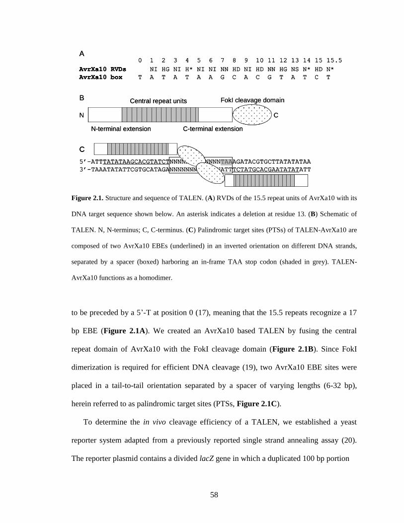
58
N-terminal extension C-terminal extension
Central repeat units FokI cleavage domain
CN
B
0 1 2 3 4 5 6 7 8 9 10 11 12 13 14 15 15.5
AvrXa10 RVDs NI HG NI H* NI NI NN HD NI HD NN HG NS N* HD N*
AvrXa10 box T A T A T A A G C A C G T A T C T
A
5’-ATTTATATAAGCACGTATCTNNNNNNNNNNNNNTAAAGATACGTGCTTATATATAA
3’-TAAATATATTCGTGCATAGANNNNNNNNNNNNNATTTCTATGCACGAATATATATT
C
N-terminal extension C-terminal extension
Central repeat units FokI cleavage domain
CN
B
0 1 2 3 4 5 6 7 8 9 10 11 12 13 14 15 15.5
AvrXa10 RVDs NI HG NI H* NI NI NN HD NI HD NN HG NS N* HD N*
AvrXa10 box T A T A T A A G C A C G T A T C T
A0 1 2 3 4 5 6 7 8 9 10 11 12 13 14 15 15.5
AvrXa10 RVDs NI HG NI H* NI NI NN HD NI HD NN HG NS N* HD N*
AvrXa10 box T A T A T A A G C A C G T A T C T
A
5’-ATTTATATAAGCACGTATCTNNNNNNNNNNNNNTAAAGATACGTGCTTATATATAA
3’-TAAATATATTCGTGCATAGANNNNNNNNNNNNNATTTCTATGCACGAATATATATT
C
5’-ATTTATATAAGCACGTATCTNNNNNNNNNNNNNTAAAGATACGTGCTTATATATAA
3’-TAAATATATTCGTGCATAGANNNNNNNNNNNNNATTTCTATGCACGAATATATATT
C
Figure 2.1. Structure and sequence of TALEN. (A) RVDs of the 15.5 repeat units of AvrXa10 with its
DNA target sequence shown below. An asterisk indicates a deletion at residue 13. (B) Schematic of
TALEN. N, N-terminus; C, C-terminus. (C) Palindromic target sites (PTSs) of TALEN-AvrXa10 are
composed of two AvrXa10 EBEs (underlined) in an inverted orientation on different DNA strands,
separated by a spacer (boxed) harboring an in-frame TAA stop codon (shaded in grey). TALEN-
AvrXa10 functions as a homodimer.
to be preceded by a 5’-T at position 0 (17), meaning that the 15.5 repeats recognize a 17
bp EBE (Figure 2.1A). We created an AvrXa10 based TALEN by fusing the central
repeat domain of AvrXa10 with the FokI cleavage domain (Figure 2.1B). Since FokI
dimerization is required for efficient DNA cleavage (19), two AvrXa10 EBE sites were
placed in a tail-to-tail orientation separated by a spacer of varying lengths (6-32 bp),
herein referred to as palindromic target sites (PTSs, Figure 2.1C).
To determine the in vivo cleavage efficiency of a TALEN, we established a yeast
reporter system adapted from a previously reported single strand annealing assay (20).
The reporter plasmid contains a divided lacZ gene in which a duplicated 100 bp portion
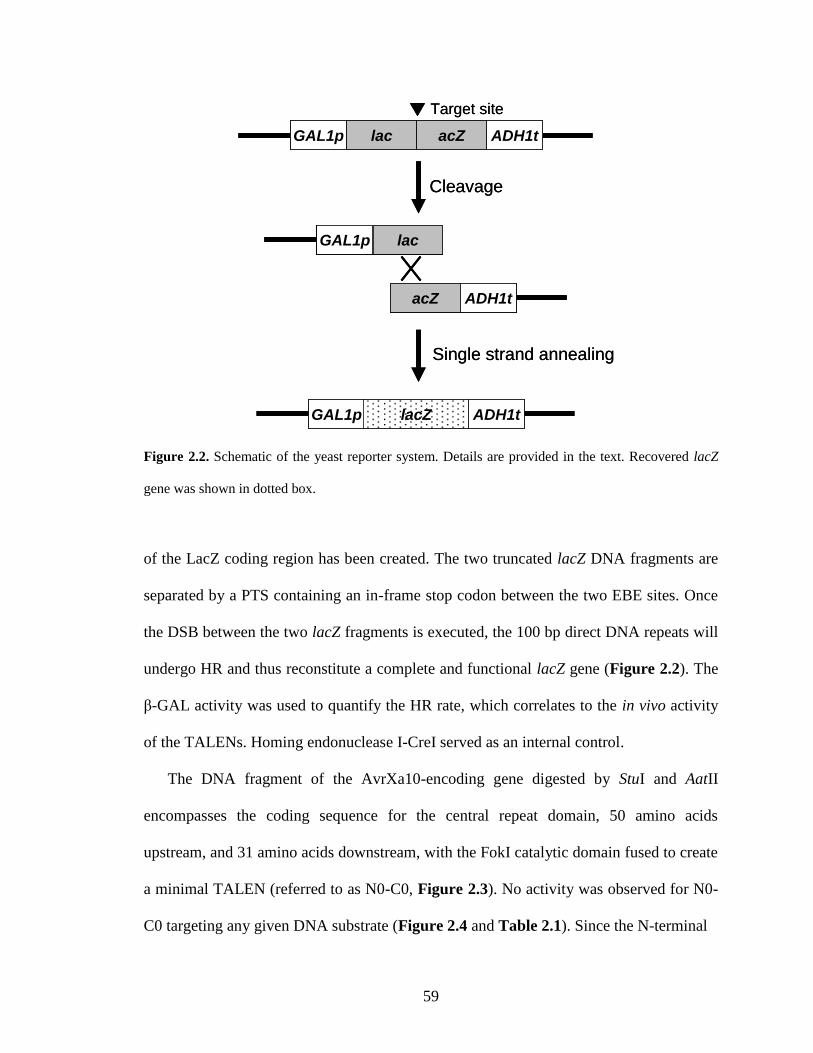
59
GAL1p lac acZ ADH1t
GAL1p lac
acZ ADH1t
GAL1p lacZ ADH1t
Target site
Cleavage
Single strand annealing
GAL1p lac acZ ADH1tGAL1p lac acZ ADH1tGAL1p lac acZ ADH1t
GAL1p lac
acZ ADH1t
GAL1p lacGAL1p lac
acZ ADH1tacZ ADH1t
GAL1p lacZ ADH1t
Target site
Cleavage
Single strand annealing
Figure 2.2. Schematic of the yeast reporter system. Details are provided in the text. Recovered lacZ
gene was shown in dotted box.
of the LacZ coding region has been created. The two truncated lacZ DNA fragments are
separated by a PTS containing an in-frame stop codon between the two EBE sites. Once
the DSB between the two lacZ fragments is executed, the 100 bp direct DNA repeats will
undergo HR and thus reconstitute a complete and functional lacZ gene (Figure 2.2). The
β-GAL activity was used to quantify the HR rate, which correlates to the in vivo activity
of the TALENs. Homing endonuclease I-CreI served as an internal control.
The DNA fragment of the AvrXa10-encoding gene digested by StuI and AatII
encompasses the coding sequence for the central repeat domain, 50 amino acids
upstream, and 31 amino acids downstream, with the FokI catalytic domain fused to create
a minimal TALEN (referred to as N0-C0, Figure 2.3). No activity was observed for N0-
C0 targeting any given DNA substrate (Figure 2.4 and Table 2.1). Since the N-terminal
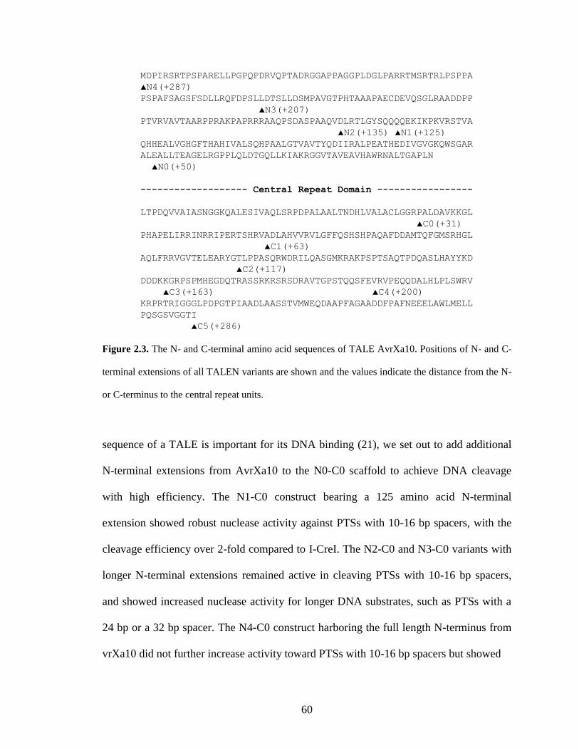
60
MDPIRSRTPSPARELLPGPQPDRVQPTADRGGAPPAGGPLDGLPARRTMSRTRLPSPPA
▲N4(+287)
PSPAFSAGSFSDLLRQFDPSLLDTSLLDSMPAVGTPHTAAAPAECDEVQSGLRAADDPP
▲N3(+207)
PTVRVAVTAARPPRAKPAPRRRAAQPSDASPAAQVDLRTLGYSQQQQEKIKPKVRSTVA
▲N2(+135) ▲N1(+125)
QHHEALVGHGFTHAHIVALSQHPAALGTVAVTYQDIIRALPEATHEDIVGVGKQWSGAR
ALEALLTEAGELRGPPLQLDTGQLLKIAKRGGVTAVEAVHAWRNALTGAPLN
▲N0(+50)
------------------- Central Repeat Domain -----------------
LTPDQVVAIASNGGKQALESIVAQLSRPDPALAALTNDHLVALACLGGRPALDAVKKGL
▲C0(+31)
PHAPELIRRINRRIPERTSHRVADLAHVVRVLGFFQSHSHPAQAFDDAMTQFGMSRHGL
▲C1(+63)
AQLFRRVGVTELEARYGTLPPASQRWDRILQASGMKRAKPSPTSAQTPDQASLHAYYKD
▲C2(+117)
DDDKKGRPSPMHEGDQTRASSRKRSRSDRAVTGPSTQQSFEVRVPEQQDALHLPLSWRV
▲C3(+163) ▲C4(+200)
KRPRTRIGGGLPDPGTPIAADLAASSTVMWEQDAAPFAGAADDFPAFNEEELAWLMELL
PQSGSVGGTI
▲C5(+286)
Figure 2.3. The N- and C-terminal amino acid sequences of TALE AvrXa10. Positions of N- and C-
terminal extensions of all TALEN variants are shown and the values indicate the distance from the N-
or C-terminus to the central repeat units.
sequence of a TALE is important for its DNA binding (21), we set out to add additional
N-terminal extensions from AvrXa10 to the N0-C0 scaffold to achieve DNA cleavage
with high efficiency. The N1-C0 construct bearing a 125 amino acid N-terminal
extension showed robust nuclease activity against PTSs with 10-16 bp spacers, with the
cleavage efficiency over 2-fold compared to I-CreI. The N2-C0 and N3-C0 variants with
longer N-terminal extensions remained active in cleaving PTSs with 10-16 bp spacers,
and showed increased nuclease activity for longer DNA substrates, such as PTSs with a
24 bp or a 32 bp spacer. The N4-C0 construct harboring the full length N-terminus from
vrXa10 did not further increase activity toward PTSs with 10-16 bp spacers but showed
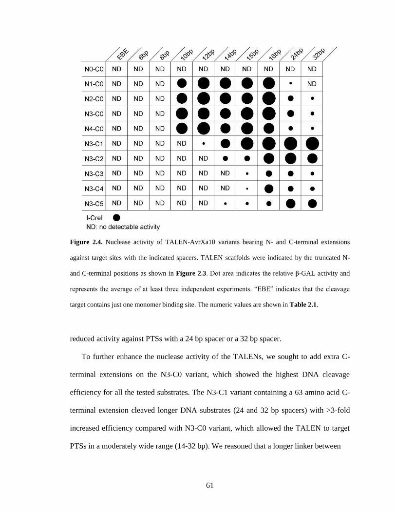
61
Figure 2.4. Nuclease activity of TALEN-AvrXa10 variants bearing N- and C-terminal extensions
against target sites with the indicated spacers. TALEN scaffolds were indicated by the truncated N-
and C-terminal positions as shown in Figure 2.3. Dot area indicates the relative β-GAL activity and
represents the average of at least three independent experiments. “EBE” indicates that the cleavage
target contains just one monomer binding site. The numeric values are shown in Table 2.1.
reduced activity against PTSs with a 24 bp spacer or a 32 bp spacer.
To further enhance the nuclease activity of the TALENs, we sought to add extra C-
terminal extensions on the N3-C0 variant, which showed the highest DNA cleavage
efficiency for all the tested substrates. The N3-C1 variant containing a 63 amino acid C-
terminal extension cleaved longer DNA substrates (24 and 32 bp spacers) with >3-fold
increased efficiency compared with N3-C0 variant, which allowed the TALEN to target
PTSs in a moderately wide range (14-32 bp). We reasoned that a longer linker between
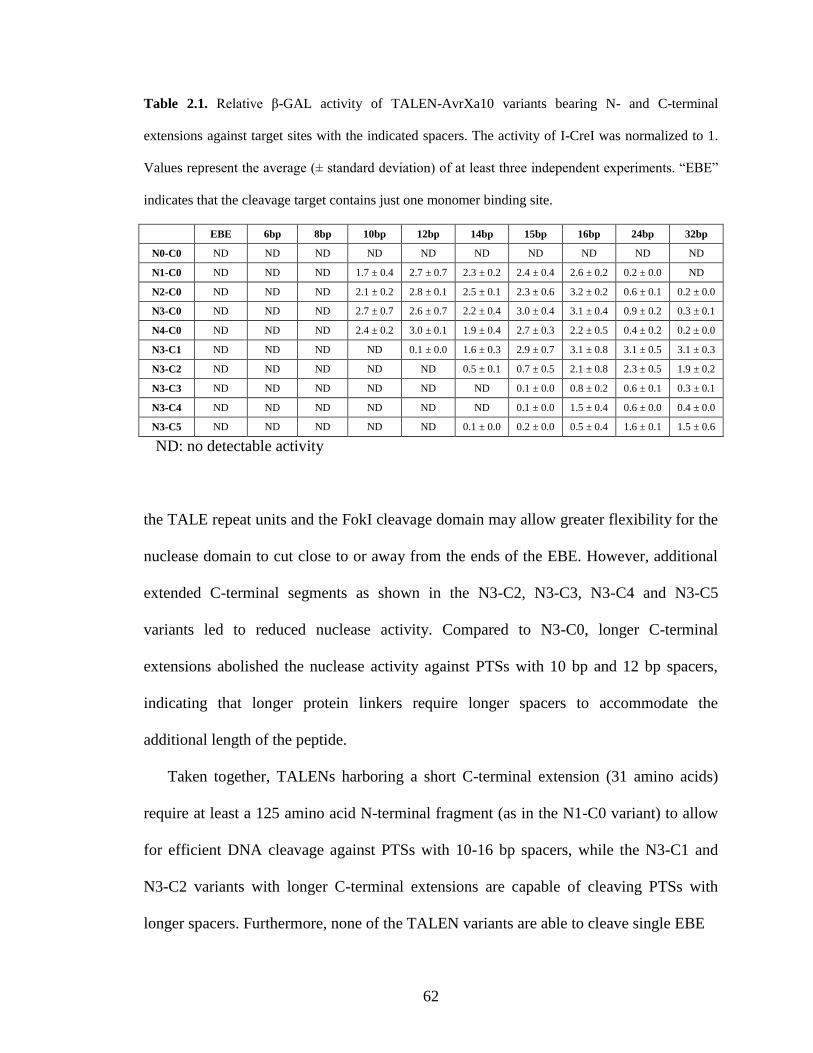
62
Table 2.1. Relative β-GAL activity of TALEN-AvrXa10 variants bearing N- and C-terminal
extensions against target sites with the indicated spacers. The activity of I-CreI was normalized to 1.
Values represent the average (± standard deviation) of at least three independent experiments. “EBE”
indicates that the cleavage target contains just one monomer binding site.
EBE 6bp 8bp 10bp 12bp 14bp 15bp 16bp 24bp 32bp
N0-C0 ND ND ND ND ND ND ND ND ND ND
N1-C0 ND ND ND 1.7 ± 0.4 2.7 ± 0.7 2.3 ± 0.2 2.4 ± 0.4 2.6 ± 0.2 0.2 ± 0.0 ND
N2-C0 ND ND ND 2.1 ± 0.2 2.8 ± 0.1 2.5 ± 0.1 2.3 ± 0.6 3.2 ± 0.2 0.6 ± 0.1 0.2 ± 0.0
N3-C0 ND ND ND 2.7 ± 0.7 2.6 ± 0.7 2.2 ± 0.4 3.0 ± 0.4 3.1 ± 0.4 0.9 ± 0.2 0.3 ± 0.1
N4-C0 ND ND ND 2.4 ± 0.2 3.0 ± 0.1 1.9 ± 0.4 2.7 ± 0.3 2.2 ± 0.5 0.4 ± 0.2 0.2 ± 0.0
N3-C1 ND ND ND ND 0.1 ± 0.0 1.6 ± 0.3 2.9 ± 0.7 3.1 ± 0.8 3.1 ± 0.5 3.1 ± 0.3
N3-C2 ND ND ND ND ND 0.5 ± 0.1 0.7 ± 0.5 2.1 ± 0.8 2.3 ± 0.5 1.9 ± 0.2
N3-C3 ND ND ND ND ND ND 0.1 ± 0.0 0.8 ± 0.2 0.6 ± 0.1 0.3 ± 0.1
N3-C4 ND ND ND ND ND ND 0.1 ± 0.0 1.5 ± 0.4 0.6 ± 0.0 0.4 ± 0.0
N3-C5 ND ND ND ND ND 0.1 ± 0.0 0.2 ± 0.0 0.5 ± 0.4 1.6 ± 0.1 1.5 ± 0.6
ND: no detectable activity
the TALE repeat units and the FokI cleavage domain may allow greater flexibility for the
nuclease domain to cut close to or away from the ends of the EBE. However, additional
extended C-terminal segments as shown in the N3-C2, N3-C3, N3-C4 and N3-C5
variants led to reduced nuclease activity. Compared to N3-C0, longer C-terminal
extensions abolished the nuclease activity against PTSs with 10 bp and 12 bp spacers,
indicating that longer protein linkers require longer spacers to accommodate the
additional length of the peptide.
Taken together, TALENs harboring a short C-terminal extension (31 amino acids)
require at least a 125 amino acid N-terminal fragment (as in the N1-C0 variant) to allow
for efficient DNA cleavage against PTSs with 10-16 bp spacers, while the N3-C1 and
N3-C2 variants with longer C-terminal extensions are capable of cleaving PTSs with
longer spacers. Furthermore, none of the TALEN variants are able to cleave single EBE
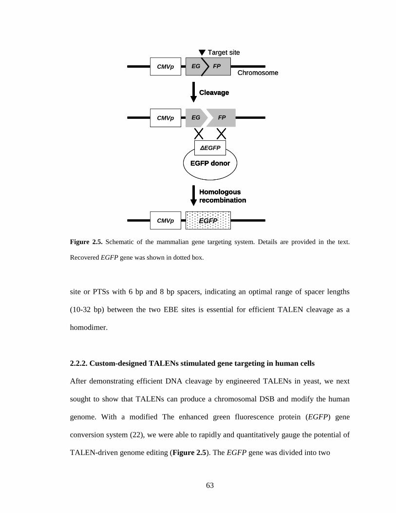
63
CMVp FPEG
CMVp EGFP
FPCMVp EG
EGFP donor
ΔEGFP
Cleavage
Homologous
recombination
Target site
ChromosomeCMVp FPEGCMVp FPEG FPEG FPEG
CMVp EGFP
FPCMVp EG
EGFP donor
ΔEGFP
EGFP donor
ΔEGFP
CleavageCleavage
Homologous
recombination
Homologous
recombination
Target site
Chromosome
Figure 2.5. Schematic of the mammalian gene targeting system. Details are provided in the text.
Recovered EGFP gene was shown in dotted box.
site or PTSs with 6 bp and 8 bp spacers, indicating an optimal range of spacer lengths
(10-32 bp) between the two EBE sites is essential for efficient TALEN cleavage as a
homodimer.
2.2.2. Custom-designed TALENs stimulated gene targeting in human cells
After demonstrating efficient DNA cleavage by engineered TALENs in yeast, we next
sought to show that TALENs can produce a chromosomal DSB and modify the human
genome. With a modified The enhanced green fluorescence protein (EGFP) gene
conversion system (22), we were able to rapidly and quantitatively gauge the potential of
TALEN-driven genome editing (Figure 2.5). The EGFP gene was divided into two
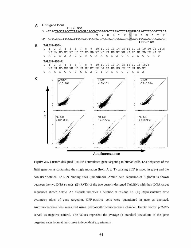
64
A
5’-TCACTAGCAACCTCAAACAGACACCATGGTGCATCTGACTCCTGTGGAGAAGTCTGCCGTTACT
M V H L T P V E K S A V T
3’-AGTGATCGTTGGAGTTTGTCTGTGGTACCACGTAGACTGAGGACACCTCTTCAGACGGCAATGA
HBB gene locus
C
TALEN-HBB-L0 1 2 3 4 5 6 7 8 9 10 11 12 13 14 15 16 17 18 19 20 21 21.5
NI NN HD NI NI HD HD HG HD NI NI NI HD NI NN NI HD NI HD HD NI N*
T A G C A A C C T C A A A C A G A C A C C A T
TALEN-HBB-R0 1 2 3 4 5 6 7 8 9 10 11 12 13 14 15 16 17 18 18.5
NI NI HD NN NN HD NI NN NI HD HG HG HD HG HD HD NI HD NI
T A A C G G C A G A C T T C T C C A C A
B
HBB-L site
HBB-R site
PE
EG
FP
-10210
010
210
310
410
5
-102
101
103
104
105
PE
EG
FP
-101
102
103
104
105
-101
102
103
104
105
PE
EG
FP
-10210
010
210
310
410
5
-102
101
103
104
105
PE
EG
FP
-101
102
103
104
105
-101
102
103
104
105
PE
EG
FP
-102-10
110
210
310
410
5
-101
102
103
104
105
PE
EG
FP
-101
102
103
104
105
-101
102
103
104
105G
FP
Autofluorescence
N1-C0
0.1±0.0 %
N0-C0
< 5×10⁻⁵
pCMV5
< 5×10⁻⁵
N3-C1
4.9±0.9 %
N4-C0
3.4±0.5 %
N3-C0
4.8±1.0 %
A
5’-TCACTAGCAACCTCAAACAGACACCATGGTGCATCTGACTCCTGTGGAGAAGTCTGCCGTTACT
M V H L T P V E K S A V T
3’-AGTGATCGTTGGAGTTTGTCTGTGGTACCACGTAGACTGAGGACACCTCTTCAGACGGCAATGA
HBB gene locus
C
TALEN-HBB-L0 1 2 3 4 5 6 7 8 9 10 11 12 13 14 15 16 17 18 19 20 21 21.5
NI NN HD NI NI HD HD HG HD NI NI NI HD NI NN NI HD NI HD HD NI N*
T A G C A A C C T C A A A C A G A C A C C A T
TALEN-HBB-R0 1 2 3 4 5 6 7 8 9 10 11 12 13 14 15 16 17 18 18.5
NI NI HD NN NN HD NI NN NI HD HG HG HD HG HD HD NI HD NI
T A A C G G C A G A C T T C T C C A C A
B
HBB-L site
HBB-R site
PE
EG
FP
-10210
010
210
310
410
5
-102
101
103
104
105
PE
EG
FP
-101
102
103
104
105
-101
102
103
104
105
PE
EG
FP
-10210
010
210
310
410
5
-102
101
103
104
105
PE
EG
FP
-101
102
103
104
105
-101
102
103
104
105
PE
EG
FP
-102-10
110
210
310
410
5
-101
102
103
104
105
PE
EG
FP
-101
102
103
104
105
-101
102
103
104
105
PE
EG
FP
-10210
010
210
310
410
5
-102
101
103
104
105
PE
EG
FP
-10210
010
210
310
410
5
-102
101
103
104
105
PE
EG
FP
-101
102
103
104
105
-101
102
103
104
105
PE
EG
FP
-101
102
103
104
105
-101
102
103
104
105
PE
EG
FP
-10210
010
210
310
410
5
-102
101
103
104
105
PE
EG
FP
-10210
010
210
310
410
5
-102
101
103
104
105
PE
EG
FP
-101
102
103
104
105
-101
102
103
104
105
PE
EG
FP
-101
102
103
104
105
-101
102
103
104
105
PE
EG
FP
-102-10
110
210
310
410
5
-101
102
103
104
105
PE
EG
FP
-102-10
110
210
310
410
5
-101
102
103
104
105
PE
EG
FP
-101
102
103
104
105
-101
102
103
104
105
PE
EG
FP
-101
102
103
104
105
-101
102
103
104
105G
FP
GF
P
AutofluorescenceAutofluorescence
N1-C0
0.1±0.0 %
N0-C0
< 5×10⁻⁵
pCMV5
< 5×10⁻⁵
N3-C1
4.9±0.9 %
N4-C0
3.4±0.5 %
N3-C0
4.8±1.0 %
Figure 2.6. Custom-designed TALENs stimulated gene targeting in human cells. (A) Sequence of the
HBB gene locus containing the single mutation (from A to T) causing SCD (shaded in grey) and the
two user-defined TALEN binding sites (underlined). Amino acid sequence of β-globin is shown
between the two DNA strands. (B) RVDs of the two custom-designed TALENs with their DNA target
sequences shown below. An asterisk indicates a deletion at residue 13. (C) Representative flow
cytometry plots of gene targeting. GFP-positive cells were quantitated in gate as depicted.
Autofluorescence was measured using phycoerythrin-fluorescence channel. Empty vector pCMV5
served as negative control. The values represent the average (± standard deviation) of the gene
targeting rates from at least three independent experiments.
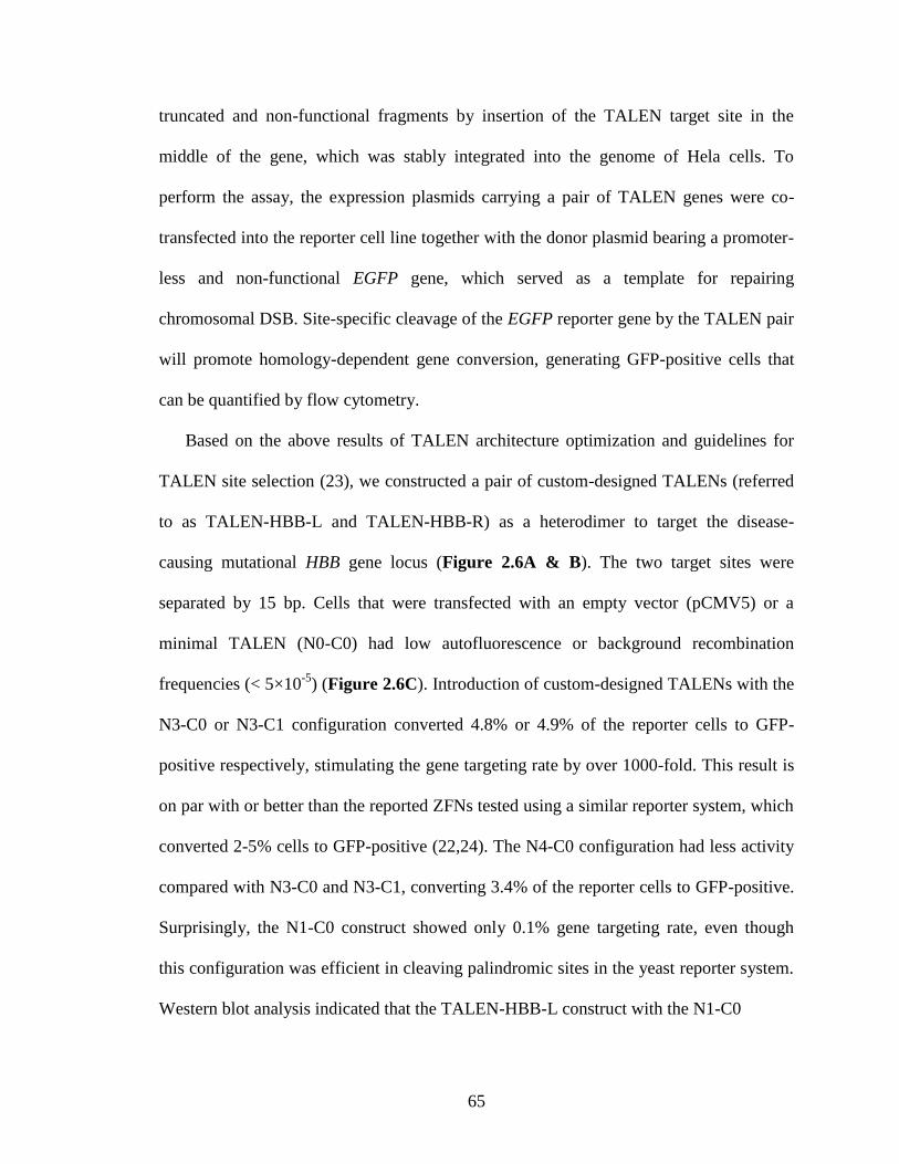
65
truncated and non-functional fragments by insertion of the TALEN target site in the
middle of the gene, which was stably integrated into the genome of Hela cells. To
perform the assay, the expression plasmids carrying a pair of TALEN genes were co-
transfected into the reporter cell line together with the donor plasmid bearing a promoter-
less and non-functional EGFP gene, which served as a template for repairing
chromosomal DSB. Site-specific cleavage of the EGFP reporter gene by the TALEN pair
will promote homology-dependent gene conversion, generating GFP-positive cells that
can be quantified by flow cytometry.
Based on the above results of TALEN architecture optimization and guidelines for
TALEN site selection (23), we constructed a pair of custom-designed TALENs (referred
to as TALEN-HBB-L and TALEN-HBB-R) as a heterodimer to target the disease-
causing mutational HBB gene locus (Figure 2.6A & B). The two target sites were
separated by 15 bp. Cells that were transfected with an empty vector (pCMV5) or a
minimal TALEN (N0-C0) had low autofluorescence or background recombination
frequencies (< 5×10⁻⁵
) (Figure 2.6C). Introduction of custom-designed TALENs with the
N3-C0 or N3-C1 configuration converted 4.8% or 4.9% of the reporter cells to GFP-
positive respectively, stimulating the gene targeting rate by over 1000-fold. This result is
on par with or better than the reported ZFNs tested using a similar reporter system, which
converted 2-5% cells to GFP-positive (22,24). The N4-C0 configuration had less activity
compared with N3-C0 and N3-C1, converting 3.4% of the reporter cells to GFP-positive.
Surprisingly, the N1-C0 construct showed only 0.1% gene targeting rate, even though
this configuration was efficient in cleaving palindromic sites in the yeast reporter system.
Western blot analysis indicated that the TALEN-HBB-L construct with the N1-C0
66
Figure 2.7. Western blot analysis for the expression of TALEN variants. (A) Expression level of
TALEN-HBB-L with different configurations. (B) Expression level of TALEN-HBB-R with different
configurations. All the TALEN variants contained a FLAG tag at the N-terminus. α-tubulin was used
as an internal control.
configuration did not express well in human cells (Figure 2.7A), which may partially
explain the lack of activity of this variant. On the other hand, all the tested TALEN-HBB-
R variants including the N1-C0 variant exhibited good expression levels (Figure 2.7B).
Based on the gene targeting system discussed above, we analyzed each TALEN’s
cleavage activity as a homodimer (Figure 2.8 and Figure 2.9). TALEN-HBB-L with the
N1-C0 configuration exhibited little activity (0.4%) against the HBB-L palindromic site
partially due to a low expression level, which is consistent with the activity as a
heterodimer. However, the low GFP signal (0.5%) from TALEN-HBB-R with the N1-C0
configuration indicated its low DNA cleavage activity, because it was expressed well in
human cells. Similar to a heterodimer, each TALEN monomer bearing the N3-C0, N4-C0
or N3-C1 configuration allowed for efficient cleavage as a homodimer, exhibiting >2%
gene targeting rate. Taken together, the TALEN pairs we created with at least 207 amino
acid N-terminal segment were capable of recognizing and cleaving preselected sites on
the HBB gene locus efficiently.
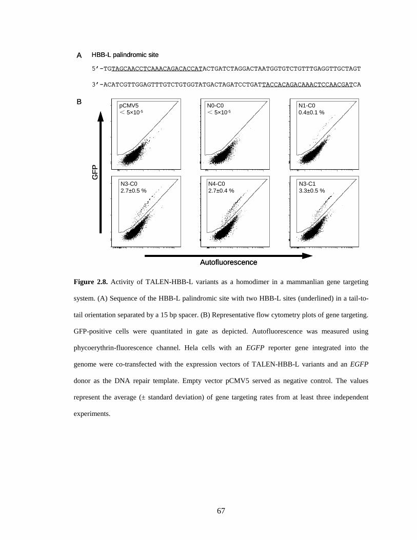
67
5’-TGTAGCAACCTCAAACAGACACCATACTGATCTAGGACTAATGGTGTCTGTTTGAGGTTGCTAGT
3’-ACATCGTTGGAGTTTGTCTGTGGTATGACTAGATCCTGATTACCACAGACAAACTCCAACGATCA
B
A HBB-L palindromic site
PE
EG
FP
-101
102
103
104
105
-101
102
103
104
105
PEE
GF
P-10
110
210
310
410
5
-101
102
103
104
105
PE
EG
FP
-102-10
110
210
310
410
5
-101
102
103
104
105
PE
EG
FP
-102-10
110
210
310
410
5-10
2
-101
102
103
104
105
PE
EG
FP
-101
102
103
104
105
-101
102
103
104
105
PE
EG
FP
-102-10
110
210
310
410
5
-101
102
103
104
105
GF
P
Autofluorescence
N1-C0
0.4±0.1 %
N0-C0
< 5×10⁻⁵
pCMV5
< 5×10⁻⁵
N3-C1
3.3±0.5 %
N4-C0
2.7±0.4 %
N3-C0
2.7±0.5 %
5’-TGTAGCAACCTCAAACAGACACCATACTGATCTAGGACTAATGGTGTCTGTTTGAGGTTGCTAGT
3’-ACATCGTTGGAGTTTGTCTGTGGTATGACTAGATCCTGATTACCACAGACAAACTCCAACGATCA
B
A HBB-L palindromic site
PE
EG
FP
-101
102
103
104
105
-101
102
103
104
105
PEE
GF
P-10
110
210
310
410
5
-101
102
103
104
105
PE
EG
FP
-102-10
110
210
310
410
5
-101
102
103
104
105
PE
EG
FP
-102-10
110
210
310
410
5-10
2
-101
102
103
104
105
PE
EG
FP
-101
102
103
104
105
-101
102
103
104
105
PE
EG
FP
-102-10
110
210
310
410
5
-101
102
103
104
105
PE
EG
FP
-101
102
103
104
105
-101
102
103
104
105
PE
EG
FP
-101
102
103
104
105
-101
102
103
104
105
PEE
GF
P-10
110
210
310
410
5
-101
102
103
104
105
PEE
GF
P-10
110
210
310
410
5
-101
102
103
104
105
PE
EG
FP
-102-10
110
210
310
410
5
-101
102
103
104
105
PE
EG
FP
-102-10
110
210
310
410
5
-101
102
103
104
105
PE
EG
FP
-102-10
110
210
310
410
5-10
2
-101
102
103
104
105
PE
EG
FP
-102-10
110
210
310
410
5-10
2
-101
102
103
104
105
PE
EG
FP
-101
102
103
104
105
-101
102
103
104
105
PE
EG
FP
-101
102
103
104
105
-101
102
103
104
105
PE
EG
FP
-102-10
110
210
310
410
5
-101
102
103
104
105
PE
EG
FP
-102-10
110
210
310
410
5
-101
102
103
104
105
GF
PG
FP
AutofluorescenceAutofluorescence
N1-C0
0.4±0.1 %
N0-C0
< 5×10⁻⁵
pCMV5
< 5×10⁻⁵
N3-C1
3.3±0.5 %
N4-C0
2.7±0.4 %
N3-C0
2.7±0.5 %
Figure 2.8. Activity of TALEN-HBB-L variants as a homodimer in a mammanlian gene targeting
system. (A) Sequence of the HBB-L palindromic site with two HBB-L sites (underlined) in a tail-to-
tail orientation separated by a 15 bp spacer. (B) Representative flow cytometry plots of gene targeting.
GFP-positive cells were quantitated in gate as depicted. Autofluorescence was measured using
phycoerythrin-fluorescence channel. Hela cells with an EGFP reporter gene integrated into the
genome were co-transfected with the expression vectors of TALEN-HBB-L variants and an EGFP
donor as the DNA repair template. Empty vector pCMV5 served as negative control. The values
represent the average (± standard deviation) of gene targeting rates from at least three independent
experiments.
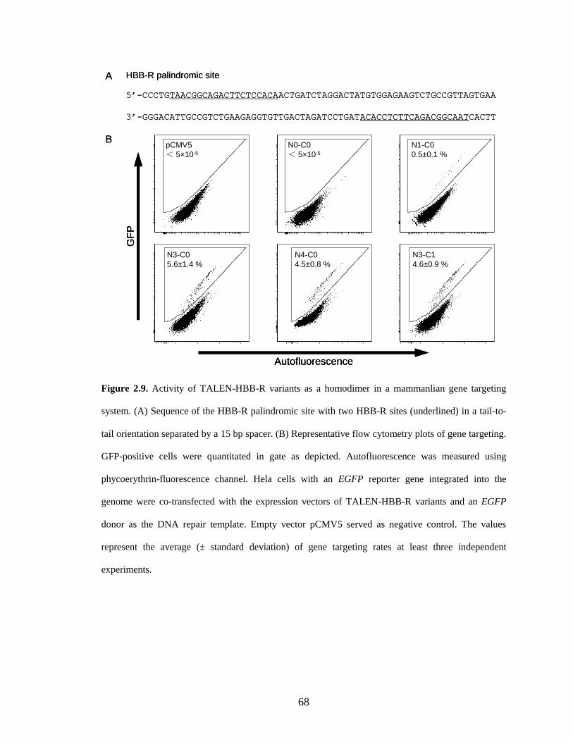
68
PE
EG
FP
-101
102
103
104
105
-101
102
103
104
105
B
A
5’-CCCTGTAACGGCAGACTTCTCCACAACTGATCTAGGACTATGTGGAGAAGTCTGCCGTTAGTGAA
3’-GGGACATTGCCGTCTGAAGAGGTGTTGACTAGATCCTGATACACCTCTTCAGACGGCAATCACTT
HBB-R palindromic site
PEE
GF
P-10
110
210
310
410
5
-101
102
103
104
105
PE
EG
FP
-101
102
103
104
105
-101
102
103
104
105
PE
EG
FP
-102-10
110
210
310
410
5
-101
102
103
104
105
PE
EG
FP
-10210
010
210
310
410
5
-102
101
103
104
105
PE
EG
FP
-102-10
110
210
310
410
5
-101
102
103
104
105
GF
P
Autofluorescence
N3-C1
4.6±0.9 %
N4-C0
4.5±0.8 %
N3-C0
5.6±1.4 %
N1-C0
0.5±0.1 %
N0-C0
< 5×10⁻⁵
pCMV5
< 5×10⁻⁵
PE
EG
FP
-101
102
103
104
105
-101
102
103
104
105
PE
EG
FP
-101
102
103
104
105
-101
102
103
104
105
B
A
5’-CCCTGTAACGGCAGACTTCTCCACAACTGATCTAGGACTATGTGGAGAAGTCTGCCGTTAGTGAA
3’-GGGACATTGCCGTCTGAAGAGGTGTTGACTAGATCCTGATACACCTCTTCAGACGGCAATCACTT
HBB-R palindromic site
PEE
GF
P-10
110
210
310
410
5
-101
102
103
104
105
PEE
GF
P-10
110
210
310
410
5
-101
102
103
104
105
PE
EG
FP
-101
102
103
104
105
-101
102
103
104
105
PE
EG
FP
-101
102
103
104
105
-101
102
103
104
105
PE
EG
FP
-102-10
110
210
310
410
5
-101
102
103
104
105
PE
EG
FP
-102-10
110
210
310
410
5
-101
102
103
104
105
PE
EG
FP
-10210
010
210
310
410
5
-102
101
103
104
105
PE
EG
FP
-10210
010
210
310
410
5
-102
101
103
104
105
PE
EG
FP
-102-10
110
210
310
410
5
-101
102
103
104
105
PE
EG
FP
-102-10
110
210
310
410
5
-101
102
103
104
105
GF
PG
FP
AutofluorescenceAutofluorescence
N3-C1
4.6±0.9 %
N4-C0
4.5±0.8 %
N3-C0
5.6±1.4 %
N1-C0
0.5±0.1 %
N0-C0
< 5×10⁻⁵
pCMV5
< 5×10⁻⁵
Figure 2.9. Activity of TALEN-HBB-R variants as a homodimer in a mammanlian gene targeting
system. (A) Sequence of the HBB-R palindromic site with two HBB-R sites (underlined) in a tail-to-
tail orientation separated by a 15 bp spacer. (B) Representative flow cytometry plots of gene targeting.
GFP-positive cells were quantitated in gate as depicted. Autofluorescence was measured using
phycoerythrin-fluorescence channel. Hela cells with an EGFP reporter gene integrated into the
genome were co-transfected with the expression vectors of TALEN-HBB-R variants and an EGFP
donor as the DNA repair template. Empty vector pCMV5 served as negative control. The values
represent the average (± standard deviation) of gene targeting rates at least three independent
experiments.

69
M0
M2
M10
pC
MV5
N3-
C0
N3-
C1
GZF3N
0
20
40
60
80
%-H
2A
X-p
osit
ive c
ell
s
(etoposide)
*
*
*
M0
M2
M10
pC
MV5
N3-
C0
N3-
C1
GZF3N
0
20
40
60
80
%-H
2A
X-p
osit
ive c
ell
s
(etoposide)
M0
M2
M10
pC
MV5
N3-
C0
N3-
C1
GZF3N
0
20
40
60
80
%-H
2A
X-p
osit
ive c
ell
s
(etoposide)
*
*
*
Figure 2.10. Nuclease-associated cytotoxicity of TALENs. The columns denote the percentage of γ-
H2AX positive cells either transfected with artificial DNA nucleases or treated using etoposide with
indicated concentration. Error bars indicate standard deviation from the average of at least three
independent experiments. *, statistically significant increase of toxicity as compared to empty vector
pCMV5 (P < 0.001).
2.2.3. Custom-designed TALENs caused no detectable cytotoxicity
The DNA binding specificity is a major determinant of cytotoxicity for ZFNs (25). Since
the novel TALEN pair we designed recognizes a 43 bp long DNA recognition sequence,
we postulated that it might not cause significant cytotoxicity due to its high specificity,
even though there is some degeneracy in RVD recognition (i.e. NN can recognize G as
well as A) (26). To assess the nuclease-associated toxicity, we monitored the intracellular
level of γ-H2AX, which is the H2AX histone phosphorylated on serine 139 as a reaction
on a chromosomal DSB (27). HEK293 cells displayed an average γ-H2AX level similar
to the background (pCMV5 control) after being transfected with the TALEN pairs under
the same conditions used to induce recombination in the gene targeting assay (Figure
2.10). In contrast, when we transfected the cells with a reported toxic ZFN (28) or treated
70
the cells with etoposide (an anti-cancer agent that causes DNA DSBs (29)) for 2 h,
significantly higher levels of H2AX phosphorylation were detected. Therefore, the
engineered TALENs caused little, if any, cytotoxicity in human cells, demonstrating their
high specificity.
2.2.4. Targeting “unnatural” TALE sites
Naturally occurring TALE recognition sites are uniformly preceded by a 5’-T (17);
therefore, all of the previously reported TALENs were designed to target DNA sequences
bearing a T at position 0. To test whether a preceding T is an essential requirement for
efficient cleavage by TALENs, we selected five TALEN-AvrXa10 variants to measure
their nuclease activities towards “unnatural” TALE sites with a preceding A, C or G
using the yeast reporter system. The spacer length between each monomer binding site
was fixed to be 16 bp, allowing for efficient cleavage for all the tested TALEN variants
(Figure 2.4). As shown in Figure 2.11, the N2-C0 and N3-C0 constructs harboring short
C-terminal extensions exhibited moderate nuclease activity against unnatural PTSs (0.5-1
fold efficiency compared with I-CreI), which is still significantly lower than that towards
the natural PTS with a T at position 0. The N4-C0 construct with a full length N-terminal
segment displayed slightly reduced DNA cleavage efficiency towards all the tested
substrates. The N3-C1 construct with additional C-terminal extension compared with the
N3-C0 construct showed much higher cleavage efficiency against unnatural substrates.
The N3-C1 construct cleaved PTSs bearing a preceding A, C or G with an efficiency
similar to that for natural PTS with a preceding T. The extended 63 amino acid peptide
linker between the DNA binding domain and the FokI cleavage domain in the N3-C1
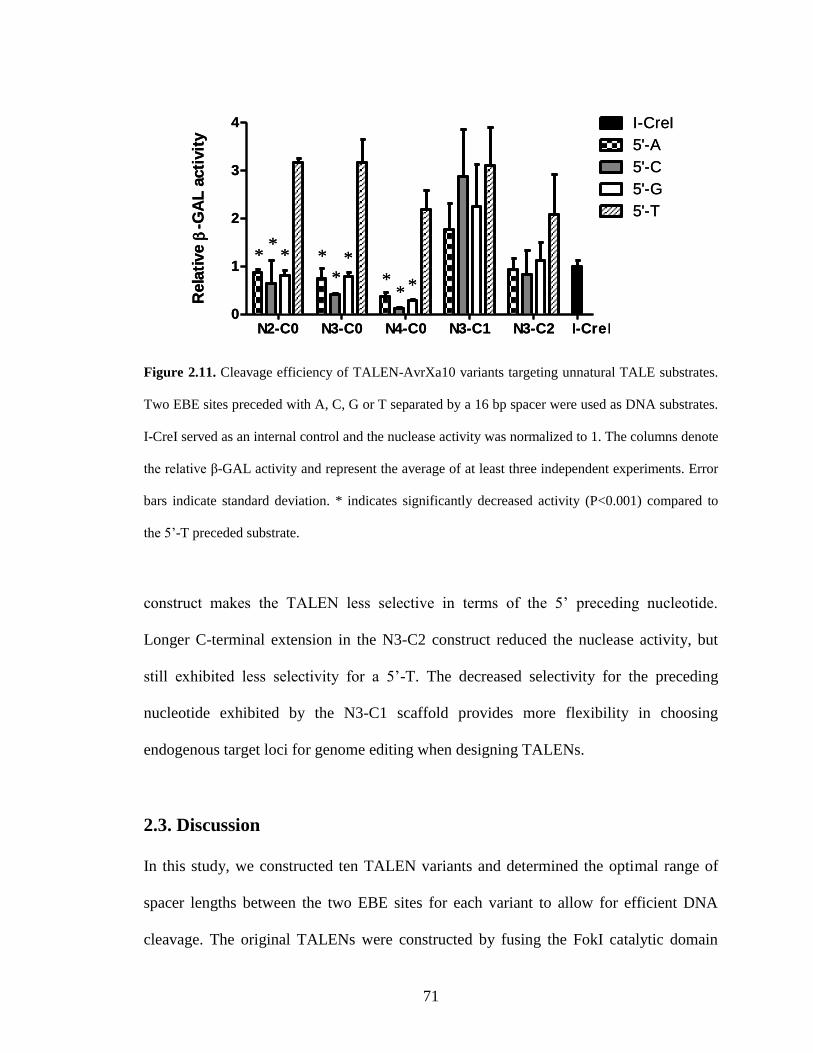
71
N2-C0 N3-C0 N4-C0 N3-C1 N3-C2 I-CreI0
1
2
3
4 I-CreI
5'-A
5'-C
5'-G
5'-TR
ela
tive
-GA
L a
cti
vit
y
**
* *
**
** *
N2-C0 N3-C0 N4-C0 N3-C1 N3-C2 I-CreI0
1
2
3
4 I-CreI
5'-A
5'-C
5'-G
5'-TR
ela
tive
-GA
L a
cti
vit
y
**
* *
**
** *
Figure 2.11. Cleavage efficiency of TALEN-AvrXa10 variants targeting unnatural TALE substrates.
Two EBE sites preceded with A, C, G or T separated by a 16 bp spacer were used as DNA substrates.
I-CreI served as an internal control and the nuclease activity was normalized to 1. The columns denote
the relative β-GAL activity and represent the average of at least three independent experiments. Error
bars indicate standard deviation. * indicates significantly decreased activity (P<0.001) compared to
the 5’-T preceded substrate.
construct makes the TALEN less selective in terms of the 5’ preceding nucleotide.
Longer C-terminal extension in the N3-C2 construct reduced the nuclease activity, but
still exhibited less selectivity for a 5’-T. The decreased selectivity for the preceding
nucleotide exhibited by the N3-C1 scaffold provides more flexibility in choosing
endogenous target loci for genome editing when designing TALENs.
2.3. Discussion
In this study, we constructed ten TALEN variants and determined the optimal range of
spacer lengths between the two EBE sites for each variant to allow for efficient DNA
cleavage. The original TALENs were constructed by fusing the FokI catalytic domain

72
with full length TALEs (30-32). Since the naturally occurring TALEs are transcription
activators from a plant bacterial pathogen, their N-terminal segment contains signal
peptides responsible for protein translocation and C-terminal segment harbors nuclei
localization signals and transcription activator domain (33). These redundant fragments
can be detrimental to the TALENs nuclease activity. Therefore, TALEN scaffold needs to
be optimized to achieve efficient cleavage. Previously, Miller et al. (26) truncated the
first 152 N-terminal residues from TALE and produced a set of TALENs with different
C-terminal truncations to maximize nuclease activity. Their variants with 28 amino acid
or 63 amino acid C-terminal extensions exhibited highest DNA cleavage efficiency,
which are similar to our N3-C0 and N3-C1 constructs, respectively. We found that N3-
C1 construct bearing 63 residues displayed efficient cleavage against DNA substrates
with long spacers (16-32 bp), while Miller et al. did not observe this activity, which may
be due to the shorter N-terminal segment in their TALEN variants. Even though they
reported undetectable nuclease activity of TALEN with 93 amino acid C-terminal
fragment, we observed efficient cleavage using N3-C4 and N3-C5 variants against DNA
substrates with longer spacers, which is in agreement with the original study on TALENs
bearing full length TALE C-terminus. More recently, Mussolino et al. (34) reported the
most active configuration was a TALEN bearing 153 amino acid N-terminal segment and
17 or 47 amino acid C-terminal segment, cleaving target sites with 12-15 bp spacer.
However, their in vivo reporter system relied on the disappearance of GFP signal, in
which the binding of TALENs to the target sites without cleavage may generate false
positive signals (e.g., some of their TALEN variants exhibited positive result targeting
single EBE site). Because the different TALEN activities in the above mentioned reports
73
were determined using different reporter systems, it is difficult to compare their results
directly with ours. However, our systematic study clearly identifies two optimized
TALEN scaffolds, with N3-C0 variant preferring PTSs with shorter (10-16 bp) spacers
and N3-C1 variant preferring PTSs with longer (14-32 bp) spacers. This indicates that our
optimized TALEN architectures have a moderately wide optimal range of spacer length
between each TALEN monomer binding site, which allows for more flexibility with the
choice of recognition sites in gene targeting study.
The single mutation in HBB gene causes sickle cell disease, against which there is no
adequate long-term treatment. Gene-insertion based gene therapy has been reported to be
effective in mouse model by introduction of an antisickling HBB gene copy (3,4).
However, immunogenicity and insertional mutagenesis caused by integrating viral
vectors are still under safety concerns, which limit the wide application of this
technology. Therefore, it is tempting to correct the mutational HBB gene in situ. In this
study, we constructed a pair of TALENs that were able to recognize a user-defined
sequence within HBB gene locus and stimulated gene targeting rate by over 1000-fold
without detectable cytotoxicity. The TALENs with N3-C0 and N3-C1 scaffolds displayed
highest cleavage efficiency, which is consistent with our result from yeast reporter
system. To be noticed, N1-C0 variant exhibited high nuclease activity against PTS with
15bp spacer in the yeast reporter system but showed little activity in the human gene
targeting system, even though Western blot analysis indicated its good expression level.
The inconsistency may be due to the difference between episomal cleavage in our yeast
reporter system and chromosomal cleavage in the more complex genome in human cells.
Taken together, our optimized TALENs with N3-C0 or N3-C1 scaffold exhibited robust
74
nuclease activity and low cytotoxicity in human cells, which can serve as a powerful tool
for gene repair in gene therapy.
A 5’-T preceding the TALE EBE sites was believed to be essential for TALE
function (17), which, based on our study, turned out not to be the case for TALE
nucleases. All the tested TALEN variants exhibited good nuclease activity against
unnatural DNA substrate (0.5-1 fold efficiency compared with I-CreI); even though
TALENs variants with short C-terminal extension still preferred to cleave natural
TALEN recognition site. Interestingly, N3-C1 TALEN variant was capable of cleaving
unnatural DNA substrates preceded by 5’-A, C or G with similar efficiency compared
with that for natural TALE site. However, the longer C-terminal extension in N3-C1
variant is at the 3’-end of the EBE, which is physically distant from the 5’-end of target
site. We reasoned that the presence of a leucine zipper-like heptad repeats closely linked
to the TALE central repeat units (35) may increase the DNA binding affinity of the
central repeat domain and thus make the 5’-T interaction less of a requirement. More
detailed structural studies will be needed to elucidate the protein-DNA interface of
TALENs and their corresponding DNA substrates in order to solve this uncertainty. Our
result indicates that the requirement for a 5’-T can be mitigated using certain scaffolds,
thus allowing greater flexibility in choosing desired target site in genome editing
endeavors.
75
2.4. Materials and methods
2.4.1. Materials
Phusion DNA polymerase, T4 DNA ligase, T4 polynucleotide kinase, antarctic
phosphatase, and restriction endonucleases were purchased from New England Biolabs
(Beverly, MA). QIAprep Spin Plasmid Miniprep Kit, QIAquick Gel Extraction Kit, and
QIAquick PCR Purification Kit were obtained from Qiagen (Valencia, CA).
Oligonucleotide primers were obtained from Integrated DNA Technologies (Coralville,
IA). All the other reagents unless specified were obtained from Sigma-Aldrich (St Louis,
MO).
2.4.2. Yeast reporter system
The yeast reporter system was constructed based on a single strand annealing assay with
slight modifications (20). The lacZ gene was divided into two truncated and non-
functional copies by insertion of a palindromic target site consisting of two AvrXa10
EBEs in a tail-to-tail orientation with various spacers. The two separated lacZ fragments
sharing a 100 bp homologous region were PCR amplified separately, digested with AvrII
and XbaI respectively and ligated overnight at 16 °C. The ligation product was
subsequently digested with a combination of AvrII and XbaI and the 3.3 kb non-cut
fragment was gel purified. The lacZ reporter gene was then cloned into the pRS414
plasmid in between the GAL1 promoter and the ADH1 terminator, whose expression was
induced by adding 2% galactose in the culture medium.
76
2.4.3. Construction of TALEN expression vectors
The DNA sequence encoding the nonspecific DNA cleavage domain of FokI restriction
endonuclease was PCR-amplified from pST1374 (plasmid 13426; Addgene, Cambridge,
MA). The PCR product was inserted into pET28a(+) vector (EMD, Madison, WI)
through NdeI and SalI recognition sites to yield plasmid pET28a-G4S-FokI. The DNA
sequence encoding the central repeat DNA binding domain of TALE AvrXa10 was
isolated from 0.7% agarose gel after digestion of the plasmid pAS202 (kindly provided
by Dr. Frank F. White from Kansas State University, Manhattan, KS) using restriction
enzymes StuI and AatII. The DNA fragment after gel extraction was cloned into the StuI
and AatII sites of plasmid pET28a-G4S-FokI to make pET28a-TALEN-Xa10. The DNA
sequences encoding the N-terminal extensions of the AvrXa10 protein with varying
length were PCR-amplified from pAS202. The PCR products were digested with NdeI
and StuI and cloned into the large fragment of the pET28a-TALEN-Xa10 vector digested
with the same enzymes. The DNA sequences encoding the C-terminal extensions of the
AvrXa10 protein with varying length were PCR-amplified using the same template and
cloned into the AatII and AflII sites of the plasmid pET28a-TALEN-Xa10. The gene
encoding TALEN-Xa10 with different configurations were subcloned into pRS415
plasmid through the NdeI and SalI sites in between the GAL10 promoter and the ADH2
terminator. The expression of TALENs in Saccharomyces cerevisiae was induced by
adding 2% galactose in the cultural medium.
The DNA sequences encoding the custom-designed TALE central repeat domains
targeting the specific sequence within the HBB gene locus were synthesized by DNA2.0,
Inc (Menlo Park, CA). The synthesized DNA sequences were codon optimized to
77
increase the protein expression level and reduce the repetitiveness among amino acid
repeats in the DNA binding domain to facilitate DNA cloning. The genes of custom-
designed TALENs with different N- and C-terminal extensions were first cloned into the
pET28a(+) vector using the same strategy as described above. Next, the TALEN genes
along with a FLAG tag sequence and a SV40 nucleus localization signal added to the N-
terminus were subcloned into the pCMV5 mammalian expression vector (36) through the
KpnI and SalI sites. The expression of these constructs is under the control of the
cytomegalovirus (CMV) promoter.
To test the in vivo cleavage activity of TALENs in the yeast reporter system, both the
pRS414 reporter plasmid and pRS415 expression plasmid were transformed into
Saccharomyces cerevisiae HZ848 (MATα, ade2-1, ade3Δ22, Δura3, his3-11,15, trp1-1,
leu2-3,112 and can1-100) using the LiAc/SS carrier DNA/PEG method (37). After 3-4
days growth at 30 °C on plates containing synthetic complete medium lacking leucine
and tryptophan (SC-Leu-Trp) with 2% glucose, three transformants were picked to grow
in SC-Leu-Trp liquid medium with 2% galactose. After cultivation for 2 days at 30 °C,
yeast cells were harvested by centrifugation and lysed by Y-PER yeast protein extraction
reagent (Pierce, Rockford, IL). The β-galactosidase (β-GAL) enzyme activity was
determined by the β-galactosidase enzyme system from Promega (Madison, WI)
according to the manufacturer's instruction. For each sample, the β-GAL activity was
measured as a function of time and normalized by total protein concentration determined
using the BCA protein assay (Pierce, Rockford, IL). Homing endonuclease I-CreI (38)
serves as an internal control and its activity was normalized to one.
78
2.4.4. Human gene targeting system
EGFP gene was divided into two fragments by insertion of a preselected sequence from
the HBB gene locus and an in-frame stop codon. The non-functional EGFP reporter gene
was cloned into the pLNCX2 retroviral vector (BD Clontech, Palo Alto, CA) under the
cytomegalovirus (CMV) promoter and stably integrated into the genome via retroviral
transduction according to the manufacturer’s protocol. Transfected Hela cells were
selected in 500 μg/ml G418 for two weeks. A donor plasmid was constructed by insertion
of a promoter-less EGFP gene with the first 37 nucleotides missing into the pNEB193
plasmid (New England Biolabs, Beverly, MA) through the HindIII and SalI sites. The
donor plasmid provides a homologous DNA segment as a template for repairing the DSB
in the EGFP reporter. Construction of TALEN expression vectors is described in the
Supplementary Data.
Reporter Hela cells were maintained in modified Eagle’s medium (MEM)
supplemented with 10% Fetal Bovine Serum (FBS; Hyclone, Logan, UT). Cells were
seeded in 12-well plates at a density of 1×10⁵ per well. After 24 hr, reporter cells were
co-transfected with 333 ng of each TALEN expression plasmid and 333 ng of donor
plasmid using FuGene HD transfection reagent (Promega, Madison, WI) under
conditions specified by the manufacturer. The transfection efficiency was determined to
be between 45% and 50%. Cells were trypsinized from their culturing plates 48h after
transfection and resuspended in 200 μL phosphate buffered saline (PBS) supplemented
with 10 mM EDTA for flow cytometry analysis. 20,000 cells were analyzed by BD
LSRII flow cytometer (BD Biosciences, San Jose, CA) to determine the percentage of
79
GFP-positive cells. The rate of gene targeting was determined by normalizing the
percentage of GFP-positive cells to the transfection efficiency.
2.4.5. Immunoblotting
Hela cells in 12-well plates were harvested 24 h after transfection with TALEN variants.
Cells were collected by centrifugation, washed with PBS and resuspended in 40 μL RIPA
lysis buffer (Santa Cruz Biotechnology, Santa Cruz, CA) with SDS-PAGE loading dye.
Proteins were resolved by 4-20% Mini-Protean TGX Precast Gel (Bio-Rad Laboratories,
Hercules, CA), transferred onto a nitrocellulose membrane, blocked for 1 hr with Tris-
buffered saline/0.05% Tween 20 containing 5% nonfat milk, followed by incubation with
anti-FLAG tag (1:500) and anti-α-tubulin (1:5000) antibodies at 4 °C overnight. After
incubation with anti-mouse horseradish peroxidase-conjugated secondary antibody
(1:25000; GeneScript, Piscataway, NJ) for 1 hr, bands were visualized using SuperSignal
West Pico Chemiluminescent Substrate (Pierce, Rockford, IL).
2.4.6. H2AX phosphorylation assay
HEK293 cells were cultured in Dulbecco’s modified Eagle’s medium (DMEM)
supplemented with 10% FBS. Cells were seeded in 12-well plates (2×10⁵
/well) and
transfected after 24 hr with 500 ng of each TALEN expression plasmid, 1 μg empty
pCMV5 vector as a negative control or 1 μg reported toxic ZFN construct GZF3N
(kindly provided by Dr. Toni Cathomen of Hannover Medical School, Hannover,
Germany) as a positive control, respectively. After 48 h post transfection, cells were
harvested, fixed, permeabilized, and stained using the H2AX phosphorylation assay kit
80
(Millipore, Watford, UK) according to the manufacturer’s protocol. Alternatively, cells
were treated with dimethyl sulfoxide (DMSO), 2 μM and 10 μM etoposide in DMSO for
2 hr before staining. Cells were then scanned in a flow cytometer to quantitate the
number of cells staining positive for phosphorylated histone H2AX.
2.5. References
1. Ashley-Koch, A., Yang, Q. and Olney, R.S. (2000) Sickle hemoglobin (HbS)
allele and sickle cell disease: a HuGE review. Am J Epidemiol, 151, 839-845.
2. Frenette, P.S. and Atweh, G.F. (2007) Sickle cell disease: old discoveries, new
concepts, and future promise. J Clin Invest, 117, 850-858.
3. Levasseur, D.N., Ryan, T.M., Pawlik, K.M. and Townes, T.M. (2003) Correction
of a mouse model of sickle cell disease: lentiviral/antisickling beta-globin gene
transduction of unmobilized, purified hematopoietic stem cells. Blood, 102, 4312-
4319.
4. Pawliuk, R., Westerman, K.A., Fabry, M.E., Payen, E., Tighe, R., Bouhassira,
E.E., Acharya, S.A., Ellis, J., London, I.M., Eaves, C.J. et al. (2001) Correction of
sickle cell disease in transgenic mouse models by gene therapy. Science, 294,
2368-2371.
5. Marshall, E. (1999) Gene therapy death prompts review of adenovirus vector.
Science, 286, 2244-2245.
6. Check, E. (2002) A tragic setback. Nature, 420, 116-118.
7. Chang, J.C., Ye, L. and Kan, Y.W. (2006) Correction of the sickle cell mutation
in embryonic stem cells. Proc Natl Acad Sci U S A, 103, 1036-1040.
81
8. Thomas, K.R. and Capecchi, M.R. (1987) Site-directed mutagenesis by gene
targeting in mouse embryo-derived stem cells. Cell, 51, 503-512.
9. Bollag, R.J., Waldman, A.S. and Liskay, R.M. (1989) Homologous recombination
in mammalian cells. Annu Rev Genet, 23, 199-225.
10. Cathomen, T. and Joung, J.K. (2008) Zinc-finger nucleases: the next generation
emerges. Mol Ther, 16, 1200-1207.
11. Urnov, F.D., Rebar, E.J., Holmes, M.C., Zhang, H.S. and Gregory, P.D. (2010)
Genome editing with engineered zinc finger nucleases. Nat Rev Genet, 11, 636-
646.
12. Carroll, D. (2008) Progress and prospects: zinc-finger nucleases as gene therapy
agents. Gene Ther, 15, 1463-1468.
13. Ramirez, C.L., Foley, J.E., Wright, D.A., Muller-Lerch, F., Rahman, S.H., Cornu,
T.I., Winfrey, R.J., Sander, J.D., Fu, F., Townsend, J.A. et al. (2008) Unexpected
failure rates for modular assembly of engineered zinc fingers. Nat Methods, 5,
374-375.
14. Kim, J.S., Lee, H.J. and Carroll, D. (2010) Genome editing with modularly
assembled zinc-finger nucleases. Nat Methods, 7, 91; author reply 91-92.
15. Radecke, S., Radecke, F., Cathomen, T. and Schwarz, K. (2010) Zinc-finger
nuclease-induced gene repair with oligodeoxynucleotides: wanted and unwanted
target locus modifications. Mol Ther, 18, 743-753.
16. Joung, J.K. and Sander, J.D. (2013) TALENs: a widely applicable technology for
targeted genome editing. Nat Rev Mol Cell Biol, 14, 49-55.
82
17. Boch, J., Scholze, H., Schornack, S., Landgraf, A., Hahn, S., Kay, S., Lahaye, T.,
Nickstadt, A. and Bonas, U. (2009) Breaking the code of DNA binding specificity
of TAL-type III effectors. Science, 326, 1509-1512.
18. Hopkins, C.M., White, F.F., Choi, S.H., Guo, A. and Leach, J.E. (1992)
Identification of a family of avirulence genes from Xanthomonas-oryzae pv.
oryzae. Mol Plant-Microbe Interact, 5, 451-459.
19. Bitinaite, J., Wah, D.A., Aggarwal, A.K. and Schildkraut, I. (1998) FokI
dimerization is required for DNA cleavage. Proc Natl Acad Sci U S A, 95, 10570-
10575.
20. Epinat, J.C., Arnould, S., Chames, P., Rochaix, P., Desfontaines, D., Puzin, C.,
Patin, A., Zanghellini, A., Paques, F. and Lacroix, E. (2003) A novel engineered
meganuclease induces homologous recombination in yeast and mammalian cells.
Nucleic Acids Res, 31, 2952-2962.
21. Gurlebeck, D., Szurek, B. and Bonas, U. (2005) Dimerization of the bacterial
effector protein AvrBs3 in the plant cell cytoplasm prior to nuclear import. Plant
J, 42, 175-187.
22. Porteus, M.H. and Baltimore, D. (2003) Chimeric nucleases stimulate gene
targeting in human cells. Science, 300, 763.
23. Cermak, T., Doyle, E.L., Christian, M., Wang, L., Zhang, Y., Schmidt, C., Baller,
J.A., Somia, N.V., Bogdanove, A.J. and Voytas, D.F. (2011) Efficient design and
assembly of custom TALEN and other TAL effector-based constructs for DNA
targeting. Nucleic Acids Res, 39, e82.
83
24. Urnov, F.D., Miller, J.C., Lee, Y.L., Beausejour, C.M., Rock, J.M., Augustus, S.,
Jamieson, A.C., Porteus, M.H., Gregory, P.D. and Holmes, M.C. (2005) Highly
efficient endogenous human gene correction using designed zinc-finger nucleases.
Nature, 435, 646-651.
25. Cornu, T.I., Thibodeau-Beganny, S., Guhl, E., Alwin, S., Eichtinger, M., Joung,
J.K. and Cathomen, T. (2008) DNA-binding specificity is a major determinant of
the activity and toxicity of zinc-finger nucleases. Mol Ther, 16, 352-358.
26. Miller, J.C., Tan, S., Qiao, G., Barlow, K.A., Wang, J., Xia, D.F., Meng, X.,
Paschon, D.E., Leung, E., Hinkley, S.J. et al. (2011) A TALE nuclease
architecture for efficient genome editing. Nat Biotechnol, 29, 143-148.
27. Rogakou, E.P., Pilch, D.R., Orr, A.H., Ivanova, V.S. and Bonner, W.M. (1998)
DNA double-stranded breaks induce histone H2AX phosphorylation on serine
139. J Biol Chem, 273, 5858-5868.
28. Szczepek, M., Brondani, V., Buchel, J., Serrano, L., Segal, D.J. and Cathomen, T.
(2007) Structure-based redesign of the dimerization interface reduces the toxicity
of zinc-finger nucleases. Nat Biotechnol, 25, 786-793.
29. Kaufmann, S.H. (1989) Induction of endonucleolytic DNA cleavage in human
acute myelogenous leukemia cells by etoposide, camptothecin, and other
cytotoxic anticancer drugs: a cautionary note. Cancer Res, 49, 5870-5878.
30. Bikard, D., Julie-Galau, S., Cambray, G. and Mazel, D. (2010) The synthetic
integron: an in vivo genetic shuffling device. Nucleic acids research, 38, e153.
31. Li, T., Huang, S., Zhao, X., Wright, D.A., Carpenter, S., Spalding, M.H., Weeks,
D.P. and Yang, B. (2011) Modularly assembled designer TAL effector nucleases
84
for targeted gene knockout and gene replacement in eukaryotes. Nucleic Acids
Res, Epub ahead of print 31 March 2011; doi: 2010.1093/nar/gkr2188.
32. Mahfouz, M.M., Li, L., Shamimuzzaman, M., Wibowo, A., Fang, X. and Zhu,
J.K. (2011) De novo-engineered transcription activator-like effector (TALE)
hybrid nuclease with novel DNA binding specificity creates double-strand breaks.
Proc Natl Acad Sci U S A, 108, 2623-2628.
33. White, F.F., Potnis, N., Jones, J.B. and Koebnik, R. (2009) The type III effectors
of Xanthomonas. Mol Plant Pathol, 10, 749-766.
34. Mussolino, C., Morbitzer, R., Lutge, F., Dannemann, N., Lahaye, T. and
Cathomen, T. (2011) A novel TALE nuclease scaffold enables high genome
editing activity in combination with low toxicity. Nucleic Acids Res, 39, 9283-
9293.
35. Yang, Y. and Gabriel, D.W. (1995) Xanthomonas avirulence/pathogenicity gene
family encodes functional plant nuclear targeting signals. Mol Plant Microbe
Interact, 8, 627-631.
36. Andersson, S., Davis, D.L., Dahlback, H., Jornvall, H. and Russell, D.W. (1989)
Cloning, structure, and expression of the mitochondrial cytochrome P-450 sterol
26-hydroxylase, a bile acid biosynthetic enzyme. J Biol Chem, 264, 8222-8229.
37. Gietz, R.D. and Schiestl, R.H. (2007) High-efficiency yeast transformation using
the LiAc/SS carrier DNA/PEG method. Nat Protoc, 2, 31-34.
38. Chevalier, B.S., Monnat, R.J., Jr. and Stoddard, B.L. (2001) The homing
endonuclease I-CreI uses three metals, one of which is shared between the two
active sites. Nat Struct Biol, 8, 312-316.
85
CHAPTER 3. SEAMLESS GENE CORRECTION OF
SICKLE CELL DISEASE MUTATION IN HUMAN
INDUCED PLURIPOTENT STEM CELLS USING TALENS
3.1. Introduction
Human induced pluripotent stem cells (hiPSCs) are genetically reprogrammed from adult
somatic cells. Similar to human embryonic stem cells (hESCs), hiPSCs can propagate
indefinitely in culture and differentiate to all somatic tissues (1). Disease-specific hiPSCs
derived from patients have invaluable therapeutic applications such as studying disease
mechanisms, screening effective drugs, and perhaps most importantly, treating diseases
via cell replacement therapies (2,3). For the treatment of genetic disorders which are
caused by genomic abnormalities, patient-derived hiPSCs can be corrected in culture and
thus provide a renewable cell source for autologous transplantations, in which modified
hiPSCs (or their differentiated progenies) can be transplanted back into the patient to
restore normal cell functions (4).
To correct genetic defects in hiPSCs, a gene-insertion based strategy has been
developed in which viral vectors are used to carry a normal gene for the substitution of
the defective endogenous counterpart (5). However, immunogenicity and insertional
mutagenesis caused by integrating viral vectors are major safety concerns. Alternatively,
the mutated gene can be corrected in situ by homologous recombination (HR), which not
only circumvents the safety concerns but also ensures the corrected gene expressed in the
natural genetic context and thus in the appropriate temporal and tissue-specific manner.
86
However, the low HR frequency (<10-6
) in human stem cells prevents its wide application
(6).
To address this limitation, custom-designed DNA endonucleases such as zinc finger
nucleases (ZFNs) and transcription activator-like (TAL) effector nucleases (TALENs)
have been constructed to introduce a site-specific DNA double-strand break (DSB) on the
chromosome and enhance HR frequency by >1000 fold (see Chapter 1). ZFNs have been
successfully applied to induce HR-based gene correction in patient-derived hiPSCs (7-10).
Yet the low specificity of the ZFNs can cause cleavage at off-target sites, resulting in
undesired genomic instability (11,12). Moreover, the limited modularity of zinc finger
domains makes it difficult for ZFNs to target many disease-causing genes. In contrast,
TALENs represent a new class of artificial nucleases with high specificity and modularity
(13,14). The central repeat domain of a TALEN consists of repeating units of 33-35
amino acids. Each repeat recognizes a single nucleotide and the specificity is conferred
by the highly variable di-residues at positions 12 and 13 (e.g. Asn-Ile recognizes A, His-
Asp recognizes C, Asn-Gly recognizes T, and Asn-Asn recognizes G and A) (15,16). The
simple DNA recognition code and the modular nature make TALENs an ideal platform
for use in targeted gene correction.
To explore the potential therapeutic application of TALENs in hiPSC-based cell and
gene therapies, we chose sickle cell disease (SCD) as a model system (see Chapter 2).
SCD is the most common autosomal recessive disorder that affects more than 300 000
individuals worldwide each year (17). A homozygous mutation (from A to T) in the sixth
codon of the human β-globin (HBB) gene converts a glutamate to a valine, which
generates abnormal β-globin proteins and results in malfunctional red blood cells (18).
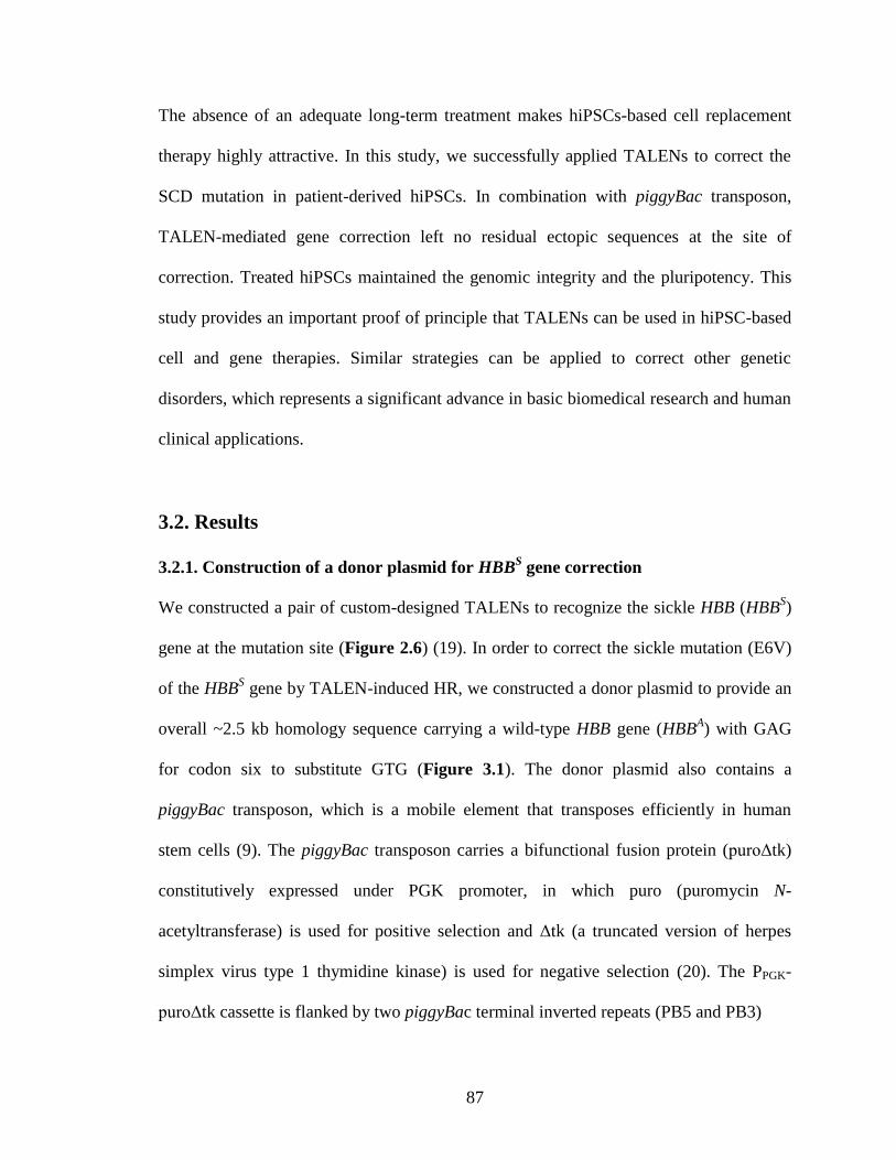
87
The absence of an adequate long-term treatment makes hiPSCs-based cell replacement
therapy highly attractive. In this study, we successfully applied TALENs to correct the
SCD mutation in patient-derived hiPSCs. In combination with piggyBac transposon,
TALEN-mediated gene correction left no residual ectopic sequences at the site of
correction. Treated hiPSCs maintained the genomic integrity and the pluripotency. This
study provides an important proof of principle that TALENs can be used in hiPSC-based
cell and gene therapies. Similar strategies can be applied to correct other genetic
disorders, which represents a significant advance in basic biomedical research and human
clinical applications.
3.2. Results
3.2.1. Construction of a donor plasmid for HBBS gene correction
We constructed a pair of custom-designed TALENs to recognize the sickle HBB (HBBS)
gene at the mutation site (Figure 2.6) (19). In order to correct the sickle mutation (E6V)
of the HBBS gene by TALEN-induced HR, we constructed a donor plasmid to provide an
overall ~2.5 kb homology sequence carrying a wild-type HBB gene (HBBA) with GAG
for codon six to substitute GTG (Figure 3.1). The donor plasmid also contains a
piggyBac transposon, which is a mobile element that transposes efficiently in human
stem cells (9). The piggyBac transposon carries a bifunctional fusion protein (puroΔtk)
constitutively expressed under PGK promoter, in which puro (puromycin N-
acetyltransferase) is used for positive selection and Δtk (a truncated version of herpes
simplex virus type 1 thymidine kinase) is used for negative selection (20). The PPGK-
puroΔtk cassette is flanked by two piggyBac terminal inverted repeats (PB5 and PB3)
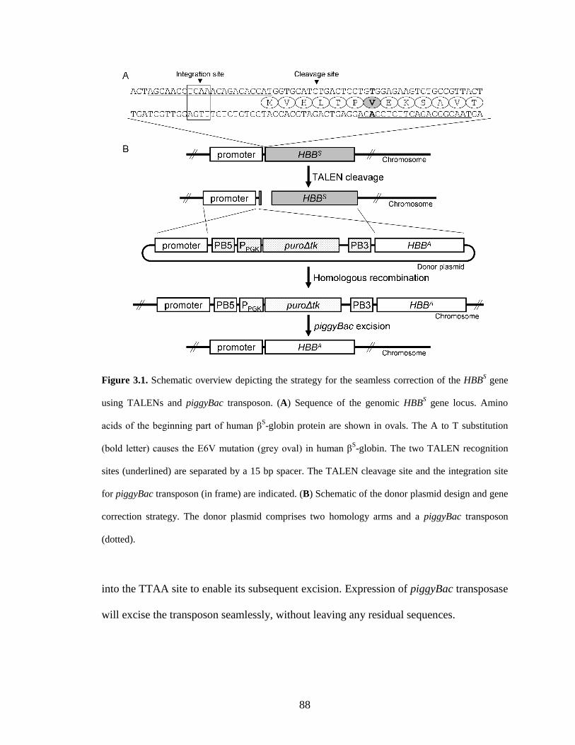
88
Figure 3.1. Schematic overview depicting the strategy for the seamless correction of the HBBS gene
using TALENs and piggyBac transposon. (A) Sequence of the genomic HBBS gene locus. Amino
acids of the beginning part of human βS-globin protein are shown in ovals. The A to T substitution
(bold letter) causes the E6V mutation (grey oval) in human βS-globin. The two TALEN recognition
sites (underlined) are separated by a 15 bp spacer. The TALEN cleavage site and the integration site
for piggyBac transposon (in frame) are indicated. (B) Schematic of the donor plasmid design and gene
correction strategy. The donor plasmid comprises two homology arms and a piggyBac transposon
(dotted).
into the TTAA site to enable its subsequent excision. Expression of piggyBac transposase
will excise the transposon seamlessly, without leaving any residual sequences.
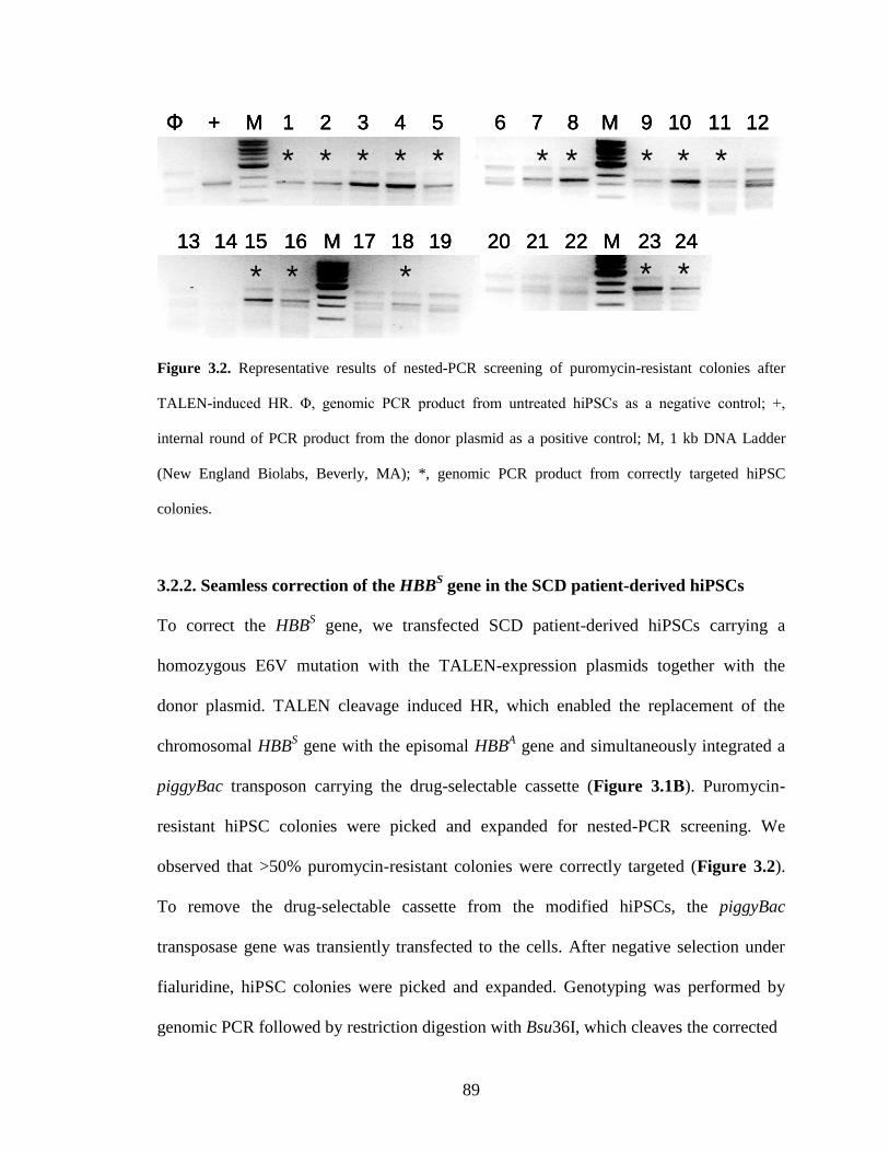
89
Φ + M 1 2 3 4 5 6 7 8 M 9 10 11 12
13 14 15 16 M 17 18 19 20 21 22 M 23 24
* * * * * * * * *
* * * * *
*
Φ + M 1 2 3 4 5 6 7 8 M 9 10 11 12
13 14 15 16 M 17 18 19 20 21 22 M 23 24
Φ + M 1 2 3 4 5 6 7 8 M 9 10 11 12
13 14 15 16 M 17 18 19 20 21 22 M 23 24
* * * * * * * * *
* * * * *
*
Figure 3.2. Representative results of nested-PCR screening of puromycin-resistant colonies after
TALEN-induced HR. Φ, genomic PCR product from untreated hiPSCs as a negative control; +,
internal round of PCR product from the donor plasmid as a positive control; M, 1 kb DNA Ladder
(New England Biolabs, Beverly, MA); *, genomic PCR product from correctly targeted hiPSC
colonies.
3.2.2. Seamless correction of the HBBS gene in the SCD patient-derived hiPSCs
To correct the HBBS gene, we transfected SCD patient-derived hiPSCs carrying a
homozygous E6V mutation with the TALEN-expression plasmids together with the
donor plasmid. TALEN cleavage induced HR, which enabled the replacement of the
chromosomal HBBS gene with the episomal HBB
A gene and simultaneously integrated a
piggyBac transposon carrying the drug-selectable cassette (Figure 3.1B). Puromycin-
resistant hiPSC colonies were picked and expanded for nested-PCR screening. We
observed that >50% puromycin-resistant colonies were correctly targeted (Figure 3.2).
To remove the drug-selectable cassette from the modified hiPSCs, the piggyBac
transposase gene was transiently transfected to the cells. After negative selection under
fialuridine, hiPSC colonies were picked and expanded. Genotyping was performed by
genomic PCR followed by restriction digestion with Bsu36I, which cleaves the corrected
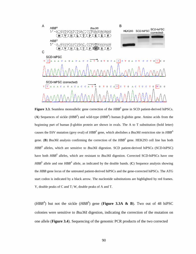
90
Figure 3.3. Seamless monoallelic gene correction of the HBBS gene in SCD patient-derived hiPSCs.
(A) Sequences of sickle (HBBS) and wild-type (HBB
A) human β-globin gene. Amino acids from the
beginning part of human β-globin protein are shown in ovals. The A to T substitution (bold letter)
causes the E6V mutation (grey oval) of HBBS gene, which abolishes a Bsu36I restriction site in HBB
A
gene. (B) Bsu36I analysis confirming the correction of the HBBS gene. HEK293 cell line has both
HBBA alleles, which are sensitive to Bsu36I digestion. SCD patient-derived hiPSCs (SCD-hiPSC)
have both HBBS alleles, which are resistant to Bsu36I digestion. Corrected SCD-hiPSCs have one
HBBS allele and one HBB
A allele, as indicated by the double bands. (C) Sequence analysis showing
the HBB gene locus of the untreated patient-derived hiPSCs and the gene-corrected hiPSCs. The ATG
start codon is indicated by a black arrow. The nucleotide substitutions are highlighted by red frames.
Y, double peaks of C and T; W, double peaks of A and T.
(HBBA) but not the sickle (HBB
S) gene (Figure 3.3A & B). Two out of 48 hiPSC
colonies were sensitive to Bsu36I digestion, indicating the correction of the mutation on
one allele (Figure 3.4). Sequencing of the genomic PCR products of the two corrected
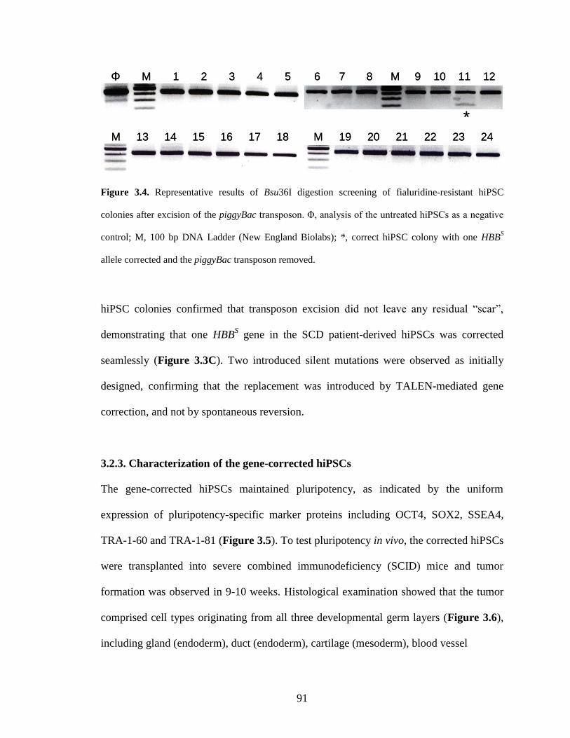
91
Φ M 1 2 3 4 5 6 7 8 M 9 10 11 12
13 14 15 16 17 18M M 19 20 21 22 23 24
*
Φ M 1 2 3 4 5 6 7 8 M 9 10 11 12
13 14 15 16 17 18M M 19 20 21 22 23 24
*
Figure 3.4. Representative results of Bsu36I digestion screening of fialuridine-resistant hiPSC
colonies after excision of the piggyBac transposon. Φ, analysis of the untreated hiPSCs as a negative
control; M, 100 bp DNA Ladder (New England Biolabs); *, correct hiPSC colony with one HBBS
allele corrected and the piggyBac transposon removed.
hiPSC colonies confirmed that transposon excision did not leave any residual “scar”,
demonstrating that one HBBS gene in the SCD patient-derived hiPSCs was corrected
seamlessly (Figure 3.3C). Two introduced silent mutations were observed as initially
designed, confirming that the replacement was introduced by TALEN-mediated gene
correction, and not by spontaneous reversion.
3.2.3. Characterization of the gene-corrected hiPSCs
The gene-corrected hiPSCs maintained pluripotency, as indicated by the uniform
expression of pluripotency-specific marker proteins including OCT4, SOX2, SSEA4,
TRA-1-60 and TRA-1-81 (Figure 3.5). To test pluripotency in vivo, the corrected hiPSCs
were transplanted into severe combined immunodeficiency (SCID) mice and tumor
formation was observed in 9-10 weeks. Histological examination showed that the tumor
comprised cell types originating from all three developmental germ layers (Figure 3.6),
including gland (endoderm), duct (endoderm), cartilage (mesoderm), blood vessel
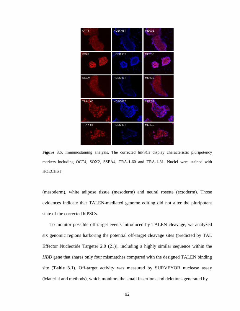
92
Figure 3.5. Immunostaining analysis. The corrected hiPSCs display characteristic pluripotency
markers including OCT4, SOX2, SSEA4, TRA-1-60 and TRA-1-81. Nuclei were stained with
HOECHST.
(mesoderm), white adipose tissue (mesoderm) and neural rosette (ectoderm). Those
evidences indicate that TALEN-mediated genome editing did not alter the pluripotent
state of the corrected hiPSCs.
To monitor possible off-target events introduced by TALEN cleavage, we analyzed
six genomic regions harboring the potential off-target cleavage sites (predicted by TAL
Effector Nucleotide Targeter 2.0 (21)), including a highly similar sequence within the
HBD gene that shares only four mismatches compared with the designed TALEN binding
site (Table 3.1). Off-target activity was measured by SURVEYOR nuclease assay
(Material and methods), which monitors the small insertions and deletions generated by
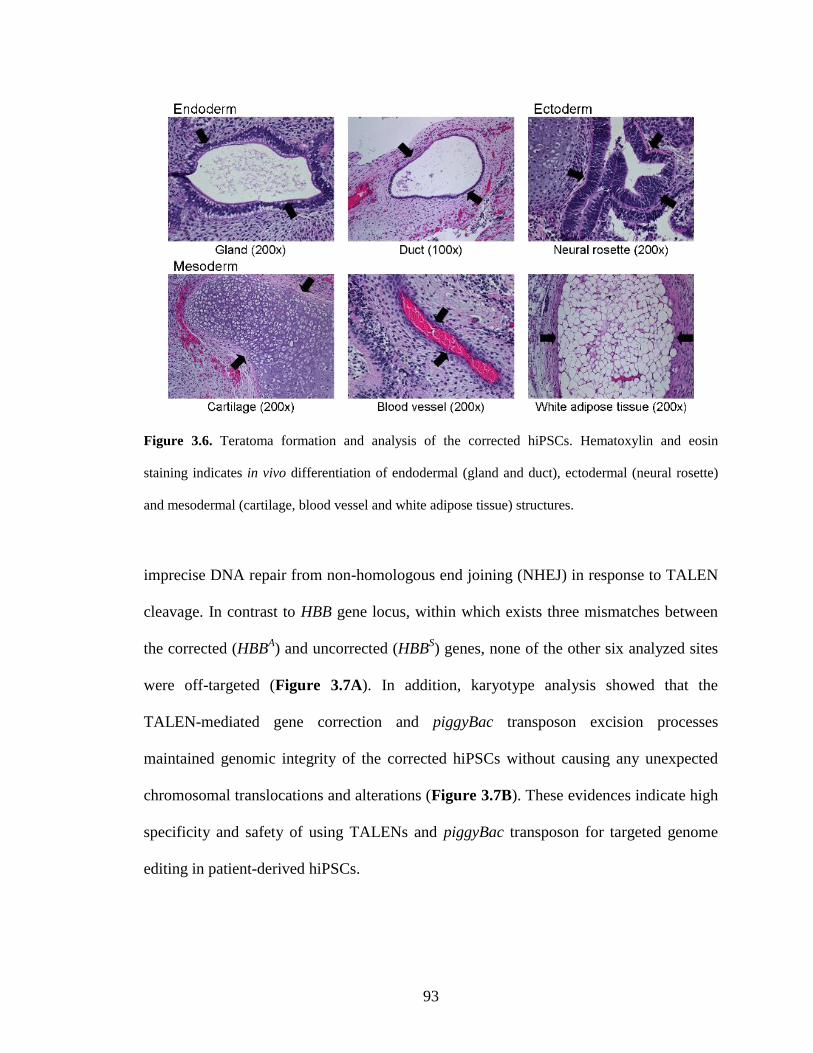
93
Figure 3.6. Teratoma formation and analysis of the corrected hiPSCs. Hematoxylin and eosin
staining indicates in vivo differentiation of endodermal (gland and duct), ectodermal (neural rosette)
and mesodermal (cartilage, blood vessel and white adipose tissue) structures.
imprecise DNA repair from non-homologous end joining (NHEJ) in response to TALEN
cleavage. In contrast to HBB gene locus, within which exists three mismatches between
the corrected (HBBA) and uncorrected (HBB
S) genes, none of the other six analyzed sites
were off-targeted (Figure 3.7A). In addition, karyotype analysis showed that the
TALEN-mediated gene correction and piggyBac transposon excision processes
maintained genomic integrity of the corrected hiPSCs without causing any unexpected
chromosomal translocations and alterations (Figure 3.7B). These evidences indicate high
specificity and safety of using TALENs and piggyBac transposon for targeted genome
editing in patient-derived hiPSCs.
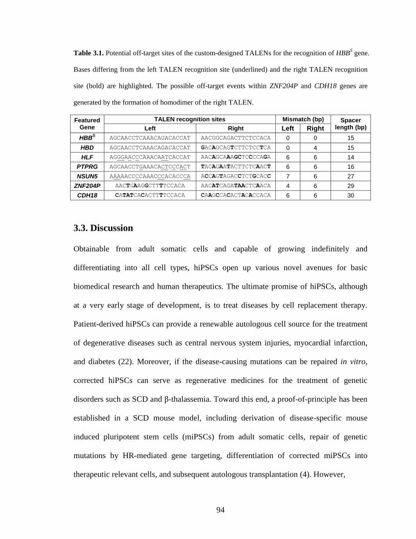
94
Table 3.1. Potential off-target sites of the custom-designed TALENs for the recognition of HBBS gene.
Bases differing from the left TALEN recognition site (underlined) and the right TALEN recognition
site (bold) are highlighted. The possible off-target events within ZNF204P and CDH18 genes are
generated by the formation of homodimer of the right TALEN.
Featured Gene
TALEN recognition sites Mismatch (bp) Spacer length (bp) Left Right Left Right
HBBS AGCAACCTCAAACAGACACCAT AACGGCAGACTTCTCCACA 0 0 15
HBD AGCAACCTCAAACAGACACCAT GACAGCAGTCTTCTCCTCA 0 4 15
HLF AGGGAACCCAAACAATCACCAT AACAGCAAAGCTCCCCAGA 6 6 14
PTPRG AGCAACCTGAAACACTCCCACT TACAGAATACTTCTCAACT 6 6 16
NSUN5 AAAAACCCCAAACCCACACCCA ACCAGTAGACCTCTGCACC 7 6 27
ZNF204P AACTGAAGGCTTTTCCACA AACATCAGATAACTCAACA 4 6 29
CDH18 CATATCACACTTTTCCACA CAAGCCACACTACACCACA 6 6 30
3.3. Discussion
Obtainable from adult somatic cells and capable of growing indefinitely and
differentiating into all cell types, hiPSCs open up various novel avenues for basic
biomedical research and human therapeutics. The ultimate promise of hiPSCs, although
at a very early stage of development, is to treat diseases by cell replacement therapy.
Patient-derived hiPSCs can provide a renewable autologous cell source for the treatment
of degenerative diseases such as central nervous system injuries, myocardial infarction,
and diabetes (22). Moreover, if the disease-causing mutations can be repaired in vitro,
corrected hiPSCs can serve as regenerative medicines for the treatment of genetic
disorders such as SCD and β-thalassemia. Toward this end, a proof-of-principle has been
established in a SCD mouse model, including derivation of disease-specific mouse
induced pluripotent stem cells (miPSCs) from adult somatic cells, repair of genetic
mutations by HR-mediated gene targeting, differentiation of corrected miPSCs into
therapeutic relevant cells, and subsequent autologous transplantation (4). However,
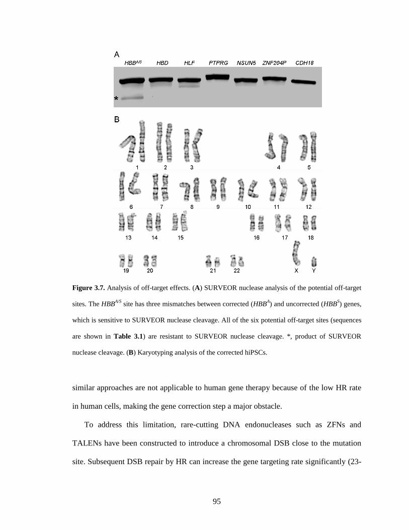
95
Figure 3.7. Analysis of off-target effects. (A) SURVEOR nuclease analysis of the potential off-target
sites. The HBBA/S
site has three mismatches between corrected (HBBA) and uncorrected (HBB
S) genes,
which is sensitive to SURVEOR nuclease cleavage. All of the six potential off-target sites (sequences
are shown in Table 3.1) are resistant to SURVEOR nuclease cleavage. *, product of SURVEOR
nuclease cleavage. (B) Karyotyping analysis of the corrected hiPSCs.
similar approaches are not applicable to human gene therapy because of the low HR rate
in human cells, making the gene correction step a major obstacle.
To address this limitation, rare-cutting DNA endonucleases such as ZFNs and
TALENs have been constructed to introduce a chromosomal DSB close to the mutation
site. Subsequent DSB repair by HR can increase the gene targeting rate significantly (23-
96
25). ZFNs have been applied to correct disease-causing mutations in hiPSCs derived
from patients with SCD (7,10), Parkinson’s disease (8), and α1-antitrypsin deficiency (9).
However, the lack of specificity of zinc finger domains raises safety concerns about
potential off-target cleavage, which might lead to undesired mutations and chromosomal
aberrations (11,12). In addition, zinc finger domains have limited modularity due to the
context-dependent DNA-binding effects. Recently, TALENs have rapidly emerged as an
alternative to ZFNs and have been widely applied for genome editing in many different
organisms and cell types (19,26-38). The modular nature of the TALE central repeat
domains enables researchers to tailor DNA recognition specificity with ease and target
essentially any desired DNA sequence (15,16). Moreover, TALENs exhibit minimal off-
target effects as determined by exome sequencing and whole-genome sequencing (37),
indicating their potential utility for human therapeutics. In this study, we sought to
explore the therapeutic potential of TALENs in hiPSCs-based cell and gene therapies.
We successfully applied TALENs to correct the SCD mutation in patient-derived hiPSCs.
The corrected hiPSCs maintain their pluripotent state. TALEN-mediated gene correction
does not introduce detectable off-target events or chromosomal alterations.
Although the chromosomal DSBs introduced by custom-designed DNA
endonucleases can significantly stimulate HR, the overall gene targeting rate is still low,
which makes it necessary to introduce a drug-selectable cassette into the chromosome for
the isolation of targeted colonies. To remove the unwanted drug-selection cassette after
positive selection, the Cre/loxP recombination system has been frequently used, which
leaves a 34 bp loxP sequence at the site of excision (7,10,35,39,40). However, the small
ectopic sequence has the potential to silence the gene expression of the targeted allele (10)
97
or interfere with transcription regulatory elements of adjacent genes (41). To address this
limitation, we inserted the drug-selectable cassette into a piggyBac transposon, which
enables the removal of the selection cassette efficiently in hiPSCs without leaving any
remnant sequences (9). After TALEN-mediated gene correction and drug selection,
transient expression of piggyBac transposase excised the drug-selectable cassette without
leaving any residual “scar” in the corrected gene. The combination of TALEN and
piggyBac technology enabled us to correct the SCD mutation in patient-derived hiPSCs
in a seamless manner.
Because robust protocols for differentiating hiPSCs into mature, enucleated red blood
cells in vitro have not been established, functional data demonstrating the recovery of the
gene-corrected red blood cells cannot be generated in this study. In addition, a
reproducible in vivo model is needed to test the efficacy and safety of using the gene-
corrected hiPSCs as a renewable autologous cell source for the treatment of SCD patients
(10,42,43). Although challenges and barriers remain, our study demonstrates an
important first step of using TALEN technology and piggBac transposon to correct the
disease-causing mutation in situ in a seamless manner, which represents a significant
advance toward hiPSC-based cell and gene therapies.
3.4. Materials and methods
3.4.1. Cell culture
SCD patient-derived hiPSCs with a homozygous E6V mutation were kindly provided by
Dr. Marius Wernig from Stanford University (Stanford, CA). Cultures were maintained
under feeder-free culture conditions on 6-well plates coated with Matrigel Basement
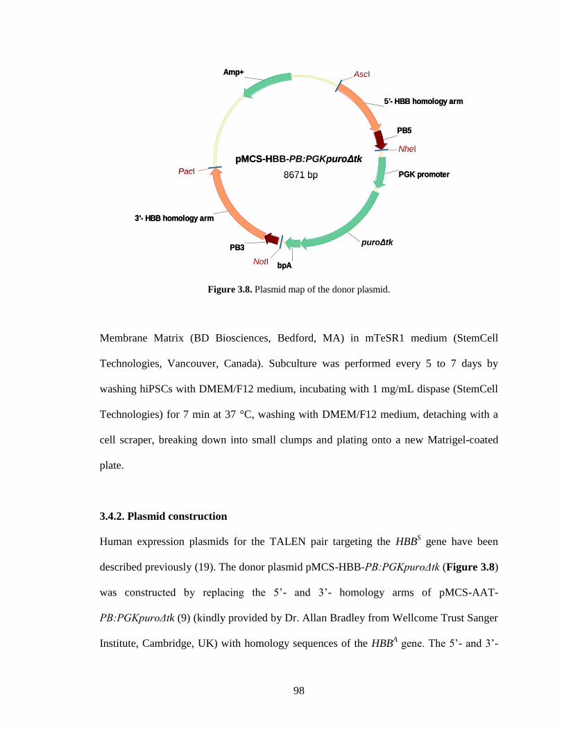
98
pMCS-HBB
8671 bp
5'- HBB homology arm
3'- HBB homology arm
puro deltaTK
bpA
Amp+
PB5
PB3
PGK promoter
Asc I (669)
Nhe I (2002)
Not I (4621)
Pac I (6373)
pMCS-HBB-PB:PGKpuroΔtk
puroΔtk
8671 bp
AscI
NheI
NotI
PacI
pMCS-HBB
8671 bp
5'- HBB homology arm
3'- HBB homology arm
puro deltaTK
bpA
Amp+
PB5
PB3
PGK promoter
Asc I (669)
Nhe I (2002)
Not I (4621)
Pac I (6373)
pMCS-HBB-PB:PGKpuroΔtk
puroΔtk
8671 bp
AscI
NheI
NotI
PacI
Figure 3.8. Plasmid map of the donor plasmid.
Membrane Matrix (BD Biosciences, Bedford, MA) in mTeSR1 medium (StemCell
Technologies, Vancouver, Canada). Subculture was performed every 5 to 7 days by
washing hiPSCs with DMEM/F12 medium, incubating with 1 mg/mL dispase (StemCell
Technologies) for 7 min at 37 °C, washing with DMEM/F12 medium, detaching with a
cell scraper, breaking down into small clumps and plating onto a new Matrigel-coated
plate.
3.4.2. Plasmid construction
Human expression plasmids for the TALEN pair targeting the HBBS gene have been
described previously (19). The donor plasmid pMCS-HBB-PB:PGKpuroΔtk (Figure 3.8)
was constructed by replacing the 5’- and 3’- homology arms of pMCS-AAT-
PB:PGKpuroΔtk (9) (kindly provided by Dr. Allan Bradley from Wellcome Trust Sanger
Institute, Cambridge, UK) with homology sequences of the HBBA gene. The 5’- and 3’-
99
HBB homology arms were amplified by PCR from the genomic DNA of HEK293 cells.
The primers AscI-HBB-5’-for (5’-act gac ggc gcg cct att ctt agt gga cta gag g-3’, AscI
site shown in italic) and PB5-HBB-5’-rev (5’-gca tgc gtc aat ttt acg cag act atc ttt cta ggg
tta agg ttg cta gtg aac aca gtt-3’) amplified the 1.0 kbp 5’- HBB homology arm. The
primers PB3-HBB-3’ (5’-ttt aac gta cgt cac aat atg att atc ttt cta ggg tta aac aga cac cat
ggt gca tct-3’) and PacI-HBB-3’-rev (5’-act gac tta att aat gga cag caa gaa agc gag c-3’,
PacI site shown in italic) amplified the 1.5 kbp 3’- HBB homology arm. The two terminal
inverted repeats (PB5 and PB3) of piggyBac transposon were amplified by PCR from
pMCS-AAT-PB:PGKpuroΔtk. PB5 was amplified by primers HBB-5’-PB5-for (5’-ctt
aca ttt gct tct gac aca act gtg ttc act agc aac ctt aac cct aga aag ata gtc-3’) and NheI-PB5-
rev (5’-act tgt gct agc att cta gtt gat atc tat aac-3’, NheI site shown in italic). PB3 was
amplified by primers NotI-PB3-for (5’-gct aga gcg gcc gca ctc gag ata tct aga c-3’, NotI
site shown in italic) and HBB-3’-PB3-rev (5’-cag act tct cct cag gag tca gat gca cca tgg
tgt ctg ttt aac cct aga aag ata atc-3’). 5’-HBB homology arm was combined with PB5 by
overlap extension-PCR (OE-PCR) (44) and inserted into AscI and NheI sites of pMCS-
AAT-PB:PGKpuroΔtk to form pMCS-Intermediate. 3’-HBB homology arm was
combined with PB3 by OE-PCR and inserted into NotI and PacI sites of pMCS-
Intermediate to form donor plasmid pMCS-HBB-PB:PGKpuroΔtk.
3.4.3. TALEN-mediated genome editing of hiPSCs
On day 0, SCD-hiPSCs were pre-treated with 2 μM of a Rho Kinase (ROCK) inhibitor
(thiazovivin, BioVision, Milpitas, CA) for at least 1 hr prior to transfection. Cells were
dissociated into single-cell suspension by washing with DMEM/F12, incubating with
100
accutase (StemCell Technologies) for 7 min at 37 °C, mixing with mTeSR1 medium
followed by vigorous pipetting. 3 × 10⁶ cells were transfected with 2 μg of each TALEN-
encoding plasmid and 2 μg of donor plasmid by Amaxa nucleofection system using
Human Stem Cell Nucleofector Kit 1 (Lonza, Cologne, Germany) according to the
manufacturer’s instruction. The treated cells were plated onto one Matrigel-coated 100
mm dish in mTeSR1 medium supplemented with 2 μM of thiazovivin for the first 24 hrs.
On day 3, puromycin (Sigma, St. Louis, MO) selection at 0.5 μg/mL was started.
Individual colonies were picked and expanded in one or two Matrigel-coated 24-well
plates on day 13-17. On day 20-24, near-confluent cells in each well were harvested
using accutase. One quarter of the cells was seeded onto a new Matrigel-coated 24-well
plate and the rest of the cells were used for nested PCR analysis.
3.4.4. Nested PCR of targeted integration
Genomic DNA was extracted from hiPSCs using QuickExtract DNA Extraction Solution
1.0 (EPICENTRE Biotechnologies, Madison, WI) according to the manufacturer’s
instructions. The external round of PCR was carried out using primers Purotest-for-PB3
(5’-ggc tcg aga tcc act agt tc-3’) and Purotest-rev-HBB3-long (5’-gat gct caa ggc cct tca
ta-3’). The external round of PCR products were used as template for the internal round
of PCR using primers Nested-for-HBB3 (5’-acg tgg atg aag ttg gtg gt-3’) and HBB-R (5’-
taa ggg tgg gaa aat aga cc-3’). Nested PCR products were then resolved on a 1.0%
agarose gel.
101
3.4.5. Transposon excision in targeted hiPSCs
On day 0, correctly targeted hiPSCs were pre-treated with 2 μM of thiazovivin for at least
1 hr prior to transfection. Cells were dissociated into single-cell suspension as described
earlier. 2 × 10⁶ cells were transfected with 4 μg of the hyperactive piggyBac transposase
expression vector (pCMV-hyPBase (45), kindly provided by Dr. Allan Bradley) by
Amaxa nucleofection system using Human Stem Cell Nucleofector Kit 1 according to the
manufacturer’s instructions. The treated cells were plated onto one Matrigel-coated 100
mm dish in mTeSR1 medium supplemented with 2 μM of thiazovivin for the first 24 hrs.
On day 3, cells were dissociated into single-cell suspension and seeded to a new
Matrigel-coated 100 mm dish at low density (~1000 cells/mL) in mTeSR1 medium
containing 2 μM of thiazovivin and 0.25 μM of fialuridine (Santa Cruz Biotechnology,
Santa Cruz, CA). On day 6, medium was changed to mTeSR1 medium supplemented
with 2.5 μM of fialuridine, which was used for daily medium change until picking
colonies. Individual colonies were picked and expanded in one or two Matrigel-coated
24-well plates on day 16-20. On day 23-27, near-confluent cells in each well were
harvested using accutase. One quarter of the cells was seeded onto a new Matrigel-coated
24-well plate and the rest of the cells were used for Bsu36I analysis and sequencing.
3.4.6. Bsu36I analysis and sequencing of genomic HBB locus
Genomic DNA from corrected hiPSCs was amplified with primers HBB-F (5’-agg tac
ggc tgt cat cac tt-3’) and HBB-R (5’-taa ggg tgg gaa aat aga cc-3’) under standard PCR
conditions, which resulted in a 402 bp product. PCR products were purified by QIAquick
Gel Extraction Kit (QIAGEN, Valencia, CA) and subjected to restriction digestion with
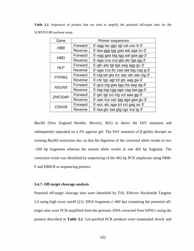
102
Table 3.2. Sequences of primers that are used to amplify the potential off-target sites for the
SURVEYOR nuclease assay.
Gene Primer sequences
HBB Forward 5'-agg tac ggc tgt cat cac tt-3'
Reverse 5'-taa ggg tgg gaa aat aga cc-3'
HBD Forward 5'-agg gaa tag tgg aat gaa gg-3'
Reverse 5'-aga cca cca gta atc tga gg-3'
HLF Forward 5'-gtc ata tgt tga aag agg gc-3'
Reverse 5'-aga cca ttc caa ata tag cag g-3'
PTPRG Forward 5'-ctg tat gta tcc aac atc ata ctg-3'
Reverse 5'-ctc tgc agt tct gtc aag ga-3'
NSUN5 Forward 5'-gca ctg gaa tgg cta aag ag-3'
Reverse 5'-tag tag cgg aga cag taa gg-3'
ZNF204P Forward 5'-gtc tgt cct ctg cct aaa gg-3'
Reverse 5'-aac tca cac tgg aga gaa gc-3'
CDH18 Forward 5'-acc atc aga tct cct gag ac-3'
Reverse 5'-taa gtc taa gtg cgc tca tg-3'
Bsu36I (New England Biolabs, Beverly, MA) to detect the E6V mutation and
subsequently separated on a 2% agarose gel. The E6V mutation of β-globin disrupts an
existing Bsu36I restriction site, so that the digestion of the corrected allele results in two
~200 bp fragments whereas the mutant allele results in one 402 bp fragment. The
correction event was identified by sequencing of the 402 bp PCR amplicons using HBB-
F and HBB-R as sequencing primers.
3.4.7. Off-target cleavage analysis
Potential off-target cleavage sites were identified by TAL Effector Nucleotide Targeter
2.0 using high score cutoff (21). DNA fragments (~400 bp) containing the potential off-
target sites were PCR-amplified from the genomic DNA extracted from hiPSCs using the
primers described in Table 3.2. Gel-purified PCR products were reannealed slowly and
103
analyzed by SURVEYOR Mutation Detection Kits (Transgenomic, Omaha, NE)
according to the manufacturer’s instructions. Off-target cleavage of TALENs can be
repaired by NHEJ, which typically generates deletions and insertions. Therefore,
genomic PCR products from TALEN-modified region are heterogeneous. After
reannealing, DNA heteroduplexes can be cleaved by SURVEYOR nucleases, which
indicates off-target cleavage of the TALENs. Because the potential off-target sites are
designed to be in the center of the genomic PCR products (~400 bp), cleavage by
SURVEYOR nucleases can generate two ~200 bp fragments. On the contrary, genomic
PCR products from TALEN-unmodified regions are homogeneous, which are resistant to
SURVEYOR nuclease.
3.4.8. Immunostaining
Expression of the five pluripotent markers including OCT4, SOX2, SSEA4, TRA-1-60
and TRA-1-80 were analyzed by Human ES/iPS Cell Characterization Kit (Applied
StemCell, Menlo Park, CA) according to the manufacturer’s instruction. HOECHST stain
was used to stain nuclei.
3.4.9. Karyotyping
Karyotyping of the gene-corrected hiPSCs was carried out by Applied StemCell Inc.
(Menlo Park, CA).
3.4.10. Teratoma formation and analysis
The gene-corrected hiPSCs were dissociated with dispase and triturated as small clumps
104
and injected into kidney capsule and testis sites (1.5-3×10⁶ cells per site) of three 6-week
old male mice (Fox Chase SCIDR-beige from Charles River Laboratories, Wilmington,
MA). Cells from a hiPSC line were injected into the same sites of one mouse as a positive
control. Tumor formation was observed in the three mice and harvested at day 74 post
injection. The tumor samples at the six sites were fixed in 10% formalin overnight,
embedded in paraffin, cut into 5-μm serial sections, and analyzed by hematoxylin and
eosin staining.
3.5. References
1. Takahashi, K., Tanabe, K., Ohnuki, M., Narita, M., Ichisaka, T., Tomoda, K. and
Yamanaka, S. (2007) Induction of pluripotent stem cells from adult human
fibroblasts by defined factors. Cell, 131, 861-872.
2. Saha, K. and Jaenisch, R. (2009) Technical challenges in using human induced
pluripotent stem cells to model disease. Cell Stem Cell, 5, 584-595.
3. Yamanaka, S. (2007) Strategies and new developments in the generation of
patient-specific pluripotent stem cells. Cell Stem Cell, 1, 39-49.
4. Hanna, J., Wernig, M., Markoulaki, S., Sun, C.W., Meissner, A., Cassady, J.P.,
Beard, C., Brambrink, T., Wu, L.C., Townes, T.M. et al. (2007) Treatment of
sickle cell anemia mouse model with iPS cells generated from autologous skin.
Science, 318, 1920-1923.
5. Raya, A., Rodriguez-Piza, I., Guenechea, G., Vassena, R., Navarro, S., Barrero,
M.J., Consiglio, A., Castella, M., Rio, P., Sleep, E. et al. (2009) Disease-corrected
105
haematopoietic progenitors from Fanconi anaemia induced pluripotent stem cells.
Nature, 460, 53-59.
6. Zwaka, T.P. and Thomson, J.A. (2003) Homologous recombination in human
embryonic stem cells. Nat Biotechnol, 21, 319-321.
7. Sebastiano, V., Maeder, M.L., Angstman, J.F., Haddad, B., Khayter, C., Yeo,
D.T., Goodwin, M.J., Hawkins, J.S., Ramirez, C.L., Batista, L.F. et al. (2011) In
situ genetic correction of the sickle cell anemia mutation in human induced
pluripotent stem cells using engineered zinc finger nucleases. Stem Cells, 29,
1717-1726.
8. Soldner, F., Laganiere, J., Cheng, A.W., Hockemeyer, D., Gao, Q., Alagappan,
R., Khurana, V., Golbe, L.I., Myers, R.H., Lindquist, S. et al. (2011) Generation
of isogenic pluripotent stem cells differing exclusively at two early onset
Parkinson point mutations. Cell, 146, 318-331.
9. Yusa, K., Rashid, S.T., Strick-Marchand, H., Varela, I., Liu, P.Q., Paschon, D.E.,
Miranda, E., Ordonez, A., Hannan, N.R., Rouhani, F.J. et al. (2011) Targeted
gene correction of alpha1-antitrypsin deficiency in induced pluripotent stem cells.
Nature, 478, 391-394.
10. Zou, J., Mali, P., Huang, X., Dowey, S.N. and Cheng, L. (2011) Site-specific gene
correction of a point mutation in human iPS cells derived from an adult patient
with sickle cell disease. Blood, 118, 4599-4608.
11. Pattanayak, V., Ramirez, C.L., Joung, J.K. and Liu, D.R. (2011) Revealing off-
target cleavage specificities of zinc-finger nucleases by in vitro selection. Nat
Methods, 8, 765-770.
106
12. Radecke, S., Radecke, F., Cathomen, T. and Schwarz, K. (2010) Zinc-finger
nuclease-induced gene repair with oligodeoxynucleotides: wanted and unwanted
target locus modifications. Mol Ther, 18, 743-753.
13. Sun, N. and Zhao, H. (2013) Transcription activator-like effector nucleases
(TALENs): A highly efficient and versatile tool for genome editing. Biotechnol
Bioeng, doi: 10.1002/bit.24890.
14. Joung, J.K. and Sander, J.D. (2013) TALENs: a widely applicable technology for
targeted genome editing. Nat Rev Mol Cell Biol, 14, 49-55.
15. Boch, J., Scholze, H., Schornack, S., Landgraf, A., Hahn, S., Kay, S., Lahaye, T.,
Nickstadt, A. and Bonas, U. (2009) Breaking the code of DNA binding specificity
of TAL-type III effectors. Science, 326, 1509-1512.
16. Moscou, M.J. and Bogdanove, A.J. (2009) A simple cipher governs DNA
recognition by TAL effectors. Science, 326, 1501.
17. Weatherall, D.J. and Clegg, J.B. (2001) Inherited haemoglobin disorders: an
increasing global health problem. Bull World Health Organ, 79, 704-712.
18. Orkin, S.H. and Higgs, D.R. (2010) Medicine. Sickle cell disease at 100 years.
Science, 329, 291-292.
19. Sun, N., Liang, J., Abil, Z. and Zhao, H. (2012) Optimized TAL effector
nucleases (TALENs) for use in treatment of sickle cell disease. Mol Biosyst, 8,
1255-1263.
20. Chen, Y.T. and Bradley, A. (2000) A new positive/negative selectable marker,
puDeltatk, for use in embryonic stem cells. Genesis, 28, 31-35.
107
21. Doyle, E.L., Booher, N.J., Standage, D.S., Voytas, D.F., Brendel, V.P., Vandyk,
J.K. and Bogdanove, A.J. (2012) TAL Effector-Nucleotide Targeter (TALE-NT)
2.0: tools for TAL effector design and target prediction. Nucleic Acids Res, 40,
W117-122.
22. Goldring, C.E., Duffy, P.A., Benvenisty, N., Andrews, P.W., Ben-David, U.,
Eakins, R., French, N., Hanley, N.A., Kelly, L., Kitteringham, N.R. et al. (2011)
Assessing the safety of stem cell therapeutics. Cell Stem Cell, 8, 618-628.
23. Jasin, M. (1996) Genetic manipulation of genomes with rare-cutting
endonucleases. Trends Genet, 12, 224-228.
24. Smih, F., Rouet, P., Romanienko, P.J. and Jasin, M. (1995) Double-strand breaks
at the target locus stimulate gene targeting in embryonic stem cells. Nucleic Acids
Res, 23, 5012-5019.
25. Sun, N., Abil, Z. and Zhao, H. (2012) Recent advances in targeted genome
engineering in mammalian systems. Biotechnol J, 7, 1074-1087.
26. Huang, P., Xiao, A., Zhou, M., Zhu, Z., Lin, S. and Zhang, B. (2011) Heritable
gene targeting in zebrafish using customized TALENs. Nat Biotechnol, 29, 699-
700.
27. Miller, J.C., Tan, S., Qiao, G., Barlow, K.A., Wang, J., Xia, D.F., Meng, X.,
Paschon, D.E., Leung, E., Hinkley, S.J. et al. (2011) A TALE nuclease
architecture for efficient genome editing. Nat Biotechnol, 29, 143-148.
28. Mussolino, C., Morbitzer, R., Lutge, F., Dannemann, N., Lahaye, T. and
Cathomen, T. (2011) A novel TALE nuclease scaffold enables high genome
108
editing activity in combination with low toxicity. Nucleic Acids Res, 39, 9283-
9293.
29. Reyon, D., Tsai, S.Q., Khayter, C., Foden, J.A., Sander, J.D. and Joung, J.K.
(2012) FLASH assembly of TALENs for high-throughput genome editing. Nat
Biotechnol, 30, 460-465.
30. Sander, J.D., Cade, L., Khayter, C., Reyon, D., Peterson, R.T., Joung, J.K. and
Yeh, J.R. (2011) Targeted gene disruption in somatic zebrafish cells using
engineered TALENs. Nat Biotechnol, 29, 697-698.
31. Sung, Y.H., Baek, I.J., Kim, D.H., Jeon, J., Lee, J., Lee, K., Jeong, D., Kim, J.S.
and Lee, H.W. (2013) Knockout mice created by TALEN-mediated gene
targeting. Nat Biotechnol, 31, 23-24.
32. Tesson, L., Usal, C., Menoret, S., Leung, E., Niles, B.J., Remy, S., Santiago, Y.,
Vincent, A.I., Meng, X., Zhang, L. et al. (2011) Knockout rats generated by
embryo microinjection of TALENs. Nat Biotechnol, 29, 695-696.
33. Bedell, V.M., Wang, Y., Campbell, J.M., Poshusta, T.L., Starker, C.G., Krug,
R.G., 2nd, Tan, W., Penheiter, S.G., Ma, A.C., Leung, A.Y. et al. (2012) In vivo
genome editing using a high-efficiency TALEN system. Nature, 491, 114-118.
34. Carlson, D.F., Tan, W., Lillico, S.G., Stverakova, D., Proudfoot, C., Christian,
M., Voytas, D.F., Long, C.R., Whitelaw, C.B. and Fahrenkrug, S.C. (2012)
Efficient TALEN-mediated gene knockout in livestock. Proc Natl Acad Sci U S
A, 109, 17382-17387.
109
35. Hockemeyer, D., Wang, H., Kiani, S., Lai, C.S., Gao, Q., Cassady, J.P., Cost,
G.J., Zhang, L., Santiago, Y., Miller, J.C. et al. (2011) Genetic engineering of
human pluripotent cells using TALE nucleases. Nat Biotechnol, 29, 731-734.
36. Wood, A.J., Lo, T.W., Zeitler, B., Pickle, C.S., Ralston, E.J., Lee, A.H., Amora,
R., Miller, J.C., Leung, E., Meng, X. et al. (2011) Targeted genome editing across
species using ZFNs and TALENs. Science, 333, 307.
37. Ding, Q., Lee, Y.K., Schaefer, E.A., Peters, D.T., Veres, A., Kim, K.,
Kuperwasser, N., Motola, D.L., Meissner, T.B., Hendriks, W.T. et al. (2013) A
TALEN genome-editing system for generating human stem cell-based disease
models. Cell Stem Cell, 12, 238-251.
38. Liu, J., Li, C., Yu, Z., Huang, P., Wu, H., Wei, C., Zhu, N., Shen, Y., Chen, Y.,
Zhang, B. et al. (2012) Efficient and specific modifications of the Drosophila
genome by means of an easy TALEN strategy. J Genet Genomics, 39, 209-215.
39. Hockemeyer, D., Soldner, F., Beard, C., Gao, Q., Mitalipova, M., DeKelver, R.C.,
Katibah, G.E., Amora, R., Boydston, E.A., Zeitler, B. et al. (2009) Efficient
targeting of expressed and silent genes in human ESCs and iPSCs using zinc-
finger nucleases. Nat Biotechnol, 27, 851-857.
40. Osborn, M.J., Starker, C.G., McElroy, A.N., Webber, B.R., Riddle, M.J., Xia, L.,
Defeo, A.P., Gabriel, R., Schmidt, M., Von Kalle, C. et al. (2013) TALEN-based
Gene Correction for Epidermolysis Bullosa. Mol Ther, doi:
10.1038/mt.2013.1056.
41. Meier, I.D., Bernreuther, C., Tilling, T., Neidhardt, J., Wong, Y.W., Schulze, C.,
Streichert, T. and Schachner, M. (2010) Short DNA sequences inserted for gene
110
targeting can accidentally interfere with off-target gene expression. Faseb J, 24,
1714-1724.
42. Kaufman, D.S. (2009) Toward clinical therapies using hematopoietic cells derived
from human pluripotent stem cells. Blood, 114, 3513-3523.
43. Lengerke, C. and Daley, G.Q. (2010) Autologous blood cell therapies from
pluripotent stem cells. Blood Rev, 24, 27-37.
44. Ho, S.N., Hunt, H.D., Horton, R.M., Pullen, J.K. and Pease, L.R. (1989) Site-
directed mutagenesis by overlap extension using the polymerase chain reaction.
Gene, 77, 51-59.
45. Yusa, K., Zhou, L., Li, M.A., Bradley, A. and Craig, N.L. (2011) A hyperactive
piggyBac transposase for mammalian applications. Proc Natl Acad Sci U S A,
108, 1531-1536.
111
CHAPTER 4. DIRECTED EVOLUTION OF TALENS WITH
IMPROVED GENOME EDITING EFFICACY
4.1. Introduction
Genetic modifications of human genomes have a wide range of applications such as
investigating human biology, studying disease mechanisms and treating diseases via gene
therapies (1,2). Although virus-based transgenesis can modify human genomes efficiently
by introduction of exogenous genes, random integration of viral transgenes poses the risk
of insertional mutagenesis and carcinogenesis (3,4). In contrast, targeted genome editing
or engineering enables researchers to tailor the human genome in a precise manner. To
achieve efficient genome editing in human cells, DNA double-strand breaks (DSBs) have
to be introduced at the specific sites of the chromosome by custom-designed DNA
endonucleases. Subsequent repair of the DSBs by non-homologous end joining (NHEJ)
or homologous recombination can generate desired genomic modifications such as gene
disruptions, gene deletions, gene insertions, gene replacements and chromosome
rearrangements (see Chapter 1).
Recently, transcription activator-like effector (TALE) nucleases (TALENs) have
emerged as an efficient and versatile tool for genome editing by introduction of
chromosomal DNA DSBs (5-7). TALENs are artificial DNA endonucleases composed of
a TALE central repeat domain as the DNA recognition module and a non-specific FokI
nuclease domain as the DNA cleavage module. The TALE central repeat domain
comprises a series of tandem repeat units, each of which typically contains 34 residues.
112
Each repeat is used to recognize a single nucleotide and the DNA recognition specificity
is determined by the highly variable residues at positions 12 and 13 (e.g. NI recognizes
adenine, HD recognizes cytosine, NG recognizes thymine, NH recognizes guanine and
NN recognizes guanine and adenine) (8-11). The simple DNA recognition code and the
modular nature of the TALE central repeat units enable researchers to tailor DNA
recognition specificity with ease and target essentially any desired DNA sequence.
Therefore, TALENs have been widely applied for genome editing in various species (7).
Due to their long recognition sequences (30-50 bp) and high DNA recognition specificity,
TALENs exhibit minimal off-target effects in the context of complex human genomes
(12-14), making them an ideal platform for human genome editing.
First-generation TALEN scaffolds have been described with different N-terminal
segments (NTSs) and C-terminal segments (CTSs) flanking the TALE central repeat
domains (13-16). Some of them have been applied for generating human stem cell-based
disease models (12) and treating human diseases (17,18), but their efficacy of modifying
human genomes is limited in targeting certain loci (16,19). Specifically, only ~5% of
cells can be converted into GFP-positive in the human cell reporter assay as shown in
Chapter 2 and only 1 copy of the HBBS gene can be corrected as shown in Chapter 3.
Those evidences indicate a high-efficiency TALEN system in human cells is highly
desirable for both scientific and therapeutic applications. Although a second-generation
TALEN platform called GoldyTALEN has been reported with improved genome editing
efficacy in zebrafish (20) and livestock (21), its efficacy in modifying human genomes
has not been demonstrated. In this chapter, we report the development and application of
a high-throughput screening system to improve TALEN activity through a directed
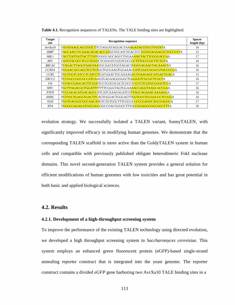
113
Table 4.1. Recognition sequences of TALENs. The TALE binding sites are highlighted.
Target
site Recognition sequence
Spacer
length (bp)
AvrXa10 TATATAAGCACGTATCTTCTAGGATAGGACTAAAGATACGTGCTTATATA 16
HBBS TAGCAACCTCAAACAGACACCATGGTGCATCTGACTCCTGTGGAGAAGTCTGCCGTTA 15
ABL1 TACCTATTATTACTTTATGGGGCAGCAGCCTGGAAAAGTACTTGGGGACCAA 17
APC TATGTACGCCTCCCTGGGCTCGGGTCCGGTCGCCCCTTTGCCCGCTTCTGTA 16
BRCA2 TTAGACTTAGGTAAGTAATGCAATATGGTAGACTGGGGAGAACTACAAACTA 16
CCND1 TGGAACACCAGCTCCTGTGCTGCGAAGTGGAAACCATCCGCCGCGCGTACCCCGA 16
CCR5 TGCTGGTCATCCTCATCCTGATAAACTGCAAAAGGCTGAAGAGCATGACTGACA 15
ERCC2 TCCGGCCGGCGCCATGAAGTGAGAAGGGGGCTGGGGGTCGCGCTCGCTA 16
FH TGTACCGAGCACTTCGGCTCCTCGCGCGCTCGCGTCCCCTCGTGCGGGCTCCA 17
MYC TGCTTAGACGCTGGATTTTTTTCGGGTAGTGGAAAACCAGGTAAGCACCGAA 16
PTEN TCCCAGACATGACAGCCATCATCAAAGAGATCGTTAGCAGAAACAAAAGGA 16
SDHC TGTTGCTGAGGTGACTTCAGTGGGACTGGGAGTTGGTGCCTGCGGCCCTCCGGA 16
SS18 TGGTGACGGCGGCAACATGTCTGTGGCTTTCGCGGCCCCGAGGCAGCGAGGCA 17
XPA TGGGCCAGAGATGGCGGCGGCCGACGGGGCTTTGCCGGAGGCGGCGGCTTTA 16
evolution strategy. We successfully isolated a TALEN variant, SunnyTALEN, with
significantly improved efficacy in modifying human genomes. We demonstrate that the
corresponding TALEN scaffold is more active than the GoldyTALEN system in human
cells and compatible with previously published obligate heterodimeric FokI nuclease
domains. This novel second-generation TALEN system provides a general solution for
efficient modifications of human genomes with low toxicities and has great potential in
both basic and applied biological sciences.
4.2. Results
4.2.1. Development of a high-throughput screening system
To improve the performance of the existing TALEN technology using directed evolution,
we developed a high throughput screening system in Saccharomyces cerevisiae. This
system employs an enhanced green fluorescent protein (eGFP)-based single-strand
annealing reporter construct that is integrated into the yeast genome. The reporter
construct contains a divided eGFP gene harboring two AvrXa10 TALE binding sites in a
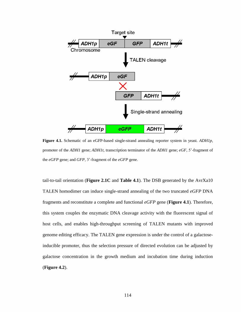
114
Figure 4.1. Schematic of an eGFP-based single-strand annealing reporter system in yeast. ADH1p,
promoter of the ADH1 gene; ADH1t, transcription terminator of the ADH1 gene; eGF, 5’-fragment of
the eGFP gene; and GFP, 3’-fragment of the eGFP gene.
tail-to-tail orientation (Figure 2.1C and Table 4.1). The DSB generated by the AvrXa10
TALEN homodimer can induce single-strand annealing of the two truncated eGFP DNA
fragments and reconstitute a complete and functional eGFP gene (Figure 4.1). Therefore,
this system couples the enzymatic DNA cleavage activity with the fluorescent signal of
host cells, and enables high-throughput screening of TALEN mutants with improved
genome editing efficacy. The TALEN gene expression is under the control of a galactose-
inducible promoter, thus the selection pressure of directed evolution can be adjusted by
galactose concentration in the growth medium and incubation time during induction
(Figure 4.2).
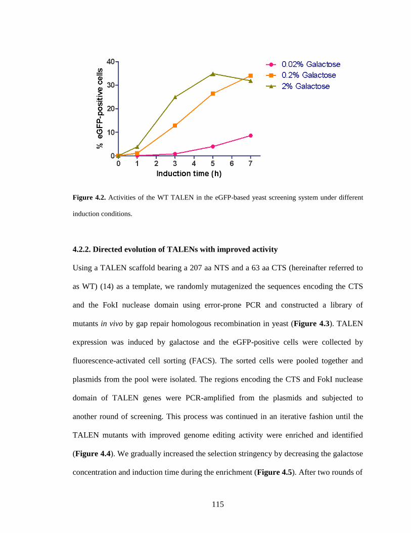
115
Figure 4.2. Activities of the WT TALEN in the eGFP-based yeast screening system under different
induction conditions.
4.2.2. Directed evolution of TALENs with improved activity
Using a TALEN scaffold bearing a 207 aa NTS and a 63 aa CTS (hereinafter referred to
as WT) (14) as a template, we randomly mutagenized the sequences encoding the CTS
and the FokI nuclease domain using error-prone PCR and constructed a library of
mutants in vivo by gap repair homologous recombination in yeast (Figure 4.3). TALEN
expression was induced by galactose and the eGFP-positive cells were collected by
fluorescence-activated cell sorting (FACS). The sorted cells were pooled together and
plasmids from the pool were isolated. The regions encoding the CTS and FokI nuclease
domain of TALEN genes were PCR-amplified from the plasmids and subjected to
another round of screening. This process was continued in an iterative fashion until the
TALEN mutants with improved genome editing activity were enriched and identified
(Figure 4.4). We gradually increased the selection stringency by decreasing the galactose
concentration and induction time during the enrichment (Figure 4.5). After two rounds of
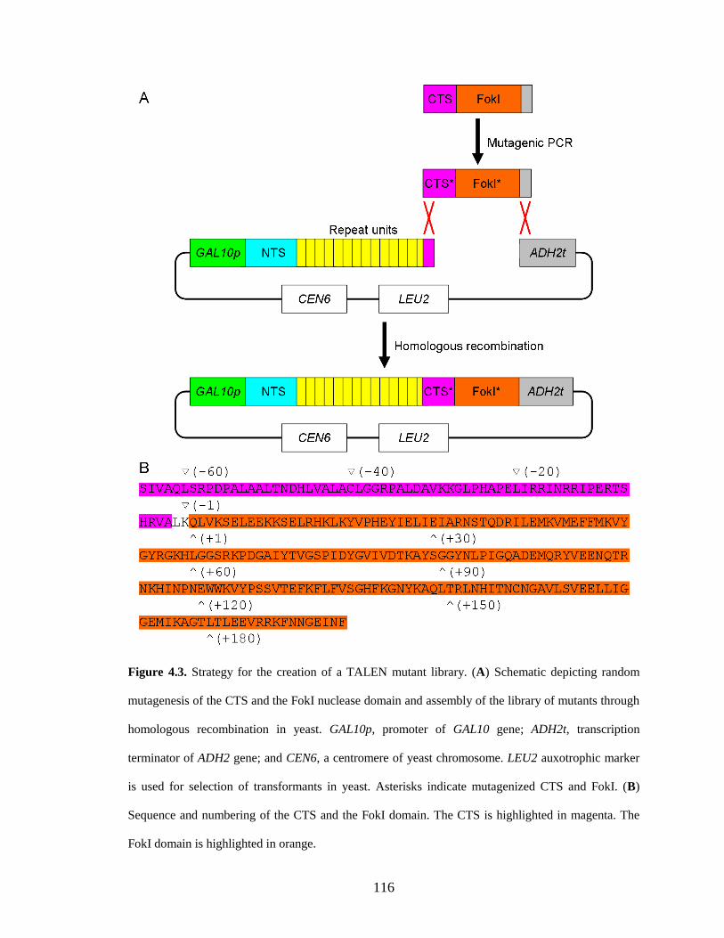
116
Figure 4.3. Strategy for the creation of a TALEN mutant library. (A) Schematic depicting random
mutagenesis of the CTS and the FokI nuclease domain and assembly of the library of mutants through
homologous recombination in yeast. GAL10p, promoter of GAL10 gene; ADH2t, transcription
terminator of ADH2 gene; and CEN6, a centromere of yeast chromosome. LEU2 auxotrophic marker
is used for selection of transformants in yeast. Asterisks indicate mutagenized CTS and FokI. (B)
Sequence and numbering of the CTS and the FokI domain. The CTS is highlighted in magenta. The
FokI domain is highlighted in orange.
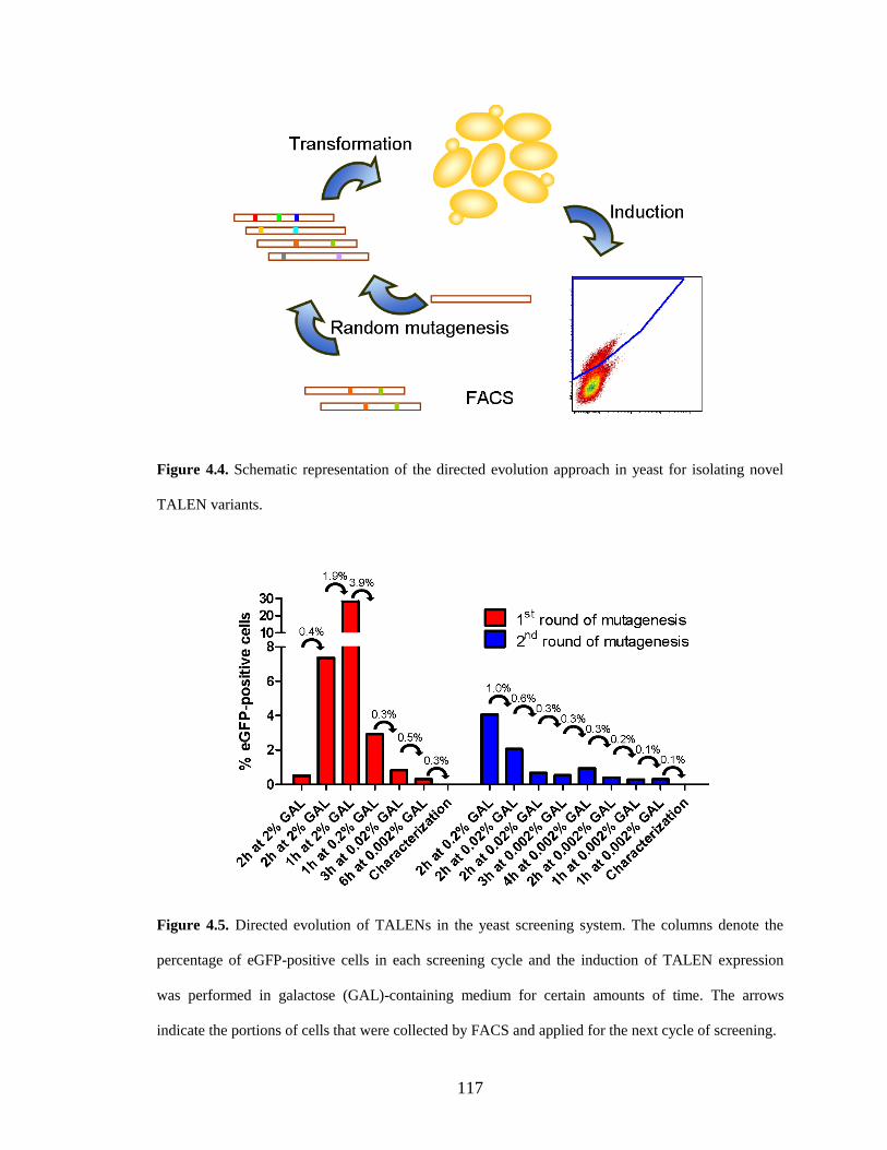
117
Figure 4.4. Schematic representation of the directed evolution approach in yeast for isolating novel
TALEN variants.
Figure 4.5. Directed evolution of TALENs in the yeast screening system. The columns denote the
percentage of eGFP-positive cells in each screening cycle and the induction of TALEN expression
was performed in galactose (GAL)-containing medium for certain amounts of time. The arrows
indicate the portions of cells that were collected by FACS and applied for the next cycle of screening.
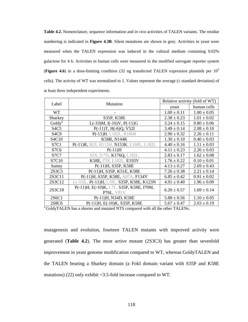
118
Table 4.2. Nomenclature, sequence information and in vivo activities of TALEN variants. The residue
numbering is indicated in Figure 4.3B. Silent mutations are shown in grey. Activities in yeast were
measured when the TALEN expression was induced in the cultural medium containing 0.02%
galactose for 4 h. Activities in human cells were measured in the modified surrogate reporter system
(Figure 4.6) in a dose-limiting condition (32 ng transfected TALEN expression plasmids per 105
cells). The activity of WT was normalized to 1. Values represent the average (± standard deviation) of
at least three independent experiments.
Label Mutation Relative activity (fold of WT)
yeast human cells
WT - 1.00 ± 0.11 1.00 ± 0.05
Sharkey S35P, K58E 2.38 ± 0.23 1.01 ± 0.02
Goldya L(-33)M, I(-16)V, P(-11)G 3.24 ± 0.15 0.80 ± 0.06
S4C5 P(-11)T, H(-6)Q, V52I 3.49 ± 0.14 2.08 ± 0.10
S4C9 P(-11)H, L182L, R186R 2.90 ± 0.32 2.26 ± 0.11
S4C10 K58R, N144K 1.30 ± 0.10 0.40 ± 0.03
S7C1 P(-11)R, I82I, H115H, N153K, L168L, L182L 4.40 ± 0.16 1.11 ± 0.03
S7C6 P(-11)H 4.11 ± 0.23 2.26 ± 0.03
S7C7 S5S, I175I, K176Q, L182L 2.83 ± 0.17 1.62 ± 0.08
S7C10 K58E, I70I, L182L, E193V 1.76 ± 0.22 0.10 ± 0.01
Sunny P(-11)H, S35P, K58E 4.13 ± 0.27 2.69 ± 0.43
2S3C3 P(-11)H, S35P, K51E, K58E 7.26 ± 0.38 2.21 ± 0.14
2S3C11 P(-11)H, S35P, K58E, A87A, F134Y 6.85 ± 0.42 0.91 ± 0.02
2S3C12 L(-33)L, P(-11)H, L18L, S35P, K58E, K123N 4.91 ± 0.40 1.96 ± 0.09
2S3C18 P(-11)H, E(-10)K, L7L, S35P, K58E, I70M,
P76L, V81V 6.20 ± 0.57 1.69 ± 0.14
2S6C1 P(-11)H, N34D, K58E 5.88 ± 0.56 1.10 ± 0.05
2S8C6 P(-11)H, E(-10)K, S35P, K58E 5.67 ± 0.47 2.03 ± 0.19 a GoldyTALEN has a shorter and mutated NTS compared with all the other TALENs.
mutagenesis and evolution, fourteen TALEN mutants with improved activity were
generated (Table 4.2). The most active mutant (2S3C3) has greater than sevenfold
improvement in yeast genome modification compared to WT, whereas GoldyTALEN and
the TALEN bearing a Sharkey domain (a FokI domain variant with S35P and K58E
mutations) (22) only exhibit <3.5-fold increase compared to WT.
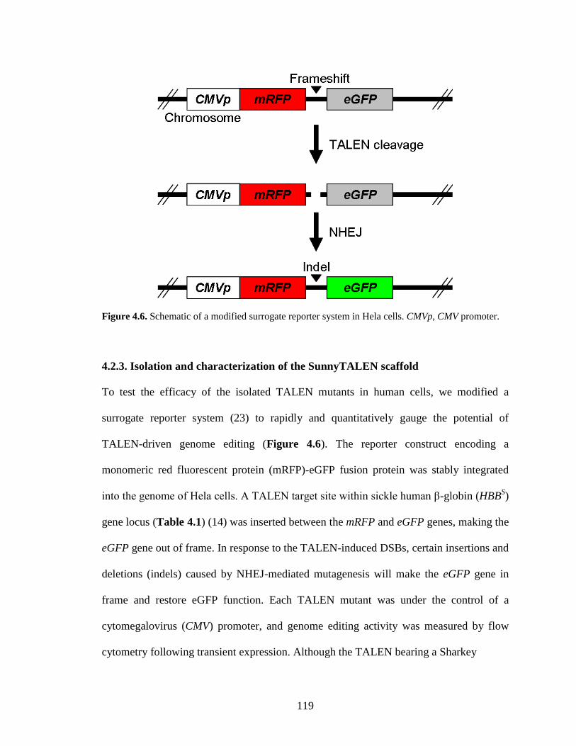
119
Figure 4.6. Schematic of a modified surrogate reporter system in Hela cells. CMVp, CMV promoter.
4.2.3. Isolation and characterization of the SunnyTALEN scaffold
To test the efficacy of the isolated TALEN mutants in human cells, we modified a
surrogate reporter system (23) to rapidly and quantitatively gauge the potential of
TALEN-driven genome editing (Figure 4.6). The reporter construct encoding a
monomeric red fluorescent protein (mRFP)-eGFP fusion protein was stably integrated
into the genome of Hela cells. A TALEN target site within sickle human β-globin (HBBS)
gene locus (Table 4.1) (14) was inserted between the mRFP and eGFP genes, making the
eGFP gene out of frame. In response to the TALEN-induced DSBs, certain insertions and
deletions (indels) caused by NHEJ-mediated mutagenesis will make the eGFP gene in
frame and restore eGFP function. Each TALEN mutant was under the control of a
cytomegalovirus (CMV) promoter, and genome editing activity was measured by flow
cytometry following transient expression. Although the TALEN bearing a Sharkey
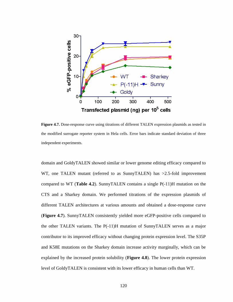
120
Figure 4.7. Dose-response curve using titrations of different TALEN expression plasmids as tested in
the modified surrogate reporter system in Hela cells. Error bars indicate standard deviation of three
independent experiments.
domain and GoldyTALEN showed similar or lower genome editing efficacy compared to
WT, one TALEN mutant (referred to as SunnyTALEN) has >2.5-fold improvement
compared to WT (Table 4.2). SunnyTALEN contains a single P(-11)H mutation on the
CTS and a Sharkey domain. We performed titrations of the expression plasmids of
different TALEN architectures at various amounts and obtained a dose-response curve
(Figure 4.7). SunnyTALEN consistently yielded more eGFP-positive cells compared to
the other TALEN variants. The P(-11)H mutation of SunnyTALEN serves as a major
contributor to its improved efficacy without changing protein expression level. The S35P
and K58E mutations on the Sharkey domain increase activity marginally, which can be
explained by the increased protein solubility (Figure 4.8). The lower protein expression
level of GoldyTALEN is consistent with its lower efficacy in human cells than WT.
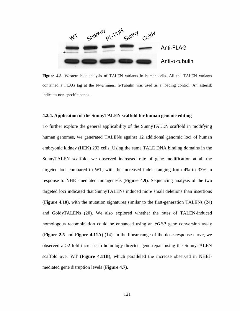
121
Figure 4.8. Western blot analysis of TALEN variants in human cells. All the TALEN variants
contained a FLAG tag at the N-terminus. α-Tubulin was used as a loading control. An asterisk
indicates non-specific bands.
4.2.4. Application of the SunnyTALEN scaffold for human genome editing
To further explore the general applicability of the SunnyTALEN scaffold in modifying
human genomes, we generated TALENs against 12 additional genomic loci of human
embryonic kidney (HEK) 293 cells. Using the same TALE DNA binding domains in the
SunnyTALEN scaffold, we observed increased rate of gene modification at all the
targeted loci compared to WT, with the increased indels ranging from 4% to 33% in
response to NHEJ-mediated mutagenesis (Figure 4.9). Sequencing analysis of the two
targeted loci indicated that SunnyTALENs induced more small deletions than insertions
(Figure 4.10), with the mutation signatures similar to the first-generation TALENs (24)
and GoldyTALENs (20). We also explored whether the rates of TALEN-induced
homologous recombination could be enhanced using an eGFP gene conversion assay
(Figure 2.5 and Figure 4.11A) (14). In the linear range of the dose-response curve, we
observed a >2-fold increase in homology-directed gene repair using the SunnyTALEN
scaffold over WT (Figure 4.11B), which paralleled the increase observed in NHEJ-
mediated gene disruption levels (Figure 4.7).
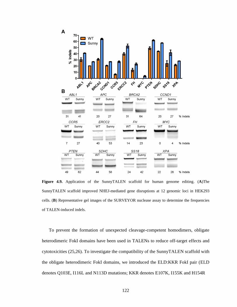
122
Figure 4.9. Application of the SunnyTALEN scaffold for human genome editing. (A)The
SunnyTALEN scaffold improved NHEJ-mediated gene disruptions at 12 genomic loci in HEK293
cells. (B) Representative gel images of the SURVEYOR nuclease assay to determine the frequencies
of TALEN-induced indels.
To prevent the formation of unexpected cleavage-competent homodimers, obligate
heterodimeric FokI domains have been used in TALENs to reduce off-target effects and
cytotoxicities (25,26). To investigate the compatibility of the SunnyTALEN scaffold with
the obligate heterodimeric FokI domains, we introduced the ELD:KKR FokI pair (ELD
denotes Q103E, I116L and N113D mutations; KKR denotes E107K, I155K and H154R
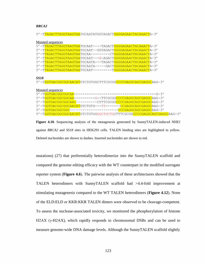
123
BRCA2
5’-TTAGACTTAGGTAAGTAATGCAATATGGTAGACTGGGGAGAACTACAAACTA-3’
Mutated sequences 5’-TTAGACTTAGGTAAGTAATGCAAT----TAGACTGGGGAGAACTACAAACTA-3’
5’-TTAGACTTAGGTAAGTAATGCAAT--GGTAGACTGGGGAGAACTACAAACTA-3’
5’-TTAGACTTAGGTAAGTAATGCAA---------CTGGGGAGAACTACAAACTA-3’
5’-TTAGACTTAGGTAAGTAATGCAAT---G-AGACTGGGGAGAACTACAAACTA-3’
5’-TTAGACTTAGGTAAGTAATGCAATA---TAGACTGGGGAGAACTACAAACTA-3’
5’-TTAGACTTAGGTAAGTAATGCAATA-----GACTGGGGAGAACTACAAACTA-3’
5’-TTAGACTTAGGTAAGTAATGCAAT---------TGGGGAGAACTACAAACTA-3’
SS18 5’-TGGTGACGGCGGCAACATGTCTGTGGCTTTCGCGGCCCCGAGGCAGCGAGGCAAG-3’
Mutated sequences 5’-TGGTGACGGCGGCAA---------------------------------------G-3’
5’-TGGTGACGGCGGCAA----------G--TTCGCGGCCCCGAGGCAGCGAGGCAAG-3’
5’-TGGTGACGGCGGCAAC----------CTTTCGCGGCCCCGAGGCAGCGAGGCAAG-3’
5’-TGGTGACGGCGGCAACATGTCTGTG---TT-------CCGAGGCAGCGAGGCAAG-3’
5’-TGGTGACGGCGGC-----------------------CCCGAGGCAGCGAGGCAAG-3’
5’-TGGTGACGGCGGCAACATGTCTGTGGGGCTGCTGCTTTCGCGGCCCCGAGGCAGCGAGGCAAG-3’
Figure 4.10. Sequencing analysis of the mutagenesis generated by SunnyTALEN-induced NHEJ
against BRCA2 and SS18 sites in HEK293 cells. TALEN binding sites are highlighted in yellow.
Deleted nucleotides are shown in dashes. Inserted nucleotides are shown in red.
mutations) (27) that preferentially heterodimerize into the SunnyTALEN scaffold and
compared the genome editing efficacy with the WT counterpart in the modified surrogate
reporter system (Figure 4.6). The pairwise analysis of these architectures showed that the
TALEN heterodimers with SunnyTALEN scaffold had >4.4-fold improvement at
stimulating mutagenesis compared to the WT TALEN heterodimers (Figure 4.12). None
of the ELD:ELD or KKR:KKR TALEN dimers were observed to be cleavage-competent.
To assess the nuclease-associated toxicity, we monitored the phosphorylation of histone
H2AX (γ-H2AX), which rapidly responds to chromosomal DSBs and can be used to
measure genome-wide DNA damage levels. Although the SunnyTALEN scaffold slightly
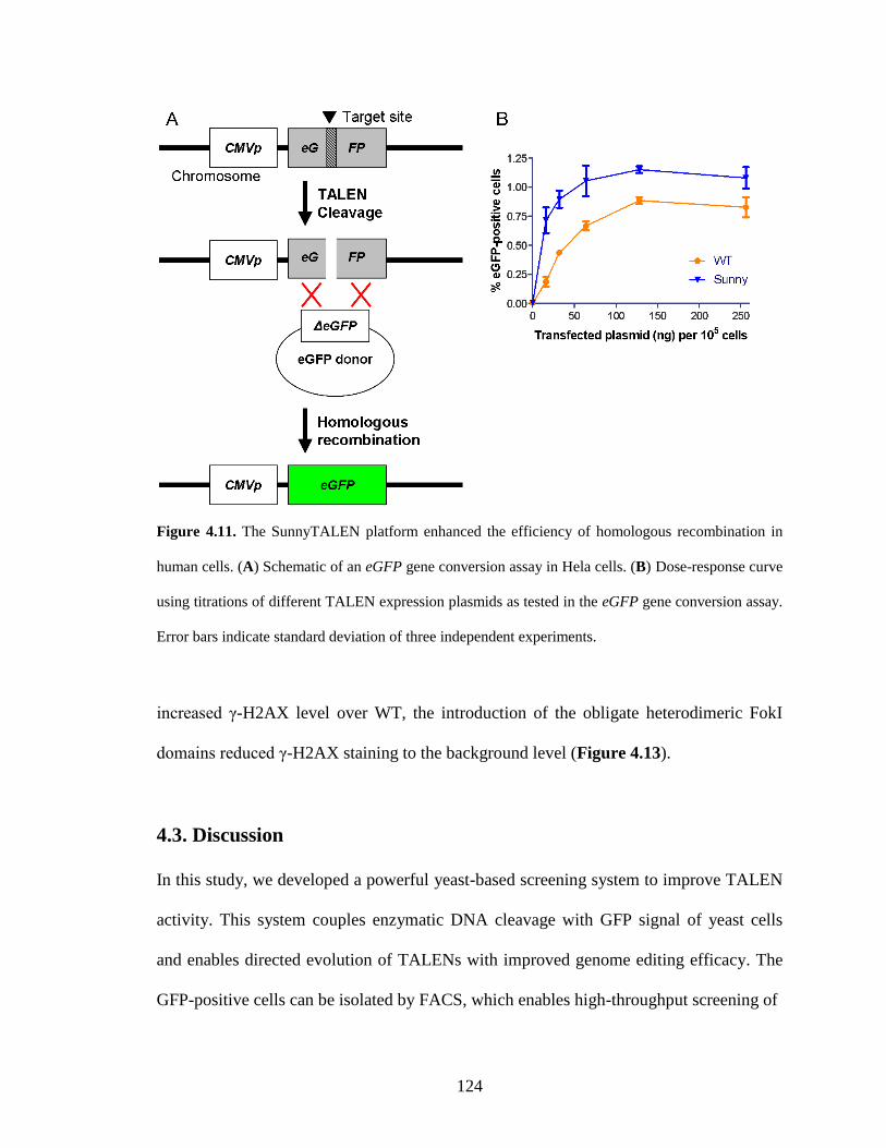
124
Figure 4.11. The SunnyTALEN platform enhanced the efficiency of homologous recombination in
human cells. (A) Schematic of an eGFP gene conversion assay in Hela cells. (B) Dose-response curve
using titrations of different TALEN expression plasmids as tested in the eGFP gene conversion assay.
Error bars indicate standard deviation of three independent experiments.
increased γ-H2AX level over WT, the introduction of the obligate heterodimeric FokI
domains reduced γ-H2AX staining to the background level (Figure 4.13).
4.3. Discussion
In this study, we developed a powerful yeast-based screening system to improve TALEN
activity. This system couples enzymatic DNA cleavage with GFP signal of yeast cells
and enables directed evolution of TALENs with improved genome editing efficacy. The
GFP-positive cells can be isolated by FACS, which enables high-throughput screening of
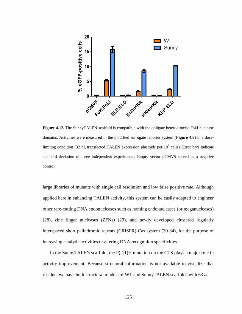
125
Figure 4.12. The SunnyTALEN scaffold is compatible with the obligate heterodimeric FokI nuclease
domains. Activities were measured in the modified surrogate reporter system (Figure 4.6) in a dose-
limiting condition (32 ng transfected TALEN expression plasmids per 105 cells). Error bars indicate
standard deviation of three independent experiments. Empty vector pCMV5 served as a negative
control.
large libraries of mutants with single cell resolution and low false positive rate. Although
applied here in enhancing TALEN activity, this system can be easily adapted to engineer
other rare-cutting DNA endonucleases such as homing endonucleases (or meganucleases)
(28), zinc finger nucleases (ZFNs) (29), and newly developed clustered regularly
interspaced short palindromic repeats (CRISPR)-Cas system (30-34), for the purpose of
increasing catalytic activities or altering DNA recognition specificities.
In the SunnyTALEN scaffold, the P(-11)H mutation on the CTS plays a major role in
activity improvement. Because structural information is not available to visualize that
residue, we have built structural models of WT and SunnyTALEN scaffolds with 63 aa
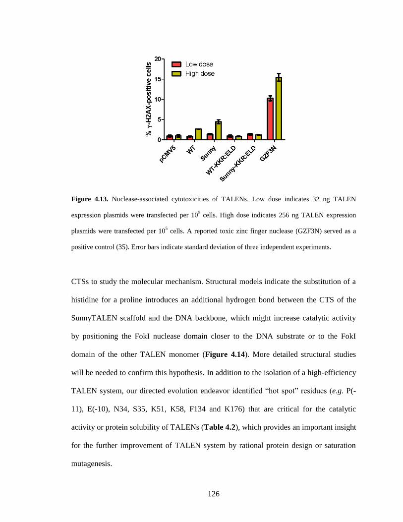
126
Figure 4.13. Nuclease-associated cytotoxicities of TALENs. Low dose indicates 32 ng TALEN
expression plasmids were transfected per 105 cells. High dose indicates 256 ng TALEN expression
plasmids were transfected per 105 cells. A reported toxic zinc finger nuclease (GZF3N) served as a
positive control (35). Error bars indicate standard deviation of three independent experiments.
CTSs to study the molecular mechanism. Structural models indicate the substitution of a
histidine for a proline introduces an additional hydrogen bond between the CTS of the
SunnyTALEN scaffold and the DNA backbone, which might increase catalytic activity
by positioning the FokI nuclease domain closer to the DNA substrate or to the FokI
domain of the other TALEN monomer (Figure 4.14). More detailed structural studies
will be needed to confirm this hypothesis. In addition to the isolation of a high-efficiency
TALEN system, our directed evolution endeavor identified “hot spot” residues (e.g. P(-
11), E(-10), N34, S35, K51, K58, F134 and K176) that are critical for the catalytic
activity or protein solubility of TALENs (Table 4.2), which provides an important insight
for the further improvement of TALEN system by rational protein design or saturation
mutagenesis.
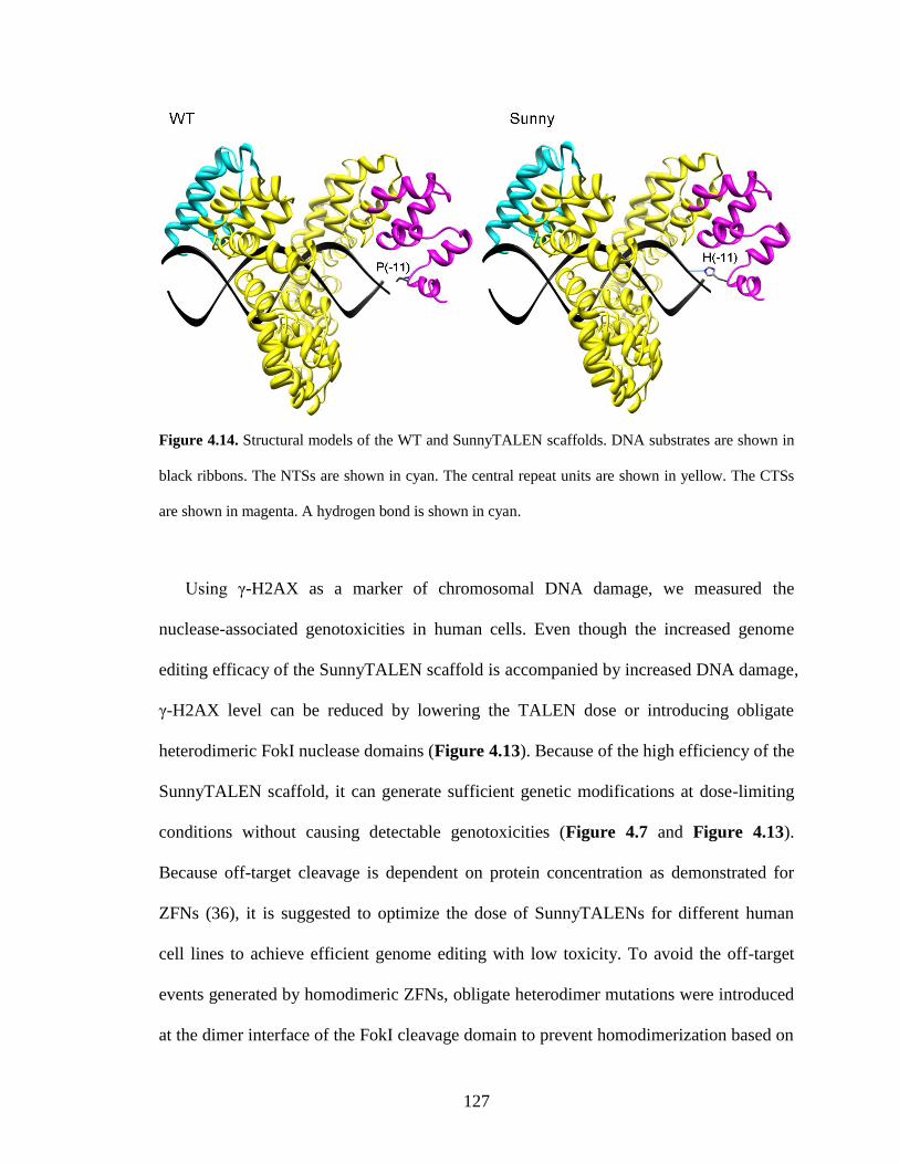
127
Figure 4.14. Structural models of the WT and SunnyTALEN scaffolds. DNA substrates are shown in
black ribbons. The NTSs are shown in cyan. The central repeat units are shown in yellow. The CTSs
are shown in magenta. A hydrogen bond is shown in cyan.
Using γ-H2AX as a marker of chromosomal DNA damage, we measured the
nuclease-associated genotoxicities in human cells. Even though the increased genome
editing efficacy of the SunnyTALEN scaffold is accompanied by increased DNA damage,
γ-H2AX level can be reduced by lowering the TALEN dose or introducing obligate
heterodimeric FokI nuclease domains (Figure 4.13). Because of the high efficiency of the
SunnyTALEN scaffold, it can generate sufficient genetic modifications at dose-limiting
conditions without causing detectable genotoxicities (Figure 4.7 and Figure 4.13).
Because off-target cleavage is dependent on protein concentration as demonstrated for
ZFNs (36), it is suggested to optimize the dose of SunnyTALENs for different human
cell lines to achieve efficient genome editing with low toxicity. To avoid the off-target
events generated by homodimeric ZFNs, obligate heterodimer mutations were introduced
at the dimer interface of the FokI cleavage domain to prevent homodimerization based on
128
electrostatic and hydrophobic interactions (27,35,37). Similar principle was applied to
TALENs and successfully reduced off-target cleavage and relieved toxicity (25). We
show here that the incorporation of obligate heterodimeric FokI domains completely
abolished the formation of cleavage-competent SunnyTALEN homodimers, resulting in
reduced genotoxicity (Figure 4.12 and Figure 4.13).
In conclusion, we isolated the high-efficiency SunnyTALEN scaffold using a directed
evolution strategy. The SunnyTALEN scaffold is portable to many TALE DNA binding
domains and compatible with heterodimer TALEN architectures. Although deployed here
in yeast and human cells, the SunnyTALEN scaffold could be effective in various
organisms and cell types. In addition to the GoldyTALEN scaffold that enables efficient
genome modifications in zebrafish and livestock, the SunnyTALEN scaffold serves as a
novel second-generation TALEN system and will aid in a variety of applications in
medicine and biology.
4.4. Materials and Methods
4.4.1. Materials
Q5 DNA polymerase, T4 DNA ligase, Antarctic phosphatase, and restriction
endonucleases were purchased from New England Biolabs (Beverly, MA). QIAprep Spin
Plasmid Miniprep Kit, QIAquick Gel Extraction Kit, and QIAquick PCR Purification Kit
were obtained from Qiagen (Valencia, CA). Oligonucleotide primers were obtained from
Integrated DNA Technologies (Coralville, IA). All the other reagents unless specified
were obtained from Sigma-Aldrich (St. Louis, MO).
129
4.4.2. Yeast reporter strain
To construct a single-strand annealing eGFP reporter, two separated eGFP fragments
sharing a 100 bp homologous region were PCR amplified separately, digested with AvrII
and XbaI, respectively, and ligated overnight at 16 °C. The ligation product was
subsequently digested with a combination of AvrII and XbaI and the 870 bp non-cut
fragment was gel-purified. The eGFP reporter gene was then cloned into the pRS414
plasmid (New England Biolabs) between the ADH1 promoter and the ADH1 terminator.
The centromere sequence of the plasmid was then removed by the PmlI digestion and
blunt ligation. This plasmid was then digested with HindIII at the TRP1 gene. The
linearized plasmid was transformed into S. cerevisiae HZ848 (MATα, ade2-1, ade3Δ22,
Δura3, his3-11,15, trp1-1, leu2-3,112 and can1-100) using the LiAc/SS carrier
DNA/PEG method (38) and stably integrated into the chromosome at the TRP1 site. The
yeast reporter strain was obtained by selection of the transformed yeast cells on plates
containing synthetic complete medium lacking tryptophan with 2% glucose at 30 °C. The
integration was confirmed by PCR amplification and DNA sequencing of the targeted
genomic region.
4.4.3. Library creation
The pRS415 yeast expression vector encoding the AvrXa10 TALEN that has been
described previously (14) was used as the template for mutagenic PCR. The sequence
encoding the CTS and FokI nuclease domain was mutagenized using the GeneMorph II
Random Mutagenesis Kit (Stratagene, La Jolla, CA) according to the manufacturer's
instruction. The following primers were used for the amplification: EP-FokI-Far-for 5’-
130
ttg ttg ccc agt tat ctc gc-3’ and EP-FokI-Far-rev 5’-cgt gaa act tcg aac act gtc-3’. Two
libraries with a low mutation rate (0-4.5 mutations/kbp) and a medium mutation rate (4.5-
9 mutations/kbp) were amplified and gel-purified, respectively. The TALEN expression
plasmid was digested with AatII and SalI and gel-purified to remove the sequences
encoding the wild-type CTS and FokI domain. The two gene libraries were mixed and
co-transformed with the linearized TALEN expression plasmids at an approximately 13:1
insert:vector molar ratio into the yeast reporter strain using the LiAc/SS carrier
DNA/PEG method (38). Through yeast homologous recombination, the pRS415 plasmid
library was created containing ~2×10⁵ transformants.
4.4.4. High-throughput screening
After transformation, the yeast reporter strain carrying the library of TALEN variants was
recovered at 30 °C with shaking for 1 h in YPA medium (1% yeast extract, 2% peptone
and 0.01% adenine hemisulphate) with 2% glucose. The cells were then centrifuged and
resuspended in synthetic complete medium lacking leucine and tryptophan (SC-Leu-Trp)
with 2% raffinose for growing at 30 °C with shaking overnight. TALEN expression was
induced by culturing cells in YPA medium with 0.002-2% galactose at 30 °C for 1-6 h
(the induction conditions for each cycle of screening are listed in Figure 4.5). The cells
were then centrifuged and resuspended in SC-Leu-Trp liquid medium with 2% glucose at
30 °C with shaking overnight. On the next day, cells were analyzed on a BD FACS Aria
III cell sorting system (BD Biosciences, San Jose, CA) and 20,000-100,000 eGFP-
positive cells were collected. After 2 days’ growth at 30 °C in SC-Leu-Trp liquid medium
with 2% glucose, plasmids from the sorted cells were extracted using Zymoprep Yeast
131
Plasmid Miniprep II Kit (Zymo Research, Orange, CA). Sequences encoding the CTS
and FokI domain were PCR amplified from the plasmids by Q5 DNA polymerase and
gel-purified for the next cycle of screening.
4.4.5. A modified surrogate reporter system
A TALEN target site within the HBBS gene was inserted between the mRFP gene and the
eGFP gene by overlap extension PCR. The fusion gene was cloned into the pLNCX2
retroviral vector (BD Clontech, Palo Alto, CA) in the presence of the CMV promoter and
stably integrated into the genome of Hela cells through retroviral transduction according
to the manufacturer’s protocol. Transfected cells were selected in 500 μg/mL G418 for
two weeks. The TALEN genes along with a FLAG tag sequence and a SV40 nucleus
localization signal added to the N-terminus were cloned into the pCMV5 mammalian
expression vector (39) through the KpnI and SalI sites.
Reporter Hela cells were routinely maintained in the modified Eagle’s medium
(MEM) supplemented with 10% Fetal Bovine Serum (FBS; Hyclone, Logan, UT). Cells
were seeded in 24-well plates at a density of 5×10⁴ per well. After 24 h, reporter cells
were transfected with certain amounts of TALEN expression plasmids using FuGene HD
transfection reagent (Promega, Madison, WI) under conditions specified by the
manufacturer. For each transfection, the overall plasmid amount was made to be 500 ng
by adding the empty pCMV5 plasmid. Cells were trypsinized from their culturing plates
48 h after transfection and resuspended in 300 μL phosphate buffered saline (PBS) for
flow cytometry analysis. 20 000 cells were analyzed by a BD LSRII flow cytometer (BD
Biosciences, San Jose, CA) to quantify the eGFP-positive cells.
132
4.4.6. Western blot analysis
Hela cells in 12-well plates were harvested 24 h after transfection with pCMV5-TALEN-
HBB-R plasmid (14). Cells were collected by centrifugation, washed with PBS and
resuspended in 40 μL whole cell lysis buffer (1 M Tris-HCl, pH 6.8, 20% sodium
dodecyl sulfate, and 0.1 M dithiothreitol). Proteins were resolved by 4-20% Mini-Protean
TGX Precast Gel (Bio-Rad Laboratories, Hercules, CA), transferred onto a nitrocellulose
membrane, blocked for 1 h with Tris-buffered saline/0.05% Tween 20 containing 5%
nonfat milk, followed by incubation with anti-FLAG tag (1: 500) and anti-α-tubulin (1:
10 000) antibodies at 4 °C overnight. After incubation with anti-mouse horseradish
peroxidase-conjugated secondary antibody (1: 25 000; GeneScript, Piscataway, NJ) for 1
h, bands were visualized using SuperSignal West Pico Chemiluminescent Substrate
(Pierce, Rockford, IL).
4.4.7. TALEN construction
The central repeat units of TALEs were assembled by the Golden Gate reactions as
previously described (40). The receiver plasmids pCMV5-GG-WT for WT TALEN and
pCMV5-GG-Sunny for the SunnyTALEN scaffold are available upon request. The NTS
and CTS of the GoldyTALEN scaffold were PCR-amplified from pC-GoldyTALEN
plasmid (Kindly provided by Dr. Daniel Carlson from University of Minnesota,
Minneapolis, MN).
4.4.8. SURVEYOR nuclease assay
HEK293 cells in 6-well plates were harvested 48 h after transfection. Genomic DNA was
133
extracted using Wizard Genomic DNA Purification Kit (Promega, Madison, WI)
according to the manufacturer’s instructions. DNA fragments containing TALEN target
sites were PCR-amplified from the genomic DNA using the primers described previously
(19). Gel-purified PCR products were re-annealed slowly and analyzed by SURVEYOR
Mutation Detection Kits (Transgenomic, Omaha, NE) according to the manufacturer’s
instructions. The product was resolved on a 2% agarose gel stained with GelStar (Lonza,
Rockland, ME). The bands were quantified by G Box using Gene Tools software
(Syngene, Frederick, MD). The apparent indel percentage of the original cell pool was
calculated using the following equations as previously described (41):
Fraction cleaved = volume of cleaved bands/(volume of cleaved bands + volume
of uncleaved bands)
% Indels = 100 × (1- (1- Fraction cleaved)1/2
)
4.4.9. H2AX phosphorylation assay
HEK293 cells were cultured in Dulbecco’s modified Eagle’s medium (DMEM)
supplemented with 10% FBS. Cells were seeded in 12-well plates (2×10⁵ per well) and
transfected after 24 h with 500 ng of each TALEN expression plasmid, 1 μg empty
pCMV5 vector as a negative control or 1 μg reported toxic ZFN construct GZF3N
(kindly provided by Dr. Toni Cathomen of Hannover Medical School, Hannover,
Germany) as a positive control. After 48 h post transfection, cells were harvested, fixed,
permeabilized, and stained using the H2AX phosphorylation assay kit (Millipore,
Watford, UK) according to the manufacturer’s protocol. Cells were then scanned in a
134
flow cytometer to quantitate the number of cells staining positive for phosphorylated
histone H2AX.
4.4.10. Sequencing analysis of endogenous gene mutations
HEK293 cells in 6-well plates were harvested 48 h after transfection with SunnyTALEN
expression plasmids. Genomic DNA was extracted using Wizard Genomic DNA
Purification Kit (Promega, Madison, WI) according to the manufacturer’s instructions.
DNA fragments containing TALEN target sites within the BRCA2 gene were PCR-
amplified from the genomic DNA using the primers KpnI-BRCA2-for 5’-act gac ggt acc
tga tct tta act gtt ctg ggt cac aaa-3’ (KpnI recognition site shown in italics) and SalI-
BRCA2-rev 5’-act gac gtc gac cgc cag gga aac tcc ttc ca-3’ (SalI recognition site shown
in italics). DNA fragments containing TALEN target sites within the SS18 gene were
PCR-amplified from the genomic DNA using the primers KpnI-SS18-for 5’-act gac ggt
acc ggg atg cag gga cgg tca ag-3’ (KpnI recognition site shown in italics) and SalI-SS18-
rev 5’-act gac gtc gac gcc gcc cca tcc cta gag aaa-3’ (SalI recognition site shown in
italics). PCR products corresponding to the two sites were cloned into pCMV5 plasmid
between KpnI and SalI restriction sites. Plasmid DNA was isolated from 20 colonies for
each transformation and then sent for Sanger sequencing using the primer pSeq-pCMV5-
for 5’-cgc aaa tgg gcg gta ggc gtg-3’.
4.4.11. An eGFP gene conversion assay
The eGFP gene was divided into two fragments by insertion of a preselected sequence
from the HBBS gene locus and an in-frame stop codon. The non-functional eGFP reporter
135
stably integrated into the genome of Hela cells as previously described (14). A donor
plasmid was constructed by insertion of a promoter-less eGFP gene with the first 37
nucleotides missing into the pNEB193 plasmid (New England Biolabs, Beverly, MA) as
previously decribed (14). The donor plasmid provides a homologous DNA segment as a
template for repairing the double-stranded break in the eGFP reporter. The reporter Hela
cells were maintained in modified Eagle’s medium (MEM) supplemented with 10% Fetal
Bovine Serum (FBS; Hyclone, Logan, UT). Cells were seeded in 12-well plates at a
density of 1×10⁵ per well. After 24 hr, reporter cells were co-transfected with a certain
amount of TALEN expression plasmids and 500 ng of donor plasmid using FuGene HD
transfection reagent (Promega, Madison, WI) under conditions specified by the
manufacturer. Cells were trypsinized from their culturing plates 48h after transfection and
resuspended in 300 μL phosphate buffered saline (PBS) for flow cytometry analysis.
20,000 cells were analyzed by BD LSRII flow cytometer (BD Biosciences, San Jose, CA)
to determine the percentage of eGFP-positive cells.
4.4.12. Computational modeling
The homology models of TALEs with 63 aa CTSs were created by the I-TASSER online
server (42) based on the crystal structure of dHax3 (PDB accession code: 3V6P) (43).
The DNA-bound models were created based on the crystal structure of DNA-bound
dHax3 (PDB accession code: 3V6T) (43) and energy minimized using the Molecular
Operating Environment (MOE, The Chemical Computing Group, 2008) software
package. The figures were generated using UCSF Chimera (44).
136
4.5. References
1. Dow, L.E. and Lowe, S.W. (2012) Life in the fast lane: mammalian disease
models in the genomics era. Cell, 148, 1099-1109.
2. Cherry, A.B. and Daley, G.Q. (2013) Reprogrammed cells for disease modeling
and regenerative medicine. Annu Rev Med, 64, 277-290.
3. Check, E. (2002) A tragic setback. Nature, 420, 116-118.
4. Marshall, E. (1999) Gene therapy death prompts review of adenovirus vector.
Science, 286, 2244-2245.
5. Mussolino, C. and Cathomen, T. (2012) TALE nucleases: tailored genome
engineering made easy. Curr Opin Biotechnol, 23, 644-650.
6. Joung, J.K. and Sander, J.D. (2013) TALENs: a widely applicable technology for
targeted genome editing. Nat Rev Mol Cell Biol, 14, 49-55.
7. Sun, N. and Zhao, H. (2013) Transcription activator-like effector nucleases
(TALENs): a highly efficient and versatile tool for genome editing. Biotechnol
Bioeng, doi: 10.1002/bit.24890.
8. Boch, J., Scholze, H., Schornack, S., Landgraf, A., Hahn, S., Kay, S., Lahaye, T.,
Nickstadt, A. and Bonas, U. (2009) Breaking the code of DNA binding specificity
of TAL-type III effectors. Science, 326, 1509-1512.
9. Moscou, M.J. and Bogdanove, A.J. (2009) A simple cipher governs DNA
recognition by TAL effectors. Science, 326, 1501.
10. Cong, L., Zhou, R., Kuo, Y.C., Cunniff, M. and Zhang, F. (2012) Comprehensive
interrogation of natural TALE DNA-binding modules and transcriptional
repressor domains. Nat Commun, 3, 968.
137
11. Streubel, J., Blucher, C., Landgraf, A. and Boch, J. (2012) TAL effector RVD
specificities and efficiencies. Nat Biotechnol, 30, 593-595.
12. Ding, Q., Lee, Y.K., Schaefer, E.A., Peters, D.T., Veres, A., Kim, K.,
Kuperwasser, N., Motola, D.L., Meissner, T.B., Hendriks, W.T. et al. (2013) A
TALEN genome-editing system for generating human stem cell-based disease
models. Cell Stem Cell, 12, 238-251.
13. Mussolino, C., Morbitzer, R., Lutge, F., Dannemann, N., Lahaye, T. and
Cathomen, T. (2011) A novel TALE nuclease scaffold enables high genome
editing activity in combination with low toxicity. Nucleic Acids Res, 39, 9283-
9293.
14. Sun, N., Liang, J., Abil, Z. and Zhao, H. (2012) Optimized TAL effector
nucleases (TALENs) for use in treatment of sickle cell disease. Mol Biosyst, 8,
1255-1263.
15. Miller, J.C., Tan, S., Qiao, G., Barlow, K.A., Wang, J., Xia, D.F., Meng, X.,
Paschon, D.E., Leung, E., Hinkley, S.J. et al. (2011) A TALE nuclease
architecture for efficient genome editing. Nat Biotechnol, 29, 143-148.
16. Kim, Y., Kweon, J., Kim, A., Chon, J.K., Yoo, J.Y., Kim, H.J., Kim, S., Lee, C.,
Jeong, E., Chung, E. et al. (2013) A library of TAL effector nucleases spanning
the human genome. Nat Biotechnol, 31, 251-258.
17. Choi, S.M., Kim, Y., Shim, J.S., Park, J.T., Wang, R.H., Leach, S.D., Liu, J.O.,
Deng, C.X., Ye, Z. and Jang, Y.Y. (2013) Efficient drug screening and gene
correction for treating liver disease using patient-specific stem cells. Hepatology,
doi: 10.1002/hep.26237.
138
18. Osborn, M.J., Starker, C.G., McElroy, A.N., Webber, B.R., Riddle, M.J., Xia, L.,
Defeo, A.P., Gabriel, R., Schmidt, M., Von Kalle, C. et al. (2013) TALEN-based
gene correction for epidermolysis bullosa. Mol Ther, doi: 10.1038/mt.2013.1056.
19. Reyon, D., Tsai, S.Q., Khayter, C., Foden, J.A., Sander, J.D. and Joung, J.K.
(2012) FLASH assembly of TALENs for high-throughput genome editing. Nat
Biotechnol, 30, 460-465.
20. Bedell, V.M., Wang, Y., Campbell, J.M., Poshusta, T.L., Starker, C.G., Krug,
R.G., 2nd, Tan, W., Penheiter, S.G., Ma, A.C., Leung, A.Y. et al. (2012) In vivo
genome editing using a high-efficiency TALEN system. Nature, 491, 114-118.
21. Carlson, D.F., Tan, W., Lillico, S.G., Stverakova, D., Proudfoot, C., Christian, M.,
Voytas, D.F., Long, C.R., Whitelaw, C.B. and Fahrenkrug, S.C. (2012) Efficient
TALEN-mediated gene knockout in livestock. Proc Natl Acad Sci U S A, 109,
17382-17387.
22. Guo, J., Gaj, T. and Barbas, C.F., 3rd. (2010) Directed evolution of an enhanced
and highly efficient FokI cleavage domain for zinc finger nucleases. J Mol Biol,
400, 96-107.
23. Kim, H., Um, E., Cho, S.R., Jung, C., Kim, H. and Kim, J.S. (2011) Surrogate
reporters for enrichment of cells with nuclease-induced mutations. Nat Methods, 8,
941-943.
24. Kim, Y., Kweon, J. and Kim, J.S. (2013) TALENs and ZFNs are associated with
different mutation signatures. Nat Methods, 10, 185.
25. Cade, L., Reyon, D., Hwang, W.Y., Tsai, S.Q., Patel, S., Khayter, C., Joung, J.K.,
Sander, J.D., Peterson, R.T. and Yeh, J.R. (2012) Highly efficient generation of
139
heritable zebrafish gene mutations using homo- and heterodimeric TALENs.
Nucleic Acids Res, 40, 8001-8010.
26. Huang, P., Xiao, A., Zhou, M., Zhu, Z., Lin, S. and Zhang, B. (2011) Heritable
gene targeting in zebrafish using customized TALENs. Nat Biotechnol, 29, 699-
700.
27. Doyon, Y., Vo, T.D., Mendel, M.C., Greenberg, S.G., Wang, J., Xia, D.F., Miller,
J.C., Urnov, F.D., Gregory, P.D. and Holmes, M.C. (2011) Enhancing zinc-
finger-nuclease activity with improved obligate heterodimeric architectures. Nat
Methods, 8, 74-79.
28. Silva, G., Poirot, L., Galetto, R., Smith, J., Montoya, G., Duchateau, P. and
Paques, F. (2011) Meganucleases and other tools for targeted genome engineering:
perspectives and challenges for gene therapy. Curr Gene Ther, 11, 11-27.
29. Carroll, D. (2011) Genome engineering with zinc-finger nucleases. Genetics, 188,
773-782.
30. Cong, L., Ran, F.A., Cox, D., Lin, S., Barretto, R., Habib, N., Hsu, P.D., Wu, X.,
Jiang, W., Marraffini, L.A. et al. (2013) Multiplex genome engineering using
CRISPR/Cas systems. Science, 339, 819-823.
31. Jinek, M., East, A., Cheng, A., Lin, S., Ma, E. and Doudna, J. (2013) RNA-
programmed genome editing in human cells. Elife, 2, e00471.
32. Mali, P., Yang, L., Esvelt, K.M., Aach, J., Guell, M., DiCarlo, J.E., Norville, J.E.
and Church, G.M. (2013) RNA-guided human genome engineering via Cas9.
Science, 339, 823-826.
140
33. Hwang, W.Y., Fu, Y., Reyon, D., Maeder, M.L., Tsai, S.Q., Sander, J.D.,
Peterson, R.T., Yeh, J.R. and Joung, J.K. (2013) Efficient genome editing in
zebrafish using a CRISPR-Cas system. Nat Biotechnol, 31, 227-229.
34. Jiang, W., Bikard, D., Cox, D., Zhang, F. and Marraffini, L.A. (2013) RNA-
guided editing of bacterial genomes using CRISPR-Cas systems. Nat Biotechnol,
31, 233-239.
35. Szczepek, M., Brondani, V., Buchel, J., Serrano, L., Segal, D.J. and Cathomen, T.
(2007) Structure-based redesign of the dimerization interface reduces the toxicity
of zinc-finger nucleases. Nat Biotechnol, 25, 786-793.
36. Pattanayak, V., Ramirez, C.L., Joung, J.K. and Liu, D.R. (2011) Revealing off-
target cleavage specificities of zinc-finger nucleases by in vitro selection. Nat
Methods, 8, 765-770.
37. Miller, J.C., Holmes, M.C., Wang, J., Guschin, D.Y., Lee, Y.L., Rupniewski, I.,
Beausejour, C.M., Waite, A.J., Wang, N.S., Kim, K.A. et al. (2007) An improved
zinc-finger nuclease architecture for highly specific genome editing. Nat
Biotechnol, 25, 778-785.
38. Gietz, R.D. and Schiestl, R.H. (2007) High-efficiency yeast transformation using
the LiAc/SS carrier DNA/PEG method. Nat Protoc, 2, 31-34.
39. Andersson, S., Davis, D.L., Dahlback, H., Jornvall, H. and Russell, D.W. (1989)
Cloning, structure, and expression of the mitochondrial cytochrome P-450 sterol
26-hydroxylase, a bile acid biosynthetic enzyme. J Biol Chem, 264, 8222-8229.
40. Cermak, T., Doyle, E.L., Christian, M., Wang, L., Zhang, Y., Schmidt, C., Baller,
J.A., Somia, N.V., Bogdanove, A.J. and Voytas, D.F. (2011) Efficient design and
141
assembly of custom TALEN and other TAL effector-based constructs for DNA
targeting. Nucleic Acids Res, 39, e82.
41. Guschin, D.Y., Waite, A.J., Katibah, G.E., Miller, J.C., Holmes, M.C. and Rebar,
E.J. (2010) A rapid and general assay for monitoring endogenous gene
modification. Methods Mol Biol, 649, 247-256.
42. Zhang, Y. (2008) I-TASSER server for protein 3D structure prediction. BMC
Bioinformatics, 9, 40.
43. Deng, D., Yan, C., Pan, X., Mahfouz, M., Wang, J., Zhu, J.K., Shi, Y. and Yan, N.
(2012) Structural basis for sequence-specific recognition of DNA by TAL
effectors. Science, 335, 720-723.
44. Pettersen, E.F., Goddard, T.D., Huang, C.C., Couch, G.S., Greenblatt, D.M.,
Meng, E.C. and Ferrin, T.E. (2004) UCSF Chimera--a visualization system for
exploratory research and analysis. J Comput Chem, 25, 1605-1612.
.
142
CHAPTER 5. DEVELOPMENT OF A SINGLE-CHAIN
TALEN ARCHITECTURE
5.1. Introduction
Transcription-activator like effector (TALE) nucleases (TALENs) have been widely used
for genome editing or engineering in various organisms during the last three years (as
reviewed in (1,2)). The long DNA binding sequences of TALE DNA binding domains
enable TALENs to target the custom-selected loci precisely with minimal off-target
effects or cyto-tyxociteis. The simple one-repeat-one-nucleotide DNA recognition code
of TALE DNA binding domains provides modular DNA reading, which enables
researchers to target essentially any genomic region.
The standard TALEN architecture is composed of TALE DNA binding domain,
truncated N- and C-terminal extension and the non-specific DNA cleavage domain from
a Type IIS restriction enzyme FokI (Figure 5.1A). Because the dimerization of the FokI
cleavage domain is necessary for DNA cleavage activity (3), standard TALEN
architecture requires two TALENs to bind to two DNA recognition regions flanking an
unspecific central spacer. The optimal spacer length is dependent on the TALE scaffold
construction (4-6). When the TALEN technology was initially developed, three
limitations have been noticed to prevent its broader applications: a) the requirement of a
5’-thymidine preceding the TALE binding sites can limit the targeting space; b) The
highly repetitive nature of the TALE DNA binding domain makes it difficult to
synthesize TALEN coding genes using traditional cloning strategy; c) The bulky size of
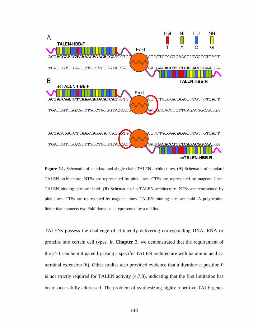
143
Figure 5.1. Schematic of standard and single-chain TALEN architectures. (A) Schematic of standard
TALEN architecture. NTSs are represented by pink lines. CTSs are represented by magenta lines.
TALEN binding sites are bold. (B) Schematic of scTALEN architecture. NTSs are represented by
pink lines. CTSs are represented by magenta lines. TALEN binding sites are bold. A polypeptide
linker that connects two FokI domains is represented by a red line.
TALENs possess the challenge of efficiently delivering corresponding DNA, RNA or
proteins into certain cell types. In Chapter 2, we demonstrated that the requirement of
the 5’-T can be mitigated by using a specific TALEN architecture with 63 amino acid C-
terminal extension (6). Other studies also provided evidence that a thymine at position 0
is not strictly required for TALEN activity (4,7,8), indicating that the first limitation has
been successfully addressed. The problem of synthesizing highly repetitive TALE genes
144
has also been solved by multiple groups by the introduction of Golden Gate cloning,
solid-phase ligation and ligation-independent cloning techniques (7,9-14).
In order to address the third limitation, i.e. reduce the TALEN size, Beurdeley and
coworkers have developed a compact TALEN (cTALEN) architecture by replacing the
requisite dimeric FokI cleavage domain with the DNA cleavage domain from the I-TevI
homing endonuclease (15). In this design, cTALENs can cleave the targeted DNA
sequence as a monomer and thus reduce the TALEN payload to half as compared with
the conventional TALEN architecture. However, unlike the non-specific FokI DNA
cleavage domain, the I-TevI DNA cleavage domain has its own DNA recognition
sequence. They discovered that the efficient DNA cleavage of cTALEN is dependent on
the consensus sequence CDDHGS (D= A, G or T; H= A, C or T; S= C or G), which is
outside the TALE binding site. Therefore, the native DNA recognition specificity of I-
TevI DNA cleavage domain severely decreases the targeting space of cTALENs. In this
chapter, I describe the development of a single-chain TALEN (scTALEN) architecture, in
which two FokI DNA cleavage domains are linked by a polypeptide linker (Figure 5.1B).
The appropriate linker is isolated from the high-throughput screening of a polypeptide
linker library. In this design, the two FokI domains on the same polypeptide can form a
catalytic active self-dimer and thus make scTALENs to function as monomers. The
scTALEN architecture I have developed reduced the TALEN payload to half as
compared to standard TALEN architecture. Moreover, because the FokI DNA cleavage
domains have no DNA recognition specificity, the DNA target sequence is solely
determined by the TALE DNA binding sites. Therefore, scTALENs can be used to target
essentially any DNA sequence.
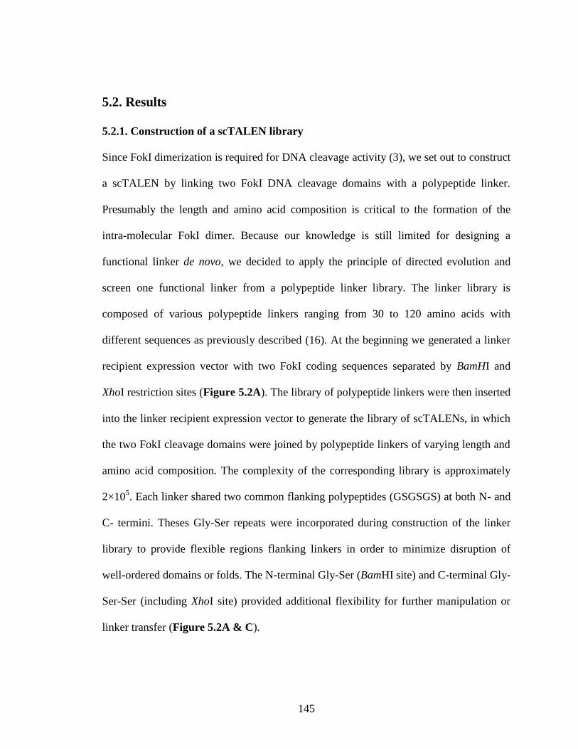
145
5.2. Results
5.2.1. Construction of a scTALEN library
Since FokI dimerization is required for DNA cleavage activity (3), we set out to construct
a scTALEN by linking two FokI DNA cleavage domains with a polypeptide linker.
Presumably the length and amino acid composition is critical to the formation of the
intra-molecular FokI dimer. Because our knowledge is still limited for designing a
functional linker de novo, we decided to apply the principle of directed evolution and
screen one functional linker from a polypeptide linker library. The linker library is
composed of various polypeptide linkers ranging from 30 to 120 amino acids with
different sequences as previously described (16). At the beginning we generated a linker
recipient expression vector with two FokI coding sequences separated by BamHI and
XhoI restriction sites (Figure 5.2A). The library of polypeptide linkers were then inserted
into the linker recipient expression vector to generate the library of scTALENs, in which
the two FokI cleavage domains were joined by polypeptide linkers of varying length and
amino acid composition. The complexity of the corresponding library is approximately
2×10⁵. Each linker shared two common flanking polypeptides (GSGSGS) at both N- and
C- termini. Theses Gly-Ser repeats were incorporated during construction of the linker
library to provide flexible regions flanking linkers in order to minimize disruption of
well-ordered domains or folds. The N-terminal Gly-Ser (BamHI site) and C-terminal Gly-
Ser-Ser (including XhoI site) provided additional flexibility for further manipulation or
linker transfer (Figure 5.2A & C).
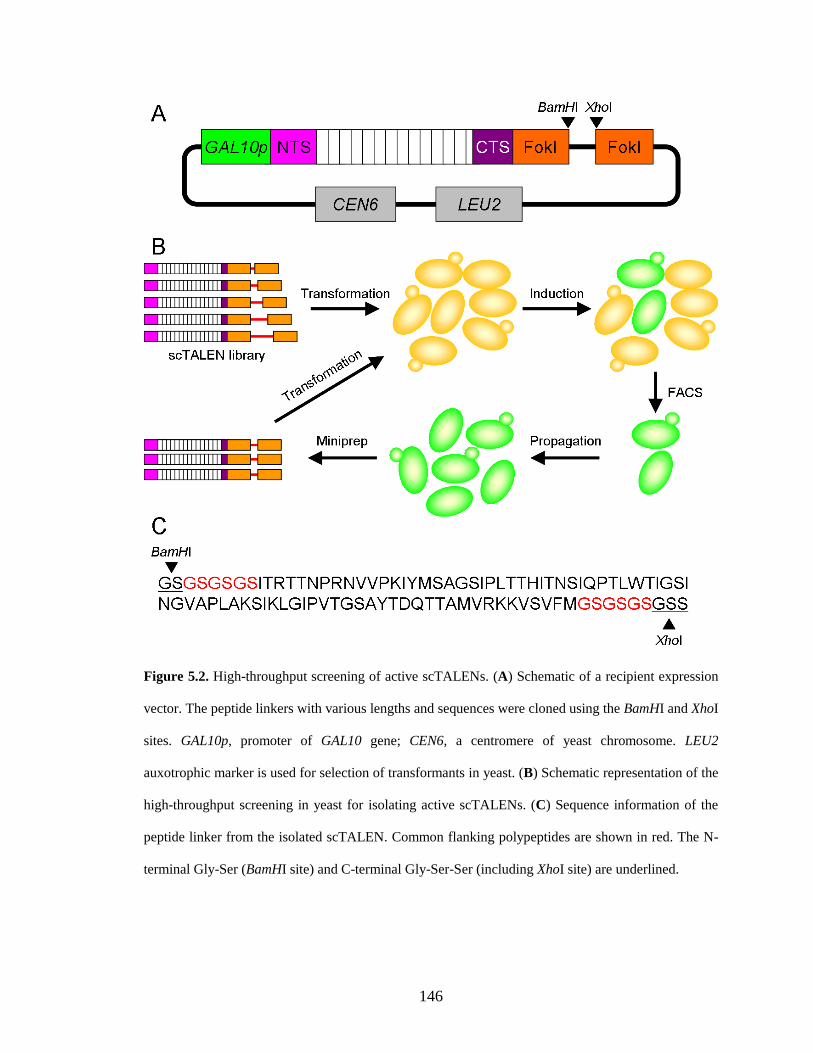
146
Figure 5.2. High-throughput screening of active scTALENs. (A) Schematic of a recipient expression
vector. The peptide linkers with various lengths and sequences were cloned using the BamHI and XhoI
sites. GAL10p, promoter of GAL10 gene; CEN6, a centromere of yeast chromosome. LEU2
auxotrophic marker is used for selection of transformants in yeast. (B) Schematic representation of the
high-throughput screening in yeast for isolating active scTALENs. (C) Sequence information of the
peptide linker from the isolated scTALEN. Common flanking polypeptides are shown in red. The N-
terminal Gly-Ser (BamHI site) and C-terminal Gly-Ser-Ser (including XhoI site) are underlined.
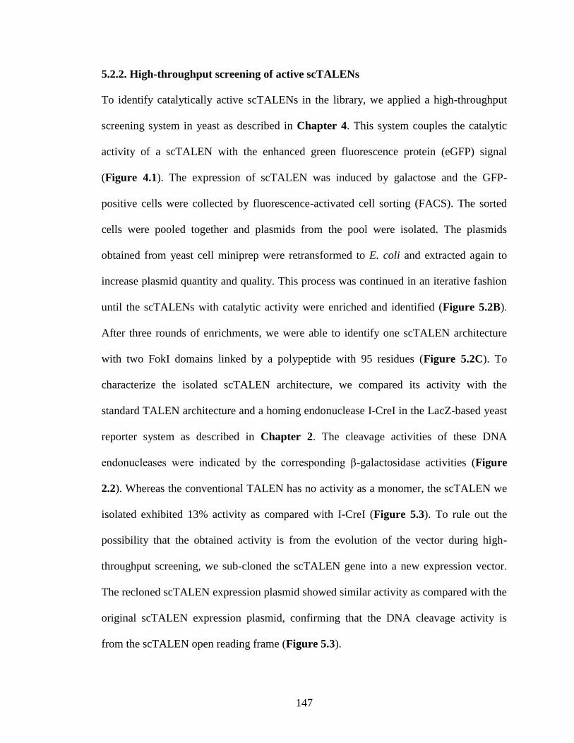
147
5.2.2. High-throughput screening of active scTALENs
To identify catalytically active scTALENs in the library, we applied a high-throughput
screening system in yeast as described in Chapter 4. This system couples the catalytic
activity of a scTALEN with the enhanced green fluorescence protein (eGFP) signal
(Figure 4.1). The expression of scTALEN was induced by galactose and the GFP-
positive cells were collected by fluorescence-activated cell sorting (FACS). The sorted
cells were pooled together and plasmids from the pool were isolated. The plasmids
obtained from yeast cell miniprep were retransformed to E. coli and extracted again to
increase plasmid quantity and quality. This process was continued in an iterative fashion
until the scTALENs with catalytic activity were enriched and identified (Figure 5.2B).
After three rounds of enrichments, we were able to identify one scTALEN architecture
with two FokI domains linked by a polypeptide with 95 residues (Figure 5.2C). To
characterize the isolated scTALEN architecture, we compared its activity with the
standard TALEN architecture and a homing endonuclease I-CreI in the LacZ-based yeast
reporter system as described in Chapter 2. The cleavage activities of these DNA
endonucleases were indicated by the corresponding β-galactosidase activities (Figure
2.2). Whereas the conventional TALEN has no activity as a monomer, the scTALEN we
isolated exhibited 13% activity as compared with I-CreI (Figure 5.3). To rule out the
possibility that the obtained activity is from the evolution of the vector during high-
throughput screening, we sub-cloned the scTALEN gene into a new expression vector.
The recloned scTALEN expression plasmid showed similar activity as compared with the
original scTALEN expression plasmid, confirming that the DNA cleavage activity is
from the scTALEN open reading frame (Figure 5.3).
148
Figure 5.3. Activity of scTALEN in yeast cells as measured by the β-galactosidase assay (schematic
of the assay is shown in Figure 2.2). Error bars indicate standard deviation of at least three
independent experiments.
5.2.3. In vivo activity in human cells
To test the in vivo activity of the isolated scTALEN in human cells, we measured the
genome editing efficiency in the modified surrogate reporter system as described in
Chapter 4. The reporter construct encoding a monomeric red fluorescent protein
(mRFP)-eGFP fusion protein was stably integrated into the genome of Hela cells. A
TALEN target site within sickle human β-globin (HBBS) gene locus (Table 4.1 and
Figure 5.1) (6) was inserted between the mRFP and eGFP genes, making the eGFP gene
out of frame. In response to the TALEN-induced DNA cleavage, certain insertions and
deletions caused by non-homologous end joining will make the eGFP gene in frame and
restore eGFP function. Pairwise comparison was performed between conventional
TALEN monomers and scTALEN architectures bearing the HBB-F and HBB-R DNA
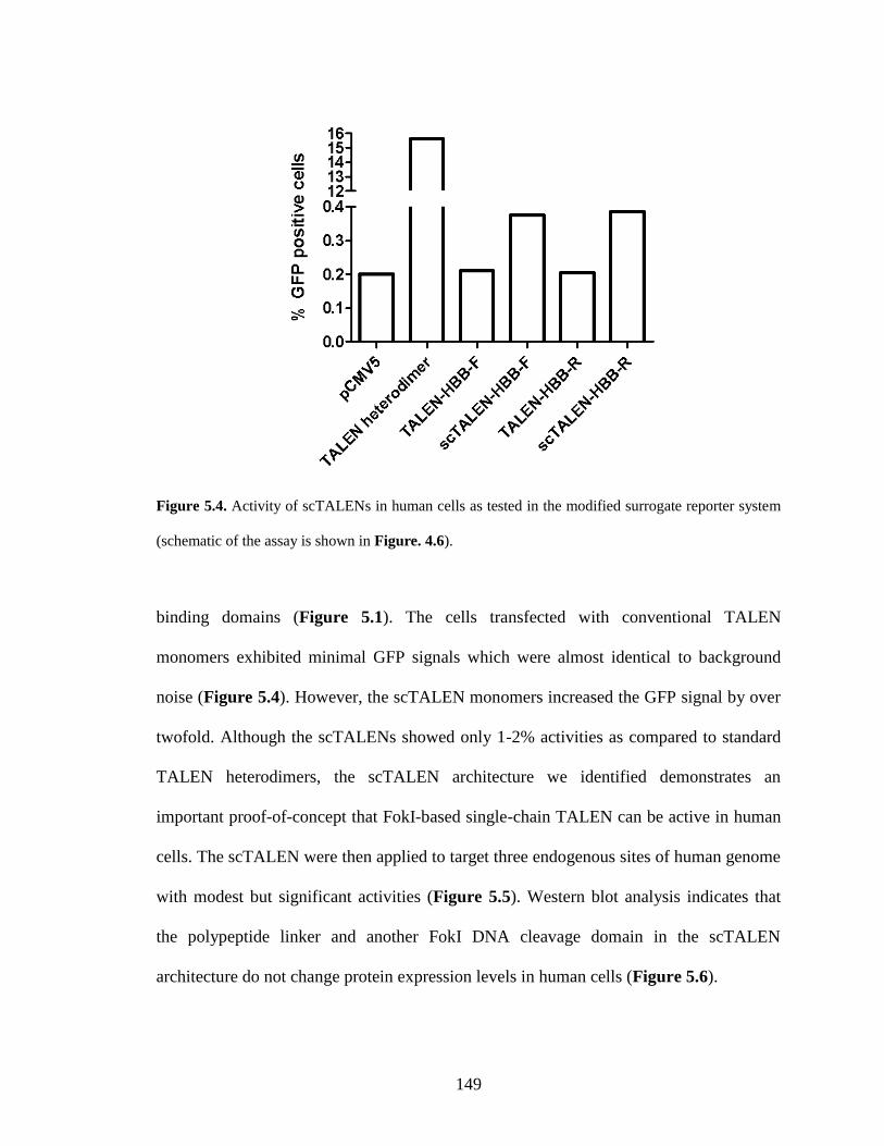
149
Figure 5.4. Activity of scTALENs in human cells as tested in the modified surrogate reporter system
(schematic of the assay is shown in Figure. 4.6).
binding domains (Figure 5.1). The cells transfected with conventional TALEN
monomers exhibited minimal GFP signals which were almost identical to background
noise (Figure 5.4). However, the scTALEN monomers increased the GFP signal by over
twofold. Although the scTALENs showed only 1-2% activities as compared to standard
TALEN heterodimers, the scTALEN architecture we identified demonstrates an
important proof-of-concept that FokI-based single-chain TALEN can be active in human
cells. The scTALEN were then applied to target three endogenous sites of human genome
with modest but significant activities (Figure 5.5). Western blot analysis indicates that
the polypeptide linker and another FokI DNA cleavage domain in the scTALEN
architecture do not change protein expression levels in human cells (Figure 5.6).
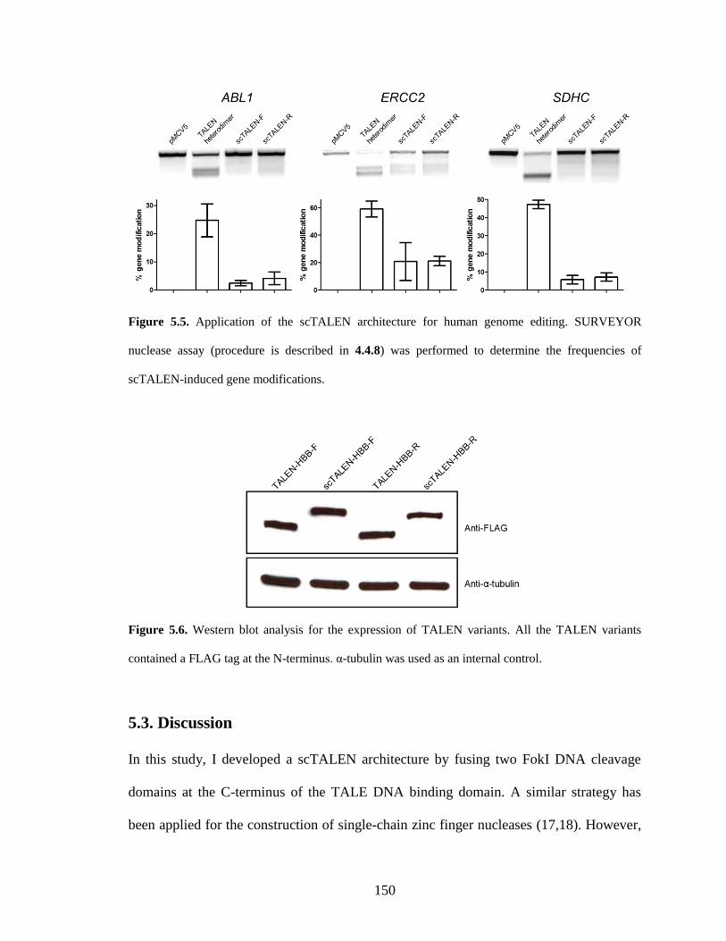
150
Figure 5.5. Application of the scTALEN architecture for human genome editing. SURVEYOR
nuclease assay (procedure is described in 4.4.8) was performed to determine the frequencies of
scTALEN-induced gene modifications.
Figure 5.6. Western blot analysis for the expression of TALEN variants. All the TALEN variants
contained a FLAG tag at the N-terminus. α-tubulin was used as an internal control.
5.3. Discussion
In this study, I developed a scTALEN architecture by fusing two FokI DNA cleavage
domains at the C-terminus of the TALE DNA binding domain. A similar strategy has
been applied for the construction of single-chain zinc finger nucleases (17,18). However,
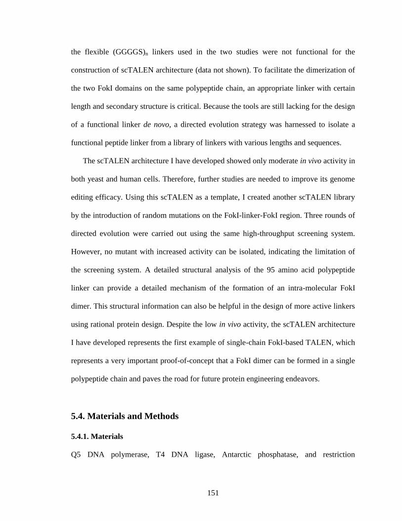
151
the flexible (GGGGS)n linkers used in the two studies were not functional for the
construction of scTALEN architecture (data not shown). To facilitate the dimerization of
the two FokI domains on the same polypeptide chain, an appropriate linker with certain
length and secondary structure is critical. Because the tools are still lacking for the design
of a functional linker de novo, a directed evolution strategy was harnessed to isolate a
functional peptide linker from a library of linkers with various lengths and sequences.
The scTALEN architecture I have developed showed only moderate in vivo activity in
both yeast and human cells. Therefore, further studies are needed to improve its genome
editing efficacy. Using this scTALEN as a template, I created another scTALEN library
by the introduction of random mutations on the FokI-linker-FokI region. Three rounds of
directed evolution were carried out using the same high-throughput screening system.
However, no mutant with increased activity can be isolated, indicating the limitation of
the screening system. A detailed structural analysis of the 95 amino acid polypeptide
linker can provide a detailed mechanism of the formation of an intra-molecular FokI
dimer. This structural information can also be helpful in the design of more active linkers
using rational protein design. Despite the low in vivo activity, the scTALEN architecture
I have developed represents the first example of single-chain FokI-based TALEN, which
represents a very important proof-of-concept that a FokI dimer can be formed in a single
polypeptide chain and paves the road for future protein engineering endeavors.
5.4. Materials and Methods
5.4.1. Materials
Q5 DNA polymerase, T4 DNA ligase, Antarctic phosphatase, and restriction
152
endonucleases were purchased from New England Biolabs (Beverly, MA). QIAprep Spin
Plasmid Miniprep Kit, QIAquick Gel Extraction Kit, and QIAquick PCR Purification Kit
were obtained from Qiagen (Valencia, CA). Oligonucleotide primers were obtained from
Integrated DNA Technologies (Coralville, IA). All the other reagents unless specified
were obtained from Sigma-Aldrich (St. Louis, MO).
5.4.2. Yeast eGFP reporter strain
The single-strand annealing eGFP reporter on pRS414 vector with two separated eGFP
fragments sharing a 100 bp homologous region was described in Chapter 4. The
centromere sequence of the plasmid was then removed by the PmlI digestion and blunt
ligation. This plasmid was then digested with HindIII within the TRP1 gene. The
linearized plasmid was transformed into S. cerevisiae HZ848 (MATα, ade2-1, ade3Δ22,
Δura3, his3-11,15, trp1-1, leu2-3,112 and can1-100) using the LiAc/SS carrier
DNA/PEG method (19) and stably integrated into the chromosome at the TRP1 site. The
yeast reporter strain was obtained by selection of the transformed yeast cells on plates
containing synthetic complete medium lacking tryptophan with 2% glucose at 30 °C. The
integration was confirmed by PCR amplification and DNA sequencing of the targeted
genomic region.
5.4.3. Creation of a scTALEN library
The yeast TALEN expression vector with pRS415 backbone was described in Chapter 2.
This vector was digested with HindIII and SalI to remove the latter part of the FokI
coding sequence. Ligation of this digested vector and a PCR product amplified by the
153
primers FokI-middle(HindIII)-for 5’-gga tac taa agc tta tag cgg agg-3’ (HindIII site
shown in italic) and SalI-XhoI-BamHI-FokI-rev 5’-atc cgc gtc gac act gac tga ctg act cga
gcc tac tga ctg tta gga tcc aaa gtt tat ctc gcc gtt’3’ (XhoI site underlined and BamHI site
shown in italic) digested with HindIII and SalI makes plasmid pRS415-GAL10P-N3-C1-
Xa10-BamHI-XhoI. An additional copy of FokI coding region was inserted into this
vector between the XhoI and SalI sites to generate the recipient expression vector (Figure
5.2A).
The polypeptide linker library was kindly provided by Dr. David Baker from the
University of Washington (Seattle, WA). The sequences encoding the linker library was
PCR amplified by primers: BamHI-linkerlib-forN 5’-act gac gga tcc ggt agc ggc tca gga-
3’ (BamHI site shown in italics) and XhoI-linkerlib-revN 5’-act gac ctc gag cc g ctt ccc
gac cca ga-3’ (XhoI site shown in italics). The PCR product has various lengths and
showed a smeared wide band on 2% agarose gel. All of the PCR products were gel-
extracted, digested with XhoI and BamHI, purified, and inserted into the recipient
expression vector that was digested with the same restriction enzymes. The ligation
products were precipitated by 1-butanol and electroporated into DH5α competent cells to
create the library of scTALENs. The library contains approximately 2×10⁵ transformants
(data not shown).
5.4.4. High-throughput screening
The plasmids were minipreped from the scTALEN library and transformed into the
reporter yeast strain as described above using the LiAc/SS carrier DNA/PEG method (19).
After transformation, the yeast cells were recovered at 30 °C with shaking for 1 h in YPA
154
medium (1% yeast extract, 2% peptone and 0.01% adenine hemisulphate) with 2%
glucose. The cells were then centrifuged and resuspended in synthetic complete medium
lacking leucine and tryptophan (SC-Leu-Trp) with 2% raffinose for growing at 30 °C
with shaking overnight. TALEN expression was induced by culturing cells in YPA
medium with 2% galactose at 30 °C for 5 h. The cells were then centrifuged and
resuspended in SC-Leu-Trp liquid medium with 2% glucose at 30 °C with shaking
overnight. On the next day, cells were analyzed on a BD FACS Aria III cell sorting
system (BD Biosciences, San Jose, CA) and 20,000-100,000 eGFP-positive cells were
collected. After 2 days’ growth at 30 °C in SC-Leu-Trp liquid medium with 2% glucose,
plasmids from the sorted cells were extracted using Zymoprep Yeast Plasmid Miniprep II
Kit (Zymo Research, Orange, CA). The plasmids were then electroporated into DH5α
competent cells for amplification. The minipreped plasmids were subjected to the next
round of screening.
5.4.5. LacZ reporter assay in yeast
The yeast reporter system based on lacZ gene was described in Chapter 2. To test the in
vivo cleavage activity of scTALENs in the yeast reporter system, both the pRS414
reporter plasmid and pRS415 expression plasmid were transformed into Saccharomyces
cerevisiae HZ848 (MATα, ade2-1, ade3Δ22, Δura3, his3-11,15, trp1-1, leu2-3,112 and
can1-100). After 3-4 days growth at 30 °C on SC-Leu-Trp plates with 2% glucose, three
transformants were picked to grow in SC-Leu-Trp liquid medium with 2% galactose.
After cultivation for 2 days at 30 °C, yeast cells were harvested by centrifugation and
lysed by Y-PER yeast protein extraction reagent (Pierce, Rockford, IL). The β-
155
galactosidase (β-GAL) enzyme activity was determined by the β-galactosidase enzyme
system from Promega (Madison, WI) according to the manufacturer's instruction. For
each sample, the β-GAL activity was measured as a function of time and normalized by
total protein concentration determined using the BCA protein assay (Pierce, Rockford,
IL). Homing endonuclease I-CreI (20) and conventional dimeric TALENs served as
positive controls.
5.4.6. In vivo activity in human cells
The pCMV5 TALEN expression plasmids were reported in Chapter 2. To construct
scTALEN expression vectors for human cells, the FokI-linker-FokI coding sequence of
scTALEN was PCR amplified from the pRS415 plasmids. The PCR products were then
digested with AflII and SalI and inserted into the AflII/SalI digested pCMV5 TALEN
expression plasmids to make pCMV5 scTALEN expression plasmids by replacing the
FokI genes with the FokI-linker-FokI coding sequences.
A modified surrogate reporter system in human cells was described in Chapter 4.
Reporter Hela cells were routinely maintained in the modified Eagle’s medium (MEM)
supplemented with 10% Fetal Bovine Serum (FBS; Hyclone, Logan, UT). Cells were
seeded in 24-well plates at a density of 5×10⁴ per well. After 24 h, reporter cells were
transfected with 1 μg of pCMV5 scTALEN expression plasmids using FuGene HD
transfection reagent (Promega, Madison, WI) under conditions specified by the
manufacturer. Cells were trypsinized from their culturing plates 48 h after transfection
and resuspended in 300 μL phosphate buffered saline (PBS) for flow cytometry analysis.
20 000 cells were analyzed by a BD LSRII flow cytometer (BD Biosciences, San Jose,
156
CA) to quantify the eGFP-positive cells.
5.4.7. Western blot analysis
Hela cells in 12-well plates were harvested 24 h after transfection with TALEN
expression plasmids. Cells were collected by centrifugation, washed with PBS and
resuspended in 40 μL whole cell lysis buffer (1 M Tris-HCl, pH 6.8, 20% sodium
dodecyl sulfate, and 0.1 M dithiothreitol). Proteins were resolved by 4-20% Mini-Protean
TGX Precast Gel (Bio-Rad Laboratories, Hercules, CA), transferred onto a nitrocellulose
membrane, blocked for 1 h with Tris-buffered saline/0.05% Tween 20 containing 5%
nonfat milk, followed by incubation with anti-FLAG tag (1: 500) and anti-α-tubulin (1:
15 000) antibodies at 4 °C overnight. After incubation with anti-mouse horseradish
peroxidase-conjugated secondary antibody (1: 15 000; GeneScript, Piscataway, NJ) for 1
h, bands were visualized using SuperSignal West Pico Chemiluminescent Substrate
(Pierce, Rockford, IL).
5.5. References
1. Sun, N. and Zhao, H. (2013) Transcription activator-like effector nucleases
(TALENs): A highly efficient and versatile tool for genome editing. Biotechnol
Bioeng, doi: 10.1002/bit.24890.
2. Joung, J.K. and Sander, J.D. (2013) TALENs: a widely applicable technology for
targeted genome editing. Nat Rev Mol Cell Biol, 14, 49-55.
157
3. Bitinaite, J., Wah, D.A., Aggarwal, A.K. and Schildkraut, I. (1998) FokI
dimerization is required for DNA cleavage. Proc Natl Acad Sci U S A, 95, 10570-
10575.
4. Miller, J.C., Tan, S., Qiao, G., Barlow, K.A., Wang, J., Xia, D.F., Meng, X.,
Paschon, D.E., Leung, E., Hinkley, S.J. et al. (2011) A TALE nuclease
architecture for efficient genome editing. Nat Biotechnol, 29, 143-148.
5. Mussolino, C., Morbitzer, R., Lutge, F., Dannemann, N., Lahaye, T. and
Cathomen, T. (2011) A novel TALE nuclease scaffold enables high genome
editing activity in combination with low toxicity. Nucleic Acids Res, 39, 9283-
9293.
6. Sun, N., Liang, J., Abil, Z. and Zhao, H. (2012) Optimized TAL effector
nucleases (TALENs) for use in treatment of sickle cell disease. Mol Biosyst, 8,
1255-1263.
7. Briggs, A.W., Rios, X., Chari, R., Yang, L., Zhang, F., Mali, P. and Church, G.M.
(2012) Iterative capped assembly: rapid and scalable synthesis of repeat-module
DNA such as TAL effectors from individual monomers. Nucleic Acids Res, 40,
e117.
8. Yu, Y., Streubel, J., Balzergue, S., Champion, A., Boch, J., Koebnik, R., Feng, J.,
Verdier, V. and Szurek, B. (2011) Colonization of rice leaf blades by an African
strain of Xanthomonas oryzae pv. oryzae depends on a new TAL effector that
induces the rice nodulin-3 Os11N3 gene. Mol Plant Microbe Interact, 24, 1102-
1113.
158
9. Cermak, T., Doyle, E.L., Christian, M., Wang, L., Zhang, Y., Schmidt, C., Baller,
J.A., Somia, N.V., Bogdanove, A.J. and Voytas, D.F. (2011) Efficient design and
assembly of custom TALEN and other TAL effector-based constructs for DNA
targeting. Nucleic Acids Res, 39, e82.
10. Ding, Q., Lee, Y.K., Schaefer, E.A., Peters, D.T., Veres, A., Kim, K.,
Kuperwasser, N., Motola, D.L., Meissner, T.B., Hendriks, W.T. et al. (2013) A
TALEN genome-editing system for generating human stem cell-based disease
models. Cell Stem Cell, 12, 238-251.
11. Kim, Y., Kweon, J., Kim, A., Chon, J.K., Yoo, J.Y., Kim, H.J., Kim, S., Lee, C.,
Jeong, E., Chung, E. et al. (2013) A library of TAL effector nucleases spanning
the human genome. Nat Biotechnol, 31, 251-258.
12. Reyon, D., Tsai, S.Q., Khayter, C., Foden, J.A., Sander, J.D. and Joung, J.K.
(2012) FLASH assembly of TALENs for high-throughput genome editing. Nat
Biotechnol, 30, 460-465.
13. Schmid-Burgk, J.L., Schmidt, T., Kaiser, V., Honing, K. and Hornung, V. (2013)
A ligation-independent cloning technique for high-throughput assembly of
transcription activator-like effector genes. Nat Biotechnol, 31, 76-81.
14. Weber, E., Gruetzner, R., Werner, S., Engler, C. and Marillonnet, S. (2011)
Assembly of designer TAL effectors by Golden Gate cloning. PLoS One, 6,
e19722.
15. Beurdeley, M., Bietz, F., Li, J., Thomas, S., Stoddard, T., Juillerat, A., Zhang, F.,
Voytas, D.F., Duchateau, P. and Silva, G.H. (2013) Compact designer TALENs
for efficient genome engineering. Nat Commun, 4, 1762.
159
16. Li, H., Pellenz, S., Ulge, U., Stoddard, B.L. and Monnat, R.J., Jr. (2009)
Generation of single-chain LAGLIDADG homing endonucleases from native
homodimeric precursor proteins. Nucleic Acids Res, 37, 1650-1662.
17. Minczuk, M., Papworth, M.A., Miller, J.C., Murphy, M.P. and Klug, A. (2008)
Development of a single-chain, quasi-dimeric zinc-finger nuclease for the
selective degradation of mutated human mitochondrial DNA. Nucleic Acids Res,
36, 3926-3938.
18. Mino, T., Aoyama, Y. and Sera, T. (2009) Efficient double-stranded DNA
cleavage by artificial zinc-finger nucleases composed of one zinc-finger protein
and a single-chain FokI dimer. J Biotechnol, 140, 156-161.
19. Gietz, R.D. and Schiestl, R.H. (2007) High-efficiency yeast transformation using
the LiAc/SS carrier DNA/PEG method. Nat Protoc, 2, 31-34.
20. Chevalier, B.S., Monnat, R.J., Jr. and Stoddard, B.L. (2001) The homing
endonuclease I-CreI uses three metals, one of which is shared between the two
active sites. Nat Struct Biol, 8, 312-316.