Embed Size (px)
Citation preview

Table 2. Bohan and Peter criteria for the diagnosis of PM and DM8
1. Symmetrical weakness of the limb girdle muscles and anterior neck flexors, progressing over weeks to months, with or without dysphagia or respiratory muscle involvement
2. Muscle biopsy evidence of necrosis of myofibers, phagocytosis, regeneration with basophils, large vesicular sarcolernmal nuclei, and prominent nucleoli, atrophy in a perifascicular distribution, variation in fiber size and an inflammatory exudate, often perivascular
3. Elevation in serum of skeletal-muscle enzymes, particularly the CK and often aldolase, aspartate aminotransferase (AST or SGOT), alanine aminotransfe1ase (ALT or SGPT) and lactate dehydrogenase (LOH)
4. Electromyographic triad of short, small, polyphasic motor units, fibrillations, positive sharp waves and insertional irritability, and bizarre, high frequency repetitive discharges
5. Any one of the characteristic dennatologic features of the rash ofnM
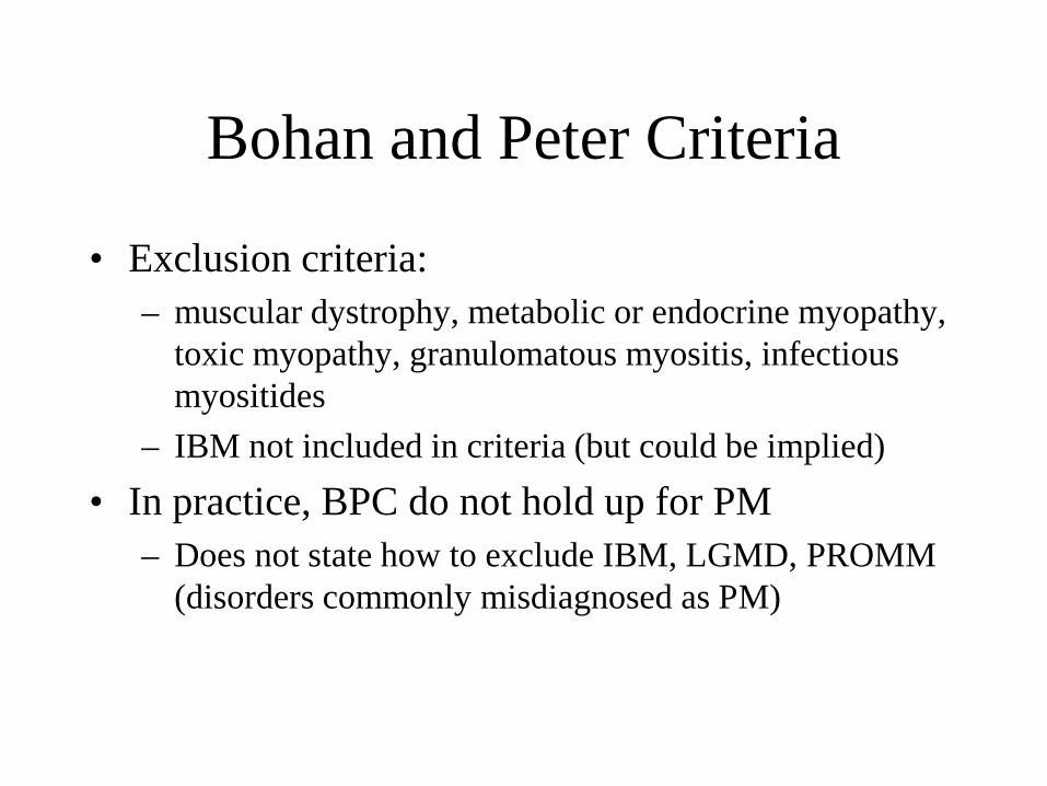
Bohan and Peter Criteria
• Exclusion criteria: – muscular dystrophy, metabolic or endocrine myopathy,
toxic myopathy, granulomatous myositis, infectious myositides
– IBM not included in criteria (but could be implied) • In practice, BPC do not hold up for PM
– Does not state how to exclude IBM, LGMD, PROMM (disorders commonly misdiagnosed as PM)
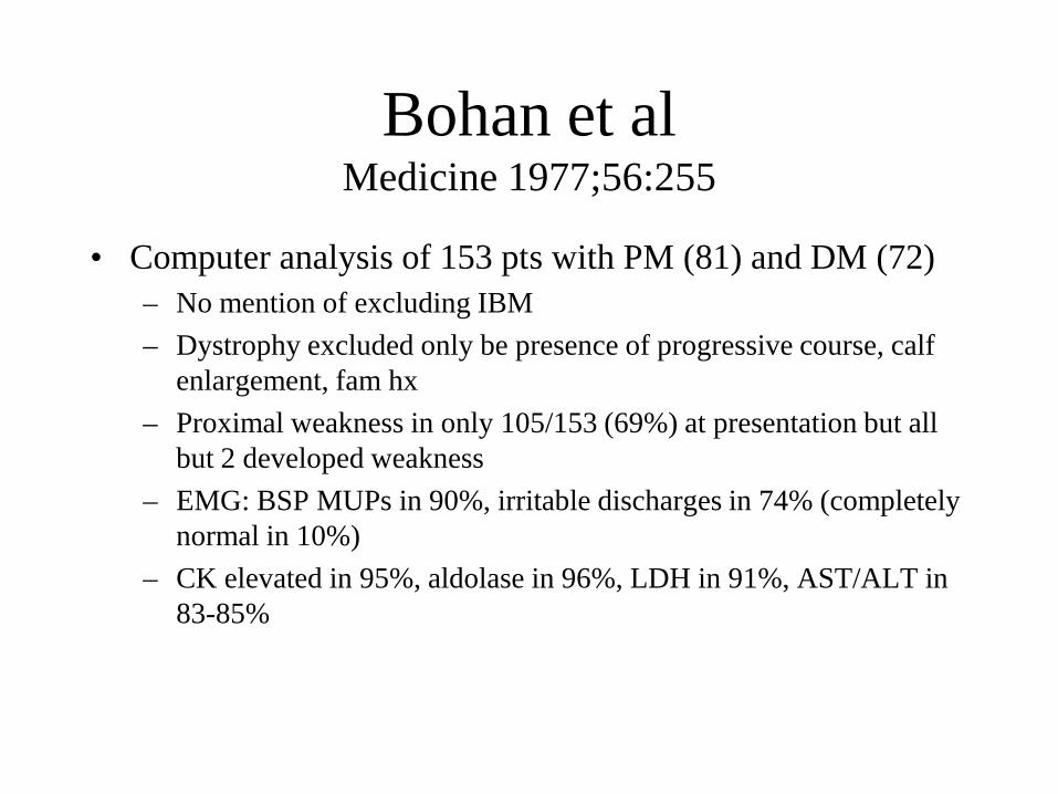
Bohan et al Medicine 1977;56:255
• Computer analysis of 153 pts with PM (81) and DM (72) – No mention of excluding IBM – Dystrophy excluded only be presence of progressive course, calf
enlargement, fam hx – Proximal weakness in only 105/153 (69%) at presentation but all
but 2 developed weakness – EMG: BSP MUPs in 90%, irritable discharges in 74% (completely
normal in 10%) – CK elevated in 95%, aldolase in 96%, LDH in 91%, AST/ALT in
83-85%
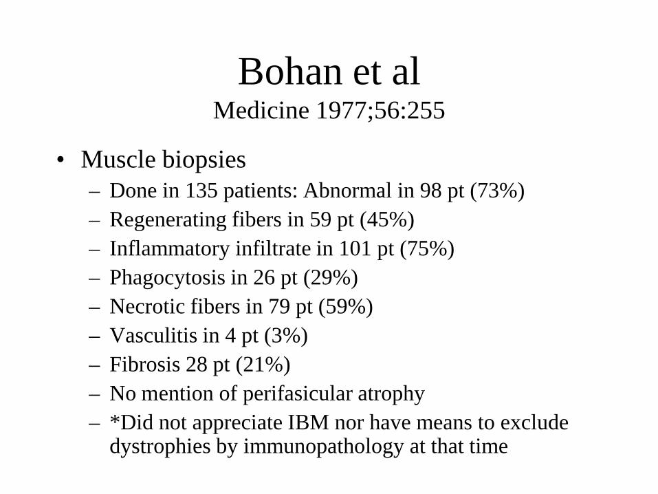
Bohan et al Medicine 1977;56:255
• Muscle biopsies – Done in 135 patients: Abnormal in 98 pt (73%) – Regenerating fibers in 59 pt (45%) – Inflammatory infiltrate in 101 pt (75%) – Phagocytosis in 26 pt (29%) – Necrotic fibers in 79 pt (59%) – Vasculitis in 4 pt (3%) – Fibrosis 28 pt (21%) – No mention of perifasicular atrophy – *Did not appreciate IBM nor have means to exclude
dystrophies by immunopathology at that time
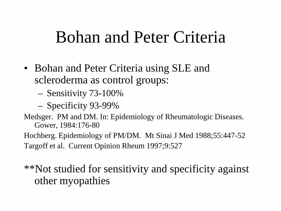
Bohan and Peter Criteria
• Bohan and Peter Criteria using SLE and scleroderma as control groups: – Sensitivity 73-100% – Specificity 93-99%
Medsger. PM and DM. In: Epidemiology of Rheumatologic Diseases. Gower, 1984:176-80
Hochberg. Epidemiology of PM/DM. Mt Sinai J Med 1988;55:447-52 Targoff et al. Current Opinion Rheum 1997;9:527
**Not studied for sensitivity and specificity against other myopathies
Tanimoto et al J Rheum 1995;22;668-74
• Questionaires inquiring about pts wit DM, PM, SLE, SSc, and non-inflammatory myopathies sent to major institutes in Japan
• DM dx by Rheumatologist and Dermatologists (not stated how in methods)
• PM dx by rheumatologist and neurologists (not stated how in methods)
• Non-inflammatory myopathies dx by neurologists based on clinical, laboratory, and histopathological features
Tanimoto et al J Rheum 1995;22;668-74
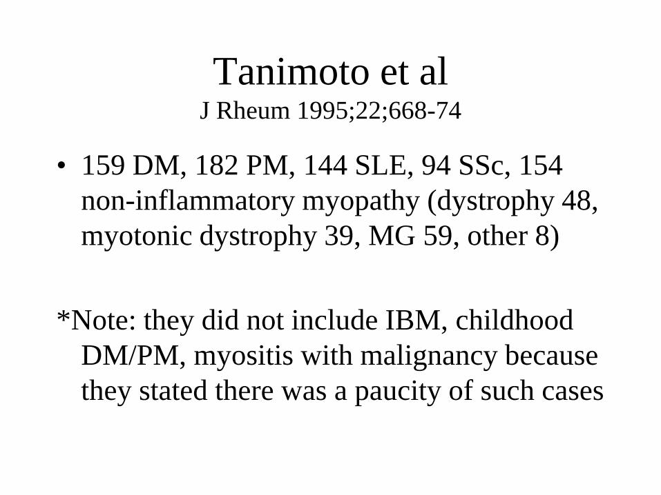
• 159 DM, 182 PM, 144 SLE, 94 SSc, 154 non-inflammatory myopathy (dystrophy 48, myotonic dystrophy 39, MG 59, other 8)
*Note: they did not include IBM, childhood DM/PM, myositis with malignancy because they stated there was a paucity of such cases
Tanimoto et al J Rheum 1995;22;668-74
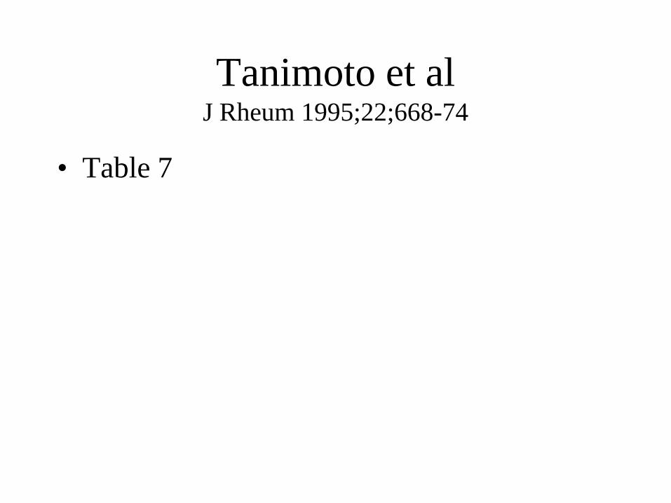
• Table 7
Tanimoto et al J Rheum 1995;22;668-74
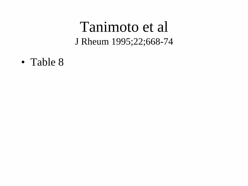
• Table 8
Tanimoto et al J Rheum 1995;22;668-74
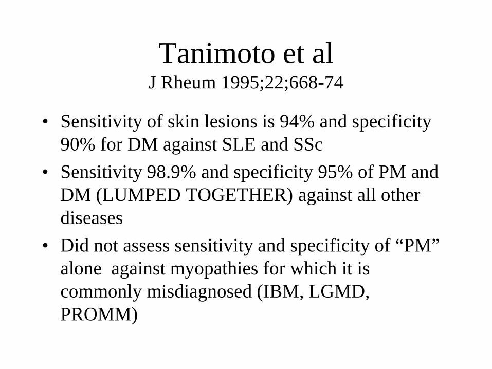
• Table 12
Tanimoto et al J Rheum 1995;22;668-74
• Sensitivity of skin lesions is 94% and specificity 90% for DM against SLE and SSc
• Sensitivity 98.9% and specificity 95% of PM and DM (LUMPED TOGETHER) against all other diseases
• Did not assess sensitivity and specificity of “PM” alone against myopathies for which it is commonly misdiagnosed (IBM, LGMD, PROMM)
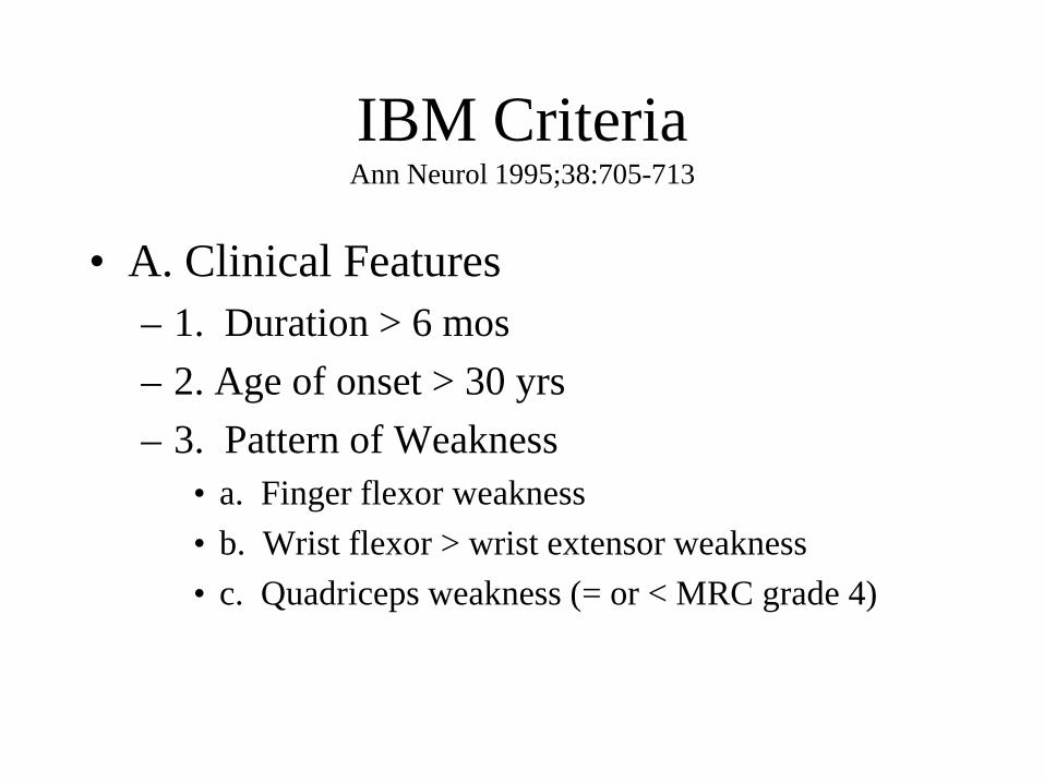
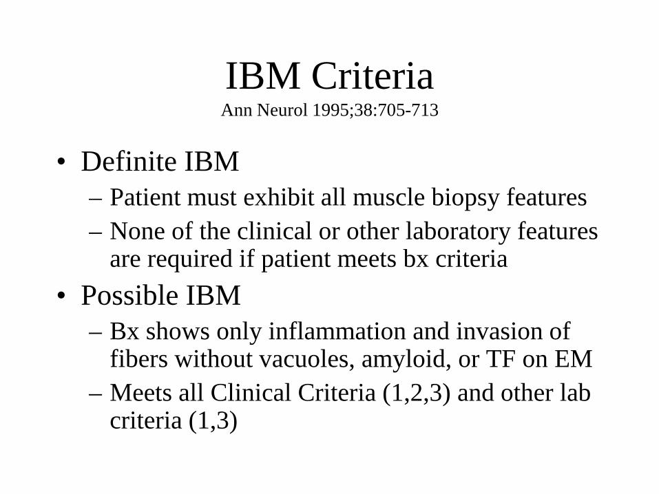
IBM Criteria Ann Neurol 1995;38:705-713
• A. Clinical Features – 1. Duration > 6 mos – 2. Age of onset > 30 yrs – 3. Pattern of Weakness
• a. Finger flexor weakness • b. Wrist flexor > wrist extensor weakness • c. Quadriceps weakness (= or < MRC grade 4)

IBM Criteria Ann Neurol 1995;38:705-713
• B. Laboratory Features – 1. Serum CK < 12 x normal – 2. Muscle biopsy
• a. mononuclear inflammatory cells invasion of non-necrotic muscle fibers
• b. vacuolated muscle fibers • c. either
– i. Intracellular amyloid deposits – ii. 15-18 nm tubulofilaments by EM
– 3. EMG • “features of an inflammatory myopathy” • May have long-duration MUAPs
IBM Criteria Ann Neurol 1995;38:705-713
• Definite IBM – Patient must exhibit all muscle biopsy features – None of the clinical or other laboratory features
are required if patient meets bx criteria • Possible IBM
– Bx shows only inflammation and invasion of fibers without vacuoles, amyloid, or TF on EM
– Meets all Clinical Criteria (1,2,3) and other lab criteria (1,3)

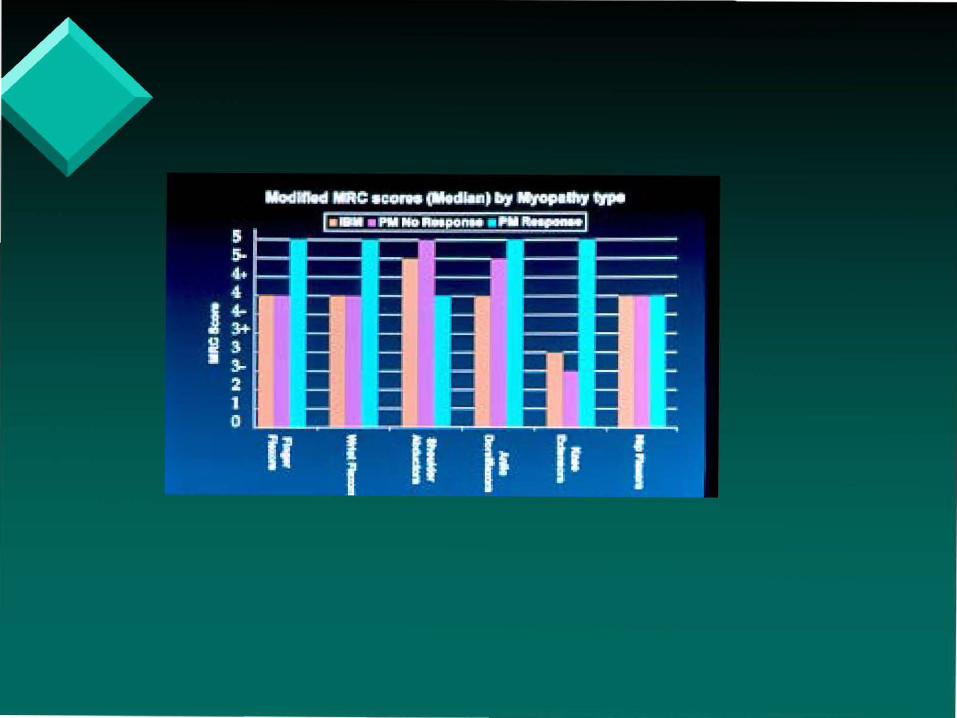
IBM: Clinical and Pathological Boundaries Amato et al. Ann Neurol 1996;40:581-586
• Table 1
IBM: Clinical and Pathological Boundaries Amato et al. Ann Neurol 1996;40:581-586
• Table 2
----- · -• ·••••

Dalakas and Hohlfeld Criteria Lancet 2003;362:971-982
• TABLE 2
ENMC Criteria
• Neuromuscular Disorders 2004;14:337-345 – Takes into account advances in understanding of the
immunopathogenesis (PM and DM), IBM as a clinical and histological dx, and under-appreciated form of myositis: immune-mediated necrotizing myopathies
– Spells out in more detail the clinical features, laboratory, and histopathological feature required for inclusion and exclusion
– May be less sensitive but will be much more specific – Reliability and validity need to be assessed in
prospective study
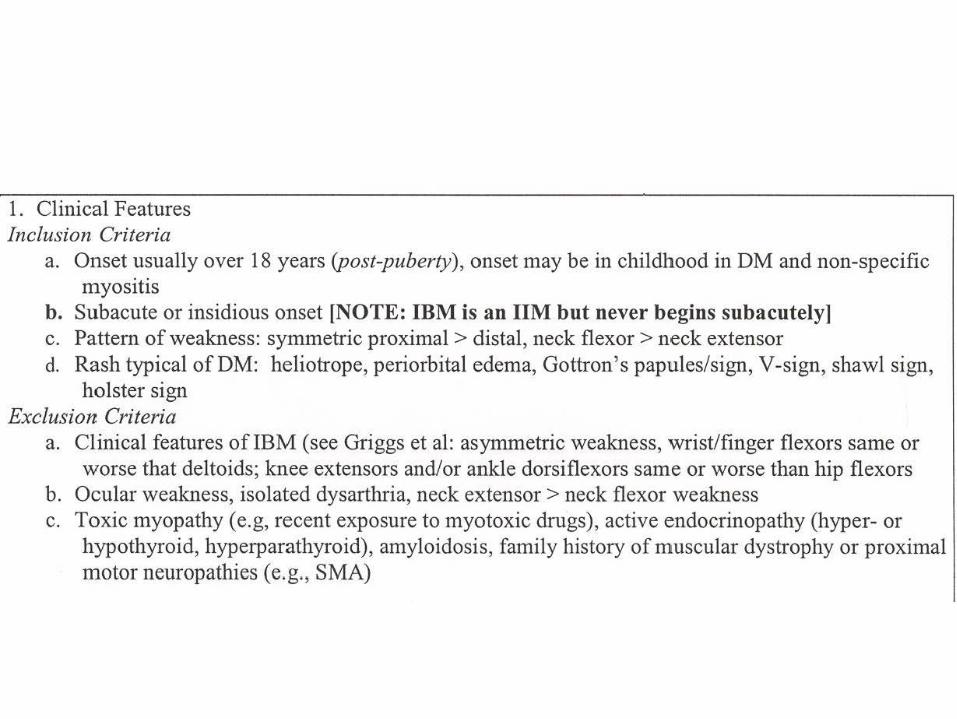
1. Clinical Features Inclusion Criteria
a. Onset usually over 18 years (post-puberty), onset may be in childhood in DM and non-specific myositis
b. Subacute or insidious onset [NOTE: IBM is an JIM but never begins subacutely] c. Pattern ofweakness: symmetric proximal > distal, neck flexor >neck extensor d. Rash typical of DM: heliotrope, periorbital edema, Gottron's papules/sign, V-sign, shawl sign,
holster sign Exclusion Criteria
a. Clinical features of IBM (see Griggs et al: asymmetric weakness, wrist/finger flexors same or worse that deltoids; knee extensors and/or ankle dorsiflexors same or worse than hip flexors
b. Ocular weakness, isolated dysarthria, neck extensor> neck flexor weakness c. Toxic myopathy (e.g, recent exposure to myotoxic drugs), active endocrinopathy (hyper- or
hypothyroid, hyperparathyroid), amyloidosis, family history ofmuscular dystrophy or proximalmotor neuropathies (e.g., SMA)

2. Elevated serum creatine kinase level
3. Other Laboratory Criteria: 1) Electromyography:
Inclusion Criteria o Increased insertional and spontaneous activity in the form of fibrillation potentials,
positive sharp waves, or complex repetitive discharges o Morphometric analysis reveals the presence of short duration, small amplitude,
polyphasic MUAPs Exclusion Criteria o Myotonic discharges that would suggest proxin1al myotonic dystrophy or other
channelopathy o Morphometic analysis reveals predominantly long duration, large amplitude MUAPs
2) MRI: diffuse or patchy increased signal (edema) within muscle tissue on STIR images 3) Myositis-specific antibodies detected in serum
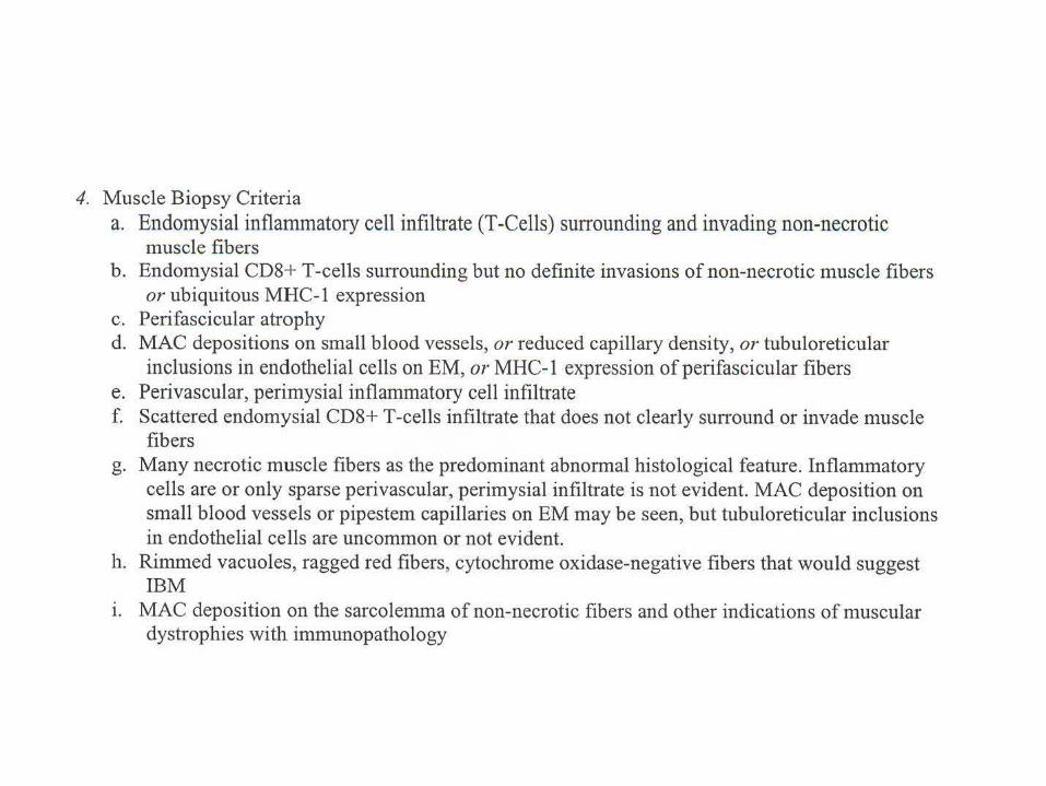
4. Muscle Biopsy Criteria a. Endomysial inflammatory cell infiltrate (I-Cells) surrounding and invading non-necrotic
muscle fibers b. Endomysial CD8+ T-cells surrounding but no definite invasions of non-necrotic muscle fibers
or ubiquitous MHC-1 expression c. Perifascicular atrophy d. MAC depositions on small blood vessels, or reduced capillary density, or tubuloreticular
inclusions in endothelial cells on EM, or MHC-1 expression of perifascicular fibers e. Perivascular, perimysial inflammatory cell infiltrate f. Scattered endomysial CD8+ T-cells infiltrate that does not clearly surround or invade muscle
fibers g. Many necrotic muscle fibers as the predominant abnormal histological feature. Inflammatory
cells are or only sparse perivascular, perimysial infiltrate is not evident. MAC deposition on small blood vessels or pipestem capillaries on EM may be seen, but tubuloreticular inclusions in endothelial cells are uncommon or not evident.
h. Rimmed vacuoles, ragged red fibers, cytochrome oxidase-negative fibers that would suggest IBM
1. MAC deposition on the sarcolemma of non-necrotic fibers and other indications of muscular dystrophies with immunopathology
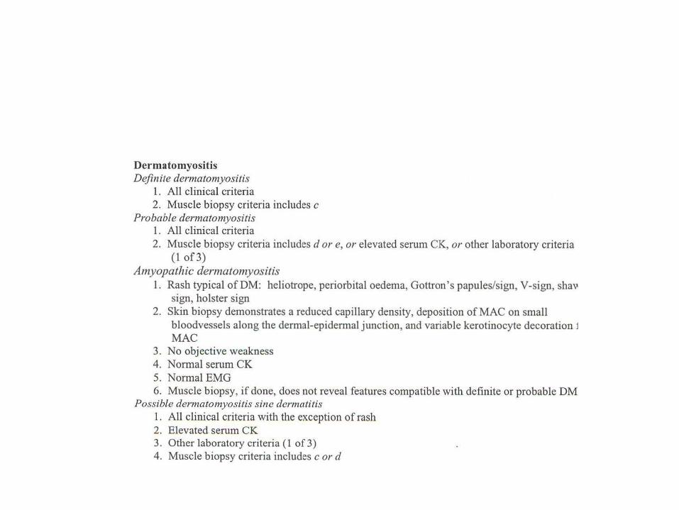
Dermatomyositis Definite dermatomyositis
1. All clinical criteria 2. Muscle biopsy criteria includes c
Probable dermatomyositis 1. All clinical criteria 2. Muscle biopsy criteria includes d or e, or elevated serum CK, or other laboratory criteria
(1of3) Amyopathic dermatomyositis
1. Rash typical of DM: heliotrope, periorbital oedema, Gottron 's papules/sign, V-sign, shav sign, holster sign
2. Skin biopsy demonstrates a reduced capillary density, deposition of MAC on small bloodvessels along the dermal-epidermal junction, and variable kerotinocyte decoration 1 MAC
3. No objective weakness 4. Normal serum CK 5. Normal EMG 6. Muscle biopsy, if done, does not reveal features compatible with definite or probable DM
Possible dermatomyositis sine dermatitis I . All clinical criteria with the exception of rash 2. Elevated serum CK 3. Other laboratory criteria ( 1 of 3) 4. Muscle biopsy criteria includes c or d
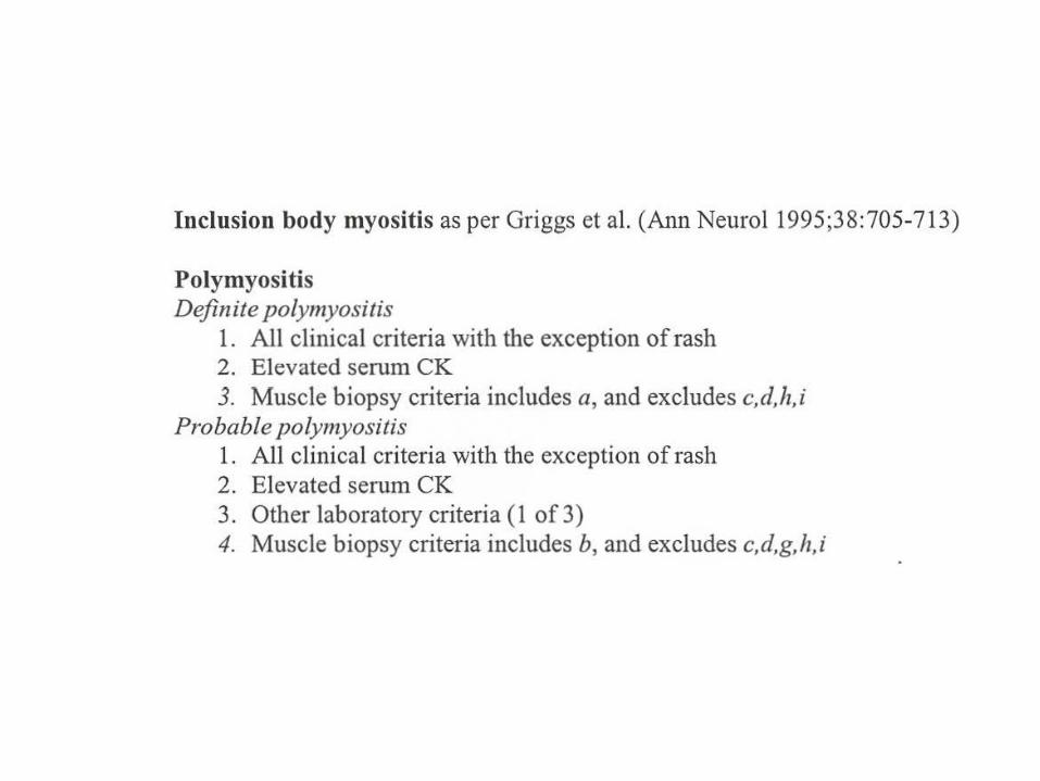
Inclusion body myositis as per Griggs et al. (Ann Neurol 1995;38:705-713)
Polymyositis Definite polymyositis
1. All clinical criteria with the exception of rash 2. Elevated serum CK 3. Muscle biopsy criteria includes a, and excludes c,d,h,i
Probable polymyositis 1. All clinical criteria with the exception of rash 2. Elevated serum CK 3. Other laboratory criteria ( 1 of 3) 4. Muscle biopsy criteria includes b, and excludes c,d,g,h,i
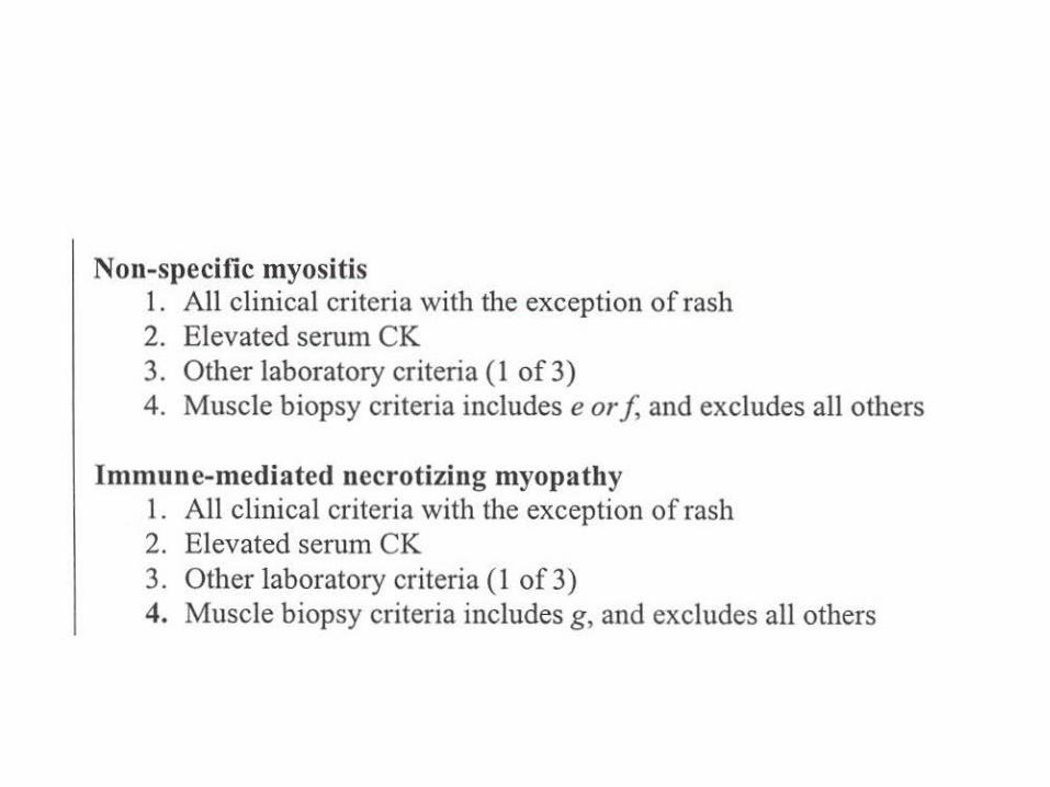
Non-specific myositis 1. All clinical criteria with the exception of rash 2. Elevated serum CK 3. Other laboratory criteria (1 of 3) 4. Muscle biopsy criteria includes e or f, and excludes all others
Immune-mediated necrotizing myopathy 1. All clinical criteria with the exception of rash 2. Elevated serum CK 3. Other laboratory criteria (1 of 3) 4. Muscle biopsy criteria includes g , and excludes all others
ENMC Criteria
• Clinical criteria not too different than others except pattern of weakness and exclusion of other disorders based on pattern of weakness should make it more specific
• Most retrospective studies have found CK elevation in 90-95% of IIM (DM can be normal)
• EMG abnormal in 90% of retrospective studies. New criteria helps exclude proximal myotonic myopathy

ENMC Criteria
• Skeletal muscle MRI – Fraser et al. J Rheum 1991;18:1693-1700
• 17 PM, 10 DM, 13 IBM, and 10 controls (6 SLE without myopathy, 1 steroid myopathy, 1 EDMD, and 2 post-polio)
• MRI was not as specific as muscle bx (89 vs. 66%); PPV 97%; NPP 64%
• Pts with IBM may have more focal, quadriceps involvement • Control group is not sufficient to assess if MRI is useful in
distinguishing PM from LGMD

ENMC Criteria
• “Myositis-Specific Antibodies” – All MSA have low sensitive for IIM or for any subtype – None have been studied prospectively in regards to prognosis – Jo-1 antibodies are found in approximately 20% of IMM
• Associated with ILD, arthritis, mechanic hands, Raynauds • can be seen in ILD without myositis • More common in PM by BPC but is usually DM or non-specific by
ENMC – Mi-2 is almost exclusively associated with DM (will not help with
dx of PM) – SRP has been associated with an acute, severe myositis/myocarditis
• usually PM by BPC but not by Dalakas or ENMC (microangiopathy)

ENMC Criteria
• Histopathology – Criteria used to help distinguish DM, PM, IBM,
necrotizing myopathy and otherwise nonspecific myopathies
PM • PM is characterized by CD8+ cells invading non-
necrotic muscle fibers expression MHC-1antigen
• MHC-1 expression is diffuse on muscle fibers • Similar findings in IBM • MHC-1 expression on muscle fibers is seen in
immune-mediated necrotizing myopathies
• Rarely, will see CD8+ cells invading non-necrotic MHC-1 muscle fibers in dystrophies (MHC-1 expression is not diffuse)
DM • Predominantly, perivascular/perimysial infiltrate CD4+>CD8+ • Also may have mild endomysial infiltrate but no invasion of
non-necrotic muscle fibers • Perifascicular atrophy is rather specific- can be seen in SLE (?
Overlap with DM) and I have seen in GVHD; Not very sensitive (at least in adults 9/14 my experience in past 3 yrs)
• Kissel et al. NEJM 1986;314:329-334 and Arch Neurol 1991;48:26-30 – MAC deposition precedes other abnormalities – MAC on vessels 10/13 childhood and 11/26 adults
• Emslie-Smith & Engel. Ann Neurol 1990:27:343-46 – Diminished capillary density is earliest histological feature (9/10); MAC
on vessels in 13/15 cases (mean #MAC + vessels was 9.7-13.3%) (early/late DM)
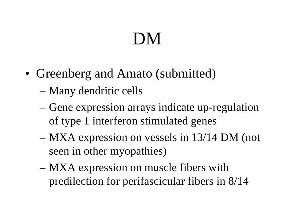
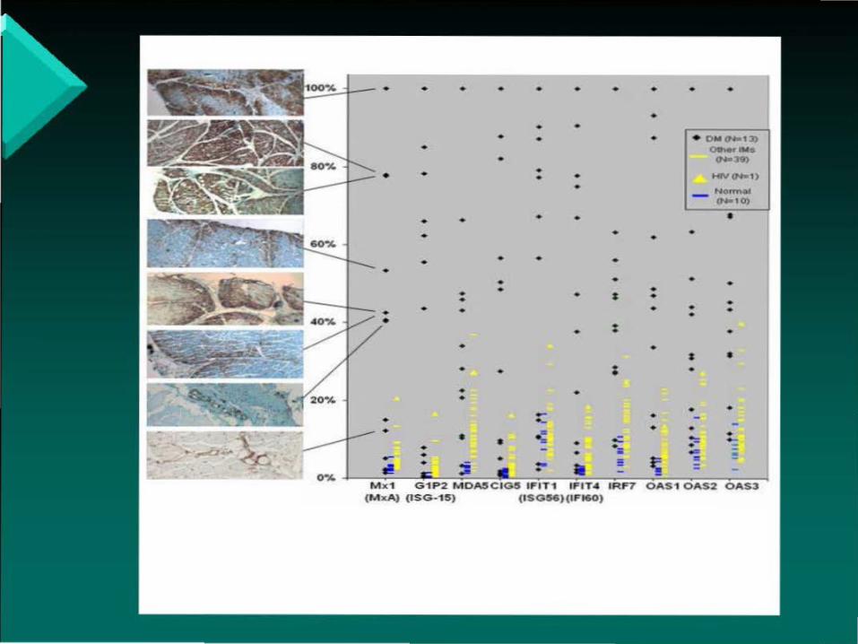
DM
• Greenberg and Amato (submitted) – Many dendritic cells – Gene expression arrays indicate up-regulation
of type 1 interferon stimulated genes – MXA expression on vessels in 13/14 DM (not
seen in other myopathies) – MXA expression on muscle fibers with
predilection for perifascicular fibers in 8/14
• • • • • • • • • •
• • • • •
• • • •
• • • • • •
• • • •
• • • • •
• • • • • • • • •
M x 'I G"'IP2 MDA5C IG5 IFIT"'I IFIT' IRF7 O AS1 OAS2 OAS3 (MxA) (ISG-15) (I SG56)(1Fl60)
•
• •
• •
•
• •• • • I ••
20•4 • •• - ••
• • • .: - • .::
•.,... • • ·-•
0•4 • ...,. • -• II = '!
•
•
••
I •
• • .....= -
• OM (N•l:J) Olner-..:s
(Na;J9)
Hlll-1)
NormM (Nal 0)
•
• •• •
•
•
•..... !!!


![RESEARCHARTICLE EffectsofUrbanLandscapePatternonPM …hub.hku.hk/bitstream/10722/227869/1/Content.pdf · 2016. 7. 21. · tion[13]and health riskassessment ofPM 2.5 [14],attemptingtomakeclear](https://img.pdfslide.us/doc/110x75/6010e1a3debb210d6d49b06b/researcharticle-effectsofurbanlandscapepatternonpm-hubhkuhkbitstream107222278691.jpg)