1
defect in CD. Intestinal removal of necrotic/apoptotic cells occurs through sloughing into the gut lumen and through engulfment by professional (e.g. macrophages) phagocytes. The potential role of non-professional phagocytes, e.g. epithelial cells, fibroblasts, etc., has not been examined within the context of cellular clearance in CD. Work from our laboratory and others have identified a group of molecules shared by both professional and non- professional phagocytes as being critical in engulfment of apoptotic targets. These include the phosphatidyl recognition receptor BAI1 and its downstream signaling module ELMO/ Dock180/CrkII/Rac, as well as a second module involving LRP1/GULP/ABCA1. Our microar- ray data show gene expression of key engulfment molecules in purified murine intestinal epithelial cells (IEC), and protein levels in three human IEC lines. Engulfment assays using apoptotic thymocytes indicate the ability of IECs to engulf In Vitro. This, coupled with their numerical abundance, may represent an important homeostatic mechanism, especially in CD. Furthermore, stimulation of IECs with apoptotic cells results in the active production of the immunosuppressive cytokine TGF-β. TGF-β production by CaCo2 IECs following stimulation with apoptotic thymocytes T1717 Gender Influences Intestinal Toll-Like Receptor Expression Under Normal and Inflammatory Conditions Mirjam A. Looijer-van Langen, Levinus A. Dieleman, Karen Madsen Gender influences immune responses and severity of infectious and inflammatory diseases. Toll-like Receptors (TLRs) are critical in maintaining gut homeostasis and mediating host response to pathogens. We have previously shown gender differences in gut permeability and severity of inflammation in the HLA-B27 rat model of colitis and hypothesize that these gender differences are related to alterations in TLR expression. Aim: To examine the effect of gender on colonic TLR expression under normal and inflammatory conditions and deter- mine the role of estrogen in mediating these effects. Methods: As a model of acute inflamma- tion, estrogen receptor-β (ERβ) expressing human colonic epithelial cells (HT-29) were incubated with TNFα ± 17β-estradiol (E2). To determine the effects of chronic inflammation on TLR expression, colons were collected from 12 wk HLA-B27 non-transgenic (NTG) and transgenic (TG) rats. TLR2, 4, 9 and ERβ mRNA levels were measured in HT-29 cells and rat colon by qRT-PCR. As a measurement of inflammation in rats, colonic IL-1β levels were measured by ELISA. Results: In HLAB27 rats, males showed higher TLR2 and TLR9 and lower TLR4 expression as compared with females (Table). Under chronic inflammatory conditions in males, TLR2 and TLR4 expression increased, while TLR9 expression decreased. In contrast, under chronic inflammatory conditions, females showed increased TLR2 and TLR9, and decreased TLR4 levels. This was associated with decreased IL-1β secretion in females. ERβ expression was down-regulated under chronic inflammatory conditions in both males and females. HT-29 cells grown in the presence of E2 had increased expression of TLR4 and TLR9. The addition of TNFα resulted in a down-regulation of TLR2 and TLR4, suggesting an interaction between estrogen and TNFα signaling on TLR expression Conclu- sion: In HLAB27 rats, males and females exhibit different patterns of expression of colonic TLRs under normal conditions and respond to inflammation differentially, although both groups demonstrate a significant decrease in ERβ expression. The down-regulation of TLR4 in females under inflammatory conditions may contribute to the reduced severity of disease in females in this model. Although estrogen effects TLR expression in epithelial cells, it is unlikely to be completely accountable for the observed difference in TLR expression. T1718 Natural Killer Cell Responses in Both Acute and Chronic Phases of a Murine Model of Experimental Colitis Lindsay J. Hall, Aoife Quinlan, Fergus Shanahan, Kenneth Nally, Silvia Melgar Introduction/Aim: Natural killer (NK) cells contribute to host resistance against viruses, bacteria and certain parasites as well as providing immune surveillance against the develop- ment of tumours. Recently NK cells have also be implicated in the modulation of various autoimmune diseases. In this study we have investigated the recruitment and functional characteristics of NK cells in an experimental model of colitis. Methods: Female C57BL/6 mice were exposed to 6 days of dextran sulphate sodium (DSS) followed by 20 days of A-565 AGA Abstracts water. Organs were harvested in the acute phase (day 1, 3, 5, 8, and 12 post DSS) and in the chronic phase (day 25 post DSS) of colitis. Healthy mice (controls) received only water. Cells from spleens and mesenteric lymph nodes (MLN) were isolated to determine the percentages (DX5+/CD3+ cells), activation status (CD69, CD25) and cytokine/cytotoxic profile (IFN-γ, IL-6, TNF-α, IL-17, IL-10, IL-4, perforin and granzyme B) of NK cells using flow cytometry. In addition, frozen colonic tissue was used to analyse NK cell number and distribution in relation to other cellular populations along disease progression. Results: We observed that as early as 1 day post DSS, animals were found to have significant increases in both the percentage and activation status of NK cells in comparison to controls, which continued throughout the acute phase of the study. Interestingly, NK cell percentages were significantly increased within the spleens of chronic animals; however these cells did not appear to be activated. In contrast, NK cells in MLN were significantly activated but the percentages were similar to control animals. With regards to their cytotoxic profile we observed significantly less perforin+ NK cells at days 3 and 7, whereas granzyme B producing NK cells were significantly increased at every time-point, reaching their peak at day 25 post- DSS. Regarding their cytokine profile, the percentage of anti-inflammatory cytokine (IL-4 and IL-10) producing NK cells was significantly decreased throughout the study. Conversely, significant increases in the percentage of NK cells producing pro-inflammatory cytokines particularly TNF-α, IL-6 and IL-17 was observed. Interestingly even though NK cells are known to be potent produces of IFN-γ, no increases in IFN-γ+ NK cells was observed. In fact, NK cells were significantly less capable of producing IFN-γ in mice with acute inflamma- tion when compared to controls. Conclusions: Collectively, our data suggest that NK cells might play a prominent role in the establishment of immune responses in DSS-induced colitis and therefore may have a consequent impact on the pathogenesis of IBD. T1719 Nutritional Activation of the Afferent Vagus Modulates Inflammation and Preserves Intestinal Integrity in Rats Tim Lubbers, Jacco-Juri De Haan, Misha Luyer, M'hamed Hadfoune, Cornelis Dejong, Wim Buurman, Jan Willem Greve Background and Aims: Administration of enteral nutrition is vital to maintain intestinal structure and function. Recently, it was demonstrated that the autonomic nervous system is involved in the beneficial effects of dietary lipids on inflammation and gut barrier integrity following hemorrhagic shock in rats. The current study investigates the activation of this nutritional anti-inflammatory pathway. Methods: Rats underwent a hemorrhagic shock either fasted or fed lipid-rich enteral nutrition (containing 30% phospholipids). Prior to shock, rats were treated with selective receptor antagonists to the predominantly peripheral located cholecystokinin-1 and mainly central located cholecystokinin-2 receptor or were vagally deafferented by perineural application of capsaicin. Furthermore, sulfated cholecystokinin- 8 was intravenously infused in fasted rats starting 30 minutes before shock until sacrifice. A Mann-Whitney U test was used for between group analysis. Results: Pretreatment with the cholecystokinin-1 and cholecystokinin-2 receptor antagonists both abrogated the protect- ive effect of lipid-rich nutrition on shock-induced inflammation (TNF-α: both p<0.001 vs vehicle) and loss of intestinal integrity (leakage of horseradish peroxidase: both p<0.001 vs vehicle). Next, deafferentation significantly reduced the inhibitory effect of lipid-rich nutrition on inflammation (TNF-α: p<0.01 vs sham) and gut barrier failure (leakage of horseradish peroxidase: p<0.01 vs sham). Intravenous infusion of sulfated cholecystokinin-8 demon- strated no beneficial effect on systemic inflammation and intestinal integrity. Conclusion: Enteral lipid-rich nutrition modulates inflammation and prevents intestinal tissue damage via local activation of the afferent vagus nerve by cholecystokinin. These data reveal that the enteral presence of nutrition activates a vagal pathway leading to modulation of the inflammatory response. This feedback-loop may contribute to our understanding of the unresponsiveness of intestinal immune cells to luminal antigens. T1720 Regulation of the Pro-Resolution Mediators Prostaglandin D 2 and Lipoxin A 4 in Quiescent Human Colitis Linda Vong, Jose G. Ferraz, Ida Rabbani, Remo Panaccione, Paul L. Beck, John L. Wallace The etiology and pathogenesis of Crohn's disease and ulcerative colitis remains unclear, however dysregulation of the inflammatory response plays a central role in enciting tissue damage and promoting intestinal dysfunction. Several anti-inflammatory mediators act as important braking-signals, and are key to coordinating the resolution phase of inflammation. Aim: To characterise the profiles of two anti-inflammatory mediators, prostaglandin D 2 (PGD 2 ) and lipoxin A 4 (LXA 4 ) in active and quiescent ulcerative colitis. Human colonic mucosal biopsies were collected from ulcerative colitis patients with endoscopically active or quiescent disease (with and without maintenance therapy) as well as from healthy controls undergoing colonoscopy for colorectal cancer screening. A combination of molecular and biochemical techniques were employed to examine the changes in (1) protein levels of PGD 2 and LXA 4 , (2) mRNA expression of enzymes involved in the production of these mediators and (3) receptors for these mediators. Results: COX-2 and PGD 2 synthase gene expression were up-regulated in biopsies from patients with active disease (n=4-16 per group; P<0.05 vs. Ctrl). However, in the setting of quiescent disease, COX-2 expression was significantly down-regulated in patients in remission and no maintenance therapy versus patients on maintenance with oral/topical 5-ASA or topical steroids/azathioprine/infliximab (n=4-16 per group; P<0.05). Mucosal PGD 2 levels were 3-fold higher in patients with quiescent disease relative to controls, but not in those requiring maintenance therapy (n=3-8 per group; P<0.05). In contrast, colonic LXA 4 levels were higher (n=4-21; P<0.05 vs Ctrl, active UC) in patients on maintenance therapy compared to patients on no therapy at all, suggesting that maintenance therapy to keep patients in remission may actually drive the production of pro-resolution mediators such as LXA 4 . Conclusion: The results indicate that the local production of anti-inflammatory mediators such as PGD 2 and LXA 4 play an important role in the resolution phase of inflammation in ulcerative colitis. Differential colonic COX-2 expression and LXA 4 production in UC patients in remission on maintenance therapy may be the turning point in achieving normalization of tissue homeostasis. These findings provide clues to tailoring effective therapies for induction of remission in IBD. AGA Abstracts

T1719 Nutritional Activation of the Afferent Vagus Modulates Inflammation and Preserves Intestinal Integrity in Rats

Embed Size (px)

Citation preview

defect in CD. Intestinal removal of necrotic/apoptotic cells occurs through sloughing intothe gut lumen and through engulfment by professional (e.g. macrophages) phagocytes. Thepotential role of non-professional phagocytes, e.g. epithelial cells, fibroblasts, etc., has notbeen examined within the context of cellular clearance in CD. Work from our laboratoryand others have identified a group of molecules shared by both professional and non-professional phagocytes as being critical in engulfment of apoptotic targets. These includethe phosphatidyl recognition receptor BAI1 and its downstream signaling module ELMO/Dock180/CrkII/Rac, as well as a second module involving LRP1/GULP/ABCA1. Our microar-ray data show gene expression of key engulfment molecules in purified murine intestinalepithelial cells (IEC), and protein levels in three human IEC lines. Engulfment assays usingapoptotic thymocytes indicate the ability of IECs to engulf In Vitro. This, coupled with theirnumerical abundance, may represent an important homeostatic mechanism, especially inCD. Furthermore, stimulation of IECs with apoptotic cells results in the active productionof the immunosuppressive cytokine TGF-β.

TGF-β production by CaCo2 IECs following stimulation with apoptotic thymocytes

T1717

Gender Influences Intestinal Toll-Like Receptor Expression Under Normal andInflammatory ConditionsMirjam A. Looijer-van Langen, Levinus A. Dieleman, Karen Madsen

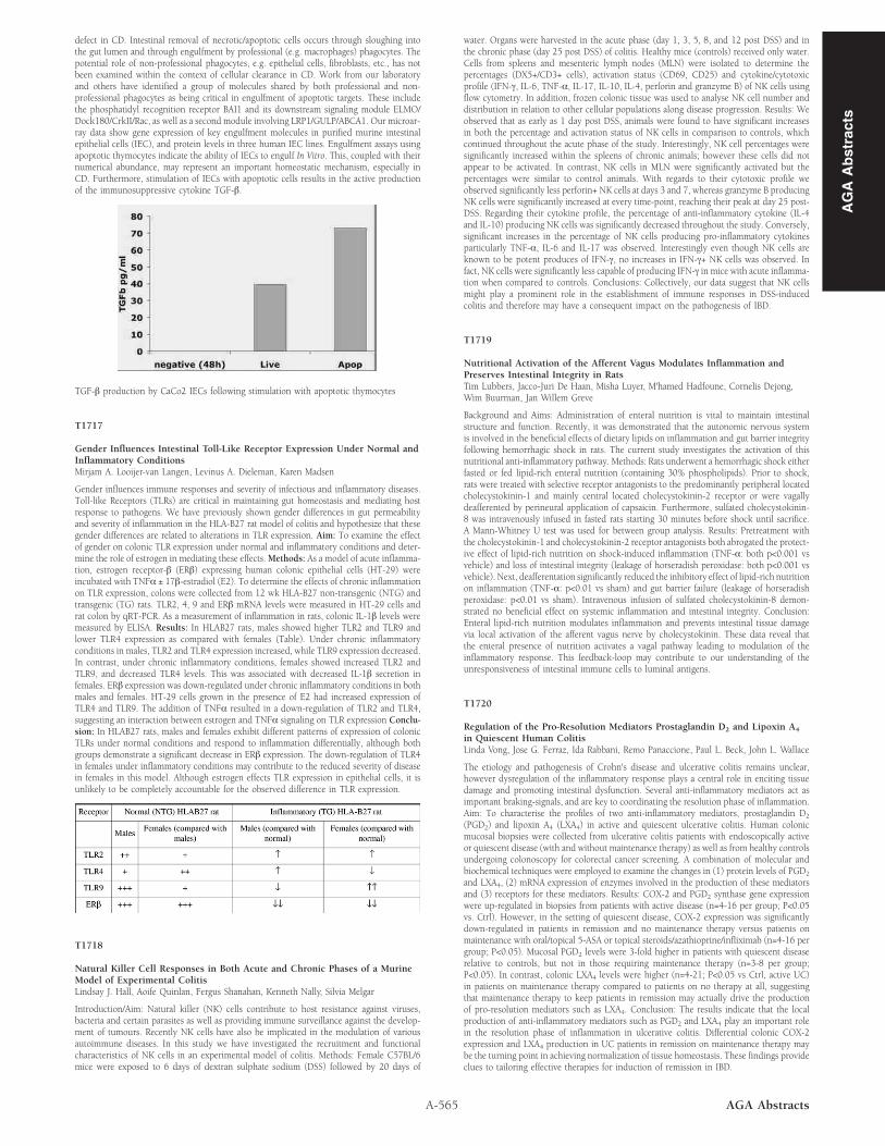
Gender influences immune responses and severity of infectious and inflammatory diseases.Toll-like Receptors (TLRs) are critical in maintaining gut homeostasis and mediating hostresponse to pathogens. We have previously shown gender differences in gut permeabilityand severity of inflammation in the HLA-B27 rat model of colitis and hypothesize that thesegender differences are related to alterations in TLR expression. Aim: To examine the effectof gender on colonic TLR expression under normal and inflammatory conditions and deter-mine the role of estrogen in mediating these effects. Methods: As a model of acute inflamma-tion, estrogen receptor-β (ERβ) expressing human colonic epithelial cells (HT-29) wereincubated with TNFα ± 17β-estradiol (E2). To determine the effects of chronic inflammationon TLR expression, colons were collected from 12 wk HLA-B27 non-transgenic (NTG) andtransgenic (TG) rats. TLR2, 4, 9 and ERβ mRNA levels were measured in HT-29 cells andrat colon by qRT-PCR. As a measurement of inflammation in rats, colonic IL-1β levels weremeasured by ELISA. Results: In HLAB27 rats, males showed higher TLR2 and TLR9 andlower TLR4 expression as compared with females (Table). Under chronic inflammatoryconditions in males, TLR2 and TLR4 expression increased, while TLR9 expression decreased.In contrast, under chronic inflammatory conditions, females showed increased TLR2 andTLR9, and decreased TLR4 levels. This was associated with decreased IL-1β secretion infemales. ERβ expression was down-regulated under chronic inflammatory conditions in bothmales and females. HT-29 cells grown in the presence of E2 had increased expression ofTLR4 and TLR9. The addition of TNFα resulted in a down-regulation of TLR2 and TLR4,suggesting an interaction between estrogen and TNFα signaling on TLR expression Conclu-sion: In HLAB27 rats, males and females exhibit different patterns of expression of colonicTLRs under normal conditions and respond to inflammation differentially, although bothgroups demonstrate a significant decrease in ERβ expression. The down-regulation of TLR4in females under inflammatory conditions may contribute to the reduced severity of diseasein females in this model. Although estrogen effects TLR expression in epithelial cells, it isunlikely to be completely accountable for the observed difference in TLR expression.

T1718

Natural Killer Cell Responses in Both Acute and Chronic Phases of a MurineModel of Experimental ColitisLindsay J. Hall, Aoife Quinlan, Fergus Shanahan, Kenneth Nally, Silvia Melgar

Introduction/Aim: Natural killer (NK) cells contribute to host resistance against viruses,bacteria and certain parasites as well as providing immune surveillance against the develop-ment of tumours. Recently NK cells have also be implicated in the modulation of variousautoimmune diseases. In this study we have investigated the recruitment and functionalcharacteristics of NK cells in an experimental model of colitis. Methods: Female C57BL/6mice were exposed to 6 days of dextran sulphate sodium (DSS) followed by 20 days of

A-565 AGA Abstracts

water. Organs were harvested in the acute phase (day 1, 3, 5, 8, and 12 post DSS) and inthe chronic phase (day 25 post DSS) of colitis. Healthy mice (controls) received only water.Cells from spleens and mesenteric lymph nodes (MLN) were isolated to determine thepercentages (DX5+/CD3+ cells), activation status (CD69, CD25) and cytokine/cytotoxicprofile (IFN-γ, IL-6, TNF-α, IL-17, IL-10, IL-4, perforin and granzyme B) of NK cells usingflow cytometry. In addition, frozen colonic tissue was used to analyse NK cell number anddistribution in relation to other cellular populations along disease progression. Results: Weobserved that as early as 1 day post DSS, animals were found to have significant increasesin both the percentage and activation status of NK cells in comparison to controls, whichcontinued throughout the acute phase of the study. Interestingly, NK cell percentages weresignificantly increased within the spleens of chronic animals; however these cells did notappear to be activated. In contrast, NK cells in MLN were significantly activated but thepercentages were similar to control animals. With regards to their cytotoxic profile weobserved significantly less perforin+ NK cells at days 3 and 7, whereas granzyme B producingNK cells were significantly increased at every time-point, reaching their peak at day 25 post-DSS. Regarding their cytokine profile, the percentage of anti-inflammatory cytokine (IL-4and IL-10) producing NK cells was significantly decreased throughout the study. Conversely,significant increases in the percentage of NK cells producing pro-inflammatory cytokinesparticularly TNF-α, IL-6 and IL-17 was observed. Interestingly even though NK cells areknown to be potent produces of IFN-γ, no increases in IFN-γ+ NK cells was observed. Infact, NK cells were significantly less capable of producing IFN-γ in mice with acute inflamma-tion when compared to controls. Conclusions: Collectively, our data suggest that NK cellsmight play a prominent role in the establishment of immune responses in DSS-inducedcolitis and therefore may have a consequent impact on the pathogenesis of IBD.

T1719

Nutritional Activation of the Afferent Vagus Modulates Inflammation andPreserves Intestinal Integrity in RatsTim Lubbers, Jacco-Juri De Haan, Misha Luyer, M'hamed Hadfoune, Cornelis Dejong,Wim Buurman, Jan Willem Greve

Background and Aims: Administration of enteral nutrition is vital to maintain intestinalstructure and function. Recently, it was demonstrated that the autonomic nervous systemis involved in the beneficial effects of dietary lipids on inflammation and gut barrier integrityfollowing hemorrhagic shock in rats. The current study investigates the activation of thisnutritional anti-inflammatory pathway. Methods: Rats underwent a hemorrhagic shock eitherfasted or fed lipid-rich enteral nutrition (containing 30% phospholipids). Prior to shock,rats were treated with selective receptor antagonists to the predominantly peripheral locatedcholecystokinin-1 and mainly central located cholecystokinin-2 receptor or were vagallydeafferented by perineural application of capsaicin. Furthermore, sulfated cholecystokinin-8 was intravenously infused in fasted rats starting 30 minutes before shock until sacrifice.A Mann-Whitney U test was used for between group analysis. Results: Pretreatment withthe cholecystokinin-1 and cholecystokinin-2 receptor antagonists both abrogated the protect-ive effect of lipid-rich nutrition on shock-induced inflammation (TNF-α: both p<0.001 vsvehicle) and loss of intestinal integrity (leakage of horseradish peroxidase: both p<0.001 vsvehicle). Next, deafferentation significantly reduced the inhibitory effect of lipid-rich nutritionon inflammation (TNF-α: p<0.01 vs sham) and gut barrier failure (leakage of horseradishperoxidase: p<0.01 vs sham). Intravenous infusion of sulfated cholecystokinin-8 demon-strated no beneficial effect on systemic inflammation and intestinal integrity. Conclusion:Enteral lipid-rich nutrition modulates inflammation and prevents intestinal tissue damagevia local activation of the afferent vagus nerve by cholecystokinin. These data reveal thatthe enteral presence of nutrition activates a vagal pathway leading to modulation of theinflammatory response. This feedback-loop may contribute to our understanding of theunresponsiveness of intestinal immune cells to luminal antigens.

T1720

Regulation of the Pro-Resolution Mediators Prostaglandin D2 and Lipoxin A4

in Quiescent Human ColitisLinda Vong, Jose G. Ferraz, Ida Rabbani, Remo Panaccione, Paul L. Beck, John L. Wallace

The etiology and pathogenesis of Crohn's disease and ulcerative colitis remains unclear,however dysregulation of the inflammatory response plays a central role in enciting tissuedamage and promoting intestinal dysfunction. Several anti-inflammatory mediators act asimportant braking-signals, and are key to coordinating the resolution phase of inflammation.Aim: To characterise the profiles of two anti-inflammatory mediators, prostaglandin D2

(PGD2) and lipoxin A4 (LXA4) in active and quiescent ulcerative colitis. Human colonicmucosal biopsies were collected from ulcerative colitis patients with endoscopically activeor quiescent disease (with and without maintenance therapy) as well as from healthy controlsundergoing colonoscopy for colorectal cancer screening. A combination of molecular andbiochemical techniques were employed to examine the changes in (1) protein levels of PGD2

and LXA4, (2) mRNA expression of enzymes involved in the production of these mediatorsand (3) receptors for these mediators. Results: COX-2 and PGD2 synthase gene expressionwere up-regulated in biopsies from patients with active disease (n=4-16 per group; P<0.05vs. Ctrl). However, in the setting of quiescent disease, COX-2 expression was significantlydown-regulated in patients in remission and no maintenance therapy versus patients onmaintenance with oral/topical 5-ASA or topical steroids/azathioprine/infliximab (n=4-16 pergroup; P<0.05). Mucosal PGD2 levels were 3-fold higher in patients with quiescent diseaserelative to controls, but not in those requiring maintenance therapy (n=3-8 per group;P<0.05). In contrast, colonic LXA4 levels were higher (n=4-21; P<0.05 vs Ctrl, active UC)in patients on maintenance therapy compared to patients on no therapy at all, suggestingthat maintenance therapy to keep patients in remission may actually drive the productionof pro-resolution mediators such as LXA4. Conclusion: The results indicate that the localproduction of anti-inflammatory mediators such as PGD2 and LXA4 play an important rolein the resolution phase of inflammation in ulcerative colitis. Differential colonic COX-2expression and LXA4 production in UC patients in remission on maintenance therapy maybe the turning point in achieving normalization of tissue homeostasis. These findings provideclues to tailoring effective therapies for induction of remission in IBD.

AG

AA

bst

ract

s