Embed Size (px)
Citation preview
Copyright 2012 American Clinical Neurophysiology Society
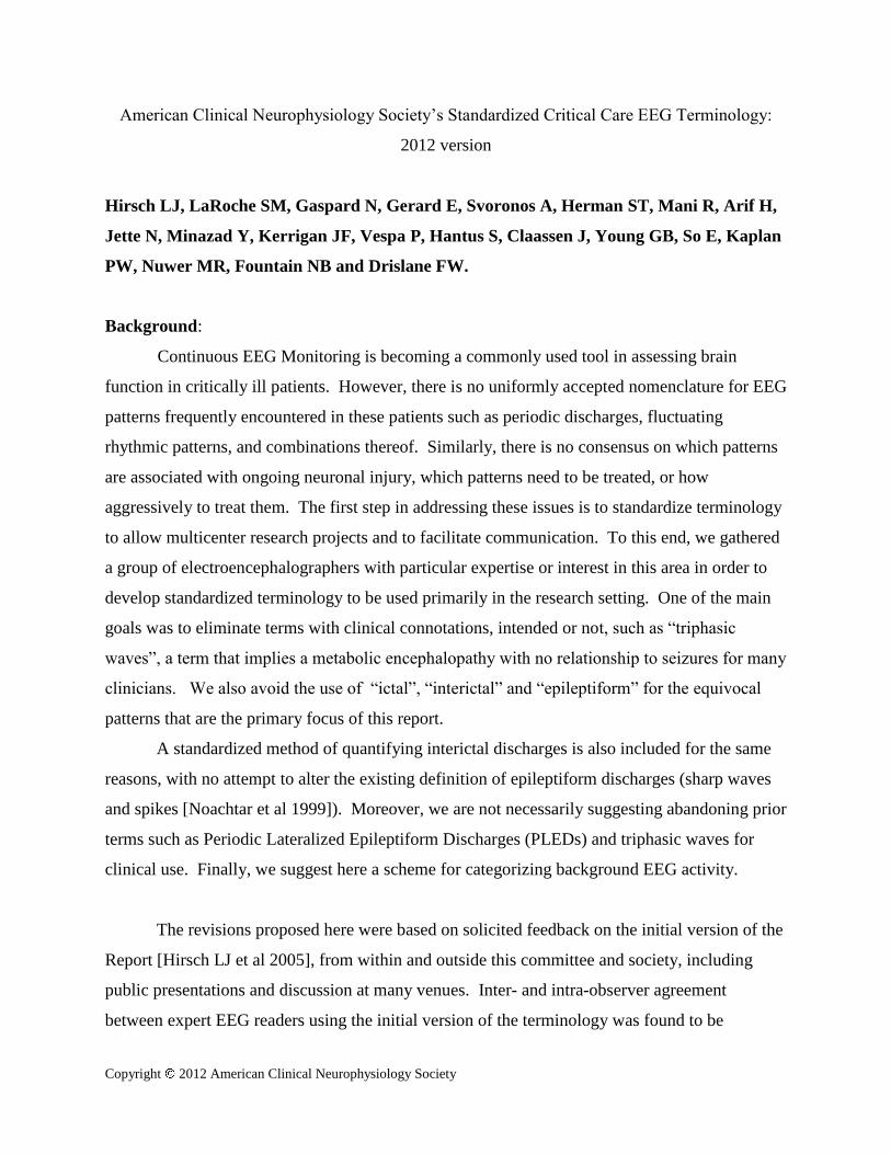
American Clinical Neurophysiology Society’s Standardized Critical Care EEG Terminology:
2012 version
Hirsch LJ, LaRoche SM, Gaspard N, Gerard E, Svoronos A, Herman ST, Mani R, Arif H,
Jette N, Minazad Y, Kerrigan JF, Vespa P, Hantus S, Claassen J, Young GB, So E, Kaplan
PW, Nuwer MR, Fountain NB and Drislane FW.
Background:
Continuous EEG Monitoring is becoming a commonly used tool in assessing brain
function in critically ill patients. However, there is no uniformly accepted nomenclature for EEG
patterns frequently encountered in these patients such as periodic discharges, fluctuating
rhythmic patterns, and combinations thereof. Similarly, there is no consensus on which patterns
are associated with ongoing neuronal injury, which patterns need to be treated, or how
aggressively to treat them. The first step in addressing these issues is to standardize terminology
to allow multicenter research projects and to facilitate communication. To this end, we gathered
a group of electroencephalographers with particular expertise or interest in this area in order to
develop standardized terminology to be used primarily in the research setting. One of the main
goals was to eliminate terms with clinical connotations, intended or not, such as “triphasic
waves”, a term that implies a metabolic encephalopathy with no relationship to seizures for many
clinicians. We also avoid the use of “ictal”, “interictal” and “epileptiform” for the equivocal
patterns that are the primary focus of this report.
A standardized method of quantifying interictal discharges is also included for the same
reasons, with no attempt to alter the existing definition of epileptiform discharges (sharp waves
and spikes [Noachtar et al 1999]). Moreover, we are not necessarily suggesting abandoning prior
terms such as Periodic Lateralized Epileptiform Discharges (PLEDs) and triphasic waves for
clinical use. Finally, we suggest here a scheme for categorizing background EEG activity.
The revisions proposed here were based on solicited feedback on the initial version of the
Report [Hirsch LJ et al 2005], from within and outside this committee and society, including
public presentations and discussion at many venues. Inter- and intra-observer agreement
between expert EEG readers using the initial version of the terminology was found to be
Copyright 2012 American Clinical Neurophysiology Society
moderate for major terms but only slight to fair for modifiers. [Gerber PA et al 2008] A second
assessment was performed on an interim version after extensive changes were introduced. This
assessment showed a significant improvement with an inter-rater agreement superior to 95% for
main terms and superior to 80% for the evaluated modifiers (amplitude, frequency and plus
modifiers). [Mani R et al 2012] Minor modifications were introduced after this second
assessment to further improve reliability.
Objective: To standardize terminology of periodic and rhythmic EEG patterns in the critically
ill in order to aid future research involving such patterns. Our goal is to avoid terms with clinical
connotations and to define terms thoroughly enough to ensure adequate inter-rater reliability.
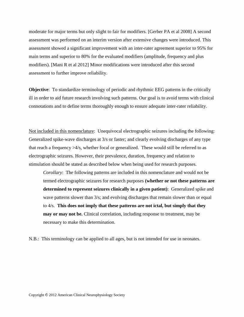
Not included in this nomenclature: Unequivocal electrographic seizures including the following:
Generalized spike-wave discharges at 3/s or faster; and clearly evolving discharges of any type
that reach a frequency >4/s, whether focal or generalized. These would still be referred to as
electrographic seizures. However, their prevalence, duration, frequency and relation to
stimulation should be stated as described below when being used for research purposes.
Corollary: The following patterns are included in this nomenclature and would not be
termed electrographic seizures for research purposes (whether or not these patterns are
determined to represent seizures clinically in a given patient): Generalized spike and
wave patterns slower than 3/s; and evolving discharges that remain slower than or equal
to 4/s. This does not imply that these patterns are not ictal, but simply that they
may or may not be. Clinical correlation, including response to treatment, may be
necessary to make this determination.
N.B.: This terminology can be applied to all ages, but is not intended for use in neonates.
Copyright 2012 American Clinical Neurophysiology Society
PROPOSED NOMENCLATURE
A. RHYTHMIC OR PERIODIC PATTERNS
All terms consist of main term # 1 followed by #2, with modifiers added as appropriate.
Main term 1: G, L, BI, or Mf:
Generalized (G; for this purpose, the term “generalized” refers to any bilateral,
bisynchronous and symmetric pattern, even if it has a restricted field [e.g. bifrontal])
Lateralized (L; includes unilateral and bilateral synchronous but asymmetric;
includes focal, regional and hemispheric patterns)
Bilateral Independent (BI; refers to the presence of 2 independent [asynchronous]
lateralized patterns, one in each hemisphere)
Multifocal (Mf; refers to the presence of at least three independent lateralized
patterns with at least one in each hemisphere)
a. Additional localizing information
i. For Generalized patterns
1. Frontally predominant (defined as having an amplitude in anterior
derivations that is at least 50% greater than that in posterior
derivations on an ipsilateral ear, average, or non-cephalic
referential recording),
2. Occipitally predominant (defined as having an amplitude in
posterior derivations that is at least 50% greater than that in
anterior derivations on an ipsilateral ear, average, or non-cephalic
referential recording),
3. Midline predominant (defined as having an amplitude in midline
derivations that is at least 50% greater than in parasagittal
derivations on an average or non-cephalic referential recording),
4. Generalized, not otherwise specified.
ii. For Lateralized patterns
1. Specify unilateral vs. bilateral asymmetric
Copyright 2012 American Clinical Neurophysiology Society
a. Patterns that are purely unilateral are termed “Lateralized,
unilateral”.
b. Patterns seen bilaterally and synchronous but clearly more
prominent on one side would be called “Lateralized,
bilateral asymmetric”
2. Specify lobe(s) most involved (F, P, T, O, or hemispheric if more
specific localization is not possible)
iii. For Bilateral Independent and Multifocal patterns:
1. Specify symmetric vs. asymmetric
a. Patterns that are bilateral and asynchronous but clearly
more prominent on one side would be called “Bilateral
Independent, asymmetric”, or “Multifocal, asymmetric”
b. Patterns that are bilateral, asynchronous and symmetric
would be called “Bilateral Independent, symmetric”, or
“Multifocal, symmetric”
2. Specify lobes most involved in both hemispheres (F, P, T, O, or
hemispheric if more specific localization is not possible).
Copyright 2012 American Clinical Neurophysiology Society
Main Term 2: PDs, RDA or SW:
Periodic Discharges (PDs): Periodic = repetition of a waveform with relatively uniform
morphology and duration with a quantifiable inter-discharge interval between
consecutive waveforms and recurrence of the waveform at nearly regular intervals.
Discharges: These are defined as waveforms with no more than 3 phases (i.e. crosses the
baseline no more than twice) or any waveform lasting 0.5 seconds or less, regardless of
number of phases. This is as opposed to bursts, defined as waveforms lasting more than
0.5 seconds and having at least 4 phases (i.e. crosses the baseline at least 3 times).
o “Nearly regular intervals” is defined as having a cycle length (i.e., period)
varying by <50% from one cycle to the next in the majority (>50%) of cycle
pairs.
Rhythmic Delta Activity (RDA): Rhythmic = repetition of a waveform with relatively
uniform morphology and duration, and without an interval between consecutive
waveforms. RDA = rhythmic activity < 4 Hz. The duration of one cycle (i.e., the period)
of the rhythmic pattern should vary by <50% from the duration of the subsequent cycle
for the majority (>50%) of cycle pairs to qualify as rhythmic.
Spike-and-wave or Sharp-and-wave (SW) = polyspike, spike or sharp wave
consistently followed by a slow wave in a regularly repeating and alternating pattern
(spike-wave-spike-wave-spike-wave), with a consistent relationship between the spike
(or polyspike or sharp wave) component and the slow wave; and with no interval between
one spike-wave complex and the next (if there is an interval, this would qualify as PDs,
where each discharge is a spike-and- wave).
NOTE 1: A pattern can qualify as rhythmic or periodic as long as it continues for at least
6 cycles (e.g. 1/s for 6 seconds, or 3/s for 2 seconds).
NOTE 2: If a pattern qualifies as both GPDs and RDA simultaneously, it should be
coded as GPDs+ rather than RDA+ (see modifier 8 below for description of “+”).
Copyright 2012 American Clinical Neurophysiology Society
Most of the following sections can be applied to research on any EEG phenomenon.
Although many of the categorizations are arbitrary, our hope is that standardization will allow
systematic, scientific and collaborative investigation of these EEG features.
Modifiers:
1. Prevalence: Specify percent of record or epoch that includes the pattern (see section
B below). This should be based on the percent of seconds that include or are within
the pattern. The time between widely spaced periodic discharges counts as part of the
pattern duration. For example, 2Hz LPDs present for 1 minute every 10 minutes is
10% prevalence, and a 0.25 Hz. pattern present for 1 minute every 10 minutes is also
10% prevalence. When categorizing or using qualitative terms, follow the cutoffs
listed below for each term. Suggested equivalent clinical terms are given as well. If
2 or more patterns are equally or almost equally prevalent (e.g. ~30% GRDA, 30%
GPDs, and 40% BIPDs) record the presence and prevalence of each one.
a. >90% of record/epoch (“Continuous”)
b. 50-89% of record/epoch (“Abundant”)
c. 10-49% of record/epoch (“Frequent”)
d. 1-9% of record/epoch (“Occasional”)
e. <1% of record/epoch (“Rare”)
2. Duration: Specify typical duration of pattern if not continuous. When categorizing
or using qualitative terms, follow the cutoffs listed below for each term. Also record
the longest continuous duration.
a. >1 hour (“Very long”)
b. 5-59 minutes (“Long”)
c. 1-4.9 minutes (“Intermediate duration”)
d. 10-59 seconds (“Brief”)
e. <10 seconds (“Very brief”)
3. Frequency = rate per second: Specify typical rate and range for all patterns, e.g., 1/s
and 0.5-2/s;
Copyright 2012 American Clinical Neurophysiology Society
When categorizing, use the following (record typical, minimum and maximum
frequency).
<0.5/s, 0.5/s, 1/s, 1.5/s, 2/s, 2.5/s, 3/s, 3.5/s and >4/s
4. Number of phases = number of baseline crossings of the typical discharge (in
longitudinal bipolar and in the channel in which it is most readily appreciated).
Applies to PDs and the entire spike-and-wave or sharp-and-wave complex of SW
(include the slow wave). This does not apply to RDA. Categorize as follows:
a. 1, 2, 3 or >3.
5. Sharpness: Specify for both the predominant phase (phase with greatest amplitude)
and the sharpest phase if different. For both phases, describe the typical discharge.
Applies only to PDs and the spike/sharp component of SW, not RDA. Categorize as one
of the following:
a. Spiky (duration of that component, measured at the EEG baseline, is <70 ms)
b. Sharp (duration of that component is 70-200 ms)
c. Sharply contoured: used for theta or delta waves that have a sharp wave
morphology (steep slope to one side of the wave and/or pointy at inflection point[s]),
but are too long in duration to qualify as a sharp wave.
d. Blunt: having smooth or sinusoidal morphology.
6. Amplitude:
a. Absolute: Typical amplitude measured in standard longitudinal bipolar 10-20
recording in the channel in which the pattern is most readily appreciated. For PDs, this
refers to the highest amplitude component. For SW, this refers to the spike/sharp wave.
Amplitude should be measured from peak to trough (not peak to baseline). Specify for
RDA as well.
Categorize amplitude as:
i. <20 µV (“very low”)
ii. 20-49 µV (“low”)
ii. 50-199 µV (“medium”)
Copyright 2012 American Clinical Neurophysiology Society
iii. >200 µV (“high”)
b. Relative: For PDs only (PDs require 2 amplitudes, absolute and relative). Typical
ratio of amplitude of the highest amplitude component to the amplitude of the typical
background between discharges, measured in the same channel and montage as absolute
amplitude. Categorize as <2 or >2.
7. Polarity: Specify for the predominant phase (phase with the greatest amplitude) only.
Should be determined in a referential montage. Describe the typical discharge. Applies
only to PDs and the spike/sharp component of SW, not RDA. Categorize as one of the
following:
a. Positive
b. Negative
c. Dipole, horizontal/tangential
d. Unclear
8. Stimulus-Induced (SI) = reproducibly brought about by an alerting stimulus, with or
without clinical alerting; may also be seen spontaneously. If never clearly induced by
stimulation, report as spontaneous. If unknown, unclear or untested, report as
“unknown”. Specify type of stimulus (auditory, light tactile, patient care and other non-
noxious stimulations, suction, sternal rub, nostril tickle or other noxious stimulations).
9. Evolving OR Fluctuating: both terms refer to changes in either frequency, location or
morphology. If neither term applies, report as static.
Evolving is defined as follows: at least 2 unequivocal, sequential changes in frequency,
morphology or location defined as follows: Evolution in frequency is defined as at least 2
consecutive changes in the same direction by at least 0.5/s, e.g. from 2 to 2.5 to 3/s, or
from 3 to 2 to 1.5/s; Evolution in morphology is defined as at least 2 consecutive changes
to a novel morphology; Evolution in location is defined as sequentially spreading into or
sequentially out of at least two different standard 10-20 electrode locations.
In order to qualify as present, a single frequency or location must persist at
least 3 cycles (e.g. 1/s for 3 seconds, or 3/s for 1 second). Thus, the following
Copyright 2012 American Clinical Neurophysiology Society
pattern would qualify as evolving: 3/s for > 1 second, then 2/s for > 1.5
seconds (the first change), then 1.5/s for > 2 seconds (the 2nd change). To
qualify as evolution in morphology, each different morphology or each
morphology plus its transitional forms must last at least 3 cycles. Thus the
following examples would both qualify as evolving in morphology:
- spiky 4-phase PDs for 3 cycles then sharp 2-3 phase PDs for 3 cycles then
blunt diphasic PDs for 3 cycles
- 1 blunt triphasic PD then 2 blunt biphasic PDs then 2 sharply contoured
biphasic PDs then 2 sharp biphasic PDs then 3 sharp monophasic PDs.
The criteria for evolution must be reached without the pattern remaining
unchanged in frequency, morphology or location for 5 or more minutes. Thus,
the following pattern would not qualify as evolving: 3/s for 1 minute, then 2/s
for 7 minutes, then 1.5/s for 2 minutes.
Fluctuating is defined as follows: >3 changes, not more than one minute apart, in
frequency (by at least 0.5/s), >3 changes in morphology, or >3 changes in location (by at
least 1 standard inter-electrode distance), but not qualifying as evolving. This includes
patterns fluctuating from 1 to 1.5 to 1 to 1.5/s; spreading in and out of a single electrode
repeatedly; or alternating between 2 morphologies repeatedly.
The following would not qualify as fluctuating: 2/s for 30 seconds, then 1.5/s
for 30 seconds, then 2/s for 3 minutes, then 1.5/s for 30 seconds, then 2/s for 5
minutes. The changes are too far apart (>1 minute).
The following would qualify as fluctuating: 2/s for 10 seconds, then 2.5/s for
30 seconds, then 2/s for 5 seconds, then 2.5/s for 5 seconds.
Change in amplitude alone would not qualify as evolving or fluctuating.
a. For data entry, if evolving or fluctuating, a minimum and maximum frequency
should be specified under the “frequency” modifier above. For non-generalized
patterns, specify degree of spread (none, unilateral, or bilateral).
10. Plus (+) = additional feature which renders the pattern more ictal-appearing than the
usual term without the plus. (Does not apply to SW)
Copyright 2012 American Clinical Neurophysiology Society
Periodic discharges (PDs): includes superimposed fast activity (theta or faster,
rhythmic or not) with each discharge (+F), or superimposed rhythmic or quasi-rhythmic
delta activity (+R).
Rhythmic delta activity (RDA): includes superimposed fast activity (+F) or
frequent intermixed sharp waves or spikes (+S; “frequent” is defined as more than one
sharp wave or spike every 10 seconds, but not periodic and not SW) or RDA that is
sharply contoured (also +S). If absent, indicate as “no +”.
a. Subtyping of “+”: all cases with “+” should be subtyped as follows into
+F, +R, +FS, or +FR:
i. “+F”: superimposed fast activity. Can be used with PDs or RDA.
ii. “+R”: superimposed rhythmic or quasi-rhythmic delta activity; applies to
PDs only.
iii. “+S”: superimposed sharp waves or spikes, or sharply contoured; applies
to RDA only.
iv. It is possible to have “+FR” for PDs, or “+FS” for RDA
NOTE 3: Re: Bilateral “+” vs. unilateral: If a pattern is bilateral and qualifies as plus on
one side, but not on the other, the overall main term should include the plus (even though
one side does not warrant a plus). For example, bilateral independent periodic discharges
with fast activity superimposed in one hemisphere only (PD on one side, and PD+F on
the other) would qualify for BIPDs+F. Similarly, generalized rhythmic delta activity
with superimposed spikes in one hemisphere only (RDA on one side and RDA+S on the
other) would qualify for GRDA+S.
NOTE 4: Re: +F: If a pattern qualifying as RDA or PDs has superimposed continuous
fast frequencies (theta or faster), this can and should be coded as +F if the fast activity is
not present in the background activity when the RDA or PDs is not present. In other
words, code as +F if the superimposed fast activity is part of the RDA or PD pattern and
not simply part of the background activity.
Copyright 2012 American Clinical Neurophysiology Society
Minor Modifiers:
1. Quasi-: Used to modify rhythmic or periodic, as in quasi-periodic or quasi-rhythmic.
(Quasi preferred over pseudo- or semi-). This distinction between quasi- and not
quasi is to be applied only if determined by quantitative computer analysis (not by
visual impression). Quasi is defined as having a cycle length (i.e., period) varying by
25-50% from one cycle to the next in the majority (>50%) of cycle pairs. If >50%
variation in the majority of cycles, the pattern would not qualify as rhythmic or
periodic and would not be included in this nomenclature. If the variation is <25%,
the modifier quasi- would not be appropriate. When not using computer analysis,
quasiperiodic is coded as periodic, and quasirhythmic as rhythmic.
2. Sudden onset OR gradual onset (sudden onset preferred over paroxysmal). Sudden
onset is defined as progressing from absent to well developed within 3 seconds.
3. “Triphasic” morphology: Applies to PDs and SW. Either two or three phases, with
each phase longer than the previous, and the positive phase of highest amplitude. If
three phases, this must be negative-positive-negative in polarity; if two phases,
positive-negative. Note that a biphasic waveform may be categorized as “triphasic”
by this definition.
4. Anterior-posterior lag or posterior-anterior lag: Applies if a consistent measurable
delay of > 100 ms exists from the most anterior to the most posterior derivation in
which is seen; specify typical delay in ms from anterior to posterior (negative =
posterior to anterior) in both a longitudinal bipolar and a referential montage,
preferably with an ipsilateral ear reference.
B. Minimal time epochs to be reported documented separately:
1. First ~30 minutes (equivalent to a “routine” EEG).
2. Each 24 hour period.
If significant changes occur in the record during this time period, report
additional epochs separately as needed.
Copyright 2012 American Clinical Neurophysiology Society
C. Quantification and categorization of sporadic (non-rhythmic and non-periodic)
epileptiform discharges (includes sharp waves and spikes as previously defined [Noachtar et al
1999]).
>1/10s, but not periodic (“Abundant”)
>1/min but less than 1/10s (“Frequent”)
> 1/h but less than 1/min (“Occasional”)
<1/h (“Rare”)
______________________________________________________________________
D. Background EEG:
Symmetry: 1. Symmetric; 2. Mild asymmetry (consistent asymmetry in amplitude on referential
recording of <50%, or consistent asymmetry in frequency of 0.5 - 1 Hz); 3. Marked asymmetry
(>50% amplitude or >1 Hz frequency asymmetry).
Breach effect (note presence, absence, or unclear)
When any of the following features are asymmetric, they should be
described separately for each hemisphere.
Posterior dominant “alpha” rhythm: Must be demonstrated to attenuate with eye opening.
Specify frequency (to the nearest 0.5 Hz) or absence.
Predominant background EEG frequency: Delta, Theta, and/or >Alpha (including beta). If 2 or
3 frequency bands are equally prominent, record each one.
Anterior-posterior (AP) gradient: Present, absent or reverse. An AP gradient is present if at any
point in the epoch, there is a clear and persistent (at least 1 continuous minute) anterior to
posterior gradient of voltages and frequencies such that lower amplitude, faster frequencies are
seen in anterior derivations, and higher amplitude, slower frequencies are seen in posterior
derivations A reverse AP gradient is defined identically but with a posterior to anterior gradient
of voltages and frequencies.”

Copyright 2012 American Clinical Neurophysiology Society
Variability: Yes, No, or Unknown/unclear/not applicable. The last choice might apply, for
example, in a 30 minute wake record.
Reactivity: Change in cerebral EEG activity to stimulation: Yes, No, or Unclear/unknown/not
applicable. This may include change in amplitude or frequency, including attenuation of
activity. Strength and/or nature of stimulus should be noted. Appearance of muscle activity or
eye blink artifacts does not qualify as reactive.
If the only form of reactivity is SI-RDA, SI-PDs or SI-seizures, categorize as “Reactive,
SIRPIDs only”)
Voltage: 1. Normal; 2. Low (most or all activity <20 µV in longitudinal bipolar with standard
10-20 electrodes, [measured from peak to trough]); or 3. Suppressed (all activity <10 µV). If
the background is discontinuous, this refers to the higher amplitude portion.
Stage II sleep transients (K-complexes and spindles): 1. Normal (K-complexes and spindles both
present and normal); 2. Present (at least one) but abnormal; or 3. Absent (both absent).
Continuity:
1. Continuous.
2. Nearly Continuous: continuous, but with occasional (<10% of the record) periods of
attenuation or suppression. Describe typical duration of attenuation/suppression as
above.
a. Nearly continuous with attenuation: periods of lower voltage are >10µV but
<50% of the background voltage.
b. Nearly continuous with suppression: periods of lower voltage are <10 µV;
c. If suppressions/attenuations are stimulus-induced, code as “nearly continuous with
SI-attenuation” or “…with SI-suppression”;
3. Discontinuous: 10-49% of the record consisting of attenuation or suppression, as defined
above.
Copyright 2012 American Clinical Neurophysiology Society
4. Burst-attenuation/Burst-suppression: more than 50% of the record consisting of
attenuation or suppression, as defined above, with bursts alternating with attenuation or
suppression; specify the following:
a. Typical duration of bursts and interburst intervals;
b. Sharpest component of a typical burst using the sharpness categories defined
above under modifiers;
c. Presence or absence of Highly Epileptiform Bursts: Present if multiple
epileptiform discharges (traditional definition) are seen within the majority
(>50%) of bursts and occur at an average of 1/s or faster; record typical frequency
(using categories above) and location (G, L, BI or Mf). Also present if a
rhythmic, potentially ictal-appearing pattern occurs at 1/s or faster within the
majority (>50%) of bursts; record frequency and location as well.
5. Suppression: entirety of the record consisting of suppression (<10 µV, as defined above).
NOTE 5: Bursts must average more than 0.5 seconds; if shorter, they should be
considered single discharges (as defined above under main term 2). Bursts within burst-
suppression or burst-attenuation can last up to 30 seconds.
E. Other Terms for Research Use:
“Daily Pattern Duration” is defined as total duration of a pattern per 24 hours. e.g. if
GPDs were present for 33% of the record for 12 hours, then 10% of the record for 12 hours, the
Daily GPD Duration would be 4 hours + 1.2 hours = 5.2 hours. Daily Seizure Duration can be
calculated similarly: e.g. six 30-second seizures in one day would have a Daily Seizure Duration
of 3minutes.
“Daily Pattern Index” is defined as Daily Duration X Mean Frequency (Hz). In the above
example, if GPDs were at 1.5 Hz, the Daily GPD Index would be 5.2 h x 1.5 Hz = 7.8 Hz-hours.
Examples of appropriate terms:
Continuous 1-2/s fluctuating GPDs
Occasional 30-60 second periods of 1.5/s SI-LRDA
Abundant 1-3 minute periods of 0.5-1.5/s LPDs+F
Occasional 10-second periods of 1/s BIPDs
Other examples of corresponding new terms for older terms (some could have alternative new
terms depending on exact pattern):
Copyright 2012 American Clinical Neurophysiology Society
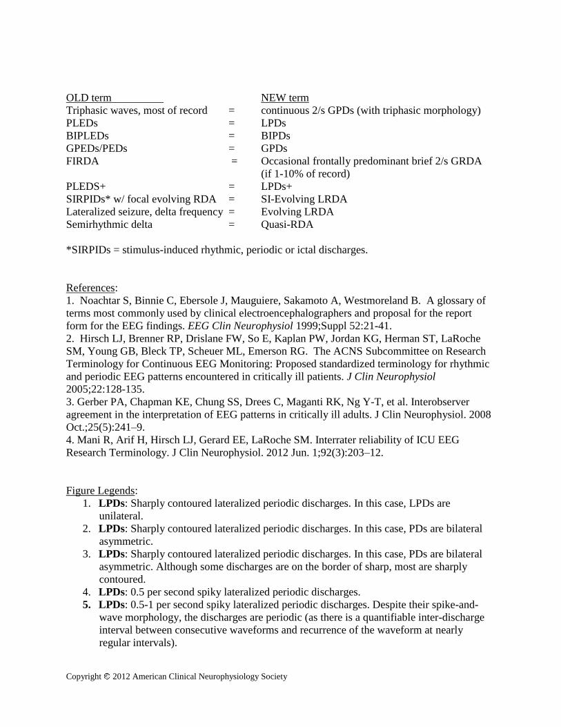
OLD term NEW term
Triphasic waves, most of record = continuous 2/s GPDs (with triphasic morphology)
PLEDs = LPDs
BIPLEDs = BIPDs
GPEDs/PEDs = GPDs
FIRDA = Occasional frontally predominant brief 2/s GRDA
(if 1-10% of record)
PLEDS+ = LPDs+
SIRPIDs* w/ focal evolving RDA = SI-Evolving LRDA
Lateralized seizure, delta frequency = Evolving LRDA
Semirhythmic delta = Quasi-RDA
*SIRPIDs = stimulus-induced rhythmic, periodic or ictal discharges.
References:
1. Noachtar S, Binnie C, Ebersole J, Mauguiere, Sakamoto A, Westmoreland B. A glossary of
terms most commonly used by clinical electroencephalographers and proposal for the report
form for the EEG findings. EEG Clin Neurophysiol 1999;Suppl 52:21-41.
2. Hirsch LJ, Brenner RP, Drislane FW, So E, Kaplan PW, Jordan KG, Herman ST, LaRoche
SM, Young GB, Bleck TP, Scheuer ML, Emerson RG. The ACNS Subcommittee on Research
Terminology for Continuous EEG Monitoring: Proposed standardized terminology for rhythmic
and periodic EEG patterns encountered in critically ill patients. J Clin Neurophysiol
2005;22:128-135.
3. Gerber PA, Chapman KE, Chung SS, Drees C, Maganti RK, Ng Y-T, et al. Interobserver
agreement in the interpretation of EEG patterns in critically ill adults. J Clin Neurophysiol. 2008
Oct.;25(5):241–9.
4. Mani R, Arif H, Hirsch LJ, Gerard EE, LaRoche SM. Interrater reliability of ICU EEG
Research Terminology. J Clin Neurophysiol. 2012 Jun. 1;92(3):203–12.
Figure Legends:
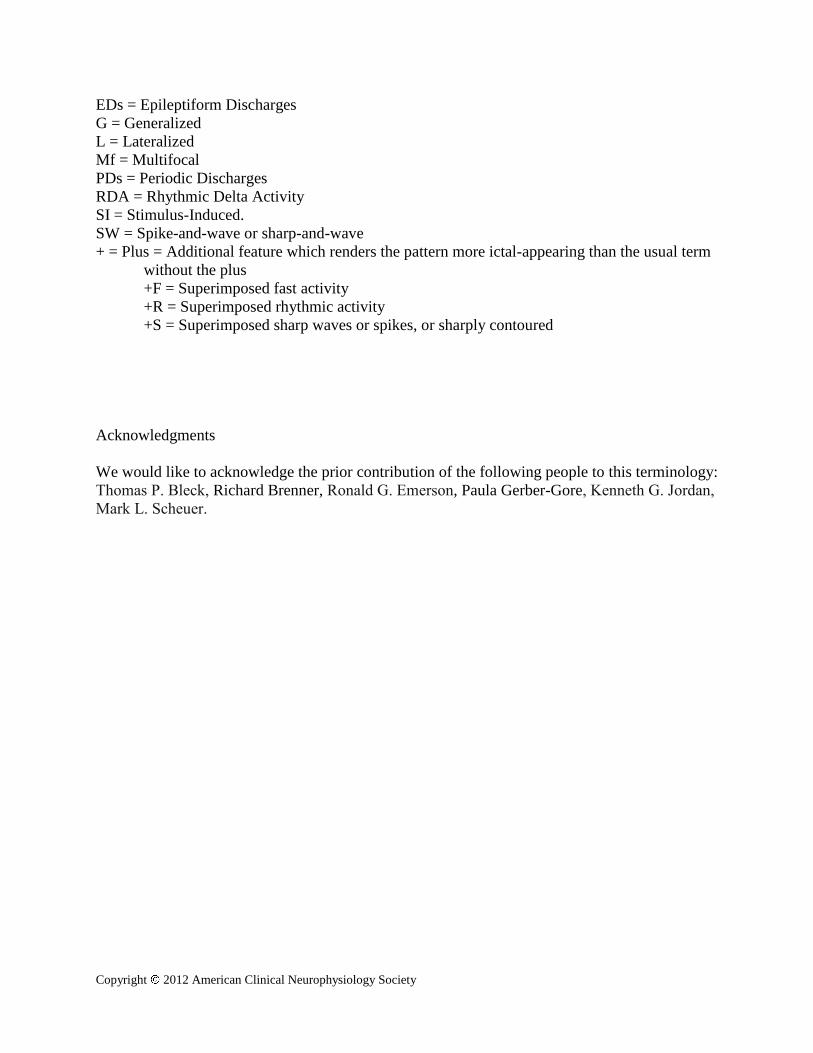
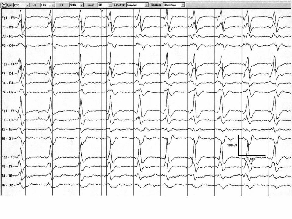
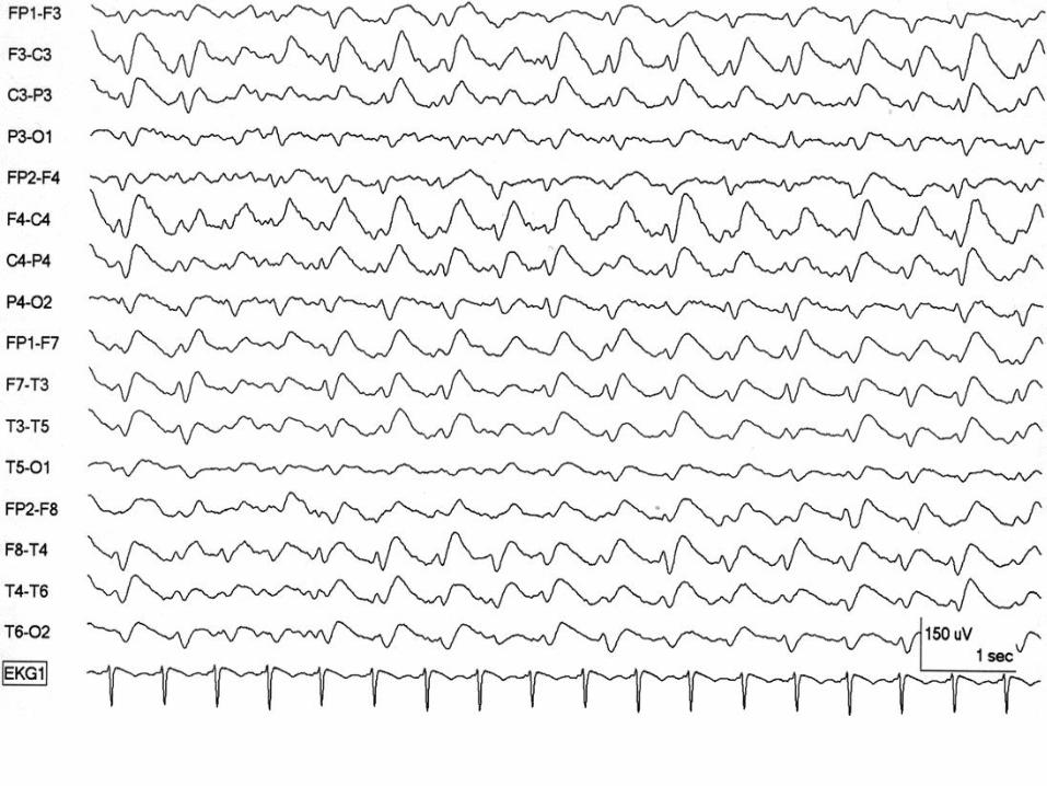
1. LPDs: Sharply contoured lateralized periodic discharges. In this case, LPDs are
unilateral.
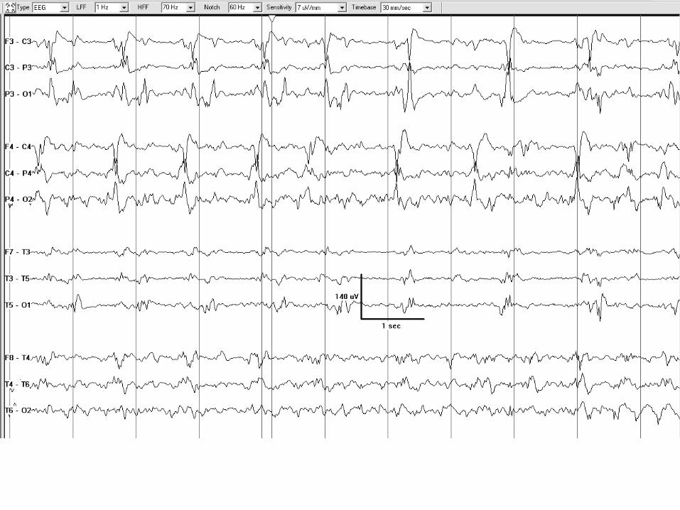
2. LPDs: Sharply contoured lateralized periodic discharges. In this case, PDs are bilateral
asymmetric.
3. LPDs: Sharply contoured lateralized periodic discharges. In this case, PDs are bilateral
asymmetric. Although some discharges are on the border of sharp, most are sharply
contoured.
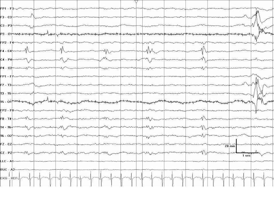
4. LPDs: 0.5 per second spiky lateralized periodic discharges.
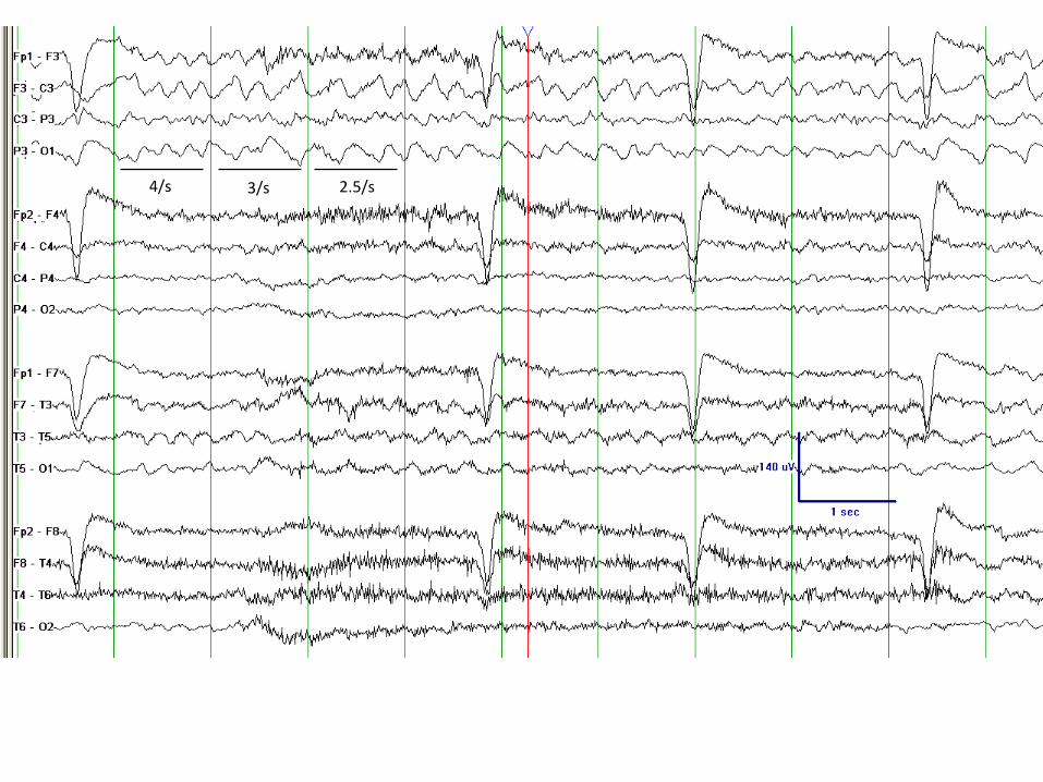
5. LPDs: 0.5-1 per second spiky lateralized periodic discharges. Despite their spike-and-
wave morphology, the discharges are periodic (as there is a quantifiable inter-discharge
interval between consecutive waveforms and recurrence of the waveform at nearly
regular intervals).

Copyright 2012 American Clinical Neurophysiology Society
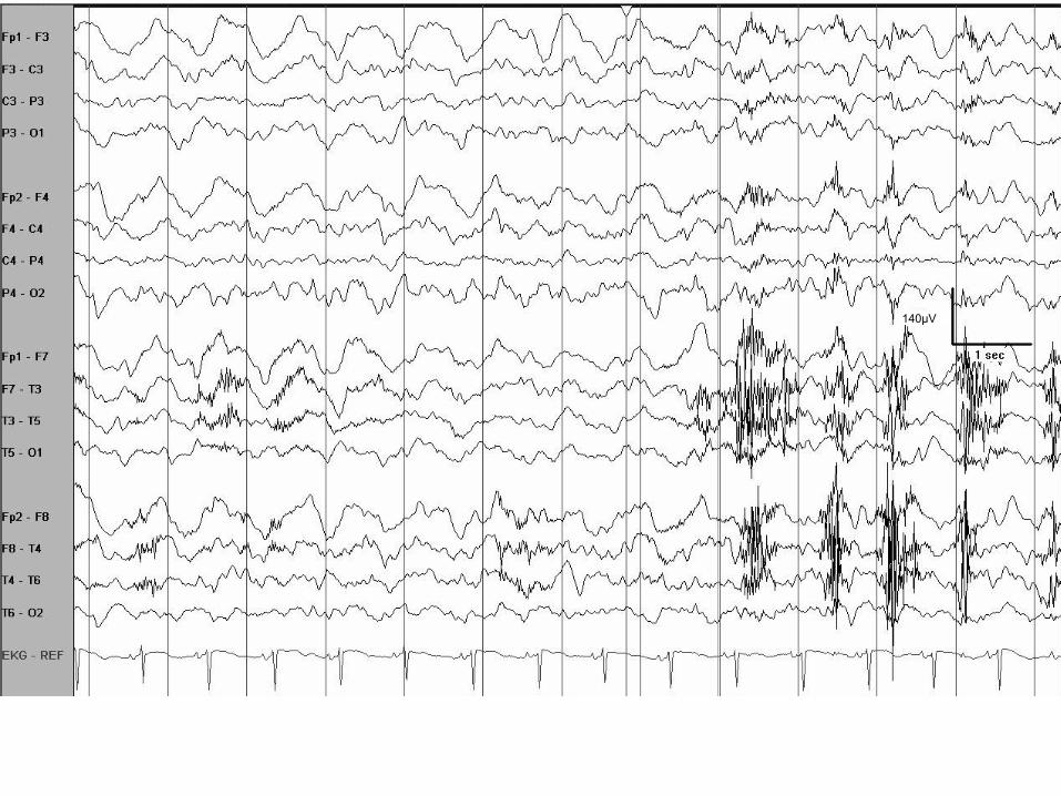
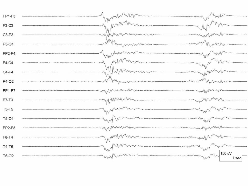
6. LPDs+F: 0.5 to 1 per second spiky LPDs with superimposed burst of low amplitude fast
activity (highlighted in boxes).
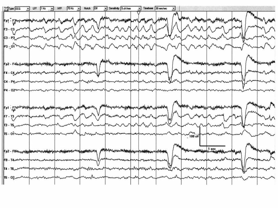
7. LPDs+R: Irregular (in morphology and repetition rate) 0.5-1 per second quasi-periodic
discharges with superimposed quasi-rhythmic delta activity in the right hemisphere with
occasional spread to the left. Less “stable” pattern and more ictal-appearing than LPDs
alone; compare with Figure 1.
8. Fluctuating LPDs: Lateralized periodic discharges that fluctuate in frequency between
0.5 and 1 per second.
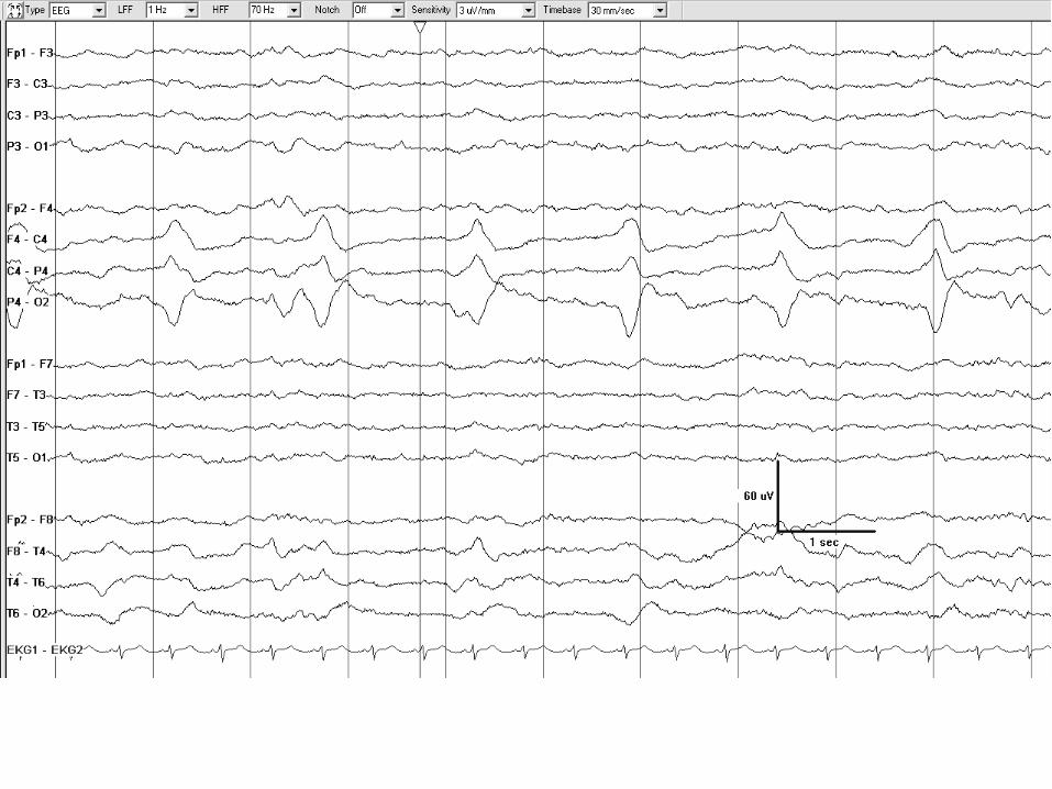
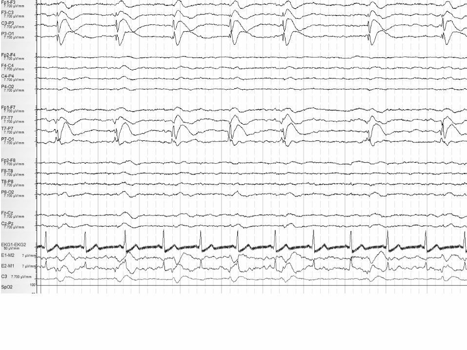
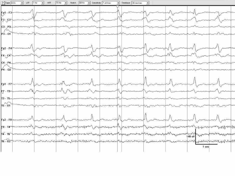
9. GPDs: One per second sharp generalized periodic discharges.
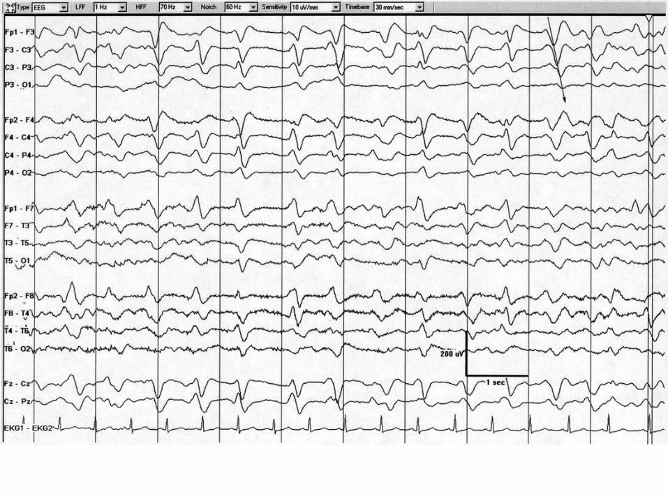
10. GPDs with triphasic morphology and A-P lag: Generalized periodic discharges at just
under 1.5 per second. In this case there is also a triphasic morphology and an anterior-
posterior lag, highlighted with the diagonal line in the upper right of the figure.
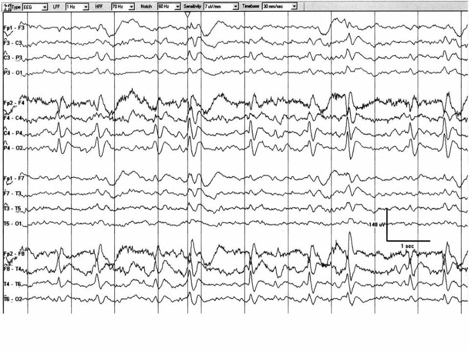
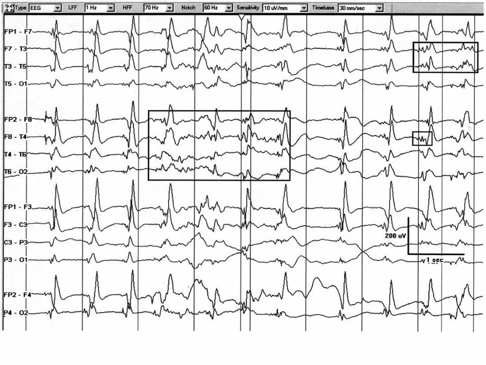
11. GPDs+F: 1-1.25 per second sharp GPDs with superimposed low amplitude quasi-
rhythmic sharp activity (highlighted in boxes).
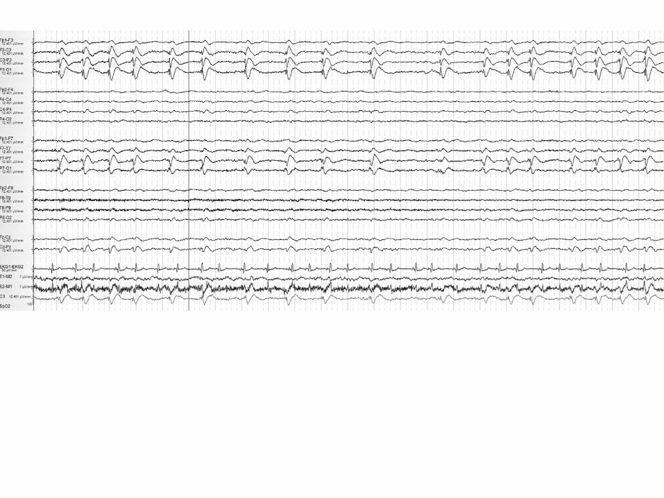
12. GPDs: One per second generalized periodic discharges, characterized by a marked
frontal predominance and a sharp morphology. Despite background attenuation, the
discharges last less than 500ms and thus do not qualify as bursts.
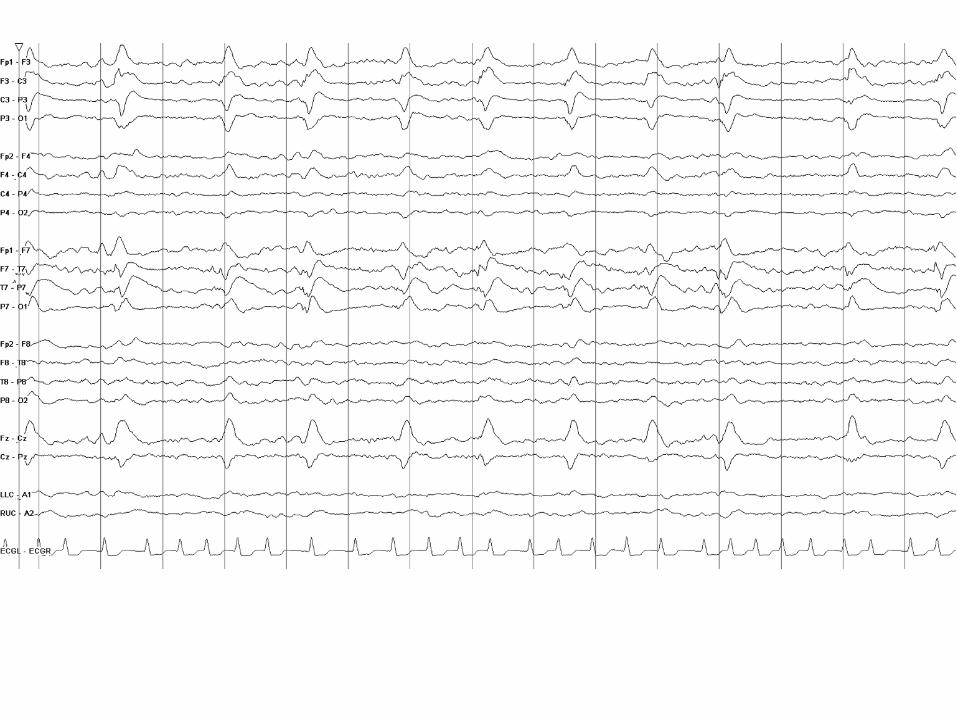
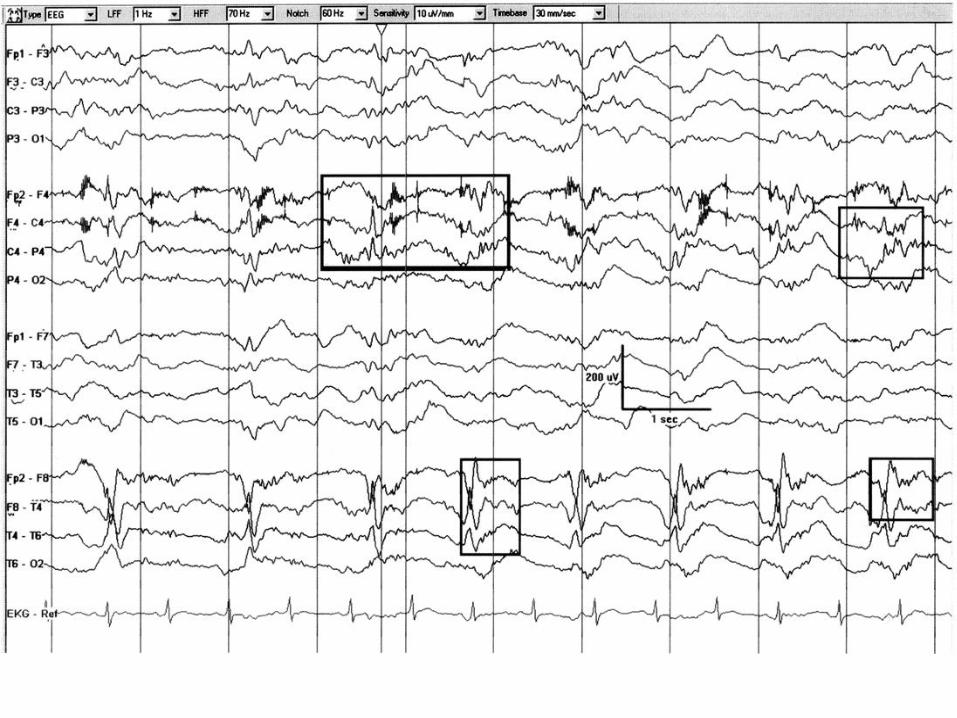
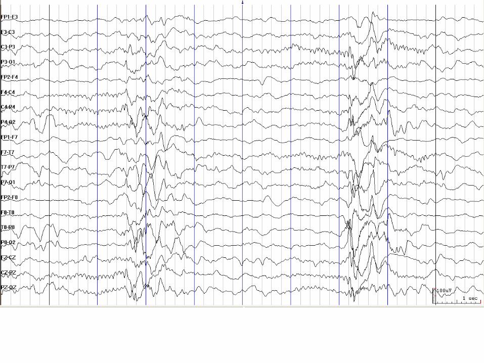
13. BIPDs+F: Bilateral independent periodic discharges at 0.5-1 per second, most prominent
centroparietally on both sides. The periodic discharges have a sharp morphology and are
associated with low amplitude sharply contoured quasi-rhythmic fast activity, especially
posteriorly, and more prominent on the right where the fast activity is nearly continuous.
14. GRDA: Generalized rhythmic delta activity, frontally predominant.
15. SI-GRDA: Stimulus-induced generalized rhythmic delta activity. In this case, the
pattern was elicited by suctioning the patient.
16. Evolving LRDA: Lateralized rhythmic delta activity that evolves in morphology and
frequency. It begins as low voltage sharply contoured 1.5 Hz delta in the left parasagittal
region, evolves to 3 Hz rhythmic delta, then again slows.
17. Evolving LRDA: Lateralized rhythmic delta activity that evolves in frequency and
morphology from a 4 per second blunt RDA to a 2.5 per second sharply contoured RDA.
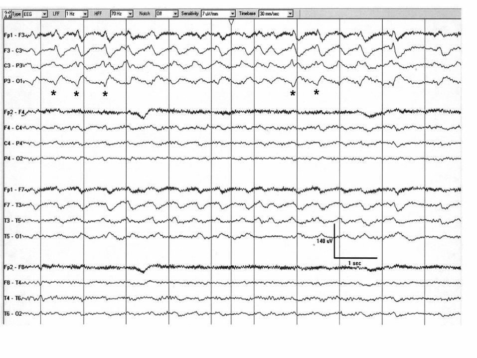
18. LRDA+S: Two per second lateralized rhythmic delta activity with superimposed
repetitive sharp waves (several marked with asterisks). The superimposed low amplitude
fast activity is also present on the right hemisphere and should not be recorded as +F.
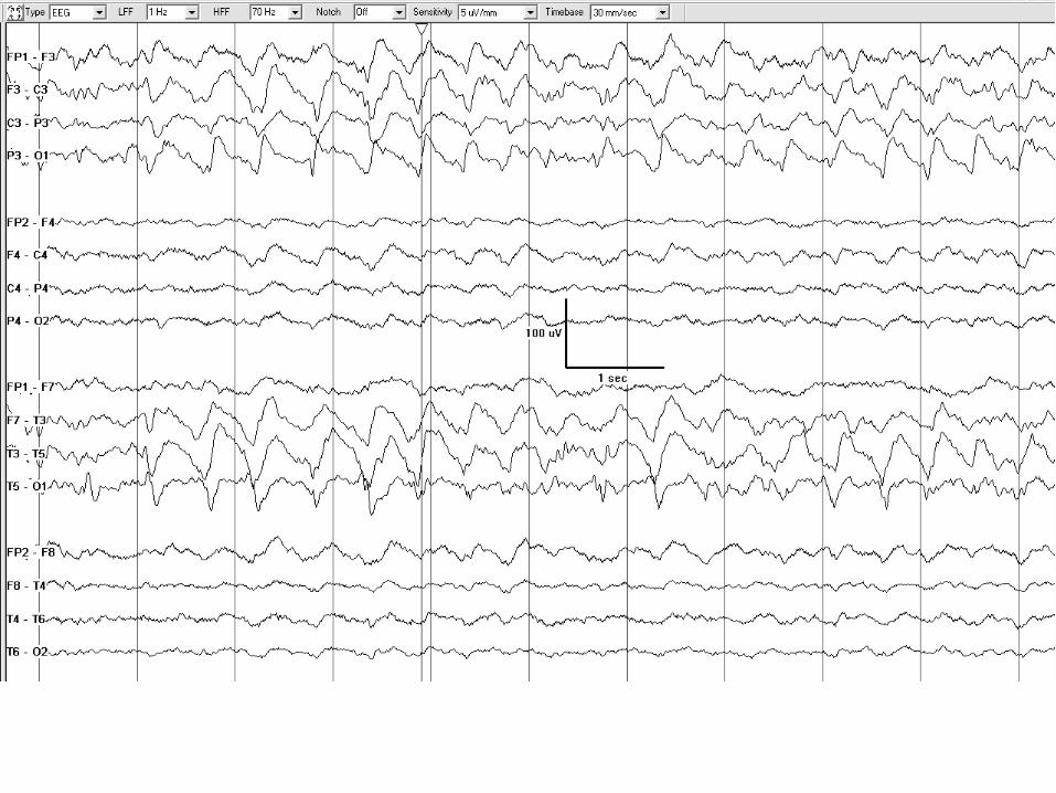
19. LRDA+S: Two per second lateralized rhythmic delta activity with superimposed sharp
waves most prominent in the left parasagittal region. The superimposed low amplitude
fast activity is also present on the right hemisphere and could be recorded as +F if not
present in the background (i.e., in the absence of the rhythmic delta activity).
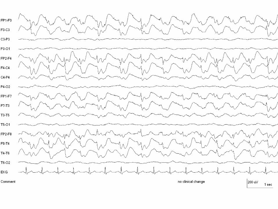
20. GSW: 1.5 per second generalized polyspike-and-wave, frontally predominant. A
polyspike precedes every slow wave and there is no inter-discharge interval; thus this
pattern does not qualify for GRDA+S or GPDs+R.
21. GSW: 1.5 per second generalized spike-and-wave.
22. Burst-suppression pattern: Bursts (>500ms AND >3phases) of generalized activity on a
suppressed background.
23. Burst-attenuation pattern: In between bursts of generalized activity, there is low
amplitude background activity.
Abbreviation list:
BI = Bilateral Independent
Copyright 2012 American Clinical Neurophysiology Society
EDs = Epileptiform Discharges
G = Generalized
L = Lateralized
Mf = Multifocal
PDs = Periodic Discharges
RDA = Rhythmic Delta Activity
SI = Stimulus-Induced.
SW = Spike-and-wave or sharp-and-wave
+ = Plus = Additional feature which renders the pattern more ictal-appearing than the usual term
without the plus
+F = Superimposed fast activity
+R = Superimposed rhythmic activity
+S = Superimposed sharp waves or spikes, or sharply contoured
Acknowledgments
We would like to acknowledge the prior contribution of the following people to this terminology:
Thomas P. Bleck, Richard Brenner, Ronald G. Emerson, Paula Gerber-Gore, Kenneth G. Jordan,
Mark L. Scheuer.
140µV

4/s 3/s 2.5/s
P1: OTA/XYZ P2: ABCc02 JWBK388-Hirsch October 24, 2009 13:6 Printer Name: To Come
EEG IN ENCEPHALOPATHY 73
Figure 2.25 Suppression-burst. A suppression-burst pattern is present in this 55-year-old man s/p cardiac arrest.
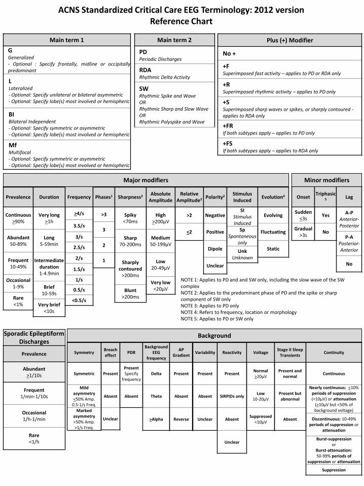
Main term 1
G Generalized - Optional : Specify frontally, midline or occipitally predominant
L Lateralized - Optional: Specify unilateral or bilateral asymmetric - Optional: Specify lobe(s) most involved or hemispheric
BI Bilateral Independent - Optional: Specify symmetric or asymmetric - Optional: Specify lobe(s) most involved or hemispheric
Mf Multifocal - Optional: Specify symmetric or asymmetric - Optional: Specify lobe(s) most involved or hemispheric
Main term 2
PD Periodic Discharges
RDA Rhythmic Delta Activity
SW Rhythmic Spike and Wave OR Rhythmic Sharp and Slow Wave OR Rhythmic Polyspike and Wave
Plus (+) Modifier
No +
+F Superimposed fast activity – applies to PD or RDA only
+R Superimposed rhythmic activity – applies to PD only
+S Superimposed sharp waves or spikes, or sharply contoured - applies to RDA only
+FR If both subtypes apply – applies to PD only
+FS If both subtypes apply – applies to RDA only
Prevalence
Continuous >90%
Abundant 50-89%
Frequent 10-49%
Occasional 1-9%
Rare <1%
Duration
Very long >1h
Long 5-59min
Intermediate duration 1-4.9min
Brief 10-59s
Very brief <10s
Frequency
>4/s
3.5/s
3/s
2.5/s
2/s
1.5/s
1/s
0.5/s
<0.5/s
Phases1
>3
3
2
1
Sharpness2
Spiky <70ms
Sharp 70-200ms
Sharply contoured
>200ms
Blunt >200ms
Absolute Amplitude
High >200µV
Medium 50-199µV
Low 20-49µV
Very low <20µV
Relative Amplitude3
>2
<2
Polarity2
Negative
Positive
Dipole
Unclear
Stimulus Induced
SI Stimulus Induced
Sp Spontaneous
only
Unk Unknown
Evolution4
Evolving
Fluctuating
Static
Major modifiers
Onset
Sudden <3s
Gradual >3s
Triphasic5
Yes
No
Lag
A-P Anterior-Posterior
P-A Posterior-Anterior
No
Minor modifiers
NOTE 1: Applies to PD and and SW only, including the slow wave of the SW complex NOTE 2: Applies to the predominant phase of PD and the spike or sharp component of SW only NOTE 3: Applies to PD only NOTE 4: Refers to frequency, location or morphology NOTE 5: Applies to PD or SW only
Background
Symmetry
Symmetric
Mild asymmetry <50% Amp.
0.5-1/s Freq. Marked
asymmetry >50% Amp. >1/s Freq.
Breach effect
Present
Absent
Unclear
PDR
Present Specify
frequency
Absent
Background EEG
frequency
Delta
Theta
>Alpha
Variability
Present
Absent
Unclear
Reactivity
Present
SIRPIDs only
Absent
Unclear
Voltage
Normal >20µV
Low 10-20µV
Suppressed <10µV
Stage II Sleep Transients
Present and normal
Present but abnormal
Absent
Continuity
Continuous
Nearly continuous: <10% periods of suppression (<10µV) or attenuation
(>10µV but <50% of background voltage)
Discontinuous: 10-49% periods of suppression or
attenuation
Burst-suppression or
Burst-attenuation: 50-99% periods of
suppression or attenuation
Suppression
Sporadic Epileptiform Discharges
Prevalence
Abundant >1/10s
Frequent 1/min-1/10s
Occasional 1/h-1/min
Rare <1/h
ACNS Standardized Critical Care EEG Terminology: 2012 version Reference Chart
AP Gradient
Present
Absent
Reverse
Response to public comments on
American Clinical Neurophysiology Society’s Standardized Critical Care EEG Terminology: 2012 version
June 3, 2012
Hirsch LJ, Gaspard N, Laroche SM
Public Reviewer Comments are in standard font, and our responses are in italics. The revised manuscript
has the major changes highlighted in yellow.
Comment #1: From my standpoint there are numerous problems with this classification scheme. The
main issue is that since we do not know which patterns are associated with what structural lesions,
there is no reason to change our language for describing these patterns at the present time. If we want
to do research it is better to say that a record has generalized periodic complexes of 20uV in amplitude
with a duration of 200-300msec occurring at a rate of 1/second than to use the new proposed
descriptive terms such as "abundant", "long", "very long", etc. Because this scheme is very complex and
has no clinical correlate, it will be largely ignored even if the ACNS endorses it. We should wait until we
know which waveforms are correlated with specific pathologies as we know what that pathologic
corrlelate of a PLED is.
Comment #2: Sounds reasonable but there is replacement of ingrained traditional terms (e.g. PLEDs,
triphasic waves, GPEDs) and there is a significant risk of miscommunication (not that those terms are
not open to misinterpretation also) when using the new terms. Before switching to the new terms I
would suggest mentioning in parentheses (previously called -----) for a few years until there is general
understanding of the new terminology.
>>Response to comments #1 and #2: This lack of knowledge of the meaning of these patterns is the exact
reason we have created an objective, logical nomenclature with input from as many people as possible
throughout the world over many years. The descriptive terms (“abundant”, “long”, etc) are only
provided for those who prefer them, but the actual numbers/categories can be used instead. However,
for those who use descriptive terms, we think it is important that they are standardized. Hence, they are
included and defined, albeit somewhat arbitrarily. We understand that this nomenclature represents a
change from classic terminology; thus, we have provided a table of the equivalent “old” and “new”
terms. We believe the changes are all improvements, or at least more accurate descriptions, and that
they are more amenable to consistent use and research (including dropping the E from “PLEDs”).
Comment #3: SECTION "MODIFIERS"- #5 Sharpness- What is the difference between c "sharply
contoured" and d. Blunt? If none then d should be included in the c description -OR- d should be
defined.
>> Response to comment 3: We have revised this definition as follows: Sharply contoured: having sharp
morphology (sharp inflection at its peak or trough, or steep upslope or downslope (such as saw-tooth
morphology), but the duration of the wave at the baseline is >200 msec and thus does not qualify as
“sharp”. Blunt: having smooth or sinusoidal morphology. We have also included examples of both
sharply contoured and blunt waves in the figures.
Comment #4: My question is about the requirement for ictal events to exceed 4 Hz. We often see
events in ICU recordings that look ictal when slowed down to 30 sec per page, either by virtue of
rudimentary spike-wave runs or by evolution. Sometimes we see clinical correlations suggestive of
seizures. I am coming to think of these as "slow seizures" in sick brains. I am early in the process of
gathering a series for publication. These events are distinct from intermittent rhythmic non-evolving
delta without spikes. Is there a way to encompass such events in the terminology and is there any
consensus on such slow events (sometimes) being ictal? I can attach a figure with an example, if you tell
me which email.
>>Response to comment 4: It is our opinion too that seizure activity might demonstrate slower
frequency in the critically ill. This nomenclature aims to create a common language to be used in studies
in the field and applies to all equivocal EEG patterns whose nature might or might not be ictal. Runs of
rhythmic delta activity with evolution and with embedded spikes would be described in the nomenclature
as evolving RDA+S. This would not exclude that they are ictal, simply that this pattern is included in this
nomenclature and warrants investigation. We attempted to make it clear that patterns in this
nomenclature may still be definite seizures based on evolution, clinical correlate, etc. The corollary in the
background section in the current form of the nomenclature already states the following to address this
point [bolding added]:
“Corollary: The following patterns are included in this nomenclature and would not be
termed electrographic seizures for research purposes (whether or not these patterns are
determined to represent seizures clinically in a given patient): Generalized spike and wave
patterns slower than 3/s; and evolving discharges that remain slower than or equal to 4/s.
This does not imply that these patterns are not ictal, but simply that they may or may not
be. Clinical correlation, including response to treatment, may be necessary to make this
determination.”
As this is indeed an important point, we have bolded these parts in the new version, and removed the
parentheses.
Comment 5: Some minor comments: 1. For localizations in main term 1, would suggest including
"central" and/or "vertex". I have seen adults with spikes/seizures there, and would make terminology
extend to neonates more easily.
>> 1. A similar point could be made about bifrontal and bioccipital discharges; they are actually focal,
but are included them in the term “generalized” in this nomenclature for simplicity, with a defined
subcategory for them. Based on this comment, we have added a subcategory under “generalized”
entitled “Midline Predominant”, defined as “having an amplitude in midline derivations that is at least
50% greater than in parasagittal derivations on an average or non-cephalic referential recording”.
Comment #6: Number 6 (amplitude definitions): I would generally consider 20-49 uV as being
moderate or medium, not low amplitude (and in fact you call it that when discussing background activity
later on). I think terms should be consistent across types of activities.
Response to comment #6: We think that the amplitude scale for the main terms such as periodic
discharges has to be necessarily different than the scale for background as they have to stand out of the
background. Indeed, 40µV LPDs over a 40µV background would not be noticeable. We agree that a
background of 40 µV is not low amplitude, and it would be categorized as “normal” voltage background
in this nomenclature (>20 µV). However, for PDs, 40 µV is indeed in the low range for peak to trough
amplitude based on the literature (usually closer to 100-150 µV; see Kalamangalam et al., Epilepsia
2007. We have added a comment in each location that this applies only to the one situation and not the
other (either describes Main Term #2 or the background EEG, but different scales).
Comment #7: Pocket guide is great, could have a copy hanging on the EEG machine in ICU. I would find
it helpful to show an example of an actual clinical report built using these terms to help people set up
their lab report formats. By the same token, figure legends could be not just the description, but actually
the way you would state the finding in a report. 4. Don't be dismayed by those resistant to new
terminology (if they aren't already exhausted over the ILAE classifications!). But would be helpful to
offer some sample boilerplate that could be appended to reports to explain significance of findings to
the clinicians. Also a teaching file/slide show that EEGers could use to educate their referring docs and
trainees.
Response to comment #7: This is a good idea. We will try to incorporate something like this in the
future, but not as part of the formal ACNS guideline.
Comment #8: The definitions of Generalized and Lateralized include serious contradictions: generalized
is explicitely defined as possibly not generalized and similarly for lateralized. According to the
definitions, I think the terms "symmetrical" and "asymmetrical" would appear to be more appropriate;
they would not include contradictions.
Response to Comment 8: Generalized and lateralized are widely accepted terms in the EEG literature,
including when referring to non-generalized patterns. GPEDs, as well as generalized epileptiform
discharges are in fact rarely generalized; and PLEDs are often bilateral synchronous and asymmetric
(rather than truly unilateral). In order to make the “generalized” term more accurate, one would have to
say “bilateral; synchronous or time-locked; and symmetric”, and possibly widely distributed. We have
simplified this by referring to all as “generalized”, then having subcategories. We also included this
explanation in the initial definition of “generalized”, as follows:
“for this purpose, the term “generalized” refers to any bilateral, bisynchronous and symmetric
pattern, even if it has a restricted field [e.g. bifrontal])”
Simply stating “symmetrical” would not suffice, as bilateral independent patterns are often symmetrical.
Comment #9: On main term Rhythmic Delta Activity (RDA). In the second line: uniform morphology and
duration, and no interval between, it could better say: uniform morphology and duration, but with no
regular interval between.
Response to comment 9: We have fixed this as follows: “…uniform morphology and duration, and
without an interval between…”.
Comment #10: On modifier # 8 Stimulus-Induced (SI): In the first line the word: reproducibly does not
exist in the English language; also, the second part of the phrase: brought about by an alerting stimulus,
could be changed to: brought about by a sensory stimulus….
Response to comment #10: The adverb “reproducibly” does appear in both the Oxford and Collins
dictionaries. We thus believe it is correct to use it. Concerning the stimulus, its nature is not as relevant
as the fact that it is able, or potentially able, to arouse the patients. All stimuli are by definition sensory
but many of them may not be ‘alerting’ (odors, tastes, quiet sounds).
Comment #11: On modified # 9 Evolving: In line number six: sequentially out of at least two standard 10-
20 electrode locations, it could better say: sequentially out of at least two standard 10-20 electrode
locations, it could say better: sequentially out of at least two different standard 10-20 electrode
locations.
Response to comment #11: We have rephrased this as suggested by the reviewer.
Comment #12: On minor modifier # 4: Anterior-posterior lag or posterior-anterior lag: The paragraph:
applies if a consistent measurable delay of > 100 ms appears to be present from the most anterior
derivation in which it is seen to the most posterior derivation in which is seen; specify typical delay in ms
from anterior to posterior (negative = posterior to anterior) in both longitudinal bipolar and in a
referential montage, preferably with an ipsilateral ear reference, could be more clear as: applies if a
consistent measurable delay of > 100 ms exists from the most anterior to the most posterior derivation
in which is seen; specify typical delay in ms from anterior to posterior (negative = posterior to anterior)
in both a longitudinal bipolar and a referential montage, preferably with an ipsilateral ear reference.
Response to comment #12: We have rephrased this as suggested by the reviewer.
Comment #13: On D. Background EEG, in the continuity characteristic, numeral 4. Burst-
attenuation/Burst-supression, in section c. Presence or absence of Highly Epileptiform Bursts: At the end
of the second line the word “majority” is used, without specifying what that majority refers to: (>50%)?.
Also, at the end of the fifth line the word “majority” is used, without specifying again what that majority
refers to: (>50%)?. In line 3, bursts and occur an average of 1/s, could be more clear if it would say:
bursts and occur at an average of 1/s.
Response to comment #13: We have now defined majority as >50% and rephrased as suggested by the
reviewer.
Comment #14: On Figure Legend # 5. In the second line in parenthesis: (as there is an inter-discharge
interval) would not it be better?: (as there is an approximately constant inter-discharge interval).
Response to comment #14: We have rephrased this as follows: “ … as there is a quantifiable inter-
discharge interval between consecutive waveforms and recurrence of the waveform at nearly regular
intervals.”
Comment #15: I think a discussion of the organization of the background, including the anterior to
posterior gradient is actually the most important aspect of the background and should be added.
Response to comment #15: We have now added this to the background EEG description as “AP
gradient: present or absent”. We have defined it as follows: “AP gradient is present if there are clear and
persistent lower amplitude, faster frequencies in anterior derivations compared to posterior derivations”.
Comment #16: I think the term "spiky" does not sound right. It is kind of cute, and I think we need
something more professional like "with admixed spikes".
Response to comment #16: We also are not enamored with the term “spiky”. We have tried to find an
alternative term (“apiculate”, “pointy”, etc.) without success, and another word would lose the well
known correlation with “spike”, an important advantage of this word. The cuteness of the word is
subjective and relative; for instance, the example in Webster’s dictionary of its use is in “spiky barbed
wire”. We believe that people will get accustomed to its use. “Spiky” has a different meaning from “with
admixed spikes”. The first term applies to periodic discharges and refers to the degree of sharpness of a
single discharge, whereas the latter, included in the “Plus S” modifiers, refers to the presence of spikes
embedded within runs of rhythmic delta activity.
Comment #17: This is an excellent series of documents! The only major suggestion I would have is to
consider including how artifact is defined both for practical use and research use (ie daily index). It is
highly relevant to the interpretation of cEEG and perhaps including similar prevalence values as a major
modifier this would make the paper more practical.
Response to comment #17: The purpose of this nomenclature was to address the need for an objective
description of EEG patterns in the ICU. The description of artifacts is beyond its scope, and would be more
appropriate in an educational publication rather than a guideline.
Comment #18: Minor suggestions would be to eliminate "blunt" from the "sharpness" category.
Response to comment #18: Blunt was included in the “sharpness” modifier to allow the description of the
full spectrum of sharpness. In a sense it can be seen as the degree zero (or lack) of sharpness.
Comment #19: In the 1b category under EEG background change "or" to "and" to ensure that the minor
asymmetries are an abnormality and therefore worthy of mention or eliminate the category in favor of
symmetry v asymmetry.
Response to comment #19: Changing “or” to “and” would exclude asymmetries in frequency or
amplitude alone.