Embed Size (px)
Citation preview
ISSN 0034-7000 REV. ARG. CARDIOL., 49, NO 4,155-171
T elDas de actualidad
Los hallazgos con ecocardiografía bidimensional en eI prolapso de válvula mitral
The two-dimensional echocardiographic features of mitral valve prolapse
HARRY RAKOWSKI, M.D. RANDOLPH P. MARTIN, M.D. RICHARD L. pOPP, M.D. Division of Cardiology, Stanford University School of Medicine, Stanford, California, U.S.A.
This work was supported in part by NIH Grant NO HL-5866. Este trabajo fue parcialmente financiado por una beca del NIH NOHL-5866.
Dr. Rakowski was supported by Ontario Heart Foundation Research Fellowship. El Dr. Rakowski fue financiado por un "Fellowship" de la Ontario Heart Foun- dation.
Two-dimensional wide angle ultrasonic sector scans (2D echo) were done in 41 patients with M-mode echocardiographic features of mitral valve prolapse and in 30 patients without echo- cardiographic or angiographic mitral valve pro- lapse. One or both of the following 2D echo findings were seen in all mitral valve prolapse patients: (a) systolic leaflet arching above- the level of the A V groove; (b) exaggerated motion of the inferior portion of the mitral annulus and posterobaseÛ left ventricular myocardium. The latter abnormality appeared to undermine the systolic support for the posterior mitral leaflet with resultant end-systolic leaflet position above the level of the A V groove. Interestingly, only patients with this latter finding had infero- lateral ECG lead ST- T wave changes. Systolic anterior mitral motion was seen on M-mode studies in 3/41 pacients with mitral valve prolap-
Dirección postal: Correspondence to: Richard L. Popp, M.D. Cardiology Division
Stanford University Medical Center Stanford, CA. 94305 - U.S.A.
Se realizaron ecocardiogramas bidimensionales (2D eco) con rastreador sectorial ultrasónico de
gran ángulo en 41 pacientes con características ecocardiográficas en modo M de prolapso de la válvula mitral y también en 30 casos sin evi- dencias ecocardiográficas 0 angiográficas de pro- lapso dé la válvula mitral. Uno 0 ambos de los siguientes hallazgos ecocardiográficos bidimen- sionales fueron observados en tòdos los pacien- tes con prolapso de la válvula mitral: a) arquea- mien to sistólico de una valva por arriba del nivel del surco A V; b) exagerado movimiento de la porción inferior del anillo mitral y del mio- cardio posterobasal del ventrículo izquierdo. La última anormalidad debilita el soporte sis- tólico para la valva mitral posterior, llevando al resultado de una Pdsición telesistólica en la cual la valva mitral está por arriba del surco A V. Llamativamente, solamente los casas con esta última alteración tenían cambios del ST- T en las derivaciones ECG de cara inferolateral.
Movimiento anterior sistólico de la mitral fue observado en los estudios de modo M de 3 de 41 casos con prolapso de la válvula mitral; estudios ecocardiográficos bidimensionales mostraron
156 REYIST A ARGENTINA DE CARDIOLOGIA, JULIO-AGOSTO 1981, YOLo 49, NO 4
se. Two-dimensional echo studies showed this
was due to systolic anterior buckling motion of chordae tendineae. The spatial orientation pro- vided by wide angle two,dimensional echocar- diography helped us to better understand the M-mode patterns of mitral valve prolapse. Both valvular and myocardial-annular abnormalities may contribute to the M-mode echocardiogra- phic pattern of leaflet prolapse. Key Words: Cardiomyopathy. Non-invasive methods. Ultra- sound.
The syndrome mitral :valve prolapse has gene- rated a great deal of interest since 1961, when Reid 31
refocused attention on the mitral valve apparatus as the site of the abnormality. Since that time numerous reports have detailed the various and variable features of this condition. Left ventricular cineangiography provided the first objective way of demonstrating billowing of one or both mitral leaflets toward the left atrium in systole, with or without associated
mitral regurgitation.4,8 Controversy exists re- garding the primary pathophysiology of this
syndrome. The underlying problems has been attributed to abnormal valvular apparatus, seg-
mental left ventricular contraction abnormalities (with or without localized myopathy), and abnormal mitral annulus structure. The use of time-motion (M-mode) echocardiography has
provided a convenient non-invasive method of establishing the diagnosis of mitral valve pro- lapse. Mid-systolic or holosystolic posterior dis-
placement çf mitral valve echoes has correlated well with angiographic evidence for this condi- tion 26 if strict criteria for posterior echocardio- graphic displacement22 are used and if care is
taken to avoid inferior angulation of the ultra- sound transducer.21 M-mode echocardiography provides only a .narrow field of view without spatial orientation and some apparently false negative studies are seen.
Wide-angle, phased array two-dimensional ul- trasonic sector scanning (2D echo) is a technique capable of producing dynamic images of the heart in multiple cross sectional planes. Thus, motion of most valvular structures can be
appreciated throughout the cardiac cycle. The purpose of this study was to use 2D echo:
que esto se debia a un movimiento de "e nru la-
mien to "
sistólico anterior de las cuerdas tendi- nosas. La orientación espacial proporcionada por el ecocardiograma bidimensional de gran ángulo sirvió para comprender mejor las imá- genes caracteristicas del prolapso de la válvula mitral en el modo M. Tanto las anormalidades valvulares como las anulomiocárdicas pueden contribuir para determinar el patrón ecocardio- gráfico modo M del prolapso valvular.
EI síndrome del prolapso de la válvula mitral ha origin ado una onda de gran interés desde
1961, cuando Reid 31 reenfocó la atención
sobre el aparato valvular mitral como el sitio de esta anormalidad. Desde entonces, numerosas comunicaciones han detallado las característi- cas múltiples y variables de esta condición. La cineangioventriculografía izquierda fue el pri-
mer procedimiento para la demostración obje-
tiva del arqueamiento de una 0 ambas valvas de la mitral haciå la aurícula izquierda durante la sístole, con 0 sin regurgitación mitral asocia- da.4,8 Existen controversias sobre la fisiopato- logía primaria de es.te síndrome. Los .problemas subyacentes fueron atribuidos a: aparato valvu- lar anormal, anomalías segmentarias en la con- tracción ventricular izquierda (con 0 sin miopa- tía localizada),estructura anormal del anillo mitral. El empleo de ecocardiografía modo M
facilitó un procedimiento no invasivo convenien- te para establecer el diagnóstico del prolapso de la válvula mitral. El desplazamiento posterior mesosistólico u holosistólico de los ecos de la válvula mitral se ha correlacionado bien con las evidencias angiográficas de esta condición 26
siempre que se respeten los criterios estrictos para el desplazamiento ecocardiográfico poste- rior22 y si se toma cuidado para evitar una angulación inferior del transductor ultrasóni- CO.21 El ecocardiograma modo M provee sola- mente un campo de vista muy estrecho y sin
orien tación espacial; en tonces, algunos aparen- temente falsos negativos se han observado.
El rastreo sectorial ultrasónico, de alineación electrónica en fase y gran ángulo (ecocardio- gram a 2D) es una técnica capaz de producir imágenes dinámicas del corazón en múltiples pIanos de cortes sectoriales. Es así que el mo-
PROLAPSO VALVULAR MITRAL: ECO BIDIMENSIONAL / Harry Rakowski y co\. 157
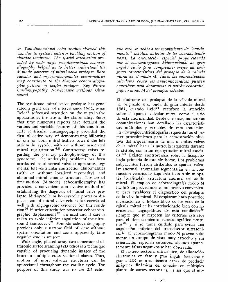
LONG AXIS
ANT
RV
POST
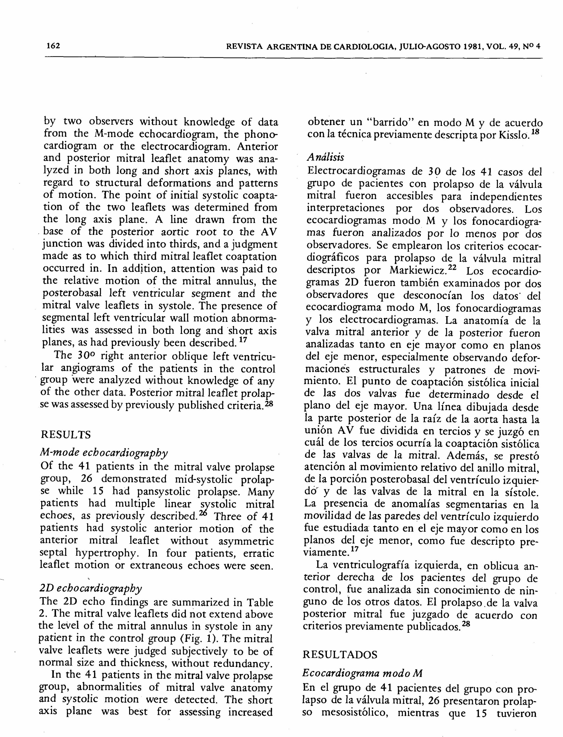
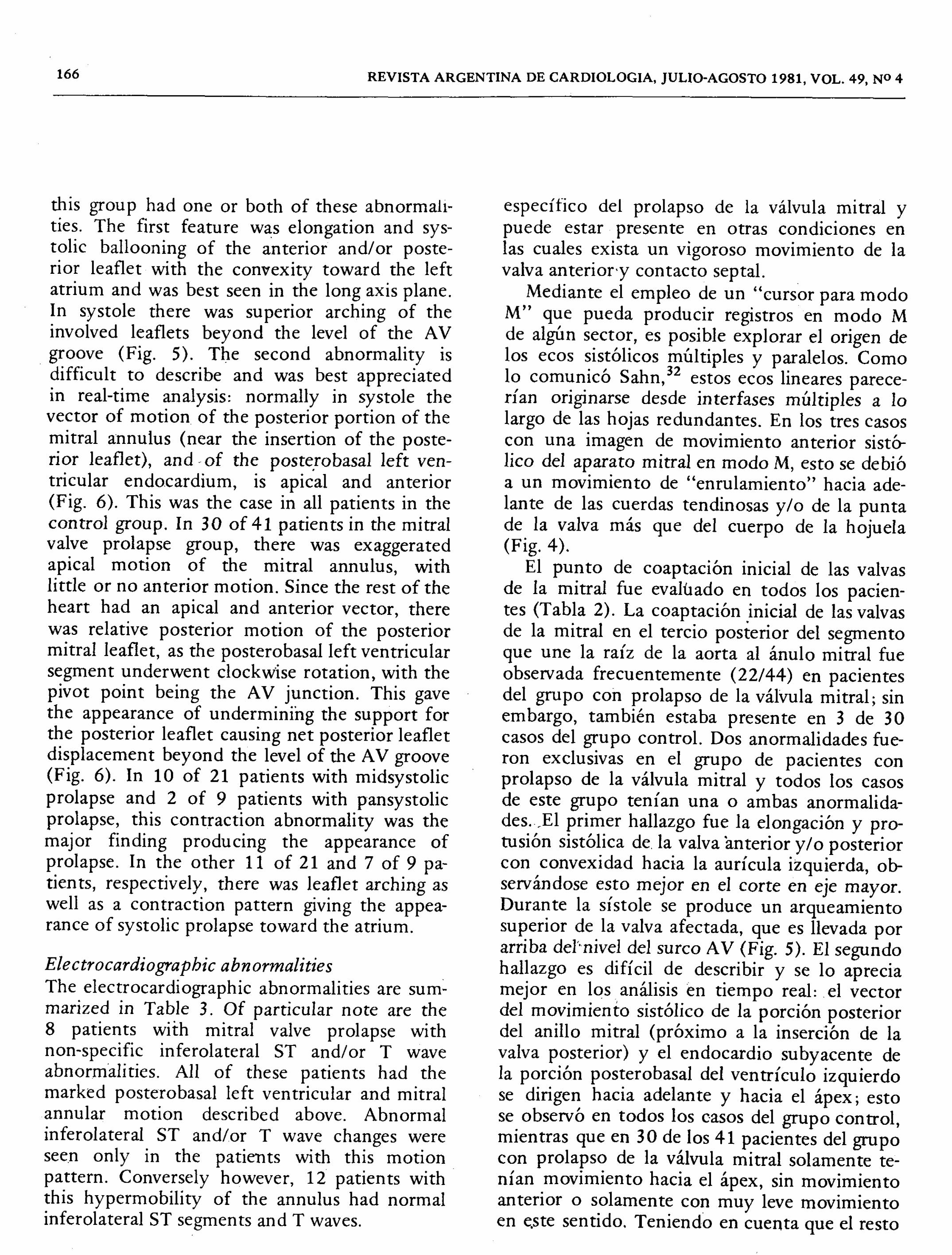
Fig. 1. Diagrama de una vista del eje mayor en sístole. La línea de puntos señala el anillo mitral. ANT = anterior, POST= posterior, RV=ventrículo derecho, LV=ventrículo izquierdo, IVS=sep- turn interventricular, PW= pared posterior del ventrículo iz- quierdo, Ao = aorta, LA = aurícula izquierda.
Fig. 1. A line diagram of a systolic normal long axis view. The dotted line outlines the mitraJ annuJus. ANT = anterior, POST =
posterior, RV=right ventricle; LV = left ventricle, IVS=inter- ventricular septum, PW = posterior left ventricular 'wall, Ao =
aorta, LA = left atrium.
(a) to better understand the findings seen on M-mode echocardiography and (b) attempt to better delineate the mechanisms contributing to mitral valve prolapse.
METHODS Patients selection Two groups of patients were studied. The mitral valve prolapse grou p consisted of 41 patients with typical M-mode echocardiographic evidence of mitral valve prolapse associated with at least one of the following: a non-ejection click, a
late systolic murmur, or pansystolic apical mur- mur of mitral regurgitation. Since the diagnosis of mitral valve prolapse normally can be made by clinical plus echocardiographic criteria, only five of 41 patients had left ventricular cineangio- graphy. This was done to rule out associated
coronary artery disease or to assess the severity of mitral regurgitation.
The control group consisted of 30 patients that had M-mode echo, 2D echo, and left ventricular cineangiographic studies without
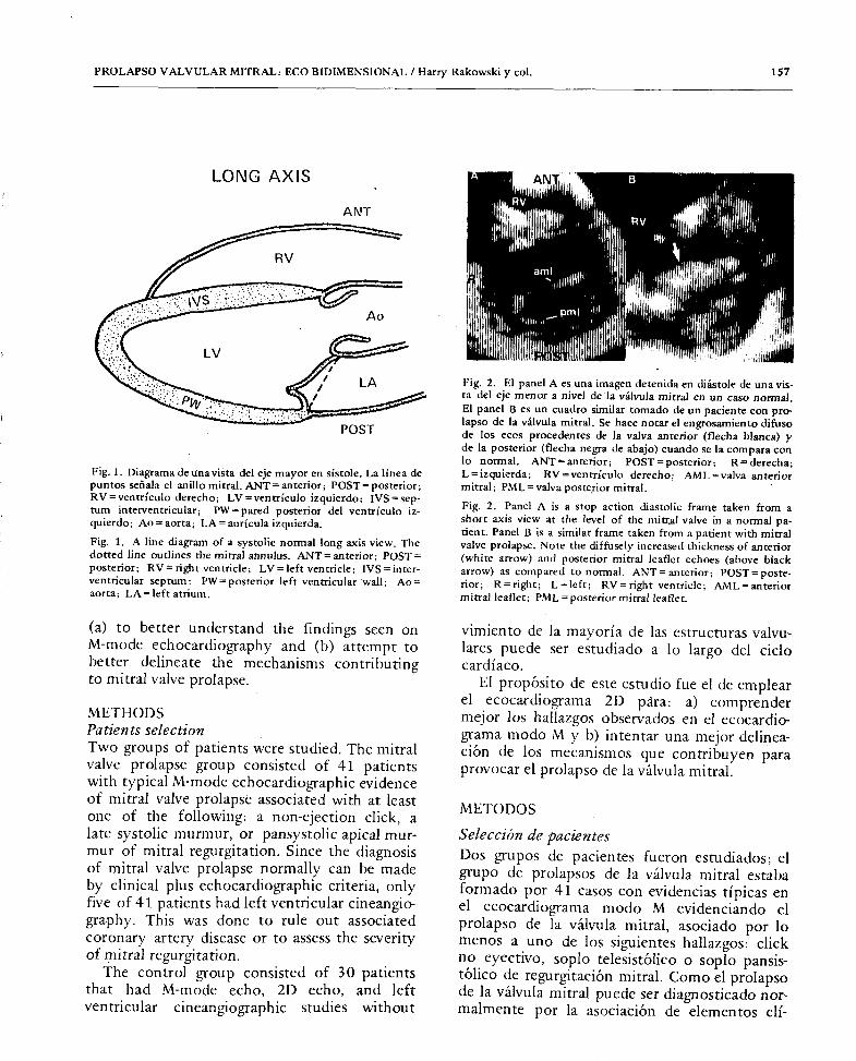
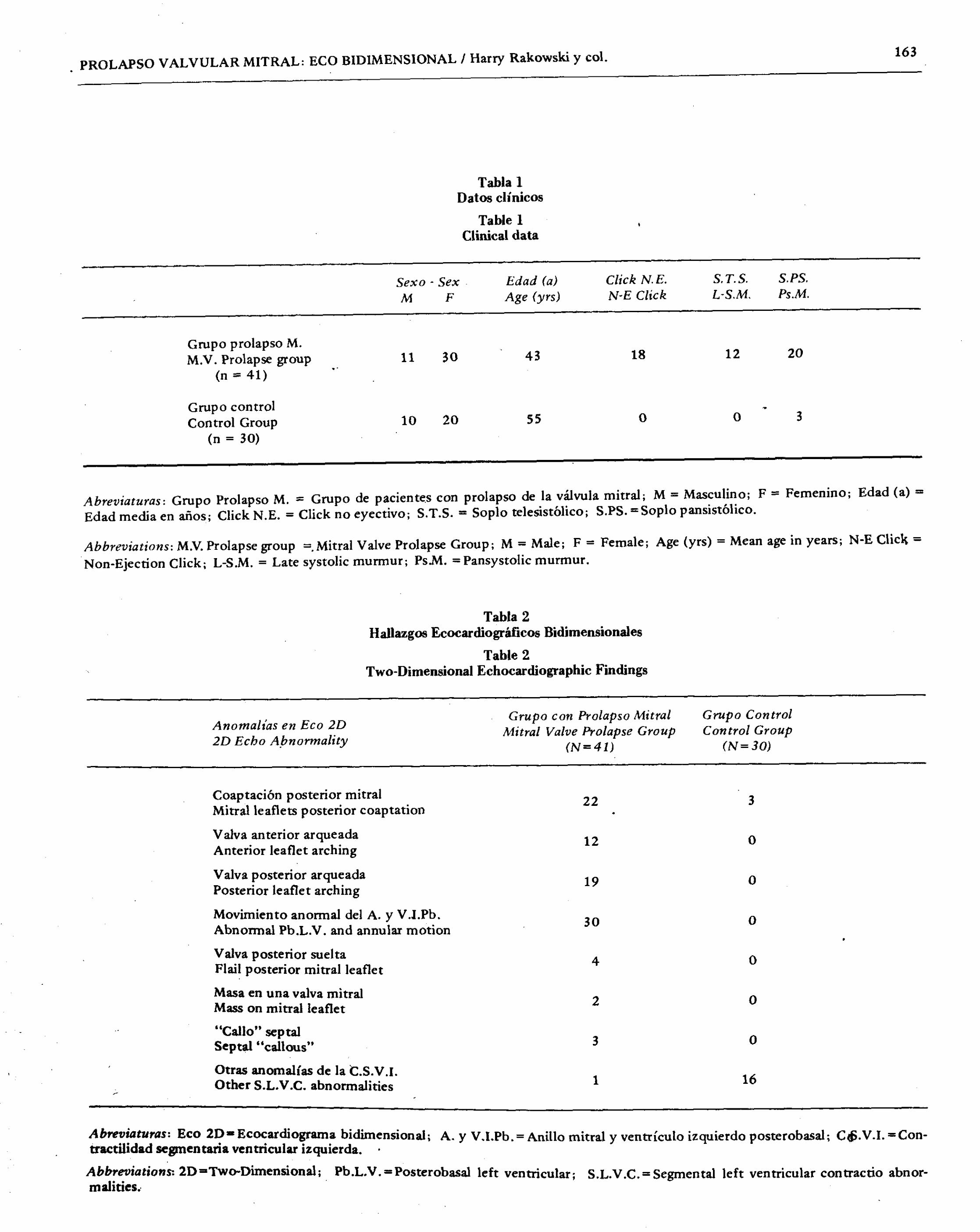
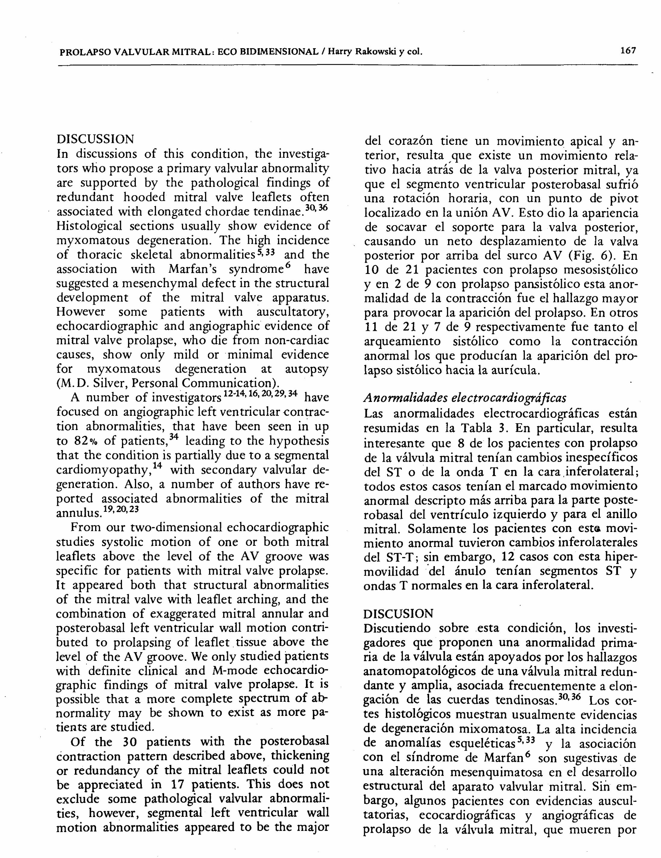
Fig. 2. EI panel A es una imagen detenida en diástole de una vis- ta del eje menor a nivel de la váJvula mitral en un caso normal. EI panel B es un cuadro similar tornado de un paciente con pro- lapso de la válvula mitral. Se hace notar el engrosamiento difuso de los ecos procedentes de Ja valva anterior (flecha hlanca) y de la posterior (flecha negra de abajo) cuando se la compara con 10 normal. ANT = an terior, POST = posterior, R = derecha, L=izquierda, RV=ventrículo derecho, AML=valva anterior mitral, PML = valva poste.rior mitral.
.
Fig. 2. Panel A is a stop action diastolic frame taken from a
short axis view at the level of the mitr.al valve in a normal pa- tient. Panel B is a similar frame taken from a patient with mitral valve prolapse. Note the diffusely increased thickness of anterior (white arrow) and posterior mitral leaflet echoes (above black arrow) as compared to normal. ANT=anterior, POST=poste- rior, R=right; L=left, RV=right ventricle; AML=anterior mitraJ leaflet, PML = posterior mitralleatlet.
vimiento de la mayoría de las estructuras valvu- lares puede ser estudiado a 10 largo del ciclo cardíaco.
El propósito de este estudio fue el de emplear el ecocardiograma. 2D pàra: a) comprender mejor 10s halIazgos observados en el ecocardio- grama modo M y b) intentar una mejor delinea- ción de 10s mecanismos que contribuyen para provocar el prolapso de la válvula mitral.
METODOS
Selección de pacientes Dos grupos de pacientes fueron estudiados; el grupo de prolapsos de la válvula mitral estaba formado por 41 casos con evidencias tí picas en el ecocardiograma modo M evidenciando el prolapso de la válvula mitral, asociado por 10
menos a uno de 10s siguientes hallazgos: click no eyectivo, soplo telesistólico a soplo pansis- tólico de regurgitación mitral. Como el prolapso de la válvula mitral puede ser diagnosticado nor- malmente por la asociación de elementos clí-
158 REVIST A ARGENTINA DE CARDIOLOGIA, JULIO-AGOSTO 1981, VOL. 49, NO 4
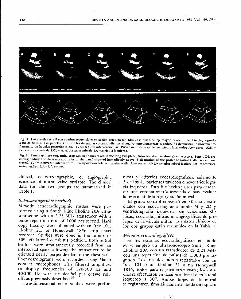
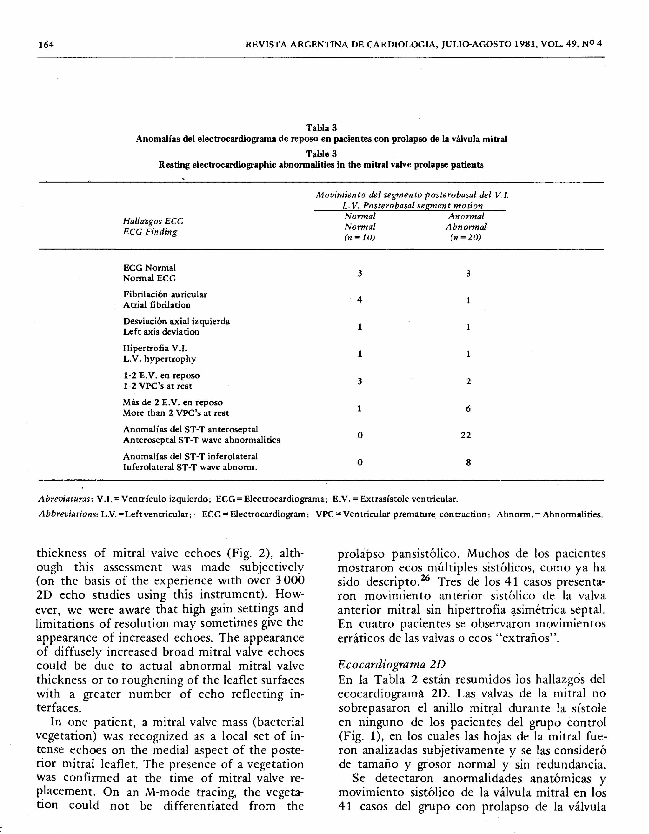
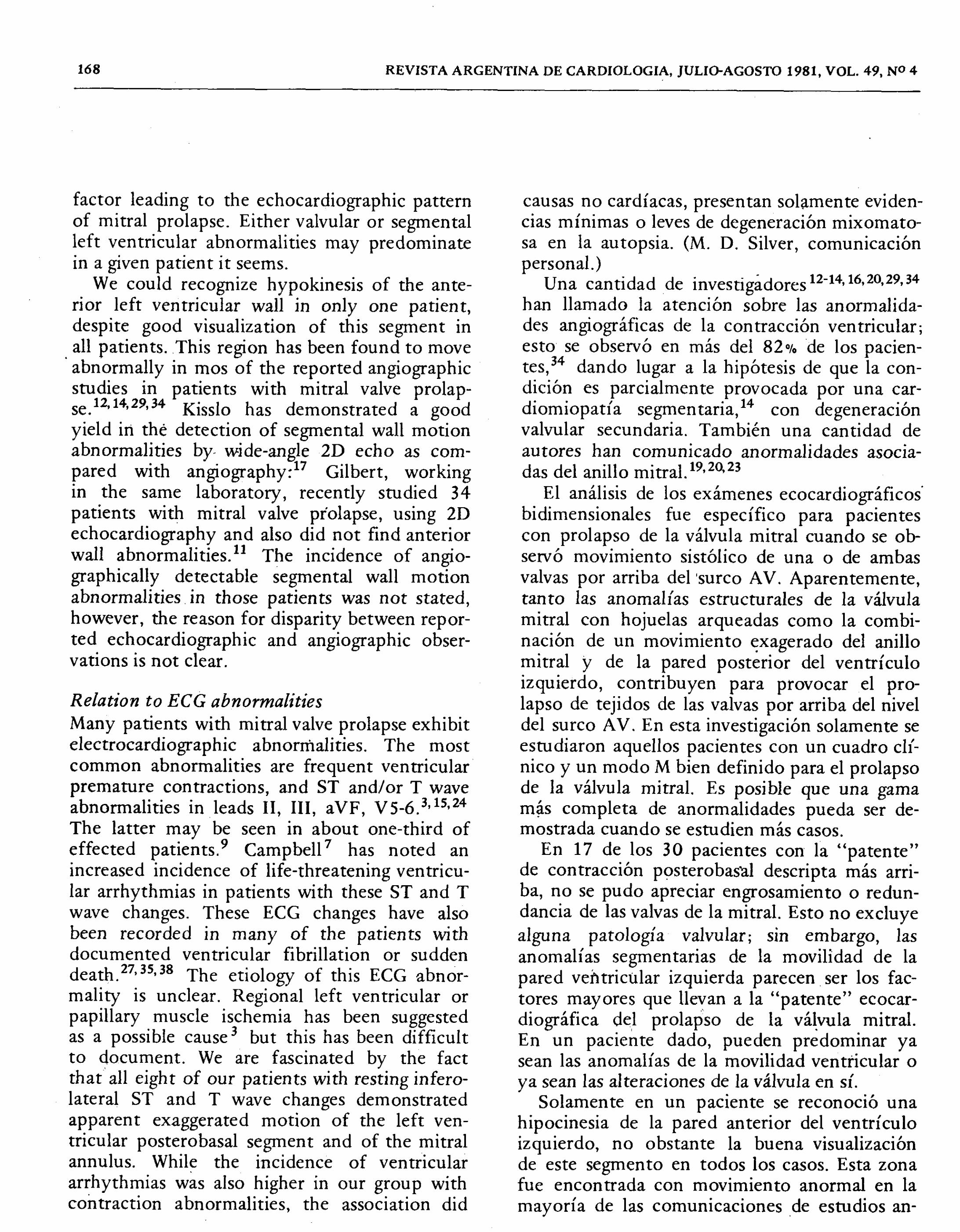
Fig. 3. Los paneles A a F son cuadros secuenciales en acción detenida tornados en el pIano del eje mayor, desde fin de diástole, llegando a fin de sístole. Los paneles GaL son los diagram as correspondientes al cuadro inmediatamente superior. Se demuestra un movimiento "flamean te de la valva posterior mitral. IVS = septum in terventricular; PW = pared posterior del ven trículo izquierdo; Ao = aorta; AML =
valva anterior mitral; PML = valva posterior mitral; LA = aurícula izquierda.
Fig. 3. Panels A-F are sequential stop action frames taken in the long axis plane, from late diastole through end-systole. Panels G-L are corresponding line "diagrams and refer to the panel situated immediately above. Flail motion of the posterior mitral leaflet is demons- trated. IVS = interventricular septum; PW = posterior left ventricular wall; Ao = aorta; AML = anterior mitral leaflet; PML = posterior mitral leaflet; LA = left atrium.
clinical, echocardiographic, or angiographic evidence of mitral valve prolapse. The clinical data for the two groups are summarized in Table 1.
Echocardiographic methods M-mode echocardiographic studies were per- formed using a Smith Kline Ekoline 20A ultra~ sonoscope with a 2.25 MHz transducer with a
pulse repetition rate of 1000 per second. Hard copy tracings were obtained with an Irex 101, Ekoline 21, or Honeywell 1856 strip chart recorder. Studies were done in the su pine or 300 left lateral decubitus position. Both mitral leaflets were simultaneously recorded from an intercostal space allowing the transducer to be oriented nearly perpendicular to the chest wall. Phonocardiograms were recorded using Maico contact microphones with filtered amplifiers to display frequencies of 120-500 Hz and 40-200 Hz with six decibel per octave roll- off, as previously described. 37
Two-dimensional echo studies were perfor-
nicos y criterios ecocardiográficos, "solamente 5 de los 41 pacientes tuvieron cineventriculogra- fía izquierda. Esto fue hecho ya sea para descar-
tar una coronariopatía asociada 0 para evaluar la sevéridad de la regurgitación mitral.
EI grupo control consistió en 30 casos estu- diados con ecocardiograma modo M y 2D Y
ventriculografía izquierda, sin evidencias clí- nicas, ecocardiográficas ni angiográficas de pro- lapso de la válvula mitral. Los datos clínicos de
los dos grupos están resumidos en la Tabla 1.
Métodos ecocardiográficos
Para los estudios ecocardiográficos en modo M se empleó un ultrasonoscopio Smith Kline Ekoline 20A con un transductor de 2,25 MHz con una repetición de pulsos de 1.000 por se-
gundo. Los trazados fueron registrados con un Irex 101 0 un Ekoline 21 0 un Honeywell 1856, todos para registro strip chart; los estu- dios se efectuaron en decúbito dorsal 0 en lateral izquierdo a 300. Ambas hojas de la mitral se registraron simultáneamente desde un espacio
. ""
PROLAPSO VALVULAR MITRAL: ECO BIDIMENSIONAL / Harry Rakowski y col. 159
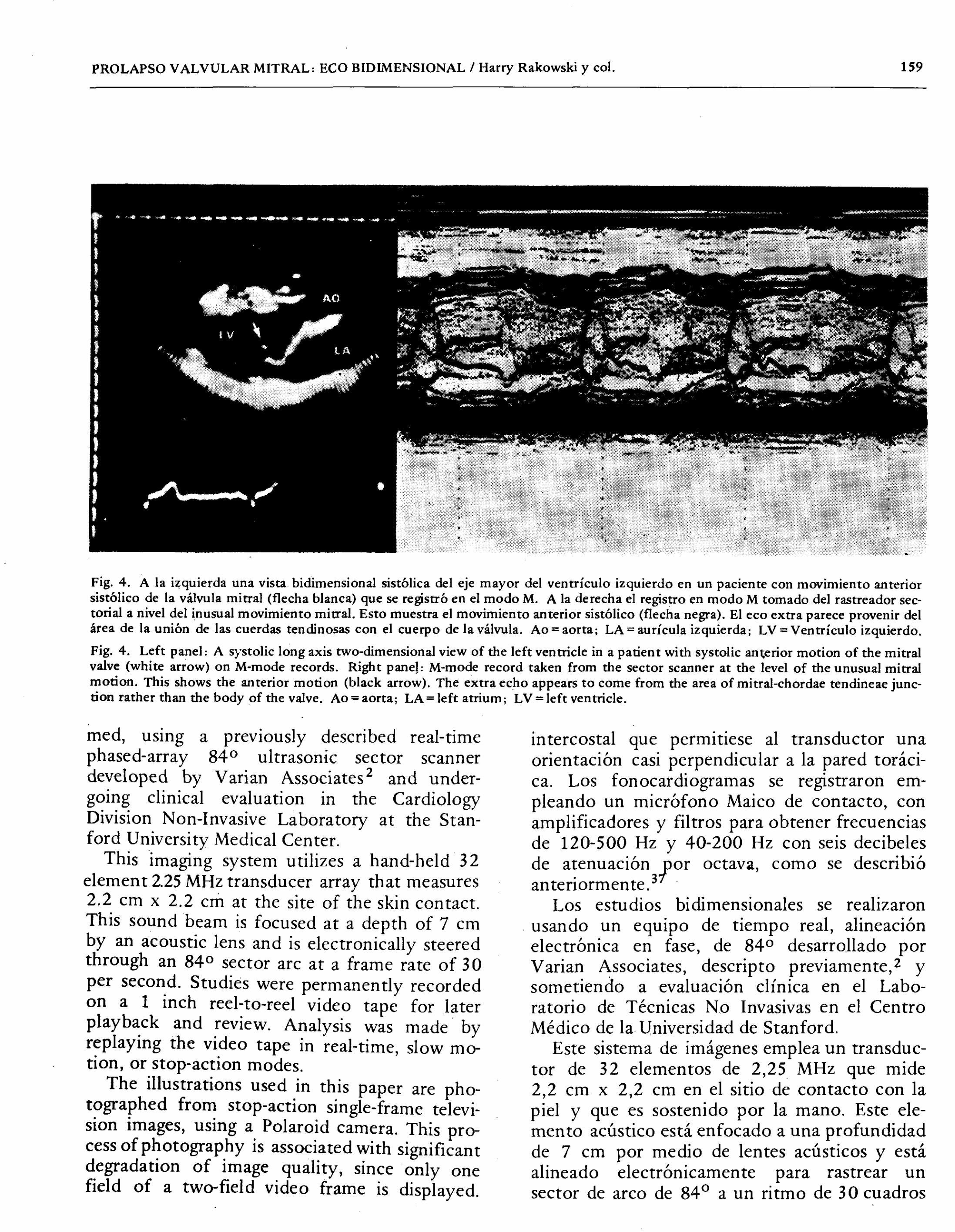
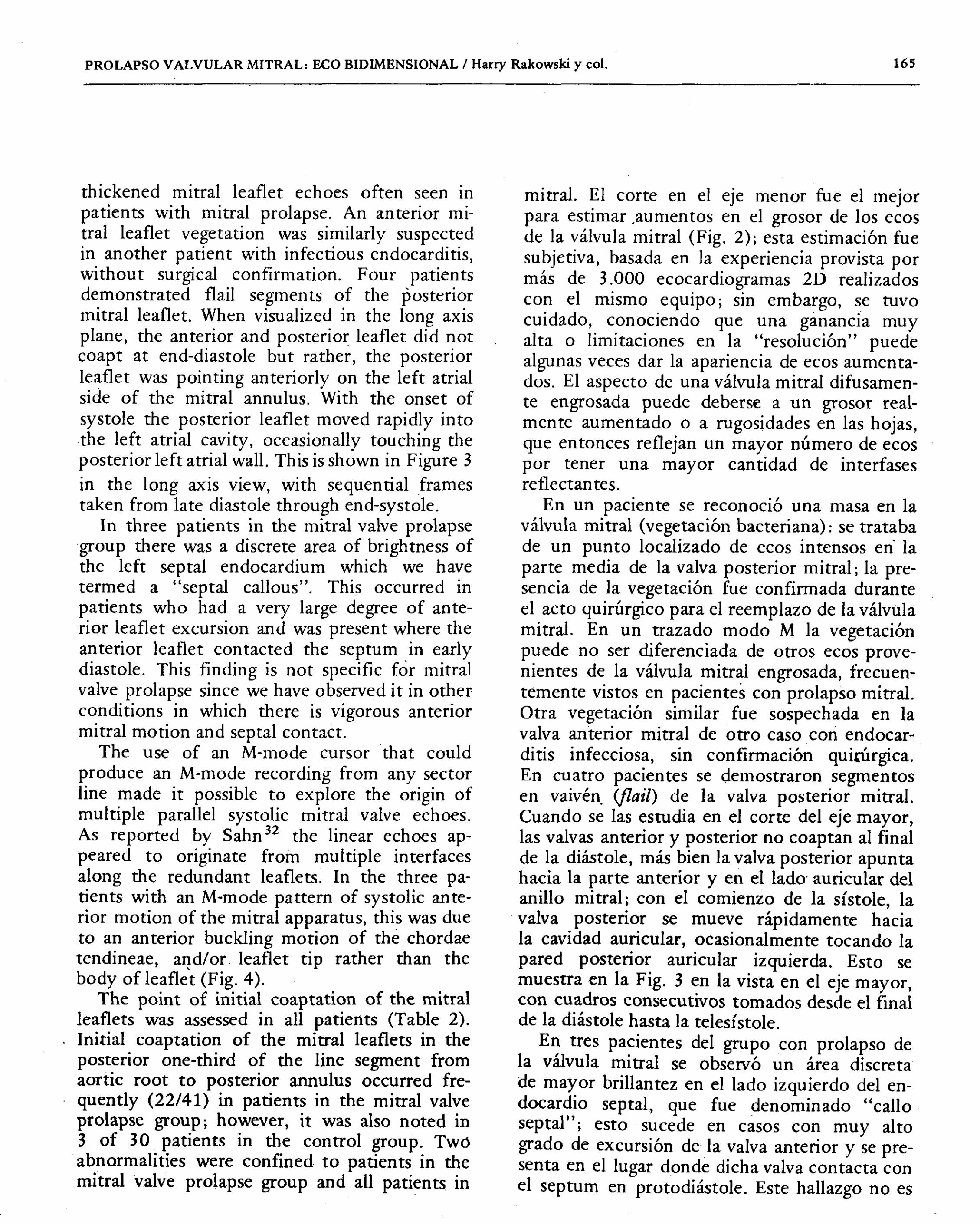
Fig. 4. A la i~quierda una vista bidimensional sistólica del eje mayor del ventrículo izquierdo en un paciente con movimiento anterior sistólico de la válvula mitral (flecha blanca) que se registró en el modo M. Ala derecha el registro en modo M tornado del rastreador sec- torial a nivel del inusual movimiento mitral. Esto muestra eI movimiento anterior sistólico (flecha negra). EI eco extra parece provenir del área de la unión de las cuerdas tendinosas con el cuerpo de la válvula. Ao = aorta; LA = aurícula izquierda; LV = Ventrículo izquierdo.
Fig. 4. Left panel: A systolic long axis two-dimensional view of the left ventricle in a patient with systolic an~erior motion of the mitral valve (white arrow) on M-mode records. Right pane!: M-mode record taken from the sector scanner at the level of the unusual mitral motion. This shows the anterior motion (black arrow). The extra echo appears to come from the area of mitral-chordae tendineae junc- tion rather than the body of the valve. Ao = aorta; LA = left atrium ; LV=left ventricle.
med, using a previously described real-time phased-array 840 ultrasonic sector scanner developed by Varian Associates
2 and under-
going clinical evaluation in the Cardiology Division Non-Invasive Laboratory at the Stan- ford University Medical Center.
This imaging system utilizes a hand-held 32
element 2.25 MHz transducer array that measures 2.2 cm x 2.2 cm at the site of the skin contact. This sound beam is focused at a depth of 7 cm by an acoustic lens and is electronically steered through an 840 sector arc at a frame rate of 30 per second. Studies were permanently recorded on a 1 inch reel-to-reel video tape for later playback and review. Analysis was made. by replaying the video tape in real-time, slow mo- tion, or stop-action modes.
The illustrations used in this paper are pho- tographed from stop-action single-frame televi- sion images, using a Polaroid camera. This pro- cess of photography is associated with significant degradation of image quality, since only one field of a two-field video frame is displayed.
intercostal que permitiese al transductor una orientación casi perpendicular a la pared toráci- ca. Los fonocardiogramas se registraron em- pleando un micrófono Maico de contacto, con amplificadores y filtros para obtener frecuencias de 120-500 Hz y 40-200 Hz con seis decibeles de atenuación lor octava, como se describió
anteriormente.3 Los estudios bidimensionales se realizaron
usando un equipo de tiempo real, alineación electrónica en fase, de 840 desarrollado por Varian Associates, descripto previamente,2 y sometiendo a evaluación clínica en el Labo- ratorio de Técnicas No Invasivas en el Centro Médico de la Universidad de Stanford.
Este sistema de imágenes emplea un transduc- tor de 32 elementos de 2,25 MHz que mide 2 2 cm x 22 cm en el sitio de contacto con la piel y que ~s sostenido por la mano. Este ele-
men to acústico está enfocado a una profundidad de 7 cm por medio de lentes acústicos y está alineado electrónicamente para rastrear un sector de arco de 840 a un ritmo de 30 cuadros
160 REVISTA ARGENTINA DE CARDIOLOGIA, JULIO-AGOSTO 1981, VOL. 49, NO 4
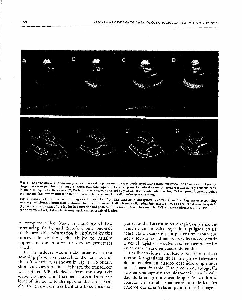
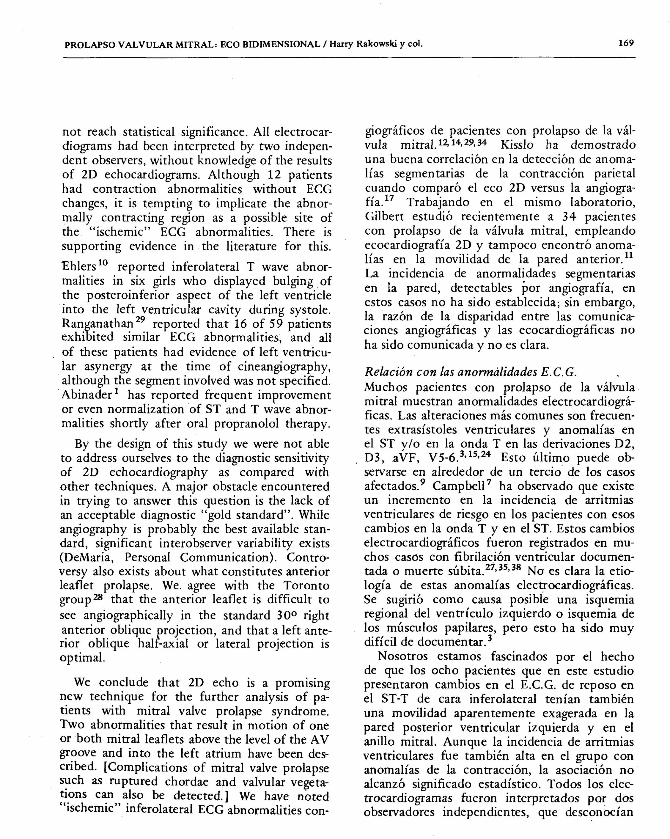
Fig. 5. Los paneles A a D son imágenes detenidas del eje mayor tomadasdesde telediástOle hasta telesístole. Los paneles E a H son los diagramas correspondientes al cuadra inmediatamente superior. La valva posterior mitral es marcadamente redundante y convex a hacia la aurícu/a izquierda. En sístole (C, D) la valva se arquea hacia arriba y atrás. RV=ventrículo derecho; IVS = septum interventricular; Ao= aorta; PML= valva mitral posterior; LA =aurícula izquierda; AML = valva anterior mitral. Fig. 5. Panels A-D are stop-action, long axis frames taken from late diastoié to late systOle. Panels E-H are line diagrams corresponding to the panel situated immediately above. The posterior mitral leaflet is markedly redundant and is convex to the left atrium. In systole (C, D) there is arching of the leaflet in a superior and posterior direction. RV'" right ventricle; IVS '" interventricular septum; PW = pos- terior mitral leaflet; LA = left atrium; AML = anterior mitral leaflet.
A complete video frame is made up of two interlacing fields, and therefore only one-half of the available information is displayed by this
process. In addition, the ability to visually appreciate the motion of cardiac structures is lost.
The transducer was initially oriented so the scanning plane was parallel to the long axis of the left ventricle, as shown in Fig. 1. To obtain short axis views of the left heart, the transducer was rotated 900 clockwise from the long axis view. . To record a shon axis sweep from the level of the aorta to the apex of the left ventri- cle, the transducer was held at a fixed locus on
por segundö. Los estudios se registran permanen- temente en un video tape de 1 pulgada en sis-
tema carrete-carrete para posteriores proyeccio- nes y revisiones. EI análisis se efectuó volviendo aver el registro de video tape en tiempo real 0
en cámara lenta 0 en cuadro detenido. Las ilustraciones empleadas en este trabajo
fueron fotografiadas de la imagen de televisión de un cuadro en cuadro detenido, empleando una cámara Polaroid. Este proceso de fotografía acarrea una significativa degradación en la cali- dad de la imagen, a causa de que de esta forma aparece en pantalla solamente uno de los dos cuadro~ que se entrelazan para formar la imagen,
PRO LAP SO VALVULAR MITRAL: ECO BIDIMENSIONAL / Harry Rakowski y col. 161
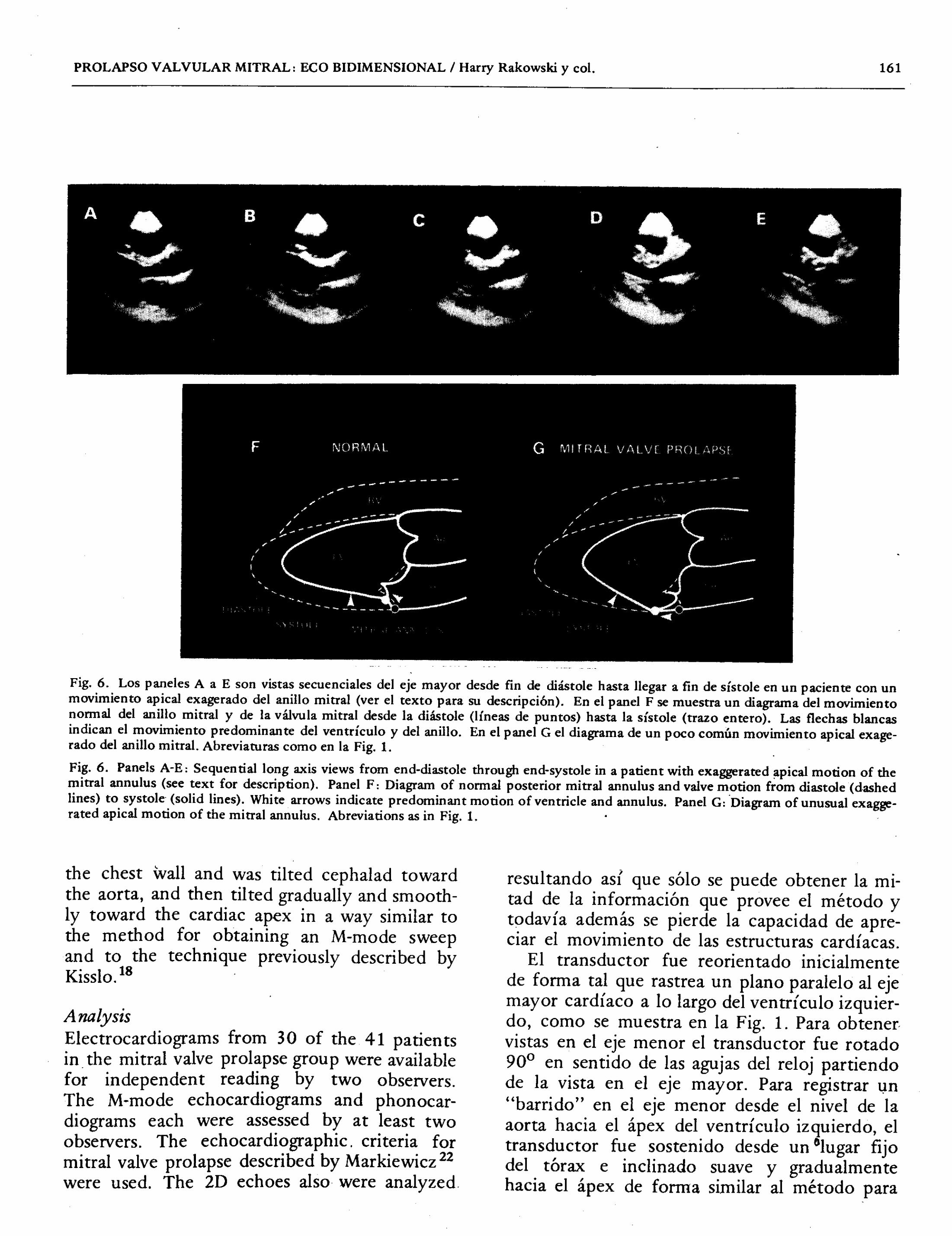
Fig. 6. Los paneles A a E son vistas secuenciales del eje mayor desde fin de diástole hasta llegar a fin de sístole en un paciente con un movimiento apical exagerado del anillo mitral (ver el texto para su descripción). En el panel F se muestra un diagrama del movimiento normal del anillo mitral y de la válvula mitral desde la diástole (líneas de puntos) hasta la sístole (trazo entero). Las flechas blancas indican eI movimiento predominante del ventrículo y del anillo. En el panel Gel diagram a de un poco común movimiento apical exage- rado del anillo mitral. Abreviaturas como en la Fig. 1.
Fig. 6. Panels A-E: Sequential long axis views from end-diastole through end-systole in a patient with exaggerated ~pical motion of the mitral annulus (see text for description). Panel F: Diagram of normal posterior mitral annulus and valve motion from diastole (dashed lines) to systole (solid lines). White arrows indicate predominant motion of ventricle and annulus. Panel G: >Diagram of unusual exagge- rated apical motion of the mitral annulus. Abreviations as in Fig. 1.
the chest wall and was tilted cephalad toward the aorta, and then tilted gradually and smooth- ly toward the cardiac apex in a way similar to the method for obtaining an M-mode sweep and to the technique previously described by Kisslo.18 >
Analysis
Electrocardiograms from 30 of the 41 patients in the mitral valve prolapse group were available for independent reading by two observers. The M-mode echocardiograms and phonocar- diograms each were assessed by at least two observers. The echocardiographic. criteria for mitral valve prolapse described by Markiewicz 22
were used. The 2D echoes also were analyzed
resultando asÍ que sólo se puede obtener la mi- tad de la información que provee el método y todavía además se pierde la capacidad de apre- ciar el movimiento de las estructuras cardíacas.
El transductor fue reorientado inicialmente de forma tal que rastrea un pIano paralelo al eje mayor cardíaco a 10 largo del ventrículo izquier- do, como se muestra en la Fig. 1. Para obtener vistas en el eje menor el transductor fue rotado 900 en sentido de las agujas del reloj partiendo de la vista en el eje mayor. Para registrar l,ln "barrido" en el eje menor desde el nivel de la aorta hacia el ápex del ventrículo izquierdo, el transductor fue sostenido desde un Olugar fijo del tórax e inc1inado suave y gradualmente hacia el ápex de forma similar al método para
162 REVISTA ARGENTINA DE CARDIOLOGIA, JULIO-AGOSTO 1981, VOL. 49, NO 4
by two observers without knowledge of data from the M-mode echocardiogram, the phono- cardiogram or the electrocardiogram. Anterior and posterior mitral leaflet anatomy was ana- lyzed in both long and short axis planes, with regard to structural deformations and patterns of motion. The point of initial systolic coapta- tion of the two leaflets was determined from the long axis plane. A line drawn from the
. base of the posterior aortic root to the A V
junction was divided into thirds, and a judgment made as to which third mitral leaflet coaptation occurred in. In addjtion, attention was paid to the relative motion of the mitral annulus, the posterobasal left ventricular segment and the mitral valve leaflets in systole. The presence of segmental left ventricular wall motion abnorma- lities was assessed in both long and short axis planes, as had previously been described. 17
The 300 right anterior oblique left ventricu- lar angiograms of the patients in the control
.
group were analyzed without knowledge of any of the other data. Posterior mitral leaflet prolap- se was assessed by previously published criteria. 28
RESULTS
M-mode echocardiography Of the 41 patients in the mitral valve prolapse
group, 26 demonstrated mid-systolic prolap- se while 15 had pansystolic prolapse. Many patients had multiple linear ilstolic mitral echoes, as previously described. Three of 41 patients had systolic anterior motion of the anterior mitral leaflet without asymmetric septal hypertrophy. In four patients, erratic leaflet motion or extraneous echoes were seen.
2D echo cardiography The 2D echo findings are summarized in Table 2. The mitral valve leaflets did not extend above the level of the mitral annulus in systole in any patient in the control group (Fig. 1). The mitral valve leaflets were judged subjectively to be of normal size and thickness, without redundancy.
In the 41 patients in the mitral valve prol~pse
group, abnormalities of mitral valve anatomy and systolic motion were detected. The short axis plane was best for assessing increased
obtener un "barrido" en modo M y de acuerdo con la técnica previamente descripta por Kisslo. 18
A nálisis
Electrocardiogramas de 30 de los 41 casos del grupo de pacientes con prolapso de la válvula mitral fueron accesibles para independientes interpretaciones por dos observadores. Los ecocardiogramas modo M y los fonocardiogra- mas fueron analizados por 10 menos por dos observadores. Se emplearon los criterios ecocar- diográficos para prolapso de la válvula mitral descriptos por Markiewicz. 22
Los ecocardio- gram as 2D fueron también examinados por dos observadores que desconocían los datos' del ecocardiograma modo M, los fonocardiogramas y los electrocardiogram as. La anatomía de la valva mitral anterior y de la posterior fueron analizadas tanto en eje mayor como en pIanos del eje menor, especialmente observando de for- maciom~s estructurales y patrones de movi- mien to. El punto de coaptación sistólica inicial de las dos valvas fue determinado desde eI pIano del eje mayor. Una línea dibujada desde la parte posterior de la raíz de la aorta hasta la unión A V fue dividida en tercios y se juzgó en cuál de los tercios ocurría la coaptación sistólica de las valvas de la mitral. Además, se prestó atención al movimiento relativo del anillo mitral, de la porción posterobasal del ventrículo izquier- dd y de las valvas de la mitral en la sístole. La presencia de anomalías segmentarias en la movilidad de las paredes del ventrículo izquierdo fue estudiada tanto en el eje mayor como en los pIanos del eje menor, como fue descripto pre- viamente.17
La ventriculografía izquierda, en oblicua an- terior derecha de los pacientes del grupo de control, fue analizada sin conocimiento de nin- guno de 10s otros datos. El prolapso .de la valva posterior mitral fue juzgado de acuerdo con criterios previamente publicados.28
RESULTADOS
Ecocardiograma modo M
En el grupo de 41 pacientes del grupo con pro- lapso de la válvula mitral, 26 presentaron prolap- so mesosistólico, mientras que 15 tuvieron
. PROLAPSO VALVULAR MITRAL: ECO BIDIMENSIONAL I Harry Rakowski y co\.
163
Tabla 1
Datos cIí nicos
Table 1
Clinical data
Sexo - Sex Edad (a) Click N. E. S.T.S. S.PS.
M F Age (yrs) N-E Click L-S.M. Ps.M.
Grupo prolapso M. M.V. Prolapse group 11 30 43 18 12 20
(n '" 41)
Grupo conrrol Control Group 10 20 55 0 0 3
(n'" 30)
Abreviaturas: Grupo Prolapso M. '" Grupo de paciente.s con prolapso de la válvula mitral; M'" Masculino; F'" Femenino; Edad (a) '"
Edad media en años; Click N.E. '" Click no eyectivo; S.T.S. '" Soplo telesistólico; S.PS. "'Soplo pansistólico.
Abbreviations: M.V. Prolapse group "'.Mitral Valve Prolapse Group; M = Male; F'" Female; Age (yrs) = Mean age in years; N-E Clic~ '"
Non-Ejection Click; L-S.M. = Late systolic murmur; PsM. = Pansystolic murmur.
Tabla 2
Hallazgos Ecocardiográficos Bidimensionales
Table 2
Two-Dimensional Echocardiographic Findings
Anomalias en Eco 2D 2D Echo A,bnormality
Grupo con Prolapso Mitral Mitral Valve Prolapse Group
(N"'41)
Grupo Control Con trol Group
(N=30)
Coaptación postetior mitral 22 3 Mitral leaflets posterior coaptation
Valva anterior arqueada 12 0 Anterior leaflet arching
Valva posterior arqueada 19 0 Posterior leaflet arching
Movimiento anormal del A. y V.I.Pb. 30 0 Abnormal Pb.L.V. and annular motion
Valva posterior suelta 4 Flail posterior mitral leaflet
0
Masa en una valva mitral Mass on mitral leaflet
2 0
"Callo" septal Septal "callous"
3 0
Otras anomalías de la t.S.V.I. Other S.L.V.C. abnormalities
1 16
AbrevÎaturas: Eco 2D- Ecocardiograma bidimensional' A
tractilidad segmentaria ventricular izquierda.' .. y V.I.Pb. = Anillo mitral y ventrículo izquierdo posterobasal; C~. V.I. = Con-
Abbre'lliations: 2D=Two-Dimensionai. Pb LV =Poste ball f . I .
malities.. . ... ro as e t ventncu at; S.L.V.C.'" Segmental left ventricular contractio abnor-
164 REVISTA ARGENTINA DE CARDIOLOGIA, JULIO-AGOSTO 1981, VOL. 49, NO 4
Tabla 3
Anomalías del electrocardiograma de reposo en pacientes con prolapso de la válvula mitral
Table 3
Resting electrocardiographic abnonnaJjties in the mitral valve prolapse patients
Hallazgos ECG ECG Finding
Movimiento del segmento posterobasal del V.I. L. V. Posterobasal sewnent motion Normal Anormal Normal Abnormal (n = 10) (n = 20)
ECG Normal Normal ECG
Fibrilación auricular Atrial fibrilation
Desviación axial izquierda Left axis deviation
Hipertrofia V.I. L.V. hypertrophy
1-2 E.V. en reposo 1-2 VPC's at rest
Más de 2 E.V. en reposo More than 2 VPC's at rest
Anomalías del ST-T anteroseptal Anteroseptal ST-T wave abnormalities
Anomalías del ST-T inferolateral Inferolateral ST-T wave abnorm.
3 3
4
1 1
3 2
6
o 22
o 8
AbreviatUras: V.I. = Ventrículo izquierdo; ECG = Electrocardiograma; E. V. = Extrasístole ventricular.
Abbreviations: L.V. = Left ventricular;: ECG = Electrocardiogram; VPC = Ventricular premature con traction; Abnorm. = Abnormalities.
thickness of mitral valve echoes (Fig. 2), alth- ough this assessment was made subjectively
(on the basis of the experience with over 3000 2D echo studies using this instrument). How- ever, we were aware that high gain settings and
limitations of resolution may sometimes give the
appearance of increased echoes. The appearance of diffusely increased broad mitral valve echoes could be due to actual abnormal mitral valve thickness or to roughening of the leaflet surfaces with a greater number of echo reflecting in- terfaces.
In one patient, a mitral valve mass (bacterial vegetation) was recognized as a local set of in- tense echoes on the medial aspect of the poste- rior mitral leaflet. The presence of a vegetation was confirmed at the time of mitral valve re- placement. On an M-mode tracing, the vegeta- tion could not be differentiated from the
prolaþso pansistólico. Muchos de los pacientes mostraron ecos múltiples sistólicos, como ya ha sido descripto.26 Tres de los 41 casos presenta- ron movimiento anterior sistólico de la valva anterior mitral sin hipertrofia ~simétrica septal. En cuatro pacientes se observaron movimientos erráticos de las valvas 0 ecös "extraños".
Ecocardiograma 2D En la Tabla 2 están resumidos los hallazgos del ecocardiograma. 2D. Las valvas de la mitral no sobrepasaron el anillo mitral durante la sístole en ninguno de los, pacientes del grupo control (Fig. 1), en los cuales las hojas de la mitral fue- ron analizadas subjetivamente y se las consideró de tamaño y grosor normal y sin redundancia.
Se detectaron anormalidades anatómicas y movimiento sistólico de la válvula mitral en los 41 casos del grupo con prolapso de la válvula
PROLAPSO VALVULAR MITRAL: ECO BIDIMENSIONAL / Harry Rakowski y co!. 165
thickened mitral leaflet echoes often seen in patients with mitral prolapse. An anterior mi- tral leaflet vegetation was similarly suspected in another patient with infectious endocarditis, without surgical confirmation. Four patients
demonstrated flail segments of the posterior mitral leaflet. When visualized in the long axis
plane, the anterior and posterior leaflet did not coapt at end-diastole but rather, the posterior leaflet was pointing anteriorly on the left atrial side of the mitral annulus. With the onset of systole the posterior leaflet moved rapidly into the left atrial cavity, occasionally touching the posterior left atrial wall. This is shown in Figure 3
in the long axis view, with sequential frames taken from late diastole through end-systole.
In three patients in the mitral valve prolapse
group there was a discrete area of brightness of the left septal endocardium which we have termed a "septal callous". This occurred in patients who had a very large degree of ante- rior leaflet excursion and was present where the anterior leaflet contacted the septum in early diastole. This finding is not specific fòr mitral valve prolapse since we have observed it in other conditions in which there is vigorous anterior mitral motion and septal contact.
The use of an M-mode cursor that could produce an M-mode recording from any sector line made it possible to explore the origin of multiple parallel systolic mitral valve echoes. As reported by Sahn 32 the linear echoes ap- peared to originate from multiple interfaces along the redundant leaflets. In the three pa- tients with an M-mode pattern of systolic ante- rior motion of the mitral apparatus, this was due
to an anterior buckling motion of the chordae tendineae, and/or leaflet tip rather than the body of leafle't (Fig. 4).
The point of initial coaptation of the mitral leaflets was assessed in all patients (Table 2).
. Initial coaptation of the mitral leaflets in the posterior one-third of the line segment from aortic root to posterior annulus occurred fre- quently (22/41) in patients in the mitral valve prolapse group; however, it was also noted in 3 of 30. patients in the control group. Twö abnormalities were confined to patients in the mitral valve prolapse group and all patients in
mitral. EI corte en el eje menor fue el mejor para estimar .aumentos en el grosor de los ecos de la válvula mitral (Fig. 2); esta estimación fue subjetiva, basada en la experiencia provista por más de 3.000 ecocardiogramas 2D realizados con el mismo equipo; sin embargo, se tuvo cuidado, conociendo que una ganancia muy alta 0 limitaciones en la "resolución" puede algunas veces dar la apariencia de ecos aumenta- dos. EI aspecto de una válvula mitral difusamen- te engrosada puede deberse a un grosor real- mente aumentado 0 a rugosidades en las hojas, que entonces reflejan un mayor número de ecos
por tener una mayor cantidad de interfases reflectan tes.
En un paciente se reconoció una masa en la válvula mitral (vegetación bacteriana): se trataba de un punto localizado de ecos intensos en' la parte media de la valva posterior mitral; la pre- sencia de la vegetación fue confirmada durante el acto quirúrgico para el reemplazo de la válvula mitral. En un trazado modo M la vegetación puede no ser diferenciada de otros ecos prove- nientes de la válvula mitral engrosada, frecuen- temente vistos en pacientes con prolapso mitral. Otra vegetación similar fue sospechada en la valva anterior mitral de otro caso con endocar- ditis infecciosa, sin confirmación quiriIrgica. En cuatro pacientes se demostraron segmentos en vaivén. (flail) de la valva posterior mitral. Cuando se las estudia en el corte del eje mayor, las valvas anterior y posterior no coaptan al final de la diástole, más bien la valva posterior apunta hacia la parte anterior y en ellado' auricular del an ill 0 mitral; con el comienzo de la sístole, la valva posterior se mueve rápidamente hacia la cavidad auricular, ocasionalmente tocando la pared posterior auricular izquierda. Esto se
muestra en la Fig. 3 en la vista en el eje mayor, con cuadros consecutivos tornados desde el final de la diástole hasta la telesístole.
En tres pacientes del grupo con prolapso de la vá1vula mitral se observó un área discreta de mayor brillantez en el lado izquierdo del en- docardio septal, que fue denominado "callo septal"; esto sucede en casos con muy alto grado de excursión de la valva anterior y se pre- senta en ellugar donde dicha valva contacta con el septum en protodiástole. Este hallazgo no es
166 REVISTA ARGENTINA DE CARDIOLOGIA, JULIO-AGOSTO 1981, VOL. 49, NO 4
this group had one or both of these abnormali- ties. The first feature was elongation and sys- tolic ballooning of the anterior and/or poste- rior leaflet with the convexity toward the left atrium and was best seen in the long axis plane. In systole there was superior arching of the involved leaflets beyond the level of the A V
groove (Fig. 5). The second abnormality is
difficult to describe and was best appreciated in real-time analysis: normally in systole the vector of motion of the posterior portion of the mitral annulus (near the insertion of the poste- rior leaflet), and. of the poste.robasal left ven- tricular endocardium, is apical and anterior (Fig. 6). This was the case in all patients in the control group. In 30 of 41 patients in the mitral valve prolapse group, there was exaggerated apical motion of the mitral annulus, with little or no anterior motion. Since the rest of the heart had an apical and anterior vector, there was relative posterior motion of the posterior mitral leaflet, as the posterobasal left ventricular segment underwent clockwise rotation, with the pivot point being the A V junction. This gave the appearance of undermining the support for the posterior leaflet causing net posterior leaflet displacement beyond the level of the A V groove (Fig. 6). In 10 of 21 patients with midsystolic prolapse and 2 of 9 patients with pansystolic prolapse, this contraction abnormality was the major finding producing the appearance of prolapse. In the other 11 of 21 and 7 of 9 pa- tients, respectively, there was leaflet arching as
well as a contraction pattern giving the appea- rance of systolic prolapse toward the atrium.
Electrocardiographic abnormalities The electrocardiographic abnormalities are sum- marized in Table 3. Of particular note are the 8 patients with mitral valve prolapse with non-specific inferolateral ST and/or T wave abnormalities. All of these patients had the marked posterobasal left ventricular and mitral annular motion described above. Abnormal inferolateral ST and/or T wave changes were seen only in the patie11ts with this motion pattern. Conversely however, 12 patients with this hypermobility of the annulus had normal inferolateral ST segments and T waves.
específico del prolapso de la válvula mitral y puede estar presente en otras condiciones en las cuales exista un vigoroso movimiento de la valva anterior.y contacto septal.
Mediante el empleo de un "cursor para modo M" que pueda producir registros en modo M
de algún sector, es posible explorar el origen de los ecos sistólicos múltiples y paralelos. Como 10 comunicó Sahn,32 estos ecos lineares parece- rían originarse des de interfases múltiples a 10 largo de las hojas redundantes. En los tres Casos
con una imagen de movimiento anterior sistó- lico del aparato mitral en modo M, esto se debió a un movimiento de "enrulamiento" hacia ade- lante de las cuerdas tendinosas y/o de la punta de la valva más que del cuerpo de la hojuela (Fig. 4).
El punto de coaptación inicial de las valvas de la mitral fue evaluado en todos los pacien- tes (Tabla 2). La coaptación inicial de las valvas de la mitral en el tercio posterior del segmento que une la raíz de la aorta al ánulo mitral fue observada frecuentemente (22/44) en pacientes del grupo con prolapso de la válvula mitral; sin
embargo, también estaba presente en 3 de 30 casos del grupo control. Dos anormalidades fue- ron exclusivas en el grupo de pacientes con prolapso de la válvula mitral y todos los casos de este grupo tenían una 0 ambas anormalida- des..EI primer hallazgo fue la elongación y pro- tusión sistólica de.la valva anterior y/o posterior con convexidad hacia la aurícula izquierda, ob- servándose esto mejor en el corte en eje mayor. Durante la sístole se produce un arqueamiento superior de la valva afectada, que es llevada por arriba de1"nivel del surco A V (Fig. 5). El segundo hallazgo es difícil de describir y se 10 aprecia mejor en los. análisis en tiempo real: el vector del movimiento sistólico de la porción posterior del anillo mitral (próximo a la inserción de la valva posterior) y el endocardio subyacente de la porción posterobasal del ventrículo izquierdo se dirigen hacia adelante y hacia el ápex; esto se observó en todos los easos del grupo control, mientras que en 30 de los 41 pacientes del grupo con prolapso de la válvula mitral solamente te- nían movimiento hacia el ápex, sin movimiento anterior 0 solamente con muy leve movimiento en e.ste sentido. Teniendo en cuenta que el resto
PROLAPSO VALVULAR MITRAL: ECO BIDIMENSIONAL / Harry Rakowski y col. 167
DISCUSSION In discussions of this condition, the investiga-
tors who propose a primary valvular abnormality are supported by the pathological findings of redundant hooded mitral valve leaflets often associated with elongated chordae tendinae. 30,36
Histological sections usually show evidence of myxomatous degeneration. The high incidence of thoracic skeletal abnormalities 5, 33 and the association with Marfan's syndrôme6 have suggested a mesenchymal defect in the structural development of the mitral valve apparatus. However some patients with auscultatory, echocardiographic and angiographic evidence of mitral valve prolapse, who die from non-cardiac causes, show only mild or minimal evidence for myxomatous degeneration at autopsy (M. D. Silver, Personal Communication).
A number of investigators 12-14, 16, 20,29,34 have focused on angiographic left ventricular <:ontrac- tion abnormalities, that have been seen in up to 82% of patients,34 leading to the hypothesis
that the condition is partially due to a segmental cardiomyopathy,14 with secondary valvular de-
generation. Also, a number of authors have re- ported associated abnormalities of the mitral annulus. 19,20,23
From our two-dimensional echocardiographic
studies systolic motion of one or both mitral leaflets above the level of the A V groove was specific for patients with mitral valve prolapse.
It appeared both that structural abnormalities of the mitral valve with leaflet arching, and the combination of exaggerated mitral annular and posterobasal left ventricular wall motion contri- buted to prolapsing of leaflet tissue above the level of the A V groove. We only studied patients
with .
definite clinical and M-mode echocardio- graphic findings of mitral valve prolapse. It is
possible that a more complete s.pectrum of ab-
normality may be shown to eXIst as more pa- tients are studied.
Of the 30 patients with the posterobasal
contraction pattern described above, thickening
or redundancy of the mitral leaflets could not be appreciated in 17 patients. This does not exclude some pathological valvular abnormali- ties, however, segmental left ventricular wall motion abnormalities appeared to be the major
del corazón tiene un movimiento apical y an- terior, resulta ,que existe un movimiento rela- tivo hacia atrás de la valva posterior mitral, ya que el segmento ventricular posterobasal sufrió
una rotación horaria, con un punto de pivot localizado en la unión A V. Esto dio la apariencia de socavar el soporte para la valva posterior, causando un neto desplazamiento de la valva posterior por arriba del surco A V (Fig. 6). En 10 de 21 pacientes con prolapso mesosistólico y en 2 de 9 con prolapso pansistólico esta anor- malidad de la contracción fue el hallazgo mayor para provocar la aparición del prolapso. En otros 11 de 21 y 7 de 9 respectivamente fue tanto el
arqueamiento sistólico como la contracción anormal los que producían la aparición del pro- lapso sistólico hacia la aurícula.
Anormalidades electrocardiográftcas
Las anormalidades electrocardiográficas están resumidas en la Tabla 3. En particular, resulta
interesante que 8 de los pacientes con prolapso de la válvula mitral tenían earn bios inespecíficos
del ST 0 de la onda T en la cara ,inferolateral; todos estos casos tenían el marcado movimiento anormal descripto más arriba para la parte poste- robasal del ventrículo izquierdo y para el anillo mitral. Solamente los pacientes con estCi movi- miento anorrnal tuvieron cambios inferolaterales del ST-T; sin embargo, 12 casos con esta hiper-
movilidad 'del ánulo tenían segmentos ST y
ondas T normales en la cara inferolateral.
DISCUSION Discutiendo sobreesta condición, los investi- gadores que proponen una anorrnalidad prima- ria de la válvula están apoyados por los hallazgos anatomopatológicos de una válvula mitral redun- dante y amplia, asociada frecuentemente a elon- gación de las cuerdas tendinosas.30, 36 Los cor- tes histológicos muestran usualmente evidencias de degeneración mixomatosa. La alta incidencia de anomalías esqueléticas 5, 33 Y la asociación con el síndrome de Marfan
6 son sugestivas. de
una alteración mesenquimatosa en el desarrollo estructural del aparato valvular mitral. SiÍl em- bargo, algunos pacientes con evidencias auscul- tatorias, ecocardiográficas y angiográficas de prolapso de la válvula mitral, que mueren por
168 REVISTA ARGENTINA DE CARDlOLOGIA, JULIO-AGOSTO 1981, VOL. 49, NO 4
factor leading to the echocardiographic pattern of mitral prolapse. Either valvular or segmental left ventricular abnormalities may predominate in a given patient it seems.
We could recognize hypokinesis of the ante- rior left ventricular wall in only one patient, despite good visualization of this segment in
.
all patients. This region has been found to move abnormally in mos of the reported angiographic studies in patients with mitral valve prolap- se.12,14,29,34 Kisslo has demonstrated a good yield in thë detection of segmental wall motion abnormalities by- wide-angle 2D echo as com- pared with angiography:17 Gilbert, working in the same laboratory, recently studied 34 patients with mitral valve prolapse, using 2D echocardiography and also did not find anterior wall abnormalities.ll The incidence of angio- graphically detectable segmental wall motion abnormalities in those patients was not stated, however, the reason for disparity between repor- ted echocardiographic and angiographic obser- vations is not clear.
Relation to ECG abnormalities Many patients with mitral valve prolapse exhibit electrocardiographic abnormalities. The most common abnormalities are frequent ventricular premature contractions, and ST and/or T wave abnormalities in leads II, III, aVF, VS-6.3,IS,24 The latter may be seen in about one-third of effected patiems.9 Campbell7 has noted an increased incidence of life-threatening ventricu- lar arrhythmias in patients with these ST and T
wave changes. These ECG changes have also
been recorded in many of the patients with documented ventricular fibrillation or sudden death.27,3s,38 The etiology of this ECG abnor- mality is unclear. Regional left ventricular or papillary muscle ischemia has been suggested as a possible cause
3 but this has been difficult
to document. We are fascinated by the fact thatall eight of our patients with resting infero- lateral ST and T wave changes demonstrated apparent exaggerated motion of the left ven- tricular posterobasal segment and of the mitral annulus. While the incidence of ventricular arrhythmias was also higher in our group with contraction abnormalities, the association did
causas no cardíacas, presentan solamente eviden- cias mínimas 0 leves de degeneración mixomato- sa en la autopsia. (M. D. Silver, comunicación personal.)
-
Una cantidad de investigadores 12-14, 16,20,29,34
han llamado la atención sobre las anormalida- des angiográficas de la contracción ventricular; esto se observó en más del 82% de los pacien- tes,34 dando lugar a la hipótesis de que la con- dición es parcialmente provocada por una car- diomiopatía segmentaria,14 con degeneración valvular secundaria. También una cantidad de
autores han comunicado anormalidades asocia- das del anillo mitral. 19,20,23
El análisis de los exámenes ecocardiográficos'
bidimensionales fue específico para pacientes
con prolapso de la válvula mitral cuando se ob- servó movimiento sistólico de una 0 de ambas valvas por arriba del'surco A V. Aparentemente, tanto las anomalías estructurales de la váIvula mitral con hojuelas arqueadas como la combi- nación de un movimiento e;xagerado del anillo mitral y de la pared posterior del ventrículo izquierdo, contribuyen para provocar el pro- lapso de tejidos de las valvas por arriba del nivel del surco A V. En esta investigación solamente se
estudiaron aquellos pacientes con un cuadro clÍ- nico y un modo M bien definido para el prolapso de la válvula mitral. Es posible que una gama m~s completa de anormalidades pueda ser de- mostrada cuando se estudien más casos.
En 17 de los 30 pacientes con la "patente" de contracción posterobasal descripta más arri- ba, no se pudo apreciar engrosamiento 0 redun- dancia de las valvas de la mitral. Esto no excluye alguna patología valvular; sin embargo, las
anomalías segmentarias de la movilidad de la pared veritricular izquierda parecen ser los fac- tores mayores que llevan a la "patente" ecocar- diográfica del prolapso de la vá~vula mitral. En un paciente dado, pueden predominar ya sean las anomalías de la movilidad ventricular 0
ya sean las alteraciones de la válvula en sÍ. Solamente en un paciente se reconoció una
hipocinesia de la pared anterior del ventrículo izquierdo, no obstante la buena visualización de este segmento en todos los casos. Esta zona fue encontrada con movimiento anormal en la
mayoría de las comunicaciones de estudios an-
PROLAPSO VALVULAR MITRAL: ECO BIDIMENSIONAL / Harry Rakowski y co!. 169
not reach statistical significance. All electrocar- diograms had been interpreted by two indepen-
dent observers, without knowledge of the results of 2D echocardiograms. Although 12 patients had contraction abnormalities without ECG changes, it is tempting to implicate the abnor- mally contracting region as a possible site of the "ischemic" ECG abnormalities. There is
supporting evidence in the literature for this.
'Ehlers 10 reported inferolateral T wave abnor-
malities in six girls who displayed bulging. of the posteroinferior aspect of the left ventricle into the left ventricular cavity during systole.
Ranganathan 29
reported that 16 of 59 patients exhibited similar ECG abnormalities, and all of these patients had evidence of left ventricu- lar asynergy at the time of cineangiography,
although the segment involved was not specified. .
Abinader 1 has reported frequent improvement
or even normalization of ST and T wave abnor- malities shortly after oral propranolol therapy.
By the design of this study we were not able
to address ourselves to the diagnostic sensitivity
of 2D echocardiography as compared with other techniques. A major obstacle encountered in trying to answer this question is the lack of an acceptable diagnostic "gold standard". While angiography is probably the best available stan- dard, significant interobserver variability exists (DeMaria, Personal Communication). Contro- versy also exists about what constitutes anterior leaflet prolapse. We. agree with the Toronto group 28 that the anterior leaflet is difficult to see angiographically in the standard 300 right
anterior oblique projection, and that a left ante- rior oblique half-axial or lateral projection is
optimal.
We conclude that 2D echo is a promising
new technique for the further analysis of pa- tients with mitral valve prolapse syndrome. Two abnormalities that result in motion of one or both mitral leaflets above the level of the A V
groove and into the left atrium have been des- cribed. [Complications of mitral valve prolapse such as ruptured chordae and valvular vegeta- tions can also be detected.] We have noted "ischemic" inferolateral ECG abnormalities con-
giográficos de pacientes con prolapso de la vál- vula mitrap2,14.29,34 Kisslo ha demostrado una buena correlación en la detección de anoma- Has segmentarias de la contracción parietal
cuando comparó el eco 2D versus la angiogra- fía.l? Trabajando en el mismo laboratorio, Gilbert estudió recientemente a 34 pacientes
con prolapso de la válvula mitral, empleando ecocardiografía 2D y tampoco encontró anoma- Has en la movilidad de la pared anterior.ll La incidencia de anormalidades segmentarias
en la pared, detectables por angiografía, en estos cas os no ha sido establecida; sin embargo, la razón de la disparidad entre las comunica- ciones angiográficas y las ecocardiográficas no ha sido comunicada y no es clara.
Relación con las anormálidades E.C.C. Muchos pacientes con prolapso de la válvula mitral muestran anormalidades electrocardiográ- ficas. Las alteraciones más comunes son frecuen- tes extrasístoles ventriculares y an omaHas en el ST y/o en la onda T en las derivaciones D2,
. D3,aVF, V5-6.3,15,24 Esto último puede ob- servarse en alrededor de un tercio de los casos
afectados.9 Campbell? ha observado que existe un incremento en la incidencia de arritmias ventriculares de riesgo en los pacientes con esos cambios en la onda T y en el ST. Estos cambios electrocardiográficos fueron registrados en mu- chos casas con fibrilación ventricular documen- tada 0 muerte súbita.27,35,38 No es clara la etio- logía de estas anomalías electrocardiográficas. Se sugirió como causa posible una isquemia
regional del ventrÍculo izquierdo 0 isquemia de
los músculos papilares, pero esto ha sido muy difí cil de documentar.
3
Nosotros estamos fascinados por el hecho de que los ocho pacientes que en este estudio presentaron cambios en el E.C.G. de reposo en el ST-T de cara inferolateral tenían también una movilidad aparentemente exagerada en la pared posterior ventricular izquierda y en el
anillo mitral. Aunque la incidencia de arritmias ventriculares fue también alta en el grupo con anomalías de la contracción, la asociación no alcanzó significado estadístico. Todos los elec-
trocardiogramas fueron interpretados por dos
observadores independientes, que desconocían
170 REVISTA ARGENTINA DE CARDIOLOGIA, JULIO-AGOSTO 1981, VOL. 49, NO 4
fined to patients with abnormal posterobasal
left ventricular and mitral annular motion suggesting"an interrelationship.
.
REFERENCES (BIBLIOGRAFIA)
1. Abinader EG: Adrenergic beta blockade and ECG changes in the systolic click murmur syndrome. Am Heart J 91: 297-302, 1976.
2. Anderson WA, Arnold JT, Clark LD, Davids WT, Hillard WJ, Lehr WJ, Zitelli LT: A new real time phased-array sec-
tor scanner for imaging the entire adult human heart. In White D (ed): Ultrasound in Medicine, Vol 3, p 1547, 1977.
3. Barlow JB, Bosman CK:.Aneurysmal protrusion of the pos- rerior leaflet of the mitral valve. Am Heart J 71: 166-172, 1966.
4. Barlow JB, Pocock WA, Marchand P, Denny M: The signifi-
cance of late systolic murmurs. Am Heart J 66: 443-452, 1963.
"5. Bontempo CR, Ronan JA, deLeon AC, Twigg HL: Radio- graphic appearance of the thorax in systolic click-late systolic murmur syndrome. Am J Cardiol 36: 27-31, 1975.
6. Brown OR, DeMots H, Kloster FE, Roberts A, Menashe
VD, Beals RK: Aortic root dilatation and mitral valve pro- lapse in Marfan's syndrome. Circulation 52: 651-657, 1975.
7. Campbell RWF, Godman MG, Fiddler GI, Marquis RM, Julian DG: Ventricular arrhythmias in the auscultatory- electrocardiographic variant of the balloon mitral valve syndrome. Am J Cardiol37: 126,1976 (abstract).
8. Criley JM, Lewis KB, Humpbries JO, Ross RS: Prolapse of .
the mitral valve: Clinical and cineangiographic findings. Brit Heart J 28: 488-496, 1966.
9. Devereux RB, Perloff JK, Reichek N, Josephson ME: Mi- tral valve prolapse. Circulation 54: 3-14,1976.
10. Ehlers KH, Engle MA, Levin AR, Grossman H, Fleming RJ: Left ventricular abnormality with late mitral insuffi- ciency and abnormal electrocardiogram. Am J Cardiol26: 333-340, 1970.
11. Gilbert BW, Schatz RA, von Ramrn OT, Behar VS, Kisslo JA: Mitral valve prolapse: Two-dimensional echocardiogra- phic and angiographic correlation. Circulation 54: 716- 723, 1976.
12. Gooch AS, Vicencio F, Maranhao V, Goldberg H: Arrhyth- mias and left ventricular asynergy in the prolapsing mitral leaflet syndrome. Am J Cardiol 29: 611-620, 1972.
13. Grossman H, Fleming RJ, Engle MA, Levin AR, Ehlers KH: Angiocardiography in the apical systolic click syndrome. Radiology 91: 898-908, 1968.
"
14. Gulotta SJ, Gulco L, Padmanabhan V, Miller S: The syn- drome of systolic click, murmur and mitral valve prolapse -
a cardiomyopathy? Circulation 49: 717-728, 1974. 15. Hancock EW, Cohn K: The syndrome associated with mid-
"systolic click and late systolic mùrmur. Am J Med 41: 183- 196,1966.
16. Jeresaty RM: Ballooning of the mitral valve leaflets. Radio-
logy 100, 45-56, 1971. 17. Kisslo JA, Robertson D, Gilbert BW, von Ramm OT, Behar
VS: A comparison of real time, tWo dimensional echocar- diography and cineangiography in detecting left ventricular asynergy. Circulation 55: 134-141,1977.
el resultado de los ecocardiogramas 20. Sin
embargo, 12 casos con anormalidades en la
contracción no tuvieron alteraciones en el
ECG; resulta atractiva la idea de implicar a la región con contracción anormal como el sitio posible del origen de los cambios "isquémicos" .
del ECG. Existe bibliografía apoyando esta
idea. Ehlers 10 comunicó alteraciones en la
onda T inferolateral en seis niñas que mostra- ban curvatura de la parte posteroinferior del
ventrículo izquierdo, protruyendo hacia la cavi- dad ventricular durante la sÍstole. Rangana- than 29 observó que 16 de 59 pacientes con alteraciones electrocardiográficas similares te- nían evidencias cineangiográficas de asinergias,
no obstante que el segmento implicado no era específico. Abinader 1
comunicó la frecuente mejoría e inclusive normalización poco después de la administración oral de propanolol.
A causa del diseño de este estudio, no es po- sible comparar la sensibilidad diagnóstica del
ecocardiograma 20 con otras técnicas. Al tra- tar de responder esta pregunta, el mayor obs-
táculo fue la ausencia de un aceptable gold
standard de uso diagnóstico: existe significativa
variabilidad entre interobservadores (OeMaría, comunicación personal). Existen también con- troversias sobre qué constituye el prolapso de la
valva anterior. Los autores coinciden con el
grupo de Toronto 28 en que es difícil ver la valva' anterior por angiografía en oblicua anterior derecha a 300 Y que es mejor la hemiaxial oblicua anterior izquierda 0 la proyección la-
teral. Se concluye que el ecocardiograma 20 es
una prometedora técnica nueva para un más amplio análisis de pacientes con el síndrome del prolapso de la válvula mitral. Oos anorma- lidades que llevan al movimiento de una 0
ambas valvas de la mitral por arriba del surco A V Y hacia la cavidad de la aurícula izquierda
son descriptas. (También pueden detectarse complicaciones del prolapso de la válvula mitral como ruptura de cuerdas 0 vegetaciones valvu- lares,) Se observaron alteraciones ECG "isqué-
micas" en cara inferolateral aisladas al grupo de pacientes con movilidad anormal en la cara pos-
terobasal del ventrículo izquierdo y del anillo mitral, sugiriendo una interrelación.
PROLAPSO VALVULAR MITRAL: ECO BIDIMENSIONAL / Harry Rakowski y col. 171
18. Kisslo JA, von Ramm OT, Thurstone FL: Cardiac imaging using a phased array ultrasound system. II. Clinical techni- que and application. Circulation 53: 262-267, 1976.
19. Leachman RD, DeFranceschi A, Zamalloa 0: Late systolic
murmur and clicks associated with abnormal mitral valve
ring. Am J Cardiol23: 679-683, 1969. 20. Liedtke AJ, Gault JH, Leaman OM, Blumenthal MS: Gecr
me try of left ventricular contraction in the systolic click
syndrome. Circulation 47: 27-35,1973. 21. Markiewicz W, Stoner J, London E, Hunt S, Popp RL:
Effect of transducer placement on echocardiographic mitral valve systolic motion. Eur J CardioI4/3: 359-366, 1976.
22. Markiewicz W, Stoner J, London E, Hunt S; Popp RL: Mi- tral valve prolapse in one hundred presumably healthy young females. Circulation 53: 464-473, 1976.
23. Mathews E Jr, Henry WL, Ronan JA, Griffith JM: Twcr dimensional echo evaluation of mittal valve prolapse -
an explanation of the patterns seen wi!h M-mode echocardicr
grams. Circulation 54 (Suppl 11): 11-235, 1976. 24. Pocock WA,Bariow JB: Etiology and electrocardiographic
features of the billowing posterior mitral leaflet syndrome. Am J Med 51: 731-739, 1971.
25. Pomerance A: Ballooning deformity (mucoid degeneration) of atricrventricular valves. Brit Heart J 31: 343-348,1969.
26. Popp RL, Brown OR, Silverman JF, Harrison DC: Echocar- diographic abnormalities in the mitral valve prolapse syn'-
dròme. Circulation 49: 42&433, 1974. 27. Rakowski H, Waxman MB, Wald R, Wigle ED: Ventricular
fibrillation and mitral valve prolapse. Circulation 52 (Suppl
11): 11-93, 1975. 28. Ranganathan N, Silver MD, Robinson TI, Kostuk WJ, Fel-
derhof CH, Patt NL, Wilson JK, Wigle ED: Angi'ographic- morphological correlation in patients with severe mitral regurgitation due to prolapse of the posterior mitral valve
leaflet. Circulation 48: 514-518,1973. 29. Ranganathan N, Silver MD, Robinson TI, Wilson JK: Idicr
pathic prolapsed mitral leaflet syndrome: Angiographic- clinical correlations. Circulation 54: 707-716, 1976.
30. Read CR, Thai AP, Wendt VE: Symptomatic valvular my- xomatous transformation (the floppy valve syndrome). Circulation 32: 897-910, 1965.
31. Reid JVO: Mid-systolic clicks. S Afr Med J 35: 353-360, 1961.
32. Sahn OJ, Allert HD, Goldberg SJ, Friedman WJ: Mitral val- ve prolapse in -children: A problem defined by real-time cross-sectiOl~al echocardiography. Circulation 53: 651-657, 1976.
33. Saloman J, Shah PM, Heinle RA: Thoracic skeletal abnor- malities in idiopathic mitral valve prolapse. Am J Cardiol 36: 32-36,1975.
34. Scampardonis G, Van SS, Maranhao V, Goldberg H, Gooch AS: Left ventricular abnormalities in prolapsed mitral leaflet syndrome. Circulation 48: 287-297, 1973.
35. Shell WE, Walton JA, Clifford ME, Willis PW III: The fa- milial occurrence of the syndrome of mid-late systolic click and late systolic murmur. Girculation 39: 327-337, 1969.
36. Trent JK, Adelman AG, Wigle ED, Silver MD: Morphology of a prolapsed posterior mitral valve leaflet. Am Heart J 79: 539-551, 1970.
37. Winkle RA, Goodman OJ, Popp RL: Simultaneous echocar- diographic-phonocardiographic recordings at rest and during amyl nitrite administration in patients with mitral valve
prolapse. Circulation 51: 522-529,1975. 38. Winkle RA, Lopes MG, Popp RL, Hancock EW: Life
threatening arrhythmias in the mitral valve prolapse syn-
drome. Am J Med 60: 961-967, 1976.
ACKNOWLEDGEMENT The authors gratefully acknowledge the assistance of
Gretchen Houd,' Dorothy McCain, Gretchen Selzer, and
Sue Slattery in the preparation of this manuscript.
AGRADECIMIENTO Los autores agradecen y reconocen la ayuda prestada
por Gretchen Houd, Dorothy McCain, Gretchen Selzer y Sue Slattery para la preparación de este trabajo.