Embed Size (px)
Citation preview
Systems/Circuits
Transient Pupil Response Is Modulated by Contrast-BasedSaliency
Chin-An Wang,1 Susan E. Boehnke,1 Laurent Itti,2 and Douglas P. Munoz1
1Centre for Neuroscience Studies, Queen’s University, Kingston, Ontario K7L 3N6, Canada, and 2Department of Computer Science, University of SouthernCalifornia, Los Angeles, California 90089-0781
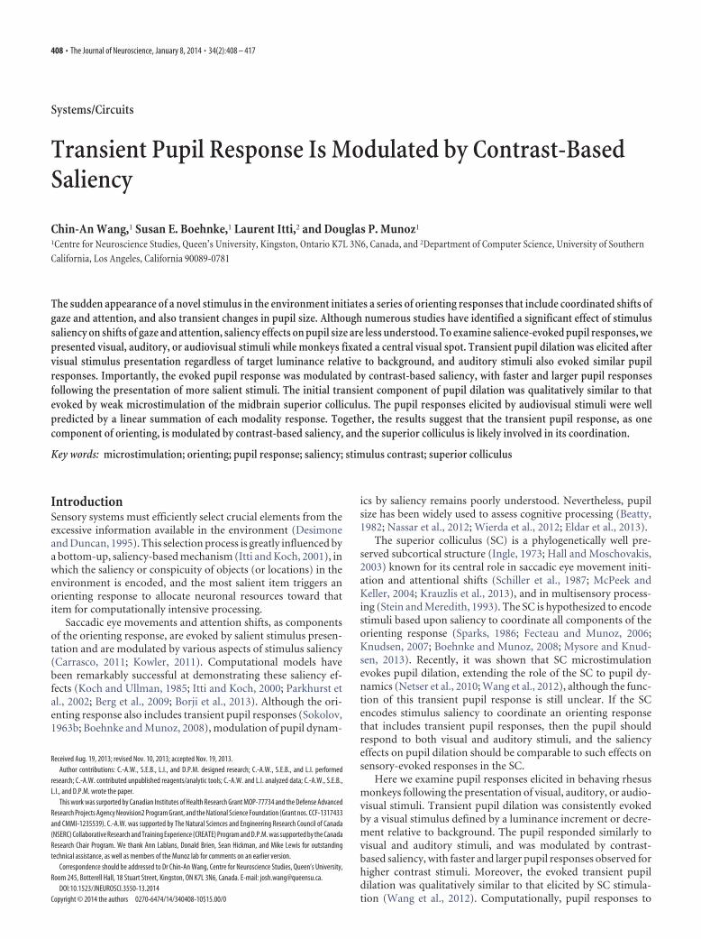
The sudden appearance of a novel stimulus in the environment initiates a series of orienting responses that include coordinated shifts ofgaze and attention, and also transient changes in pupil size. Although numerous studies have identified a significant effect of stimulussaliency on shifts of gaze and attention, saliency effects on pupil size are less understood. To examine salience-evoked pupil responses, wepresented visual, auditory, or audiovisual stimuli while monkeys fixated a central visual spot. Transient pupil dilation was elicited aftervisual stimulus presentation regardless of target luminance relative to background, and auditory stimuli also evoked similar pupilresponses. Importantly, the evoked pupil response was modulated by contrast-based saliency, with faster and larger pupil responsesfollowing the presentation of more salient stimuli. The initial transient component of pupil dilation was qualitatively similar to thatevoked by weak microstimulation of the midbrain superior colliculus. The pupil responses elicited by audiovisual stimuli were wellpredicted by a linear summation of each modality response. Together, the results suggest that the transient pupil response, as onecomponent of orienting, is modulated by contrast-based saliency, and the superior colliculus is likely involved in its coordination.
Key words: microstimulation; orienting; pupil response; saliency; stimulus contrast; superior colliculus
IntroductionSensory systems must efficiently select crucial elements from theexcessive information available in the environment (Desimoneand Duncan, 1995). This selection process is greatly influenced bya bottom-up, saliency-based mechanism (Itti and Koch, 2001), inwhich the saliency or conspicuity of objects (or locations) in theenvironment is encoded, and the most salient item triggers anorienting response to allocate neuronal resources toward thatitem for computationally intensive processing.
Saccadic eye movements and attention shifts, as componentsof the orienting response, are evoked by salient stimulus presen-tation and are modulated by various aspects of stimulus saliency(Carrasco, 2011; Kowler, 2011). Computational models havebeen remarkably successful at demonstrating these saliency ef-fects (Koch and Ullman, 1985; Itti and Koch, 2000; Parkhurst etal., 2002; Berg et al., 2009; Borji et al., 2013). Although the ori-enting response also includes transient pupil responses (Sokolov,1963b; Boehnke and Munoz, 2008), modulation of pupil dynam-
ics by saliency remains poorly understood. Nevertheless, pupilsize has been widely used to assess cognitive processing (Beatty,1982; Nassar et al., 2012; Wierda et al., 2012; Eldar et al., 2013).
The superior colliculus (SC) is a phylogenetically well pre-served subcortical structure (Ingle, 1973; Hall and Moschovakis,2003) known for its central role in saccadic eye movement initi-ation and attentional shifts (Schiller et al., 1987; McPeek andKeller, 2004; Krauzlis et al., 2013), and in multisensory process-ing (Stein and Meredith, 1993). The SC is hypothesized to encodestimuli based upon saliency to coordinate all components of theorienting response (Sparks, 1986; Fecteau and Munoz, 2006;Knudsen, 2007; Boehnke and Munoz, 2008; Mysore and Knud-sen, 2013). Recently, it was shown that SC microstimulationevokes pupil dilation, extending the role of the SC to pupil dy-namics (Netser et al., 2010; Wang et al., 2012), although the func-tion of this transient pupil response is still unclear. If the SCencodes stimulus saliency to coordinate an orienting responsethat includes transient pupil responses, then the pupil shouldrespond to both visual and auditory stimuli, and the saliencyeffects on pupil dilation should be comparable to such effects onsensory-evoked responses in the SC.
Here we examine pupil responses elicited in behaving rhesusmonkeys following the presentation of visual, auditory, or audio-visual stimuli. Transient pupil dilation was consistently evokedby a visual stimulus defined by a luminance increment or decre-ment relative to background. The pupil responded similarly tovisual and auditory stimuli, and was modulated by contrast-based saliency, with faster and larger pupil responses observed forhigher contrast stimuli. Moreover, the evoked transient pupildilation was qualitatively similar to that elicited by SC stimula-tion (Wang et al., 2012). Computationally, pupil responses to
Received Aug. 19, 2013; revised Nov. 10, 2013; accepted Nov. 19, 2013.Author contributions: C.-A.W., S.E.B., L.I., and D.P.M. designed research; C.-A.W., S.E.B., and L.I. performed
research; C.-A.W. contributed unpublished reagents/analytic tools; C.-A.W. and L.I. analyzed data; C.-A.W., S.E.B.,L.I., and D.P.M. wrote the paper.
This work was surported by Canadian Institutes of Health Research Grant MOP-77734 and the Defense AdvancedResearch Projects Agency Neovision2 Program Grant, and the National Science Foundation (Grant nos. CCF-1317433and CMMI-1235539). C.-A.W. was supported by The Natural Sciences and Engineering Research Council of Canada(NSERC) Collaborative Research and Training Experience (CREATE) Program and D.P.M. was supported by the CanadaResearch Chair Program. We thank Ann Lablans, Donald Brien, Sean Hickman, and Mike Lewis for outstandingtechnical assistance, as well as members of the Munoz lab for comments on an earlier version.
Correspondence should be addressed to Dr Chin-An Wang, Centre for Neuroscience Studies, Queen’s University,Room 245, Botterell Hall, 18 Stuart Street, Kingston, ON K7L 3N6, Canada. E-mail: [email protected].
DOI:10.1523/JNEUROSCI.3550-13.2014Copyright © 2014 the authors 0270-6474/14/340408-10$15.00/0
408 • The Journal of Neuroscience, January 8, 2014 • 34(2):408 – 417
audiovisual stimuli are well approximated by the sum of the re-sponses evoked by the stimulus components. Overall, our resultsdemonstrate the effects of contrast-based saliency modulation ontransient pupil responses, and suggest that the SC may coordinatesuch behavior.
Materials and MethodsAnimal preparation and equipment. All protocols used in this study wereapproved by Queen’s University Animal Care Committee in accordancewith the Canadian Council on Animal Care policies on the use of labo-ratory animals. Experiments were performed on four male rhesus mon-keys (Macaca mulatta; 10 –12 kg). Specifically, monkeys U and D weretrained to perform the visual and auditory stimulus experiments. Themicrostimulation experiment was performed on monkeys Q and Y, andthese data were published previously (Wang et al., 2012). The methods ofsurgical procedures and data collection have been described in detailpreviously (Marino et al., 2008). Eye position was measured by the scleralsearch coil technique (Robinson, 1963), and horizontal and vertical eyepositions were digitized at 1000 Hz. Pupil diameter was measured usinga video-based eye tracker (Eyelink-II, SR Research) at a rate of 500 Hz.
To calibrate actual pupil size derived from output pupil diameter val-ues recorded from the Eyelink II, we used the following method (Steinerand Barry, 2011; Wang et al., 2012). We made a number of different-sizedfalse pupils (6 pupils from 1 to 6 mm in diameter) and placed them at theexact same position as the monkey’s pupil position during data record-ing. Eyelink pupil values from false pupils were used to transform Eyelinkpupil values recorded from real monkeys to actual pupil diameter,and pupil size resolution was �0.01 mm. Critically, pupil-size datacan be distorted by eye movements because the size of the pupildepends on the subject’s gaze angle in a video-based eye tracker. Inaddition, saccade generation could confound our test of the role ofstimulus saliency on the evoked pupil responses, because any ob-served differences in pupil response between different salient condi-tions could be triggered by saccadic eye movement itself, rather thanstimulus saliency per se. To maintain an accurate measure of pupilsize before, during, and after stimulation, and to avoid an influence ofthe saccadic eye movements, monkeys were required to maintainvisual fixation on a point at the center of the screen throughout thetrial except for the trials that required saccadic eye movements.
Stimulus presentation and data acquisition were controlled by aUNIX-based real-time data control system (REX; Hays et al., 1982). Eyeposition and pupil diameter were recorded in a multichannel data acqui-sition system (Plexon). Stimuli were presented on a CRT monitor at ascreen resolution of 1024 � 768 pixels (75 Hz noninterlaced), subtendinga viewing angle of 54° � 44°. Luminance measures for visual stimuli weredetermined using a luminance meter (CS100, Konica Minolta), andsound pressure levels (SPLs) for auditory stimuli were determined usinga sound level meter (type 2239, Bruel and Kjaer).
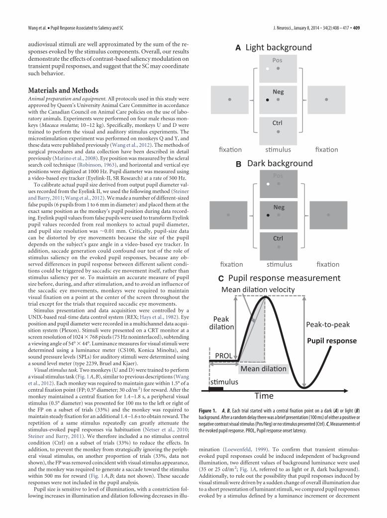
Visual stimulus task. Two monkeys (U and D) were trained to performa visual stimulus task (Fig. 1 A, B), similar to previous descriptions (Wanget al., 2012). Each monkey was required to maintain gaze within 1.5° of acentral fixation point (FP; 0.5° diameter; 30 cd/m 2) for reward. After themonkey maintained a central fixation for 1.4 –1.8 s, a peripheral visualstimulus (0.5° diameter) was presented for 100 ms to the left or right ofthe FP on a subset of trials (33%) and the monkey was required tomaintain steady fixation for an additional 1.4 –1.6 s to obtain reward. Therepetition of a same stimulus repeatedly can greatly attenuate thestimulus-evoked pupil responses via habituation (Netser et al., 2010;Steiner and Barry, 2011). We therefore included a no stimulus controlcondition (Ctrl) on a subset of trials (33%) to reduce the effects. Inaddition, to prevent the monkey from strategically ignoring the periph-eral visual stimulus, on another proportion of trials (33%, data notshown), the FP was removed coincident with visual stimulus appearance,and the monkey was required to generate a saccade toward the stimuluswithin 500 ms for reward (Fig. 1 A, B; data not shown). These saccaderesponses were not included in the pupil analysis.
Pupil size is sensitive to level of illumination, with a constriction fol-lowing increases in illumination and dilation following decreases in illu-
mination (Loewenfeld, 1999). To confirm that transient stimulus-evoked pupil responses could be induced independent of backgroundillumination, two different values of background luminance were used(35 or 25 cd/m 2; Fig. 1A, referred to as light or B, dark background).Additionally, to rule out the possibility that pupil responses induced byvisual stimuli were driven by a sudden change of overall illumination dueto a short presentation of luminant stimuli, we compared pupil responsesevoked by a stimulus defined by a luminance increment or decrement
Light background
s�mulus fixa�onfixa�on
Pos
Neg
Ctrl
Dark background
s�mulus fixa�onfixa�on
Pos
Neg
Ctrl
Pupil response measurement
Time
Mean dila�on velocity
Peakdila�on
PROL
Pupil response
Peak-to-peak
Mean dila�on
s�mulus
A
B
C
Figure 1. A, B, Each trial started with a central fixation point on a dark (A) or light (B)background. After a random delay there was a brief presentation (100 ms) of either a positive ornegative contrast visual stimulus (Pos/Neg) or no stimulus presented (Ctrl). C, Measurements ofthe evoked pupil response. PROL, Pupil response onset latency.
Wang et al. • Pupil Response Associated to Saliency and SC J. Neurosci., January 8, 2014 • 34(2):408 – 417 • 409
(Fig. 1 A, B) relative to background (positive/negative contrast: 58 or 16cd/m 2 on a light background, and 40 or 12 cd/m 2 on a dark background).If pupil responses elicited by salient stimuli were driven by illuminationchanges, the pupil responses induced by a positive or negative contraststimulus should be qualitatively different because one decreases but an-other increases overall illumination. All conditions were randomly inter-leaved. Data collected across days/sessions were collapsed for analysis.
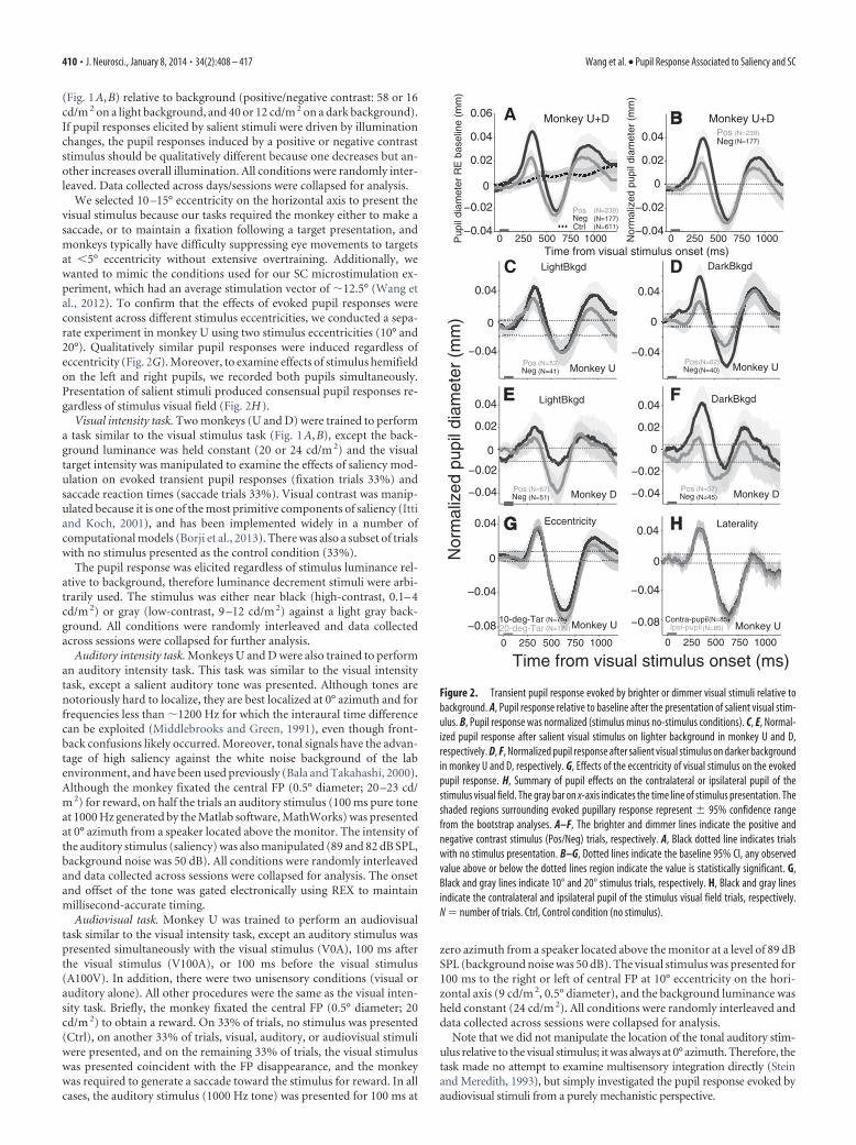
We selected 10 –15° eccentricity on the horizontal axis to present thevisual stimulus because our tasks required the monkey either to make asaccade, or to maintain a fixation following a target presentation, andmonkeys typically have difficulty suppressing eye movements to targetsat �5° eccentricity without extensive overtraining. Additionally, wewanted to mimic the conditions used for our SC microstimulation ex-periment, which had an average stimulation vector of �12.5° (Wang etal., 2012). To confirm that the effects of evoked pupil responses wereconsistent across different stimulus eccentricities, we conducted a sepa-rate experiment in monkey U using two stimulus eccentricities (10° and20°). Qualitatively similar pupil responses were induced regardless ofeccentricity (Fig. 2G). Moreover, to examine effects of stimulus hemifieldon the left and right pupils, we recorded both pupils simultaneously.Presentation of salient stimuli produced consensual pupil responses re-gardless of stimulus visual field (Fig. 2H ).
Visual intensity task. Two monkeys (U and D) were trained to performa task similar to the visual stimulus task (Fig. 1 A, B), except the back-ground luminance was held constant (20 or 24 cd/m 2) and the visualtarget intensity was manipulated to examine the effects of saliency mod-ulation on evoked transient pupil responses (fixation trials 33%) andsaccade reaction times (saccade trials 33%). Visual contrast was manip-ulated because it is one of the most primitive components of saliency (Ittiand Koch, 2001), and has been implemented widely in a number ofcomputational models (Borji et al., 2013). There was also a subset of trialswith no stimulus presented as the control condition (33%).
The pupil response was elicited regardless of stimulus luminance rel-ative to background, therefore luminance decrement stimuli were arbi-trarily used. The stimulus was either near black (high-contrast, 0.1– 4cd/m 2) or gray (low-contrast, 9 –12 cd/m 2) against a light gray back-ground. All conditions were randomly interleaved and data collectedacross sessions were collapsed for further analysis.
Auditory intensity task. Monkeys U and D were also trained to performan auditory intensity task. This task was similar to the visual intensitytask, except a salient auditory tone was presented. Although tones arenotoriously hard to localize, they are best localized at 0° azimuth and forfrequencies less than �1200 Hz for which the interaural time differencecan be exploited (Middlebrooks and Green, 1991), even though front-back confusions likely occurred. Moreover, tonal signals have the advan-tage of high saliency against the white noise background of the labenvironment, and have been used previously (Bala and Takahashi, 2000).Although the monkey fixated the central FP (0.5° diameter; 20 –23 cd/m 2) for reward, on half the trials an auditory stimulus (100 ms pure toneat 1000 Hz generated by the Matlab software, MathWorks) was presentedat 0° azimuth from a speaker located above the monitor. The intensity ofthe auditory stimulus (saliency) was also manipulated (89 and 82 dB SPL,background noise was 50 dB). All conditions were randomly interleavedand data collected across sessions were collapsed for analysis. The onsetand offset of the tone was gated electronically using REX to maintainmillisecond-accurate timing.
Audiovisual task. Monkey U was trained to perform an audiovisualtask similar to the visual intensity task, except an auditory stimulus waspresented simultaneously with the visual stimulus (V0A), 100 ms afterthe visual stimulus (V100A), or 100 ms before the visual stimulus(A100V). In addition, there were two unisensory conditions (visual orauditory alone). All other procedures were the same as the visual inten-sity task. Briefly, the monkey fixated the central FP (0.5° diameter; 20cd/m 2) to obtain a reward. On 33% of trials, no stimulus was presented(Ctrl), on another 33% of trials, visual, auditory, or audiovisual stimuliwere presented, and on the remaining 33% of trials, the visual stimuluswas presented coincident with the FP disappearance, and the monkeywas required to generate a saccade toward the stimulus for reward. In allcases, the auditory stimulus (1000 Hz tone) was presented for 100 ms at
zero azimuth from a speaker located above the monitor at a level of 89 dBSPL (background noise was 50 dB). The visual stimulus was presented for100 ms to the right or left of central FP at 10° eccentricity on the hori-zontal axis (9 cd/m 2, 0.5° diameter), and the background luminance washeld constant (24 cd/m 2). All conditions were randomly interleaved anddata collected across sessions were collapsed for analysis.
Note that we did not manipulate the location of the tonal auditory stim-ulus relative to the visual stimulus; it was always at 0° azimuth. Therefore, thetask made no attempt to examine multisensory integration directly (Steinand Meredith, 1993), but simply investigated the pupil response evoked byaudiovisual stimuli from a purely mechanistic perspective.
0 250 500 750 1000−0.04
−0.02
0
0.02
0.04
0.06
Pup
il di
amet
er R
E b
asel
ine
(mm
)
0 250 500 750 1000−0.04
−0.02
0
0.02
0.04
Nor
mal
ized
pup
il di
amet
er (
mm
)
−0.04
0
0.04
−0.04
0
0.04
−0.04
−0.02
0
0.02
0.04
Nor
mal
ized
pup
il di
amet
er (
mm
)
−0.04
−0.02
0
0.02
0.04
B B
C D
EE F
CtrlNegPos
NegPos
Monkey U+D Monkey U+D
Monkey U
LightBkgd DarkBkgd
LightBkgd DarkBkgd
0 250 500 750 1000
−0.08
−0.04
0
0.04
Time from visual stimulus onset (ms)
20-deg-Tar10-deg-Tar
Monkey U
GG
0 250 500 750 1000
−0.08
−0.04
0
0.04
Ipsi-pupilContra-pupil
HH
Monkey U
(N=239)(N=177)(N=611)
(N=239)(N=177)
NegPos(N=62)
(N=40)Monkey UNegPos (N=53)
(N=41)
Monkey DNegPos (N=67)
(N=51) Monkey DNegPos (N=57)
(N=45)
(N=122)(N=75)
(N=85)(N=85)
Eccentricity Laterality
Time from visual stimulus onset (ms)
A
Figure 2. Transient pupil response evoked by brighter or dimmer visual stimuli relative tobackground. A, Pupil response relative to baseline after the presentation of salient visual stim-ulus. B, Pupil response was normalized (stimulus minus no-stimulus conditions). C, E, Normal-ized pupil response after salient visual stimulus on lighter background in monkey U and D,respectively. D, F, Normalized pupil response after salient visual stimulus on darker backgroundin monkey U and D, respectively. G, Effects of the eccentricity of visual stimulus on the evokedpupil response. H, Summary of pupil effects on the contralateral or ipsilateral pupil of thestimulus visual field. The gray bar on x-axis indicates the time line of stimulus presentation. Theshaded regions surrounding evoked pupillary response represent � 95% confidence rangefrom the bootstrap analyses. A–F, The brighter and dimmer lines indicate the positive andnegative contrast stimulus (Pos/Neg) trials, respectively. A, Black dotted line indicates trialswith no stimulus presentation. B–G, Dotted lines indicate the baseline 95% CI, any observedvalue above or below the dotted lines region indicate the value is statistically significant. G,Black and gray lines indicate 10° and 20° stimulus trials, respectively. H, Black and gray linesindicate the contralateral and ipsilateral pupil of the stimulus visual field trials, respectively.N � number of trials. Ctrl, Control condition (no stimulus).
410 • J. Neurosci., January 8, 2014 • 34(2):408 – 417 Wang et al. • Pupil Response Associated to Saliency and SC
SC microstimulation task. Aspects of the microstimulation data reportedherein including detailed methods were published previously using differentanalysis techniques (Wang et al., 2012). Briefly, two monkeys (Q and Y) weretrained to perform a simple fixation task, except instead of presenting asensory stimulus, a train of microstimulation pulses (100 ms, 300 Hz; range,5–50 �A) was delivered to the intermediate layers of the SC (SCi) on a subsetof trials (50%). To get an accurate estimation of pupil change linked tomicrostimulation, we used current values that were 50–70% of the thresholdcurrent to evoke saccades, so that no eye movements were triggered. Notethat the latency of microstimulation evoked pupil dilation would be shorterif suprathreshold microstimulation was used. On another 50% of trials, therewas no microstimulation and the monkey was required to maintain centralfixation for reward. Luminance levels for FP and background were identicalto the visual stimulus task, and all conditions were randomly interleaved.Microstimulation was delivered to 28 sites in the SCi (18 and 10 in monkeysQ and Y, respectively).
Data analysis. The initial transient component of the evoked pupilresponse was of primary interest here (Fig. 1C). Therefore, the analysesfocused on the initial transient pupil response. Trials with blinks or aneye position deviation of �1.5° from the central FP during the requiredperiod of fixation were excluded from analysis. Because the pupil re-sponse was confirmed to be consensual, only pupil diameter of the righteye was recorded for data analysis. The same procedure was used toquantify pupil responses previously (Wang et al., 2012). For each trial,original pupil diameter values were subtracted from the baseline pupildiameter value determined by averaging pupil size from 250 ms before tothe onset of stimulation (visual/auditory/electrical; Bala and Takahashi,2000; Moresi et al., 2008). Because pupil size increased slightly even whenthere was no stimulus presented (Fig. 2A, dotted line), to simplify datapresentation and quantification, we normalized pupil diameter values bycontrasting the sensory/electrical stimulation (Fig. 2A, solid line) versusno-stimulation (Fig. 2A, dotted line) conditions directly. Specifically,
pupil values from each stimulation trial were contrasted to the averagepupil value from all no-stimulation trials (Fig. 2 A, B).
In contrast to our previous study (Wang et al., 2012), we used a bootstrapmethod to inform the statistical significance of the comparison by perform-ing a random sampling of pupil values derived from each recording trial with1500 repetitions. This resulted in a normally distributed cluster of pointscentered on the mean of selected pupil values (clusters not shown, normaldistribution was verified by the Kolmogorov–Smirnov test). The 95% con-fidence interval (CI) derived from the bootstrap analysis was depicted inshading (Fig. 3A) or in error bar (Fig. 3B), and the planned comparison wasconsidered to be statistically significant when two compared CIs did notoverlap. The baseline CI (Fig. 2B–H, dotted lines) was defined by the onsetpupil value calculated by averaging pupil values from higher or lower 95% CIrange from 25 ms before to 25 ms after stimulation (�25 to �25 ms), and apupil value at any given time exceeded the baseline CI range was consideredto be statistically significant.
Figure 1C shows a number of measurements we extracted to capturedynamics of evoked transient pupil responses (Beatty and Lucero-Wagoner, 2000). The pupil response onset latency (PROL) was com-puted using the average of all trials in a given condition rather thantrial-by-trial due to a low signal-to-noise ratio. PROL was defined as theearliest point at which average pupil magnitude exceeded the baseline CIrange. The remaining pupil measurements were computed on a trial-by-trial basis. We also characterized several size-related pupil measure-ments. The mean dilation was defined as the average of the pupil sizefrom PROL to the time where the pupil reached to the highest valuewithin 400 ms (arbitrarily selected from visual inspection) after the stim-ulation onset (visual/auditory/electrical). The peak dilation was definedas the maximum value observed within 400 ms after the stimulationonset. The peak-to-peak pupil response was defined as the peak dilationminus the pupil size at the time where the pupil reached to the lowestvalue within 350 ms after the peak dilation (arbitrarily selected from
0 250 500 750 1000
−0.04
−0.02
0
0.02
0.04
Time from visual stimulus onset (ms)Nor
mal
ized
pup
il di
amet
er (
mm
)
0 250 500 750 1000
−0.04
−0.02
0
0.02
0.04
High Low
150
170
190
SR
T (
ms)
Visual contrast
PROL
200
240
280
Res
pons
e la
tenc
y (m
s)
Mean Peak PeakToPeak
0.02
0.07
0.12R
espo
nse
size
(m
m)
Mean Velocity
0.2
0.3
0.4
Dila
tion
velo
city
(m
m/s
)
Monkey U
Monkey D
*
*
Monkey U Monkey D
LowHigh
LowHigh
(N=110)
(N=109)
Monkey U+D
LowHigh
(N=146)
(N=132)
LowHigh
Monkey U+D
LowHigh
Monkey U+D
N=642
*
*o
*
A B C
D E F
Figure 3. Effect of visual contrast-based saliency modulation on transiently evoked pupil responses. A, B, Normalized pupil dynamics after two levels of visual stimulus presentation (high-contrast and low-contrast) in monkey U and D, respectively. C, Saccade reaction times between the high and the low contrast conditions on different monkeys. D–F, Modulation of stimulus contraston the PROL (D); the mean size of pupil dilation, the peak size of pupil dilation, and peak-to-peak size of pupil response (E); and the mean velocity of pupil dilation (F ). A, B, Black and gray linesindicate the high and low contrast stimulus trials, respectively. Shaded regions surrounding evoked pupillary response traces represent � 95% CI. Gray bar on x-axis indicates the time line ofstimulus presentation and the dotted lines indicate the baseline � 95% confidence range. C, E, F, Error bars represent �95% CI. D–F, Dark and light bars indicate the high- and low-stimuluscontrast condition, respectively. *Differences are statistically significant (not overlap in the compared CIs). SRT, Saccade reaction times; High, high-contrast stimulus; Low, low-contrast stimulus;N � number of trials.
Wang et al. • Pupil Response Associated to Saliency and SC J. Neurosci., January 8, 2014 • 34(2):408 – 417 • 411
visual inspection). The mean dilation velocity was computed using thesame time window for the mean pupil dilation (positive and negativevalues indicate the dilating and constricting process, respectively).
Computational modeling. We considered a simple linear model to an-alyze pupil responses to audiovisual stimuli. This “additive” model sim-ply summed the responses obtained separately from each sensorymodality, after proper temporal alignment of each component (e.g., de-lay the visual response by 100 ms when the visual stimulus was presented100 ms after the auditory one).
ResultsVisual stimuli elicited transient pupil dilationThe sudden appearance of a visual stimulus resulted in a multi-phasic pupil response (Fig. 2A). Transient pupil dilation was firstelicited, followed by a constriction, and then second dilation,before pupil size returned to the control condition (no stimulus).For subsequent illustration and analysis, pupil responses evokedby stimuli (Fig. 2A, solid lines) were subtracted from the meanpupil response of the control condition (Fig. 2A, dotted line, B;see Materials and Methods).
Pupil size is sensitive to global changes in luminance (Loewen-feld, 1999), and a brief presentation of luminant stimuli could alterglobal luminance and therefore influence pupil size. If true, then thepupil responses induced by contrast increment- or decrement-stimuli should be different. The presentation of positive contraststimuli should result in a constriction because it increases overallillumination, whereas the presentation of negative contrast stimulishould cause initial dilation because it decreases overall illumination.
The results showed that initial transient pupil dilation was evokedregardless of stimulus luminance defined by positive or negativecontrast (Pos/Neg) relative to background (Fig. 2B, brighter or dim-mer solid lines, respectively), although statistically larger pupil dila-tion was elicited by the presentation of negative contrast stimuli.Moreover, a similar response was produced regardless of backgroundluminanceandacross twodifferentmonkeys(Fig.2C–F).Together, theresults suggest that theevokedtransientpupil responsewasmediatedbya mechanism dissociable from the pupil light reflex pathway that regu-lates pupil size according to the global luminance (Loewenfeld, 1999;Gamlin, 2006). Given that the pupil responded similarly for each mon-key regardless of background luminance and direction of visual con-trast, these data were collapsed for subsequent analyses.
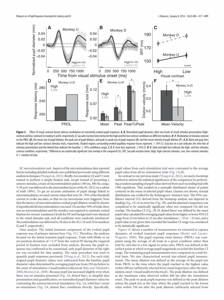
Pupil response scaled with visual intensityBrief presentation of a visual stimulus produced transient pupil re-sponses that were greater when high-contrast (more salient) stimuliwere presented (Fig. 3A). The effects of stimulus contrast on theevoked pupil response were consistent across two monkeys (Fig.3A,B). Comparable effects on SRT were evident on saccade trials(Fig. 3C; high/low: monkey U, 150 � 3 ms/158 � 5 ms; monkey D,176�4 ms/188�3 ms;�values were defined by�95% confidenceinterval and the comparison was statistically significant when twoCIs did not overlap; see Materials and Methods), as shown previ-ously (Marino and Munoz, 2009).
Visual stimulus contrast modulated the PROL, and the pupilresponded faster for high-contrast stimuli (Fig. 3D; PROL: high-
0 250 500 750 1000
−0.02
0
0.02
0.04
Time from auditory stimulus onset (ms)
Nor
mal
ized
pup
il di
amet
er (
mm
)
0 250 500 750 1000
−0.02
0
0.02
0.04
PROL
150
190
230
Res
pons
e la
tenc
y (m
s)
Mean Peak PeakToPeak
0.02
0.05
0.08
Res
pons
e si
ze (
mm
)
Mean Velocity
0.1
0.2
Dila
tion
velo
city
(m
m/s
)
Monkey U
LowHigh
LowHigh
LowHigh
Monkey D
Monkey U+D Monkey U+D Monkey U+D
LowHigh
(N=172)(N=171)
LowHigh
(N=182)(N=176)
A
C D E
B
Figure 4. Effect of auditory contrast-based saliency modulation on transiently evoked pupil responses. A, B, Pupil response evoked by auditory stimuli (high- and low-intensity) in monkey U andD, respectively. C–E, Modulation of auditory stimulus intensity on the PROL (C); the mean size of pupil dilation, the peak size of pupil dilation, and peak-to-peak size of pupil response (D); and themean velocity of pupil dilation (E). A, B, Black and gray lines indicate the high- and low-contrast stimulus trials, respectively. Shaded regions surrounding evoked pupillary response tracesrepresent � 95% CI. Gray bar on x-axis indicates the time line of stimulus presentation and the dotted lines indicate the baseline �95% confidence range. D, E, Error bars represent � 95% CI. C–E,Dark and light bars indicate the high and low auditory-stimulus intensity condition, respectively. High, High-auditory intensity stimulus; Low, low-auditory intensity stimulus; N � number of trials.
412 • J. Neurosci., January 8, 2014 • 34(2):408 – 417 Wang et al. • Pupil Response Associated to Saliency and SC
contrast, 222 ms; low-contrast, 259 ms). Moreover, the size-related pupil responses were also influenced by stimulus contrast.High-contrast visual stimuli elicited statistically larger peak dila-tion (Fig. 3E; high contrast, 0.057 � 0.007 mm; low-contrast,0.041 � 0.008 mm) and peak-to-peak response (Fig. 3E; highcontrast, 0.13 � 0.007 mm; low-contrast, 0.11 � 0.007 mm), andthe difference in mean dilation was marginally significant (Fig.3E; high-contrast, 0.036 � 0.008 mm; low-contrast, 0.025 �0.008 mm). The mean velocity of dilation was also modulated byvisual contrast (Fig. 3F; high-contrast, 0.36 � 0.04 mm/s; low-contrast, 0.23 � 0.07 mm/s).
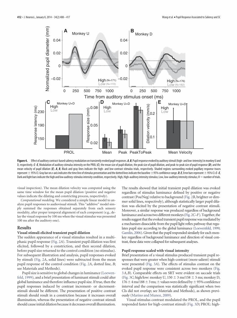
Auditory stimuli elicited transient pupil dilationIf the above evoked pupil responses were driven by stimulus sa-liency, they should also be elicited with nonvisual stimuli. Figure4A illustrates the effects of auditory stimulus presentation on thepupil. Presentation of an auditory stimulus also resulted in amultiphasic pupil response that was similar to that elicited by avisual stimulus. The initial phasic response was evoked, withtransient pupil dilation followed by a constriction, and secondarydilation before the pupil size merged with the no-stimulus con-dition. Two monkeys showed a similar pattern of auditory sa-liency modulation (Fig. 4A,B).
Manipulation of auditory intensity had similar effects onpupil size, although they did not reach significance. Auditorystimulus intensity influenced the timing of pupil responses:PROL was 159 ms for high-intensity auditory stimuli, and 223 msfor low-intensity stimuli (Fig. 4C; the observed pupil values onthe low-intensity condition overlapped with the upper baselinepupil values, so we selected the time with the highest observeddifference). Furthermore, presentation of a high-intensity audi-tory stimulus elicited a larger mean dilation (Fig. 4D; high-intensity, 0.028 � 0.006 mm; low-intensity, 0.021 � 0.007 mm),peak dilation (Fig. 4D; high-intensity, 0.042 � 0.006 mm; low-
intensity, 0.035 � 0.007 mm), and thepeak-to-peak response (Fig. 4D; high-intensity, 0.082 � 0.005 mm; low-intensity, 0.075 � 0.005 mm). However,there was no difference in the mean veloc-ity of dilation (Fig. 4E; high-intensity,0.17 � 0.05 mm/s; low-intensity, 0.17 �0.06 mm/s).
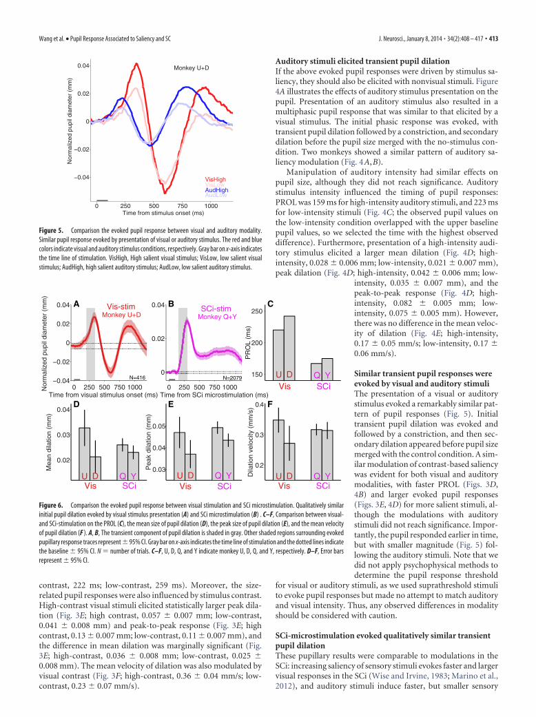
Similar transient pupil responses wereevoked by visual and auditory stimuliThe presentation of a visual or auditorystimulus evoked a remarkably similar pat-tern of pupil responses (Fig. 5). Initialtransient pupil dilation was evoked andfollowed by a constriction, and then sec-ondary dilation appeared before pupil sizemerged with the control condition. A sim-ilar modulation of contrast-based saliencywas evident for both visual and auditorymodalities, with faster PROL (Figs. 3D,4B) and larger evoked pupil responses(Figs. 3E, 4D) for more salient stimuli, al-though the modulations with auditorystimuli did not reach significance. Impor-tantly, the pupil responded earlier in time,but with smaller magnitude (Fig. 5) fol-lowing the auditory stimuli. Note that wedid not apply psychophysical methods todetermine the pupil response threshold
for visual or auditory stimuli, as we used suprathreshold stimulito evoke pupil responses but made no attempt to match auditoryand visual intensity. Thus, any observed differences in modalityshould be considered with caution.
SCi-microstimulation evoked qualitatively similar transientpupil dilationThese pupillary results were comparable to modulations in theSCi: increasing saliency of sensory stimuli evokes faster and largervisual responses in the SCi (Wise and Irvine, 1983; Marino et al.,2012), and auditory stimuli induce faster, but smaller sensory
0 250 500 750 1000
−0.04
−0.02
0
0.02
0.04
Time from stimulus onset (ms)
Nor
mal
ized
pup
il di
amet
er (
mm
)Monkey U+D
VisLowVisHigh
AudLowAudHigh
Figure 5. Comparison the evoked pupil response between visual and auditory modality.Similar pupil response evoked by presentation of visual or auditory stimulus. The red and bluecolors indicate visual and auditory stimulus conditions, respectively. Gray bar on x-axis indicatesthe time line of stimulation. VisHigh, High salient visual stimulus; VisLow, low salient visualstimulus; AudHigh, high salient auditory stimulus; AudLow, low salient auditory stimulus.
0 250 500 750 1000−0.04
−0.02
0
0.02
0.04
Time from visual stimulus onset (ms)
Nor
mal
ized
pup
il di
amet
er (
mm
)
0 250 500 750 1000
0
0.02
0.04
Time from SCi microstimulation (ms)
0.02
0.03
0.04
Mea
n di
latio
n (m
m)
0.03
0.04
0.05
Pea
k di
latio
n (m
m)
Vis SCi150
200
250
PR
OL
(ms)
0.2
0.3
0.4
Dila
tion
velo
city
(m
m/s
)
Vis SCiVis SCi Vis SCi
U D Q Y
U D Q Y U D Q Y U D Q Y
SCi-stimMonkey Q+YMonkey U+D
Vis-stim
N=2079N=416
A B C
D E F
Figure 6. Comparison the evoked pupil response between visual stimulation and SCi microstimulation. Qualitatively similarinitial pupil dilation evoked by visual stimulus presentation (A) and SCi microstimulation (B) . C–F, Comparison between visual-and SCi-stimulation on the PROL (C), the mean size of pupil dilation (D), the peak size of pupil dilation (E), and the mean velocityof pupil dilation (F ). A, B, The transient component of pupil dilation is shaded in gray. Other shaded regions surrounding evokedpupillary response traces represent � 95% CI. Gray bar on x-axis indicates the time line of stimulation and the dotted lines indicatethe baseline � 95% CI. N � number of trials. C–F, U, D, Q, and Y indicate monkey U, D, Q, and Y, respectively. D–F, Error barsrepresent � 95% CI.
Wang et al. • Pupil Response Associated to Saliency and SC J. Neurosci., January 8, 2014 • 34(2):408 – 417 • 413
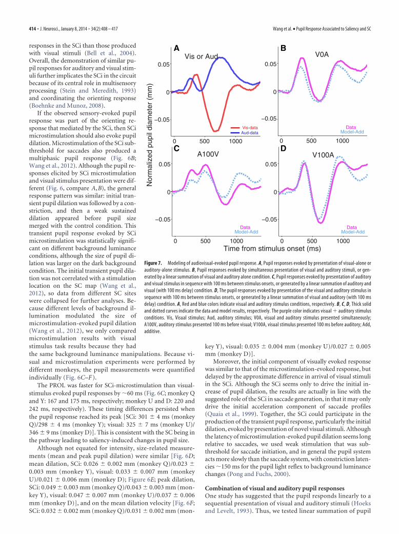
responses in the SCi than those producedwith visual stimuli (Bell et al., 2004).Overall, the demonstration of similar pu-pil responses for auditory and visual stim-uli further implicates the SCi in the circuitbecause of its central role in multisensoryprocessing (Stein and Meredith, 1993)and coordinating the orienting response(Boehnke and Munoz, 2008).
If the observed sensory-evoked pupilresponse was part of the orienting re-sponse that mediated by the SCi, then SCimicrostimulation should also evoke pupildilation. Microstimulation of the SCi sub-threshold for saccades also produced amultiphasic pupil response (Fig. 6B;Wang et al., 2012). Although the pupil re-sponses elicited by SCi microstimulationand visual stimulus presentation were dif-ferent (Fig. 6, compare A,B), the generalresponse pattern was similar: initial tran-sient pupil dilation was followed by a con-striction, and then a weak sustaineddilation appeared before pupil sizemerged with the control condition. Thistransient pupil response evoked by SCimicrostimulation was statistically signifi-cant on different background luminanceconditions, although the size of pupil di-lation was larger on the dark backgroundcondition. The initial transient pupil dila-tion was not correlated with a stimulationlocation on the SC map (Wang et al.,2012), so data from different SC siteswere collapsed for further analyses. Be-cause different levels of background il-lumination modulated the size ofmicrostimulation-evoked pupil dilation(Wang et al., 2012), we only comparedmicrostimulation results with visualstimulus task results because they hadthe same background luminance manipulations. Because vi-sual and microstimulation experiments were performed bydifferent monkeys, the pupil measurements were quantifiedindividually (Fig. 6C–F ).
The PROL was faster for SCi-microstimulation than visual-stimulus evoked pupil responses by �60 ms (Fig. 6C; monkey Qand Y: 167 and 175 ms, respectively; monkey U and D: 220 and242 ms, respectively). These timing differences persisted whenthe pupil response reached its peak [SCi: 301 � 4 ms (monkeyQ)/298 � 4 ms (monkey Y); visual: 325 � 7 ms (monkey U)/346 � 9 ms (monkey D)]. This is consistent with the SC being inthe pathway leading to saliency-induced changes in pupil size.
Although not equated for intensity, size-related measure-ments (mean and peak pupil dilation) were similar [Fig. 6D;mean dilation, SCi: 0.026 � 0.002 mm (monkey Q)/0.023 �0.003 mm (monkey Y), visual: 0.033 � 0.007 mm (monkeyU)/0.021 � 0.006 mm (monkey D); Figure 6E; peak dilation,SCi: 0.049 � 0.003 mm (monkey Q)/0.043 � 0.003 mm (mon-key Y), visual: 0.047 � 0.007 mm (monkey U)/0.037 � 0.006mm (monkey D)], and on the mean dilation velocity [Fig. 6F;SCi: 0.032 � 0.002 mm (monkey Q)/0.031 � 0.002 mm (mon-
key Y), visual: 0.035 � 0.004 mm (monkey U)/0.027 � 0.005mm (monkey D)].
Moreover, the initial component of visually evoked responsewas similar to that of the microstimulation-evoked response, butdelayed by the approximate difference in arrival of visual stimuliin the SCi. Although the SCi seems only to drive the initial in-crease of pupil dilation, the results are actually in line with thesuggested role of the SCi in saccade generation, in that it may onlydrive the initial acceleration component of saccade profiles(Quaia et al., 1999). Together, the SCi could participate in theproduction of the transient pupil response, particularly the initialdilation, evoked by presentation of novel visual stimuli. Althoughthe latency of microstimulation-evoked pupil dilation seems longrelative to saccades, we used weak stimulation that was sub-threshold for saccade initiation, and in general the pupil systemacts more slowly than the saccade system, with constriction laten-cies �150 ms for the pupil light reflex to background luminancechanges (Pong and Fuchs, 2000).
Combination of visual and auditory pupil responsesOne study has suggested that the pupil responds linearly to asequential presentation of visual and auditory stimuli (Hoeksand Levelt, 1993). Thus, we tested linear summation of pupil
0 500 1000
0
0 500 1000
−0.05
0
0.05
0 500 1000
−0.05
0
0.05
V0A
V100AA100V
DataModel-Add
0 500 1000
−0.05
0
0.05
Vis-dataAud-data
Vis or Aud
−0.05
0.05
DataModel-Add
DataModel-Add
Time from stimulus onset (ms)
Nor
mal
ized
pup
il di
amet
er (
mm
)
A B
DC
Figure 7. Modeling of audiovisual-evoked pupil response. A, Pupil responses evoked by presentation of visual-alone orauditory-alone stimulus. B, Pupil responses evoked by simultaneous presentation of visual and auditory stimuli, or gen-erated by a linear summation of visual and auditory alone condition. C, Pupil responses evoked by presentation of auditoryand visual stimulus in sequence with 100 ms between stimulus onsets, or generated by a linear summation of auditory andvisual (with 100 ms delay) condition. D, The pupil responses evoked by presentation of the visual and auditory stimulus insequence with 100 ms between stimulus onsets, or generated by a linear summation of visual and auditory (with 100 msdelay) condition. A, Red and blue colors indicate visual and auditory stimulus conditions, respectively. B, C, D, Thick solidand dotted curves indicate the data and model results, respectively. The purple color indicates visual auditory stimulusconditions. Vis, Visual stimulus; Aud, auditory stimulus; V0A, visual and auditory stimulus presented simultaneously;A100V, auditory stimulus presented 100 ms before visual; V100A, visual stimulus presented 100 ms before auditory; Add,additive.
414 • J. Neurosci., January 8, 2014 • 34(2):408 – 417 Wang et al. • Pupil Response Associated to Saliency and SC
responses elicited by the simultaneous or temporally offsetpresentation of visual and auditory stimuli. To examine theadditive model, a separate experiment was conducted withmonkey U, in which visual and auditory stimuli were eitherpresented alone or in combination. The presentation of a uni-modal visual or auditory stimulus evoked the typical pupilresponse (Fig. 6A). The presentation of visual and auditorystimuli simultaneously induced similar pupil responses, andcritically, the evoked pupil responses were comparable tothose derived from summing the visual- and auditory-onlypupil responses (Fig. 6B, Model-Add curve). We further testedthe linear addition assumption by presenting two stimuli insequence with 100 ms temporal asynchrony. The pupil re-sponses between actual data and time-shifted addition ofunimodal responses were similar regardless of the order ofmodality (Fig. 6C, Model-Add, auditory stimulus first; D,Model-Add, visual stimulus first). The simple additive modelprovided a good fit to the audiovisual data (root mean squareerrors: V0A, 0.29; A100V, 0.34; V100A, 0.28), although somequalitative features of the data were not predicted by themodel. In conclusion, our modeling results suggest that thepupil, in general, responded in a linearly additive manner toaudiovisual stimuli. Moreover, because the location of theauditory stimulus was not spatially aligned with the visualstimulus, it is possible that SCi activity induced by stimulipresented from different spatial locations could be integratedin a spatially nonlocalized manner to generate a coordinatedpupil response.
DiscussionThe orienting system responds to salient changes in the environ-ment by initiating a coordinated orienting response that includestransient pupil dilation (Sokolov, 1963a; Lynn, 1966). In mon-keys, the brief presentation of a visual or auditory stimulusevoked a reliable triphasic pupil response that included an initialdilation followed by a constriction and then a second dilation.The response was dissociated from illumination-dependent pu-pil modulation because it was evoked by a visual stimulus thatwas either a luminance increment or decrement relative to back-
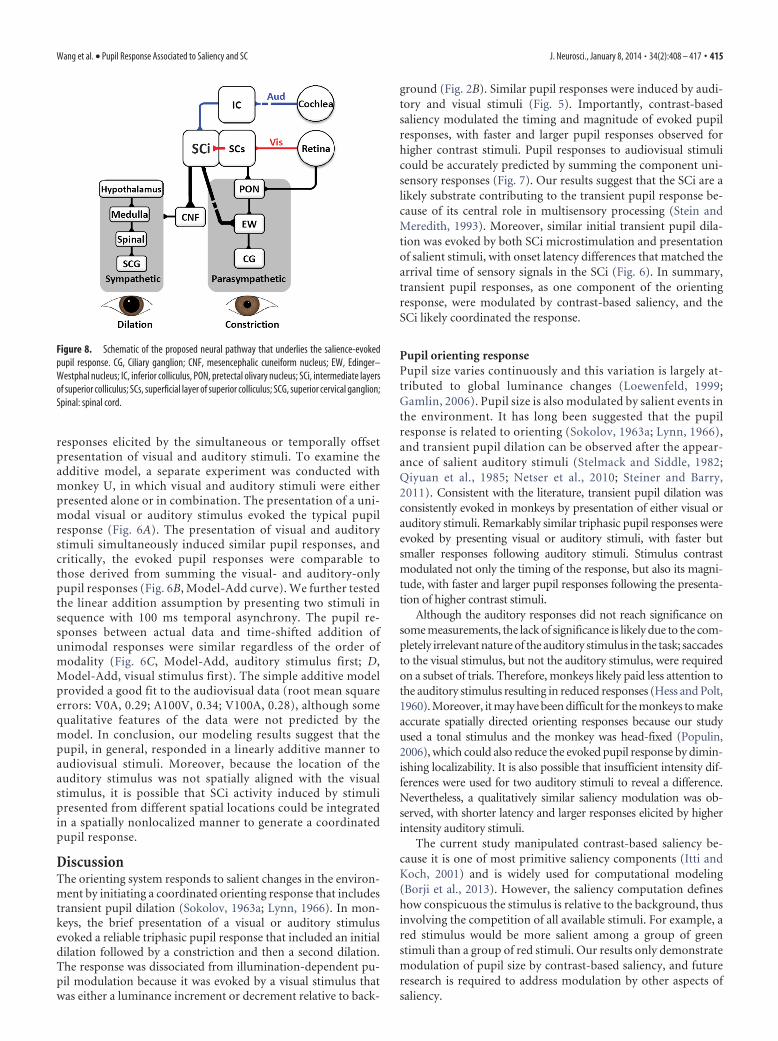
ground (Fig. 2B). Similar pupil responses were induced by audi-tory and visual stimuli (Fig. 5). Importantly, contrast-basedsaliency modulated the timing and magnitude of evoked pupilresponses, with faster and larger pupil responses observed forhigher contrast stimuli. Pupil responses to audiovisual stimulicould be accurately predicted by summing the component uni-sensory responses (Fig. 7). Our results suggest that the SCi are alikely substrate contributing to the transient pupil response be-cause of its central role in multisensory processing (Stein andMeredith, 1993). Moreover, similar initial transient pupil dila-tion was evoked by both SCi microstimulation and presentationof salient stimuli, with onset latency differences that matched thearrival time of sensory signals in the SCi (Fig. 6). In summary,transient pupil responses, as one component of the orientingresponse, were modulated by contrast-based saliency, and theSCi likely coordinated the response.
Pupil orienting responsePupil size varies continuously and this variation is largely at-tributed to global luminance changes (Loewenfeld, 1999;Gamlin, 2006). Pupil size is also modulated by salient events inthe environment. It has long been suggested that the pupilresponse is related to orienting (Sokolov, 1963a; Lynn, 1966),and transient pupil dilation can be observed after the appear-ance of salient auditory stimuli (Stelmack and Siddle, 1982;Qiyuan et al., 1985; Netser et al., 2010; Steiner and Barry,2011). Consistent with the literature, transient pupil dilation wasconsistently evoked in monkeys by presentation of either visual orauditory stimuli. Remarkably similar triphasic pupil responses wereevoked by presenting visual or auditory stimuli, with faster butsmaller responses following auditory stimuli. Stimulus contrastmodulated not only the timing of the response, but also its magni-tude, with faster and larger pupil responses following the presenta-tion of higher contrast stimuli.
Although the auditory responses did not reach significance onsome measurements, the lack of significance is likely due to the com-pletely irrelevant nature of the auditory stimulus in the task; saccadesto the visual stimulus, but not the auditory stimulus, were requiredon a subset of trials. Therefore, monkeys likely paid less attention tothe auditory stimulus resulting in reduced responses (Hess and Polt,1960). Moreover, it may have been difficult for the monkeys to makeaccurate spatially directed orienting responses because our studyused a tonal stimulus and the monkey was head-fixed (Populin,2006), which could also reduce the evoked pupil response by dimin-ishing localizability. It is also possible that insufficient intensity dif-ferences were used for two auditory stimuli to reveal a difference.Nevertheless, a qualitatively similar saliency modulation was ob-served, with shorter latency and larger responses elicited by higherintensity auditory stimuli.
The current study manipulated contrast-based saliency be-cause it is one of most primitive saliency components (Itti andKoch, 2001) and is widely used for computational modeling(Borji et al., 2013). However, the saliency computation defineshow conspicuous the stimulus is relative to the background, thusinvolving the competition of all available stimuli. For example, ared stimulus would be more salient among a group of greenstimuli than a group of red stimuli. Our results only demonstratemodulation of pupil size by contrast-based saliency, and futureresearch is required to address modulation by other aspects ofsaliency.
Figure 8. Schematic of the proposed neural pathway that underlies the salience-evokedpupil response. CG, Ciliary ganglion; CNF, mesencephalic cuneiform nucleus; EW, Edinger–Westphal nucleus; IC, inferior colliculus, PON, pretectal olivary nucleus; SCi, intermediate layersof superior colliculus; SCs, superficial layer of superior colliculus; SCG, superior cervical ganglion;Spinal: spinal cord.
Wang et al. • Pupil Response Associated to Saliency and SC J. Neurosci., January 8, 2014 • 34(2):408 – 417 • 415
Possible functions of the pupil orienting responseThe orienting response facilitates allocation of neuronal re-sources toward potentially important objects or locations toprepare the body for appropriate actions. It has been suggestedthat transient pupil dilation can slightly increase visual sensi-tivity (Sokolov, 1963a; Stelmack and Siddle, 1982), althoughthere is no direct evidence to support this. Importantly, it isnot clear how the different orienting components are coordi-nated to optimize performance. Our results showed that thePROL for visual stimuli was �225 ms, which is slightly longerthan the SRTs elicited by similar salient visual stimuli, �150 –200 ms (Marino and Munoz, 2009). Furthermore, the timerequired for a spatial attention shift induced by a peripheralcue (stimulus-driven) was regularly within 100 ms (Carrasco,2011). It is thus possible that the appearance of a salient visualstimulus first leads to a shift of attention and gaze to the objectto enhance its recognition. Immediately after foveation, thepupil starts to dilate, presumably to slightly increase visualsensitivity of the targeted object and its surrounding locations,providing a possible benefit for pupil dilation latency to belonger than saccade latency. This coordination can facilitateprocessing of the selected object to optimize an organism’sperformance.
The role of the SC in the pupil orienting responseThe SC is organized into a retinotopic map of contralateralvisual space with functionally and anatomically differentiatedlayers. The superficial layers receive inputs from early visualareas exclusively, including the retina and the primary visualcortex, whereas the SCi receives inputs from the superficial SCas well as multisensory and frontal-parietal areas, and thenprojects directly to the brainstem and the spinal cord to exe-cute the orienting response (White and Munoz, 2011). Be-cause the SCi integrates sensory-related and goal-directedsignals from cortical and subcortical areas, it is hypothesizedto encode stimuli based upon saliency and relevance to coor-dinate the orienting response (Sparks, 1986; Fecteau and Mu-noz, 2006; Knudsen, 2007; Boehnke and Munoz, 2008; Mysoreand Knudsen, 2013). Microstimulation of the SCi not onlyinduces saccades (Robinson, 1972) and biases spatial attention(Kustov and Robinson, 1996; Cavanaugh and Wurtz, 2004;Muller et al., 2005), but also evokes transient pupil responses(Netser et al., 2010; Wang et al., 2012). The similar pupilresponses evoked by presentation of both salient visualand auditory stimuli and the qualitatively similar transientpupil dilation elicited by SCi microstimulation suggests thatthe SCi is likely involved in control of salience-evoked pupilresponses.
Consistent with our observations on evoked pupil responses,changing the level of stimulus contrast modulates the timing andmagnitude of sensory responses in the SCi (Wise and Irvine,1983; Marino et al., 2012), with more salient stimuli inducingfaster and larger SC sensory responses. Sensory responses in theSCi are also faster but smaller following the presentation of audi-tory compared with visual stimuli (Bell et al., 2004), consistentwith our observations of the effect of modality on pupil modula-tion. Given these consistencies, we propose that the sensory re-sponses in the SCi induced by different modalities and differentlevels of stimulus contrast systematically influence the timing andmagnitude of orienting pupil responses.
Possible anatomical pathways underlying thesalience-related pupil responsePupil size is controlled by the interactions between the parasym-pathetic and sympathetic pathways (Loewenfeld, 1999). Theparasympathetic pathway underlies the illumination-dependentpupil modulation to optimize visual acuity (Leibowitz, 1952;Campbell and Gregory, 1960; Woodhouse, 1975; Laughlin,1992). Information about illumination is carried directly fromthe retina to the pretectal olivary nucleus (PON). PON neuronsproject bilaterally to the Edinger–Westphal (EW) nucleus, whichcontains the parasympathetic, preganglionic neurons that projectto the ciliary ganglion to control pupillary constriction muscles ofthe iris (Loewenfeld, 1999; Gamlin, 2006). In addition, pupil sizeis also controlled by the dilator muscle, innervated by sympatheticnerves from the superior cervical ganglion, which is driven by a sympa-thetic circuit originating in the hypothalamus (Loewenfeld, 1999).
Because weak microstimulation of the superficial SC did notevoke pupil dilation (Wang et al., 2012), pupil dilation should bemediated via connections from the SCi to the pupil pathways(Fig. 8). The SCi could inhibit parasympathetic pathways via in-direct inhibitory projections to the EW (Edwards and Henkel,1978; Harting et al., 1980; Grantyn and Grantyn, 1982) or canactivate parasympathetic pathways via direct excitatory projec-tions to the EW (Harting et al., 1980). The SCi also projects to themesencephalic cuneiform nucleus (CNF) (Harting, 1977; Huertaand Harting, 1984; May, 2006), a brainstem area regulatingstress-related and defensive responses (Dean et al., 1989; Korte etal., 1992). Stimulation of the CNF activates sympathetic vasomo-tor outflow (Verberne, 1995), which also influences pupil dila-tion (Loewenfeld, 1999). We propose that projections from theSCi to the EW and the CNF may underlie this response either byinhibiting the parasympathetic (pupil constriction) pathway, ac-tivating the sympathetic (pupil dilation) pathway, or both. TheSC contrast-based saliency coding could be translated to the pu-pil through the suggested pathways.
ReferencesBala AD, Takahashi TT (2000) Pupillary dilation response as an indicator of
auditory discrimination in the barn owl. J Comp Physiol A 186:425– 434.CrossRef Medline
Beatty J (1982) Task-evoked pupillary responses, processing load, and thestructure of processing resources. Psychol Bull 91:276 –292. CrossRefMedline
Beatty J, Lucero-Wagoner B (2000) The pupillary system. In: The handbookof psychophysiology (Caccioppo J, Tassinary LG, Berntson G, eds). Cam-bridge: UP.
Bell AH, Fecteau JH, Munoz DP (2004) Using auditory and visual stimuli toinvestigate the behavioral and neuronal consequences of reflexive covertorienting. J Neurophysiol 91:2172–2184. CrossRef Medline
Berg DJ, Boehnke SE, Marino RA, Munoz DP, Itti L (2009) Free viewing ofdynamic stimuli by humans and monkeys. J Vis 9(5):19 1–15. CrossRefMedline
Boehnke SE, Munoz DP (2008) On the importance of the transient visualresponse in the superior colliculus. Curr Opin Neurobiol 18:544 –551.CrossRef Medline
Borji A, Sihite DN, Itti L (2013) Quantitative analysis of human-modelagreement in visual saliency modeling: a comparative study. IEEE TransImage Process 22:55– 69. CrossRef Medline
Campbell FW, Gregory AH (1960) Effect of size of pupil on visual acuity.Nature 187:1121–1123. CrossRef Medline
Carrasco M (2011) Visual attention: the past 25 years. Vision Res 51:1484 –1525. CrossRef Medline
Cavanaugh J, Wurtz RH (2004) Subcortical modulation of attention coun-ters change blindness. J Neurosci 24:11236 –11243. CrossRef Medline
Dean P, Redgrave P, Westby GW (1989) Event or emergency? two responsesystems in the mammalian superior colliculus. Trends Neurosci 12:137–147. CrossRef Medline
416 • J. Neurosci., January 8, 2014 • 34(2):408 – 417 Wang et al. • Pupil Response Associated to Saliency and SC
Desimone R, Duncan J (1995) Neural mechanisms of selective visual atten-tion. Annu Rev Neurosci 18:193–222. CrossRef Medline
Edwards SB, Henkel CK (1978) Superior colliculus connections with theextraocular motor nuclei in the cat. J Comp Neurol 179:451– 467.CrossRef Medline
Eldar E, Cohen JD, Niv Y (2013) The effects of neural gain on attention andlearning. Nat Neurosci 16:1146 –1153. CrossRef Medline
Fecteau JH, Munoz DP (2006) Salience, relevance, and firing: a priority mapfor target selection. Trends Cogn Sci 10:382–390. CrossRef Medline
Gamlin PD (2006) The pretectum: connections and oculomotor-relatedroles. Prog Brain Res 151:379 – 405. CrossRef Medline
Grantyn A, Grantyn R (1982) Axonal patterns and sites of termination of catsuperior colliculus neurons projecting in the tecto-bulbo-spinal tract. ExpBrain Res 46:243–256. CrossRef Medline
Hall WC, Moschovakis A (2004) he superior colliculus: new approaches forstudying sensorimotor integration. Boca Raton, FL: CRC.
Harting JK (1977) Descending pathways from the superior collicullus: anautoradiographic analysis in the rhesus monkey (Macaca mulatta).J Comp Neurol 173:583– 612. CrossRef Medline
Harting JK, Huerta MF, Frankfurter AJ, Strominger NL, Royce GJ (1980)Ascending pathways from the monkey superior colliculus: an autoradio-graphic analysis. J Comp Neurol 192:853– 882. CrossRef Medline
Hays AV, Richmond BJ, Optican LM (1982) A UNIX-based multiple pro-cess system for real-time data acquisition and control. WESCON ConfProc 2:1–10.
Hess EH, Polt JM (1960) Pupil size as related to interest value of visualstimuli. Science 132:349 –350. CrossRef Medline
Hoeks B, Levelt WJ (1993) Pupillary dilation as a measure of attention: aquantitative system analysis. Behav Res Meth Instr Comput 25:16 –26.CrossRef
Huerta M, Harting J (1984) Connectional organization of the superior col-liculus. Trends Neurosci 7:286 –289. CrossRef
Ingle D (1973) Evolutionary perspectives on the function of the optic tec-tum. Brain Behav Evol 8:211–237. CrossRef Medline
Itti L, Koch C (2000) A saliency-based search mechanism for overt and co-vert shifts of visual attention. Vision Res 40:1489 –1506. CrossRef Medline
Itti L, Koch C (2001) Computational modelling of visual attention. Nat RevNeurosci 2:194 –203. CrossRef Medline
Knudsen EI (2007) Fundamental components of attention. Annu Rev Neu-rosci 30:57–78. CrossRef Medline
Koch C, Ullman S (1985) Shifts in selective visual attention: towards theunderlying neural circuitry. Hum Neurobiol 4:219 –227. Medline
Korte SM, Jaarsma D, Luiten PG, Bohus B (1992) Mesencephalic cuneiformnucleus and its ascending and descending projections serve stress-relatedcardiovascular responses in the rat. J Auton Nerv Syst 41:157–176.CrossRef Medline
Kowler E (2011) Eye movements: the past 25 years. Vision Res 51:1457–1483. CrossRef Medline
Krauzlis RJ, Lovejoy LP, Zenon A (2013) Superior colliculus and visual spa-tial attention. Annu Rev Neurosci 36:165–182. CrossRef Medline
Kustov AA, Robinson DL (1996) Shared neural control of attentional shiftsand eye movements. Nature 384:74 –77. CrossRef Medline
Laughlin SB (1992) Retinal information capacity and the function of thepupil. Ophthalmic Physiol Opt 12:161–164. Medline
Leibowitz H (1952) The effect of pupil size on visual acuity for photometri-cally equated test fields at various levels of luminance. J Opt Soc Am42:416 – 422. CrossRef Medline
Loewenfeld IE (1999) The pupil: anatomy, physiology, and clinical applica-tions. Boston: Butterworth-Heinemann.
Lynn R (1966) Attention, arousal and the orientation reaction. Oxford, UK:Pergamon.
Marino RA, Munoz DP (2009) The effects of bottom-up target luminanceand top-down spatial target predictability on saccadic reaction times. ExpBrain Res 197:321–335. CrossRef Medline
Marino RA, Rodgers CK, Levy R, Munoz DP (2008) Spatial relationships ofvisuomotor transformations in the superior colliculus map. J Neuro-physiol 100:2564 –2576. CrossRef Medline
Marino RA, Levy R, Boehnke S, White BJ, Itti L, Munoz DP (2012) Linkingvisual response properties in the superior colliculus to saccade behavior.Eur J Neurosci 35:1738 –1752. CrossRef Medline
May PJ (2006) The mammalian superior colliculus: laminar structure andconnections. Prog Brain Res 151:321–378. CrossRef Medline
McPeek RM, Keller EL (2004) Deficits in saccade target selection after inac-tivation of superior colliculus. Nat Neurosci 7:757–763. CrossRefMedline
Middlebrooks JC, Green DM (1991) Sound localization by human listeners.Annu Rev Psychol 42:135–159. CrossRef Medline
Moresi S, Adam JJ, Rijcken J, Van Gerven PW (2008) Cue validity effects inresponse preparation: a pupillometric study. Brain Res 1196:94 –102.CrossRef Medline
Muller JR, Philiastides MG, Newsome WT (2005) Microstimulation of thesuperior colliculus focuses attention without moving the eyes. Proc NatlAcad Sci U S A 102:524 –529. CrossRef Medline
Mysore SP, Knudsen EI (2013) A shared inhibitory circuit for both exoge-nous and endogenous control of stimulus selection. Nat Neurosci 16:473–478. CrossRef Medline
Nassar MR, Rumsey KM, Wilson RC, Parikh K, Heasly B, Gold JI (2012)Rational regulation of learning dynamics by pupil-linked arousal systems.Nat Neurosci 15:1040 –1046. CrossRef Medline
Netser S, Ohayon S, Gutfreund Y (2010) Multiple manifestations of micro-stimulation in the optic tectum: eye movements, pupil dilations, andsensory priming. J Neurophysiol 104:108 –118. CrossRef Medline
Parkhurst D, Law K, Niebur E (2002) Modeling the role of salience in theallocation of overt visual attention. Vision Res 42:107–123. CrossRefMedline
Pong M, Fuchs AF (2000) Characteristics of the pupillary light reflex in themacaque monkey: discharge patterns of pretectal neurons. J Neuro-physiol 84:964 –974. Medline
Populin LC (2006) Monkey sound localization: head-restrained versushead-unrestrained orienting. J Neurosci 26:9820 –9832. CrossRefMedline
Qiyuan J, Richer F, Wagoner BL, Beatty J (1985) The pupil and stimulusprobability. Psychophysiology 22:530 –534. CrossRef Medline
Quaia C, Lefèvre P, Optican LM (1999) Model of the control of saccades bysuperior colliculus and cerebellum. J Neurophysiol 82:999 –1018.Medline
Robinson DA (1963) A method of measuring eye movement using a scleralsearch coil in a magnetic field. IEEE Trans Biomed Eng 10:137–145.Medline
Robinson DA (1972) Eye movements evoked by collicular stimulation inthe alert monkey. Vision Res 12:1795–1808. CrossRef Medline
Schiller PH, Sandell JH, Maunsell JH (1987) The effect of frontal eye fieldand superior colliculus lesions on saccadic latencies in the rhesus monkey.J Neurophysiol 57:1033–1049. Medline
Sokolov EN (1963a) Perception and the conditioned reflex. Oxford:Pergamon.
Sokolov EN (1963b) Higher nervous functions; the orienting reflex. AnnuRev Physiol 25:545–580. CrossRef Medline
Sparks DL (1986) Translation of sensory signals into commands for controlof saccadic eye movements: role of primate superior colliculus. PhysiolRev 66:118 –171. Medline
Stein B, Meredith M (1993) The merging of the senses. Cambridge, MA:MIT.
Steiner GZ, Barry RJ (2011) Pupillary responses and event-related poten-tials as indices of the orienting reflex. Psychophysiology 48:1648 –1655.CrossRef Medline
Stelmack RM, Siddle DA (1982) Pupillary dilation as an index of the orient-ing reflex. Psychophysiology 19:706 –708. CrossRef Medline
Verberne AJ (1995) Cuneiform nucleus stimulation produces activation ofmedullary sympathoexcitatory neurons in rats. Am J Physiol 268:R752–R758. Medline
Wang CA, Boehnke SE, White BJ, Munoz DP (2012) Microstimulation ofthe monkey superior colliculus induces pupil dilation without evokingsaccades. J Neurosci 32:3629 –3636. CrossRef Medline
White BJ, Munoz DP (2011) The superior colliculus. In: Oxford handbookof eye movements (Liversedge S, Gilchrist I, Everling S, eds). Oxford:Oxford UP.
Wierda SM, van Rijn H, Taatgen NA, Martens S (2012) Pupil dilation de-convolution reveals the dynamics of attention at high temporal resolu-tion. Proc Natl Acad Sci U S A 109:8456 – 8460. CrossRef Medline
Wise LZ, Irvine DR (1983) Auditory response properties of neurons in deeplayers of cat superior colliculus. J Neurophysiol 49:674 – 685. Medline
Woodhouse JM (1975) The effect of pupil size on grating detection at vari-ous contrast levels. Vision Res 15:645– 648. CrossRef Medline
Wang et al. • Pupil Response Associated to Saliency and SC J. Neurosci., January 8, 2014 • 34(2):408 – 417 • 417