Embed Size (px)
Citation preview
SYSTEMIC PATHOLOGY
Pathology of Muscle
Laboratory
Paul Hanna Winter 2018
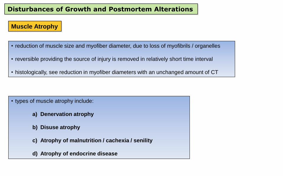
Disturbances of Growth and Postmortem Alterations
Muscle Atrophy
• reduction of muscle size and myofiber diameter, due to loss of myofibrils / organelles
• reversible providing the source of injury is removed in relatively short time interval
• histologically, see reduction in myofiber diameters with an unchanged amount of CT
• types of muscle atrophy include:
a) Denervation atrophy
b) Disuse atrophy
c) Atrophy of malnutrition / cachexia / senility
d) Atrophy of endocrine disease
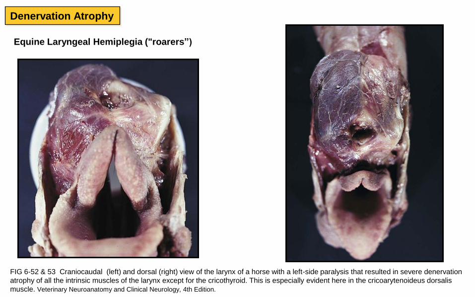
Equine Laryngeal Hemiplegia ("roarers”)
FIG 6-52 & 53 Craniocaudal (left) and dorsal (right) view of the larynx of a horse with a left-side paralysis that resulted in severe denervation
atrophy of all the intrinsic muscles of the larynx except for the cricothyroid. This is especially evident here in the cricoarytenoideus dorsalis
muscle. Veterinary Neuroanatomy and Clinical Neurology, 4th Edition.
Denervation Atrophy
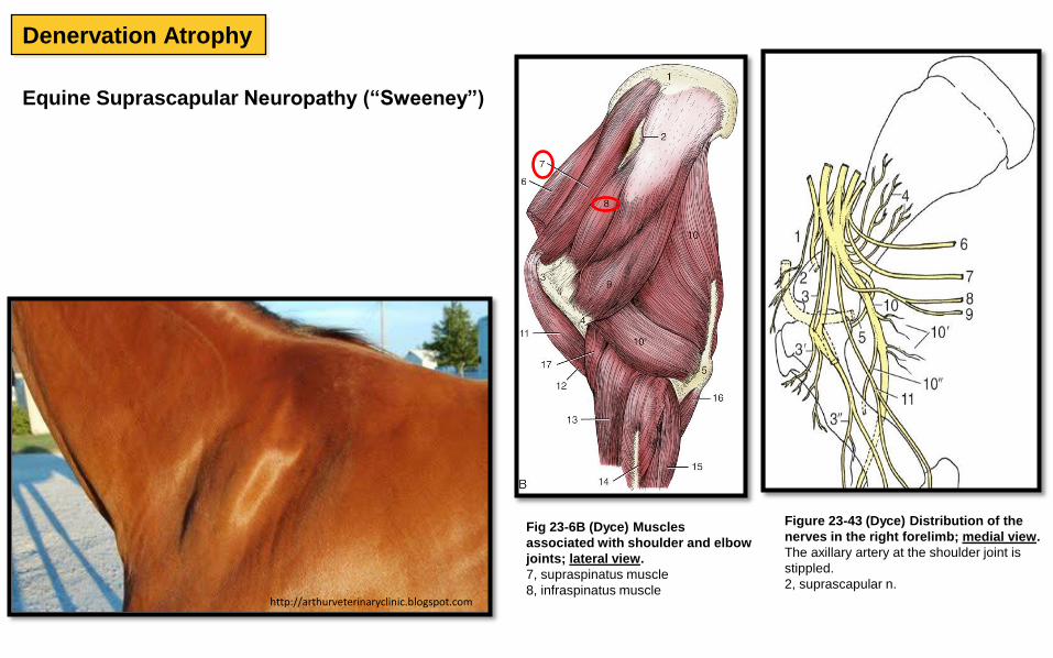
http://arthurveterinaryclinic.blogspot.com
Fig 23-6B (Dyce) Muscles
associated with shoulder and elbow
joints; lateral view.
7, supraspinatus muscle
8, infraspinatus muscle
Equine Suprascapular Neuropathy (“Sweeney”)
Figure 23-43 (Dyce) Distribution of the
nerves in the right forelimb; medial view.
The axillary artery at the shoulder joint is
stippled.
2, suprascapular n.
Denervation Atrophy
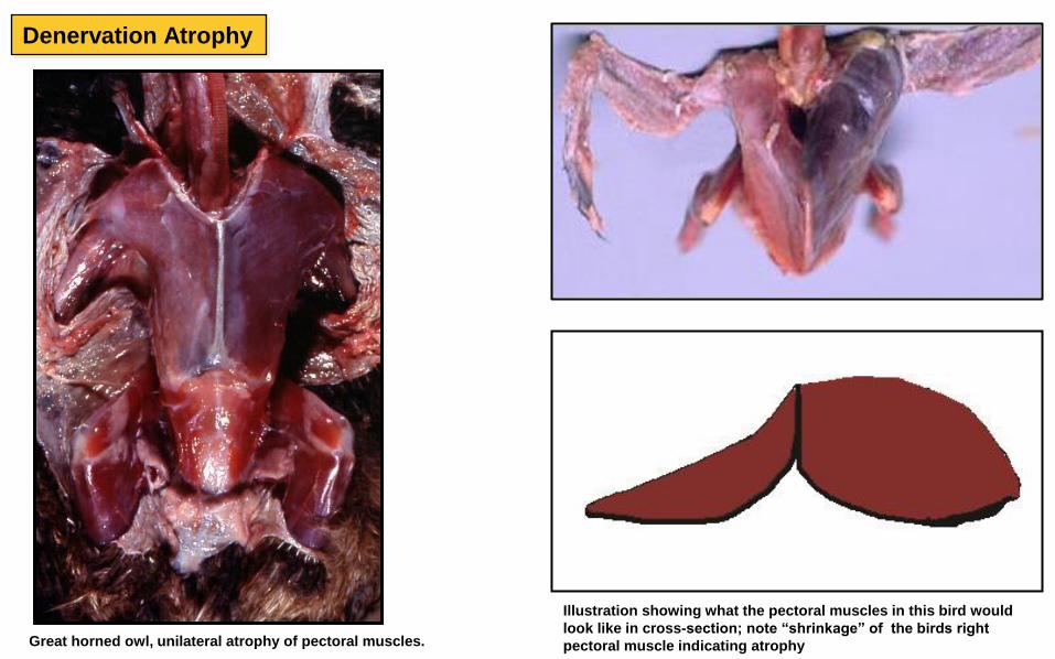
Great horned owl, unilateral atrophy of pectoral muscles.
Illustration showing what the pectoral muscles in this bird would
look like in cross-section; note “shrinkage” of the birds right
pectoral muscle indicating atrophy
Denervation Atrophy
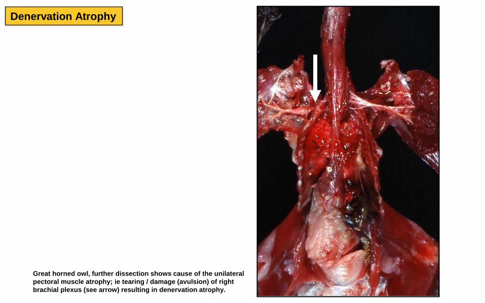
Great horned owl, further dissection shows cause of the unilateral
pectoral muscle atrophy; ie tearing / damage (avulsion) of right
brachial plexus (see arrow) resulting in denervation atrophy.
Denervation Atrophy
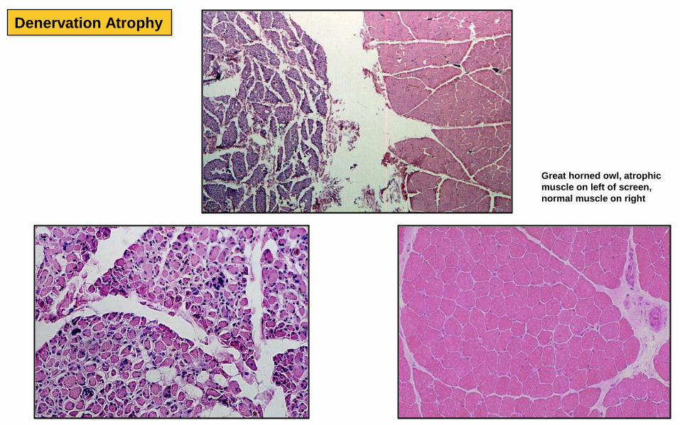
Great horned owl, atrophic
muscle on left of screen,
normal muscle on right
Denervation Atrophy
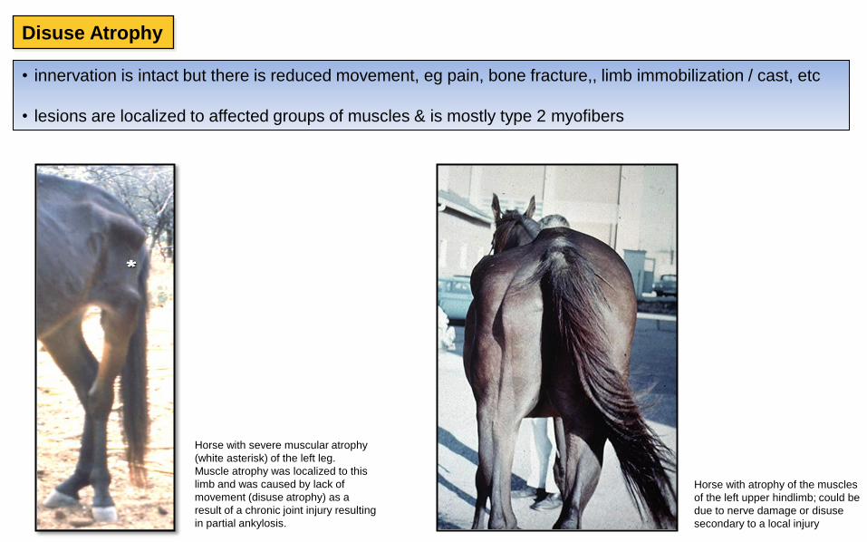
Disuse Atrophy
• innervation is intact but there is reduced movement, eg pain, bone fracture,, limb immobilization / cast, etc
• lesions are localized to affected groups of muscles & is mostly type 2 myofibers
Horse with severe muscular atrophy
(white asterisk) of the left leg.
Muscle atrophy was localized to this
limb and was caused by lack of
movement (disuse atrophy) as a
result of a chronic joint injury resulting
in partial ankylosis.
Horse with atrophy of the muscles
of the left upper hindlimb; could be
due to nerve damage or disuse
secondary to a local injury
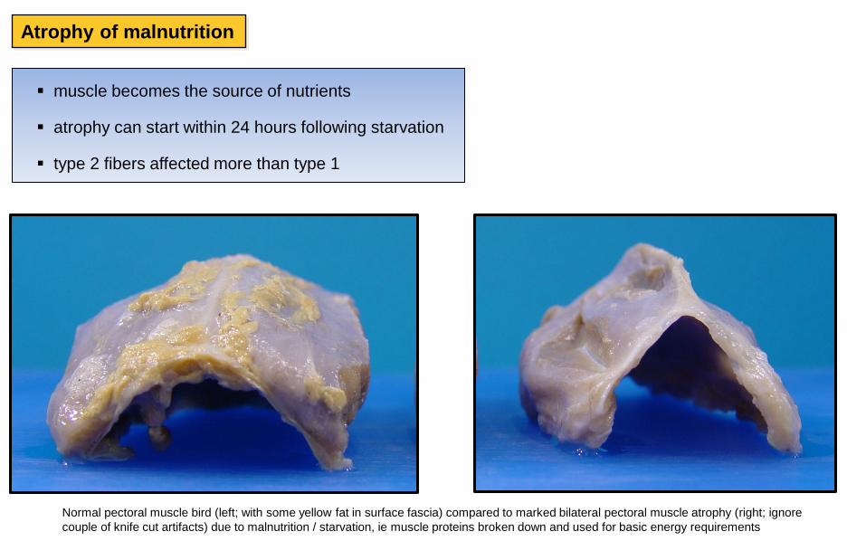
Atrophy of malnutrition
muscle becomes the source of nutrients
atrophy can start within 24 hours following starvation
type 2 fibers affected more than type 1
Normal pectoral muscle bird (left; with some yellow fat in surface fascia) compared to marked bilateral pectoral muscle atrophy (right; ignore
couple of knife cut artifacts) due to malnutrition / starvation, ie muscle proteins broken down and used for basic energy requirements

Carcass of dog showing severe muscle atrophy due to starvation (owner was
charged by the police). Note the extensive atrophy of scapular muscles (arrow) and
intercostal muscles (asterisks). Intercostal muscles are so atrophic that the lungs
can be seen right through them.
Atrophy of malnutrition
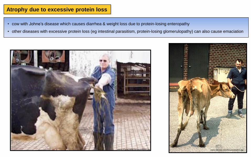
• cow with Johne’s disease which causes diarrhea & weight loss due to protein-losing enteropathy
• other diseases with excessive protein loss (eg intestinal parasitism, protein-losing glomerulopathy) can also cause emaciation
www.nd.gov/ndda/JohnesInAction.jpg www.vetnext.com/fotos/cowjohn1.jpg
Atrophy due to excessive protein loss
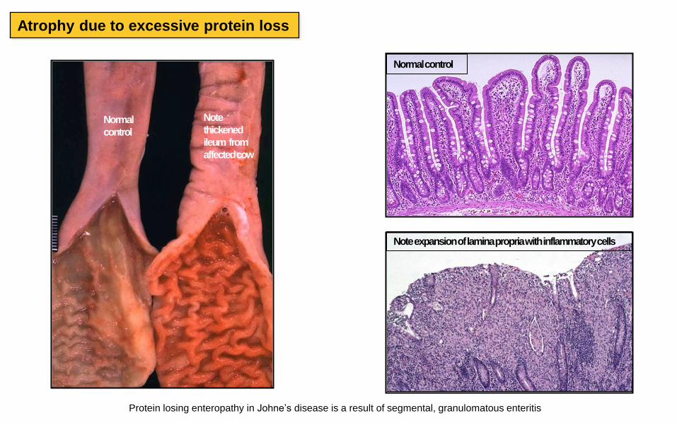
Protein losing enteropathy in Johne’s disease is a result of segmental, granulomatous enteritis
Normal
Note expansion of lamina propria with inflammatory cells
Normal control
Normal
control
Note
thickened
ileum from
affected cow
Atrophy due to excessive protein loss
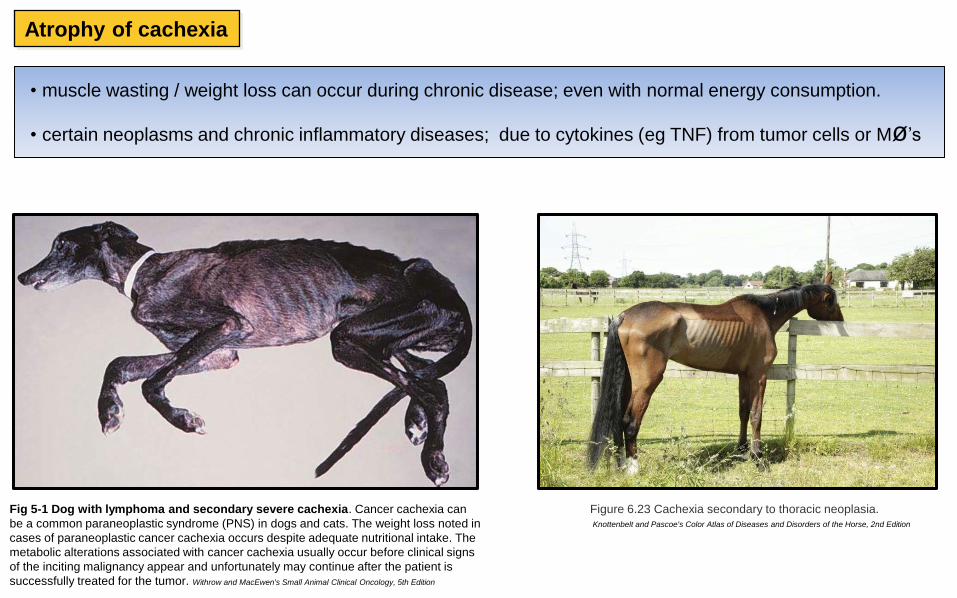
• muscle wasting / weight loss can occur during chronic disease; even with normal energy consumption.
• certain neoplasms and chronic inflammatory diseases; due to cytokines (eg TNF) from tumor cells or Mø’s
Figure 6.23 Cachexia secondary to thoracic neoplasia. Knottenbelt and Pascoe's Color Atlas of Diseases and Disorders of the Horse, 2nd Edition
Fig 5-1 Dog with lymphoma and secondary severe cachexia. Cancer cachexia can
be a common paraneoplastic syndrome (PNS) in dogs and cats. The weight loss noted in
cases of paraneoplastic cancer cachexia occurs despite adequate nutritional intake. The
metabolic alterations associated with cancer cachexia usually occur before clinical signs
of the inciting malignancy appear and unfortunately may continue after the patient is
successfully treated for the tumor. Withrow and MacEwen's Small Animal Clinical Oncology, 5th Edition
Atrophy of cachexia
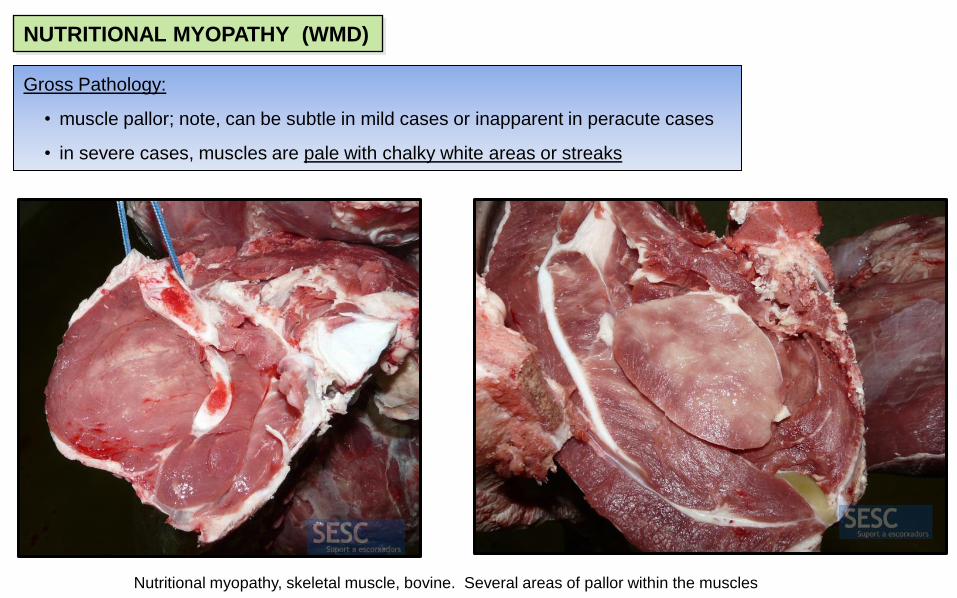
NUTRITIONAL MYOPATHY (WMD)
Gross Pathology:
• muscle pallor; note, can be subtle in mild cases or inapparent in peracute cases
• in severe cases, muscles are pale with chalky white areas or streaks
Nutritional myopathy, skeletal muscle, bovine. Several areas of pallor within the muscles
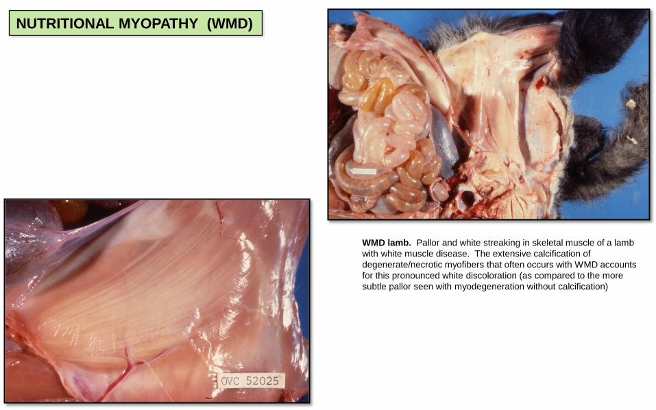
WMD lamb. Pallor and white streaking in skeletal muscle of a lamb
with white muscle disease. The extensive calcification of
degenerate/necrotic myofibers that often occurs with WMD accounts
for this pronounced white discoloration (as compared to the more
subtle pallor seen with myodegeneration without calcification)
NUTRITIONAL MYOPATHY (WMD)
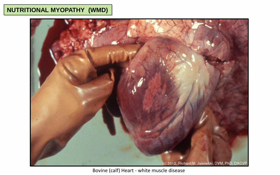
Bovine (calf) Heart - white muscle disease
NUTRITIONAL MYOPATHY (WMD)
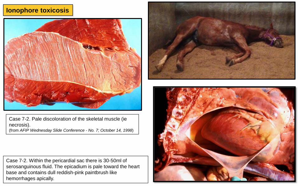
Ionophore toxicosis
Case 7-2. Pale discoloration of the skeletal muscle (ie
necrosis). (from AFIP Wednesday Slide Conference - No. 7; October 14, 1998)
Case 7-2. Within the pericardial sac there is 30-50ml of
serosanguinous fluid. The epicadium is pale toward the heart
base and contains dull reddish-pink paintbrush like
hemorrhages apically.
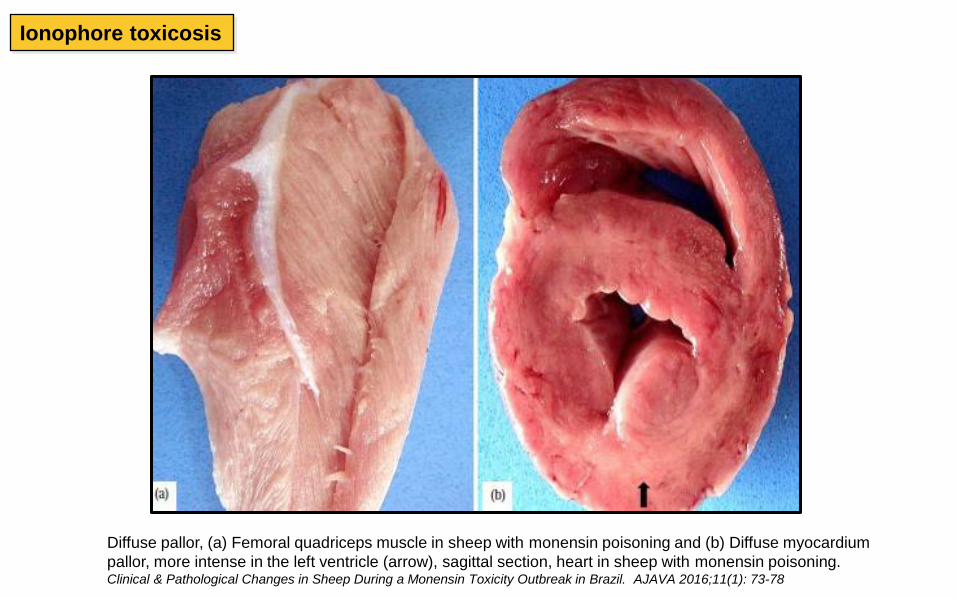
Ionophore toxicosis
Diffuse pallor, (a) Femoral quadriceps muscle in sheep with monensin poisoning and (b) Diffuse myocardium
pallor, more intense in the left ventricle (arrow), sagittal section, heart in sheep with monensin poisoning. Clinical & Pathological Changes in Sheep During a Monensin Toxicity Outbreak in Brazil. AJAVA 2016;11(1): 73-78
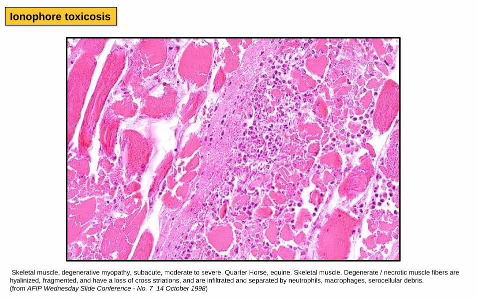
Skeletal muscle, degenerative myopathy, subacute, moderate to severe, Quarter Horse, equine. Skeletal muscle. Degenerate / necrotic muscle fibers are
hyalinized, fragmented, and have a loss of cross striations, and are infiltrated and separated by neutrophils, macrophages, serocellular debris.
(from AFIP Wednesday Slide Conference - No. 7 14 October 1998)
Ionophore toxicosis
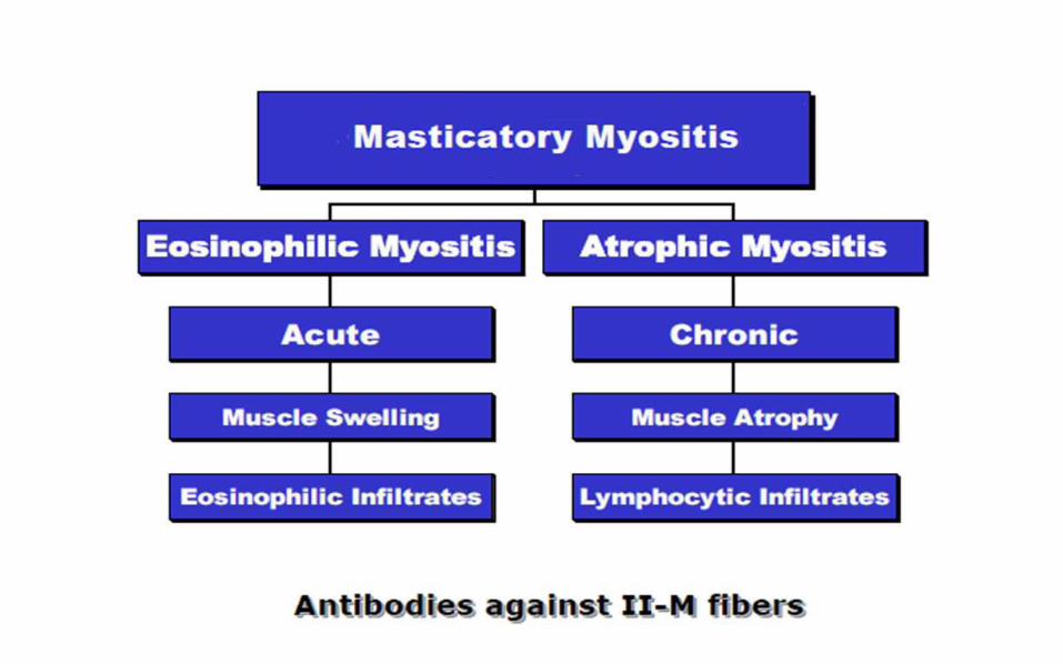
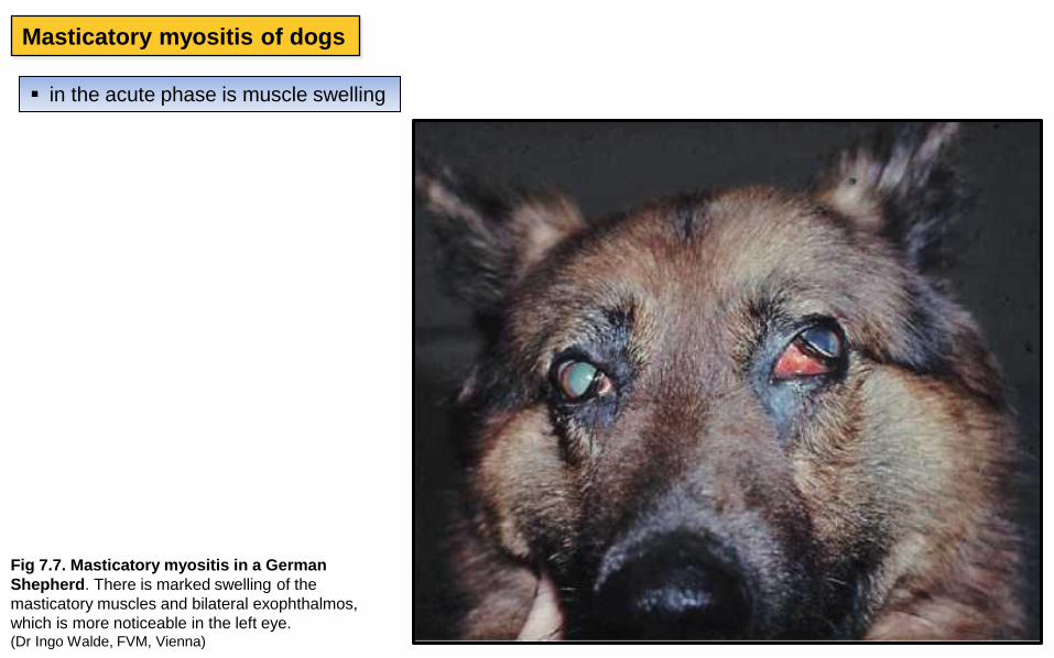
Masticatory myositis of dogs
Fig 7.7. Masticatory myositis in a German
Shepherd. There is marked swelling of the
masticatory muscles and bilateral exophthalmos,
which is more noticeable in the left eye. (Dr Ingo Walde, FVM, Vienna)
in the acute phase is muscle swelling
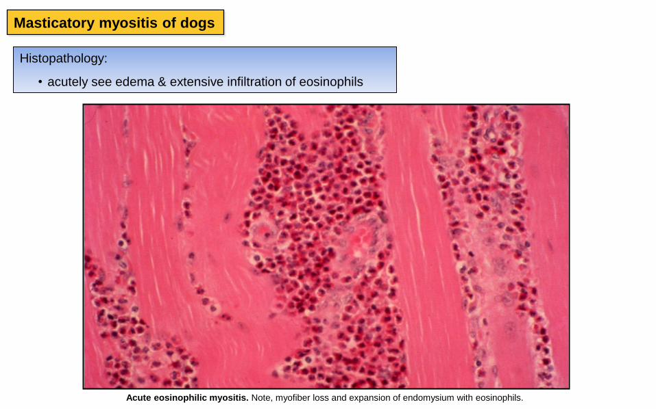
Acute eosinophilic myositis. Note, myofiber loss and expansion of endomysium with eosinophils.
Histopathology:
• acutely see edema & extensive infiltration of eosinophils
Masticatory myositis of dogs
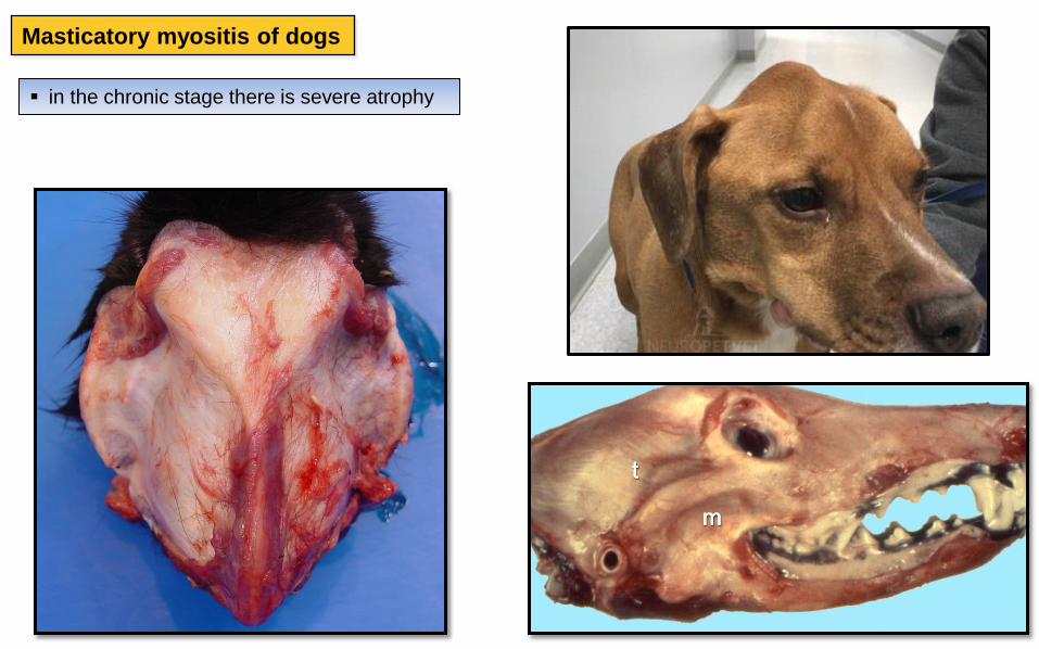
Masticatory myositis of dogs
in the chronic stage there is severe atrophy
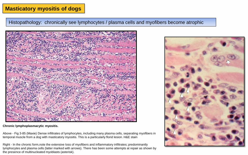
Histopathology: chronically see lymphocytes / plasma cells and myofibers become atrophic
Chronic lymphoplasmacytic myositis.
Above - Fig 3-85 (Maxie) Dense infiltrates of lymphocytes, including many plasma cells, separating myofibers in
temporal muscle from a dog with masticatory myositis. This is a particularly florid lesion. H&E stain
Right - In the chronic form,note the extensive loss of myofibers and inflammatory infiltrates; predominantly
lymphocytes and plasma cells (latter marked with arrows). There has been some attempts at repair as shown by
the presence of multinucleated myoblasts (asterisk).
Masticatory myositis of dogs
Blackleg
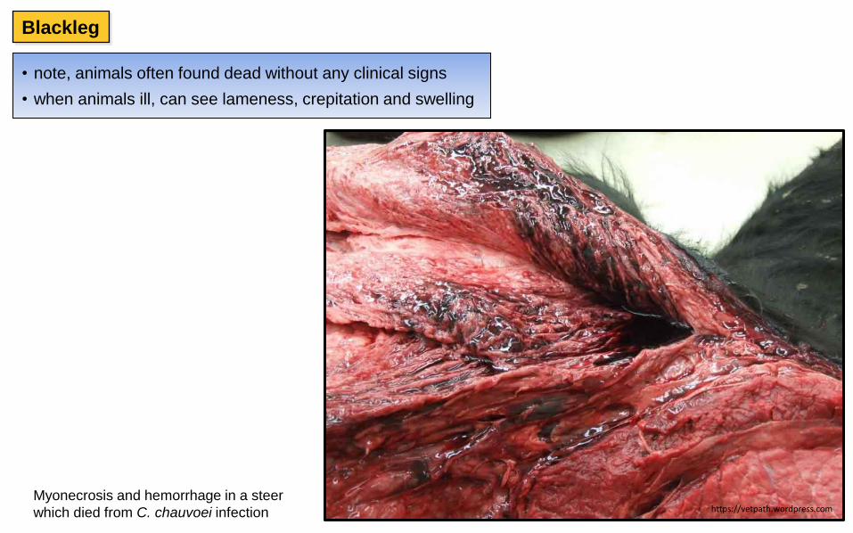
• note, animals often found dead without any clinical signs
• when animals ill, can see lameness, crepitation and swelling
Myonecrosis and hemorrhage in a steer
which died from C. chauvoei infection https://vetpath.wordpress.com
Blackle
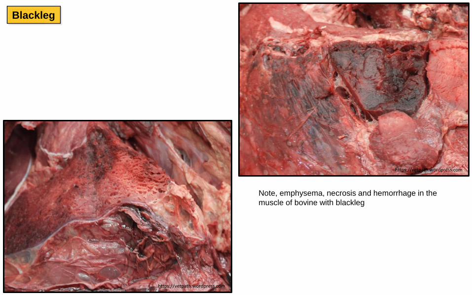
Note, emphysema, necrosis and hemorrhage in the
muscle of bovine with blackleg
Blackleg
https://vetpath.wordpress.com
https://vetpath.wordpress.com
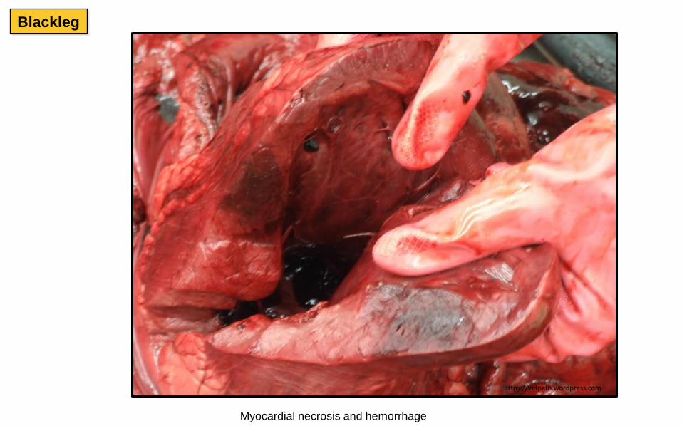
Myocardial necrosis and hemorrhage
Blackleg
https://vetpath.wordpress.com
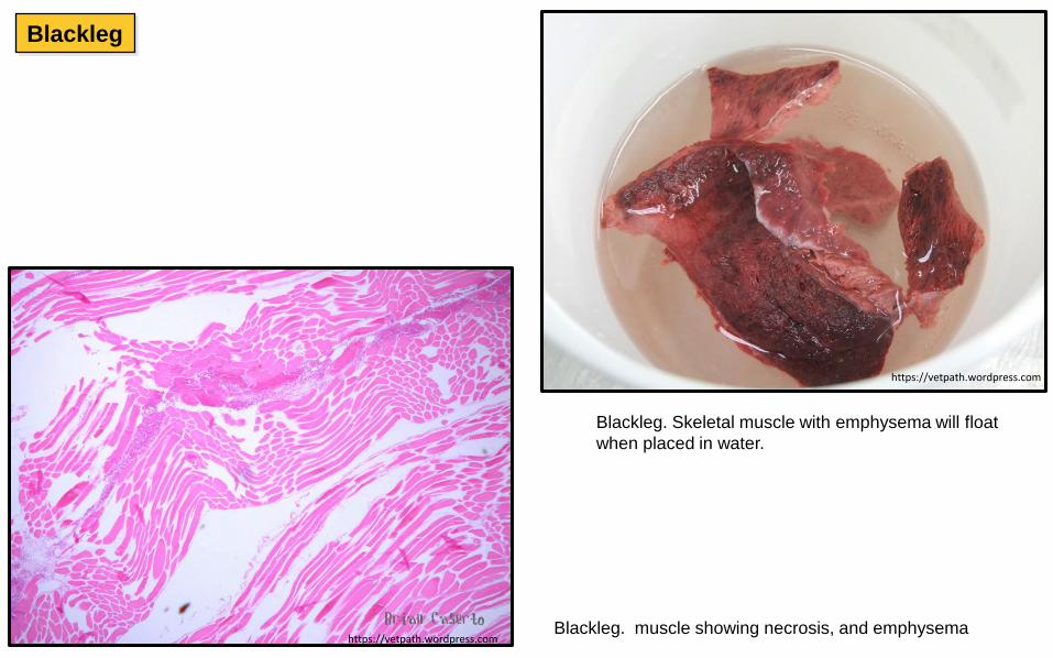
Blackleg. muscle showing necrosis, and emphysema
Blackleg. Skeletal muscle with emphysema will float
when placed in water.
https://vetpath.wordpress.com
https://vetpath.wordpress.com
Blackleg
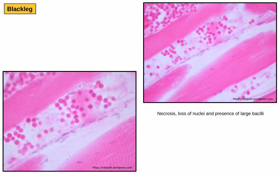
Necrosis, loss of nuclei and presence of large bacilli
https://vetpath.wordpress.com
https://vetpath.wordpress.com
Blackleg
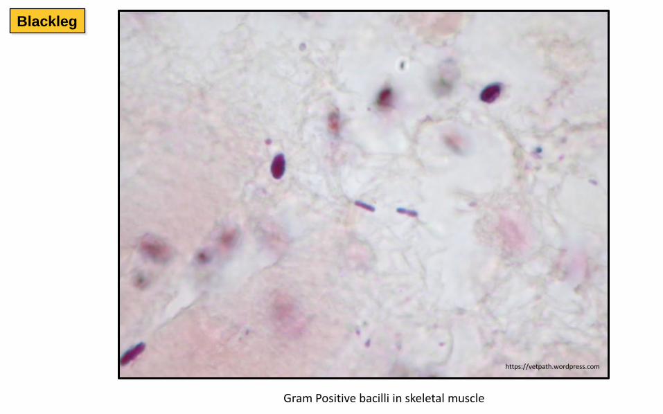
Gram Positive bacilli in skeletal muscle
https://vetpath.wordpress.com
Blackleg

Fluorescent Antibody Test (FA test) Note the positive
fluorescence for Clostridium chauvoei. Remember to
submit fresh (non-fixed) tissues for this confirmatory test
• Use FAT or anaerobic culture to confirm Cl. chauvoei
Figure 16.3 Direct fluorescent antibody technique showing C. chauvoei in
muscle tissue from a case of blackleg in a heifer. (×400). Clinical Veterinary Microbiology, 2nd ed.
Blackleg
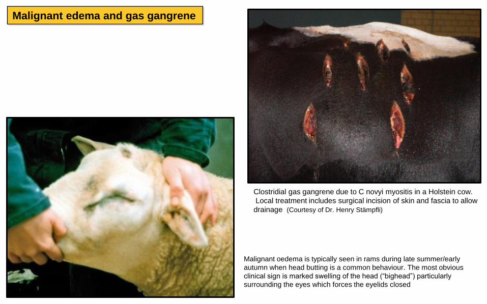
Clostridial gas gangrene due to C novyi myositis in a Holstein cow.
Local treatment includes surgical incision of skin and fascia to allow
drainage (Courtesy of Dr. Henry Stämpfli)
Malignant oedema is typically seen in rams during late summer/early
autumn when head butting is a common behaviour. The most obvious
clinical sign is marked swelling of the head (“bighead”) particularly
surrounding the eyes which forces the eyelids closed
Malignant edema and gas gangrene
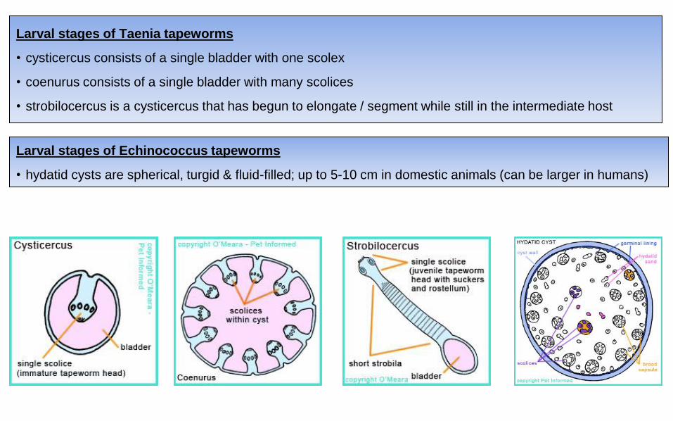
Larval stages of Taenia tapeworms
• cysticercus consists of a single bladder with one scolex
• coenurus consists of a single bladder with many scolices
• strobilocercus is a cysticercus that has begun to elongate / segment while still in the intermediate host
Larval stages of Echinococcus tapeworms
• hydatid cysts are spherical, turgid & fluid-filled; up to 5-10 cm in domestic animals (can be larger in humans)
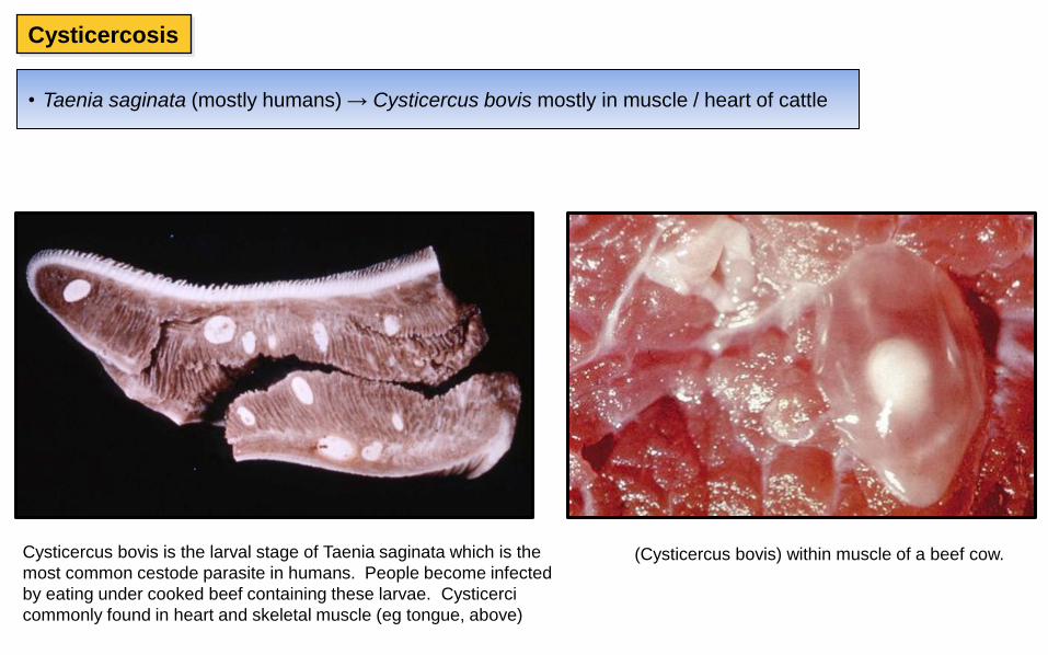
• Taenia saginata (mostly humans) → Cysticercus bovis mostly in muscle / heart of cattle
Cysticercosis
Cysticercus bovis is the larval stage of Taenia saginata which is the
most common cestode parasite in humans. People become infected
by eating under cooked beef containing these larvae. Cysticerci
commonly found in heart and skeletal muscle (eg tongue, above)
(Cysticercus bovis) within muscle of a beef cow.
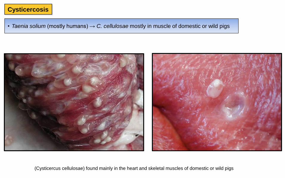
• Taenia solium (mostly humans) → C. cellulosae mostly in muscle of domestic or wild pigs
Cysticercosis
(Cysticercus cellulosae) found mainly in the heart and skeletal muscles of domestic or wild pigs
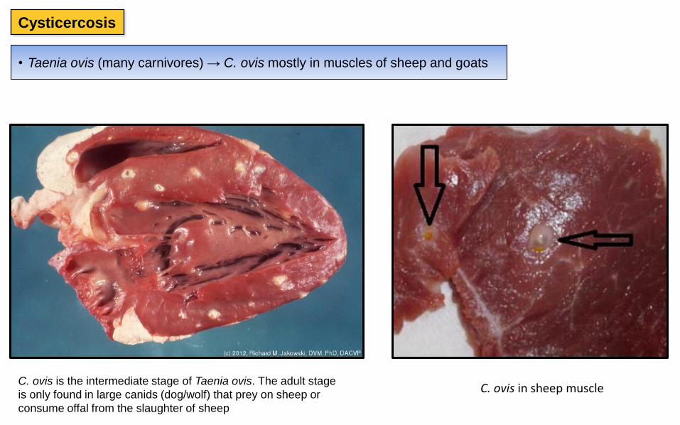
• Taenia ovis (many carnivores) → C. ovis mostly in muscles of sheep and goats
Cysticercosis
C. ovis is the intermediate stage of Taenia ovis. The adult stage
is only found in large canids (dog/wolf) that prey on sheep or
consume offal from the slaughter of sheep
C. ovis in sheep muscle
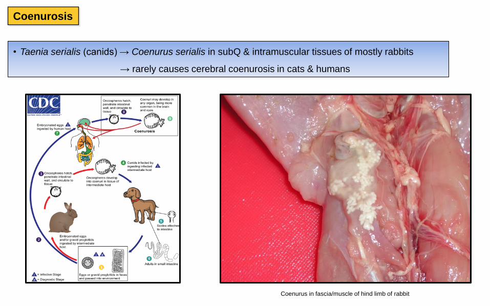
• Taenia serialis (canids) → Coenurus serialis in subQ & intramuscular tissues of mostly rabbits
→ rarely causes cerebral coenurosis in cats & humans
Coenurosis
Coenurus in fascia/muscle of hind limb of rabbit
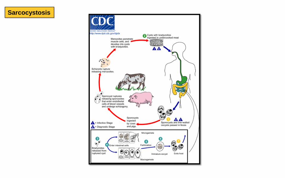
Sarcocystosis
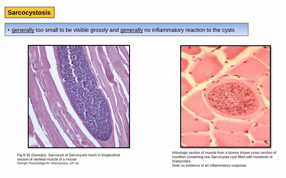
• generally too small to be visible grossly and generally no inflammatory reaction to the cysts
Histologic section of muscle from a bovine shows cross-section of
myofiber containing one Sarcocystis cyst filled with hundreds of
bradyzoites.
Note no evidence of an inflammatory response.
Fig 8-32 (Georgis) Sarcocyst of Sarcocystis muris in longitudinal
section of skeletal muscle of a mouse Georgis' Parasitology for Veterinarians, 10th ed.
Sarcocystosis
Sarcocystosis
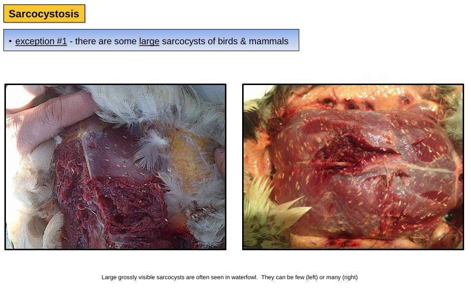
• exception #1 - there are some large sarcocysts of birds & mammals
Large grossly visible sarcocysts are often seen in waterfowl. They can be few (left) or many (right)

Sarcocytis gigantea in esophagus and diaphragm of a sheep; about the size of a grain of rice
http://www.uobabylon.edu.iq/
Int J Mol Cell Med 2014; 3 (9) 1-6
Sarcocystosis
• exception #1 - there are some large sarcocysts of birds & mammals
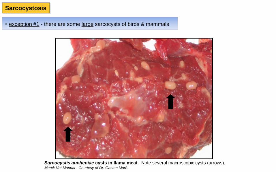
Sarcocystis aucheniae cysts in llama meat. Note several macroscopic cysts (arrows). Merck Vet Manual - Courtesy of Dr. Gaston Moré.
Sarcocystosis
• exception #1 - there are some large sarcocysts of birds & mammals
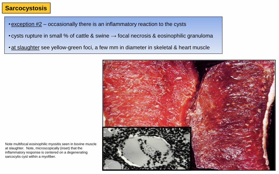
• exception #2 – occasionally there is an inflammatory reaction to the cysts
• cysts rupture in small % of cattle & swine → focal necrosis & eosinophilic granuloma
• at slaughter see yellow-green foci, a few mm in diameter in skeletal & heart muscle
Note multifocal eosinophilic myositis seen in bovine muscle
at slaughter. Note, microscopically (inset) that the
inflammatory response is centered on a degenerating
sarcocytis cyst within a myofiber.
Sarcocystosis
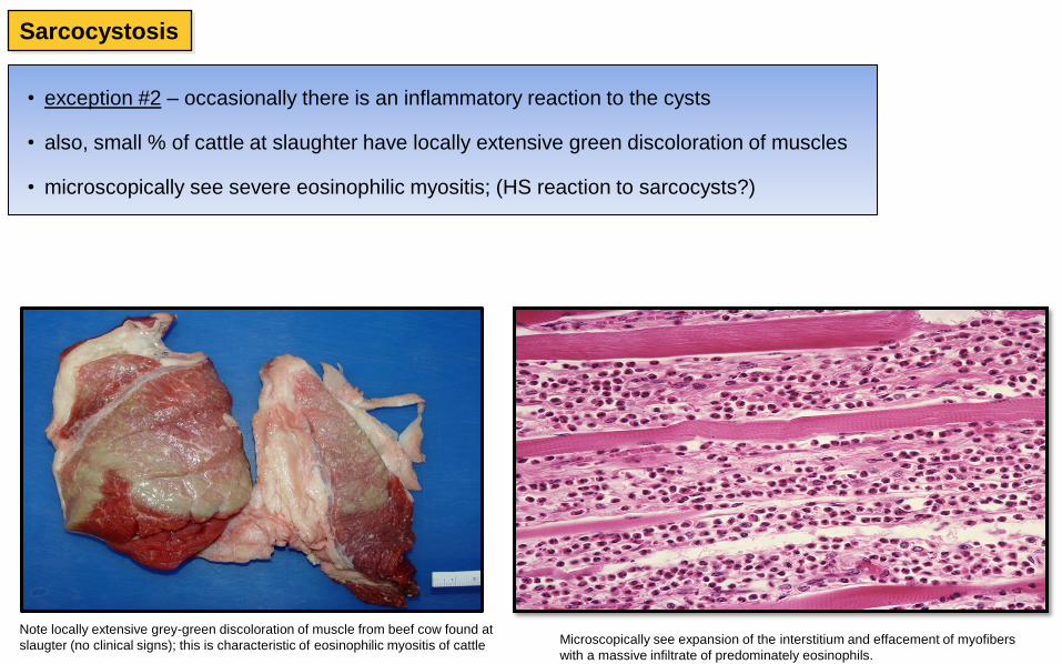
• exception #2 – occasionally there is an inflammatory reaction to the cysts
• also, small % of cattle at slaughter have locally extensive green discoloration of muscles
• microscopically see severe eosinophilic myositis; (HS reaction to sarcocysts?)
Note locally extensive grey-green discoloration of muscle from beef cow found at
slaugter (no clinical signs); this is characteristic of eosinophilic myositis of cattle Microscopically see expansion of the interstitium and effacement of myofibers
with a massive infiltrate of predominately eosinophils.
Sarcocystosis