Embed Size (px)
Citation preview
Pathology of Neuromuscular Disease Part 1: muscle
Zarife Sahenk, MD. PhD. Research Institute at Nationwide Children’s Hospital
Center for gene therapy, Neuromuscular Program Experimental & Clinical Neuromuscular Laboratories
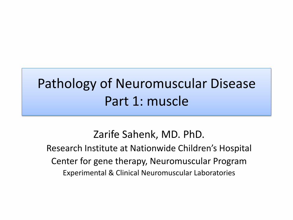
MUSCLE BIOPSY DESCRIPTION OF SPECIMENS, PROCEDURES & STAINS
• 2 blocks of skeletal muscle, frozen in isopentane cooled in liquid nitrogen. 12 μm thick sections are cut using a cryostat.
• The following routine stains are done : • Basic histopathological stains: H & E and Gomori trichrome • Special Stains:, oil red O, PAS, Congo red. • Enzyme Histochemistry: NADH, SDH, COX, and ATPase, at pH 9.4,
4.6, 4.2. (Myophosphorylase, MAD, acid phosphatase if needed) • Immune staining: carried out if needed
– CD3, CD4, CD8, CD20 and CD68 cell markers, MAC – dystrophin (dys 1, 2, 3), sarcoglycans (α, β, γ, δ), dystroglycans (α, β), dysferlin,
caveolin 3, laminin alpha 2 (merosin), utrophin, spectrin , collagen VI – specific antibodies for protein aggregates
• EM piece placed in glutaraldehyde for further processing • A separate piece of muscle frozen for biochemical/genetic studies
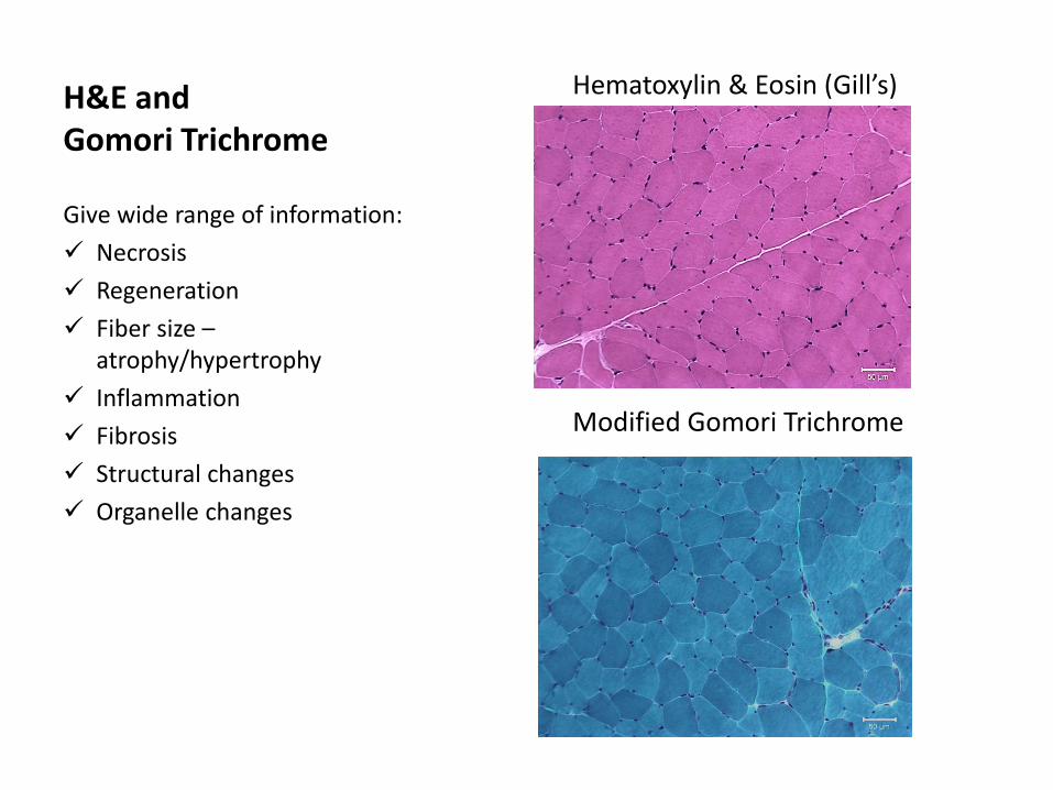
H&E and Gomori Trichrome
Hematoxylin & Eosin (Gill’s) Modified Gomori Trichrome
Give wide range of information: Necrosis Regeneration Fiber size –
atrophy/hypertrophy Inflammation Fibrosis Structural changes Organelle changes
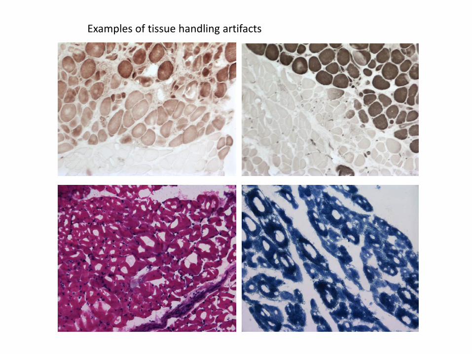
Examples of tissue handling artifacts
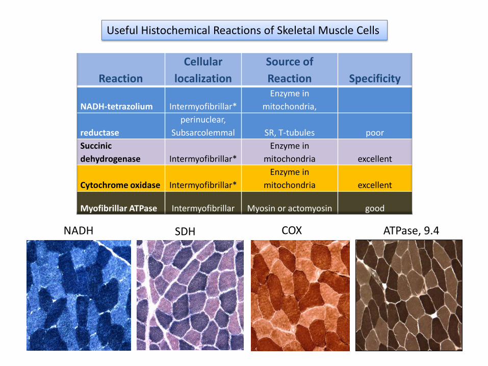
Reaction Cellular
localization Source of Reaction Specificity
NADH-tetrazolium Intermyofibrillar* Enzyme in
mitochondria,
reductase perinuclear,
Subsarcolemmal SR, T-tubules poor Succinic dehydrogenase Intermyofibrillar*
Enzyme in mitochondria excellent
Cytochrome oxidase Intermyofibrillar* Enzyme in
mitochondria excellent
Myofibrillar ATPase Intermyofibrillar Myosin or actomyosin good
Useful Histochemical Reactions of Skeletal Muscle Cells
NADH SDH COX ATPase, 9.4
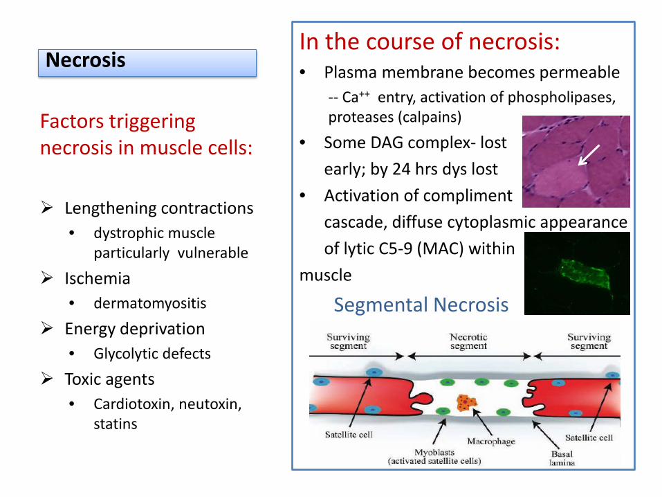
Necrosis In the course of necrosis: • Plasma membrane becomes permeable
-- Ca++ entry, activation of phospholipases, proteases (calpains)
• Some DAG complex- lost early; by 24 hrs dys lost • Activation of compliment cascade, diffuse cytoplasmic appearance of lytic C5-9 (MAC) within muscle
Segmental Necrosis
Factors triggering necrosis in muscle cells:
Lengthening contractions
• dystrophic muscle particularly vulnerable
Ischemia • dermatomyositis
Energy deprivation • Glycolytic defects
Toxic agents • Cardiotoxin, neutoxin,
statins
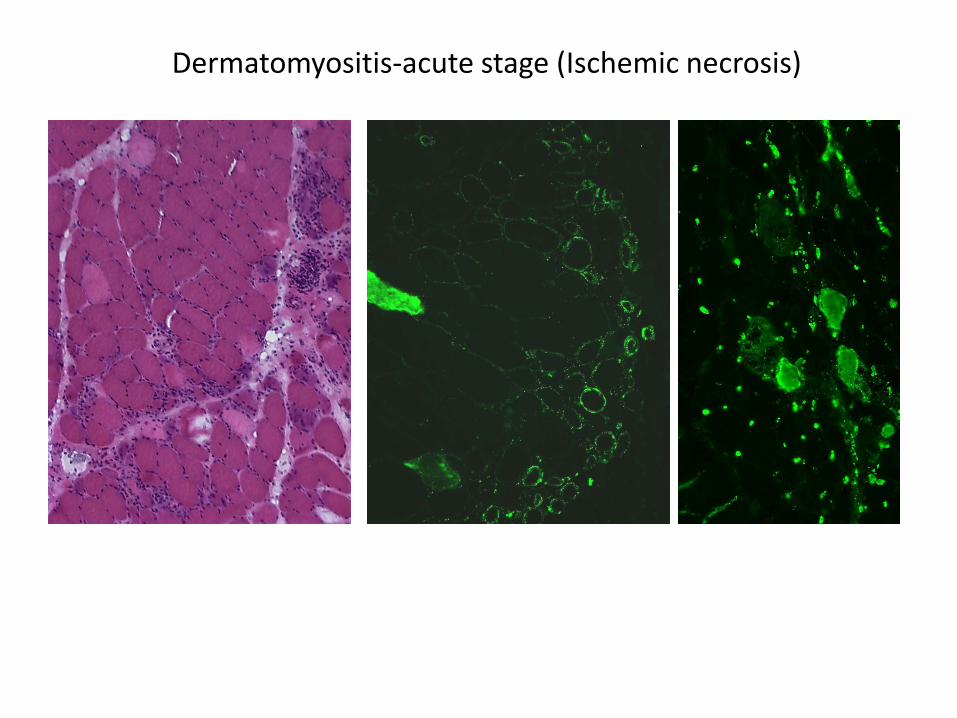
Dermatomyositis-acute stage (Ischemic necrosis)
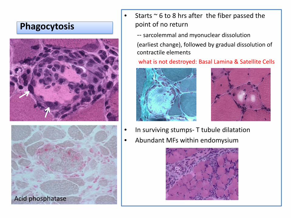
Phagocytosis • Starts ~ 6 to 8 hrs after the fiber passed the
point of no return -- sarcolemmal and myonuclear dissolution (earliest change), followed by gradual dissolution of contractile elements what is not destroyed: Basal Lamina & Satellite Cells
• In surviving stumps- T tubule dilatation • Abundant MFs within endomysium
Acid phosphatase

Patterns of inflammation
H&E
H&E
H&E
Perivascular inflammation
• Variation in muscle fiber size
• Small rounded fibers
Perivascular & Perimysial inflammation
• Mononuclear cell
Endomysial inflammation
Often associated with focal invasion of muscle fibers
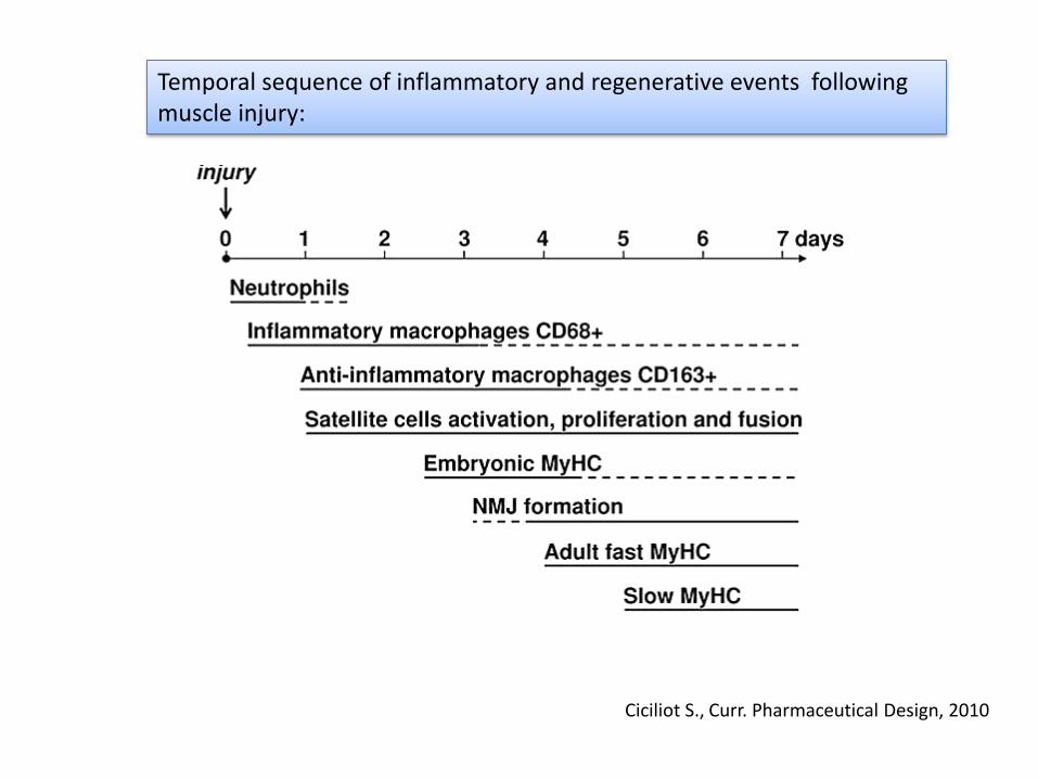
Temporal sequence of inflammatory and regenerative events following muscle injury:
Ciciliot S., Curr. Pharmaceutical Design, 2010
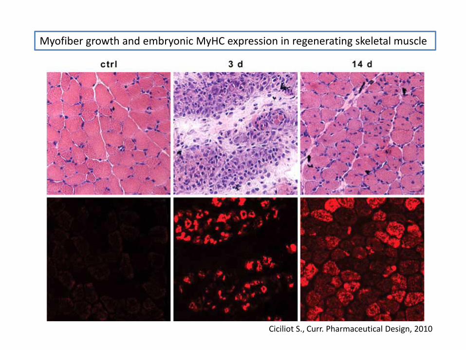
Myofiber growth and embryonic MyHC expression in regenerating skeletal muscle
Ciciliot S., Curr. Pharmaceutical Design, 2010
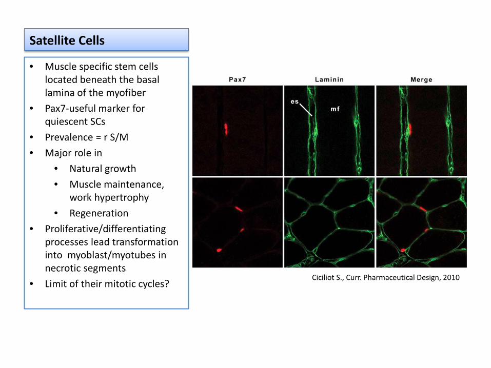
Satellite Cells
• Muscle specific stem cells located beneath the basal lamina of the myofiber
• Pax7-useful marker for quiescent SCs
• Prevalence = r S/M • Major role in
• Natural growth • Muscle maintenance,
work hypertrophy • Regeneration
• Proliferative/differentiating processes lead transformation into myoblast/myotubes in necrotic segments
• Limit of their mitotic cycles? Ciciliot S., Curr. Pharmaceutical Design, 2010
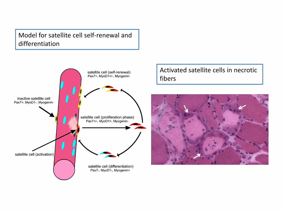
Model for satellite cell self-renewal and differentiation
Activated satellite cells in necrotic fibers
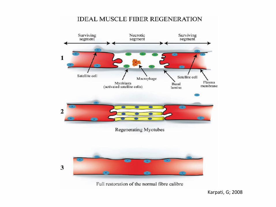
Karpati, G; 2008
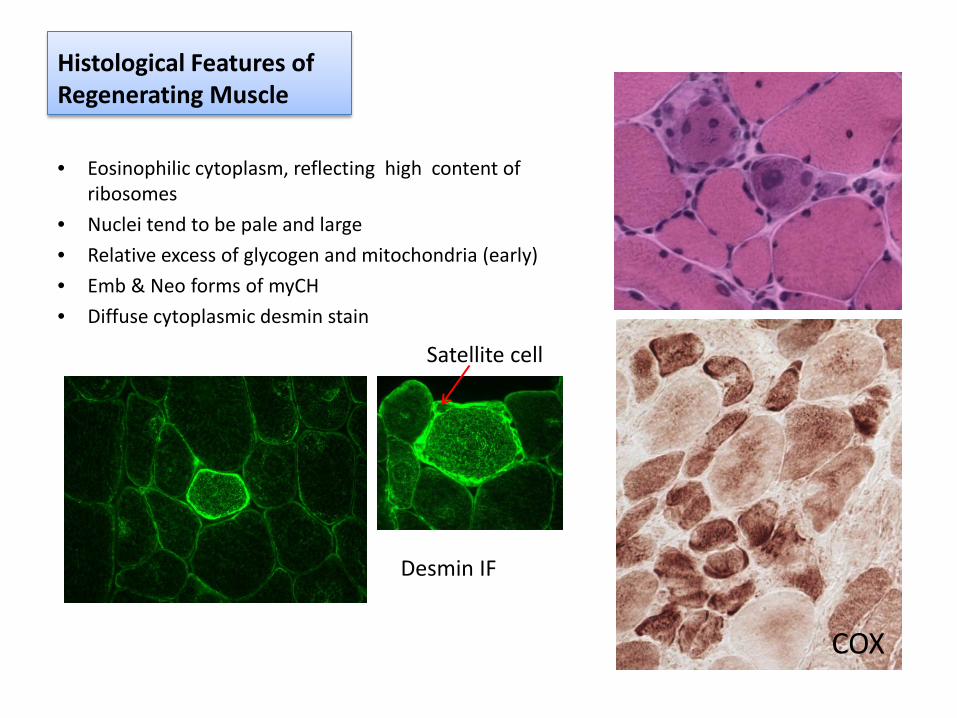
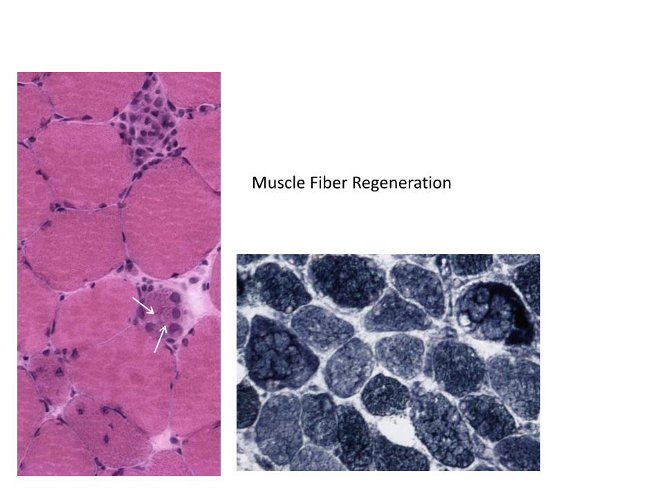
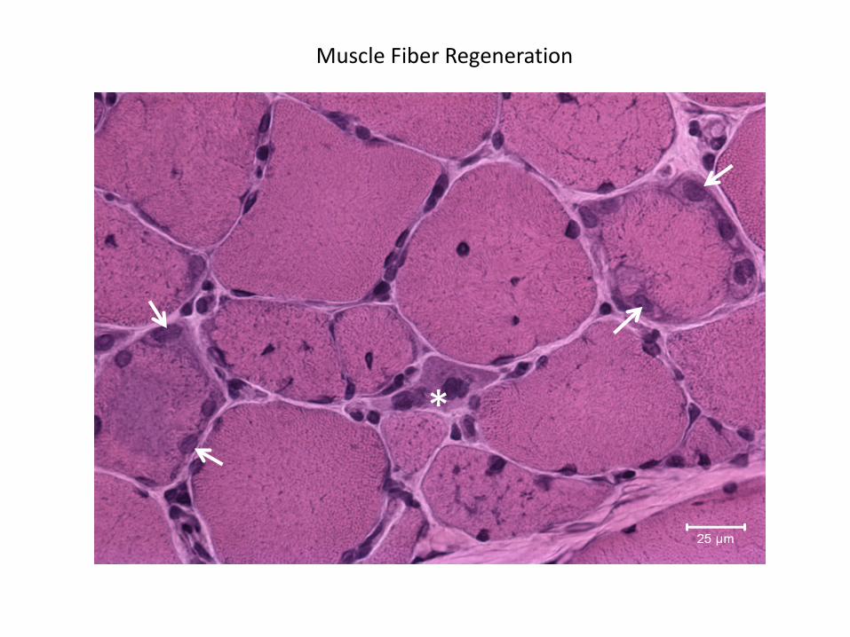
Histological Features of Regenerating Muscle
• Eosinophilic cytoplasm, reflecting high content of ribosomes
• Nuclei tend to be pale and large • Relative excess of glycogen and mitochondria (early) • Emb & Neo forms of myCH • Diffuse cytoplasmic desmin stain
COX
Satellite cell
Desmin IF
NADH
Muscle Fiber Regeneration
*
Muscle Fiber Regeneration
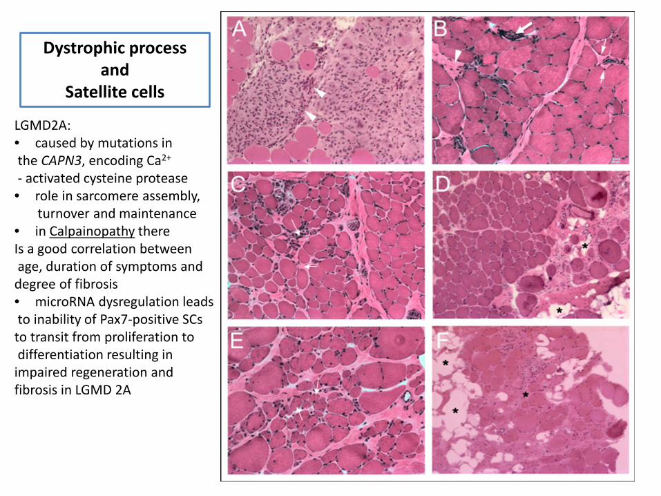
LGMD2A: • caused by mutations in the CAPN3, encoding Ca2+
- activated cysteine protease • role in sarcomere assembly, turnover and maintenance • in Calpainopathy there Is a good correlation between age, duration of symptoms and degree of fibrosis • microRNA dysregulation leads to inability of Pax7-positive SCs to transit from proliferation to differentiation resulting in impaired regeneration and fibrosis in LGMD 2A
Dystrophic process and
Satellite cells
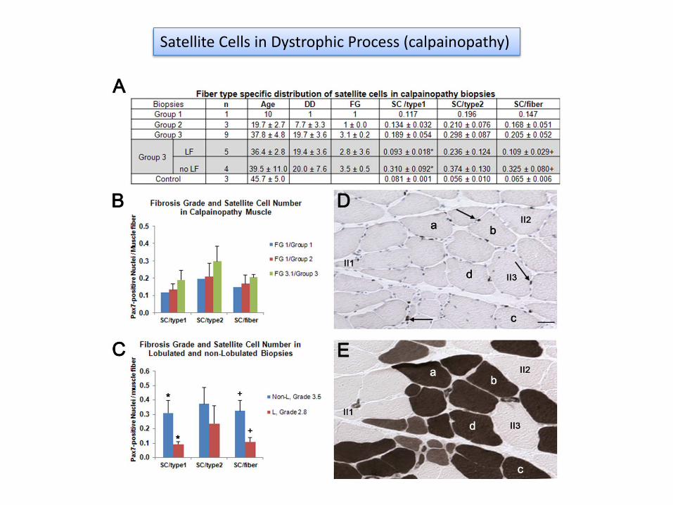
Satellite Cells in Dystrophic Process (calpainopathy)
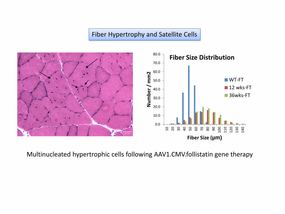
Fiber Hypertrophy and Satellite Cells
Multinucleated hypertrophic cells following AAV1.CMV.follistatin gene therapy
0.0
10.0
20.0
30.0
40.0
50.0
60.0
70.0
80.0
10 20 30 40 50 60 70 80 90 100
110
120
130
140
Num
ber /
mm
2
Fiber Size (µm)
Fiber Size Distribution
WT-FT12 wks-FT36wks-FT
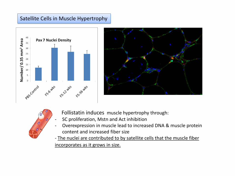
Follistatin induces muscle hypertrophy through: - SC proliferation, Mstn and Act inhibition - Overexpression in muscle lead to increased DNA & muscle protein
content and increased fiber size - The nuclei are contributed to by satellite cells that the muscle fiber incorporates as it grows in size.
Satellite Cells in Muscle Hypertrophy
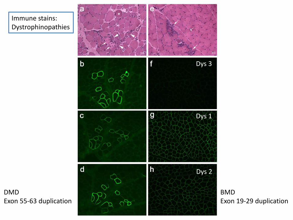
Dys 3
Dys 1
Dys 2
BMD Exon 19-29 duplication
DMD Exon 55-63 duplication
Immune stains: Dystrophinopathies
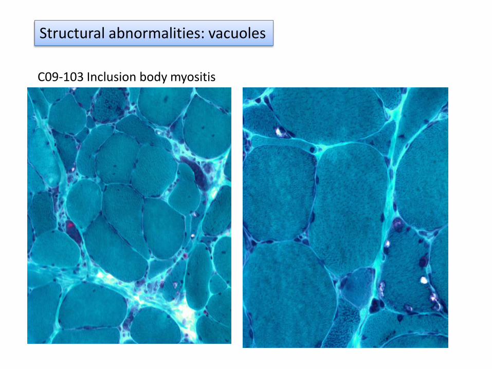
C09-103 Inclusion body myositis
Structural abnormalities: vacuoles
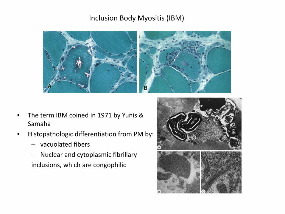
Inclusion Body Myositis (IBM)
• The term IBM coined in 1971 by Yunis & Samaha
• Histopathologic differentiation from PM by: – vacuolated fibers – Nuclear and cytoplasmic fibrillary inclusions, which are congophilic
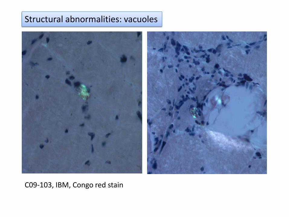
C09-103, IBM, Congo red stain
Structural abnormalities: vacuoles
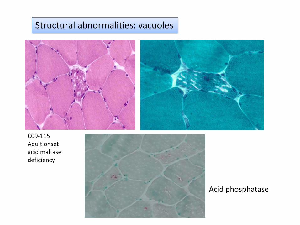
Acid phosphatase
C09-115 Adult onset acid maltase deficiency
Structural abnormalities: vacuoles
NADH SDH
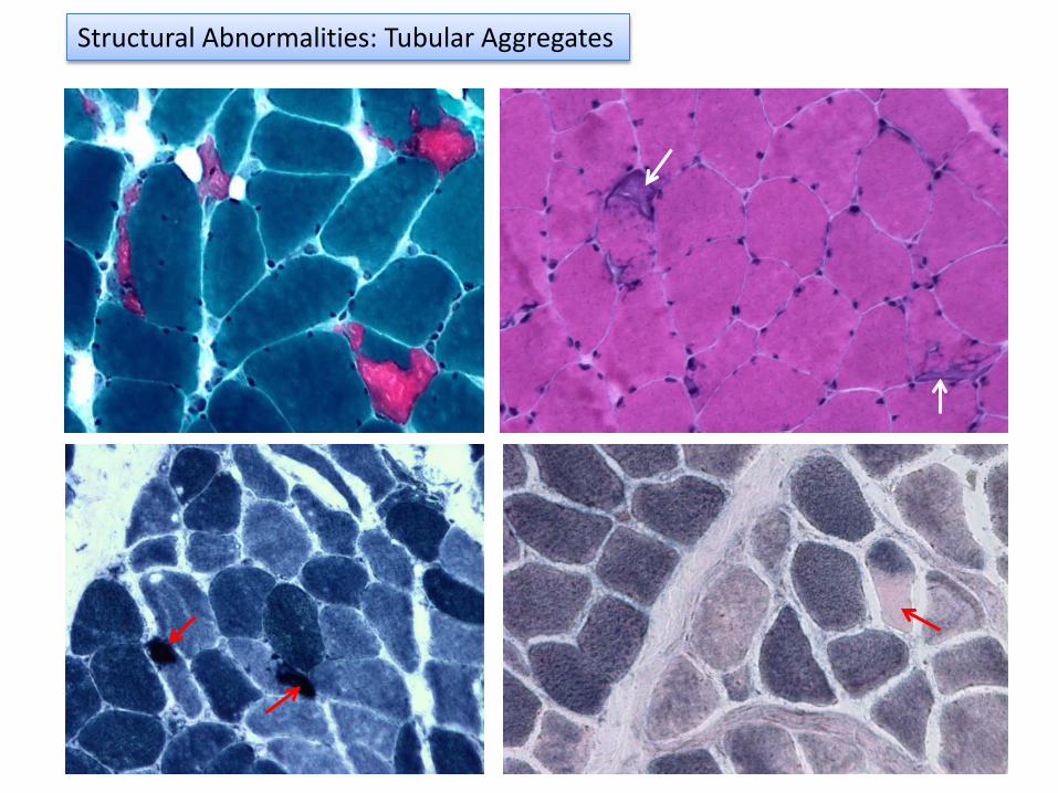
Structural Abnormalities: Tubular Aggregates
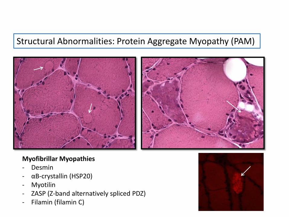
Structural Abnormalities: Protein Aggregate Myopathy (PAM)
Myofibrillar Myopathies - Desmin - αB-crystallin (HSP20) - Myotilin - ZASP (Z-band alternatively spliced PDZ) - Filamin (filamin C)
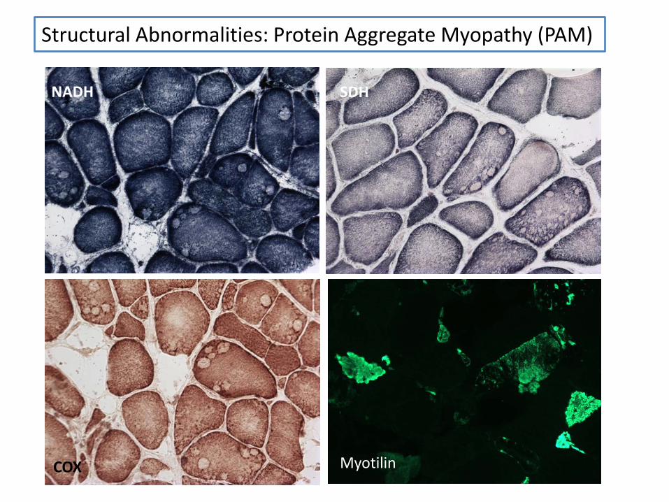
Structural Abnormalities: Protein Aggregate Myopathy (PAM)
Myotilin
NADH SDH
COX
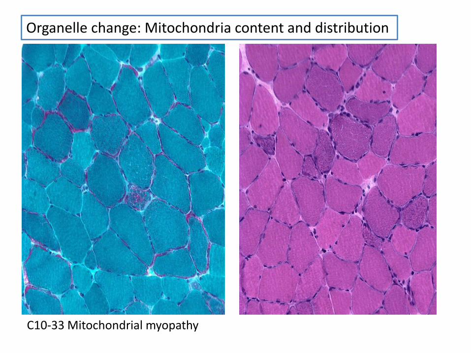
C10-33 Mitochondrial myopathy
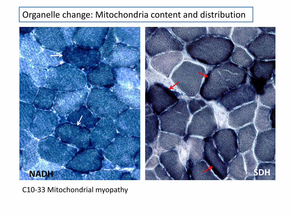
Organelle change: Mitochondria content and distribution
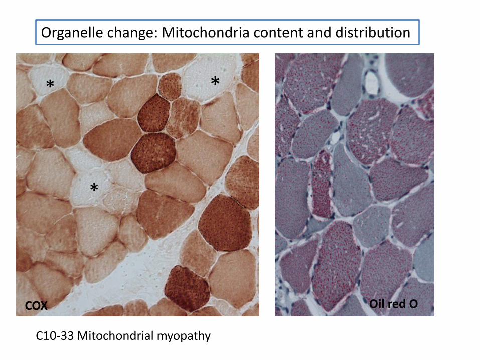
Organelle change: Mitochondria content and distribution
C10-33 Mitochondrial myopathy
NADH SDH
Oil red O
Organelle change: Mitochondria content and distribution
C10-33 Mitochondrial myopathy
COX
*
* *
Oil red O
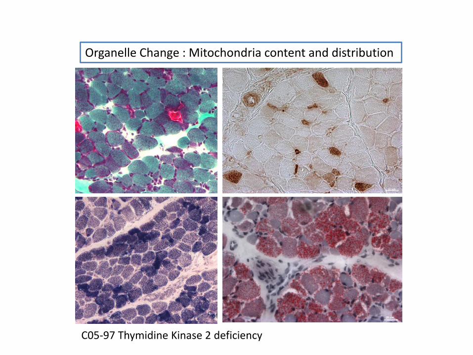
Organelle Change : Mitochondria content and distribution
C05-97 Thymidine Kinase 2 deficiency
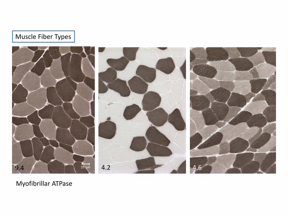
Myofibrillar ATPase
9.4 4.6 4.2
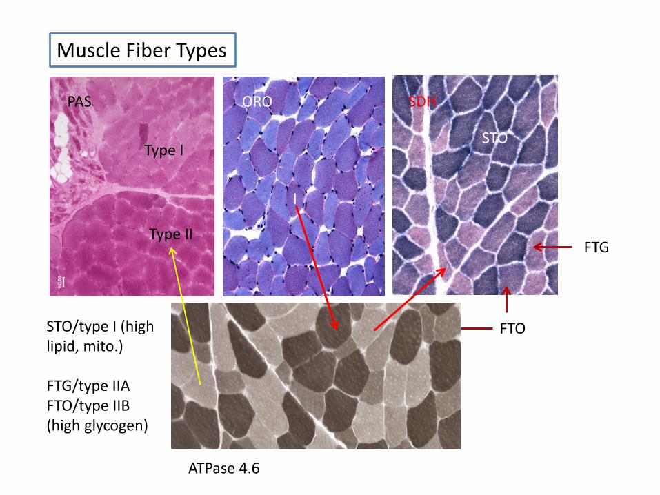
Muscle Fiber Types
I
Type II
Type I STO
FTO
FTG
ATPase 4.6
Muscle Fiber Types
STO/type I (high lipid, mito.) FTG/type IIA FTO/type IIB (high glycogen)
PAS ORO SDH
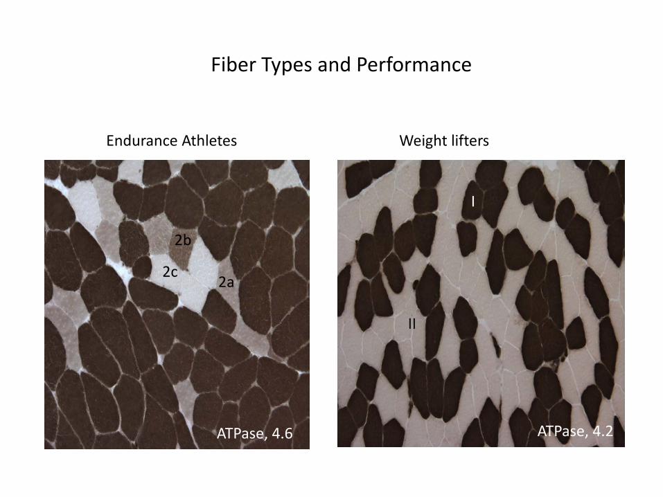
Fiber Types and Performance
Endurance Athletes
ATPase, 4.6
2c
2b
2a
ATPase, 4.2
I
II
Weight lifters
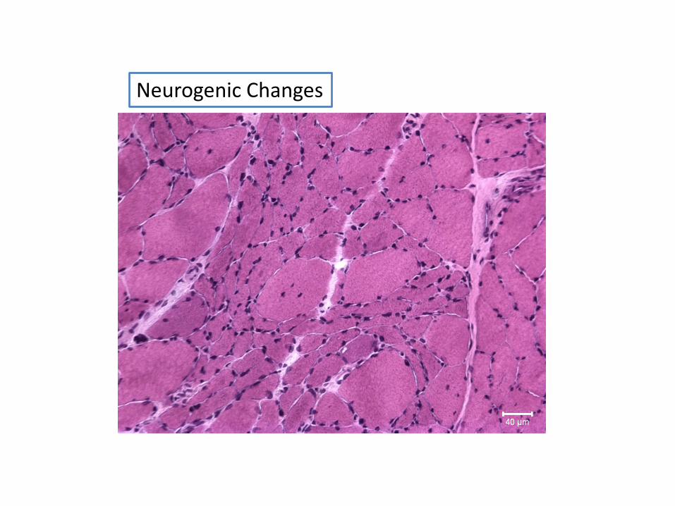
Neurogenic Changes
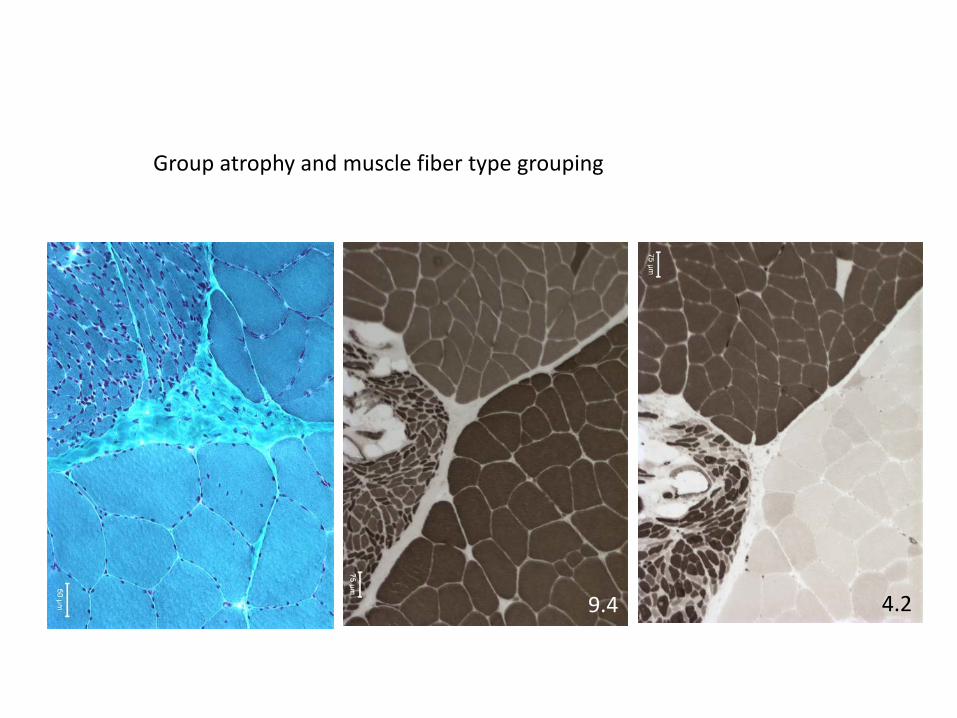
Group atrophy and muscle fiber type grouping
9.4 4.2
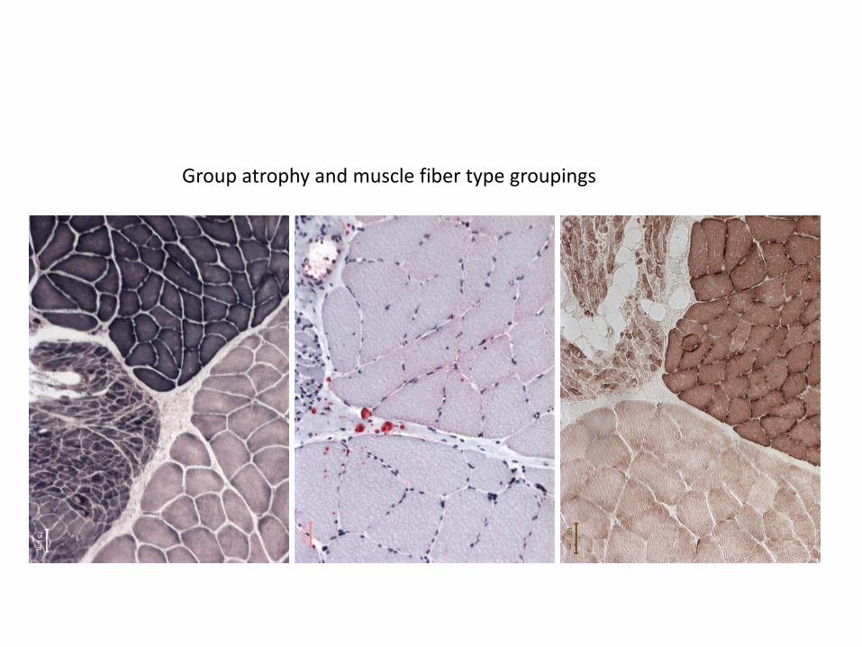
Group atrophy and muscle fiber type groupings
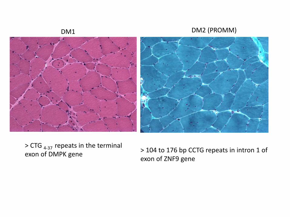
DM1 DM2 (PROMM)
> CTG 4-37 repeats in the terminal exon of DMPK gene
> 104 to 176 bp CCTG repeats in intron 1 of exon of ZNF9 gene
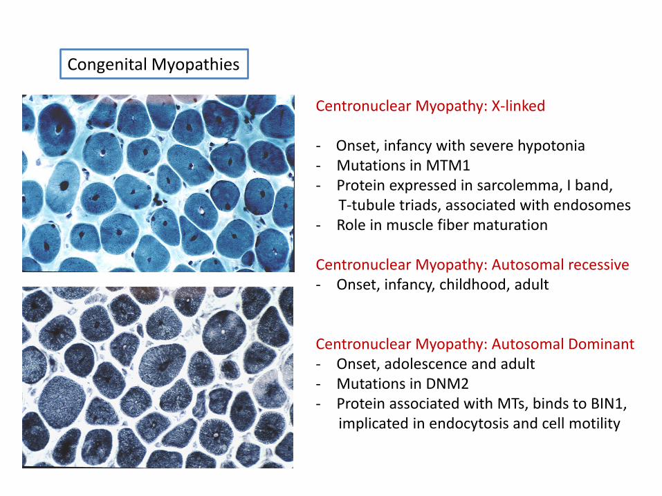
Centronuclear Myopathy: X-linked - Onset, infancy with severe hypotonia - Mutations in MTM1 - Protein expressed in sarcolemma, I band, T-tubule triads, associated with endosomes - Role in muscle fiber maturation Centronuclear Myopathy: Autosomal recessive - Onset, infancy, childhood, adult
Centronuclear Myopathy: Autosomal Dominant - Onset, adolescence and adult - Mutations in DNM2 - Protein associated with MTs, binds to BIN1, implicated in endocytosis and cell motility
Congenital Myopathies