Embed Size (px)
Citation preview

Systemic MycosesSystemic Mycoses
Prepared By:
Hodaifa S. Al AbssyHodaifa S. Al Abssy
Supervised By:
Dr. Abd El Raouf Al ManaamaDr. Abd El Raouf Al Manaama
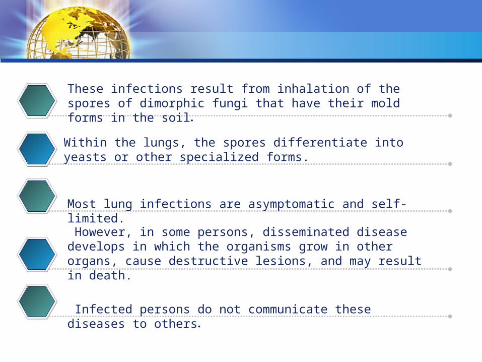
These infections result from inhalation of the spores of dimorphic fungi that have their mold forms in the soil.
Within the lungs, the spores differentiate into yeasts or other specialized forms.
Most lung infections are asymptomatic and self-limited.
However, in some persons, disseminated disease develops in which the organisms grow in other organs, cause destructive lesions, and may result in death.
Infected persons do not communicate these diseases to others.
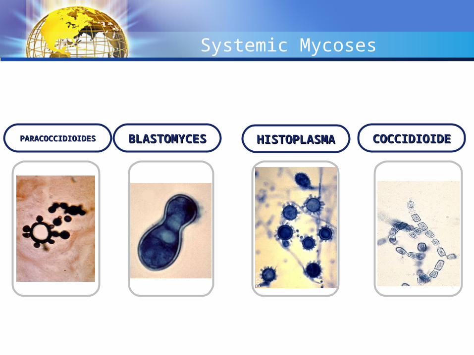
COCCIDIOIDECOCCIDIOIDEHISTOPLASMAHISTOPLASMABLASTOMYCESBLASTOMYCESPARACOCCIDIOIDESPARACOCCIDIOIDES
Systemic Mycoses
COCCIDIOIDECOCCIDIOIDE
COCCIDIOIDE
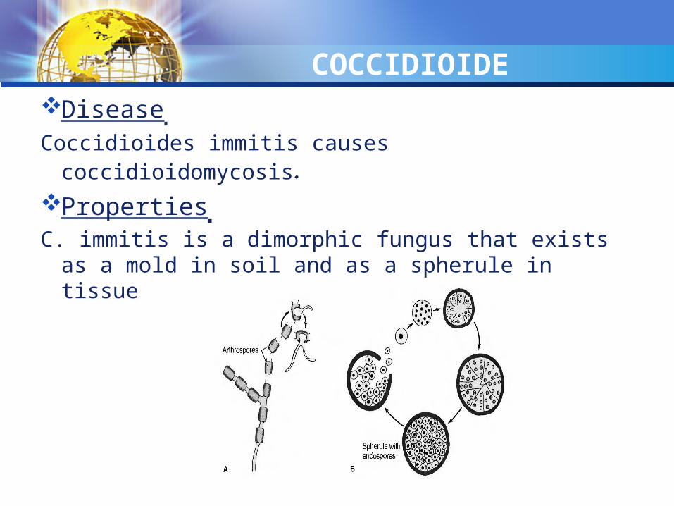
Disease
Coccidioides immitis causes coccidioidomycosis. Properties C. immitis is a dimorphic fungus that exists as a
mold in soil and as a spherule in tissue
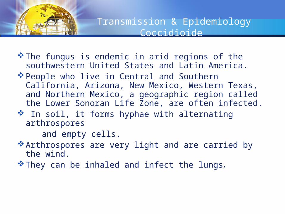
Transmission & Epidemiology Coccidioide
The fungus is endemic in arid regions of the southwestern United States and Latin America.
People who live in Central and Southern California, Arizona, New Mexico, Western Texas, and Northern Mexico, a geographic region called the Lower Sonoran Life Zone, are often infected.
In soil, it forms hyphae with alternating arthrospores and empty cells.Arthrospores are very light and are carried by the wind. They can be inhaled and infect the lungs.
Pathogenesis of Coccidioide
In the lungs, arthrospores form spherules that are large, have a thick, doubly refractive wall, and are filled with endospores.
Upon rupture of the wall, endospores are released and differentiate to form new spherules. The organism can spread within a person by direct
extension or via the bloodstream. Granulomatous lesions can occur in virtually any organ
but are found primarily in bones and the central nervous system (meningitis)
Dissemination from the lungs to other organs occurs in people who have a defect in cell-mediated immunity.
Pathogenesis of CoccidioideMost people who are infected by C. immitis
develop a cell-mediated (delayed hypersensitivity) immune response that restricts the growth of the organism.
One way to determine whether a person has produced adequate cell-mediated immunity to the organism is to do a skin test (see below).
In general, a person who has a positive skin test reaction has developed sufficient immunity to prevent disseminated disease from occurring.
If, at a later time, a person's cellular immunity is suppressed by drugs or disease, disseminated disease can occur.

Clinical Findings of Coccidioide Infection of the lungs is often asymptomatic and is
evident only by a positive skin test and the presence of antibodies.
Some infected persons have an influenza like illness with fever and cough.
About. 50% have changes in the lungs (infiltrates, adenopathy, or effusions) as seen on chest x-ray.
10% develop erythema nodosum (see below) or arthralgias.
This syndrome is called "valley fever" or "desert rheumatism"; it tends to subside spontaneously.
Disseminated disease can occur in almost any organ; the meninges, bone, and skin are important sites.
Clinical Findings of CoccidioideThe overall incidence of dissemination in
persons infected with C. imrnitis is 1%, although the incidence in Filipinos and African Americans is 10 times higher.
Women in the third trimester of pregnancy also have a markedly increased incidence of dissemination.
Erythema nodosum (EN) manifests as red, tender nodules ("desert bumps") on extensor surfaces such as the shins.
It is a delayed (cell-mediated) hypersensitivity response to fungal antigens and thus is an indicator of a good prognosis.
Clinical Findings of Coccidioide
There are no organisms in these lesions; they are not a sign of disseminated disease. EN is not specific for coccidioidomycosis; it occurs in other granulomatous diseases, eg, histoplasmosis, tuberculosis, and leprosy.
In infected persons, skin tests with fungal extracts cause at least a 5mm induration 48 hours after injection (delayed hypersensitivity reaction).
Skin tests become positive within 2-4 weeks of infection and remain so for years but are often negative in patients with disseminated disease.
Laboratory Diagnosis of Coccidioide
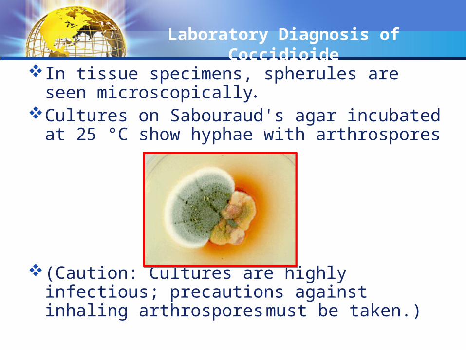
In tissue specimens, spherules are seen microscopically.
Cultures on Sabouraud's agar incubated at 25 °C show hyphae with arthrospores
(Caution: Cultures are highly infectious; precautions against inhaling arthrospores must be taken.)
Laboratory Diagnosis of Coccidioide
In serologic tests, [gM and IgG precipitins appear within 2-4 weeks of infection and then decline in subsequent months.
Complement-fixing antibodies occur at low titer initially, but the titer rises greatly if dissemination occurs
Treatment & Prevention of Coccidioide
No treatment is needed in asymptomatic or mild primary infection.
Amphotericin B (Fungizone) or itraconazole is used for persisting lung lesions or disseminated disease.
Ketoconazole is also effective in lung disease.Fluconazole is the drug of choice for meningitis.Intrathecal amphotericin B may be required and
may induce remission, but long-term results are often poor.
There are no means of prevention except avoiding travel to endemic areas.
HISTOPLASMA HISTOPLASMA
Disease of Histoplasma
Histoplasma capsulatum causes histoplasmosis.
Properties of Histoplasma
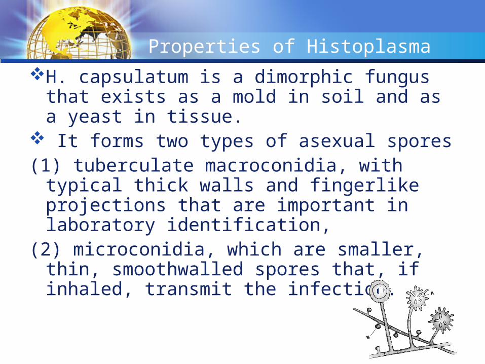
H. capsulatum is a dimorphic fungus that exists as a mold in soil and as a yeast in tissue.
It forms two types of asexual spores(1) tuberculate macroconidia, with typical thick
walls and fingerlike projections that are important in laboratory identification,
(2) microconidia, which are smaller, thin, smoothwalled spores that, if inhaled, transmit the infection.
Transmission & Epidemiology of Histoplasma
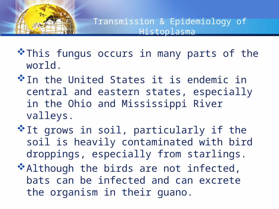
This fungus occurs in many parts of the world.In the United States it is endemic in central and
eastern states, especially in the Ohio and Mississippi River valleys.
It grows in soil, particularly if the soil is heavily contaminated with bird droppings, especially from starlings.
Although the birds are not infected, bats can be infected and can excrete the organism in their guano.
Transmission & Epidemiology of Histoplasma
In areas of endemic infection, excavation of the soil during construction or exploration of bat-infested caves has resulted in a significant number of infected individuals.
In several tropical African countries, histoplasmosis is caused by Histoplasrna duboisii.
The clinical picture is different from that caused by H. capsulatum.A description of the differences between African
histoplasmosis and that seen in the United States is beyond the scope of this book.
Pathogenesis & Clinical Findings of Histoplasma
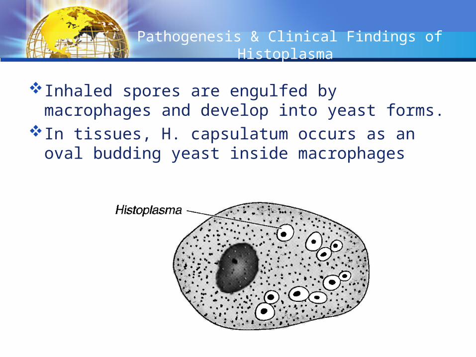
Inhaled spores are engulfed by macrophages and develop into yeast forms.
In tissues, H. capsulatum occurs as an oval budding yeast inside macrophages

Pathogenesis & Clinical Findings of Histoplasma
The yeasts survive within the phagolysosome of the macrophage by producing alkaline substances, such as bicarbonate and ammonia, that raise the pH and thereby inactivate the degradative enzymes of the phagolysosome
The organisms spread widely throughout the body; especially to the liver and spleen, but most infections remain asymptomaric, and the small grantdomatous foci heal by calcification.
Pathogenesis & Clinical Findings of Histoplasma
With intense exposure (eg, in a chicken house or batinfested cave), pneumonia may become clinically manifest.
Severe disseminated histoplasmosis develops in a small minority of infected persons, especially infants and individuals with reduced cell-mediated immunity, such as AIDS patients.
In AIDS patients, ulcerated lesions on the tongue are typica] of disseminated histoplasmosis. In immunocompetent people, EN can occur (see description of EN in Coccidioides above).
Pathogenesis & Clinical Findings of Histoplasma
EN is a sign that cell-mediated immunity is active and the organism will probably be contained.
A skin test using histoplasmin (a mycelial extract) becomes positive, ie, shows at least 5 mm of induration, within 2-3 weeks after infection and remains positive for many years.
However, because there are many false-positive reactions (due to cross-reactivity) and many false-negative reactions (in disseminated disease), the skin test is not useful for diagnosis.
Pathogenesis & Clinical Findings of Histoplasma
Furthermore, the skin test can stimulate an antibody response and confuse the serologic tests.
The skin test is useful for epidemiologic studies, and up to 90% of individuals have positive results in areas of endemic infection.

Laboratory Diagnosis of Histoplasma
In tissue biopsy specimens or bone marrow aspirates, oval yeast cells within macrophages are seen microscopically.
Cultures on Sabouraud's agar show hyphae with tuberculate macroconidia when grown at low temperature, eg, 25°C and yeasts when grown at 37°C.
Tests that detect Histoplasma antigens by radioimmunoassay and Histoplasma RNA with DNA probes are also useful.
Laboratory Diagnosis of Histoplasma
An antibody titer of 1:32 in the CF test with yeast phase antigens is considered to be diagnostic.
However, cross-reactions with other fungi, especially Blastomyces, occur.
CF titers fall when the disease becomes inactive and rise in disseminated disease.
The ID test detects precipitating antibodies (precipitins) by forming two bands, M and H, in an agar-gel diffusion assay.
The ID test is more specific but less sensitive than the CF test.
Treatment & Prevention of Histoplasma
No therapy is needed in asymptomatic or mild primary infections.
With progressive lung lesions, oral itraconazole is beneficial.
In disseminated disease, arnphotericin B is the treatment of choice.
In meningitis,fluconazole is often used because it penetrates the spinal fluid well.
Treatment & Prevention of Histoplasma
Oral itraconazole is used to treat pulmonary or disseminated disease, as well as for chronic suppression in patients with AIDS.
There are no means of prevention except avoiding exposure in areas of endemic infection.

BLASTOMYCESBLASTOMYCES
Disease of Blastomyces
Blastomyces dermatitidis causes blastomycosis, known as North American blastomycosis.
Properties of Blastomyces
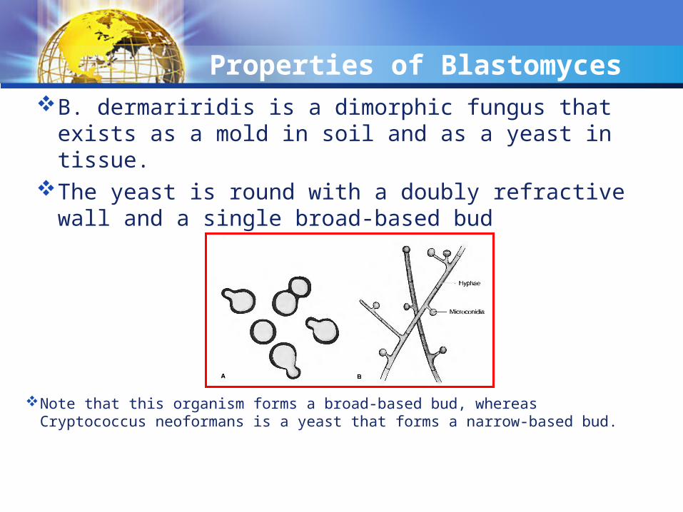
B. dermariridis is a dimorphic fungus that exists as a mold in soil and as a yeast in tissue.
The yeast is round with a doubly refractive wall and a single broad-based bud
Note that this organism forms a broad-based bud, whereas Cryptococcus neoformans is a yeast that forms a narrow-based bud.
Transmission & Epidemiology of Blastomyces
This fungus is endemic primarily in eastern North America, especially in the region bordering the Ohio, Mississippi, and St. Lawrence rivers, and the Great Lakes region.
Less commonly, blastomycosis has also occurred in Central and South America, Africa, and the Middle East. It grows in moist soil rich in organic material, forming hyphae with small pear-shaped conidia.
Inhalation of the conidia causes human infection.
Pathogenesis & Clinical Findings of Blastomyces
Infection occurs mainly via the respiratory tract. Asymptomatic or mild cases are rarely
recognized. Dissemination may result in ulcerated
granulomas of skin, bone, or other sites.
Laboratory Diagnosis of Blastomyces
In tissue biopsy specimens, thick-walled yeast cells with single broad-based buds are seen microscopically.
Hyphae with small pear-shaped conidia are visible on culture.
The skin test lacks specificity and has little value. Serologic tests have little value.
Treatment & Prevention of Blastomyces
Itraconazole is the drug of choice for most patients
Amphotericin B should be used to treat severe disease.
Surgical excision may be helpful. There are no means of prevention.
PARACOCCIDIOIDESPARACOCCIDIOIDES

Disease of Paracoccidioides
Paracoccidioides brasiliensis causes paracoccidioidomycosis, also known as South American blastomycosis.

Properties of Paracoccidioides
P. brasiliensis is a dimorphic fungus that exists as a mold in soil and as a yeast in tissue.
The yeast is thick walled with multiple buds, in contrast to B. derrnatitidis, which has a single bud .
Transmission & Epidemiology of Paracoccidioides
The spores are inhaled, and early lesions occur in the lungs.
Asymptomatic infection is common. Alternatively oral mucous membrane lesions,
lymph node enlargement, and sometimes dissemination to many organs develop.

Laboratory Diagnosis of Paracoccidioides
In pus or tissues, yeast cells with multiple buds are seen microscopically.
A specimen cultured for 2-4 weeks may grow typical organisms.
Skin tests are rarely helpful. Serologic testing shows that when significant
antibody titers (by immunodiffusion or complement fixation) are found, active disease is present.

Pathogenesis & Clinical Findings of Paracoccidioides
This fungus grows in the soil and is endemic in rural Latin America. Disease occurs only in that region.
Treatment & Prevention of Paracoccidioides
The drug of choice is itraconazole taken orally for several months.
There are no means of prevention.

Hodaifa S. Al AbssyHodaifa S. Al Abssy