Embed Size (px)
Citation preview

Systemic ModuleMSS
“Anatomy”
Gluteal Region
Dr. Ayman AlzubiFaculty of Medicine, Yarmouk University

Gluteal Region
• The gluteal region is an anatomical area lies on
the posterolateral aspect of the pelvis.
• It is occupied by large powerful muscles.
• The muscles in this region move the lower limb
at the hip joint.
• Several important nerves and vessels traverse
this area

Gluteal Region - Boundaries
• Superiorly: iliac crest (at L4)
• Medially: intergluteal cleft
• Laterally: Greater trochanter
• Inferiorly: gluteal fold

Bones of the Gluteal Region
• Posterior aspect of:
1. Hip bone2. Proximal end of Femur3. Hip joint

Ligaments of the Gluteal Region
• 2 ligaments:
1. Sacrospinous, connecting sacrum toischial spine
2. Sacrotuberous, connecting sacrum toischial tuberosity
• Both ligaments share in transformationof the greater and lesser sciatic notchesinto corresponding foramina.


Muscles of the Gluteal Region
• The muscles of the gluteal region can be divided into two groups:
1. Superficial abductors and extenders – group of large muscles that abduct and
extend the femur. Includes the gluteus maximus, gluteus medius, gluteus minimus
and tensor fascia lata.
2. Deep lateral rotators – group of smaller muscles that mainly act to laterally rotate
the femur. Includes the quadratus femoris, piriformis, gemellus superior, gemellus
inferior and obturator internus.

Superficial Abductors and Extenders
• Gluteus maximus• Gluteus medius• Gluteus minimus• Tensor fascia lata

Gluteus Maximus
• Largest and most superficial muscle
• Origin: Originates from the gluteal
surface of the ilium, back of sacrum
and coccyx, back of sacrotuberous
ligament .
• Insertion: 75% of fibers into the
iliotibial tract and 25% into the
gluteal tuberosity of the femur.


Inferior gluteal nerve
G. Maximus• Nerve supply: Inferior gluteal nerve
• Action: It is the main extensor of
the thigh, and assists with lateral
rotation.
• It is only used when force is required
• It is used in standing up from a sitting position,
running & climbing up stairs.
• Paralysis of muscle – inability to raise the trunk
from sitting or stooping positions
Gluteus Maximus

Gluteus Medius
• Lies beneath Gluteus Maximus
• Origin: Gluteal surface of ilium.
• Insertion: Lateral surface of
greater trochanter
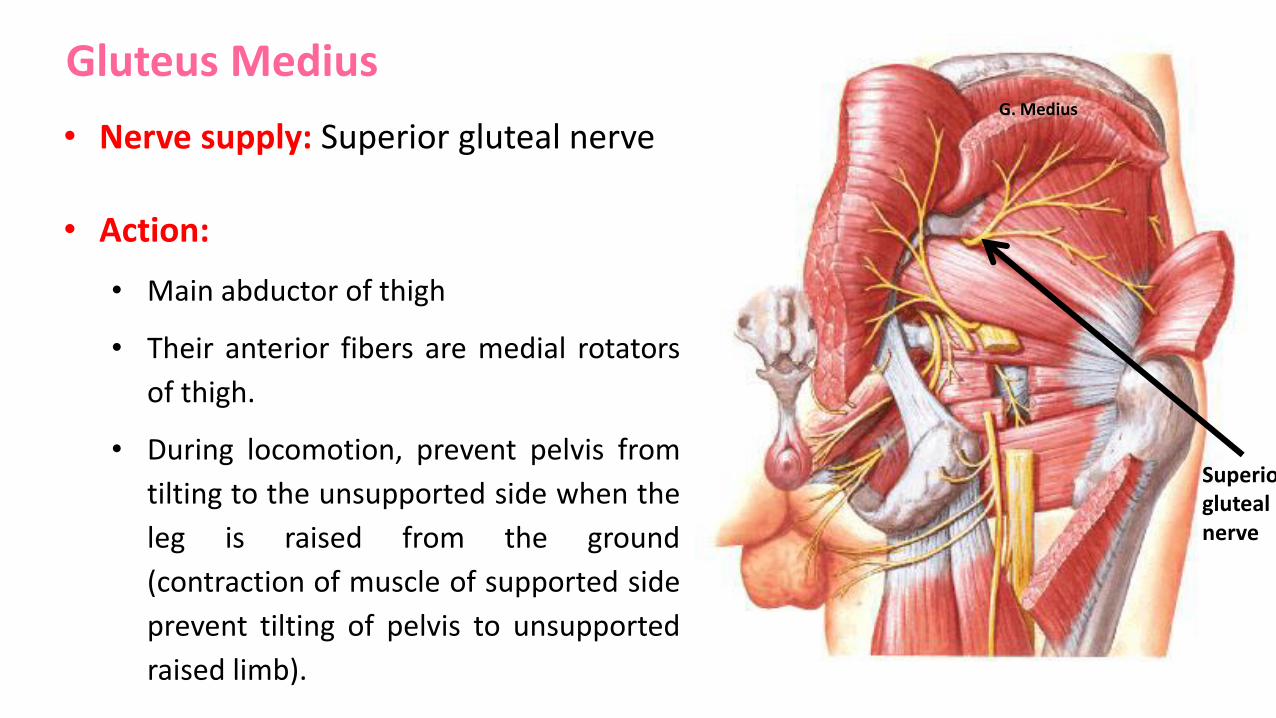
Superior gluteal nerve
G. Medius
• Nerve supply: Superior gluteal nerve
• Action:
• Main abductor of thigh
• Their anterior fibers are medial rotators
of thigh.
• During locomotion, prevent pelvis from
tilting to the unsupported side when the
leg is raised from the ground
(contraction of muscle of supported side
prevent tilting of pelvis to unsupported
raised limb).
Gluteus Medius

Gluteus Minimus
• The deepest and smallest of the
superficial gluteal muscles.
• Origin: Gluteal surface of ilium.
• Insertion: Anterior surface (front) of
greater trochanter of femur.

Superior gluteal nerve
G. Minimus
• Nerve supply: Superior gluteal nerve
• Action:
• Main abductor of thigh
• Their anterior fibers are medial rotators
of thigh.
• Holds opposite side of pelvis horizontally
when foot is off the ground.
Gluteus Minimus
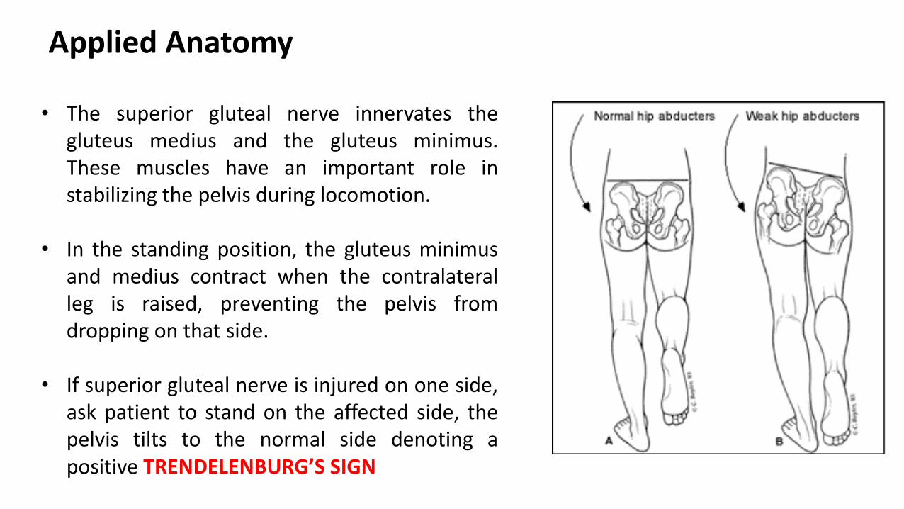
• The superior gluteal nerve innervates thegluteus medius and the gluteus minimus.These muscles have an important role instabilizing the pelvis during locomotion.
• In the standing position, the gluteus minimusand medius contract when the contralateralleg is raised, preventing the pelvis fromdropping on that side.
• If superior gluteal nerve is injured on one side,ask patient to stand on the affected side, thepelvis tilts to the normal side denoting apositive TRENDELENBURG’S SIGN
Applied Anatomy


Tensor Fascia Lata
• Origin: Anterior 5 cm of outer
lip of iliac crest..
• Insertion: Into the iliotibial
tract, which itself attaches to
the lateral condyle of the tibia.
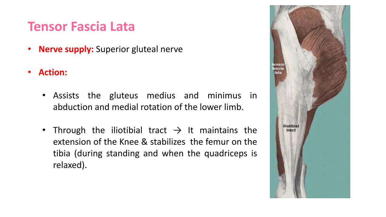
• Nerve supply: Superior gluteal nerve
• Action:
• Assists the gluteus medius and minimus inabduction and medial rotation of the lower limb.
• Through the iliotibial tract → It maintains theextension of the Knee & stabilizes the femur on thetibia (during standing and when the quadriceps isrelaxed).
Tensor Fascia Lata
Deep Lateral Rotators
• Piriformis• Obturator internus.• Gemellus superior• Gemellus inferior• Quadratus femoris

Piriformis
• It is a key landmark in the gluteal region
(vessels and nerves)
• Origin: The anterior surface of the sacrum.
• Insertion: The upper border of greater
trochanter of the femur.
• Nerve supply : Sacral nerve S1 and S2
• Action:
• Lateral rotator of thigh.
• Assists in stabilizing hip joint especially
in abduction.

Obturator Internus
• Forms the lateral walls of the pelvic cavity.
• Origin: pelvic surface of obturator membrane
& margins of obturator foramen.
• Insertion: Upper border of greater trochanter
along with gemelli.
• Nerve supply : Nerve to obturator internus
(L4,S1)
• Action: Lateral rotator of thigh.

Piriformis
Obturator Internus

Gamellus SuperiorOrigin-spine of ischiumInsertion-tendon of OBT intNerve supply- Nerve to OBT internus
Gamellus InferiorOrigin-ischial tuberosityInsertion-tendon of OBT internusNerve supply- nerve to Quadratus femoris
Quadratus FemorisOrigin-ischial tuberosityInsertion-quadrate tubercleNerve supply: Nerve to Quad. Femoris
All are Lateral Rotators of the thigh

Nerve to obturator internus Nerve to quadratus femoris

Structures Passing Through Greater Sciatic Foramen
• Greater sciatic foramen is a gatewaysbetween the pelvic cavity and the glutealregion .
• Structures passing through this foramen canbe grouped into:
1. Structures passing above the Piriformis2. Structures passing below the Piriformis

Piriformis: an important landmark
Above the piriformis:
Superior gluteal vessels & nerve
Below the piriformis:
Inferior gluteal vessels & nerve
Sciatic nerve
Posterior cutaneous nerve of thigh
Pudendal nerve & Internal pudendal vessels
Nerve to obturator internus
Nerve to quadratus femoris


Structures Passing Through Lesser Sciatic Foramen
• Lesser sciatic foramen is a gatewaybetween the gluteal region and theperineum.
1. Tendon of obturator internus2. Nerve to obturator internus3. Internal pudendal vessels4. Pudendal nerve

Safe Area for Intramuscular Injection
• Intramuscular injection enables a largeamount of a drug to be introduced atonce but absorbed gradually.
• The injection site must be carefullyselected to avoid injury to theunderlying large vessels and nerves.
• Outer upper quadrant of the buttockis the safe area for intramuscularinjection to avoid injury to theunderlying sciatic nerve

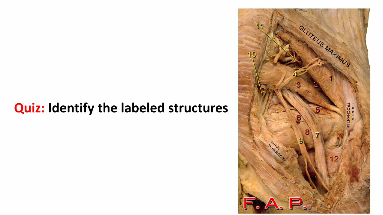
Quiz: Identify the labeled structures