Embed Size (px)
Citation preview
Research ArticleSystemic Alterations of Wnt Inhibitors in Patients with ProstateCancer and Bone Metastases
Stefan Aufderklamm,1 Jörg Hennenlotter,1 Phillip Leidenberger,1 Steffen Rausch ,1
Andrea Hohneder,1 Ursula Kühs,1 Moritz Maas,1 Christian Schwentner,2 Jens Bedke,1
Arnulf Stenzl ,1 and Tilman Todenhöfer 1
1Department of Urology, Eberhard Karls University of Tübingen, Tübingen, Germany2Department of Urology, Diakonie-Klinikum Stuttgart, Stuttgart, Germany
Correspondence should be addressed to Tilman Todenhöfer; [email protected]
Received 24 April 2018; Accepted 5 July 2018; Published 18 July 2018
Academic Editor: Frank Tacke
Copyright © 2018 Stefan Aufderklamm et al. This is an open access article distributed under the Creative CommonsAttribution License, which permits unrestricted use, distribution, and reproduction in any medium, provided the originalwork is properly cited.
Purpose. Dickkopf-1 (DKK-1) and sclerostin seem to inhibit osteoblast activity by blocking the Wnt pathway, which leads toprogression of metastatic prostate cancer (PC). However, it is unknown whether serum levels of these proteins are altered in PCpatients with or without metastasis. The aim of this study was to assess DKK-1 and sclerostin serum levels in PC patients,including patients with bone metastases. Methods. The study cohort (N = 143) consisted of 53 controls with benign prostatichyperplasia (BPH), 43 with localized PC (PC cM0), and 47 had PC with metastasis (PC cM1). Serum levels of DKK-1 andsclerostin were measured by enzyme-linked immunosorbent assay. Results were compared using the Kruskal-Wallis tests;post hoc analysis was performed using the Tukey-Kramer test. Results. Mean DKK-1 levels in patients with BPH(2809.4 pg/ml) (p < 0 001) as well as PC cM1 (2575.5 pg/ml) (p = 0 001) were significantly higher than in patients with PCcN0 cM0 (1551.8 pg/ml). Among PC cM1 patients, median DKK-1 levels were significantly lower in patients withcastration-resistant disease compared to those with hormone-sensitive PC (p = 0 02); in contrast, sclerostin concentrationswere elevated (p = 0 04). DKK-1 correlated with PSA in the cM1 group (p = 0 03) and sclerostin correlated with PSA inthe PC group (0.01). Conclusions. DKK-1 is involved in the progression of PC. DKK-1-mediated inhibition of osteoblasts,which contributes to tumor progression and osteolytic metastases, may also play a role in the development of metastaseswith osteoblastic features. The use of DKK-1 antibodies should be considered for studies including metastatic PC patients.
1. Introduction
Prostate cancer (PC) is the most common cancer in elderlymen in western countries [1]. Patients with advanced PCcommonly develop bone metastases [2]. Metastatic bone dis-ease provokes loss of bone mass, and patients may sufferfrom pathological fractures and other skeletal-related events(SREs) [3]. Recently, the role of the Wnt pathway in progres-sion of osteolytic disease has been investigated [4]. In thiscontext, antagonistic proteins Dickkopf-1 (DKK-1) andsclerostin were highlighted because they both inhibit osteo-blast activity by blockingWnt pathway signaling [4]. In othermalignancies, such as multiple myeloma or breast cancer,
higher levels of DKK-1 were associated with increased bonelesions [5, 6].
Expression of DKK-1 in PC tissue has been demon-strated to be elevated compared with benign tissue andappears to be associated with worse survival [7]. By contrast,downregulation of DKK-1 seems to delay the developmentof bone metastases in PC [8]. Furthermore, elevated DKK-1 expression is an early event in PC and with tumor progres-sion DKK-1 expression declines, particularly in advancedbone metastases.
The Wnt pathway inhibitor sclerostin has been alsodiscussed as a therapeutic target for cancer-related bone dis-ease. In men with PC undergoing antihormonal treatment,
HindawiDisease MarkersVolume 2018, Article ID 1874598, 5 pageshttps://doi.org/10.1155/2018/1874598
circulating sclerostin levels are elevated. However, the role ofsclerostin in progression of malignant bone disease is largelyunknown [9].
The aim of this study was to assess systemic alterations ofDKK-1 and sclerostin in patients with different stages of PC.Moreover, we assessed serum levels in patients with benignprostatic hyperplasia (BPH).
2. Material and Methods
2.1. Patients’ Assessment. A total of 143 patients wereincluded in this study. Serum samples were obtained between2011 and 2013 after receiving informed consent frompatients and approval of the institutional review board ofthe Eberhard Karls University of Tübingen (number 034/2011BO2 and 113/2012BO2). Bone scintigraphy was per-formed in all patients with PC. We included 53 patients withBPH prior to transurethral resection of the prostate (TUR-P),43 patients with histologically proven PC before radical pros-tatectomy, and 47 patients with metastatic PC. Serum levelsof DKK-1 and sclerostin were measured by enzyme-linkedimmunosorbent assay (ELISA).
2.2. Measurements of DKK-1 and Sclerostin. Serum sampleswere stored at −80°C until use. ELISA kits for both DKK-1and sclerostin were provided by Biomedica (Vienna,Austria), and analysis was performed according to themanufacturer’s protocol.
Briefly, for DKK-1, 20μl of serum was incubated with50μl of biotinylated DKK-1 antibody for 2 hours at roomtemperature. After repeated washing steps with wash buffer,100μl of conjugate was added into each well of a 96-well plateand incubated for 1 hour at room temperature. Anotherwashing followed before 100μl of substrate was added andincubated at room temperature.
For sclerostin, 20μl of serum was incubated with 50μlof biotinylated antisclerostin antibody for 18–24 hours atroom temperature. After washing with 300μl diluted washbuffer, 200μl of conjugate was added to each well andincubated for 1 hour at room temperature in the dark.Thereafter, another washing step followed and 200μl ofsubstrate were added and incubated for 30 minutes atroom temperature. Finally, 50μl of stop solution wasadded to each well and absorbance was measured immedi-ately at 450nm with reference at 630nm. An Anthos 2010system (Anthos Mikrosysteme GmbH, Krefeld, Germany)was used for all measurements.
2.3. Statistical Analysis. Serum concentrations were com-pared among different clinical groups (BPH, PC cM0, andPC cM1). Planned subgroup analysis included PC M1patients with castration-sensitive (CS) and castration-resistant (CR) disease. Associations between groups wereassessed by Kruskal-Wallis tests, with the Tukey-Kramermethod used in post hoc analyses. Correlations betweencontinuous variables were performed with linear regres-sion analyses. JMP 7.0® software was used, and p valuesp < 0 05 were considered significant.
3. Results
3.1. Patients. Patient characteristics and clinicopathologicalfeatures of defined subgroups of PC (cM0 and cM1) aresummarized in Table 1. Median ages of all patients,patients with benign prostate histology, PC cM0, and PCcM1 were 73 (range 44–89), 74 (44–89), 69 (48–85), and74 years (52–84), respectively.
Of 43 cM0 patients with radical prostatectomy, 41(95.3%) were pN0 and 2 were pN1 (4.7%). Median PSAwas 6.96 ng/ml (range 0.31–44 ng/ml). The median GleasonScore was 7 (Table 1).
In the PC cM1 group, all 47 patients had radiologicevidence of bone metastases. In addition, 25 (53.2%)patients were cN1 and 8 (17.0%) were cM1c. Lymph nodestatus could not be assessed in two (4.3%) patients. Themedian PSA value in PC cM1 patients was 80.4 ng/ml (range0.5–674.0 ng/ml). The median Gleason Score was 9. In thePC M1 group, 34 patients (72.3%) were castration-resistant(CR) and 11 patients (23.4%) showed ongoing response toantihormonal treatment; this information was unavailablefor the other two (4.3%) patients (Table 1).
Table 1: Characteristics of patients with prostate cancer withoutmetastases (PC cM0) or with metastases (PC cM1).
PC cM0 PC cM1
N 43 47
Age (median; range) 69 (48–85) 74 (52–84)
PSA, ng/ml (median; range) 6.96 (0.31–44) 80.4 (0.52–674)
Gleason Score, n (%) —
≤6 7 (16.3) —
7 31 (72.1) 1 (2.1)
≥8 5 (11.6) 4 (8.5)
T stage, n (%) —
pT2a 6 (14.0) —
pT2b 1 (2.3) —
pT2c 24 (55.8) —
pT3a 9 (20.9) —
pT3b 3 (7.0) —
N stage, n (%)
N0 41 (95.3) 20 (42.5)
N+ 2 (4.7) 25 (53.2)
Nx — 2 (4.3)
M stage, n (%)
cM1b — 47 (100)
cM1c — 8 (17.0)
Castrate level, n (%)
CSPC — 11 (23.4)
CRPC — 34 (72.3)
n.a. 2 (4.3)
T stage = primary tumor; N stage = lymph nodes; M stage = distant metastases;N0 =no regional lymph node metastasis; N1=metastasis in regional lymphnode(s); Nx = regional lymph nodes were not assessed; cM1b= bonemetastases; cM1c = other metastases; CSPC= castration-sensitive prostatecancer; CRPC= castration-resistant prostate cancer; n.a. = not applicable.
2 Disease Markers
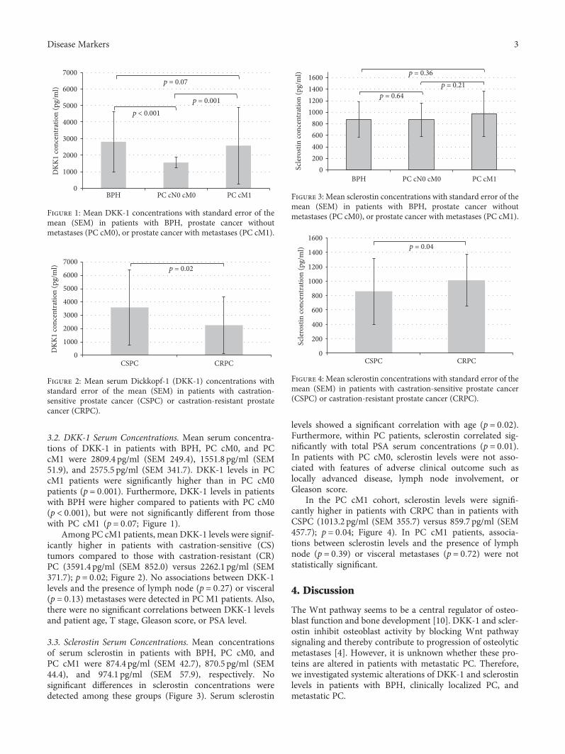
3.2. DKK-1 Serum Concentrations. Mean serum concentra-tions of DKK-1 in patients with BPH, PC cM0, and PCcM1 were 2809.4 pg/ml (SEM 249.4), 1551.8 pg/ml (SEM51.9), and 2575.5 pg/ml (SEM 341.7). DKK-1 levels in PCcM1 patients were significantly higher than in PC cM0patients (p = 0 001). Furthermore, DKK-1 levels in patientswith BPH were higher compared to patients with PC cM0(p < 0 001), but were not significantly different from thosewith PC cM1 (p = 0 07; Figure 1).
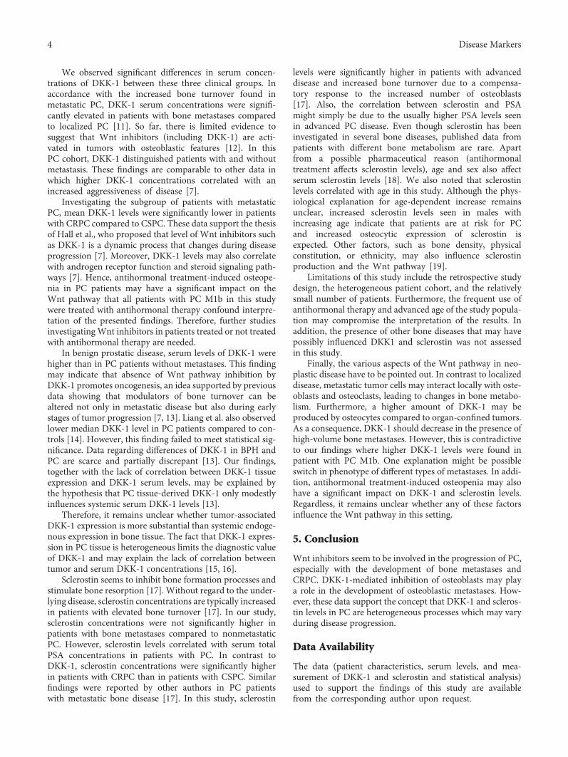
Among PC cM1 patients, mean DKK-1 levels were signif-icantly higher in patients with castration-sensitive (CS)tumors compared to those with castration-resistant (CR)PC (3591.4 pg/ml (SEM 852.0) versus 2262.1 pg/ml (SEM371.7); p = 0 02; Figure 2). No associations between DKK-1levels and the presence of lymph node (p = 0 27) or visceral(p = 0 13) metastases were detected in PC M1 patients. Also,there were no significant correlations between DKK-1 levelsand patient age, T stage, Gleason score, or PSA level.
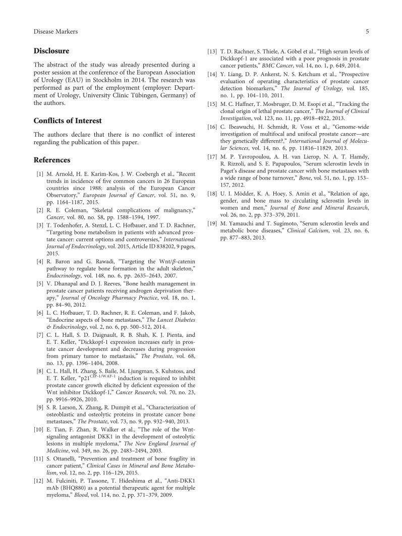
3.3. Sclerostin Serum Concentrations. Mean concentrationsof serum sclerostin in patients with BPH, PC cM0, andPC cM1 were 874.4 pg/ml (SEM 42.7), 870.5 pg/ml (SEM44.4), and 974.1 pg/ml (SEM 57.9), respectively. Nosignificant differences in sclerostin concentrations weredetected among these groups (Figure 3). Serum sclerostin
levels showed a significant correlation with age (p = 0 02).Furthermore, within PC patients, sclerostin correlated sig-nificantly with total PSA serum concentrations (p = 0 01).In patients with PC cM0, sclerostin levels were not asso-ciated with features of adverse clinical outcome such aslocally advanced disease, lymph node involvement, orGleason score.
In the PC cM1 cohort, sclerostin levels were signifi-cantly higher in patients with CRPC than in patients withCSPC (1013.2 pg/ml (SEM 355.7) versus 859.7 pg/ml (SEM457.7); p = 0 04; Figure 4). In PC cM1 patients, associa-tions between sclerostin levels and the presence of lymphnode (p = 0 39) or visceral metastases (p = 0 72) were notstatistically significant.
4. Discussion
The Wnt pathway seems to be a central regulator of osteo-blast function and bone development [10]. DKK-1 and scler-ostin inhibit osteoblast activity by blocking Wnt pathwaysignaling and thereby contribute to progression of osteolyticmetastases [4]. However, it is unknown whether these pro-teins are altered in patients with metastatic PC. Therefore,we investigated systemic alterations of DKK-1 and sclerostinlevels in patients with BPH, clinically localized PC, andmetastatic PC.
0
1000
2000
3000
4000
5000
6000
7000
BPH PC cN0 cM0 PC cM1
DKK
1 co
ncen
trat
ion
(pg/
ml)
p = 0.07
p = 0.001p < 0.001
Figure 1: Mean DKK-1 concentrations with standard error of themean (SEM) in patients with BPH, prostate cancer withoutmetastases (PC cM0), or prostate cancer with metastases (PC cM1).
0
1000
2000
3000
4000
5000
6000
7000
CSPC CRPC
DKK
1 co
ncen
trat
ion
(pg/
ml) p = 0.02
Figure 2: Mean serum Dickkopf-1 (DKK-1) concentrations withstandard error of the mean (SEM) in patients with castration-sensitive prostate cancer (CSPC) or castration-resistant prostatecancer (CRPC).
0200400600800
1000120014001600
BPH PC cN0 cM0 PC cM1
Scle
rost
in co
ncen
trat
ion
(pg/
ml)
p = 0.64p = 0.21
p = 0.36
Figure 3: Mean sclerostin concentrations with standard error of themean (SEM) in patients with BPH, prostate cancer withoutmetastases (PC cM0), or prostate cancer with metastases (PC cM1).
0
200
400
600
800
1000
1200
1400
1600
CSPC CRPC
Scle
rost
in co
ncen
trat
ion
(pg/
ml) p = 0.04
Figure 4: Mean sclerostin concentrations with standard error of themean (SEM) in patients with castration-sensitive prostate cancer(CSPC) or castration-resistant prostate cancer (CRPC).
3Disease Markers
We observed significant differences in serum concen-trations of DKK-1 between these three clinical groups. Inaccordance with the increased bone turnover found inmetastatic PC, DKK-1 serum concentrations were signifi-cantly elevated in patients with bone metastases comparedto localized PC [11]. So far, there is limited evidence tosuggest that Wnt inhibitors (including DKK-1) are acti-vated in tumors with osteoblastic features [12]. In thisPC cohort, DKK-1 distinguished patients with and withoutmetastasis. These findings are comparable to other data inwhich higher DKK-1 concentrations correlated with anincreased aggressiveness of disease [7].
Investigating the subgroup of patients with metastaticPC, mean DKK-1 levels were significantly lower in patientswith CRPC compared to CSPC. These data support the thesisof Hall et al., who proposed that level of Wnt inhibitors suchas DKK-1 is a dynamic process that changes during diseaseprogression [7]. Moreover, DKK-1 levels may also correlatewith androgen receptor function and steroid signaling path-ways [7]. Hence, antihormonal treatment-induced osteope-nia in PC patients may have a significant impact on theWnt pathway that all patients with PC M1b in this studywere treated with antihormonal therapy confound interpre-tation of the presented findings. Therefore, further studiesinvestigating Wnt inhibitors in patients treated or not treatedwith antihormonal therapy are needed.
In benign prostatic disease, serum levels of DKK-1 werehigher than in PC patients without metastases. This findingmay indicate that absence of Wnt pathway inhibition byDKK-1 promotes oncogenesis, an idea supported by previousdata showing that modulators of bone turnover can bealtered not only in metastatic disease but also during earlystages of tumor progression [7, 13]. Liang et al. also observedlower median DKK-1 level in PC patients compared to con-trols [14]. However, this finding failed to meet statistical sig-nificance. Data regarding differences of DKK-1 in BPH andPC are scarce and partially discrepant [13]. Our findings,together with the lack of correlation between DKK-1 tissueexpression and DKK-1 serum levels, may be explained bythe hypothesis that PC tissue-derived DKK-1 only modestlyinfluences systemic serum DKK-1 levels [13].
Therefore, it remains unclear whether tumor-associatedDKK-1 expression is more substantial than systemic endoge-nous expression in bone tissue. The fact that DKK-1 expres-sion in PC tissue is heterogeneous limits the diagnostic valueof DKK-1 and may explain the lack of correlation betweentumor and serum DKK-1 concentrations [15, 16].
Sclerostin seems to inhibit bone formation processes andstimulate bone resorption [17]. Without regard to the under-lying disease, sclerostin concentrations are typically increasedin patients with elevated bone turnover [17]. In our study,sclerostin concentrations were not significantly higher inpatients with bone metastases compared to nonmetastaticPC. However, sclerostin levels correlated with serum totalPSA concentrations in patients with PC. In contrast toDKK-1, sclerostin concentrations were significantly higherin patients with CRPC than in patients with CSPC. Similarfindings were reported by other authors in PC patientswith metastatic bone disease [17]. In this study, sclerostin
levels were significantly higher in patients with advanceddisease and increased bone turnover due to a compensa-tory response to the increased number of osteoblasts[17]. Also, the correlation between sclerostin and PSAmight simply be due to the usually higher PSA levels seenin advanced PC disease. Even though sclerostin has beeninvestigated in several bone diseases, published data frompatients with different bone metabolism are rare. Apartfrom a possible pharmaceutical reason (antihormonaltreatment affects sclerostin levels), age and sex also affectserum sclerostin levels [18]. We also noted that sclerostinlevels correlated with age in this study. Although the phys-iological explanation for age-dependent increase remainsunclear, increased sclerostin levels seen in males withincreasing age indicate that patients are at risk for PCand increased osteocytic expression of sclerostin isexpected. Other factors, such as bone density, physicalconstitution, or ethnicity, may also influence sclerostinproduction and the Wnt pathway [19].
Limitations of this study include the retrospective studydesign, the heterogeneous patient cohort, and the relativelysmall number of patients. Furthermore, the frequent use ofantihormonal therapy and advanced age of the study popula-tion may compromise the interpretation of the results. Inaddition, the presence of other bone diseases that may havepossibly influenced DKK1 and sclerostin was not assessedin this study.
Finally, the various aspects of the Wnt pathway in neo-plastic disease have to be pointed out. In contrast to localizeddisease, metastatic tumor cells may interact locally with oste-oblasts and osteoclasts, leading to changes in bone metabo-lism. Furthermore, a higher amount of DKK-1 may beproduced by osteocytes compared to organ-confined tumors.As a consequence, DKK-1 should decrease in the presence ofhigh-volume bone metastases. However, this is contradictiveto our findings where higher DKK-1 levels were found inpatient with PC M1b. One explanation might be possibleswitch in phenotype of different types of metastases. In addi-tion, antihormonal treatment-induced osteopenia may alsohave a significant impact on DKK-1 and sclerostin levels.Regardless, it remains unclear whether any of these factorsinfluence the Wnt pathway in this setting.
5. Conclusion
Wnt inhibitors seem to be involved in the progression of PC,especially with the development of bone metastases andCRPC. DKK-1-mediated inhibition of osteoblasts may playa role in the development of osteoblastic metastases. How-ever, these data support the concept that DKK-1 and scleros-tin levels in PC are heterogeneous processes which may varyduring disease progression.
Data Availability
The data (patient characteristics, serum levels, and mea-surement of DKK-1 and sclerostin and statistical analysis)used to support the findings of this study are availablefrom the corresponding author upon request.
4 Disease Markers
Disclosure
The abstract of the study was already presented during aposter session at the conference of the European Associationof Urology (EAU) in Stockholm in 2014. The research wasperformed as part of the employment (employer: Depart-ment of Urology, University Clinic Tübingen, Germany) ofthe authors.
Conflicts of Interest
The authors declare that there is no conflict of interestregarding the publication of this paper.
References
[1] M. Arnold, H. E. Karim-Kos, J. W. Coebergh et al., “Recenttrends in incidence of five common cancers in 26 Europeancountries since 1988: analysis of the European CancerObservatory,” European Journal of Cancer, vol. 51, no. 9,pp. 1164–1187, 2015.
[2] R. E. Coleman, “Skeletal complications of malignancy,”Cancer, vol. 80, no. S8, pp. 1588–1594, 1997.
[3] T. Todenhofer, A. Stenzl, L. C. Hofbauer, and T. D. Rachner,“Targeting bone metabolism in patients with advanced pros-tate cancer: current options and controversies,” InternationalJournal of Endocrinology, vol. 2015, Article ID 838202, 9 pages,2015.
[4] R. Baron and G. Rawadi, “Targeting the Wnt/β-cateninpathway to regulate bone formation in the adult skeleton,”Endocrinology, vol. 148, no. 6, pp. 2635–2643, 2007.
[5] V. Dhanapal and D. J. Reeves, “Bone health management inprostate cancer patients receiving androgen deprivation ther-apy,” Journal of Oncology Pharmacy Practice, vol. 18, no. 1,pp. 84–90, 2012.
[6] L. C. Hofbauer, T. D. Rachner, R. E. Coleman, and F. Jakob,“Endocrine aspects of bone metastases,” The Lancet Diabetes& Endocrinology, vol. 2, no. 6, pp. 500–512, 2014.
[7] C. L. Hall, S. D. Daignault, R. B. Shah, K. J. Pienta, andE. T. Keller, “Dickkopf-1 expression increases early in pros-tate cancer development and decreases during progressionfrom primary tumor to metastasis,” The Prostate, vol. 68,no. 13, pp. 1396–1404, 2008.
[8] C. L. Hall, H. Zhang, S. Baile, M. Ljungman, S. Kuhstoss, andE. T. Keller, “p21CIP-1/WAF-1 induction is required to inhibitprostate cancer growth elicited by deficient expression of theWnt inhibitor Dickkopf-1,” Cancer Research, vol. 70, no. 23,pp. 9916–9926, 2010.
[9] S. R. Larson, X. Zhang, R. Dumpit et al., “Characterization ofosteoblastic and osteolytic proteins in prostate cancer bonemetastases,” The Prostate, vol. 73, no. 9, pp. 932–940, 2013.
[10] E. Tian, F. Zhan, R. Walker et al., “The role of the Wnt-signaling antagonist DKK1 in the development of osteolyticlesions in multiple myeloma,” The New England Journal ofMedicine, vol. 349, no. 26, pp. 2483–2494, 2003.
[11] S. Ottanelli, “Prevention and treatment of bone fragility incancer patient,” Clinical Cases in Mineral and Bone Metabo-lism, vol. 12, no. 2, pp. 116–129, 2015.
[12] M. Fulciniti, P. Tassone, T. Hideshima et al., “Anti-DKK1mAb (BHQ880) as a potential therapeutic agent for multiplemyeloma,” Blood, vol. 114, no. 2, pp. 371–379, 2009.
[13] T. D. Rachner, S. Thiele, A. Göbel et al., “High serum levels ofDickkopf-1 are associated with a poor prognosis in prostatecancer patients,” BMC Cancer, vol. 14, no. 1, p. 649, 2014.
[14] Y. Liang, D. P. Ankerst, N. S. Ketchum et al., “Prospectiveevaluation of operating characteristics of prostate cancerdetection biomarkers,” The Journal of Urology, vol. 185,no. 1, pp. 104–110, 2011.
[15] M. C. Haffner, T. Mosbruger, D. M. Esopi et al., “Tracking theclonal origin of lethal prostate cancer,” The Journal of ClinicalInvestigation, vol. 123, no. 11, pp. 4918–4922, 2013.
[16] C. Ibeawuchi, H. Schmidt, R. Voss et al., “Genome-wideinvestigation of multifocal and unifocal prostate cancer—arethey genetically different?,” International Journal of Molecu-lar Sciences, vol. 14, no. 6, pp. 11816–11829, 2013.
[17] M. P. Yavropoulou, A. H. van Lierop, N. A. T. Hamdy,R. Rizzoli, and S. E. Papapoulos, “Serum sclerostin levels inPaget’s disease and prostate cancer with bone metastases witha wide range of bone turnover,” Bone, vol. 51, no. 1, pp. 153–157, 2012.
[18] U. I. Mödder, K. A. Hoey, S. Amin et al., “Relation of age,gender, and bone mass to circulating sclerostin levels inwomen and men,” Journal of Bone and Mineral Research,vol. 26, no. 2, pp. 373–379, 2011.
[19] M. Yamauchi and T. Sugimoto, “Serum sclerostin levels andmetabolic bone diseases,” Clinical Calcium, vol. 23, no. 6,pp. 877–883, 2013.
5Disease Markers

Stem Cells International
Hindawiwww.hindawi.com Volume 2018
Hindawiwww.hindawi.com Volume 2018
MEDIATORSINFLAMMATION
of
EndocrinologyInternational Journal of
Hindawiwww.hindawi.com Volume 2018
Hindawiwww.hindawi.com Volume 2018
Disease Markers
Hindawiwww.hindawi.com Volume 2018
BioMed Research International
OncologyJournal of
Hindawiwww.hindawi.com Volume 2013
Hindawiwww.hindawi.com Volume 2018
Oxidative Medicine and Cellular Longevity
Hindawiwww.hindawi.com Volume 2018
PPAR Research
Hindawi Publishing Corporation http://www.hindawi.com Volume 2013Hindawiwww.hindawi.com
The Scientific World Journal
Volume 2018
Immunology ResearchHindawiwww.hindawi.com Volume 2018
Journal of
ObesityJournal of
Hindawiwww.hindawi.com Volume 2018
Hindawiwww.hindawi.com Volume 2018
Computational and Mathematical Methods in Medicine
Hindawiwww.hindawi.com Volume 2018
Behavioural Neurology
OphthalmologyJournal of
Hindawiwww.hindawi.com Volume 2018
Diabetes ResearchJournal of
Hindawiwww.hindawi.com Volume 2018
Hindawiwww.hindawi.com Volume 2018
Research and TreatmentAIDS
Hindawiwww.hindawi.com Volume 2018
Gastroenterology Research and Practice
Hindawiwww.hindawi.com Volume 2018
Parkinson’s Disease
Evidence-Based Complementary andAlternative Medicine
Volume 2018Hindawiwww.hindawi.com
Submit your manuscripts atwww.hindawi.com


![differenziamento Mina fin [modalità compatibilità] · OBs Sclerostin Myeloma cells through sclerostin secretion contribute to MM Cells OBs Sclerostin OPG RANKL 1)Inhibit OB formation](https://img.pdfslide.us/doc/110x75/5ac3ff867f8b9aae1b8d18c6/differenziamento-mina-fin-modalit-compatibilit-sclerostin-myeloma-cells-through.jpg)