Embed Size (px)
Citation preview
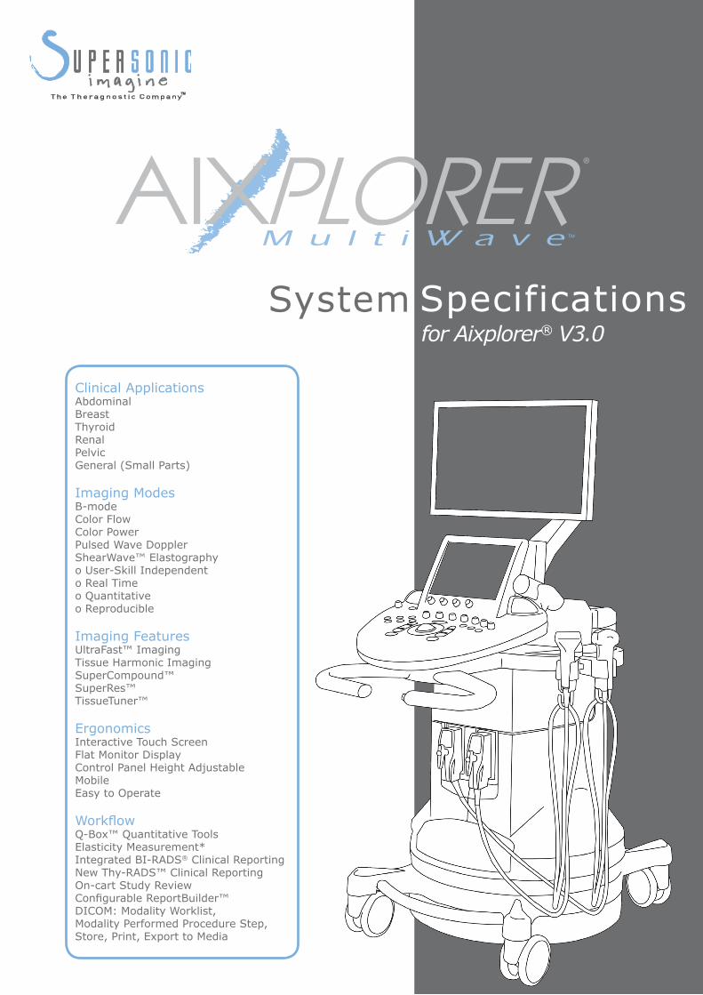
System for Aixplorer® V3.0Specifications
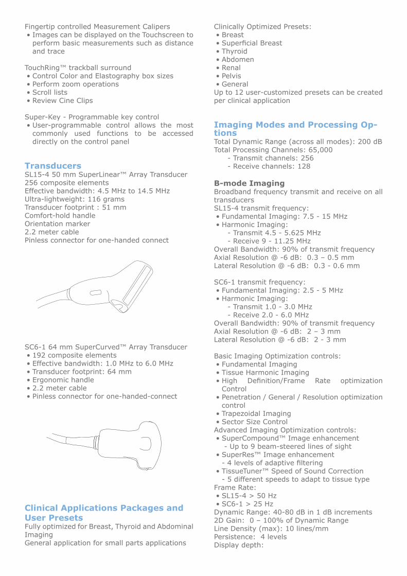
Clinical ApplicationsAbdominalBreastThyroidRenalPelvicGeneral (Small Parts)
Imaging ModesB-modeColor FlowColor PowerPulsed Wave DopplerShearWave™ Elastographyo User-Skill Independento Real Timeo Quantitativeo Reproducible
Imaging FeaturesUltraFast™ ImagingTissue Harmonic ImagingSuperCompound™SuperRes™TissueTuner™
Ergonomics Interactive Touch Screen Flat Monitor DisplayControl Panel Height AdjustableMobileEasy to Operate
Workflow Q-Box™ Quantitative ToolsElasticity Measurement*Integrated BI-RADS® Clinical ReportingNew Thy-RADS™ Clinical ReportingOn-cart Study ReviewConfigurable ReportBuilder™DICOM: Modality Worklist, Modality Performed Procedure Step,Store, Print, Export to Media

Aixplorer® is a next-generation ultrasound system from SuperSonic Imagine introducing impeccable image quality and a new concept of imaging, ShearWave™ Elastography.
Revolutionary Architecture:HardwareIntel i7 Octo Core CPU Processor6 GB of on-board RAMNVIDIA 285 GTX graphics processorWorkStation Class Motherboard256 x 256 imaging channels
Software64-bit Linux based Operating System, Debian derivedSonicSoftware™ Beamforming and ScanConversionUltraFast™ Imaging for ShearWave™Elastography: • Up to 20,000 frames per second acquisitionData transfer rate: > 3 Gbytes/second
PerformanceCold Boot-time: < 90 secondsShut down time: < 20 secondsTransducer select time (typical): < 1 secData access time: < 1 sec
Cart Design and ErgonomicsThin, sleek body styleAble to pass through a 70 cm (28”) doorwayMobile designAdjustable height handles for improved postureand mobilityFour-wheel steering for excellent mobilityTwo-wheel brakingDual ergonomically accessible transducer portsTransducer holder with cable managementOn-cart storage areaExpansion bays for two OEM devicesTwo removable and washable gel holdersBuilt-in footrestProgrammable Footswitch
Flat-Monitor Display and Articulated Arm20.1” (51 cm) EIZO FlexScan LCD Flat PanelDisplayLow-glare hard coating; flicker-free to reduceeye-strainUltra-wide viewing angle: ± 178°Pixel-sharp high resolution: 1680x1050Dot Pitch 0.258 mm x 0.258 mmDisplay Colors 16.77 million colorsContrast Ratio: 900:1Brightness: 300 cd/m2Response time: 8 msMonitor is mounted on a fully articulated arm
Tilt, pitch and height adjustableOverall height adjustment from 129 cm (51”) to168 cm (66”)Arm and monitor fold down to reduce overall heightto 129 cm (51”) for transport
Control PanelAdjustable control panel height for operator comfort standing or sittingVertical articulation: 84 to 98 cm± 45° swivel articulationAdjustable control panel backlightingLarge simple controls for ease of operationEasy-touch dual function knobs to access major modesCenter trackball with TouchRing™ for unparalleled ease of fine adjustmentIntegrated 10 ¼” Touch ScreenIntegrated wrist restMagnetic stylus holderIntegrated stereo speakersDocument holder with integrated holder for iPod® or PDA cradle
Conveniently holds patient charts, records, • etc.iPod• ® or PDA cradle allows user to view Podcasts while operating the system
Advanced User Interface FeaturesInteractive 10 ¼” Touch Screen
Resolution: 1024 x 768• Operates by touch, even with gloves• Stylus friendly non-scratch coating•
Touch-sensitive on-screen keyboardSupport for 5 language keyboard types•
Time-Gain Compensation ControlsTouch-sensitive ManualTouch™ TGC controls• 7 levels of TGC control in depth dimension• One push Auto TGC control•
Fingertip controlled Measurement CalipersImages can be displayed on the Touchscreen to • perform basic measurements such as distance and trace
TouchRing™ trackball surroundControl Color and Elastography box sizes• Perform zoom operations• Scroll lists• Review Cine Clips•
Super-Key - Programmable key controlUser-programmable control allows the most • commonly used functions to be accessed directly on the control panel
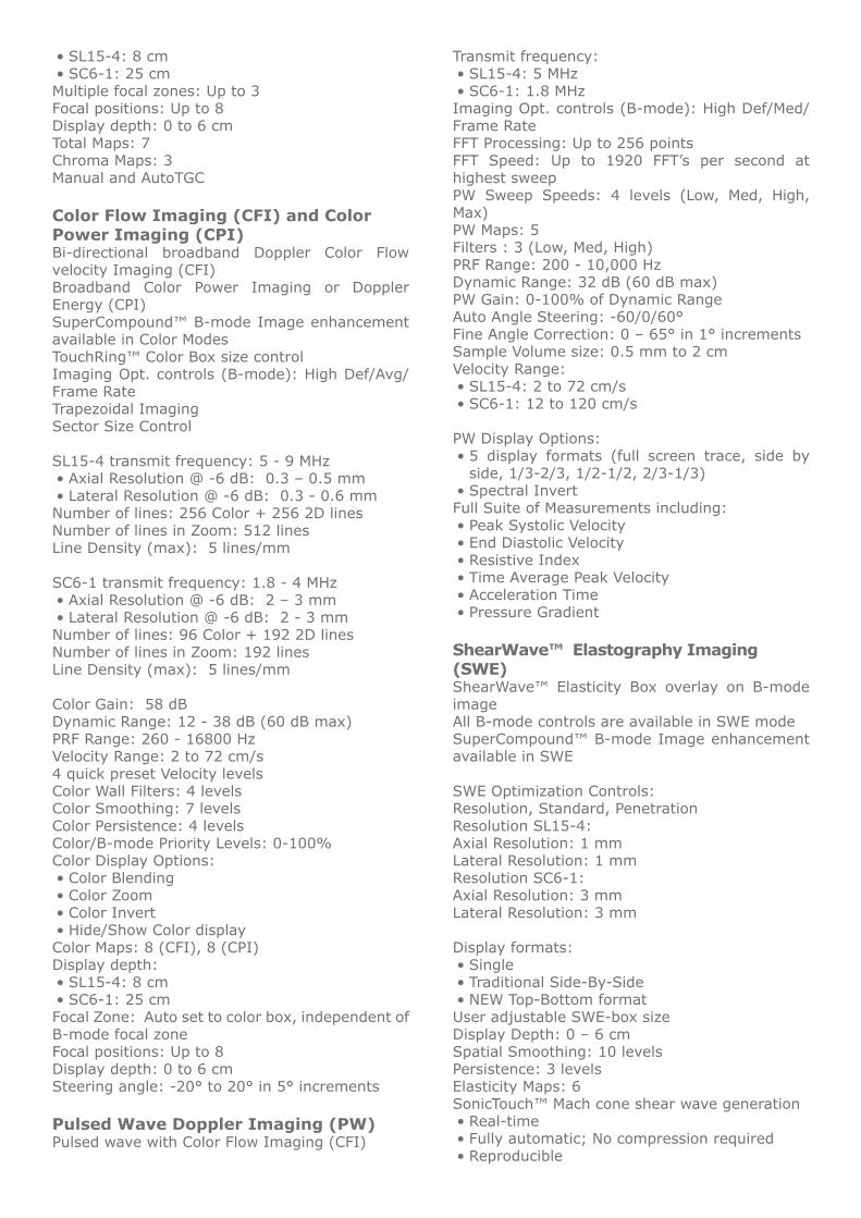
TransducersSL15-4 50 mm SuperLinear™ Array Transducer256 composite elementsEffective bandwidth: 4.5 MHz to 14.5 MHzUltra-lightweight: 116 gramsTransducer footprint : 51 mm Comfort-hold handleOrientation marker2.2 meter cable Pinless connector for one-handed connect
SC6-1 64 mm SuperCurved™ Array Transducer192 composite elements• Effective bandwidth: 1.0 MHz to 6.0 MHz• Transducer footprint: 64 mm• Ergonomic handle• 2.2 meter cable• Pinless connector for one-handed-connect•
Clinical Applications Packages and User PresetsFully optimized for Breast, Thyroid and Abdominal ImagingGeneral application for small parts applications
Clinically Optimized Presets:Breast• Superficial Breast• Thyroid• Abdomen• Renal• Pelvis• General•
Up to 12 user-customized presets can be created per clinical application
Imaging Modes and Processing Op-tionsTotal Dynamic Range (across all modes): 200 dB Total Processing Channels: 65,000
- Transmit channels: 256- Receive channels: 128
B-mode ImagingBroadband frequency transmit and receive on all transducersSL15-4 transmit frequency:
Fundamental Imaging: 7.5 - 15 MHz• Harmonic Imaging: •
- Transmit 4.5 - 5.625 MHz- Receive 9 - 11.25 MHz
Overall Bandwidth: 90% of transmit frequency Axial Resolution @ -6 dB: 0.3 – 0.5 mmLateral Resolution @ -6 dB: 0.3 - 0.6 mm
SC6-1 transmit frequency: Fundamental Imaging: 2.5 - 5 MHz• Harmonic Imaging: •
- Transmit 1.0 - 3.0 MHz- Receive 2.0 - 6.0 MHz
Overall Bandwidth: 90% of transmit frequency Axial Resolution @ -6 dB: 2 – 3 mmLateral Resolution @ -6 dB: 2 - 3 mm
Basic Imaging Optimization controls:Fundamental Imaging• Tissue Harmonic Imaging• High Definition/Frame Rate optimization • ControlPenetration / General / Resolution optimization • controlTrapezoidal Imaging• Sector Size Control•
Advanced Imaging Optimization controls: SuperCompound™ Image enhancement• - Up to 9 beam-steered lines of sightSuperRes™ Image enhancement• - 4 levels of adaptive filteringTissueTuner™ Speed of Sound Correction• - 5 different speeds to adapt to tissue type
Frame Rate: SL15-4 > 50 Hz• SC6-1 > 25 Hz•
Dynamic Range: 40-80 dB in 1 dB increments2D Gain: 0 – 100% of Dynamic RangeLine Density (max): 10 lines/mmPersistence: 4 levelsDisplay depth:
SL15-4: 8 cm• SC6-1: 25 cm•
Multiple focal zones: Up to 3Focal positions: Up to 8Display depth: 0 to 6 cmTotal Maps: 7Chroma Maps: 3Manual and AutoTGC
Color Flow Imaging (CFI) and Color Power Imaging (CPI)Bi-directional broadband Doppler Color Flow velocity Imaging (CFI)Broadband Color Power Imaging or Doppler Energy (CPI) SuperCompound™ B-mode Image enhancement available in Color ModesTouchRing™ Color Box size controlImaging Opt. controls (B-mode): High Def/Avg/Frame RateTrapezoidal ImagingSector Size Control
SL15-4 transmit frequency: 5 - 9 MHzAxial Resolution @ -6 dB: 0.3 – 0.5 mm• Lateral Resolution @ -6 dB: 0.3 - 0.6 mm•
Number of lines: 256 Color + 256 2D linesNumber of lines in Zoom: 512 linesLine Density (max): 5 lines/mm
SC6-1 transmit frequency: 1.8 - 4 MHzAxial Resolution @ -6 dB: 2 – 3 mm• Lateral Resolution @ -6 dB: 2 - 3 mm•
Number of lines: 96 Color + 192 2D linesNumber of lines in Zoom: 192 linesLine Density (max): 5 lines/mm
Color Gain: 58 dBDynamic Range: 12 - 38 dB (60 dB max)PRF Range: 260 - 16800 HzVelocity Range: 2 to 72 cm/s4 quick preset Velocity levelsColor Wall Filters: 4 levelsColor Smoothing: 7 levelsColor Persistence: 4 levelsColor/B-mode Priority Levels: 0-100%Color Display Options:
Color Blending• Color Zoom• Color Invert• Hide/Show Color display•
Color Maps: 8 (CFI), 8 (CPI)Display depth:
SL15-4: 8 cm• SC6-1: 25 cm•
Focal Zone: Auto set to color box, independent of B-mode focal zoneFocal positions: Up to 8Display depth: 0 to 6 cmSteering angle: -20° to 20° in 5° increments
Pulsed Wave Doppler Imaging (PW)Pulsed wave with Color Flow Imaging (CFI)
Transmit frequency: SL15-4: 5 MHz• SC6-1: 1.8 MHz•
Imaging Opt. controls (B-mode): High Def/Med/Frame RateFFT Processing: Up to 256 pointsFFT Speed: Up to 1920 FFT’s per second at highest sweepPW Sweep Speeds: 4 levels (Low, Med, High, Max) PW Maps: 5Filters : 3 (Low, Med, High)PRF Range: 200 - 10,000 HzDynamic Range: 32 dB (60 dB max)PW Gain: 0-100% of Dynamic RangeAuto Angle Steering: -60/0/60°Fine Angle Correction: 0 – 65° in 1° incrementsSample Volume size: 0.5 mm to 2 cmVelocity Range:
SL15-4: 2 to 72 cm/s• SC6-1: 12 to 120 cm/s•
PW Display Options:5 display formats (full screen trace, side by • side, 1/3-2/3, 1/2-1/2, 2/3-1/3)Spectral Invert•
Full Suite of Measurements including:Peak Systolic Velocity• End Diastolic Velocity• Resistive Index• Time Average Peak Velocity• Acceleration Time• Pressure Gradient•
ShearWave™ Elastography Imaging (SWE)ShearWave™ Elasticity Box overlay on B-mode imageAll B-mode controls are available in SWE modeSuperCompound™ B-mode Image enhancement available in SWE
SWE Optimization Controls:Resolution, Standard, PenetrationResolution SL15-4:Axial Resolution: 1 mmLateral Resolution: 1 mmResolution SC6-1:Axial Resolution: 3 mmLateral Resolution: 3 mm
Display formats:Single• Traditional Side-By-Side• NEW Top-Bottom format•
User adjustable SWE-box sizeDisplay Depth: 0 – 6 cmSpatial Smoothing: 10 levelsPersistence: 3 levelsElasticity Maps: 6SonicTouch™ Mach cone shear wave generation
Real-time• Fully automatic; No compression required• Reproducible•
UltraFast™ Data Acquisition Technology for SWE:
SWE Data Frame Rate: 20 kHz • Real-time Display Frame Rate: up to 4 Hz•
Q-Box™ pixel accurate Elasticity quantification* Range of Elasticity Displayed: 0 – 300 kPa Optimized default Elasticity scaleQ-BoxTM Precision: +/-15% of displayed value*
Composite Imaging Modes:Composite imaging modes include:
Simultaneous B-mode & Color Flow Imaging • (CFI) Simultaneous B-mode & Color Power Imaging • (CPI)Simultaneous B-mode & PW• Simultaneous B-mode & SWE• B-mode, Color & PW Doppler• HD Zoom (high-resolution zoom)• o Available in B-mode, Color, PW and SWE modes o Up to 512 scan lines of resolution
Dual ImagingFull featured Dual Imaging Mode with Independent controls and measures in side-by-side panes:
Dual B-mode• Dual B-mode & Color•
Cine and Review FeaturesFrozen Cine ReviewFrame-by-frame image review while frozenTrackball play, fast-forward play and frame reverse via trackball controlAvailable in all Imaging modes including dual Cine buffer size: 1000 frames (approx)CFI/CPI Cine buffer size: 500 frames (approx)PW Cine buffer size: 500 columns (approx)Post-processing controls available while in frozen review:B-mode: Gain, Dynamic Range, TGC, 2D Maps, Measurements, Annotations, Body marksCFI/CPI: Transparency, Color Map, Color Priority, Hide/Show Color, Baseline, InvertPW: Cine Review: Display Format, PW Map, Angle Correct, Baseline, InvertSWE: Display Format, Blending, Elasticity Map, Elasticity Range, Persistence
Annotation and Body MarkersAnnotationsFull annotation packages for Breast, Thyroid, Abdominal, Renal, Pelvic and General ApplicationQuick access to the last 12 annotations usedFully user-editable annotationsCustom home cursor position or choice of 5 preset locations
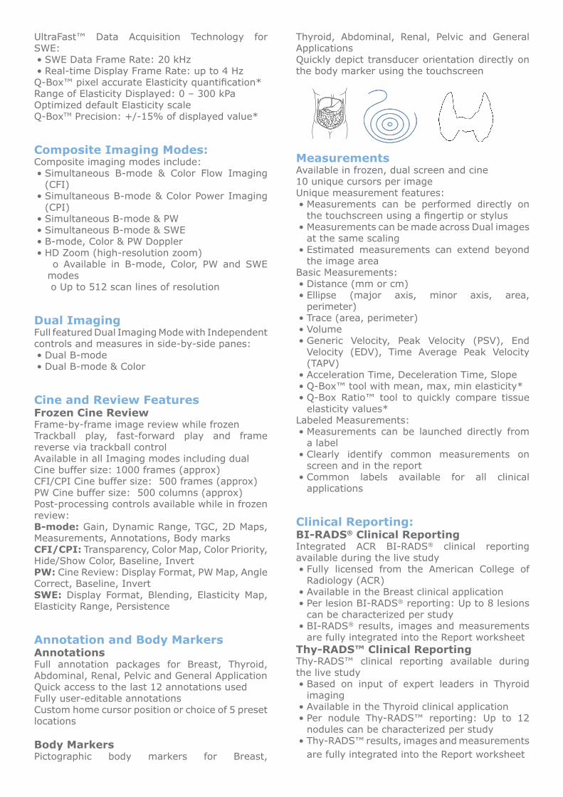
Body MarkersPictographic body markers for Breast,
Thyroid, Abdominal, Renal, Pelvic and General ApplicationsQuickly depict transducer orientation directly on the body marker using the touchscreen
MeasurementsAvailable in frozen, dual screen and cine10 unique cursors per imageUnique measurement features:
Measurements can be performed directly on • the touchscreen using a fingertip or stylusMeasurements can be made across Dual images • at the same scalingEstimated measurements can extend beyond • the image area
Basic Measurements:Distance (mm or cm)• Ellipse (major axis, minor axis, area, • perimeter)Trace (area, perimeter)• Volume• Generic Velocity, Peak Velocity (PSV), End • Velocity (EDV), Time Average Peak Velocity (TAPV)Acceleration Time, Deceleration Time, Slope• Q-Box™ tool with mean, max, min elasticity*• Q-Box Ratio™ tool to quickly compare tissue • elasticity values*
Labeled Measurements:Measurements can be launched directly from • a labelClearly identify common measurements on • screen and in the reportCommon labels available for all clinical • applications
Clinical Reporting:BI-RADS® Clinical ReportingIntegrated ACR BI-RADS® clinical reporting available during the live study
Fully licensed from the American College of • Radiology (ACR)Available in the Breast clinical application• Per lesion BI-RADS• ® reporting: Up to 8 lesions can be characterized per studyBI-RADS• ® results, images and measurements are fully integrated into the Report worksheet
Thy-RADS™ Clinical ReportingThy-RADS™ clinical reporting available during the live study
Based on input of expert leaders in Thyroid • imagingAvailable in the Thyroid clinical application• Per nodule Thy-RADS™ reporting: Up to 12 • nodules can be characterized per studyThy-RADS™ results, images and measurements • are fully integrated into the Report worksheet
Study ReviewQuick Study Review
Image thumbnails on main display allow quick • reviewPreview, Open or Delete images instantly•
Full Study ReviewSelectable study list with TouchRing™• Display study images in 1, 2, 6, 12 and 20-up • formatsReplay cine-clips in real-time• Export images directly to USB in JPEG format • Export cine-clips directly to USB in MPEG • format
Configurable ReportReportBuilder™ allows the user configurability of the information presented in the Report worksheet:
User-uploadable hospital logo for Report • headerIntegrated patient history from Patient Data • entry screensFully editable measurements and comments• Per image reporting with data reconciliation • toolsMeasurements hyper-linked to study images • for quick reviewGenerous freeform text areas for exam • comments and conclusionsReport preview• Export reports directly to USB as a Portable • Document Format (PDF) file
ReportBuilder™ configurable components:Patient history, Images, Measurements, BI-RADS®, Thy-RADS™, Reports, Comments
Data ManagementInternal hard drive(s) for image and data storageRaid mirror configuration: 2 hard drives for fail-safe storage and securityHard disk capacity: 160 Gb x 2Image storage: 20,000 images (estimated)Study storage: 2000 typical studies (10 images and data)
Data ExportExport images to CD, DVD, USB memory device or Ethernet
JPEG/MPEG Export to USB memory, CD/DVD•
Export reports directly to USB as a Portable Document Format (PDF) file
DICOM & Connectivity10/100/1000 baseT Ethernet compliant connectivity
DICOM Storage Service ClassAllows connectivity to PACS• Allows “save-as-you-scan” or “end of exam” • transfer of study data
DICOM Export to Media:Export studies in DICOM format to CD/DVD • and USB
DICOM PrintAllows “print-as-you-scan” or “end-of-exam” • printing to DICOM print devicesCompatible with the most common DICOM • printers (AGFA, KODAK, etc.)
DICOM Modality WorklistAuto-population of Patient Data Entry screen • from hospital HIS/RIS serverSort or filter Worklist according to patient • information (name, ID, date/time, etc.)
DICOM Modality Performed Procedure Step (MPPS)
System receives and transmits info relating to • the patient study and care cycle
DICOM Storage Commitment Procedure (SCP)Provides commitment from the storage • device that study data has been successfully transferred
HIPAA/Data Protection FeaturesExport images with or without patient sensitive identification“Hide” patient identification on-screen during the exam
Peripherals & PortsPrinterOn-board thermal image printers supported:
Sony Black & White model UP-897• Sony Color printer model UP-D23MD•
External plain-paper image/report laser printers supported:
Xerox Phaser model 8560AN• HP CP2025dn Color Laser Printer• HP20255dn Black & White Laser Printer• OfficeJet Pro K5400DTN Color Laser Printer• Generic Postscript printer compatible•
DICOM PrintersGreen Print Capability: 8 printer layouts to conserve resources
CD/DVDIntegrated 28x CD/DVD read/write player/burner
USB Ports3 USB ports allow image export to memory stick
1 convenience port on rear of control panel• 1 footswitch port on front side• 1 port on rear side of cart•
FootswitchTwo-function footswitch
Easily connects to front-side USB port• Programmable from a set of frequently used • operations
System ConfigurationPersonalized Institution Header for ReportsFlexible Regional settings for Language, Keyboard and On-board HelpTime & Date can be auto synchronized from the internetAdjustable Control Panel lightingUser-friendly Touch screen calibrationConfigurable Annotation LabelsConfigurable Clinical PresetsImage Export Options: clip length and CompressionAutomatic Hard Disk MaintenanceConnectivity Association and SetupsDiagnostics and Service Access
SonicResearch™ PackagePer-channel RF data accessUltraFast™ and Conventional RF data acquisition availableConfigurable transmit and receive parametersOutput 2D data in RAW or Beamformed IQ data formatsData is exported as a binary file and XML file via a USB connectionMATLAB® script compatible for reading and analyzing RF data
Language SupportUser Controls supported in five languages: English, French, German, Italian and SpanishOn-screen User’s Guide (Help) available in five languages: English, French, German, Italian and SpanishOn-screen keyboards supported in five languages: English, French, German, Italian and Spanish
Electrical/ Environmental Specifica-tionsDual Switching Power SupplyPower consumption:100-120 VAC, 50 Hz / 60 Hz, 1500 W220-240 VAC 50 Hz / 60 Hz, 1500 WTemperature Workload >> 5118 BTU
Temperature Range: Operating: 10-35°C (50-95°F); Storage: -20–60°C (-4–140°F)Humidity Range:Operating: 30-80%; Storage: 30-80%Pressure Range:Operating: 525-795 mmHg; Storage: 375–795 mmHg
Standards ComplianceSuperSonic Imagine is an ISO 9001, ISO 13485 certified company.Aixplorer is a Class IIa Medical Device per the European Medical Device Directive, and is compliant with the following Quality Standards for Electrical, Electromagnetic Interference and General Safety:
UL 60601-1• CAN/CSA-C22.2 No. 601.1-M90• IEC/EN 60601-1, 60601-1-1, 60601-1-4, • 60601-2-37IEC/EN 61340-5-1, 5-2• IEC 62304 • NEMA UD 2, UD 3 • EN 50419•
Aixplorer® is CE mark approved and FDA cleared. Canadian licensing for Aixplorer® is pending.
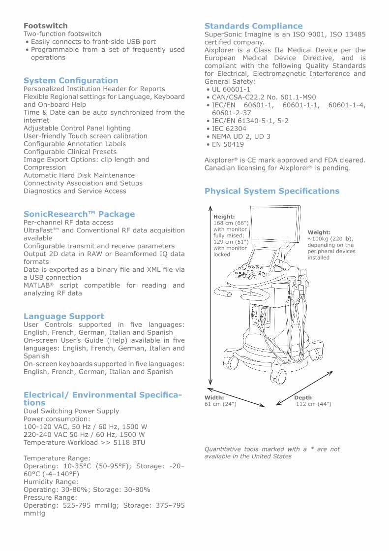
Physical System Specifications
Weight:~100kg (220 lb),depending on the peripheral devices installed
Height: 168 cm (66”) with monitor fully raised;129 cm (51”) with monitor locked
Width:61 cm (24”)
Depth: 112 cm (44”)
Quantitative tools marked with a * are not available in the United States
SuperSonic Imagine FranceLes jardins de la Duranne, Bât E & F510, rue René Descartes13 857 Aix-en-Provence, CedexFrance +33 (0)4 42 99 24 32 +33 (0)4 42 52 59 21 [email protected]
SuperSonic Imagine USAWestpark - Building FRedmond, WA 98052USA +1 (425) 284 6610 +1 (425) 284 6623 [email protected]
SuperSonic Imagine Ltd. UK18 Upper WalkVirginia WaterSurrey GU25 4SN United Kingdom +44 (0)845 643 4516 [email protected]
SuperSonic Imagine GmbH GermanyDietlindenstr. 1580802 MünchenGermany +49 89 36036 844 +49 89 36036 700 [email protected]
SuperSonic Imagine Asian Distribution Network +33 (0)4 42 99 24 32 +33 (0)4 42 52 59 21 [email protected]
SSID01459-01