Embed Size (px)
Citation preview

ORIGINAL PAPER
Synthesis of hierarchically porous bioactive glasses using naturalplants as template for bone tissue regeneration
Xiaofeng Li • Fengyu Qu • Wang Li •
Huiming Lin • Yingxue Jin
Received: 25 November 2011 / Accepted: 12 May 2012 / Published online: 23 May 2012
� The Author(s) 2012. This article is published with open access at Springerlink.com
Abstract A series of highly ordered hierarchically porous
silica and bio-glasses materials with macropore size of
8–1,000 lm and mesopore size of 3.1–5.6 nm have been
synthesized using six plant based materials as templates.
However, the as-obtained porous structure was reported for
the first time with interconnected 3D macropore up to
1,000 lm. The porous silica materials were used as the
host for drug loading and release, which showed a good
sustained delivery function. The as-synthesized bio-glasses
materials indicated the highly bioactive capability in the
bone regeneration. This method can be utilized to synthe-
size other multi-porous bioactive glasses using different
plants as templates for bone tissue repairing.
Keywords Hierarchically pore silica � Bio-glasses � Plant
template � Drug release � Osteogenic property
1 Introduction
Hierarchically porous materials have received enormous
attention due to the multiple dimensions of their pore
structures, high surface area and complex morphologies
[1]. These materials combine the advantages of the two
kinds of pores: macropores can improve diffusion and
transport of large molecules, and the high surface area and
large pore volumes of mesopores are beneficial for loading
large amounts of guest molecules. Thus, the materials will
have promising prospects for industrial processes involving
catalysis, adsorption, separation, chemical sensing, storage
of fluids and gases in transportation [2–4], and enzyme
immobilization [5]. Generally, hierarchical porous materi-
als have been prepared by multiple template methods,
including hard templates for macropores [6–13], and soft
templates for preparation of meso-/micro-pores [14].
Constructing novel hierarchically porous materials with
natural biological templates is an brand-new field. They
have many advantages compared to artificial ones due to
their abundant, renewable, and environmentally friendly
properties. They also have various structures, splendid
morphologies, and good biocompatibilities [1, 15–17]. At
present, all kinds of biological materials have been used as
hard template such as plants: wood [18], bamboo [19],
diatoms [20]; animal tissue: cuttlebone [21], echinus bone
[22–29]. However, the materials prepared by these natural
templates contain only 1–5 lm wide macropores in gen-
eral. So far, few highly ordered hierarchically porous
materials have been synthesized with macropores bigger
than 10 lm [18, 19] using natural plants as template.
Recently, these hierarchically porous materials have
been used in tissue regeneration. Ideally, a scaffold for
bone repaired should have three important characteristics:
(1) an interconnected framework with large pores
([10 lm) to enable tissue growth and nutrient delivery to
the center of the regenerated tissue; (2) a large specific
BET surface area provided by a microporous or mesopor-
ous phase to promote cell adhesion, drug storage and
delivery, and adsorption of biologic metabolites [30–33];
(3) a favorable bio-compatibility (i.e. the formation capa-
bility of hydroxyapatite (HAP) for repairing of bones).
Bioactive glasses (BGs) have been an interesting topic
since the pioneering work by Hench et al. [34]. To date
X. Li (&) � F. Qu � W. Li � H. Lin � Y. Jin
College of Huarui, Northeast Petroleum University, Harbin
150025, People’s Republic of China
e-mail: [email protected]
X. Li � F. Qu � W. Li � H. Lin � Y. Jin
College of Chemistry and Chemical Engineering, Harbin Normal
University, Harbin 150025, People’s Republic of China
123
J Sol-Gel Sci Technol (2012) 63:416–424
DOI 10.1007/s10971-012-2803-x

macro-/meso-porous bioactive glasses (MMBGs) have
been studied by several research groups for bone tissue
regeneration [35–38]. These reports focus mainly on the
synthesis of BG materials using granular polyethylene
glycol, methyl cellulose and polyurethane sponges as
macropore templates and nonionic block copolymers as
mesopore templates. Osteogenic properties of multi-level
pore materials were also studied. Till now, no BGs with
macropore size larger than 10 lm have been synthesized
using natural templates.
In this paper, we successfully synthesized a series of
highly ordered hierarchically porous silica materials with
macropore sizes ranging between 8 and 1,000 lm and
mesopore sizes between 3.1 and 5.6 nm using six plants as
templates for macropores and the block copolymer P123 as
mesopore template. To the best of our knowledge this is the
first report about hierarchically pore structures with inter-
connected 3D macropores up to 1,000 lm. In addition,
ibuprofen (IBU) was employed as a model drug to study
the drug loading/release profiles of these silica materials.
Furthermore, we achieved the first synthesis of a hierar-
chical porous bioactive glass scaffold using plants and
P123 as co-template by adding calcium and phosphate ions
during synthesis of the silica materials. The BGs also
exhibit a hierarchical structure with interconnected mac-
ropores (about 20–200 lm) and 3.1–4.1 nm wide mesop-
ores, the bioactivity of the BGs for bone tissue regeneration
was simultaneously investigated revealing superior in vitro
bone-forming bioactivities of the prepared BGs.
2 Experimental section
2.1 Materials
All the chemicals were purchased from commercial sour-
ces and used without further purification: EO20PO70EO20
(P123, Aldrich Chemical Co., USA), Tetraethoxysilane
(TEOS, Tiantai Co., Tianjin China), hydrochloric acid and
ethanol (EtOH, Harbin Chemical Co., Harbin, China),
calcium nitrate tetrahydrate (Ca (NO3)2�4H2O, Tianjin
Chemical Co., Tianjin, China), triethyl phosphate (TEP,
Shenyang Chemical Co., Shenyang, China) and ibuprofen
(IBU, Tianjin Chemical Co., Nanjing). Plants were
obtained from Harbin, China.
2.2 Characterization
Samples were characterized by X-ray diffraction (XRD)
using a SIEMENS D5005 diffractometer with Cu K�radiation at 40 kV and 30 mA. N2 adsorption/desorption
isotherms were measured at liquid nitrogen temperature
using a Micromeritics ASAP 2010M system. The pore
sizes distributions were calculated from the adsorption
branches of the N2 adsorption isotherms using the Barrett–
Joyner–Halenda (BJH) model. Scanning electron micros-
copy (SEM) was performed using a Hitachi S-4800
instrument operated at an accelerating voltage of 200 kV.
An SEM–EDS accessory was used to observe the HAP
growth on the surfaces of samples. Transmission electron
microscopy (TEM) images were recorded on JEOL 2010 F
and Philips CM200 FEG instruments with an acceleration
voltage of 20 kV. UV–vis spectra were measured on a 752
Spectrophotometer made in Shanghai. The concentrations
of Ca, P, and Si in simulated body fluid (SBF) solutions
were determined by inductively coupled plasma atomic
emission spectroscopy (ICP-AES; Varian Co., USA)
before and after soaking of plant.
2.3 Synthesis of hierarchical porous materials
In a typical procedure, a surfactant solution was prepared
by adding 0.9 g P123 to a mixed solution containing 10.0 g
of ethanol, 0.8 g of water and 0.1 g of 2 mol L-1 hydro-
chloric acid, and then 2.08 g TEOS was added. The mix-
ture was stirred for 2 h, the dried plants were cut into
1 9 1 cm3 including calla peel, paulownia stem, poplar
stem, abutilon stem, artichoke stem and calla stem, and
than they were soaked in the solution for 3 days at 60 �C in
a sealed polypropylene container. The plant based materi-
als were then removed from the solution and re-soaked in a
new solution for another 3 days at 60 �C. Finally, the
samples were taken out from the sol-like mixture, air-dried,
and calcined at 550 �C for 5 h in air. The obtained hier-
archical porous materials were named as HPM1 (calla
peel), HPM2 (paulownia stem), HPM3 (poplar stem),
HPM4 (abutilon stem), HPM5 (artichoke stem) and HPM6
(calla stem), respectively.
2.4 Drug load and release profiles
For the studies of IBU release, HPM4 and HPM5 were
selected randomly as samples. A typical drug release
experiment was performed as follows: 206 mg of porous
composite material and 206 mg of IBU were dispersed in
10 ml of n-hexane solution by stirring for 2 h at room
temperature. Then the loaded samples were separated from
the solution by vacuum filtration and dried under ambient
conditions. The filtrates were diluted by n-hexane solution
and the amount of loaded IBU was measured with the UV–
vis spectrophotometer.
The release profiles of the samples were obtained by
soaking the drug-loaded powders in phosphate buffer
solution (pH = 6.8). The release experiment was per-
formed at 37 �C. At predetermined time intervals, 3 ml of
sample was withdrawn and another 3 ml of fresh phosphate
J Sol-Gel Sci Technol (2012) 63:416–424 417
123
buffer solution was added immediately. The withdrawn
samples were diluted to 25 ml and the drug concentration
in the sampled fluid was measured with the UV–Vis
spectrophotometer.
2.5 Preparation of the MMBG scaffolds
The mesopore/macropore bioactive glass scaffolds (MMBGs)
were synthesized by using nonionic block copolymer P123
and plant peelings as co-templates. In a typical synthesis,
P123 (4.0 g), TEOS (6.7 g), Ca(NO3)2�4H2O (1.4 g), TEP
(0.73 g), and 0.5 mol L-1 HCl (1.0 g) were dissolved in
ethanol (60 g) and stirred at room temperature for 1 day.
Afterwards, paulownia stem, artichoke stem, and abutilon
stem were immersed into the solution for 3 days at 60 �C
in a sealed polypropylene container. After evaporating the
solution for 24 h at room temperature, the samples were
re-soaked in a new solution for another 3 days at 60 �C.
Finally, the samples were taken out from the sol-like
mixture, air-dried, and heated at a slow rate of 2 �C/min to
550 �C to obtain the final MMBG scaffolds named as
MMBG1 (paulownia stem), MMBG2 (artichoke stem) and
MMBG3 (abutilon stem).
2.6 In vitro bioactivity of the MMBG scaffolds in SBF
The assessment of the in vitro bioactivity of the MMBG
scaffolds was carried out in SBF. The SBF solution had a
composition and ionic concentrations similar to those of
human plasma [39]. MMBG1, MMBG2 and MMBG3 were
used to investigate the bioactivity. Each type of MMBG was
soaked in 100 ml SBF solution in a polyethylene bottle at
37 �C. The ratio of MMBG powder weight to SBF solution
volume was 1.5 mg/ml [40]. The samples were taken out
from the SBF solutions after soaking for 1, 3, 5 or 7 days,
then rinsed with acetone and air-dried at room temperature.
3 Results and discussion
3.1 Characterization of the porous composite materials
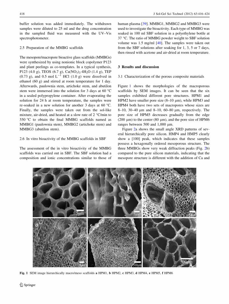
Figure 1 shows the morphologies of the macroporous
scaffolds by SEM images. It can be seen that the six
samples exhibited different pore structures. HPM1 and
HPM2 have smaller pore size (8–10 lm), while HPM3 and
HPM4 both have two sets of macropores whose sizes are
8–10, 30–40 lm and 8–10, 60–80 lm, respectively. The
pore size of HPM5 decreases gradually from the edge
(200 lm) to the center (80 lm), and the pore size of HPM6
ranges between 500 and 1,000 lm.
Figure 2a shows the small angle XRD patterns of sev-
eral hierarchically pore silicon. HMP4 and HMP5 clearly
show a [100] peak, which indicates that these samples
possess a hexagonally ordered mesoporous structure. The
three MMBGs show very weak diffraction peaks (Fig. 2b)
compared to the pure silicon materials, indicating that the
mesopore structure is different with the addition of Ca and
Fig. 1 SEM image hierarchically macro/meso scaffolds a HPM1, b HPM2, c HPM3, d HPM4, e HPM5, f HPM6
418 J Sol-Gel Sci Technol (2012) 63:416–424
123
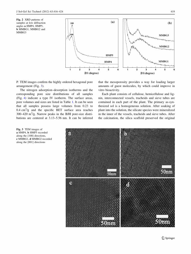
P. TEM images confirm the highly ordered hexagonal pore
arrangement (Fig. 3).
The nitrogen adsorption–desorption isotherms and the
corresponding pore size distributions of all samples
(Fig. 4) indicate a type IV isotherm. The surface areas,
pore volumes and sizes are listed in Table 1. It can be seen
that all samples possess large volumes from 0.23 to
0.4 cm3/g and the specific BET surface area reaches
300–420 m2/g. Narrow peaks in the BJH pore-size distri-
butions are centered at 3.13–5.56 nm. It can be inferred
that the mesoporosity provides a way for loading larger
amounts of guest molecules, by which could improve in
vitro bioactivity.
Each plant consists of cellulose, hemicellulose and lig-
nin, interconnected vessels, tracheids and sieve tubes are
contained in each part of the plant. The primary as-syn-
thesized sol is a homogeneous solution. After soaking of
plant into the solution, the silicate species were mineralized
in the inner of the vessels, tracheids and sieve tubes. After
the calcination, the silica scaffold preserved the original
1 2 3 4 5 6
100
(a)
HMP5
HMP4
100
2 θ (degree)1 2 3 4 5 6
(b)
MMBG3
MMBG2
MMBG1
2 θ (degree)
Fig. 2 XRD patterns of
samples at low diffraction
angles a HMP4, HMP5,
b MMBG1, MMBG2 and
MMBG3
Fig. 3 TEM images of
a HMP4, b HMP5 recorded
along the [100] directions,
c MMBG1, d MMBG2 recorded
along the [001] directions
J Sol-Gel Sci Technol (2012) 63:416–424 419
123

morphologies and structure of the plants, and ‘‘tube’’ types
have been replicated so that macroporous materials were
obtained [41].
3.2 Drug release of hierarchically pore silica
Figure 5 shows the high angle XRD patterns of IBU, the
mechanical mixture of IBU and HPM4, IBU stored in
HPM4. It can be seen that the IBU and the mechanical
mixture exhibited obvious XRD spectrum peak of the drug,
it indicated that the drug molecules exist still crystal form,
and the assembly does not appear the diffraction peaks of
the drug, it indicated that the drug molecules has loaded
into the mesopores of the material [42].
Figure 6 shows the cumulative release profile of the
samples in buffer solution of pH = 6.8. It can be observed
that IBU showed a similar, two-step release behavior for
both samples with an initially fast and a relatively slow
subsequent release through the whole period. About 20 and
45 wt% of the IBU were released from HPM4 to HPM5
within 1 h, respectively, but the IBU release reached sim-
ilar values of 58.3 and 59.1 wt% when the release rate
approached zero after 48 h. Maybe this is because that the
a
0.0 0.2 0.4 0.6 0.8 1.050
100
150
200
250
300
HPM5
HPM4
Relative Pressure/p/p0
Vol
ume
Ads
orbe
d (c
m3 /g
at
STP
)
0 5 10 15 20
0.0
0.2
0.4
0.6
0.8
1.0
1.2
HPM5
HPM4
Pore size(nm)
volu
me
Ads
orbe
d(cm
3g-1
)
0 5 10 15 20
0.0
0.4
0.8
1.2
1.6
2.0
2.4
2.8
MMBG3
MMBG2
MMBG1Vol
ume
Ads
orbe
d(cm
3g
-1)
Pore size(nm)
0.0 0.2 0.4 0.6 0.8 1.0
50
100
150
200
250
300
350
400
MMBG3
MMBG2
MMBG1
Vol
ume
Ads
orbe
d (c
m3 /g
at
STP
)
Relative Pressure/p/p0
bFig. 4 Nitrogen adsorption/
desorption isotherm and
mesopore distribution of the
samples a HMP4, HMP5,
b MMBG1, MMBG2 and
MMBG3
Table 1 BET surface area pore volume and average pores diameter
of the samples
Sample BET surface
area (m2/g)
Pore volume
(cm3/g)
Average pore
diameter (nm)
HPM4 306.03 0.40 5.56
HPM5 347.56 0.23 3.75
MMBG1 420.85 0.35 3.52
MMBG2 409.49 0.30 3.13
MMBG3 299.25 0.29 4.13
10 15 20 25 30 35 40
2θ /degree
c
b
a
Fig. 5 XRD patterns at high diffraction angels. a IBU, b IBU mixed
with HPM4, c IBU stored in HPM4
0 10 20 30 40 50
0
10
20
30
40
50
60
HMP4
T(h)
Wt(
%)
0 10 20 30 40 500
10
20
30
40
50
60
HMP5
Wt(
%)
T(h)
Fig. 6 Drug release kinetics of hierarchically pore silica HPM4,
HPM5
420 J Sol-Gel Sci Technol (2012) 63:416–424
123
specific surface area is mainly determined by the meso-
porous phase. The drug was mainly loaded in the meso-
porous channels, and only a little amount was adsorbed on
the macropore surface. The release of the drug from the
pore materials may involve: solvent diffusion into the
mesopores with dissolution of the drug, followed by its
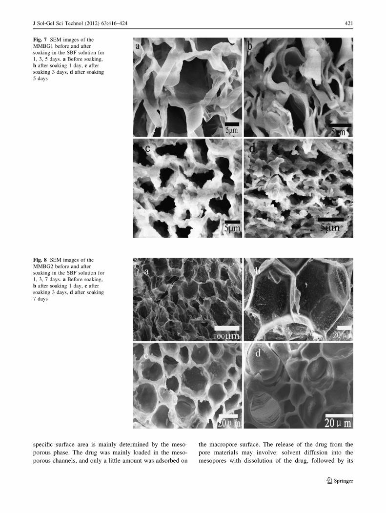
Fig. 7 SEM images of the
MMBG1 before and after
soaking in the SBF solution for
1, 3, 5 days. a Before soaking,
b after soaking 1 day, c after
soaking 3 days, d after soaking
5 days
Fig. 8 SEM images of the
MMBG2 before and after
soaking in the SBF solution for
1, 3, 7 days. a Before soaking,
b after soaking 1 day, c after
soaking 3 days, d after soaking
7 days
J Sol-Gel Sci Technol (2012) 63:416–424 421
123
release from the mesopores into the macropores, and
eventually release of the drug from the macropores to the
outside solution. Thus, the macropores shall play a buffer
role in the drug release. When the drug concentrations in
the macropores and outside medium reached a homeostatic
equilibrium, then drug molecules were no longer released
from the pores. Consequently the drug could not be
released completely.
3.3 Bioactivity of the hierarchically porous bioactive
glass scaffolds
The ability to bond with living bone through a HAP
interface layer on their surface is a significant characteristic
of MMBGs, which has been widely studied both in vitro
and in vivo [43]. The deposition/growth of HAP of the
scaffolds in vitro has been investigated here by soaking
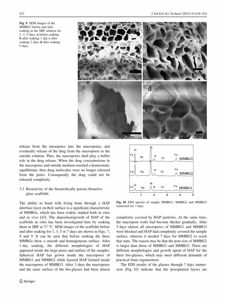
them in SBF at 37 �C. SEM images of the scaffolds before
and after soaking for 1, 3, 5 or 7 days are shown in Figs. 7,
8 and 9. It can be seen that before soaking the three
MMBGs show a smooth and homogeneous surface. After
1 day soaking, the different morphologies of HAP
appeared inside the large pores and surface of the samples.
Spherical HAP has grown inside the macropores of
MMBG1 and MMBG2 while layered HAP formed inside
the macropores of MMBG3. After 3 days the macropores
and the outer surface of the bio-glasses had been almost
completely covered by HAP particles. At the same time,
the macropore walls had become thicker gradually. After
5 days almost all macropores of MMBG1 and MMBG3
were blocked and HAP had completely covered the sample
surface, whereas it needed 7 days for MMBG2 to reach
that state. The reason may be that the pore size of MMBG2
is larger than those of MMBG1 and MMBG3. There are
different morphologies and growth speed of HAP for the
three bio-glasses, which may meet different demands of
practical bone regeneration.
The EDS results of the glasses through 7 days immer-
sion (Fig. 10) indicate that the precipitated layers are
Fig. 9 SEM images of the
MMBG3 before and after
soaking in the SBF solution for
1, 3, 5 days. a before soaking
b after soaking 1 day c after
soaking 3 days d after soaking
5 days
1 2 3 4 5
Si
Si
Ca
O
OCa
CaCa
CaCa
O
MMBG1
MMBG2
MMBG3
Cl
Cl
Cl
P
P
P
Si
Na
Na
Na
Fig. 10 EDS spectra of sample MMBG1, MMBG2 and MMBG3
immersed for 7 days
422 J Sol-Gel Sci Technol (2012) 63:416–424
123

composed of Ca and P with a Ca/P atomic ratio of 1.61
(MMBG1), 1.63 (MMBG2) and 1.72 (MMBG3), respec-
tively. The atomic ratios are close to the theoretical value
1.67 Ca/P ratio of apatite [44]. Figure 11a shows that the
concentrations of Ca, Si, and P in SBF for various
immersion periods. The results indicate that silicon was
released from the glasses, while calcium and phosphate
were deposited on their surface, as reported by Li et al.
[36]. The Si content increase with extension of soaking
time, while the concentration of P decreased continually,
because phosphorus diffused slowly from the samples in
SBF. The Ca2? concentration is controlled by both the
release of Ca2? from the sample and the formation of HAP.
The Ca2? concentration increased during the first 3 days
for the rapid calcium dissolution; and then decreased
slowly, the reason can be attributed to the rapid growth of
the apatite nuclei formed on surface of the sample, which
overcame the release rate of Ca2? to the solution. It can be
concluded that a HAP layer has formed from SBF to the
samples, and the materials can induce the growth of HAP
on their surface. On the other hand, EDS analysis of Ca, Si,
and P on the sample surfaces during different times
(Fig. 11b) roughly indicated that Ca and P increased con-
tinually while Si decreased. This also confirmed the growth
of HAP on the surface of the samples. Results indicated
that these novel bioactive glass scaffolds with good bio-
activity can induce the formation of HAP layers in SBF,
and thus may have potential application in tissue regener-
ation engineering.
4 Conclusions
In summary, for the first time we reported the synthesis of a
series of highly ordered, porous silica and bio-glasses with
large pore sizes of 8–1000 lm and mesopore sizes of
3–5 nm using six plants as templates. The novel porous
silica materials exhibit sustained drug delivery profiles, and
the porous bioactive glass scaffolds can induce the pre-
cipitation of HAP layers on their surface in SBF within
1 day, which are converted into crystalline HAP within
7 days. The morphologies and the growth speed of HAP
differ for the three MMBGs, so it is difficult to come to a
clear conclusion about the optimum macropore size for
bone regeneration. The unique interconnected multimodal
porosity distribution and excellent in vitro bioactivity of
MMBGs make them a good candidate for bone regenera-
tion and drug delivery.
Acknowledgments Financial support for this study was provided by
the National Native Science Foundation of China (20871037,
21171045, 21101046), Innovation special fund of Harbin Science and
Technology Bureau of China (2010RFXXS055), Program for Scien-
tific and Technological Innovation team Construction in Universities
of Heilongjiang province (2011TD010), and Doctoral Initiation Fund
of Harbin Normal University (KGB201006).
Open Access This article is distributed under the terms of the
Creative Commons Attribution License which permits any use, dis-
tribution, and reproduction in any medium, provided the original
author(s) and the source are credited.
References
1. Yuan ZY, Su BL (2006) J Mater Chem 16:663–667
2. Zhao DY, Yang P, Chmelka BF, Stucky GD (1999) Chem Mater
11:1174–1178
3. Sun J, Li YS, Li L (2008) J Non-Cryst Solids 354:3799–3805
4. Cai XH, Zhu GS, Zhang WW, Zhao HY, Wang C, Qiu SL, Wei Y
(2006) Eur J Inorg Chem 18:3641–3645
5. Lee YJ, Lee JS, Park YS, Yoon KB (2001) Adv Mater 13:1259–
1263
6. Andeson MW, Holmes SM, Hanif N, Cundy CS (2000) Angew
Chem Int Ed 39:2707–2710
7. Sen T, Gordon GT, John TT, Casci JL, Anderson MW (2003)
Angew Chem Int Ed 42:4649–4653
8. Holland BT, Abrams L, Stein A (1999) J Am Chem Soc
121:4308–4309
9. Sen T, Anderson MW (2004) Chem Mater 16:2044–2054
10. Zhu G, Qiu S, Tearsaki O (2001) J Mater Chem 11:1687–1693
11. Antonietti M, Berton B, Goltner C, Hentze HP (1998) Adv Mater
10:154–159
12. Lee YJ, Yoon KB (2005) Micro Meso Mater 88:176–186
13. Zhang H, Hardy GC, Rosseinsky MJ, Cooper AI (2003) Adv
Mater 15:78–81
0 5 10 15 20 25 300
20
40
60
80
100
120
140(a)
P
Si
Ca
conc
entr
atio
n (p
pm)
T(d)1 2 3 4 5 6 7
0
10
20
30
40
50(b)
P
Ca
Si
Qua
lity
Rat
io
T(d)
Fig. 11 a Ca, P, Si
concentration change with
soaking time in SBF after
MMBG1 being soaked by ICP
measurement. b Variations of
Ca, Si, P of the average quality
ratio on the surface of MMBG1
by EDS measurement
J Sol-Gel Sci Technol (2012) 63:416–424 423
123
14. Yue WB, Park RJ, Kulak AN, Meldrum FC (2006) J Cryst
Growth 294:69–77
15. Zhang BJ, Davis SA, Mann S (2000) Chem Comm 35:781–782
16. Valtchev V, Smaihi M, Vidal L (2003) Angew Chem Int Ed
42:2782–2785
17. Huang LM, Wang HT, Hayashi CY, Tian B, Zhao DY, Yan YH
(2003) J Mater Chem 13:666–668
18. Shin YS, Liu J, Chang JH, Nie ZM, Exarhos GJ (2001) Adv
Mater 13:728–732
19. Dong AG, Wang YJ, Tang Y, Ren N, Zhang YH, Yue YH, Gao Z
(2002) Adv Mater 14:926–929
20. Chaisena A, Rangsriwatananon K (2005) Mater Lett 5:1474–
1479
21. Wataru O, Wayne S, Sean AD, Stephen M (2000) Chem Mater
12:2835–2837
22. Meldrum FC, Seshadri R (2000) Chem Commun 35:29–30
23. Valtchev V, Smaihi M, Faust AC, Vidal L (2004) Chem Mater
16:1350–1355
24. Wang YJ, Tang Y, Dong AG, Wang XD, Ren N, Gao Z (2002) J
Mater Chem 12:1812–1818
25. Hall SR, Bolger H, Mann S (2003) Chem Commun 22:2784–
2785
26. Yang D, Qi LM, Ma JM (2002) Adv Mater 14:1543–1546
27. Cook G, Timms PL, Spickermann CG (2003) Angew Chem Int
Ed 42:557–559
28. Davis SA, Burkett SL, Mendelson NH, Mann S (1997) Nature
385:420–423
29. Shinye C, Jun U, Fuyuhiko T, Bruce D, Jeffrey IZ (2000) J Am
Chem Soc 122:6488–6489
30. Lei B, Chen X, Wang Y, Zhao N, Chang D, Fang L (2009) J Non-
Cryst Solids 355:2678–2681
31. Kokubo T, Matsushita T, Takadama H, Kizuki T (2009) J Eur
Ceram Soc 29:1267–1274
32. Vallet-Regı0 M, Ruiz-Gonza0lez L, Isabel-Barba I, Gonza0lez-
Calbet JM (2006) J Mater Chem 16:26–31
33. Jing Y, Wei G, Huang X, Zhao L, Zhang Q, Yu C (2008) J Sol-
Gel Sci Technol l45:115–119
34. Hench LL, Splinter RJ, Allen WC, Greenlee TK (1971) J Biomed
Mater Res 2:117–141
35. Mohamad Yunos D, Bretcanu O, Boccaccini AR, Aldo R (2008) J
Mater Sci 43:4433–4442
36. Xia L, Wang X, Chen H, Jiang P, Dong X, Shi J (2007) Chem
Mater 19:4322–4326
37. Li N, Jie Q, Zhu S, Wang R (2005) Ceram Int 31:641–646
38. Vallet-Regi M (2006) Dalton Trans 44:5211–5220
39. Kokubo T, Kushitani H, Sakk S, Kitsugi T, Yamamuro T (1990) J
Biomed Mater Res 24:721–734
40. Saravanapavan P, Jones JR, Oryce RS, Hench LL (2003) J Bio-
med Mater Res 66A:110–119
41. Li X, Jiang J, Yu W, Nie X, Qu F (2010) J Sol-Gel Sci Technol
56:75–81
42. Liu CJ, Li SG, Pang WQ (1997) Chem Commun 1:65–66
43. Izquierdo-Barbaa I, Ruiz-Gonzalezb L, Doadrioa JC, Gonzalez-
Calbetb JM, Vallet-Regı M (2005) Solid State Sci 7:983–989
44. Olmo N, Martin AI, Salinas AJ (2003) Biomaterials 24:3383–
3393
424 J Sol-Gel Sci Technol (2012) 63:416–424
123