Embed Size (px)
Citation preview
SYNTHESES AND STRUCTURAL STUDIES OF METAL COMPLEXES OF SULFA DRUGS
A thesis submitted to the Cardiff University
By
G. M. Golzar Hossain
in candidature of the degree of
Doctor of Philosophy
August 2005
Department of Chemistry
Cardiff University
UMI Number: U584873
All rights reserved
INFORMATION TO ALL USERS The quality of this reproduction is dependent upon the quality of the copy submitted.
In the unlikely event that the author did not send a complete manuscript and there are missing pages, these will be noted. Also, if material had to be removed,
a note will indicate the deletion.
Dissertation Publishing
UMI U584873Published by ProQuest LLC 2013. Copyright in the Dissertation held by the Author.
Microform Edition © ProQuest LLC.All rights reserved. This work is protected against
unauthorized copying under Title 17, United States Code.
ProQuest LLC 789 East Eisenhower Parkway
P.O. Box 1346 Ann Arbor, Ml 48106-1346
DECLARATION
This work has not previously been accepted in substance for any degree and is not being
concurrently submitted in candidature for any degree.
Signed (Candidate)
Date: O 3 / ^ 6
STATEMENT 1
This thesis is the result o f my own investigations, except where otherwise stated.
Other sources are acknowledged by in the appended bibliography.
Signed (Candidate)
Date:
STATEMENT 2
I hereby give consent for my thesis, if accepted, to be available for photocopying and for
inter-library loan, and for the title and summary to be made available to outside
organisations.
Signed (Candidate)
Date: / 3 / o 3 /
II

<tm d icate< d toJAfroza andJAsif
III
ACKNOWLEDGEMENTS
This work would not have been completed without the encouragement and patience o f my
supervisors Professor Peter G. Edwards and Dr. Angelo J. Amoroso, to whom, I will
always be grateful. I am also grateful to Dr. K. M. A. Malik for collecting crystal data and
valuable advice.
I want to express my gratitude to Dr. Ian A. Fallis and Dr. S. Aldridge for their helpful
discussion.
Many thanks go to Dr. Murphy and Dr. Farley for EPR data and discussion.
Thanks go to all the technical staffs- Rob Jenkins, Alan, Robin, Ricky, Sham, John Bowly
and Gary for helping me to work smoothly.
Special thanks go to everyone in the Department of Chemistry, Cardiff University,
specially Caroline, Ozra, Steve, Graham, Dave, Miles, Claire, Rob, Willium, Deborah,
Lisa, Amal, Chris, Andrea and Mark for making the work enjoyable.
Finally, I would like thank my wife Afroza and lovely son A sif for their support and
inspiration throughout the research works.
IV
ABSTRACT
This thesis presents the results o f the synthesis of metal complexes o f sulfa drugs
(sulfadiazine, sulfamerazine, sulfamethazine, sulfathiazole and sulfadimethoxine) with
secondary ligands, which were subjected to X-ray crystallographic studies. The aim o f this
work was to synthesise metal complexes of sulfa drugs with secondary nitrogen donor
ligands e.g. ammonia, pyridine (py), ethylenediamine (en), diethylenetriamine (dien), 2,2'-
bipyridine, 1,10-phenanthroline, 4,4'-dimethyl-2,2'-bipyridine, A(A^(3-aminopropyl)-bis-
(ethylenediamine) (apen) and oxygen donor ligands such as, water and dimethylformamide
(DMF) and to investigate the different modes of coordination of the sulfa drugs molecules
to the metal ions in the presence o f nitrogen/oxygen donor ligands.
Chapter 1, details an introduction into the fundamental concepts, procedures and equipment
involved in data collection, solution and refinement of single crystal structures.
In Chapter 2, a review o f the literature on the metal complexes of sulfa drugs along with
the main aims of this work is presented.
In Chapter 3, the general experimental procedure and synthetic methods have been
described for the metal complexes o f sulfa drugs whose results and discussion are
described in chapter 4.
In chapter 4, the crystal structure o f sulfadiazine, a new polymorph of sulfamerazine and
the syntheses of the metal complexes o f sulfadiazine, sulfamerazine and sulfamethazine,
their analytical and spectral data along with the X-ray structure determination o f the
complexes are presented. The complexes studied in this chapter are: A) Cobalt complex
{[Co(smz)2(H20)].DMF}„ (3); B) Nickel complexes [Ni(en)3(sdz)2].H20 (4) and
[Ni(smr)2(py)2].4py (5); C) copper complexes [Cu(en)2 (sdz)2 ] (6), [Cu(dien)2 (sdz)2 ] (7),
[Cu(en)2(H20 ) 2][smr)2] (8), [Cu(en)2(H20)2][smr)2].H20 (9), [Cu2(smr)4].2DMF (10),
[Cu2(smr)4] .2DMSO (11), [Cu(smz)2(apen)].3H2O.CH3OH (12) and
{[Cu(smz)2.NH3].2H20}„ (13); D) Zinc complexes [Zn(smz)2 (NH3 )2 ] (14) and
[Zn(smz)2(py)2].2py (15), E) Cadmium complexes [Cd(dien)2 (sdz)2 ].DMF (16),
V
[Cd(dien)(smr)2].H20 (17), [Cd(sdz)2(bpy)] (18), [Cd(sdz)2(phen)] (19),
[Cd(sdz)2(dmbpy)].2DMF (20), [Cd(smr)2(phen)] (21), {[Cd(smz)2(H20)].DMF}„ (22),
{[Cd(smz)2(H20)].2H 20}„ (23) and [Cd(smz)2(en)].2DMF (24); and F) Mercury
complexes [Hg(sdz)2(DMF)2] (25), [Hg(smr)2] (26) [Hg(smr)2(bpy)] (27) and
[Hg(smz)2(DMF)2] (28).
The structural results for the complexes are discussed and compared with other related
complexes. In this context, the different types o f coordination geometries, important
parameters within the coordination sphere, the various modes o f coordination o f the sulfa
drugs in the anionic form, dimeric polymeric complexes, the role o f hydrogen bonds in the
formation of the “cation-anion pair” and the packing diagram have been discussed.
In Chapter 5, we have described the syntheses and characterisation of the complexes of
sulfathiazole, their analytical and spectral data along with the X-ray structure determination
o f these complexes are presented. The complexes studied are: [(H2 stz)2 ][N 0 3 ].H 2 0 (29),
[C0 CI4 ][(H2 stz)2 ].CH 3 COOH (30) and [Cu(en)2(H20 )2][stz)2].2H20 (31). In all the
complexes the sulfathiazole molecules act as ionic species. The structural results for the
complexes are discussed and compared with other related complexes. In this context, the
role of hydrogen bonds in the formation of the “cation-anion pair” and the packing diagram
has been discussed.
Chapter 6 describes the structure o f sulfadimethoxine and synthesis and characterisation o f
a zinc complex o f sulfadimethoxine [Zn(sdm)2(NH3)2].2H20 (33) by analytical and spectral
data along with the X-ray structure determination. The structural results of the complex are
discussed and compared with other related complexes. The packing diagram of the
complex with the hydrogen bonds is discussed in this chapter.
VI
ABBREVIATIONS
A angstrom (1A = 10"10 cm)
X wavelength
V cell volume
a, b, c cell axes
a, J3, y cell angles
d interplanar spacing
Fhki structure factor
fj scattering factor
hkl general indices
(°) degree centigrade (Celcius)
cm centimeter
cm-1 wave number
g grams
ml milliliter
mmol millimolar
tsd triple sulfa drugs
Hsdz sulfadiazine
Hsmr sulfamerazine
Hsmz sulfamethazine
Hstz sulfathiazole
Hsdm sulfadimethoxine
py pyridine
DMF dimethylformamide
DMSO dimethylsulfoxide
en ethylenediamine
bpy 2,2'-bipyridine
phen 1,10-phenanthroline
dmbpy 4,4'-dimethyl-2,2'-bipyridine
dien diethylenetriamine
apen NJSF (3 -aminopropy l)-bi s(ethy lenediamine)
IR infrared
EPR Electron Paramagnetic Resonance
c. w. continuous wave
NMR nuclear magnetic resonance
s singlet
d doublet
t triplet
PABA p-aminobenzoic acid
UTI urinary tract infection
VIII
CONTENTS
TITLE I
DECLARATION II
DEDICATED TO III
ACKNOWLEDGEMENT IV
ABSTRACT V
ABBREVIATIONS VII
CHAPTER 1 - X-RAY CRYSTALLOGRAPHY 1
1.1 Historical background 1
1.2 Bragg’s equation 3
1.3 Reciprocal lattice 4
1.4 Crystal Lattice 6
1.4.1 Crystal Systems 8
1.4.2 Unit cell 8
1.4.2 Miller Indices 9
1.5 Systematic absences 10
1.6 Symmetry Elements 10
1.6.1 Point Symmetry 10
1.6.2 Translational Symmetry 10
1.6.3 Space groups 11
1.7 Structure determination 12
1.7.1 Atomic Scattering Factor 12
1.7.2 Structure F actor 12
1.7.3 Thermal motion 14
1.7.4 The Lorentz Factor, Polarisation Factor and Absorption Corrections 15
1.7.5 Thermal Ellipsoid 16
IX
1.7.6 Electron Density 16
1.7.7 Phase problem 17
1.7.8 Solutions to the phase problem 18
1.7.8.1 Direct Methods 18
1.7.8.2 Patterson Synthesis 19
1.7.9 Structure Refinement 21
1.8 Experimental Section 22
1.8.1 Data collection using a CCD area detector 23
1.8.2 The programs used for structure solution, refinement and structural drawing 25
References 26
CHAPTER 2 - INTRODUCTION 27
2.1 History 28
2.2 Pharmacology 29
2.3 Mechanism of Action 30
2.4 Pharmacokinetics 31
2.5 Clinical Uses 31
2.6 Adverse reactions 31
2.7 Previous work on triple sulfa drugs (TSD) 32
2.7.1 Sulfadiazine 33
2.7.2 Sulfamerazine 38
2.7.3 Sulfamethazine 39
2.8 Aim of the work 41
References 46
CHAPTER 3 - EXPERIMENTAL 48
3.1 Chemical and their purifications 48
3.2 Physical measurements 48
3.2.1 Elemental analysis 48
3.2.2 IR Studies 48
3.2.3 Electron Paramagnetic Resonance 48
3.2.4 'H and l3C NMR spectra 49
3.2.5 X-ray crystallography 49
3.3 Synthetic Procedure 49
X
3.4 Syntheses o f the complexes 50
3.4.1 Preparation o f the crystal of sulfadiazine (1) 50
3.4.2 Preparation o f the crystal of sulfamerazine (2) 50
3.4.3 Preparation o f the complex {[Co(smz)2(H20)].DMF}„ (3) 51
3.4.4 Preparation o f the complex [Ni(en)3(sdz)2].H20 (4) 51
3.4.5 Preparation of the complex [Ni(smr)2(py)2].4py (5) 51
3.4.6 Preparation of the complex [Cu(en)2 (sdz)2 ] (6) 52
3.4.7 Preparation of the complex [Cu(dien)(sdz)2 ] (7) 52
3.4.8 Preparation of the complex [Cu(en)2(H20)2][(smr)2] (8) 52
3.4.9 Preparation o f the complex [Cu(en)2(H20)2][(smr)2].H20 (9) 53
3.4.10 Preparation o f the complex [Cu2 (smr)4 ].DMF (10) 53
3.4.11 Preparation of the complex [Cu2 (smr)4 ].DMSO (11) 53
3.4.12 Preparation o f the complex [Cu(smz)2 (apen)].3 H2 0 .CH3 0 H (12) 54
3.4.13 Preparation o f the complex {[Cu(smz)2(NH3)].2H20}„ (13) 54
3.4.14 Preparation o f the complex [Zn(smz)2 (NH3 )2 ] (14) 54
3.4.15 Preparation o f the complex [Zn(smz)2(py)2]-2py (15) 55
3.4.16 Preparation of the complex [Cd(dien)(sdz)2 ].DMF (16) 55
3.4.17 Preparation of the complex [Cd(dien)(smr)2].H20 (17) 55
3.4.18 Preparation of the complex [Cd(sdz)2 (bpy)] (18) 56
3.4.19 Preparation o f the complex [Cd(sdz)2 (phen)] (19) 56
3.4.20 Preparation of the complex [Cd(sdz)2(dmbpy)].2DMF (20) 57
3.4.21 Preparation of the complex [Cd(smr)2 (phen)] (21) 57
3.4.22 Preparation of the complex {[Cd(smz)2(H20)].DMF}„ (22) 58
3.4.23 Preparation of the complex {[Cd(smz)2(H20)].2H20}„ (23) 58
3.4.24 Preparation of the complex [Cd(smz)2(en)].2DMF (24) 58
3.4.25 Preparation of the complex [Hg(sdz)2 (DMF)2 ] (25) 59
3.4.26 Preparation o f the complex [Hg(smr)2 ] (26) 59
3.4.27 Preparation of the complex [Hg(smr)2 (bpy)] (27) 60
3.4.28 Preparation of the complex [Hg(smz)2 (DMF)2 ] (28) 60
CHAPTER 4 - RESULTS AND DISCUSSION 61
4.1 IR spectra 61
4.2 Electron Paramagnetic Resonance 62
4.3 'H and l3C NMR spectra 67
XI
4.4. Structure of sulfadiazine (1) and sulfamerazine (2) 76
4.5 Cobalt complex {[Co(smz)2(H20)].DMF}„ (3) 82
4.6 Nickel complexes 87
4.6.1 Nickel complex of sulfadiazine [Ni(en)3][(sdz)2].H20 (4) 87
4.6.2 Nickel complex of sulfamerazine [Ni(smr)2(py)2].4py (5) 91
4.7 Copper complexes o f triple sulfa drugs 94
4.7.1 Crystal Structure of [Cu(en)2 ][(sdz)2 ] (6) 94
4.7.2 Crystal structure of [Cu(dien)(sdz)2 ] (7) 97
4.7.3 Structure o f the complexes [Cu(en)2(H20)2].2[smr]_ (8)
and {[Cu(en)2(H20 )2]. [(smr)2] }2-H20 (9) 100
4.7.4 Structures of the complexes [Cu2 (smr)4 ].DMF (10)
and [Cu2(smr)4] .DMSO (11) 105
4.7.5 Structure o f the complex [Cu(smz)2 (apen)].3 H2 0 .CH3 0 H (12) 110
4.7.6 Structure o f the complex {[Cu(smz)2(NH3)].2H20}„ (13) 113
4.8 Structure of the zinc complexes 117
4.8.1 Structure o f the complex [Zn(smz)2 (NH3 )2 ] (14) 117
4.8.2 Crystal structures of the complex [Zn(smz)2(py)2].2py (15) 120
4.9 Cadmium complexes triple sulfa drugs 123
4.9.1 Structures of [Cd(dien)(sdz)2].DMF (16) and [Cd(dien)(smr)2].2H20 (17) 123
4.9.2 Structures of (18), (19), (20) and (21) 129
4.9.3 Structures of the polymeric complexes {[Cd(smz)2(H20)].DMF}„ (22)
and {[Cd(smz)2(H20)].2H20 }„ (23) 138
4.9.4 Crystal structure of the complex (24) 144
4.10 Mercury complexes of triple sulfa drugs (TSD) 147
4.10.1 Crystal structure o f the complexes (25), (26), (27) and (28) 147
4.11 Summary 156
4.11.1 Type of coordination around the metal centers 157
4.11.2 Status of TSD anion as ligand 157
4.11.3 Role o f hydrogen bonds in the formation of crystal architecture 159
4.11.4 Conclusion 160
4.11.5 Further works 160
References 161
XII
CHAPTER 5 - COMPLEXES OF SULFATHIAZOLE 166
5.1 Introduction 166
5.2 Experimental 167
5.2.1 Preparation o f the complex [H2Stz][NC>3].H20 (29) 168
5.2.2 Preparation o f the complex [CoCl4 ][(smz)2 ]CH 3 COOH (30) 168
5.2.3 Preparation of the complex [Cu(en)2(H20)2][(stz)2].2H20 (31) 168
5.3 Results and discussion for the complexes (29), (30) and (31) 168
5.3.1 IR spectra 168
5.3.2 Crystal structures o f the complexes (29), (30) and (31) 169
5.3.2.1 Structure of the complex (29) 169
5.3.2.2 Structure o f the complex (30) 172
5.3.2.3 Structure of the complex (31) 177
5.4 Summary 180
References 181
CHAPTER 6 - COMPLEX OF SULFADIMETHOXINE 183
6.1 Introduction 183
6.2 Experimental 183
6.3 Crystal structure of sulfadimethoxine (32) and its zinc complex (33) 184
6.3.1 IR spectra 184
6.3.2 'H NMR and l3C NMR spectra 185
6.3.3 Crystal structures o f (32) and (33) 186
6.3.3.1 Structure o f sulfadimethoxine molecule (32) 186
6.3.3.2 Structure of the complex (33) 189
References 193
XIII
Chapter 1
X-RAY CRYSTALLOGRAPHY
1.1 HISTORICAL BACKGROUND
X-ray crystallography1-5 is an experimental technique that exploits the fact that X-rays are• * 2 odiffracted by crystals. X-rays have the appropriate wavelength (in the A range, ~10 cm) to
be scattered by the electron cloud of an atom of comparable size. Based on the diffraction
pattern obtained from X-ray scattering off the periodic assembly of molecules or atoms in
the crystal, the electron density can be reconstructed. Additional phase information must be
extracted either from the diffraction data or from supplementing diffraction experiments to
complete the reconstruction (the phase problem in crystallography). A model is then
progressively built into the experimental electron density, refined against the data and the
result is a quite accurate molecular structure.
The knowledge of accurate molecular structures is a prerequisite for rational drug design
and for structure based functional studies to aid the development of effective therapeutic
agents and drugs. Crystallography can reliably provide the answer to many structure related
questions, from global folds to atomic details of bonding.
The region of the electromagnetic spectrum for X-rays is between ultra-violet and gamma
radiation, with wavelengths 0.1-100 A. X-rays with these wavelengths are produced when
fast moving electrons are suddenly decelerated and their kinetic energy of motion is
converted directly or indirectly into a quantum of radiation. To generate X-rays, electrons
are accelerated by an electric field and directed against metal target, which slows them
rapidly by multiple collisions. The minimum wavelength o f this white radiation is
determined by the accelerating voltage. X-rays are produced when the cathode is a hot
filament that releases electrons which are accelerated by a high voltage applied between
cathode and anode toward the anode, called the target and the electron beam strikes its
surface. The energy o f some electrons striking the target is used to eject electrons from the
inner shells o f target atoms, if the energy is sufficient. Subsequently electrons from higher
energy shells fall into these vacancies and X-ray photons are emitted. This leads to an
1
Chapter 1
atomic line spectrum called the characteristic spectrum. When the electrons bombard the
target reach certain critical energies (threshold potentials) which are capable o f knocking
electrons out of their atomic orbitals, in particular, at energies o f about 10,000 eV (for
elements with atomic number ca. 30).
The crystal is a repeating array of closely packed atoms, molecules or ions in three
dimensions. Some solids can be crystals, usually have flat surfaces or faces that make
definite angles with one another. The orderly stacks of particles that produce these faces
also cause the solids to have highly regular shapes. Quartz and diamond are the examples
of crystalline solids.
The shapes of crystal depend on its growing, and these may be sheet like, needle like, plate
like or twin like. In general the growth should be very slow so that a regular arrangement of
molecules or ions, leading to a well-formed crystal, may be obtained.
To choosing a crystal for collecting X-ray data, two main requirements must be met:
a) It must possess uniform internal structure and
b) It must be of proper size and shape.
To fulfil the first requirement, a crystal must be pure at the molecular, ionic, or atomic
level and a single in the usual sense, it should not be twinned or composed of microscopic
subcrystals. It should not be grossly fractured, bent, or otherwise physically distorted. For
X-ray work, specimens with dimensions 0.2-0.4mm on an edge are usually appropriate.
In 1895, X-rays were discovered by Wilhelm Rontgen. It was still unknown upto 1912
whether X-rays consisted o f particles or they were electromagnetic waves. In order for the
wave hypothesis to be correct, the wavelength would have had to be of the order of lA_ o
(10 cm). It was not believed possible to use a grating to measure such short wavelengths,
due to the fact that all previous experiments had involved wavelengths o f the same order of
magnitude as the grating spacing.
There was still no direct evidence for the structure of crystals and in 1912 came the great
turning point in crystallography and the German Physicist Max Laue suggested that crystals
2
Chapter 1
might serve as diffraction gratings for X-rays. Friedrick and Knipping carried out an
experiment to test Laue’s suggestion and irradiated a crystal o f CUSO4 .5 H2 O with X-rays.
The detection o f diffraction confirmed Laue’s suggestions and launched the science of X-
ray crystallography.
1.2 BRAGG’S EQUATION
Following the discovery by Friedrick, Knipping and Laue, W. H. Bragg and his son, W. L.
Bragg developed and used X-ray diffraction techniques to determine crystal structures. The
first crystal structure for sodium chloride was published in 1913 by W. L. Bragg. The
excited electrons become themselves “secondary” scatterers of the same kind of X-
radiation. This is called coherent scattering.
Bragg showed that the coherent scattering of X-rays by crystals is mathematically
equivalent to the reflection of light from parallel planes. If an incident X-ray beam makes
an angle 6 with such a set of planes, the reflected beam makes an angle 6 with the planes.
The planes are known as the hkl set o f planes where h, k and / are the number o f parts into
which a, b and c axes o f the unit cell are divided respectively.
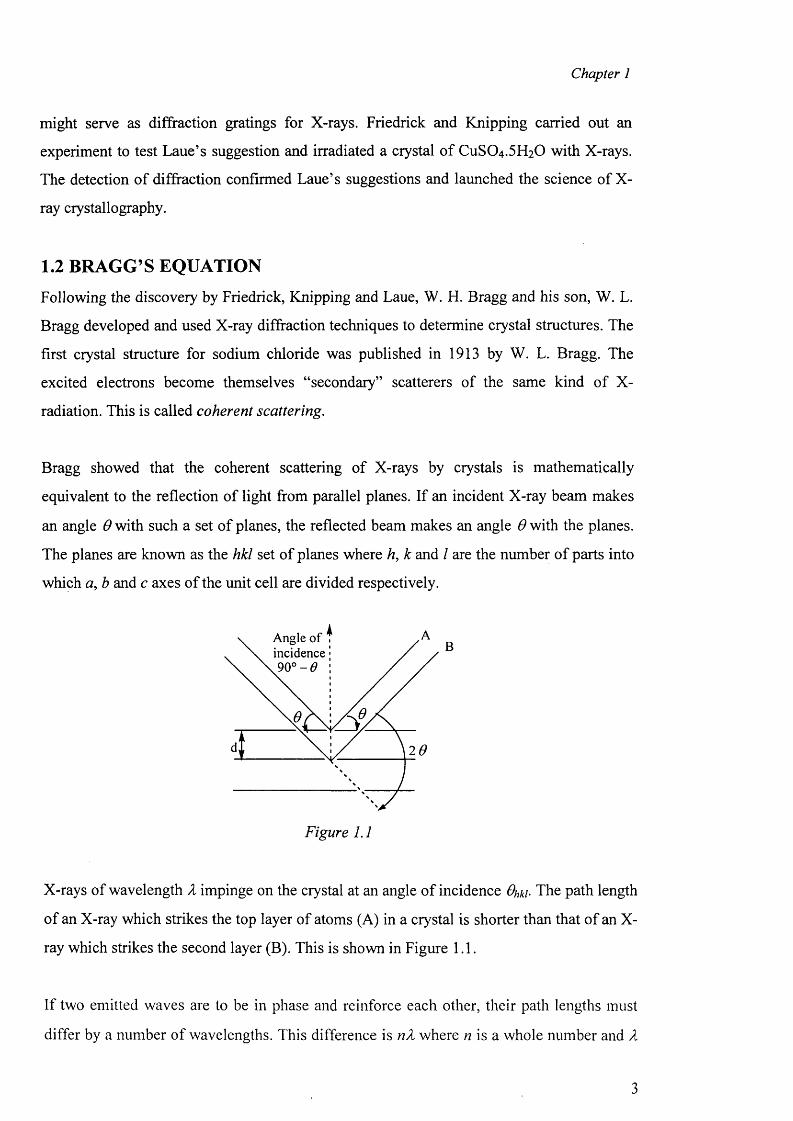
Angle of incidence .90° - 0
26
Figure 1.1
X-rays of wavelength X impinge on the crystal at an angle o f incidence Ohki• The path length
of an X-ray which strikes the top layer of atoms (A) in a crystal is shorter than that o f an X-
ray which strikes the second layer (B). This is shown in Figure 1.1.
If two emitted waves are to be in phase and reinforce each other, their path lengths must
differ by a number of wavelengths. This difference is nX where n is a whole number and X
3
Chapter 1
is the wavelength o f the radiation used. Bragg showed that the angle (#/,*/) of reflection of
X-rays could be related to the distance (dhki-the perpendicular spacing o f the hkl set of
planes) between the two layers o f atoms and is as follows;
2dhk! sinOhki = n l (1)
If equation (1) is rearranged, the spacing between the planes can be calculated as shown in
equation (2);
dhki = Z /2sm 6hki (2)
1.3 RECIPROCAL LATTICE
The concept of the reciprocal lattice was used by P. P. Ewald and extended by Laue (1913)
to describe the relationship between crystal structure and diffraction spectrum. For every
real lattice it is possible to construct a reciprocal lattice.
The advantages o f using reciprocal distances may be appreciated by considering Bragg’s
Law in the form
sin Ohki = nZJ2dhki (3)
From equation (3), it can be seen that sin# is inversely proportional to d.
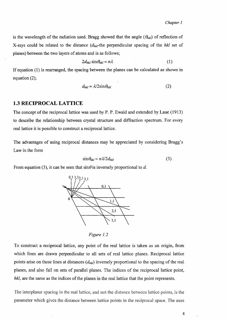
Figure 1.2
To construct a reciprocal lattice, any point of the real lattice is taken as an origin, from
which lines are drawn perpendicular to all sets of real lattice planes. Reciprocal lattice
points arise on these lines at distances {dhki) inversely proportional to the spacing of the real
planes, and also fall on sets of parallel planes. The indices o f the reciprocal lattice point,
hkl, are the same as the indices of the planes in the real lattice that the point represents.
The interplanar spacing in the real lattice, and not the distance between lattice points, is the
parameter which gives the distance between lattice points in the reciprocal space. The axes
4
Chapter 1
and angles of the unit cell in the real lattice are labelled a, b, c and a , J3, y respectively.
However in the reciprocal lattice they are labelled a , b*, c and a , /?*, y . A two
dimensional reciprocal lattice can be seen in Figure 1.2.
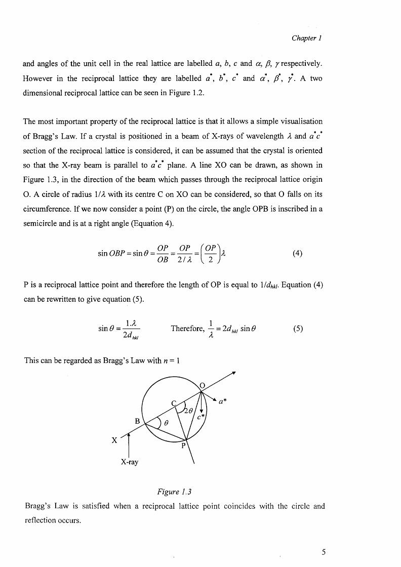
The most important property o f the reciprocal lattice is that it allows a simple visualisation
of Bragg’s Law. If a crystal is positioned in a beam of X-rays of wavelength X and a c
section of the reciprocal lattice is considered, it can be assumed that the crystal is oriented
so that the X-ray beam is parallel to a c plane. A line XO can be drawn, as shown in
Figure 1.3, in the direction of the beam which passes through the reciprocal lattice origin
O. A circle of radius MX with its centre C on XO can be considered, so that 0 falls on its
circumference. If we now consider a point (P) on the circle, the angle OPB is inscribed in a
semicircle and is at a right angle (Equation 4).
sin OBP = sin 6 =OP OP OPOB 2 /X
(4)
P is a reciprocal lattice point and therefore the length of OP is equal to Mdhkb Equation (4)
can be rewritten to give equation (5).
s in# =l.X
2d hkl
Therefore, — = 2dhkl sin 6 X
(5)
This can be regarded as Bragg’s Law with n = 1
X
X-ray
Figure 1.3
Bragg’s Law is satisfied when a reciprocal lattice point coincides with the circle and
reflection occurs.
5
Chapter 1
1.4 Crystal Lattice
A crystal contains atoms arranged in a repetitive three dimensional pattern. If each repeat
unit of this pattern is taken as a point then a three dimensional point lattice is created. Each
crystal consists o f a basic building block that repeats over and over again in all directions.
The building block which repeats in a perfectly regular array is known as the Unit Cell.
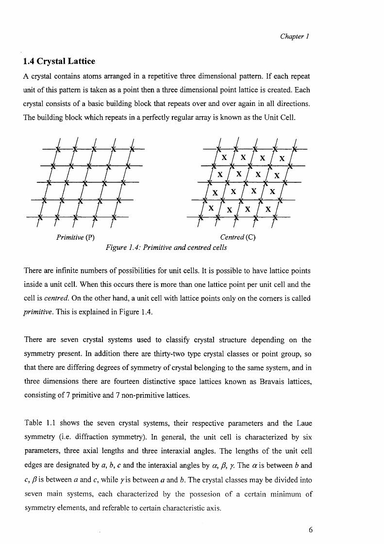
Primitive (P) Centred (C)Figure 1.4: Primitive and centred cells
There are infinite numbers of possibilities for unit cells. It is possible to have lattice points
inside a unit cell. When this occurs there is more than one lattice point per unit cell and the
cell is centred. On the other hand, a unit cell with lattice points only on the comers is called
primitive. This is explained in Figure 1.4.
There are seven crystal systems used to classify crystal structure depending on the
symmetry present. In addition there are thirty-two type crystal classes or point group, so
that there are differing degrees of symmetry of crystal belonging to the same system, and in
three dimensions there are fourteen distinctive space lattices known as Bravais lattices,
consisting of 7 primitive and 7 non-primitive lattices.
Table 1.1 shows the seven crystal systems, their respective parameters and the Laue
symmetry (i.e. diffraction symmetry). In general, the unit cell is characterized by six
parameters, three axial lengths and three interaxial angles. The lengths of the unit cell
edges are designated by a, b, c and the interaxial angles by a , p , y. The a is between b and
c, p is between a and c, while / is between a and b. The crystal classes may be divided into
seven main systems, each characterized by the possesion o f a certain minimum of
symmetry elements, and referable to certain characteristic axis.
6
Chapter 1
x
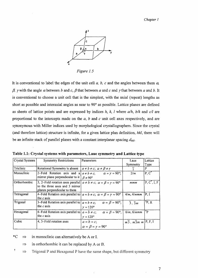
Figure 1.5
It is conventional to label the edges of the unit cell a, b, c and the angles between them a,
P, /w ith the angle a between b and c, P that between a and c and /th a t between a and b. It
is conventional to choose a unit cell that is the simplest, with the axial (repeat) lengths as
short as possible and interaxial angles as near to 90° as possible. Lattice planes are defined
as sheets of lattice points and are expressed by indices h, k, 1 where a/h, b/k and c/1 are
proportional to the intercepts made on the a , b and c unit cell axes respectively, and are
synonymous with Miller indices used by morphological crystallographers. Since the crystal
(and therefore lattice) structure is infinite, for a given lattice plan definition, hkl, there will
be an infinite stack of parallel planes with a constant interplanar spacing dhki.
Table 1.1: Crystal systems with parameters, Laue symmetry and Lattice type
Crystal Systems Symmetry Restrictions Parameters LaueSymmetry
LatticeType
Triclinic Rotational Symmetry is absent a * b * c \ a * p * y I PMonoclinic 2-Fold Rotation axis and a
mirror plane perpendicular to ita * b * c , a = / = 90°; /?* 90°
21m P, C*
Orthorhombic 3, 2-Fold rotation axes parallel to the three axes and 3 mirror planes perpendicular to them
a * b * c ; a = p = / = 90° mmm P, C*, I, F
Tetragonal 4-Fold Rotation axis parallel to the c axis
a = b * c \ a = P = / = 90° 4/m, 41 mmm P,I
Trigonal 3-Fold Rotation axis parallel to the c axis
a - b * c \ a - P - 90°; / = 120°
3 , 3m tP,R
Hexagonal 6-Fold Rotation axis parallel to the c axis
a = b * c; a - P - 90°; / = 120°
61m, 6/mmm +P
Cubic 4, 3-Fold rotation axes a = b = c; a - p - y - 90°
m3 , m3m m P, F, I
*C => in monoclinic can alternatively be A or I.
=> in orthorhombic it can be replaced by A or B.+ ^ Trigonal P and Hexagonal P have the same shape, but different symmetry
7
Chapter 1
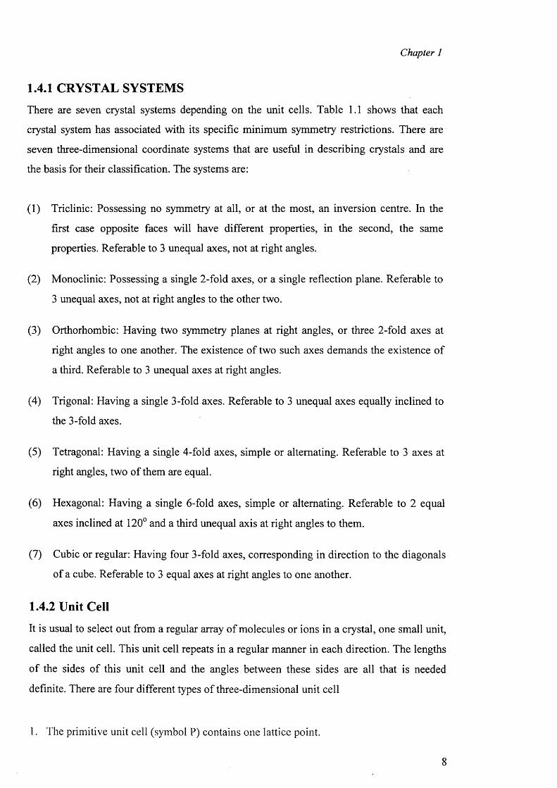
1.4.1 CRYSTAL SYSTEMS
There are seven crystal systems depending on the unit cells. Table 1.1 shows that each
crystal system has associated with its specific minimum symmetry restrictions. There are
seven three-dimensional coordinate systems that are useful in describing crystals and are
the basis for their classification. The systems are:
(1) Triclinic: Possessing no symmetry at all, or at the most, an inversion centre. In the
first case opposite faces will have different properties, in the second, the same
properties. Referable to 3 unequal axes, not at right angles.
(2) Monoclinic: Possessing a single 2-fold axes, or a single reflection plane. Referable to
3 unequal axes, not at right angles to the other two.
(3) Orthorhombic: Having two symmetry planes at right angles, or three 2-fold axes at
right angles to one another. The existence of two such axes demands the existence o f
a third. Referable to 3 unequal axes at right angles.
(4) Trigonal: Having a single 3-fold axes. Referable to 3 unequal axes equally inclined to
the 3-fold axes.
(5) Tetragonal: Having a single 4-fold axes, simple or alternating. Referable to 3 axes at
right angles, two of them are equal.
(6) Hexagonal: Having a single 6-fold axes, simple or alternating. Referable to 2 equal
axes inclined at 120° and a third unequal axis at right angles to them.
(7) Cubic or regular: Having four 3-fold axes, corresponding in direction to the diagonals
of a cube. Referable to 3 equal axes at right angles to one another.
1.4.2 Unit Cell
It is usual to select out from a regular array of molecules or ions in a crystal, one small unit,
called the unit cell. This unit cell repeats in a regular manner in each direction. The lengths
o f the sides o f this unit cell and the angles between these sides are all that is needed
definite. There are four different types of three-dimensional unit cell
1. The primitive unit cell (symbol P) contains one lattice point.
Chapter 1
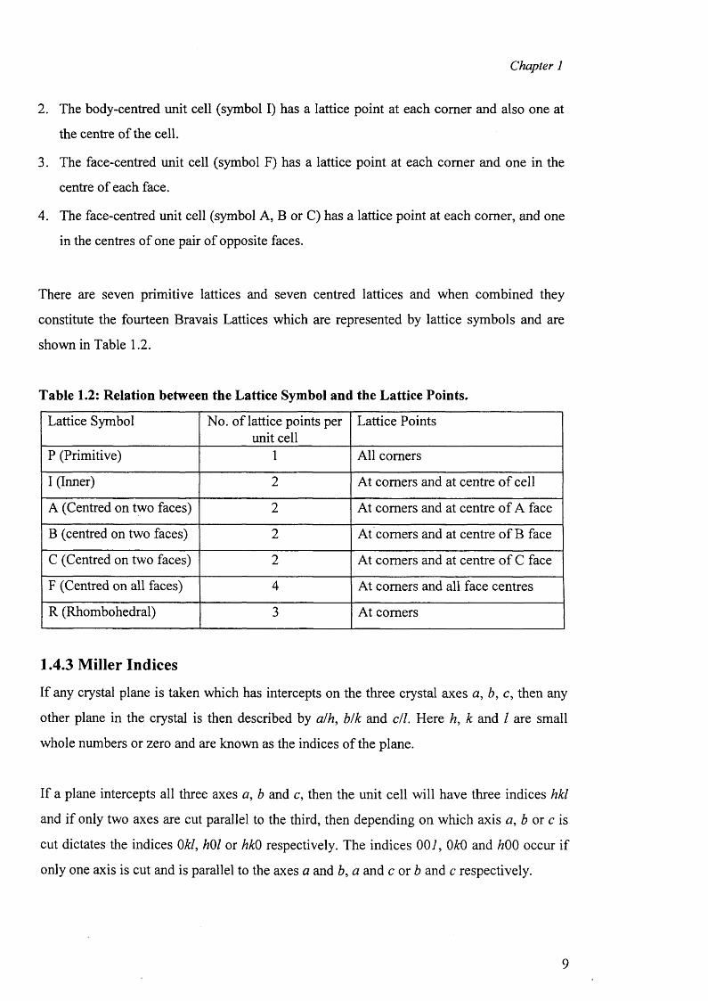
2. The body-centred unit cell (symbol I) has a lattice point at each comer and also one at
the centre of the cell.
3. The face-centred unit cell (symbol F) has a lattice point at each comer and one in the
centre of each face.
4. The face-centred unit cell (symbol A, B or C) has a lattice point at each comer, and one
in the centres of one pair of opposite faces.
There are seven primitive lattices and seven centred lattices and when combined they
constitute the fourteen Bravais Lattices which are represented by lattice symbols and are
shown in Table 1.2.
Table 1.2: Relation between the Lattice Symbol and the Lattice Points.
Lattice Symbol No. of lattice points per unit cell
Lattice Points
P (Primitive) 1 All comers
I (Inner) 2 At comers and at centre o f cell
A (Centred on two faces) 2 At comers and at centre o f A face
B (centred on two faces) 2 At comers and at centre o f B face
C (Centred on two faces) 2 At comers and at centre o f C face
F (Centred on all faces) 4 At comers and all face centres
R (Rhombohedral) 3 At comers
1.4.3 Miller Indices
If any crystal plane is taken which has intercepts on the three crystal axes a , b, c, then any
other plane in the crystal is then described by alh, b/k and c/l. Here h, k and / are small
whole numbers or zero and are known as the indices of the plane.
If a plane intercepts all three axes a, b and c, then the unit cell will have three indices hkl
and if only two axes are cut parallel to the third, then depending on which axis a, b or c is
cut dictates the indices 0kl, hOl or hkO respectively. The indices 007, OkO and h00 occur if
only one axis is cut and is parallel to the axes a and b, a and c or b and c respectively.
9
Chapter 1
1.5 SYSTEMATIC ABSENCES
Preliminary stages of a crystal structure analysis is carried out with an inspection of the
systematic absences. An absence is observed when the reflections are too weak to be
measured. The presence of systematic absences can be used to help determine the space
group of the crystal.
Two examples are given here and others are given later on in the chapter.
(1) If hOO is absent for h odd, then this indicates a 2\ axis parallel to the a axis.
(2) If OkO is absent for k odd, then this indicates a 2\ axis parallel to the b axis.
1.6 Symmetry Elements
The symmetry element is a geometrical property, such as a point, a line or a plane with
respect to which the symmetry operation is performed. There are two types o f symmetry
elements, point symmetry and translational symmetry.
1.6.1 Point Symmetry
It i s . an operation which when repeated returns the object to its original position.
Reflections, rotations and inversions are the three main types o f point group symmetry.
Firstly a rotational axis can occur at an angle of 360°/n and this corresponds to n-fold
rotations. Only 2, 3, 4 and 6 fold rotational axes are possible in crystals. Secondly an
inversion centre is a point through which an object can be moved in a straight line, so that
it finishes up at an equal distance from the inversion centre as it did originally. Thirdly a
reflection operation is the reflection of an object through a plane.
1.6.2 Translational Symmetry
Two such elements used are the screw axis and the glide plane. A screw axis combines
rotation about an axis with translation in the direction o f the axis. A three fold screw axis
would be represented as 3i, meaning a rotation about a 3-fold axis and a translation by 1/3
o f the unit cell. A glide plane combines reflections across a plane with translations parallel
to the plane. If a glide plane is parallel to the a-axis, the symbol for the glide plane is
simply “a” and for the operation is reflection in the plane and translation by a/2.
10
Chapter 1
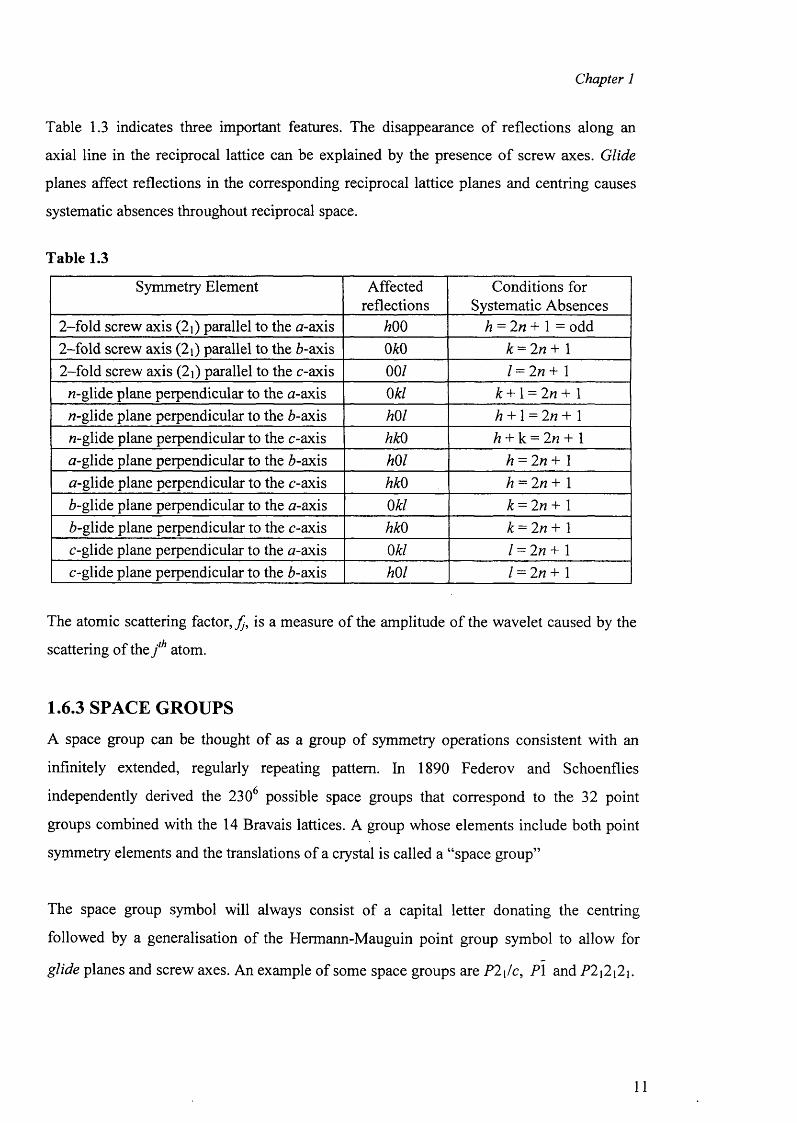
Table 1.3 indicates three important features. The disappearance o f reflections along an
axial line in the reciprocal lattice can be explained by the presence o f screw axes. Glide
planes affect reflections in the corresponding reciprocal lattice planes and centring causes
systematic absences throughout reciprocal space.
Table 1.3
Symmetry Element Affectedreflections
Conditions for Systematic Absences
2-fold screw axis (2i) parallel to the a-axis 600 6 = 2n + 1 = odd2-fold screw axis (2i) parallel to the 6-axis m 6 = 2/? + 12-fold screw axis (2j) parallel to the c-axis 00/ / = 2n + 1
/7-glide plane perpendicular to the n-axis 06/ 6 + 1 = 2n + 1/7-glide plane perpendicular to the 6-axis m i 6 + 1 = 2n + 1/7-glide plane perpendicular to the c-axis 660 6 + k = 2n + 1a-glide plane perpendicular to the 6-axis mi 6 = 2 « + 1
a-glide plane perpendicular to the c-axis 660 6 = 2/7 + 16-glide plane perpendicular to the tf-axis 06/ 6 = 2/7+ 16-glide plane perpendicular to the c-axis 660 6 = 2/7+ 1
c-glide plane perpendicular to the a-axis 06/ / = 2/7 + 1c-glide plane perpendicular to the 6-axis mi / = 2/7 + 1
The atomic scattering factor, is a measure o f the amplitude o f the wavelet caused by the
scattering of the j th atom.
1.6.3 SPACE GROUPS
A space group can be thought of as a group of symmetry operations consistent with an
infinitely extended, regularly repeating pattern. In 1890 Federov and Schoenflies
independently derived the 2306 possible space groups that correspond to the 32 point
groups combined with the 14 Bravais lattices. A group whose elements include both point
symmetry elements and the translations of a crystal is called a “space group”
The space group symbol will always consist of a capital letter donating the centring
followed by a generalisation of the Hermann-Mauguin point group symbol to allow for
glide planes and screw axes. An example of some space groups are P2\/c, PI and P2j2i2i.
11
Chapter 1
1.7 STRUCTURE DETERMINATION
It has already been shown that if a beam of X-rays is directed at a crystal consisting of a
regular array o f atoms, a pattern of reflections can be recorded.
1.7.1 Atomic Scattering Factor
Every electron in an atom is responsible for scattering and each wavelet has an amplitude.
The amplitude caused therefore by a cell o f the crystal, can be written as the sum of the unit
amplitudes o f the wavelets. Each atom has a specific number of electrons associated with it
in space and it is useful to know the general distribution of the electrons with respect to the
centre o f the atom.
The scattering power of an atom ,/, is expressed in terms of the scattering power of a single
electron. The maximum scattering by an atom is equal to its atomic number (Z). This is
observed at sin OIX = 0. If all the electrons in an atom were concentrated at one point, there
would be no destructive interference between the wavelets and also there wouldn’t be any
variation in/ with sin6!X. When the electrons are distributed, as in actual atoms, destructive
interference occurs and the larger the volume of the atom then the greater the fall-off in /
with sin 6! X.
1.7.2 Structure Factor
The diffraction from one atom cannot be considered alone. The whole of the crystal causes
diffraction and therefore all the atomic scatterings have to be accounted. This is represented
as the Structure Factor Fhki which describes the amplitude and the phase of the X-rays
scattered by a unit cell for a particular reflection, hkl.
The Structure Factor depends upon;
(1) The atomic scattering factors
(2) Arrangements of the scattering materials (including thermal motion)
(3) The direction of scattering
The intensity o f the scattered radiation from the plane, hkl, is proportional to the square of
the amplitude.
(6)
12
Chapter 1
The structure factor, F^kt), can be represented by the following equation:N
F(hki) =Y,fJexP27d(hxj+l<y j+!zj ) (?)j=i
Here F^kt) - Structure factor for hkl reflection
f j = Scattering factor for the j th atom
x ,y ,z = Positions of the j ih atom in the unit cell
i = V -l
(8)
(9)
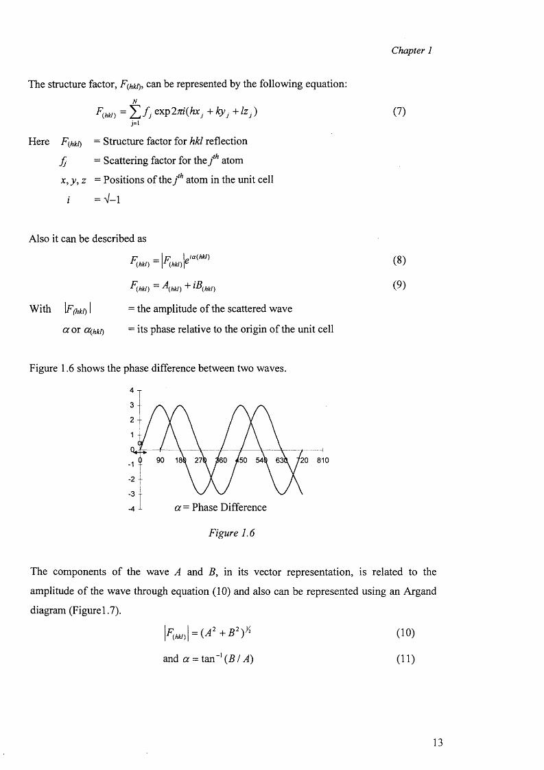
Figure 1. 6 shows the phase difference between two waves.
'20 810
-3 -a = Phase Difference
Figure 1.6
Also it can be described as
P _ \ p \Ja{hkl)r (hkl) \r (hkl ) r
F(hki) - \hki) + iB(hki)With \F(hki) I = the amplitude of the scattered wave
a or cc(hki) = its phase relative to the origin of the unit cell
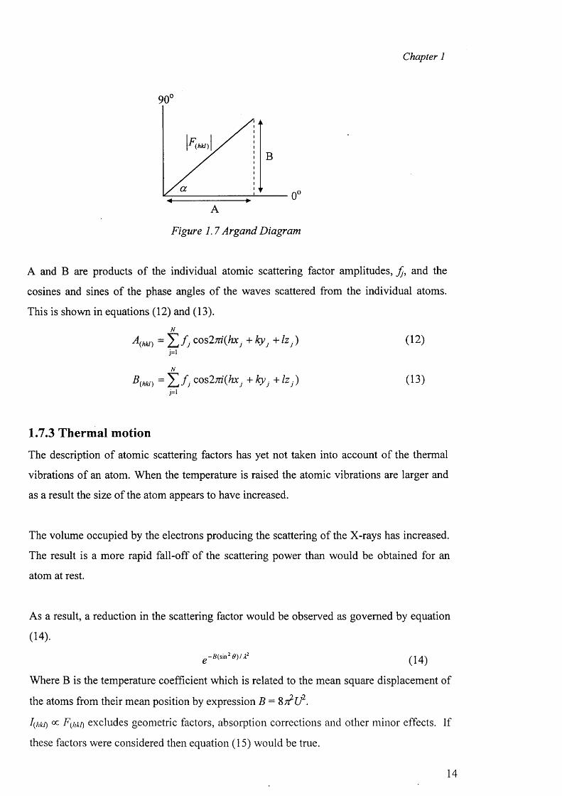
The components o f the wave A and B , in its vector representation, is related to the
amplitude o f the wave through equation (10) and also can be represented using an Argand
diagram (Figurel.7).
(10)
and a = tan - 1 ( B / A) (11)
13
Chapter 1
90°
(hkt)
0 (A
Figure 1.7 Argand Diagram
A and B are products o f the individual atomic scattering factor amplitudes, Jj, and the
cosines and sines of the phase angles of the waves scattered from the individual atoms.
This is shown in equations (12) and (13).N
A(hki) = Y j f j cos2m (hXj+kyj + /z ,) ( 1 2 )j=lN
B(hki) = Z / y cos2;n' ( ^ + ty j + Izj) (13)j=i
1.7.3 Thermal motion
The description of atomic scattering factors has yet not taken into account o f the thermal
vibrations of an atom. When the temperature is raised the atomic vibrations are larger and
as a result the size o f the atom appears to have increased.
The volume occupied by the electrons producing the scattering of the X-rays has increased.
The result is a more rapid fall-off of the scattering power than would be obtained for an
atom at rest.
As a result, a reduction in the scattering factor would be observed as governed by equation
(14).
e ~ B ( s i n 2 0 ) U 2
Where B is the temperature coefficient which is related to the mean square displacement of
the atoms from their mean position by expression B = %7?U2.
I(hki) 0 0 Ftfkt) excludes geometric factors, absorption corrections and other minor effects. If
these factors were considered then equation (15) would be true.
14
Chapter 1
V ) = 4 > ) (Lp)(Abs) (15)
Here (Lp) = an abbreviation for the geometric factors (Abs) = an abbreviation for the absorption factors and K = A Scale Factor .
For an explanation of Lp and Abs see 1.7.7 (page 20)
If an intensity, (I(COrr)), was corrected taking into account (Lp) and (Abs) it would equal
I ic„r )= I/(A b S).(Lp) (16)
I(corr) ~ I 0 )
12
hcorr) = * 1 ^ ) 1 ' exP( - 2B.so sin 2 e/X2) (18)
F(novib) is the value of F for a structure composed of no vibrating atoms.
The value of F may be reduced as a result o f thermal vibrations so that if F(n0 Vib) is the value
for the structure containing stationary atoms, the experimental values will correspond to
F hki) = F{novib) exp(-2 Blso sin2 0 / A2) (19)
and B(1S0) is the temperature factor.
1.7.4 THE LORENTZ FACTOR (L), POLARISATION FACTOR (p)
AND ABSORPTION CORRECTIONS (Abs)
When X-rays are reflected by a plane in a crystal, a reduction in the intensity of the
reflected beam occurs as a result of polarisation, scattering and absorption. The polarisation
factor (p) is a measure of this polarisation. Equation (15) shows how the intensity is
affected by L, p and Abs.
The Lorentz factor is an expression for the time a plane of a rotating crystal spends in the
reflecting position. In terms of the reciprocal lattice or reflecting sphere concept, this is the
length of time the lattice point is in contact with the sphere of reflection. This is obviously
dependent on the distance of the reciprocal lattice point from the origin.
As stated previously, the intensity o f the reflected rays decreases and if this is not corrected
for then this results in systematic errors. The effect of this which can be attributed to
absorption can be measured and corrected for using a program called SORTAV .
15
Chapter 1
1.7.5 THERMAL ELLIPSOID
We have considered the vibrations as being isotropic, however, it would be more accurate
if they were considered as anisotropic. The location of an atom in a molecule is determined
by considering the average time which the atom spends in all the positions it occupies
while the structure was being determined. The resultant structure is then often represented
in terms of thermal ellipsoids, which are probability indicators of where the atoms are most
likely to be found.
Six parameters can be used to describe an ellipsoid compared to a single parameter for a
sphere. Three are used to define the lengths of the three mutually perpendicular axes and
three to define the orientations of the ellipsoidal axes.
The ellipsoidal motion is accounted for in the structure factor equation by an anisotropic
exponential factor with six anisotropic vibration parameters by and shown in equation (2 0 ).
^ - ( b u h 2+b22^2+b-3iI 2 +bn hk+b2jkl+biihl) (20)
The mean square vibrational amplitude in any direction, specified by the cosines, 1, o f the
angles between the direction and the reciprocal axes, is given as;
U ‘ Uul* + U 21l\ + L^ 2 + 2 U nllll + 2U23l2\h + 2Ui\h l \ (21)
If the ellipsoid takes the shape of a rugby ball, then the motion of the atom is mostly back
and forth along the bond axis. If however it takes the shape o f a curling-stone, then the
motion is mostly wobbling about the bond axis. The less the thermal ellipsoid of the
molecule, the smaller o f its thermal motion.
The computer program used during this project to represent the thermal ellipsoids was
SNOOPI6.
1.7.6 Electron Density
X-rays are scattered by the electrons o f the atom and therefore it is important to know
something o f the electron density at different positions w ithin the unit cell. If a point in the
16
Chapter 1
unit cell has co-ordinates x , y, z then the electron density, p(x y z), per unit volume near the
point is given by
p (x + p ,y + q ,z + r) = p (x y z ) (2 2 )
In this equation p , q and r are any integers.
It is convenient to be able to express a function by means of a Fourier Series. This is the
sum of the sine and cosine terms with appropriate coefficients. Fourier expansions are
advantageous when the function is periodic and the electron density is one such function.
Fractional co-ordinates x„, y n, z„, are often used to give the precise location of an atom in a
unit cell. A crystal is a periodic structure and can therefore be described by the periodic
function, the Fourier Series (or Synthesis).
In three dimension the electron density at the point xn, y n, zn is
P(x„y„z„) = \ F j l . i Z ( |cos 2n(hxn + kyn + lz „ - a ( m ) (23)
Where p(xn, y n, zn) = the electron density at the points xn, y n, z„.
1 ^= the volume o f the unit cell
H h Z* £ / = the mean sum over h, k, I and F(hki)
|F(W/)| = the structure amplitude
a (Md) = phase angle of each Bragg reflection
1.7.7 PHASE PROBLEM1 12 I 12
As stated previously in 1.7.2, . However sets of values of E(M/) do not lead
to the determination of a crystal’s structure. values are needed in equation (23)
whereas, the intensities only give |e
(hk!)\
12
17
Chapter 1
If we know F(Wk/) and a (of each hkl) we could calculate the electron densities at all values
of x, y, z and plot the p(x y z) values to give a three dimensional electron density map.
However, structure factor amplitudes, | , and not phase angles, a , are obtainable from
the experimental measurements.
The problem of getting estimates of the phase angles so that an image o f the scattering
matter can be calculated is called the Phase Problem and is the central one in X-ray
crystallography.
The phase angle can be derived from values of A and B from statistical methods (trial
structures) or by analytical methods. Once the phase angle has been determined, the
structure factor amplitude can also be determined. The calculated, F c, can then be
compared with the observed amplitudes F0. F0 has to be as close to Fc as possible in order
to give a structure that best fits the calculated values.
1.7.8 SOLUTIONS TO THE PHASE PROBLEM
There are many methods which are commonly used to determine the relative phase angles
of the Bragg reflections, hkl. The two main methods of solving the Phase Problem are:
(1) by Direct Methods and
(2) by Patterson Methods.
1.7.8.1 Direct Methods
Direct methods o f structure determination, are based on two fundamental physical
principles.
Firstly, the electron density in the unit cell cannot be negative at any point, and so the large
majority of possible sets of values for the phases of various structure factors are not
allowed. A random set of phases would give positive and negative regions in the electron
density map with equal probability.
Secondly, the electron density in the cell is not randomly distributed, but is mainly
concentrated in small areas which we identify as atoms.
18
Chapter 1
A consequence of these two principles is that for certain sets o f reflections having
particular combinations of miller indices, there are theoretical probability relationships
among their phases. The phases can be assigned to some reflections usually the most
intense and then the positions of some or all of the heaviest atoms can be located.
When direct methods are used for phases determination, “normalised structure fa c to rs”
are used. The structure factor |F| is modified by the removal o f the fall-off in the scattering
fac to rs ,/ with increasing scattering angle, 20. In the normalisation factor, E, (equation 24)
the sum is taken over all atoms in the unit cell at the value of sin 6!L
where,
s= an integer for improving the space group symmetry. The values o f e can be obtained
from International Tables5.
F = Structure Factor magnitude.
Fj = Atomic scattering factor of the j th atom.
Thus showing that E is independent of sin OIL
The E values give us information about the structure of the crystal. If the mean value o f E
is 0.798 then the structure is centrosymmetric. However, if E were 0.886 then this would
indicate a noncentrosymmetric structure.
A noncentrosymmetric crystal structure has phase angles anywhere between 0° and 360°. A
centrosymmetric structure on the other hand has phase angles o f 0 ° or 180° which means
cosa= ± 1 , sinO= 0 .
1.7.8.2 Patterson Synthesis
This is the “heavy atom” method of solving structures and is very useful for transition
metal complexes, which often have one atom much heavier than the rest.
I I2In 1934, A. L. Patterson discovered that a Fourier series using values of \E{hkl)\ as
coefficients instead of Fq I) could produce useful information about the structure. If the
19
Chapter 1
electron density at point x, y, z is taken and multiplied by the electron density at the point x
+ u ,y + v ,z + w a product
p (x ,y ,z )p (x + u ,y + v ,z + w) (25)
is now formed. This is then multiplied by dxdydz and integrated over the volume of the unit
cell.
When the equation for the Fourier expansion is substituted for each electron density
function we arrive at equation (26).
1 _ I 12
p(»,v.»') = 77Z(m/)|-F(mo| c o s2 x(.hu + kv + lw) (26)
Where, P(U,v,w)= the electron density at u, v, w in the unit cell.
u, v, w - the vectors between the two atoms.
u = jq - x 2 v = y x - y 2
w = z x - z 2 V= volume of the unit cell
F(hki)= is the structure factor of the hkl set of planes
i i2Here Patterson used the squares of the structure amplitudes, LF(M/) , as Fourier coefficients
and these values were directly derivable from the primary experimental quantities. No
i i2phase information is required for this map because is independent of phase.
The peaks in the Patterson map occur at points whose distances from the origin correspond
in magnitude and direction with distances between atoms in the crystal. This results from
= v IfM *> y> + u>y+ v ’z + w)dxdydz (27)
The height [P(U,v,w)] of each peak is also proportional to the product o f the atomic numbers
of the atom giving rise to it. The very high peaks correspond to the heavy atoms and
therefore the peaks corresponding to the hydrogens are the ones appearing last in the map
and are of low intensity.
20
Chapter 1
One difficulty with the Patterson function is that there are so many interatomic vectors. If a
unit cell contains N atoms there are N vectors. There are N origin vectors and therefore
there are (N2-N) peaks other than the origin peak. Of these half are related to the other half
by a centre of symmetry so there are (N - N)/2 independent peaks. If N is 20, there are 190
independent Patterson peaks and the vector map will be very crowded. A further
complication is that the atoms are not points, but they occupy a considerable volume.
1.7.9 STRUCTURE REFINEMENT
The aim of refinement is to obtain the best agreement between the observed (F0) and the
calculated structure factors (Fc). There is sufficient information for every atom within the
unit cell to be located once the phase problem has been overcome. The most suitable sets of
parameters are those which will produce the most accurate values of interatomic distances
and bond angles. It is anticipated that the most suitable set will also give the best agreement
between the calculated and observed structure factors.
The correctness of the structure is measured in terms of a discrepancy index, R, where
N"* 1/r _ /r I
R = V i P I <28)z J ^ I
Generally, if R has a value of 0.30 then this indicates that the correct structure is near.
However, if R is 0.1 or less then F0 is close to Fc and the results are probably very reliable.
Once all the phases have been determined, a Fourier map can be calculated and the atomic
positions are taken as the locations of the maxima of the electron density function (or
Fourier Synthesis).
Alternatively, the least-squares process could be carried out. In 1806, Legendre introduced
the idea of least-squares which is a statistical method of obtaining the best fit. This is done
by minimising the sum of the square of the deviations between the exponential and the
calculated parameters.
21
Chapter 1
If the difference in the amplitudes of the observed and calculated structure factors
The sum in this equation is taken over independent observations that is the unique
diffraction maxima.
In equation (15) the scale factor (K) was introduced and this brings the observed structure
factor, F0, on to the same scale as the calculated structure factor, Fc. The relationship
between K , F0 and Fc is given in equation (30).
1.8 Experimental Section
This section presents very briefly the most important aspects of the hardware, software and
procedures used for X-ray data collection. CCD area detector diffractometer were used to
collect the crystallographic data for the compounds presented in this work.
X-ray diffractometers basically comprise three principal parts. These are:
♦> The X-ray source: This consists o f the target material, usually molybdenum or copper,
housed to that it is at the focus of an ‘electron gun’. Only about 0.1% of the power
supplied to the source is converted to X-rays, thus there is a considerable amount o f heat
produced. There are generally two types of X-ray source in laboratory use, a ‘sealed
tube’ source, where the target metal is fixed within a vacuum tube; and a rotating anode
generator, where the anode is constantly allowing heat to dissipate more effectively thus
more intense radiation can be generated. This requires constant pumping of the vacuum.
♦> A goniometer: This is a device to orientate the crystal into the desired positions for
diffraction. The goniometer head has adjustments in the vertical and horizontal
\F0\- \F C\, be simply called A|F| and the standard deviations o f the experimental values of
|F0 (M/)| be [w(hkl)]~112, then the best parameters for a structure are those corresponding to
the minimum value o f the quantity
(29)
22
Chapter 1
directions to allow alignment of the crystal within the X-ray beam. There are two main
types of goniometer in general use. The four-circle, where there are four arcs Of, fc 0 and
a>), which may be used to bring the crystal into the diffractometer position, and Kappa
goniometer consisting of a Kappa block mounted at 50° to the Omega block, orienting
the x axis at 50° to the co axis. This geometry allows the crystal to be oriented in all
available in the four-circle goniometer, without the steric restriction of the x circle.
❖ A detector. Detectors fall into two categories, single point counters and area detectors.
Single point counters such as scintillation counters can measure only one diffraction at a
time, thus giving long data collection times. Area detectors have the advantage of
measuring several diffractions concurrently, thus speeding up data collection. There are
several types of area detectors available, such as image plates, CCD and the FAST
system.
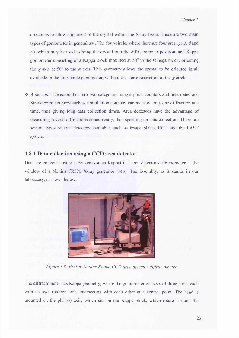
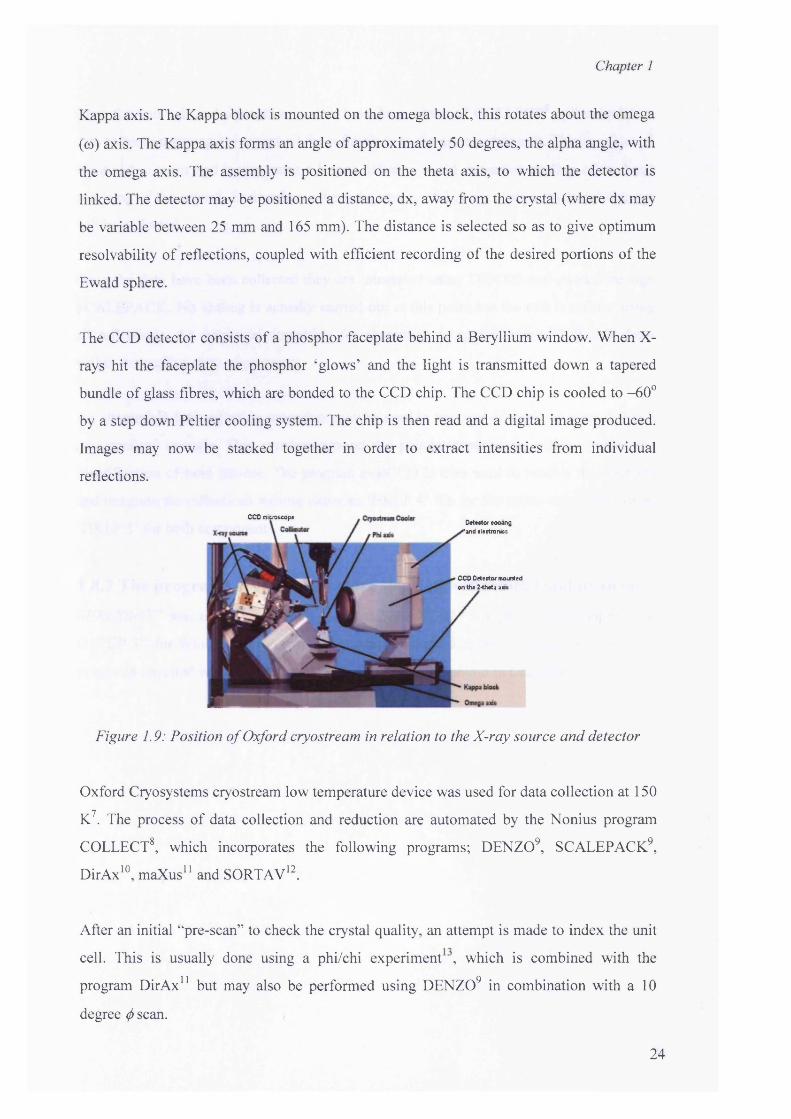
1.8.1 Data collection using a CCD area detector
Data are collected using a Bruker-Nonius KappaCCD area detector diffractometer at the
window of a Nonius FR590 X-ray generator (Mo). The assembly, as it stands in our
laboratory, is shown below.
Figure 1.8: Bruker-Nonius Kappa CCD area detector diffractometer
The diffractometer has Kappa geometry, where the goniometer consists of three parts, each
with its own rotation axis, intersecting with each other at a central point. The head is
mounted on the phi (cp) axis, which sits on the Kappa block, which rotates around the
23
Chapter 1
Kappa axis. The Kappa block is mounted on the omega block, this rotates about the omega
(co) axis. The Kappa axis forms an angle of approximately 50 degrees, the alpha angle, with
the omega axis. The assembly is positioned on the theta axis, to which the detector is
linked. The detector may be positioned a distance, dx, away from the crystal (where dx may
be variable between 25 mm and 165 mm). The distance is selected so as to give optimum
resolvability of reflections, coupled with efficient recording of the desired portions of the
Ewald sphere.
The CCD detector consists of a phosphor faceplate behind a Beryllium window. When X-
rays hit the faceplate the phosphor ‘glows’ and the light is transmitted down a tapered
bundle of glass fibres, which are bonded to the CCD chip. The CCD chip is cooled to -60°
by a step down Peltier cooling system. The chip is then read and a digital image produced.
Images may now be stacked together in order to extract intensities from individual
reflections.
Figure 1.9:
Oxford Cryosystems cryostream low temperature device was used for data collection at 150
K7. The process of data collection and reduction are automated by the Nonius program
COLLECT8, which incorporates the following programs; DENZO9, SCALEPACK9,
DirAx10, maXus11 and SORTAV12.
After an initial “pre-scan” to check the crystal quality, an attempt is made to index the unit
cell. This is usually done using a phi/chi experiment , which is combined with the
program DirAx11 but may also be performed using DENZO9 in combination with a 10
degree (/) scan.
CCD microscopeDetector cooling
'and electronics
CCD Detector mounted on the 2-theta aids
Position o f Oxford cryostream in relation to the X-ray source and detector
24
Chapter 1
Once the unit cell and orientation matrix have been obtained COLLECT8 will calculate a
data collection strategy to access all the reflections in the asymmetric unit. This is achieved
via (f> and co scans and incorporates a Kappa offset to avoid excessive cooling o f the head
and icing of the crystal. In situations where the true crystal is not clear a ‘triclinic’ data set
will be collected.
Once the data have been collected they are integrated using DENZO and passed through
SCALEPACK. No scaling is actually carried out at this point but the cell is refined using
all reflections. An empirical correction for absorption is applied using SORTAV from
within the maXus suite o f programs.
An alternative data reduction procedure may be used in the case of twins or other difficult
non-standard crystals. This revolves around the phi/chi experiment, which enables the
identification o f twin lattices. The program evalCCD is then used to resolve the overlaps
and integrate the reflections writing either an ‘HKLF 4 ’ file for the major component or an
‘HKLF 5’ for both components.
1.8.2 The programs used for structure solution, refinement and drawing14 15SHELXS-91 was used for structure solution: SHELXL-91 for structure refinement and
ORTEP-3 1 6 for Windows for drawing of crystal structural diagrams. Absorption correction
programs are cited when relevant with each individual compound in Chapters 4 - 6 .
25
Chapter 1
REFERENCES
1. G. H. Stout and L. H. Jensen; X-ray Structure Determination; A Practical Guide, McMillan, New York, 1968
2. J. P. Glusker and K. N. Trueblood; Crystal Structure Analysis, A Primer, 2nd Edition, Oxford University Press, 1985
3. D. E. Sands; Introduction to Crystallography, Benjamin Inc., New York, 1968
4. M. J. Buerger; Crystal Structure Analysis, J. Wiley and Sons, New York, 1960
5. International Tables fo r X-ray crystallography, Vol. II, J. S. Kasper and K. Lonsdale,Editors, D. Reidel Publishing Company, Dordrecht, Holland, 1969
6 . M.F.C. Ladd and R.A. Palmer; “Structure Determination by X-ray Crystallography”, 3rd Edition, Plenum Press, New York, 1960, 434
7. J. Cosier and A. M. Glazer; J. Appl. Cryst., 1986, 19, 105
8 . R. Hooft; “Collect: Data Collection Software”, Nonius B.V. 1998
9. Z. Otwinosnowski and W. Minor; “Methods in Enzymology, 1997, Vol. 276, Macromolecular Crystallography, Part A, edited by C. W. Carter, Jr. and R. M. Sweet, Academic Press, New York, 1997, 307-326
10. A. J. M. Duisenberg; J. Appl. Cryst., 1992, 25, 92
11. S. Mackay, C. J. Gilmore, C. Edwards, M. Tremayne, N. Stewart and K. Shankland; maXus, A computer program fo r the solution and refinement o f crystal structures from diffraction data.
12. (a) R. H. Blessing; Acta Cryst., 1995, A51, 33 and (b) R. H. Blessing; J. Appl. Cryst., 1997, 30, 421
13. A. J. M. Duisenberg, R. W. W. Hooft, A. M. M. Schreurs and Kroon; J. Appl. Cryst., 2000, 33, 893
14. G. M. Sheldrick; Acta Cryst., 1990, A46, 467
15. G. M. Sheldrick; SHELXL-97 Program fo r Crystal structure determination, University of Gottingen, Germany, 1997
16. L. J. Farrugia; J. App. Cryst., 1997, 30, 565
26
METAL COMPLEXES OF SULFA DRUGS - A LITERA TURE REVIEW
Chapter 2
INTRODUCTION
A sulphonamide is any member o f a class o f chemical compounds, which contain the group
— SO2 N— . The class includes several groups o f drugs used in the treatment o f bacterial
infections, diabetes mellitus, oedema, hypertension and gout . 1
Sulfonamides are analogues o f /?-aminobenzoic acid (PABA). The first sulphonamide of
clinical importance was prontosil, an azo dye, which is metabolized in vivo to
sulfanilamide. Many sulfonamides have been synthesised but they differ only slightly in
their antimicrobial activity, but vary in their pharmacokinetic properties. The sulfonamides
have been classified according to their rate o f excretion as short-, medium- or intermediate-
, long and ultra-long activity. The short-acting sulfonamides are excreted in the urine in
high concentrations and have therefore been o f particular use in the treatment of urinary-
tract infections. O f the short-acting sulfonamides most commonly used sulfadiazine has
low solubility in urine whereas sulfamerazine and sulfamethazine and their acetyl
conjugates are very soluble.
The sulfonamides are synthetic bacteriostatic antibiotics with a wide spectrum against most
gram-positive and many gram-negative organisms. However, many strains o f an individual
species may be resistant. Sulfonamides inhibit multiplication of bacteria by acting as
competitive inhibitors of p-aminobenzoic acid in the folic acid metabolism cycle. Bacterial
sensitivity is the same for the various sulfonamides, and resistance to one sulfonamide
indicates resistance to all.
These drugs are extensively used in medicine and they were the first agents to be used for
the treatment o f bacterial infection for both human and animal disease. Use of
sulfonamides today is limited to specific disease treatment in human medicine such as
urinary tract infections. However, sulfonamides are more often encountered in animal
medicine. The presence o f certain residues in animal products presents a potential health
hazard due to their allergic properties. Also, some people exhibit hypersensivity to drug
residues and/or low levels of drug residue may produce genetically altered bacteria that are
resistant to existing drug therapy . 3 In addition, a study by the National centre for
Toxicological Research indicated that sulfamethazine may be a thyroid carcinogen . 4
27
Chapter 2
2.1 History
Sulfonamides were first synthesised by Gelmo et a l 5 in 1908 while doing research into azo
dyes. Directly following this work, Hoerlien et al.6 discovered dyes containing the sulfanyl
group that had affinity for proteins of silk and wool. This led to the discovery by Eisenberg
in 1913, that chrysolidine, one of the azo dyes studied, had pronounced bactericidal action
in vitro.
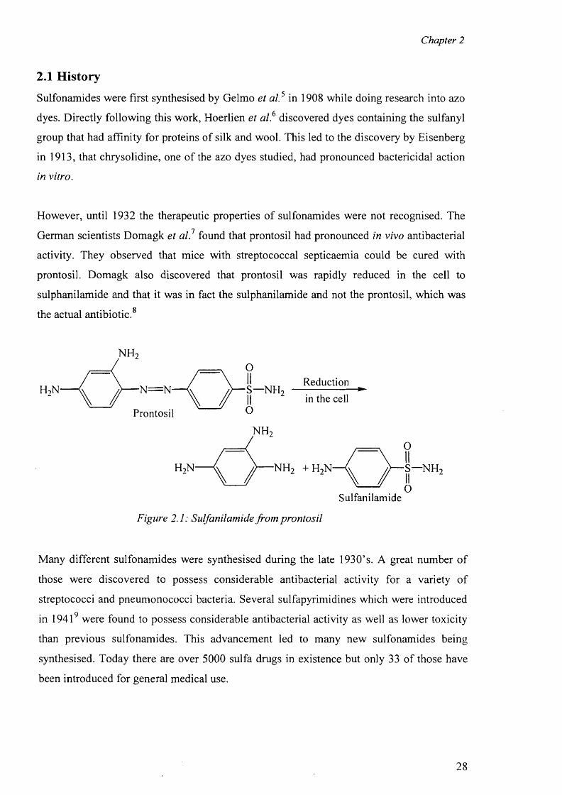
However, until 1932 the therapeutic properties o f sulfonamides were not recognised. The
German scientists Domagk et al.1 found that prontosil had pronounced in vivo antibacterial
activity. They observed that mice with streptococcal septicaemia could be cured with
prontosil. Domagk also discovered that prontosil was rapidly reduced in the cell to
sulphanilamide and that it was in fact the sulphanilamide and not the prontosil, which wasO
the actual antibiotic.
NH
Reduction■N=N- S—NH
in the cellProntosil
S—NH
Sulfanilamide
Figure 2.1: Sulfanilamide from prontosil
Many different sulfonamides were synthesised during the late 1930’s. A great number o f
those were discovered to possess considerable antibacterial activity for a variety o f
streptococci and pneumonococci bacteria. Several sulfapyrimidines which were introduced
in 19419 were found to possess considerable antibacterial activity as well as lower toxicity
than previous sulfonamides. This advancement led to many new sulfonamides being
synthesised. Today there are over 5000 sulfa drugs in existence but only 33 of those have
been introduced for general medical use.
28
Chapter 2
The use of sulphonamides as drugs dates back to the beginning o f the twentieth century,
when the discovery of the medicinal use o f sulphonamides and their derivatives was a
milestone in the history o f chemotherapy. It represented the first investigation o f synthetic
organic molecules as potential drugs to fight infection carried in the bloodstream.
This new class o f sulpha drugs was the only effective antibacterial drugs until penicillin
became available in the early 1940’s. Despite the benefits o f the sulpha drugs, they proved
ineffective against infections such as salmonella (the organism responsible for typhoid).
Problems have resulted from the way in which they are metabolised in the human body,
since toxic products are frequently obtained. This has ultimately led to the sulphonamides
being superseded in many instances by penicillin. These drugs act by inhibiting the growth
of the bacteria, rather than killing the organisms. The sulphonamides act as competitive
enzyme inhibitors and block the biosynthesis o f the vitamin folic acid in bacterial cells.
The enzyme responsible for linking together the component parts o f folic acid is inhibited
and the consequences to the cell are disastrous.
The importance o f sulphonamides within the pharmaceutical industry could even be seen
from the database search when the compound 5-Acetamido-l, 3, 4-thiadiazole-2-
sulphonamide was located. Acetazolamide, or Diamox as it is commonly known, is used in
the treatment of many types o f glaucoma (elevated pressure in the anterior chamber o f the
eye; the anterior chamber is the most forward section of the eye, behind the cornea). It is
also used as a diuretic in some problems such as heart failure, or where fluid is retained in
the body. This medication impedes the action of an enzyme, carbonic anhydrase
throughout the body. In the eye, this leads to a decrease in production o f fluid secreted in
the anterior chamber o f the eye, leading to a reduction in pressure. It acts in the kidney to
increase the amount o f bicarbonate excreted in the urine and thus increases salt and water
elimination from the body. The way it works on the central nervous system is as yet not
well understood.
2.2 Pharmacology
Most sulfonamides are readily absorbed orally. However, parenteral administration is
difficult, since the soluble sulfonamide salts are highly alkaline and irritating to the tissues.
The sulfonamides are widely distributed throughout all tissues. High levels are achieved in
pleural, peritoneal, synovial, and ocular fluids. Although these drugs are no longer used to
29
Chapter 2
treat meningitis, CSF levels are high in meningeal infections. Their antibacterial action is
inhibited by pus.
The sulfonamides are metabolized mainly by the liver to acetylated forms and
glucuronides, both o f which are therapeutically inactive. Excretion is primarily renal by
glomerular filtration with minimal tubular secretion or reabsorption. When these drugs are
given in pregnancy, high levels are achieved in the fetus. Sulfonamides are loosely and
reversibly bound in varying degrees to serum albumin. Since the bound sulfonamide is
inactive and nondiffusible, the degree o f binding can affect antibacterial effectiveness,
distribution, and excretion.
The relative insolubility o f most sulfonamides, especially their acetylated metabolites, may
cause precipitation in the renal tubules. The more soluble analogues, such as sulfisoxazole
and sulfamethoxazole, should generally be chosen, and the patient must be well hydrated.
To avoid crystalluria and renal damage, fluid intake should be sufficient to produce a
urinary output o f 1200 to 1500 mL/day. Sulfonamides should not be used in renal
insufficiency.
2.3 Mechanism of Action10
Certain microbes require p-aminobenzoic acid in order to synthesize dihydrofolic acid
which is required to produce purines and ultimately nucleic acids. Sulfonamides, chemical
analogues o f PABA, are competitive inhibitors o f dihydropteroate synthetase.
Sulfonamides are therefore reversible inhibitors of folic acid synthesis and are bacterostatic
not bacteriocidal
Spectrum of antibacterial activity and Resistance
Sulfonamides inhibit (bacteriostatic) gram-positive and gram-negative bacteria, Nocardia,
Chlamydia trachomatis and some protozoa. Enteric bacteria such as E. coli, Klebsiella,
Salmonella, Shigella and Enterobacter are also inhibited. Resistance to sulfonamides may
develop when bacterial mutations result: (i) in PABA overproduction, (ii) in a folic acid
synthesizing enzyme protein that has low affinity for sulfonamides and (iii) from a loss of
cell permeability to sulfonamides.
30
Chapter 2
2.4 Pharmacokinetics10
Sulfonamides can be classified in three groups: oral, absorbable oral, nonabsorbable topical
and oral and absorbable oral agents may be further classified as short-, medium, or long
acting sulfonamides. Sulfonamides are absorbed from the stomach and small intestine and
widely distributed to tissues, including the CNS. Sulfonamides and inactivated metabolites
are excreted by the kidney mainly through glomerular filtration.
2.5 Clinical Uses10
Sulfonamides are useful in treating urinary tract infections (UTI), but in general are rarely
used as single agents. The fixed drug combination of trimethoprim-sulfamethoxazole
(Bactrim) has supplanted many previous sulfonamide clinical uses. Examples are,
sulfisoxazole (Gantrisin) and sulfamethoxazole (Gantanol) are used almost exclusive to
treat UTI. In combination with phenazopyridine, a urinary tract anesthetic, sulfonamides
are available as Azo Gantrisin and Azo Gantanol. Sulfadiazine in combination with
pyrimethamine (Daraprim) (antiprotozoal agent, dihydrofolate reductase inhibitor) is first-
line treatment for acute toxoplasmosis. Cefpodoxime (Vantin), a long-acting sulfonamide,
is used in combination with pyrimethamine as a second-line option for treating malaria.
Oral, non-absorbable drugs: Sulfasalazine (Azulfidine, salicylazosulfapyridine) is used in
treating ulcerative colitis, enteritis and other inflammatory bowel disorders. The
antiinflammatory action is due to the release o f salicylate following splitting of
sulfasalazine by intestinal bacteria. Topical Agents such as Bacterial conjunctivitis may be
treated with sodium sulfacetamide opthalmic solution/ointment. Sodium sulfacetamide is
an adjunctive drug in treating trachoma. Mafenide acetate is used in preventing infection in
bum wounds.
2.6 Adverse reactions10
The most common such as fever, rash, exoliative dermatitis, photosensitivity, urticaria,
nausea, vomiting, diarrhea, urinary tract problems may occur due to the precipitation of
drug in urine. Sulfonamides may cause Stevens-Johnson syndrome (<1% frequency) and
hemopoietic disturbances, including hemolytic or aplastic anemia, granulcytopenia,
thrombocytopenia may also be caused by sufonamides.
31
Chapter 2
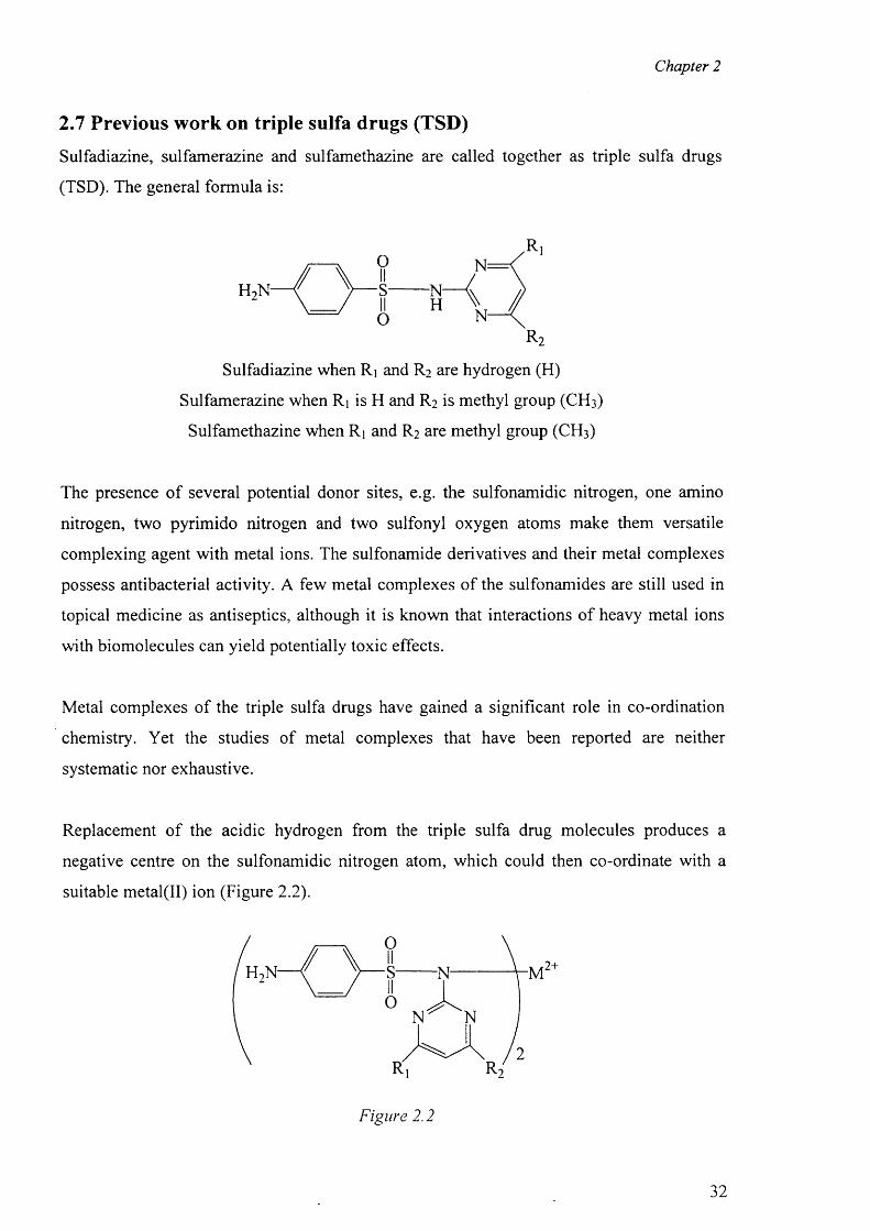
2.7 Previous work on triple sulfa drugs (TSD)
Sulfadiazine, sulfamerazine and sulfamethazine are called together as triple sulfa drugs
(TSD). The general formula is:
The presence o f several potential donor sites, e.g. the sulfonamidic nitrogen, one amino
nitrogen, two pyrimido nitrogen and two sulfonyl oxygen atoms make them versatile
complexing agent with metal ions. The sulfonamide derivatives and their metal complexes
possess antibacterial activity. A few metal complexes o f the sulfonamides are still used in
topical medicine as antiseptics, although it is known that interactions o f heavy metal ions
with biomolecules can yield potentially toxic effects.
Metal complexes of the triple sulfa drugs have gained a significant role in co-ordination
chemistry. Yet the studies o f metal complexes that have been reported are neither
systematic nor exhaustive.
Replacement o f the acidic hydrogen from the triple sulfa drug molecules produces a
negative centre on the sulfonamidic nitrogen atom, which could then co-ordinate with a
suitable metal(II) ion (Figure 2.2).
r2
Sulfadiazine when Rj and R 2 are hydrogen (H)
Sulfamerazine when Rj is H and R 2 is methyl group (CH 3 )
Sulfamethazine when Rj and R2 are methyl group (CH 3 )
Figure 2.2
32
Chapter 2
Some authors reported the preparation of several transition metal complexes o f triple sulfa
drugs in different mediums such as methanol, alcohol, acetone, DMSO etc. Cu(II), Zn(II),
Cd(II) and Hg(II) complexes were prepared by mixing the methanolic solution o f metal
chloride or metal acetate and triple sulfa drugs in the molar ratio o f 1:2. A precipitate was
formed, stirred for six hours and the precipitate collected. Based on spectrophotometric
investigations those workers suggested that Cu(II), Zn(II), Cd(II) and Hg(II) form 1:2
complexes, while Ag(I) form 1:1 complexes. The molecular compositions o f the complexes
were proposed on the basis o f chemical analyses and spectrophotometric evidences.
It is to be noted that apart from the stereochemical interest o f metal complexes o f TSD,
there are also interesting for their clinical use. For example, the silver and zinc complexes
have already found application as topical drugs for bum healing . 1 1 ' 17
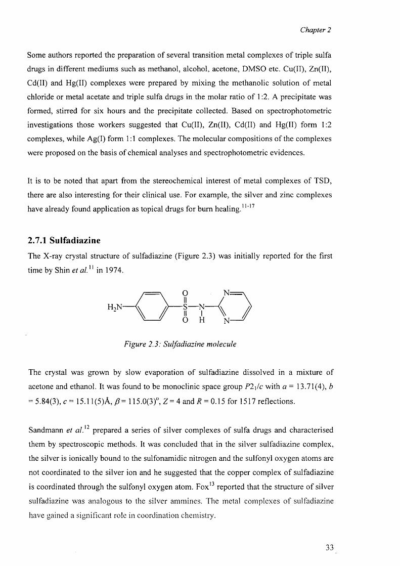
2.7.1 Sulfadiazine
The X-ray crystal structure of sulfadiazine (Figure 2.3) was initially reported for the first
time by Shin et al . 11 in 1974.
Figure 2.3: Sulfadiazine molecule
The crystal was grown by slow evaporation o f sulfadiazine dissolved in a mixture of
acetone and ethanol. It was found to be monoclinic space group P2\/c with a = 13.71(4), b
= 5.84(3), c = 15.1 l(5 )A ,/? = 115.0(3)°, Z = 4 and/? = 0.15 for 1517 reflections.
1 7Sandmann et al. prepared a series of silver complexes o f sulfa drugs and characterised
them by spectroscopic methods. It was concluded that in the silver sulfadiazine complex,
the silver is ionically bound to the sulfonamidic nitrogen and the sulfonyl oxygen atoms are
not coordinated to the silver ion and he suggested that the copper complex o f sulfadiazine1 7is coordinated through the sulfonyl oxygen atom. Fox reported that the structure o f silver
sulfadiazine was analogous to the silver ammines. The metal complexes o f sulfadiazine
have gained a significant role in coordination chemistry.
33
Chapter 2
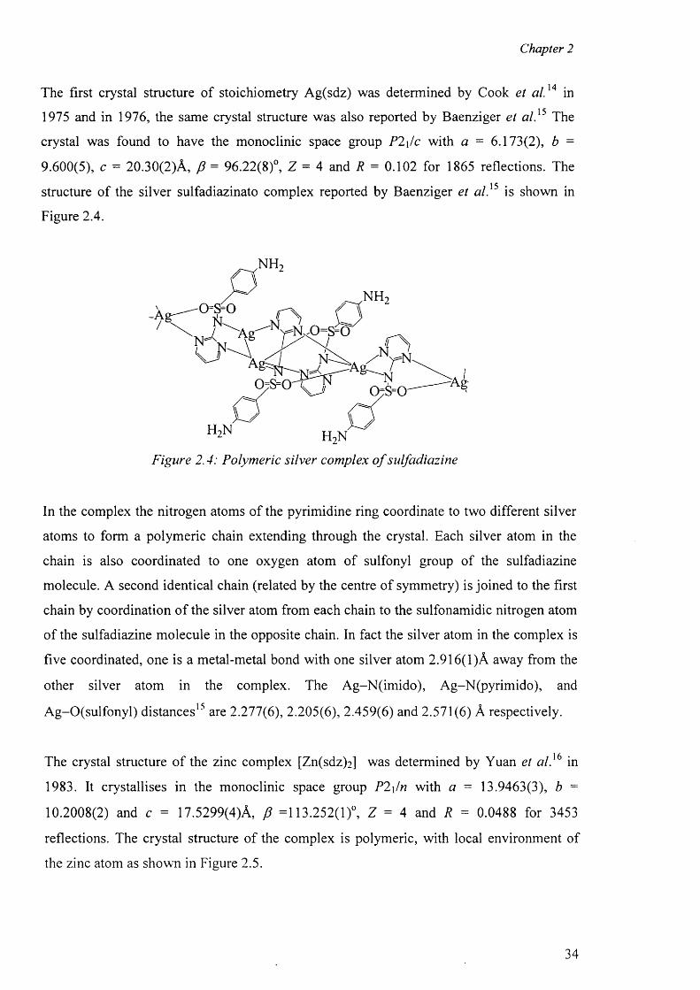
The first crystal structure of stoichiometry Ag(sdz) was determined by Cook et a l.14 in
1975 and in 1976, the same crystal structure was also reported by Baenziger et a l}5 The
crystal was found to have the monoclinic space group P2\/c with a = 6.173(2), b =
9.600(5), c = 20.30(2)A, f i = 96.22(8)°, Z = 4 and R = 0.102 for 1865 reflections. The
structure o f the silver sulfadiazinato complex reported by Baenziger et a l}5 is shown in
Figure 2.4.
Figure 2.4: Polymeric silver complex o f sulfadiazine
In the complex the nitrogen atoms of the pyrimidine ring coordinate to two different silver
atoms to form a polymeric chain extending through the crystal. Each silver atom in the
chain is also coordinated to one oxygen atom of sulfonyl group of the sulfadiazine
molecule. A second identical chain (related by the centre of symmetry) is joined to the first
chain by coordination o f the silver atom from each chain to the sulfonamidic nitrogen atom
of the sulfadiazine molecule in the opposite chain. In fact the silver atom in the complex is
five coordinated, one is a metal-metal bond with one silver atom 2.916(1 )A away from the
other silver atom in the complex. The Ag-N(im ido), Ag-N(pyrimido), and
Ag-O(sulfonyl) distances1 5 are 2.277(6), 2.205(6), 2.459(6) and 2.571(6) A respectively.
The crystal structure o f the zinc complex [Zn(sdz)2 ] was determined by Yuan et a l} 6 in
1983. It crystallises in the monoclinic space group P2\/n with a = 13.9463(3), b =
10.2008(2) and c = 17.5299(4)A, f i =113.252(1)°, Z = 4 and R = 0.0488 for 3453
reflections. The crystal structure o f the complex is polymeric, with local environment of
the zinc atom as shown in Figure 2.5.
34
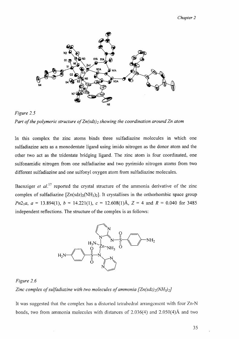
Chapter 2
Figure 2.5
Part o f the polymeric structure ofZn(sdz ) 2 showing the coordination around Zn atom
In this complex the zinc atoms binds three sulfadiazine molecules in which one
sulfadiazine acts as a monodentate ligand using imido nitrogen as the donor atom and the
other two act as the tridentate bridging ligand. The zinc atom is four coordinated, one
sulfonamidic nitrogen from one sulfadiazine and two pyrimido nitrogen atoms from two
different sulfadiazine and one sulfonyl oxygen atom from sulfadiazine molecules.
Baenziger et a l , 1 7 reported the crystal structure o f the ammonia derivative o f the zinc
complex of sulfadiazine [Zn(sdz)2 (NH 3 )2 ]. It crystallises in the orthorhombic space group
Pn2xa, a = 13.894(1), b = 14.221(1), c = 12.608(1)A, Z = 4 and R = 0.040 for 3485
independent reflections. The structure o f the complex is as follows:
-NH| N— S-
f n^NH3 0
■S—N.
Figure 2.6
Zinc complex o f sulfadiazine with two molecules o f ammonia [Zn(sdz)2 (N 1^3) 2]
It was suggested that the complex has a distorted tetrahedral arrangement with four Zn-N
bonds, two from ammonia molecules with distances o f 2.036(4) and 2.050(4)A and two
35
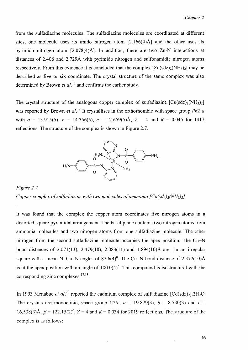
Chapter 2
from the sulfadiazine molecules. The sulfadiazine molecules are coordinated at different
sites, one molecule uses its imido nitrogen atom [2.166(4)A] and the other uses its
pyrimido nitrogen atom [2.078(4)A]. In addition, there are two Zn-N interactions at
distances o f 2.406 and 2.729A with pyrimido nitrogen and sulfonamidic nitrogen atoms
respectively. From this evidence it is concluded that the complex [Zn(sdz)2 (NH 3 )2 ] may be
described as five or six coordinate. The crystal structure o f the same complex was also1 8determined by Brown et al. and confirms the earlier study.
The crystal structure of the analogous copper complex of sulfadiazine [Cu(sdz)2 (NHs)2 ]
was reported by Brown et a l}9 It crystallises in the orthorhombic with space group Pn2\a
with a = 13.915(5), b = 14.356(5), c = 12.659(5)A, Z = 4 and R = 0.045 for 1417
reflections. The structure o f the complex is shown in Figure 2.7.
NH-Cu
•S— N.
Figure 2.7
Copper complex o f sulfadiazine with two molecules o f ammonia [Cu(sdz)2 (NHi) 2]
It was found that the complex the copper atom coordinates five nitrogen atoms in a
distorted square pyramidal arrangement. The basal plane contains two nitrogen atoms from
ammonia molecules and two nitrogen atoms from one sulfadiazine molecule. The other
nitrogen from the second sulfadiazine molecule occupies the apex position. The Cu-N
bond distances o f 2.071(13), 2.479(18), 2.083(11) and 1.894(10)A are in an irregular
square with a mean N -C u -N angles of 87.6(4)°. The C u-N bond distance of 2.377(10)A
is at the apex position with an angle o f 100.0(4)°. This compound is isostructural with the17 18corresponding zinc complexes. ’
90In 1993 Menabue et al. reported the cadmium complex o f sulfadiazine [Cd(sdz)2].2H20.
The crystals are monoclinic, space group C2/c, a = 19.879(3), b = 8.730(3) and c =
16.538(3)A, /?= 122.15(2)°, Z = 4 and R = 0.034 for 2019 reflections. The structure of the
complex is as follows:
36
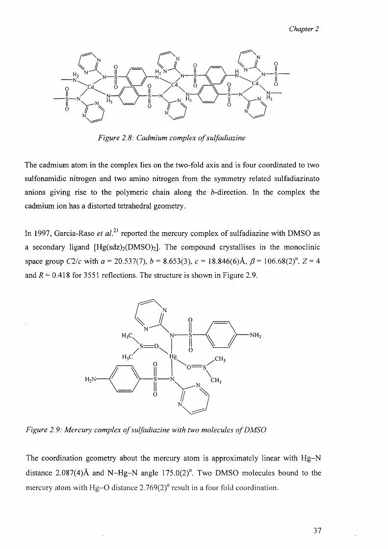
Chapter 2
Figure 2.8: Cadmium complex o f sulfadiazine
The cadmium atom in the complex lies on the two-fold axis and is four coordinated to two
sulfonamidic nitrogen and two amino nitrogen from the symmetry related sulfadiazinato
anions giving rise to the polymeric chain along the 6-direction. In the complex the
cadmium ion has a distorted tetrahedral geometry.
* 01In 1997, Garcia-Raso et al. reported the mercury complex o f sulfadiazine with DMSO as
a secondary ligand [Hg(sdz)2 (DMSO)2 ]. The compound crystallises in the monoclinic
space group C2/c with a = 20.537(7), b = 8.653(3), c = 18.846(6)A, f = 106.68(2)°, Z = 4
and R = 0.418 for 3551 reflections. The structure is shown in Figure 2.9.
Figure 2.9: Mercury complex o f sulfadiazine with two molecules o f DMSO
The coordination geometry about the mercury atom is approximately linear with H g-N
distance 2.087(4)A and N -H g-N angle 175.0(2)°. Two DMSO molecules bound to the
mercury atom with H g -0 distance 2.769(2)° result in a four fold coordination.
37
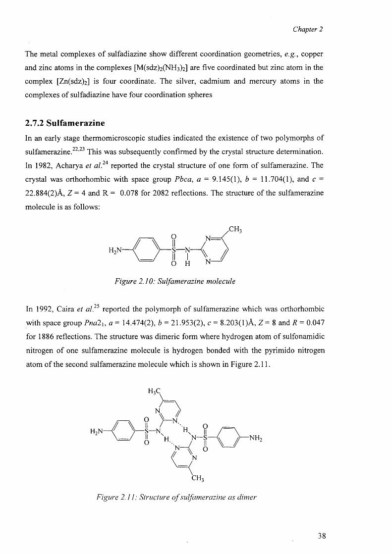
Chapter 2
The metal complexes o f sulfadiazine show different coordination geometries, e.g., copper
and zinc atoms in the complexes [M(sdz)2 (NH 3 )2 ] are five coordinated but zinc atom in the
complex [Zn(sdz)2 ] is four coordinate. The silver, cadmium and mercury atoms in the
complexes of sulfadiazine have four coordination spheres
2.7.2 Sulfamerazine
In an early stage thermomicroscopic studies indicated the existence o f two polymorphs of
sulfamerazine.22,23 This was subsequently confirmed by the crystal structure determination.
In 1982, Acharya et al.24 reported the crystal structure o f one form of sulfamerazine. The
crystal was orthorhombic with space group Pbca, a = 9.145(1), b = 11.704(1), and c =
22.884(2)A, Z = 4 and R = 0.078 for 2082 reflections. The structure of the sulfamerazine
molecule is as follows:
S— N
O H
Figure 2.10: Sulfamerazine molecule
In 1992, Caira et al.25 reported the polymorph of sulfamerazine which was orthorhombic
with space group Pna2 \, a = 14.474(2), b = 21.953(2), c = 8.203(1)A, Z = 8 and R = 0.047
for 1886 reflections. The structure was dimeric form where hydrogen atom of sulfonamidic
nitrogen o f one sulfamerazine molecule is hydrogen bonded with the pyrimido nitrogen
atom o f the second sulfamerazine molecule which is shown in Figure 2.11.
CH3
Figure 2.11: Structure o f sulfamerazine as dimer
38
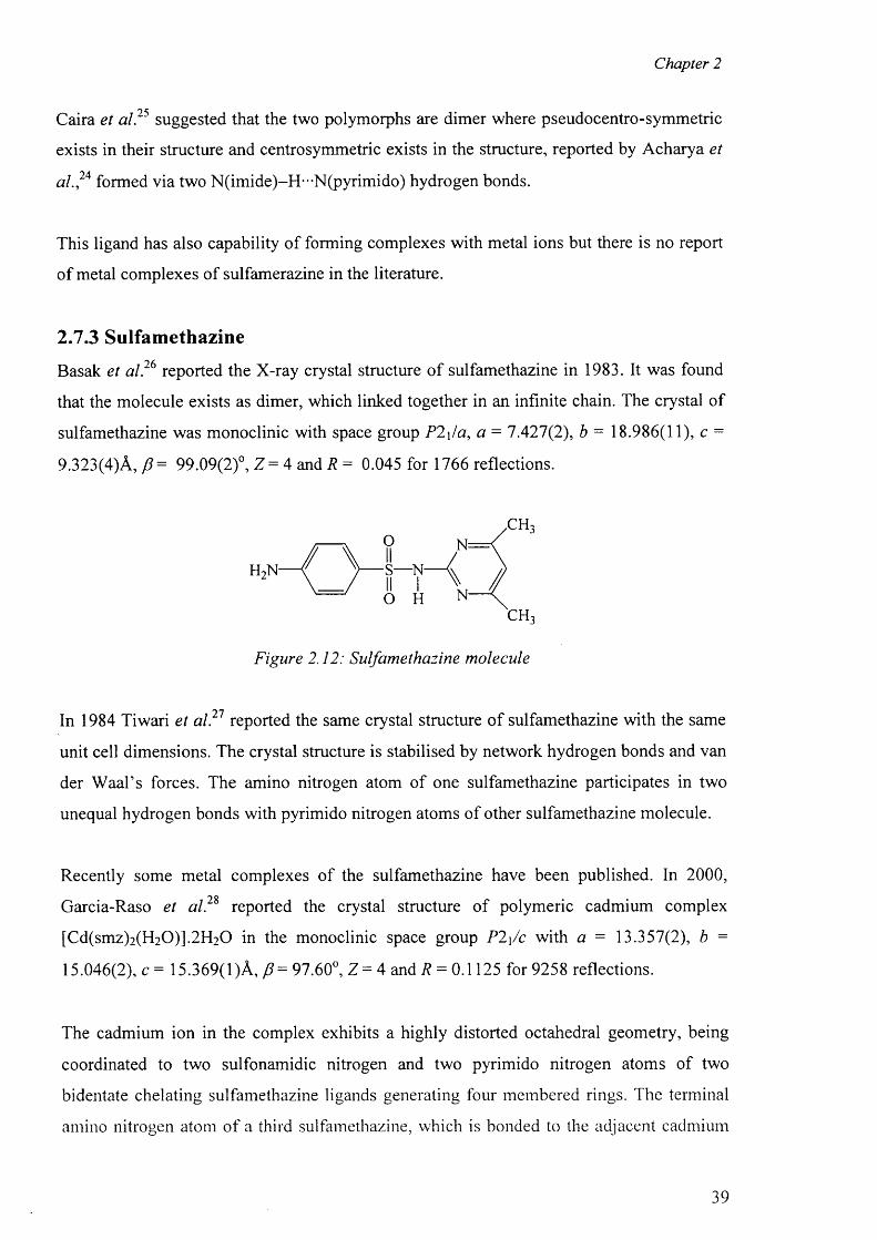
Chapter 2
Caira et a l? 5 suggested that the two polymorphs are dimer where pseudocentro-symmetric
exists in their structure and centrosymmetric exists in the structure, reported by Acharya et
al.?A formed via two N(imide)-H-"N(pyrimido) hydrogen bonds.
This ligand has also capability o f forming complexes with metal ions but there is no report
o f metal complexes of sulfamerazine in the literature.
2.7.3 Sulfamethazine
Basak et a l? 6 reported the X-ray crystal structure o f sulfamethazine in 1983. It was found
that the molecule exists as dimer, which linked together in an infinite chain. The crystal of
sulfamethazine was monoclinic with space group P2\/a, a = 7.427(2), b = 18.986(11), c =
9.323(4)A, p = 99.09(2)°, Z = 4 and R = 0.045 for 1766 reflections.
H,N S— N
Figure 2.12: Sulfamethazine molecule
In 1984 Tiwari et a l? 1 reported the same crystal structure o f sulfamethazine with the same
unit cell dimensions. The crystal structure is stabilised by network hydrogen bonds and van
der W aal’s forces. The amino nitrogen atom of one sulfamethazine participates in two
unequal hydrogen bonds with pyrimido nitrogen atoms of other sulfamethazine molecule.
Recently some metal complexes of the sulfamethazine have been published. In 2000,
Garcia-Raso et a l? % reported the crystal structure of polymeric cadmium complex
[Cd(smz)2 (H2 0 )].2 H2 0 in the monoclinic space group P2\/c with a = 13.357(2), b =
15.046(2), c = 15.369(1 )A, = 97.60°, Z = 4 and R = 0.1125 for 9258 reflections.
The cadmium ion in the complex exhibits a highly distorted octahedral geometry, being
coordinated to two sulfonamidic nitrogen and two pyrimido nitrogen atoms of two
bidentate chelating sulfamethazine ligands generating four membered rings. The terminal
amino nitrogen atom o f a third sulfamethazine, which is bonded to the adjacent cadmium
39
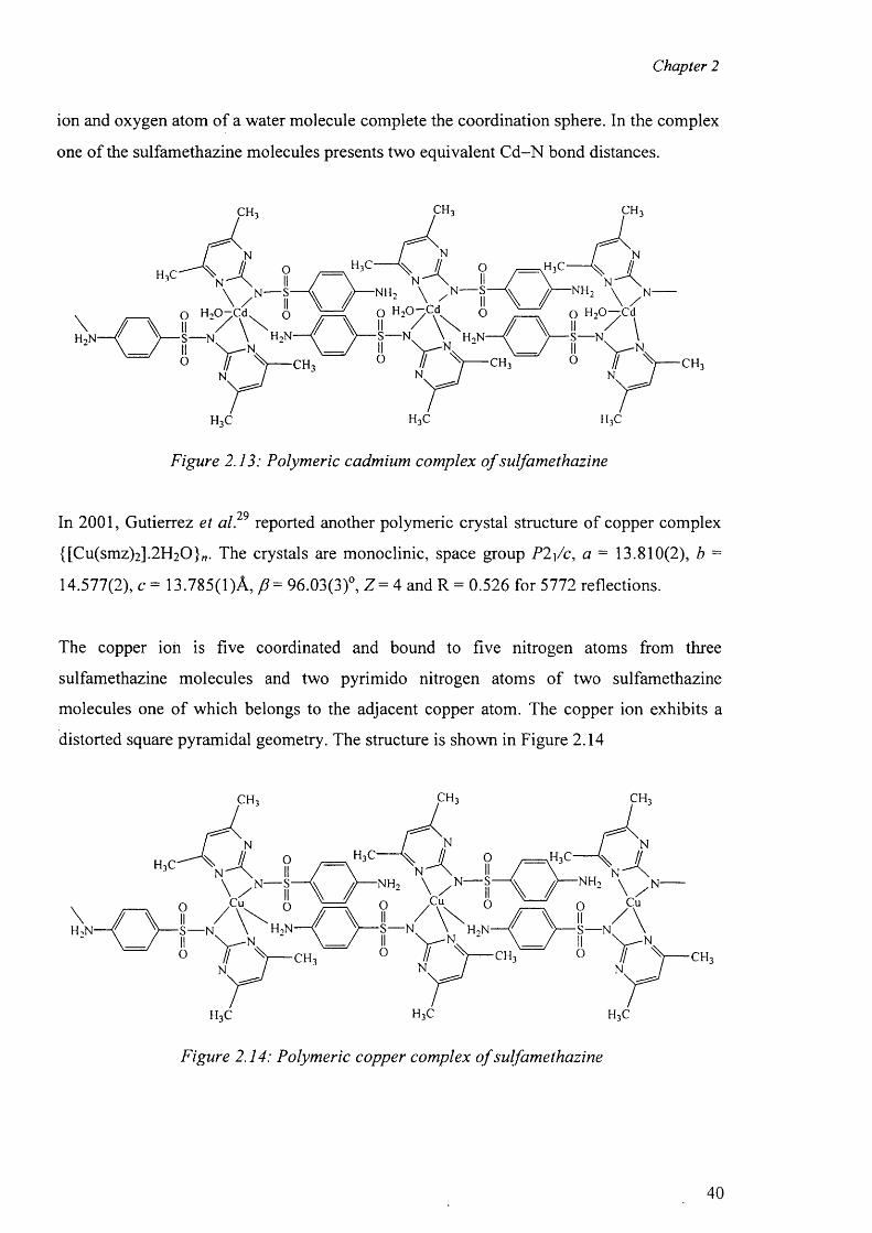
Chapter 2
ion and oxygen atom of a water molecule complete the coordination sphere. In the complex
one of the sulfamethazine molecules presents two equivalent C d-N bond distances.
h 3c h 3c h 3c
Figure 2.13: Polymeric cadmium complex o f sulfamethazine
• 7QIn 2001, Gutierrez et al. reported another polymeric crystal structure o f copper complex
{[Cu(smz)2 ].2 H 2 0 }„. The crystals are monoclinic, space group P2\/c, a = 13.810(2), b =
14.577(2), c = 13.785(1 )A, /?= 96.03(3)°, Z = 4 and R = 0.526 for 5772 reflections.
The copper ion is five coordinated and bound to five nitrogen atoms from three
sulfamethazine molecules and two pyrimido nitrogen atoms o f two sulfamethazine
molecules one o f which belongs to the adjacent copper atom. The copper ion exhibits a
distorted square pyramidal geometry. The structure is shown in Figure 2.14
h 3c h 3c h 3c
Figure 2.14: Polymeric copper complex o f sulfamethazine
40
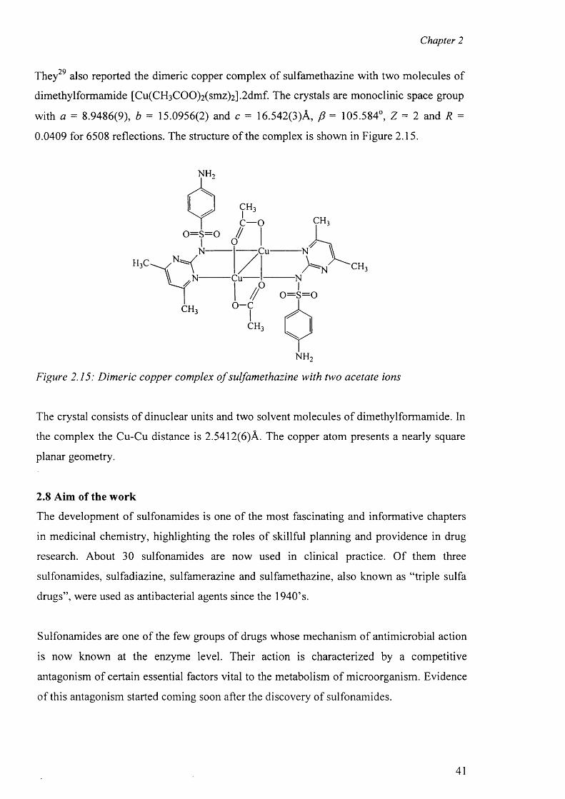
Chapter 2
They also reported the dimeric copper complex o f sulfamethazine with two molecules of
dimethylformamide [Cu(CH3 COO)2 (smz)2 ].2 dmf. The crystals are monoclinic space group
with a = 8.9486(9), b = 15.0956(2) and c = 16.542(3)A, p = 105.584°, Z = 2 and R =
0.0409 for 6508 reflections. The structure o f the complex is shown in Figure 2.15.
X T U
n h 2
Figure 2.15: Dimeric copper complex o f sulfamethazine with two acetate ions
The crystal consists o f dinuclear units and two solvent molecules o f dimethylformamide. In
the complex the Cu-Cu distance is 2.5412(6)A. The copper atom presents a nearly square
planar geometry.
2.8 Aim of the work
The development o f sulfonamides is one o f the most fascinating and informative chapters
in medicinal chemistry, highlighting the roles o f skillful planning and providence in drug
research. About 30 sulfonamides are now used in clinical practice. O f them three
sulfonamides, sulfadiazine, sulfamerazine and sulfamethazine, also known as “triple sulfa
drugs”, were used as antibacterial agents since the 1940’s.
Sulfonamides are one of the few groups o f drugs whose mechanism of antimicrobial action
is now known at the enzyme level. Their action is characterized by a competitive
antagonism of certain essential factors vital to the metabolism of microorganism. Evidence
o f this antagonism started coming soon after the discovery of sulfonamides.
41
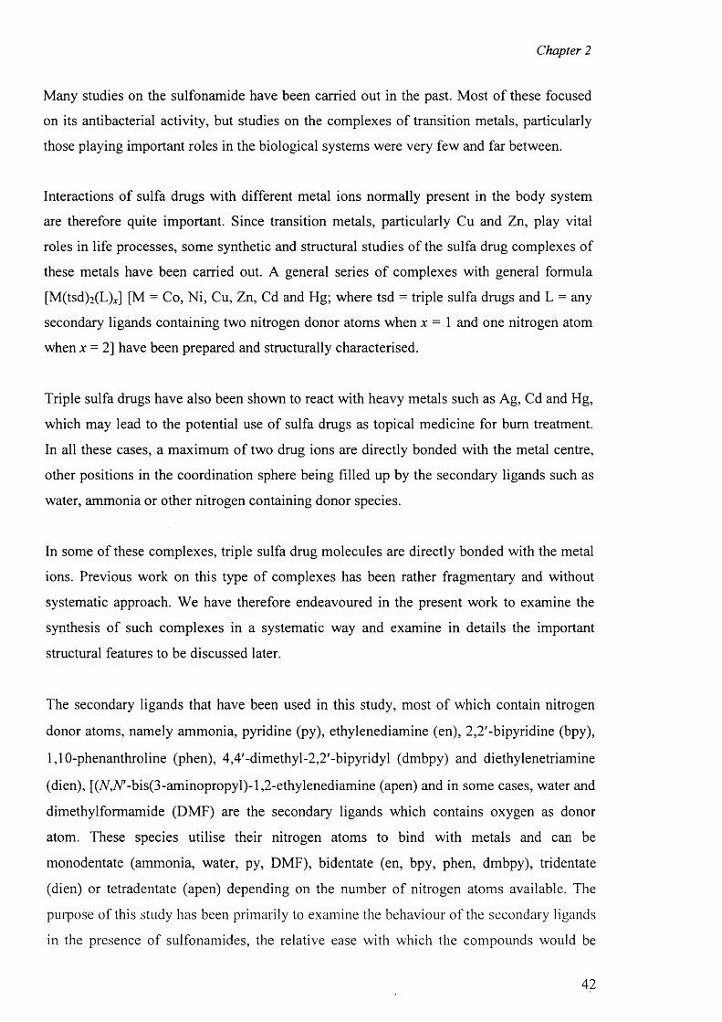
Chapter 2
Many studies on the sulfonamide have been carried out in the past. Most o f these focused
on its antibacterial activity, but studies on the complexes o f transition metals, particularly
those playing important roles in the biological systems were very few and far between.
Interactions of sulfa drugs with different metal ions normally present in the body system
are therefore quite important. Since transition metals, particularly Cu and Zn, play vital
roles in life processes, some synthetic and structural studies o f the sulfa drug complexes of
these metals have been carried out. A general series o f complexes with general formula
[M(tsd)2 (L )J [M = Co, Ni, Cu, Zn, Cd and Hg; where tsd = triple sulfa drugs and L = any
secondary ligands containing two nitrogen donor atoms when x = \ and one nitrogen atom
when x = 2 ] have been prepared and structurally characterised.
Triple sulfa drugs have also been shown to react with heavy metals such as Ag, Cd and Hg,
which may lead to the potential use o f sulfa drugs as topical medicine for bum treatment.
In all these cases, a maximum of two drug ions are directly bonded with the metal centre,
other positions in the coordination sphere being filled up by the secondary ligands such as
water, ammonia or other nitrogen containing donor species.
In some of these complexes, triple sulfa drug molecules are directly bonded with the metal
ions. Previous work on this type o f complexes has been rather fragmentary and without
systematic approach. We have therefore endeavoured in the present work to examine the
synthesis of such complexes in a systematic way and examine in details the important
structural features to be discussed later.
The secondary ligands that have been used in this study, most o f which contain nitrogen
donor atoms, namely ammonia, pyridine (py), ethylenediamine (en), 2 ,2 '-bipyridine (bpy),
1,10-phenanthroline (phen), 4,4'-dimethyl-2,2'-bipyridyl (dmbpy) and diethylenetriamine
(dien), [(ACV'-bis(3-aminopropyl)-l,2-ethylenediamine (apen) and in some cases, water and
dimethylformamide (DMF) are the secondary ligands which contains oxygen as donor
atom. These species utilise their nitrogen atoms to bind with metals and can be
monodentate (ammonia, water, py, DMF), bidentate (en, bpy, phen, dmbpy), tridentate
(dien) or tetradentate (apen) depending on the number of nitrogen atoms available. The
purpose of this study has been primarily to examine the behaviour o f the secondary ligands
in the presence of sulfonamides, the relative ease with which the compounds would be
42
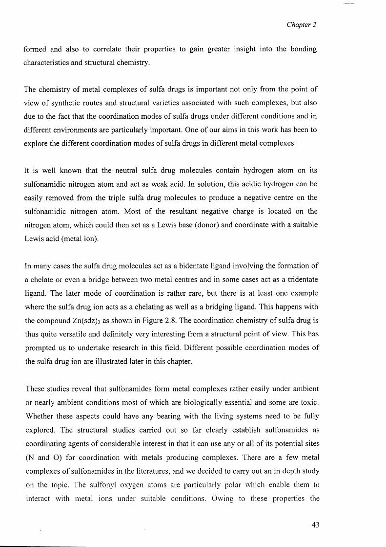
Chapter 2
formed and also to correlate their properties to gain greater insight into the bonding
characteristics and structural chemistry.
The chemistry of metal complexes of sulfa drugs is important not only from the point of
view of synthetic routes and structural varieties associated with such complexes, but also
due to the fact that the coordination modes of sulfa drugs under different conditions and in
different environments are particularly important. One o f our aims in this work has been to
explore the different coordination modes o f sulfa drugs in different metal complexes.
It is well known that the neutral sulfa drug molecules contain hydrogen atom on its
sulfonamidic nitrogen atom and act as weak acid. In solution, this acidic hydrogen can be
easily removed from the triple sulfa drug molecules to produce a negative centre on the
sulfonamidic nitrogen atom. Most o f the resultant negative charge is located on the
nitrogen atom, which could then act as a Lewis base (donor) and coordinate with a suitable
Lewis acid (metal ion).
In many cases the sulfa drug molecules act as a bidentate ligand involving the formation of
a chelate or even a bridge between two metal centres and in some cases act as a tridentate
ligand. The later mode of coordination is rather rare, but there is at least one example
where the sulfa drug ion acts as a chelating as well as a bridging ligand. This happens with
the compound Zn(sdz ) 2 as shown in Figure 2.8. The coordination chemistry of sulfa drug is
thus quite versatile and definitely very interesting from a structural point o f view. This has
prompted us to undertake research in this field. Different possible coordination modes of
the sulfa drug ion are illustrated later in this chapter.
These studies reveal that sulfonamides form metal complexes rather easily under ambient
or nearly ambient conditions most of which are biologically essential and some are toxic.
Whether these aspects could have any bearing with the living systems need to be fully
explored. The structural studies carried out so far clearly establish sulfonamides as
coordinating agents o f considerable interest in that it can use any or all o f its potential sites
(N and O) for coordination with metals producing complexes. There are a few metal
complexes o f sulfonamides in the literatures, and we decided to carry out an in depth study
on the topic. The sulfonyl oxygen atoms are particularly polar which enable them to
interact with metal ions under suitable conditions. Owing to these properties the
43
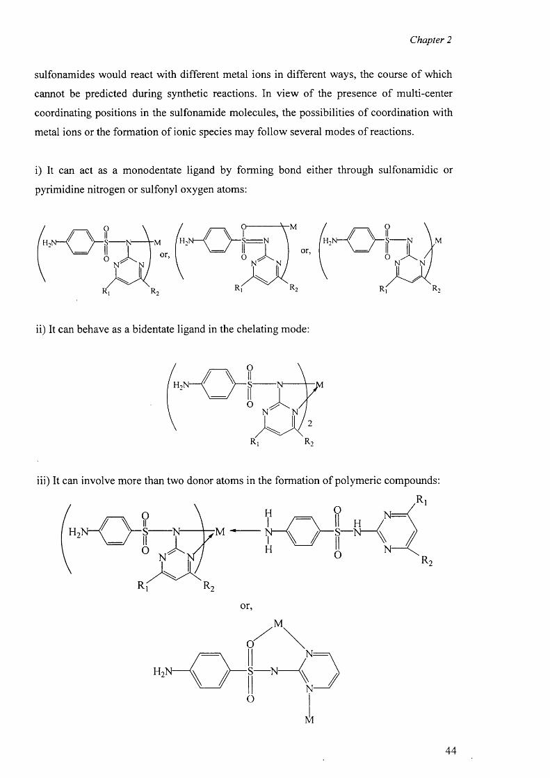
Chapter 2
sulfonamides would react with different metal ions in different ways, the course o f which
cannot be predicted during synthetic reactions. In view of the presence o f multi-center
coordinating positions in the sulfonamide molecules, the possibilities o f coordination with
metal ions or the formation of ionic species may follow several modes o f reactions.
i) It can act as a monodentate ligand by forming bond either through sulfonamidic or
pyrimidine nitrogen or sulfonyl oxygen atoms:
M
ii) It can behave as a bidentate ligand in the chelating mode:
H-
o
■s N-
O ^ /IN N
R,
2
r 2
iii) It can involve more than two donor atoms in the formation o f polymeric compounds:
R i
S— N
H,N-
or,
M
O
■s N-
O
N=
M
44
Chapter 2
The presence of secondary ligand such as ammonia could produce a variety o f metal
complexes with interesting stereochemistry and properties.
It has been mentioned earlier that the deprotonation o f neutral sulfa drug molecules leave
the negative charge (-) on the sulfonamidic nitrogen atom and this is the main reason why
the nitrogen site is preferred when bonding with metal ions. However, since the pyrimidine
nitrogen and sulfonyl oxygen atoms are also found to be involved in bonding with the
metal ions, it is very likely that the negative charge or a fraction o f it is shifted away from
the nitrogen atom along the ring bonds to the oxygen atoms. In addition to the synthetic
and structural work, we carried out theoretical calculations to investigate the charge
distribution in the sulfa drug ions and in the neutral sulfa drug, to find out if the above
assumption is true.
Finally, the roles o f weak intermolecular forces particularly the hydrogen bonds in the
formation of packing diagram and determining the properties o f materials are now well
recognised, especially in organic chemistry. One o f our particular aims in this work has
been to investigate the nature o f these interactions both within and between different
complex “molecules” that we have studied by X-ray crystallography.
45
Chapter 2
REFERENCES
1. L. A. Cates; “Sulfa Drugs in Handbook o f Chemotherapeutic Agents, Vol. 1”, M. Verderame ed., CRC Press, Boca Raton, 1986, 1
2. W. J. Blanchflower and D. A. Rice; J. Assoc. Anal. Chem., 1988, 71, 302
3. B. M. Kagan, “Antimicrobial Therapy”, W.B. Saunders, Philadelphia, 1974
4. N. Littlefield; Technical Report, National Center fo r Toxicological Research, Jeffersons, A. R., 1988
5. P. H. Long and E. A. Bliss; “The Clinical and Experimental Use o f Sulfanilamide, Sulfapyridine, and A llied Compounds'’’, The Macmillan Co., New York, 1939
6 . H. Hoerlein; Proc. Roy. Soc. Med., 1935, 29, 313
7. G. Domagk; Deutsche Med. Wehnschr., 1935, 61, 250
8 . J. Trefouel, Mme. Trefouel, F. Nittie and D. Bovet; C. R. Soc. Biol., 1935,120, 756
9. L. H. Sophian, D. L. Piper and G. H. Schneller, “The Sulfapyrimidines”, A. Colish, New York, 1952
10. H. F. Chambers and E. Jawetz; “Sulfonamides, Trimethoprim and Quinolones, in Basic and Clinical Pharmacology”, (Katzung, B.G., ed) Appleton-Lange, 1998, pp. 761-763
11. H. S. Shin, G. S. Ihn, H. S. Kim and C. H. Koo; J. Korean Chem. Soc., 1974, 18, 329
12. B. J. Sandmann, R. U. Nesbitt and R. H. Sandmann; J. Pharm. Sci., 1974, 63, 1948
13. C. L Fox, Jr., U.S.P. 1971, 1, 240, 545
14. D. S. Cook and M. F. Turner; J. Chem. Soc. Perkin Trans., 1975, 2, 1021
15. N. C. Baenziger and A. W. Struss; Inorg. Chem., 1976, 15, 1807
16. R. -X . Yuan, R. -G . Xiong, Z. -F . Chen, P. Zhang, H. -X . Ju, Z. Dai, Z. -J . Guo, H. -K . Fun and X. -Z . You; J. Chem. Soc. Dalton Trans., 2001, 774
17. N. C. Baenziger; Acta Cryst., 1983, C39, 1620
18. C. J. Brown, D. S. Cook and L. Sengier; Acta Cryst., 1987, C43, 2332
19. C. J. Brown, D. S. Cook and L. Sengier; Acta Cryst., 1985, C41, 718
20. L. Menabue and M. Saladini; J. Inorg. Biochem., 1993, 49, 201
21. A. Garcia-Raso, J. J. Fiol, G. Martorell, A. Lopez-Zafra and M. Quiros; Polyhedron, 1997, 16,613
22. M. Kuhnert-Brandstatter and S. Wunsch; Mikrochim. Acta, 1969, 6 , 1308
46
Chapter 2
23. M. Kuhnert-Brandstatter; Thermomicroscopy in the analysis o f Pharmaceuticals, Oxford: Pergamon Press, 1971, 34
24. K. R. Acharya, K.N. Kuchela and G. Kartha; J. Cryst. Spect. Res., 1982,12, 369
25. M. R. Caira and R. Mohamed; Acta Cryst., 1992, B48, 492
26. A. K. Basak, S. K. Mazumdar and S. Chaudhuri; Acta Crsyt., 1983, C39, 492
27. R. K. Tiwari, M. Haridas and T. P. Sing; Acta Cryst., 1984, C40, 655
28. A. Garcia-Raso, J. J. Fiol, S. Rigo, A. Lopez-Lopez, E. Molins, E. Espinosa, E. Borras, G. Alzuet, J. Borras and A. Castineiras; Polyhedron , 2000, 19, 991
29. L. Gutierrez, G. Alzuet, J. Borras, A. Castineiras; A. Rodriguez-Fortea and E. Ruiz; Inorg. Chem., 2001, 40, 3089
47

EXPERIMENTAL-PREPARA TION OF METAL COMPLEXES OF TRIPLE SULFA DRUGS WITH
SECONDAR Y LIGANDS
Chapter 3
EXPERIMENTAL
3.1 Chemicals and their purifications
The Analar grade chemicals (SIGMA and BDH) were purchased and used as received and
the solvents acetone, methanol, dimethylformamide and dimethylsulfoxide were also used
as received throughout the experiments.
3.2 Physical measurements
3.2.1 Elemental analysis
C, H and N were estimated microanalytically using a Perkin-Elmer 2400 CHN elemental
analyzer.
3.2.2 IR studies
The IR spectra were recorded on a Nicolet 510 FTIR spectrometer, each compound was
pressed into a KBr disk that was prepared in a sample holder by compressing a powdered
sample with a ten-fold excess of KBr, which was preheated in an oven before use.
3.2.3 Electron Paramagnetic Resonance
ENDOR (Electron Nuclear Double resonance) spectroscopy is an EMR (Electron Magnetic
Resonance) technique that is often described as EPR-Detected NMR. EPR and ENDOR
spectra recorded on a Bruker ESP 300E series spectrometer fitted with a Bruker ESP 360
DICE ENDOR system and an Oxford Instruments liquid helium variable temperature
system. X-band EPR and ENDOR spectra were recorded using a Bruker EN 801 ENDOR
cavity operating at a modulation frequency o f 12.5 kHz in both cases and the ENDOR
spectra were obtained using an ENI A-300 RF power amplifier operating at an attenuation
of 8 dB. Accurate g values were obtained at X-band using a Bruker ER 035 M NMR
gaussmeter calibrated to the perylene radical cation in concentrated H2 SO 4 , g = 2.002569.
Q-band EPR and ENDOR spectra were obtained using a Bruker ER 5106 QTE ENDOR
cavity operating at a modulation frequency of 100 kHz for the EPR experiments and 12.5
48
Chapter 3
kHz for the ENDOR. The ENDOR spectra were recorded using an ENI ER 3200L RF
power amplifier operating at a total attenuation o f 13 dB.
3.2.4 'H NMR and ,3C NMR spectra
NMR spectra were recorded on a Bruker Avance AMX-400 spectrometer. H and C
NMR spectra were obtained from a solution of the compounds in d6 -DMSO. The chemical
shifts are expressed in ppm relative to internal TMS.
3.2.5 X-ray crystallography
The crystallographic measurements for the complexes were made at 150 K on a Bruker
Nonius Kappa CCD area detector using graphite monochromatised Mo K a radiation, X —
0.71073 A. The structures were solved by direct methods (SHELXS-97) and refined on F 2
by full-matrix least squares (SHELXL-97) using all unique data and parameters. The
hydrogen atoms on the amino groups o f sulfa drugs in the complexes and water molecules
(where present) were located from the difference Fourier maps and refined freely.
3.3 Synthetic Procedure
The complexes were synthesized by the following simple reaction procedures: the sodium
salt o f sulfadiazine (Nasdz), sulfamerazine (Hsmr) and the sodium salt o f sulfamethazine
(Nasmz) were dissolved in 50 mL of hot methanol and the methanolic solution of the
respective metal salts were added slowly with constant stirring on a hot plate, a precipitate
was formed and the mixture was continued to stir for six hours. The precipitate was filtered
and dried over silica gel. The complexes of transition metals were colored whereas those of
zinc, cadmium and mercury were white powders.
Reaction scheme:
M2+ + 2(Na/Htsd)------------> M(tsd) 2 + 2Na+/2H+
[where M is any divalent metal ion and Htsd any sulfa drugs]
The precipitate was used for further reaction with secondary ligands in DMF solution to
obtain the single crystals for the complexes (3) to (28).
M(tsd ) 2 + x h ------------> M(tsd)2 (L)*
Where L is any secondary ligand, bidentate when x = 1 and monodentate when x = 2.
49
Chapter 3
All the complexes are stable in air and at room temperature. The complexes are insoluble
in water, alcohol and most organic solvents but soluble in DMF and DMSO.
We used the following metal salts:
1) Cobalt chloride (C0 CI2 .6 H2 O) (Pink precipitate)
2) Nickel chloride (NiCl2 .6 H2 0 ) (Light blue precipitate)
3) Copper chloride (CUCI2 .6 H 2 O) (Red precipitate)
4) Zinc chloride (ZnCh.bFLO) (White precipitate)
5) Cadmium acetate [(CH3 C 0 0 )2 Cd.6 H2 0 ] (White precipitate)
6 ) Mercury acetate [(CHaCOO^Hg.bFhO] (White precipitate)
These precipitates were dissolved in DMF/DMSO and a secondary ligand was added,
stirred for 30 minutes, filtered and the filtrate was left for crystallization.
3.4 Syntheses of the complexes
3.4.1 Preparation of the crystal of sulfadiazine (1)
Solid sulfadiazine (0.563 g, 1 mmol) was dissolved in a minimum amount o f DMF and
stirred for 30 minutes. It was filtered and left for crystallisation. After three days white
block crystals were obtained. IR: (KBr disc), cm-1, 3461(s), 3365(s), 3125(s), 2956(sh),
1625(vs), 1598, 1570, 1507(vs), 1465(sh), 1408, 1351, 1325(m)„ 1306, 1259, 1155(vs),
1125(s), 1106, 1093(s), 1045, 1005(s), 945(vs), 881(vw), 796(vs), 746, 680(s), 656(m),
633(vs), 589, 535(vs). ‘H N M R (DMSO-d6): 5 11.35s [1H, N (ll) ] , 8.49d [1H, C(12/14), J
= 11.7Hz], 6.59d [H, C(17/19), J = 8.7], 7.65d [1H, C(16/20), J = 8 .6 ], 6.99t [1H, C(13), J
= 4.8], 6.00s [2H (-N H 2)]. 13C NMR (DMSO-d6)\ 6 157.5 [Cl 1], 158.6 [C12/14], 153.4
[C18], 130.3 [C16/C20], 125.1 [C15], 115.9 [C13], 112.6 [C17/C19].
3.4.2 Preparation of the new polymorph of sulfamerazine (2)
Solid sulfamerazine (0.265g; 1 mmol) was dissolved in DMF (20 mL) and stirred for 30
minutes, filtered and the clear solution o f sulfamerazine was left for crystallization. White
block crystals o f sulfamerazine were obtained by slow evaporation o f the solution at room
temperature. IR: (KBr disc): vcm ”1, 3468(m), 3370(s), 3238, 3120(s), 1630(vs), 1570(vs),
1505(sh), 1446(sh), 1408(vs), 1372, 1343(m), 1331(vs), 1304(vs), 1245(sh), 1153(vs),
1107(s), 1093(s), 1040(s), 973, 965(vs), 889(vw), 836, 798(vw), 748(vs), 717(sh), 681(s),
50
Chapter 3
636(s), 616(vs), 579, 548(vs). 'H NMR (DMS0-<4): 6 11.20 [ 1 H, N ( ll) ] , 8.37d [ 1 H,
C(14), J = 4.8], 7.69d [2H, C(16)/C(20), J = 8.2], 6.95d [2H, C(17)/C(19), J = 4.9], 6.62d
[ 1 H, C(13), J = 8.2], 6.07s [2 H N(14)], 2.41s [3H (CH3)]. 13CNMR (DMSO-cfc): 5 168.0
[Cl 1], 157.5 [C12], 156.8 [C14], 152.9 [C18], 130.1 [C15], 124.9 [C16/20], 114.7
[C17/19], 112.1 [C13], 23.3 [CH3],
3.4.3 Preparation of the complex {[Co(smz)2(H20)].DMF}„ (3)
The DMF solution was filtered and left for crystallisation. Two weeks later, pink prismatic
crystals were obtained. It was filtered and dried over silica gel. The compound is soluble in
DMF and DMSO but insoluble in water and all other organic solvents. Anal. Found: C,
54.25, H, 3.40; N, 11.10%. Calc, for C3 2 H 3 0 C0 N 1 0O4 S2 : C, 54.20, H, 3.48; N, 11.15%. IR:
(KBr disc), c m '1, 3435(m), 3353(s), 3291(s), 2925, 2873, 1665(vs), 1598, 1581, 1561(vs),
1501 (sh), 1434(vs), 1395, 1373, 1344(m), 1301(vs), 1251(vs), 1178(sh), 1144(vs), 1126(s),
1081, 999(s), 974(vs), 878(vw), 835, 7 9 4 ,733(vs), 678(s), 636(s), 598, 557, 522(vs).
3.4.4 Preparation of the complex [Ni(en)3][sdz)2].H20 (4)
To the solution o f nickel precipitate o f sulfadiazine, ethylenediamine (en) (0.5 mL) was
added, stirred for 30 minutes. The solution was filtered and left for crystallisation. A few
days later, white block crystals were obtained. It was filtered and dried over silica gel. The
compound is soluble in DMF and DMSO but insoluble in water and all other organic
solvents. Anal. Found: C, 41.31, H, 5.90; N, 25.89%. Calc, for C2 6 H4 4 NiN]4 0 5 S2 : C, 41.29,
H, 5.87; N, 25.94%. IR: (KBr disc), cm-1, 3441(m), 3332(s), 3225(s), 1643(vs), 1596,
1581, 1552(vs), 1470(sh), 1448(sh), 1439(vs), 1414, 1348, 1313(m), 1298(vs), 1268(vs),
1181 (sh), 1161 (vs), 1134(s), 1102, 1091(s), 1083, 1061, 101 l(s), 975(vs), 840(vw), 828,
811, 806, 763(vs), 681(s), 651(m), 644(s), 626(vs), 584, 558, 524(vs).
3.4.5 Preparation of the complex [Ni(smr)2(py)2].4py (5)
To prepare this complex, the light blue powder nickel precipitate of sulfamerazine was
dissolved in pyridine and stirred for 30 minutes. The solution was filtered and left for
crystallisation. A week later, pink crystals were obtained. It was filtered and dried over
silica gel. The compound is soluble in DMF and DMSO but insoluble in water and all other
organic solvents. Anal. Found: C, 58.95, H, 4.90; N, 18.40. Calc, for C 5 2 Hs2 NiN]4 0 4 S2 : C,
58.93, H, 4.94; N, 18.5%. IR: (KBr disc), cm-1, 3438(m), 3349(s), 3223(s), 1632(vs),
1595(vs), 1551, 1516, 1501, 1434(sh), 141 l(sh), 1351 (vs), 1338(m), 1316, 1295(vs),
51
Chapter 3
1266(vs), 1227(sh), 1177(vs), 1131(s), 1103, 1091, 1083(s), 1011(s), 1001, 973(vs),
8 6 6 (vw), 852, 816, 801, 774(vw), 725(sh), 680(s), 645(m), 583(vs), 556(vs).
3.4.6 Preparation of the complex [Cu(en)2(sdz)2] (6)
To the DMF solution of copper precipitate o f sulfadiazine, ethylenediamine (en) (1 mL)
was added, stirred for 30 minutes. Five days later, blue block crystals were filtered and
dried over silica gel. The compound is soluble in DMF and DMSO but insoluble in water
and all other organic solvents. Anal. Found: C, 38.14, H, 4.54; N, 22.21%. Calc, for
C2 4 H3 4 CUN1 2O4 S2 : C, 38.12, H, 4.53; N, 22.24%. IR: (KBr disc), c m '1, 3439(m), 3341(s),
3229(s), 1641 (vs), 1599, 1584, 1554(vs), 1469(sh), 1446(sh), 1439(vs), 1413, 1347,
1312(m), 1297(vs), 1267(vs), 1178(sh), 1158(vs), 1131(s), 1102, 1091(s), 1081, 1058,
1008(s), 977(vs), 840(vw), 763(vs), 678(s), 651(m), 643(s), 625(vs), 583, 556, 523(vs).
3.4.7 Preparation of the complex [Cu(dien)(sdz)2] (7)
With the same procedure but diethylenetriamine (dien) (1 mmol) was added in the solution
and stirred for 30 minutes. After seven days, white block crystals were filtered and dried
over silica gel. The compound is soluble in DMF and DMSO but insoluble in water and all
other organic solvents. Anal. Found: C, 46.71, H, 5.75; N, 25.48%. Calc, for
C 2 8 H4 4 C 11N 1 4O4 S2 : C, 46.73, H, 5.73; N, 25.51%. IR: (KBr disc), cm-1, 3452(m), 3379(s),
3339(s), 3304(m), 3155(s), 1629(vs), 1598, 1578, 1537(vs), 1501(s), 1465(sh), 1417(vs),
1362, 1315, 1301(m), 1265(vs), 1240, 1221, 1173(sh), 1126(s), 1091(s), 1071, 1000(s),
968(vs), 835(vw), 809, 676(s), 661(m), 636(s), 574, 564, 528(vs).
3.4.8 Preparation of the complex [Cu(en)2(H20)2][smr)2] (8)
To the solution o f DMSO, a few drops o f ethylenediamine were added, stirred for 30
minutes. After seven days, blue prismatic crystals were obtained. It was filtered and dried
over silica gel. The compound is soluble in DMF and DMSO but insoluble in water,
alcohol and acetonitrile. Anal. Found: C, 41.84, H, 5.67; N, 22.52%. Calc, for
C 2 6 H4 2 CUN1 2O6 S2 : C, 41.81; H, 5.67; N, 22.56%. IR: (KBr disc): cm-1, 3523(m), 3387(s),
3331(s), 3174(s), 3275(s), 3174, 1662(vs), 1597(vs), 1576(vs), 1558(m), 1504(vs),
1458(sh), 1422(vs), 1400(m), 1365(s), 1317(m), 1294(sh), 1272(vs), 1228(m), 1178(m),
1122(sh), 1089(s), 1074(m), 1037(s), 1008(w), 994(m), 971(w), 898(s), 828(s), 810(s),
755(s), 719(vw), 680(w), 634(w), 625(vw), 591 (vs).
52
Chapter 3
3.4.9 Preparation of the complex [Cu2(en)4(H20)4][smr)2].H20 (9)
To the solution o f DMF, a few drops o f ethylenediamine were added, stirred for 30
minutes. After filtration it was left for crystallization. A week later, blue prismatic crystals
were obtained. It was filtered and dried over silica gel. The compound is soluble in DMF
and DMSO but insoluble in water, alcohol and acetonitrile. Anal. Found: C, 40.87, H, 5.75;
N, 22.93%. Calc, for C2 6 H 4 4 CUN1 2 O7 S2 : C, 40.83; H, 5.77; N, 21.99%. IR: (KBr disc):
c m '1, 3524(m), 3387(s), 3331(s), 3275(s), 3174(m), 1660(m), 1597(vs), 1576(vs),
1558(m), 1504(vs), 1454(sh), 1422(vs), 1394(m), 1363(s), 1318(m), 1294(sh), 1272(vs),
1228(m), 1178(m), 1122(sh), 1089(s), 1074(m), 1037(s), 1010(w), 996(m), 971(w), 898(s),
830(s), 807(s), 755(s), 719(vw), 680(w), 637(w), 621(vw), 592(vs), 555(vs).
3.4.10 Preparation of the complex [Cu2(smr)4].2DMF (10)
The DMF solution o f copper precipitate of sulfamerazine was left for crystallization. Ten
days later, deep green plate like crystals were obtained. The compound is soluble in DMF
and DMSO but insoluble in water, alcohol and acetonitrile. Anal. Found: C, 45.31, H, 4.40;
N, 18.97%. Calc, for CsoH sgC uN igO ^: C, 45.26; H, 4.42; N, 19.02%. IR: (KBr disc): v
cm"1, 3423(s), 3346(s), 3228(m), 1636(vs), 1597(vs), 1554(vs), 1502(vs), 1452(sh),
1413(vs), 1386(vs), 1319(sh), 1282(vs), 1205(m), 1177(m), 1131(sh), 1078(s), 1046(w),
971(w), 957(w), 872(w), 833, 811, 792(s), 779(vw), 716(vw), 682(s), 583(vs), 547(vs).
3.4.11 Preparation of the complex [Cu2(smr)4].2DMSO (11)
The DMSO solution o f the same precipitate was filtered and left for crystallization. Two
weeks later, deep green block crystals were obtained. The compound is soluble in DMF
and DMSO but insoluble in water, alcohol and acetonitrile. Anal. Found: C, 41.85, H, 4.60;
N, 15.00%. Calc, for C2 6 H 3 4 CuN 8 0 6 S4 : C, 41.89; H, 4.59; N, 15.06%. IR: (KBr disc): v
c m '1, 3420(s), 3341(s), 3232(m), 1639(vs), 1596(vs), 1552(vs), 1501(vs), 1450(sh),
1414(vs), 1383(vs), 1317(sh), 1281(vs), 1204(m), 1178(m), 1130(sh), 1076(s), 1045(w),
974(w), 953(w), 876(w), 830, 810, 789(s), 779(vw), 712(vw), 677(s), 589(vs).
3.4.12 Preparation of the complex [Cu(smz)2(apen)].3H2O.CH3OH (12)
To the DMSO solution o f copper precipitate o f sulfamethazine, a few drops of A,7V'(3-
aminopropyl)-bis-ethylenediamine (apen) were added and stirred for 30 minutes, filtered
and left for crystallization. After five days, blue prismatic crystals were obtained, filtered
53
Chapter 3
and dried over silica gel. The compound is soluble in DMF and DMSO but insoluble in
water, alcohol and acetonitrile. Anal. Found: C, 44.95, H, 6.75; N, 19.10%. Calc, for
C3 3 H 6 0 CUN1 2 O 8 S2 : C, 44.97; H, 6.81; N, 19.08%. IR: (KBr disc): cm-1, 3345(m), 3210(s),
2980(m), 1625(s), 1598(vs), 1570(vs), 1520(m), 1506(vs), 1483(sh), 1423(vs), 1388(m),
1350(s), 1315(m), 1294(sh), 1270(vs), 1214(m), 1182(m), 1120(sh), 1092(s), 1042(s),
1020(w), 982(m), 938(w), 905(s), 845(s), 812(s), 768(s), 692(w), 634(w), 625(vw).
3.4.13 Preparation of the complex {[Cu(smz)2.NH3].2H20 } w (13)
The copper precipitate o f sulfamethazine was dissolved in NH 3 -H2 O (1:1) and stirred for
30 minutes. A week later, green prismatic crystals were obtained. The compound is soluble
in water, DMF and DMSO. Anal. Found: C, 41.63, H, 4.65; N, 18.57. Calc, for
C2 4 H3 3 CUN9 O6 S2 : C, 42.94, H, 4.96; N, 18.78. IR: (KBr disc), cm"1, 3436(m), 3350(s),
3237(m), 1629(vs), 1597(vs), 1581(sh), 1560(m), 1501(s), 1434(m), 1397(m), 1373(m),
1322(m), 1300(m), 1250(m), 1210(s), 1184(vs), 1143(m), 1127(sh), 1081(sh), 999(sh),
975(vs), 881 (s), 832(m), 791(vw), 735(sh), 677(s), 639(s), 600(vs), 557(vs).
3.4.14 Preparation of the complex [Zn(smz)2(NH3)2] (14)
The white zinc precipitate of sulfamethazine was dissolved in 1:1 NH 3 :H2 0 solution and
stirred for 30 minutes. A week later, white block crystals were obtained. The compound is
soluble in DMF and DMSO but insoluble in water, alcohol and acetonitrile. Anal. Found:
C, 44.01; H, 4.95; N, 21.41%. Calc, for C2 4 H 3 2 Z11N 1 0O4 S2 : C, 44.03; H, 4.89; N, 21.40%.
IR: (KBr disc): cm"1, 3544(m), 3365(s), 3272(s), 3150(m), 1640(m), 1599(vs), 1557(m),
1501 (vs), 1434(sh), 1378(m), 1318(s), 1306(m), 1293(sh), 1244(vs), 1209(m), 1184(m),
1142(sh), 1117(s), 1081(m), 1050(s), 1010(w), 978(w), 885(s), 830(s), 794(s), 743(s),
677(w), 601 (vs), 548(vs). *H NMR (DMSO-<4): 5 7.58d [2H, C(16/20), J = 8.2 Hz], 6.42d
[2H, C(17/19), J = 8.1], 6.33s [1H, C(13)], 5.50s [2H (-N H 2)], 2.49s [6 H, CH3], 13C NMR
(DM SO-^): 6 167.5 [C(l 1)], 162.3 [C(12/14)], 151.2 [C(18)], 130.3 [C(l 5)], 129.8
[C( 16/20)], 111.9 [C (l7 /C l9)], 110.3 [C(13)],23.3 [CH3].
3.4.15 Preparation of the complex [Zn(smz)2(py)2].2py (15)
The same precipitate of the previous experiment was dissolved in a 1:10 C sH sN ^ O
solution and stirred for 30 minutes. Five days later, white prismatic crystals were obtained.
The compound is soluble in DMF and DMSO but insoluble in water, alcohol and
54
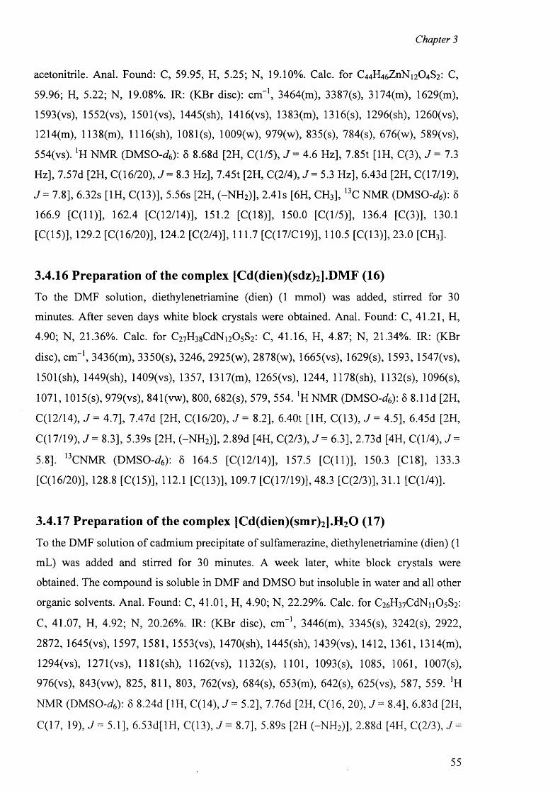
Chapter 3
acetonitrile. Anal. Found: C, 59.95, H, 5.25; N, 19.10%. Calc, for C4 4 H4 6 Z 11N 1 2O4 S2 : C,
59.96; H, 5.22; N, 19.08%. IR: (KBr disc): cm"1, 3464(m), 3387(s), 3174(m), 1629(m),
1593(vs), 1552(vs), 1501 (vs), 1445(sh), 1416(vs), 1383(m), 1316(s), 1296(sh), 1260(vs),
1214(m), 1138(m), 1116(sh), 1081(s), 1009(w), 979(w), 835(s), 784(s), 676(w), 589(vs),
554(vs). *H NMR (DMSO-<4): 6 8 .6 8 d [2H, C (l/5), J = 4.6 Hz], 7.85t [ 1 H, C(3), J = 7.3
Hz], 7.57d [2H, C(16/20), J = 8.3 Hz], 7.45t [2H, C(2/4) , J = 5.3 Hz], 6.43d [2H, C(17/19),
J = 7.8], 6.32s [1H, C(13)], 5.56s [2H, (-N H 2)], 2.41s [6 H, CH3], 13C NMR (D M SO -^): 8
166.9 [C(l 1)], 162.4 [C(12/l4)], 151.2 [C(18)], 150.0 [C(l/5)], 136.4 [C(3)], 130.1
[C (l5)], 129.2 [C( 16/20)], 124.2 [C(2/4)], 111.7 [C(17/C19)], 110.5 [C(13)],23.0 [CH3].
3.4.16 Preparation of the complex [Cd(dien)(sdz)2].DMF (16)
To the DMF solution, diethylenetriamine (dien) (1 mmol) was added, stirred for 30
minutes. After seven days white block crystals were obtained. Anal. Found: C, 41.21, H,
4.90; N, 21.36%. Calc, for C2 7H 3 8 CdN 1 2 0 5 S2 : C, 41.16, H, 4.87; N, 21.34%. IR: (KBr
disc), cm"1, 3436(m), 3350(s), 3246, 2925(w), 2878(w), 1665(vs), 1629(s), 1593, 1547(vs),
1501 (sh), 1449(sh), 1409(vs), 1357, 1317(m), 1265(vs), 1244, 1178(sh), 1132(s), 1096(s),
1071, 1015(s), 979(vs), 841(vw), 800, 682(s), 579, 554. *H NMR (DMSO-<4): 6 8 . 1 Id [2 H,
C(12/14), J = 4.7], 7.47d [2H, C(16/20), J = 8.2], 6.40t [1H, C(13), J = 4.5], 6.45d [2H,
C(17/19), J = 8.3], 5.39s [2H, (-N H 2)], 2.89d [4H, C(2/3), J = 6.3], 2.73d [4H, C (l/4), J =
5.8]. 13CNMR (DMSO-^6 ): 6 164.5 [C(12/14)], 157.5 [C(l 1)], 150.3 [C18], 133.3
[C( 16/20)], 128.8 [C (l5)], 112.1 [C(13)], 109.7 [C(17/19)], 48.3 [C(2/3)], 31.1 [C(l/4)].
3.4.17 Preparation of the complex [Cd(dien)(smr)2].H20 (17)
To the DMF solution o f cadmium precipitate o f sulfamerazine, diethylenetriamine (dien) (1
mL) was added and stirred for 30 minutes. A week later, white block crystals were
obtained. The compound is soluble in DMF and DMSO but insoluble in water and all other
organic solvents. Anal. Found: C, 41.01, H, 4.90; N, 22.29%. Calc, for C2 6 H 3 7 C dN n 0 5 S2 :
C, 41.07, H, 4.92; N, 20.26%. IR: (KBr disc), c m '1, 3446(m), 3345(s), 3242(s), 2922,
2872, 1645(vs), 1597, 1581, 1553(vs), 1470(sh), 1445(sh), 1439(vs), 1412, 1361, 1314(m),
1294(vs), 1271(vs), 1181 (sh), 1162(vs), 1132(s), 1101, 1093(s), 1085, 1061, 1007(s),
976(vs), 843(vw), 825, 811, 803, 762(vs), 684(s), 653(m), 642(s), 625(vs), 587, 559. !H
NMR (DMSO-t/6): 5 8.24d [1H, C(14), J = 5.2], 7.76d [2 H, C(16, 20), J = 8.4], 6.83d [2H,
C(17, 19), J = 5.1], 6.53d[lH , C(13), J = 8.7], 5.89s [2H (-N H 2)], 2.88d [4H, C(2/3), J =
55
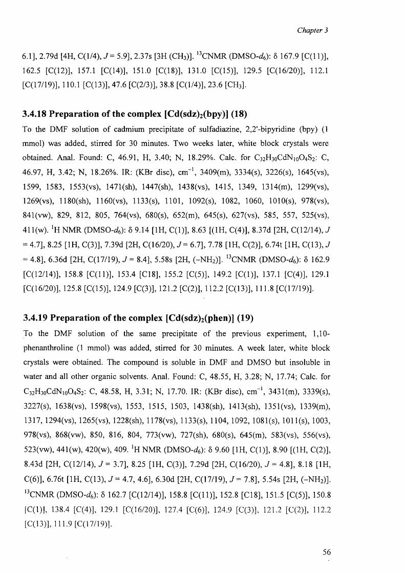
Chapter 3
6.1], 2.79d [4H, C (l/4 ) ,y = 5.9], 2.37s [3H (CH3)]. 13CNMR (DMSO-</6): 5 167.9 [C(l 1 )],
162.5 [C (l2)], 157.1 [C(14)], 151.0 [C(18)], 131.0 [C(15)], 129.5 [C(16/20)], 112.1
[C (l7/19)], 110.1 [C (l3)], 47.6 [C(2/3)], 38.8 [C(l/4)], 23.6 [CH3].
3.4.18 Preparation of the complex [Cd(sdz)2(bpy)] (18)
To the DMF solution of cadmium precipitate o f sulfadiazine, 2,2'-bipyridine (bpy) (1
mmol) was added, stirred for 30 minutes. Two weeks later, white block crystals were
obtained. Anal. Found: C, 46.91, H, 3.40; N, 18.29%. Calc, for C3 2 H 3 oCdNio0 4 S2 : C,
46.97, H, 3.42; N, 18.26%. IR: (KBr disc), cm-1, 3409(m), 3334(s), 3226(s), 1645(vs),
1599, 1583, 1553(vs), 1471(sh), 1447(sh), 1438(vs), 1415, 1349, 1314(m), 1299(vs),
1269(vs), 1180(sh), 1160(vs), 1133(s), 1101, 1092(s), 1082, 1060, 1010(s), 978(vs),
841(vw), 829, 812, 805, 764(vs), 680(s), 652(m), 645(s), 627(vs), 585, 557, 525(vs),
41 l(w). *H NMR (DMSO-<4): 5 9.14 [1H, C (l)], 8.63 [(1H, C(4)], 8.37d [2H, C(12/14), J
= 4.7], 8.25 [1H, C(3)], 7.39d [2H, C(16/20), J = 6.7], 7.78 [1H, C(2)], 6.74t [1H, C(13), J
= 4.8], 6.36d [2 H, C(17/19), J = 8.4], 5.58s [2 H, (-N H 2)]. 13CNMR (DMSO-<4): 5 162.9
[C(12/14)], 158.8 [C(l 1)], 153.4 [C18], 155.2 [C(5)], 149.2 [C(l)], 137.1 [C(4)], 129.1
[C( 16/20)], 125.8 [C (l5)], 124.9 [C(3)], 121.2 [C(2)], 112.2 [C(13)], 111.8 [C(17/19)].
3.4.19 Preparation of the complex [Cd(sdz)2(phen)] (19)
To the DMF solution of the same precipitate o f the previous experiment, 1,10-
phenanthroline (1 mmol) was added, stirred for 30 minutes. A week later, white block
crystals were obtained. The compound is soluble in DMF and DMSO but insoluble in
water and all other organic solvents. Anal. Found: C, 48.55, H, 3.28; N, 17.74; Calc, for
C 3 2 H3 0 CCIN1 0O4 S2 : C, 48.58, H, 3.31; N, 17.70. IR: (KBr disc), cm-1, 3431(m), 3339(s),
3227(s), 1638(vs), 1598(vs), 1553, 1515, 1503, 1438(sh), 1413(sh), 1351(vs), 1339(m),
1317, 1294(vs), 1265(vs), 1228(sh), 1178(vs), 1133(s), 1104, 1092, 1081(s), 1011 (s), 1003,
978(vs), 8 6 8 (vw), 850, 816, 804, 773(vw), 727(sh), 680(s), 645(m), 583(vs), 556(vs),
523(vw), 441 (w), 420(w), 409. 'H NMR (DMSO-</6): 6 9.60 [ 1 H, C (l)], 8.90 [( 1 H, C(2 )],
8.43d [2H, C(12/14), J = 3.7], 8.25 [1H, C(3)], 7.29d [2H, C(16/20), J = 4.8], 8.18 [1H,
C(6 )], 6.76t [1H, C(13), J = 4.7, 4.6], 6.30d [2H, C(17/19), J = 7.8], 5.54s [2H, (-N H 2)].
I3CNMR (DMSO-rf6): 5 162.7 [C(12/14>], 158.8 [C (ll)], 152.8 [C18], 151.5 [C(5)], 150.8
[C(l)], 138.4 [C(4)], 129.1 [C(16/20)], 127.4 [C(6 )], 124.9 [C(3>], 1 2 1 . 2 [C(2)], 112.2
[C (l3)], 111.9 [C (l7/19)].
56
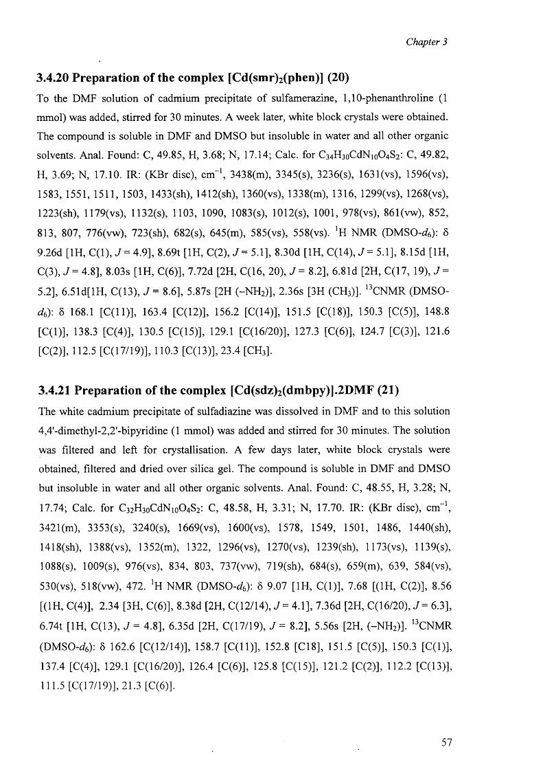
Chapter 3
3.4.20 Preparation of the complex [Cd(smr)2(phen)] (20)
To the DMF solution o f cadmium precipitate of sulfamerazine, 1,10-phenanthroline (1
mmol) was added, stirred for 30 minutes. A week later, white block crystals were obtained.
The compound is soluble in DMF and DMSO but insoluble in water and all other organic
solvents. Anal. Found: C, 49.85, H, 3.68; N, 17.14; Calc, for C3 4 H 3 oCdNio0 4 S2 : C, 49.82,
H, 3.69; N, 17.10. IR: (KBr disc), cm"1, 3438(m), 3345(s), 3236(s), 1631 (vs), 1596(vs),
1583, 1551, 1511, 1503, 1433(sh), 1412(sh), 1360(vs), 1338(m), 1316, 1299(vs), 1268(vs),
1223(sh), 1179(vs), 1132(s), 1103, 1090, 1083(s), 1012(s), 1001, 978(vs), 861(vw), 852,
813, 807, 776(vw), 723(sh), 682(s), 645(m), 585(vs), 558(vs). ‘H NMR (DMSO-<4): 8
9.26d [1H, C (l), J = 4.9], 8.69t [1H, C(2), J = 5.1], 8.30d [1H, C(14), J = 5.1], 8.15d [1H,
C(3), J = 4.8], 8.03s [1H, C(6 )], 7.72d [2H, C(16, 20), J = 8.2], 6.81d [2H, C(17, 19), 7 =
5.2], 6.51d[lH, C(13), J = 8 .6 ], 5.87s [2H (-N H 2)], 2.36s [3 H (CH3)]. 13CNMR (DMSO-
d6): 8 168.1 [C (ll)] , 163.4 [C(12)], 156.2 [C(14)], 151.5 [C(18)], 150.3 [C(5)], 148.8
[C(l)], 138.3 [C(4)], 130.5 [C(15)], 129.1 [C(16/20)], 127.3 [C(6 )], 124.7 [C(3)], 121.6
[C(2)], 112.5 [C( 17/19)], 110.3 [C(13)], 23.4 [CH3].
3.4.21 Preparation of the complex [Cd(sdz)2(dmbpy)].2DMF (21)
The white cadmium precipitate o f sulfadiazine was dissolved in DMF and to this solution
4,4'-dimethyl-2,2'-bipyridine (1 mmol) was added and stirred for 30 minutes. The solution
was filtered and left for crystallisation. A few days later, white block crystals were
obtained, filtered and dried over silica gel. The compound is soluble in DMF and DMSO
but insoluble in water and all other organic solvents. Anal. Found: C, 48.55, H, 3.28; N,
17.74; Calc, for C 3 2 H 3 oCdNio0 4 S2 : C, 48.58, H, 3.31; N, 17.70. IR: (KBr disc), cm-1,
342l(m), 3353(s), 3240(s), 1669(vs), 1600(vs), 1578, 1549, 1501, 1486, 1440(sh),
1418(sh), 1388(vs), 1352(m), 1322, 1296(vs), 1270(vs), 1239(sh), 1173(vs), 1139(s),
1088(s), 1009(s), 976(vs), 834, 803, 737(vw), 719(sh), 684(s), 659(m), 639, 584(vs),
530(vs), 518(vw), 472. NMR (DMSO-<4): S 9.07 [ 1 H, C (l)], 7.68 [(1 H, C(2 )], 8.56
[(1H, C(4)], 2.34 [3H, C(6 )], 8.38d [2H, C(12/14), J = 4.1], 7.36d [2H, C(16/20), J = 6.3],
6.74t [1H, C(13), J = 4.8], 6.35d [2H, C(17/19), J = 8.2], 5.56s [2H, (-N H 2)]. 13CNMR
(DMSO-^6 ): 6 162.6 [C(12/14)], 158.7 [C(l 1 )], 152.8 [C18], 151.5 [C(5)], 150.3 [C(l)],
137.4 [C(4)], 129.1 [C (l6/20)], 126.4 [C(6 )], 125.8 [C(15)], 121.2 [C(2)], 112.2 [C(13)],
111.5 [C (l7/19)], 21.3 [C(6 )].
57
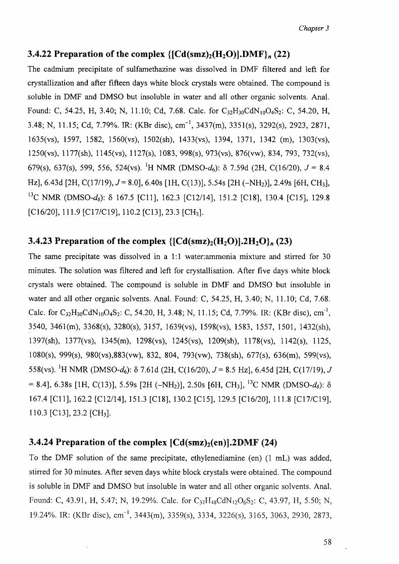
Chapter 3
3.4.22 Preparation of the complex {[Cd(smz)2(H20)].D M F}w (22)
The cadmium precipitate o f sulfamethazine was dissolved in DMF filtered and left for
crystallization and after fifteen days white block crystals were obtained. The compound is
soluble in DMF and DMSO but insoluble in water and all other organic solvents. Anal.
Found: C, 54.25, H, 3.40; N, 11.10; Cd, 7.68. Calc, for C3 2 H 3 oCdNio0 4 S2 : C, 54.20, H,
3.48; N, 11.15; Cd, 7.79%. IR: (KBr disc), c m '1, 3437(m), 3351(s), 3292(s), 2923, 2871,
1635(vs), 1597, 1582, 1560(vs), 1502(sh), 1433(vs), 1394, 1371, 1342 (m), 1303(vs),
1250(vs), 1177(sh), 1145(vs), 1127(s), 1083, 998(s), 973(vs), 876(vw), 834, 793, 732(vs),
679(s), 637(s), 599, 556, 524(vs). lH NMR (DMSO-^6): 6 7.59d (2 H, C(16/20), J = 8.4
Hz], 6.43d [2H, C(17/19), J= 8.0], 6.40s [1H, C(13)], 5.54s [2H (-N H 2)], 2.49s [6 H, CH3],
13C NMR (D M SO -^): 8 167.5 [C ll] , 162.3 [C12/14], 151.2 [C18], 130.4 [C15], 129.8
[C16/20], 111.9 [C l7/C 19], 110.2 [C13], 23.3 [CH3].
3.4.23 Preparation of the complex {[Cd(smz)2(H20)].2H 20 } w (23)
The same precipitate was dissolved in a 1:1 water.ammonia mixture and stirred for 30
minutes. The solution was filtered and left for crystallisation. After five days white block
crystals were obtained. The compound is soluble in DMF and DMSO but insoluble in
water and all other organic solvents. Anal. Found: C, 54.25, H, 3.40; N, 11.10; Cd, 7.68.
Calc, for C3 2 H3 0 CCIN1 0O4 S2 : C, 54.20, H, 3.48; N, 11.15; Cd, 7.79%. IR: (KBr disc), cm '1,
3540, 3461 (m), 3368(s), 3280(s), 3157, 1639(vs), 1598(vs), 1583, 1557, 1501, 1432(sh),
1397(sh), 1377(vs), 1345(m), 1298(vs), 1245(vs), 1209(sh), 1178(vs), 1142(s), 1125,
1080(s), 999(s), 980(vs),883(vw), 832, 804, 793(vw), 738(sh), 677(s), 636(m), 599(vs),
558(vs). ‘H NMR (D M SO -*): 8 7.61d (2H, C(16/20), J = 8.5 Hz], 6.45d [2H, C(17/19), J
= 8.4], 6.38s [1H, C(13)], 5.59s [2H (-N H 2)], 2.50s [6 H, CH3], 13C NMR (D M SO -*): 8
167.4 [C ll] , 162.2 [C12/14], 151.3 [C18], 130.2 [C15], 129.5 [C16/20], 111.8 [C17/C19],
110.3 [C l3], 23.2 [CH3],
3.4.24 Preparation of the complex [Cd(smz)2(en)].2DMF (24)
To the DMF solution o f the same precipitate, ethylenediamine (en) (1 mL) was added,
stirred for 30 minutes. After seven days white block crystals were obtained. The compound
is soluble in DMF and DMSO but insoluble in water and all other organic solvents. Anal.
Found: C, 43.91, H, 5.47; N, 19.29%. Calc, for C3 2 H4 8 CdNi2 0 6 S2: C, 43.97, H, 5.50; N,
19.24%. IR: (KBr disc), c m '1, 3443(m), 3359(s), 3334, 3226(s), 3165, 3063, 2930, 2873,
58
Chapter 3
2843, 1675(s), 1634(vs), 1595, 1579, 1562(vs), 1501(sh), 1432(sh), 1398, 1376, 1342(m),
1291 (vs), 1262(vs), 1209, 1178(sh), 1139(vs), 1122(s), 1079(s), 994(s), 974(vs), 873(vw),
835, 794(vs), 743, 718, 675(s), 636(s), 596, 555. NMR (DMSO-d6): 6 7.63d [2H,
C( 16/20), J = 8.5 Hz], 6.48d [2H, C(17/19), J = 8 .6 ], 6.42s [1H, C(13)], 5.58s [2H
(-N H 2)], 2 .73s [4H, C(l/2)], 2 .51s [6 H, CH3], 13C NMR (D M SO -^): 6 167.7 [C(l 1)],
162.2 [C(12/14)], 151.5 [C(18)], 130.0 [C(15)], 129.9 [C(16/20)], 112.0 [C(17/C19)],
110.2 [C (l3)], 45.3 [C(l/2)], 23.4 [CH3].
3.4.25 Preparation of the complex [Hg(sdz)2(dmf)2] (25)
To the DMF solution o f mercury precipitate o f sulfadiazine, a few drops o f ammonia was
added and stirred for 30 minutes. A few days later, white block crystals were obtained. The
compound is soluble in DMF and DMSO but insoluble in water, alcohol and acetonitrile.
Anal. Found: C, 36.89, H, 3.85; N, 16.53%. Calc, for C^H^HgNjoOsS^ C, 36.94; H, 3.82;
N, 16.57%. IR: (KBr disc): cm-1, 3467(s), 3365(s), 3048(m), 2930, 1655(s), 1614(vs),
1588(vs), 1557(vs), 1506(vs), 1440(sh), 1414(vs), 1342(s), 1291(vs), 1184(w), 1127(sh),
1086(s), 994(w), 958(w), 907(s), 820, 682(s), 651(w), 574(vs), 547(vs), 518(vw), 457(vw),
416(w). *H NMR (DMSO-<4): 5 8.50d [2H, C(12/14), J = 4.9], 7.78d [2H, C(16/20), J =
8.5], 6.98t [1H, C(13), J = 4.8], 6.56d [2H, C(17/19), J = 8 .6 ], 5.92s [2H, (-N H 2)]. 13C
NMR (DMSO-^6 ): 6 162.7 [C(12/14)], 158.8 [C(l 1 )], 153.0 [C18], 130.6 [C(16/20)], 126.1
[C (l5)], 114.7 [C(13)], 112.4 [C(17/19)].
3.4.26 Preparation of the complex [Hg(smr)2] (26)
To the DMF solution o f mercury precipitate o f sulfamerazine, a few drops o f ammonia was
added and stirred for 30 minutes. A few days later, white block crystals were obtained.
The compound is soluble in DMF and DMSO but insoluble in water, alcohol and
acetonitrile. Anal. Found: C, 36.89, H, 3.85; N, 16.53%. Calc, for C2 2 H 3 2 HgNg0 4 S2: C,
36.94; H, 3.82; N, 16.57%. IR: (KBr disc): cm-1, 3436(s), 3317(s), 3228(m), 1632(vs),
1598(vs), 1571 (vs), 1501(vs), 1465(sh), 1435(vs), 1392(vs), 1371(s), 1332(m), 1310(sh),
1281 (vs), 1259(m), 1189(m), 1135(sh), 1093(s), 1010(w), 971(w), 945(w), 917(s), 869(w),
823, 792(s), 769(vw), 716(vw), 689(s), 635(w), 582 (vs) and 546. !H NMR (DMSO-<4): §
8.37d [1H, C(14), J - 5.1], 7.84d [2H, C(16, 20), J = 8.5], 6.92d [2H, C(17, 19), J = 5.1],
6.63d[lH, C(13), J = 8 .8 ], 5.97s [2 H (-N H 2)], 2.39s [3H (CH3)]. ,3CNMR (DMSO-</6): 5
59
Chapter 3
168.4 [C(l 1)], 161.4 [C (l2)], 158.3 [C(14)], 152.9 [C(18)], 130.8 [C(15)], 126.2
[C( 16/20)], 114.1 [C(17/19)], 112.2 [C(13)],23.6 [CH3].
3.4.27 Preparation of the complex [Hg(smr)2(bpy)] (27)
To the DMF solution o f mercury precipitate o f sulfamerazine, 2,2'-bipyridine (0.157g, 1
mmol) was added, stirred for 30 minutes. After seven days, white block crystals were
obtained. The compound is soluble in DMF and DMSO but insoluble in water, alcohol and
acetonitrile. Anal. Found: C, 43.55, H, 3.40; N, 15.90%. Calc, for C32H3oHgNio04S2i C,
43.51; H, 3.42; N, 15.86%. IR: (KBr disc): cm-1, 3445 (s), 3349 (s), 3241 (m), 1637 (vs),
1599 (vs), 1577(vs), 1504(vs), 1459 (sh), 1432 (vs), 1389(vs), 1370(s), 1333(m), 1309(sh),
1286 (vs), 1258(m), 1194 (w), 1180 (m), 1131(sh), 1093(s), 1009 (w), 971(w), 944 (w),
915 (s), 869(w), 823, 792(s), 769 (vw), 716 (vw), 693 (w), 682 (s), 635 (w), 582 (vs), 546
(vs). !H NMR (DMSO-<4): S 8.32d [1H, C(14), J = 4.9], 7.77d [2H, C(16, 20), 8.3],
6.87d [2H, C(17, 19), J = 5.0], 6.57d[lH, C(13), J = 8.4], 5.90s [2H (-N H 2)], 2.33s [3H
(CH3)]. 13CNMR (DMSO-<4): 6 167.9 [C(l 1)], 159.9 [C(12)], 157.7 [C(14)], 152.8
[C (l8)], 130.1 [C( 15)], 126.7 [C(16/20)], 113.1 [C(17/19)], 111.8 [C(13)],23.2 [CH3].
3.4.28 Preparation of the complex [Hg(smz)2(DMF)2] (28)
To the DMF solution of mercury precipitate o f sulfamethazine, a few drops o f ammonia
was added and stirred for 30 minutes. After three weeks, white block crystals were
obtained, filtered and dried over silica gel. The compound is soluble in DMF and DMSO
but insoluble in water, alcohol and acetonitrile. Anal. Found: C, 34.62, H, 3.58; N, 15.54%.
Calc, for C26 H3 2 HgN10O6S2 : C, 34.65; H, 3.51; N, 15.50%. IR: (KBr disc): c m '1, 3457(s),
3360(s), 3247(m), 2919, 1660(vs), 1599(vs), 1577(vs), 1547(vs), 1506, 1429(sh), 1393(vs),
1373(s), 1332(m), 1292(vs), 1258(m), 1194 (w), 1137(sh), 1086(s), 994(w), 965, 897(w),
820, 779(vw), 682(s), 635(w), 589(vs), 549(vs). ‘H NMR (DMSO-d6): 5 7.80d [2H,
C( 16/20), J = 8.6 Hz], 6.57d [2H, C(17/19), J = 8.7], 6.69s [1H, C(13)], 5.90s [2H
(-N H 2)], 2 .51s [6H, CH3]. 13CN M R (DMSO-rfs): 6 167.9 [C(l 1)], 159.6 [C(12/14)], 152.8
[C(18)], 131.0 [C( 15)], 126.3 [C(16/20)], 113.1 [C(17/C19)], 112.0 [C(13)],23.5 [CH3].
60
METAL COMPLEXES OF TRIPLE SULFA DRUGS WITH
SECONDAR Y LIGANDS
Chapter 4
RESULTS AND DISCUSSION
The complexes were characterized by analytical and spectroscopic methods such as IR,
EPR and NMR as well as by X-ray diffraction methods.
4.1 IR spectra
The infrared spectra o f the complexes taken in the region 4000 - 400 cm- 1 were compared
with those o f the free ligands. The bands that appear between 3500 and 3400 cm - 1 due to1 9
<3 5 >7 tt(NH2 ) and sym(NH 2 ) vibrations of the amino (-N H 2 ) group ’ are modified with
respect to those o f the free respective ligands. In general, these vibration modes appear at
higher and lower wave numbers, compared with those o f the free ligands. When the amino
nitrogen atom does not interact with the metal ions, these modifications are consequently
due to the hydrogen bonds involving the amino groups. The vibrations for the water
molecule present in the complexes appear in the same region as well.
The peak for the sulfonamidic (N-H) group in the free ligands at around 3125 cm-1, are not
present in the spectra o f the complexes, confirming the deprotonation of the -S O 2 N H -
moiety. The scissoring vibrations for the amino (-N H 2 ) groups appear in the range of 1614
- 1662 cm - 1 and peaks due to phenyl ring appear at around 1547 and 1600 cm-1. The peaks
around 1325 and 1331 cm - 1 are assigned to v^SCE), and those at 1155 - 1157 cm - 1 to
^(SCE), show important changes upon complexation. The first one splits into two peaks at
1291 - 1386 cm - 1 and the second one appears at around 1131 - 1194 cm - 1 in the
complexes.
1 O TThe 945-965 cm bands in the ligands are assigned to v(S-N) ’ and changed to higher
frequencies (958 - 1046 cm-1) in all cases as a consequence of coordination to the metal.
This shift to higher frequencies is in accordance with the shortening o f the S-N bond
lengths, which have been observed in the crystal structures o f complexes.
61
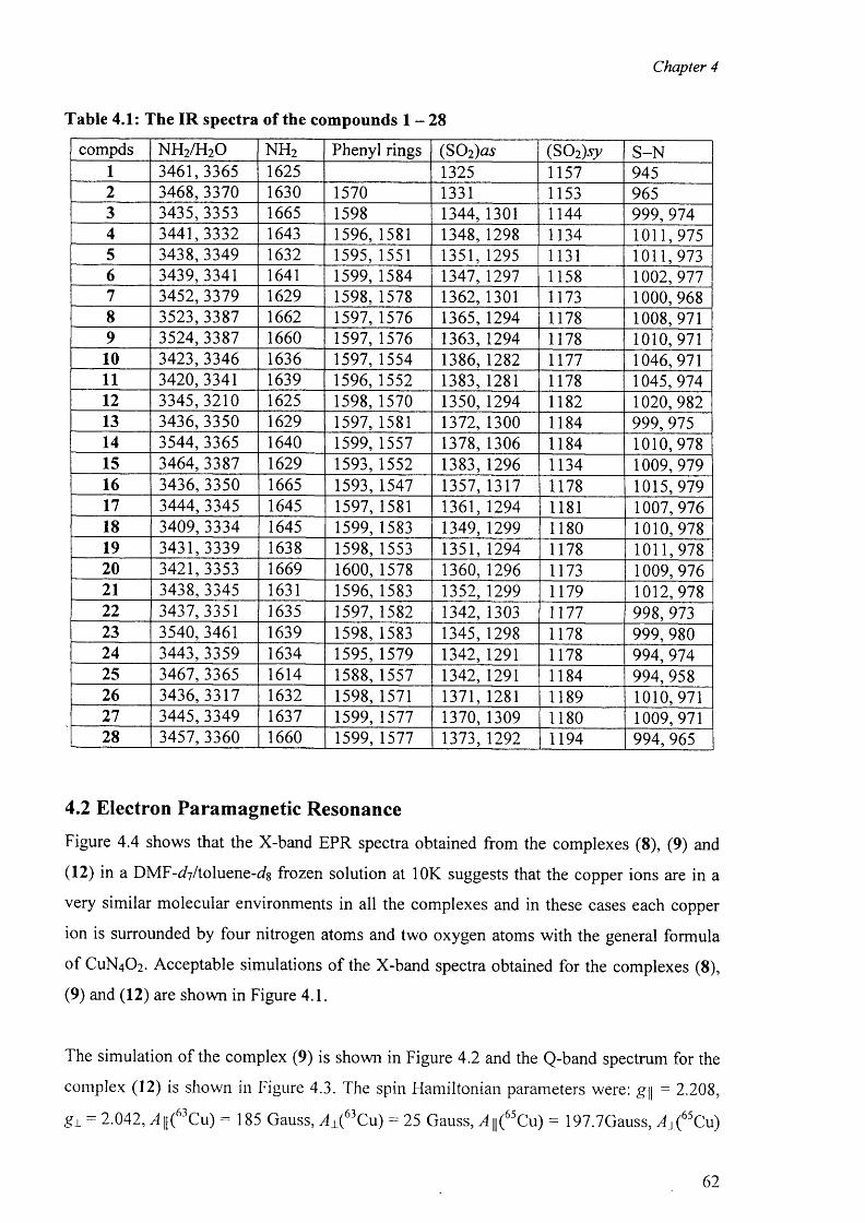
Chapter 4
Table 4.1: The IR spectra of the compounds 1 —28
compds NH 2 /H20 n h 2 Phenyl rings (S 0 2)as (SO 2)sy S -N1 3461,3365 1625 1325 1157 9452 3468, 3370 1630 1570 1331 1153 9653 3435,3353 1665 1598 1344, 1301 1144 999,9744 3441,3332 1643 1596, 1581 1348, 1298 1134 1011,9755 3438,3349 1632 1595, 1551 1351,1295 1131 1011,9736 3439, 3341 1641 1599, 1584 1347, 1297 1158 1002,9777 3452,3379 1629 1598, 1578 1362, 1301 1173 1000,9688 3523,3387 1662 1597, 1576 1365, 1294 1178 1008, 9719 3524,3387 1660 1597, 1576 1363, 1294 r ll7 8 1010, 97110 3423, 3346 1636 1597, 1554 1386,1282 1177 1046, 97111 3420, 3341 1639 1596, 1552 1383,1281 1178 1045,97412 3345,3210 1625 1598, 1570 1350, 1294 1182 1020,98213 3436,3350 1629 1597, 1581 1372, 1300 1184 999,97514 3544,3365 1640 1599, 1557 1378, 1306 1184 1010, 97815 3464, 3387 1629 1593,1552 1383,1296 1134 1009, 97916 3436, 3350 1665 1593, 1547 1357,1317 1178 1015, 97917 3444, 3345 1645 1597, 1581 1361, 1294 1181 1007, 97618 3409, 3334 1645 1599, 1583 1349,1299 1180 1010, 97819 3431,3339 1638 1598,1553 1351, 1294 1178 1011,97820 3421,3353 1669 1600,1578 1360, 1296 1173 1009, 97621 3438, 3345 1631 1596,1583 1352,1299 1179 1012, 97822 3437,3351 1635 1597,1582 1342, 1303 1177 998, 97323 3540, 3461 1639 1598, 1583 1345,1298 1178 999,98024 3443,3359 1634 1595, 1579 1342, 1291 1178 994,97425 3467, 3365 1614 1588,1557 1342, 1291 1184 994, 95826 3436,3317 1632 1598, 1571 1371, 1281 1189 1010, 97127 3445, 3349 1637 1599, 1577 1370,1309 1180 1009, 97128 3457, 3360 1660 1599,1577 1373, 1292 1194 994,965
4.2 Electron Paramagnetic Resonance
Figure 4.4 shows that the X-band EPR spectra obtained from the complexes (8 ), (9) and
( 1 2 ) in a DM F-^/toluene-ds frozen solution at 1 0 K suggests that the copper ions are in a
very similar molecular environments in all the complexes and in these cases each copper
ion is surrounded by four nitrogen atoms and two oxygen atoms with the general formula
o f CUN4 O2 . Acceptable simulations of the X-band spectra obtained for the complexes (8 ),
(9) and (12) are shown in Figure 4.1.
The simulation o f the complex (9) is shown in Figure 4.2 and the Q-band spectrum for the
complex (12) is shown in Figure 4.3. The spin Hamiltonian parameters were: g|| = 2.208,
g± = 2.042, ^ ||( 6 3 Cu) = 185 Gauss, ^ i ( 6 3 Cu) = 25 Gauss, /f ||( 6 5 Cu) = 197.7Gauss, /f i( 6 5 Cu)
62
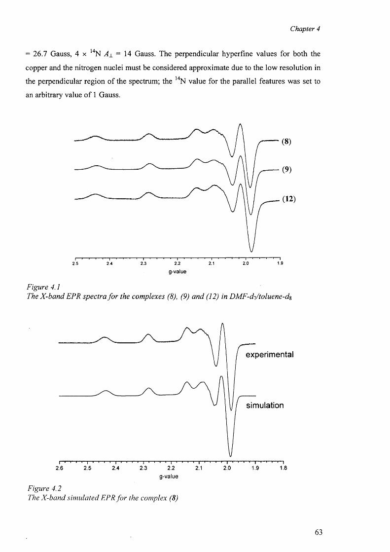
Chapter 4
= 26.7 Gauss, 4 x 14N A± = 14 Gauss. The perpendicular hyperfine values for both the
copper and the nitrogen nuclei must be considered approximate due to the low resolution in
the perpendicular region of the spectrum; the 14N value for the parallel features was set to
an arbitrary value o f 1 Gauss.
(12)
1.92.1 2.02.3 2.22.5 2.4
g-value
Figure 4.1The X-band EPR spectra fo r the complexes (8 ), (9) and (12) in D M F-dftoluene-dg
experimental
simulation
I"'- '1'2.6
i i i | ‘ ” r
2.1i > r " | i i i— r
2.0T - t — r — r— T— 1
1.81 1 1 I 1 1 1 I
2.4I I I I | I
2.21 I r
2.5 2.3 1.9g-value
Figure 4.2The X-band simulated EPR fo r the complex (8 )
63
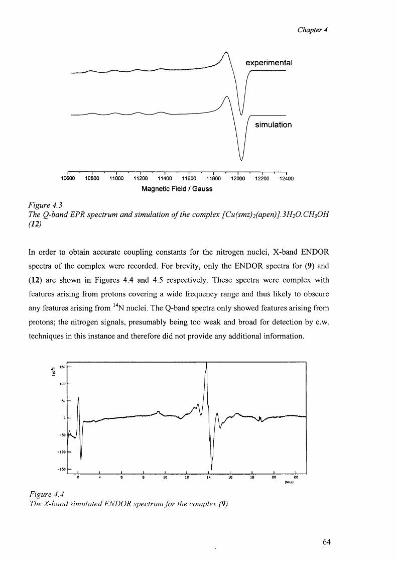
Chapter 4
experimental
simulation
T T T TT T T T10600 10800 11000 11200 11400 11600 11800 12000 12200 12400
Magnetic Field / Gauss
Figure 4.3The Q-band EPR spectrum and simulation o f the complex [Cu(smz) 2 (apen)] .3 H2 O.CH3 OH (12)
In order to obtain accurate coupling constants for the nitrogen nuclei, X-band ENDOR
spectra of the complex were recorded. For brevity, only the ENDOR spectra for (9) and
(12) are shown in Figures 4.4 and 4.5 respectively. These spectra were complex with
features arising from protons covering a wide frequency range and thus likely to obscure
any features arising from 14N nuclei. The Q-band spectra only showed features arising from
protons; the nitrogen signals, presumably being too weak and broad for detection by c.w.
techniques in this instance and therefore did not provide any additional information.
_ 150
100
-50
-100
-150
2 20 226 B 10 12 16 18*
Figure 4.4The X-band simulated ENDOR spectrum fo r the complex (9)
64
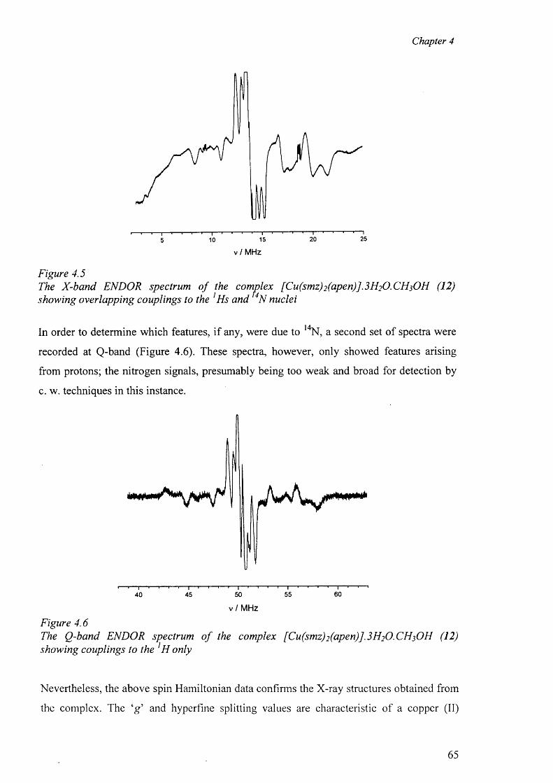
Chapter 4
v /M H z
Figure 4.5The X-band ENDOR spectrum o f the complex [ Cu(smz)2 (apen)]. 3 H2 O. CH 3 OH (12) showing overlapping couplings to the 1 Hs and 4N nuclei
In order to determine which features, if any, were due to 1 4N, a second set of spectra were
recorded at Q-band (Figure 4.6). These spectra, however, only showed features arising
from protons; the nitrogen signals, presumably being too weak and broad for detection by
c. w. techniques in this instance.
60555040 45
v / M H z
Figure 4 . 6
The Q-band ENDOR spectrum o f the complex [ Cu(smz)2 (apen)J.3 H 2 0 . CH 3 OH (12) showing couplings to the ]H only
Nevertheless, the above spin Hamiltonian data confirms the X-ray structures obtained from
the complex. The ‘g ’ and hyperfine splitting values are characteristic of a copper (II)
65
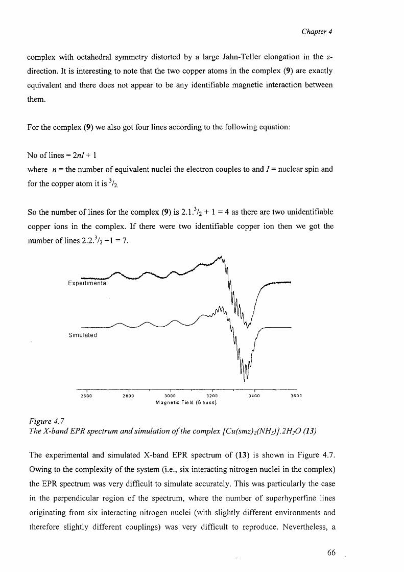
Chapter 4
complex with octahedral symmetry distorted by a large Jahn-Teller elongation in the z-
direction. It is interesting to note that the two copper atoms in the complex (9) are exactly
equivalent and there does not appear to be any identifiable magnetic interaction between
them.
For the complex (9) we also got four lines according to the following equation:
No of lines = 2n l + 1
where n = the number o f equivalent nuclei the electron couples to and I = nuclear spin and
for the copper atom it is I2 .
So the number o f lines for the complex (9) is 2 .1 . 3 / 2 + 1 = 4 as there are two unidentifiable
copper ions in the complex. If there were two identifiable copper ion then we got the
number o f lines 2 2 ? 12 + 1 = 7 .
E x p e r t i m e n t a l
S i m u l a t e d
2600 2800 3000 3200 3400 3600M a g n e t i c F i e l d ( G a u s s )
Figure 4.7The X-band EPR spectrum and simulation o f the complex [Cu(smz)2(NHs)].2H20 (13)
The experimental and simulated X-band EPR spectrum of (13) is shown in Figure 4.7.
Owing to the complexity of the system (i.e., six interacting nitrogen nuclei in the complex)
the EPR spectrum was very difficult to simulate accurately. This was particularly the case
in the perpendicular region of the spectrum, where the number o f superhyperfine lines
originating from six interacting nitrogen nuclei (with slightly different environments and
therefore slightly different couplings) was very difficult to reproduce. Nevertheless, a
66
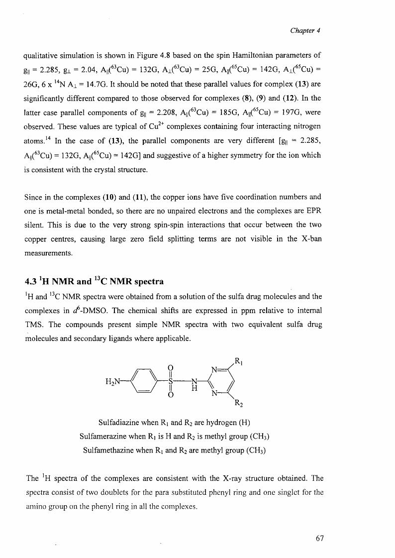
Chapter 4
qualitative simulation is shown in Figure 4.8 based on the spin Hamiltonian parameters of
g,l = 2.285, gj. = 2.04, A,|(6 3 Cu) = 132G, Aj.(6 3 Cu) = 25G, A,|(65Cu) = 142G, Aj.(65Cu) =
26G, 6 x l4N A i = 14.7G. It should be noted that these parallel values for complex (13) are
significantly different compared to those observed for complexes (8 ), (9) and (12). In the
latter case parallel components o f gy = 2.208, A||(63Cu) = 185G, A||(65Cu) = 197G, were
observed. These values are typical of Cu2+ complexes containing four interacting nitrogen
atoms . 14 In the case o f (13), the parallel components are very different [gy = 2.285,
A,|(6 3 Cu) = 132G, Ay(65Cu) = 142G] and suggestive o f a higher symmetry for the ion which
is consistent with the crystal structure.
Since in the complexes (10) and (11), the copper ions have five coordination numbers and
one is metal-metal bonded, so there are no unpaired electrons and the complexes are EPR
silent. This is due to the very strong spin-spin interactions that occur between the two
copper centres, causing large zero field splitting terms are not visible in the X-ban
measurements.
4.3 NMR and 13C NMR spectra
*H and 13C NMR spectra were obtained from a solution of the sulfa drug molecules and the
complexes in </-DMSO. The chemical shifts are expressed in ppm relative to internal
TMS. The compounds present simple NMR spectra with two equivalent sulfa drug
molecules and secondary ligands where applicable.
R 2
Sulfadiazine when Ri and R2 are hydrogen (H)
Sulfamerazine when R\ is H and R2 is methyl group (CH 3 )
Sulfamethazine when R\ and R2 are methyl group (CH 3 )
The !H spectra of the complexes are consistent with the X-ray structure obtained. The
spectra consist o f two doublets for the para substituted phenyl ring and one singlet for the
amino group on the phenyl ring in all the complexes.
67
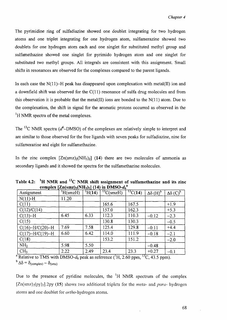
Chapter 4
The pyrimidine ring o f sulfadiazine showed one doublet integrating for two hydrogen
atoms and one triplet integrating for one hydrogen atom, sulfamerazine showed two
doublets for one hydrogen atom each and one singlet for substituted methyl group and
sulfamethazine showed one singlet for pyrimido hydrogen atom and one singlet for
substituted two methyl groups. All integrals are consistent with this assignment. Small
shifts in resonances are observed for the complexes compared to the parent ligands.
In each case the N (11)-H peak has disappeared upon complexation with metal(II) ion and
a downfield shift was observed for the C ( l l) resonance o f sulfa drug molecules and from
this observation it is probable that the metal(II) ions are bonded to the N (11) atom. Due to
the complexation, the shift in signal for the aromatic protons occurred as observed in the
’H NMR spectra of the metal complexes.
The 13C NMR spectra ((/-D M SO ) of the complexes are relatively simple to interpret and
are similar to those observed for the free ligands with seven peaks for sulfadiazine, nine for
sulfamerazine and eight for sulfamethazine.
In the zinc complex [Zn(smz)2 (NH3 )2 ] (14) there are two molecules o f ammonia as
secondary ligands and it showed the spectra for the sulfamethazine molecules.
Table 4.2: XH NMR and 13C NMR shift assignment of sulfamethazine and its zinc
Assignment 'H(smzH) AH(14) UC(smzH) UC(14) A8 (H)b A8 (C)bN (ll)-H 1 1 . 2 0
C (ll) 165.6 167.5 +1.9C(12)/C(14) 157.0 162.3 +5.3C(13)-H 6.45 6.33 112.3 110.3 - 0 . 1 2 -2.3C(15) 130.8 130.3 -0.5C( 16)-H /C(20)-H 7.69 7.58 125.4 129.8 - 0 . 1 1 +4.4C(17)-H/C(19)-H 6.60 6.42 114.0 111.9 -0.18 - 2 . 1
C(18) 153.2 151.2 - 2 . 0
n h 2 5.98 5.50 -0.48c h 3 2 . 2 2 2.49 23.4 23.3 +0.27 - 0 . 1
b AS_A 8 S (co m p lex ) S (srn z )
Due to the presence o f pyridine molecules, the NMR spectrum of the complex
[Zn(smz)2 (py)2 ].2 py (15) shows two additional triplets for the meta- and para- hydrogen
atoms and one doublet for 6>/+/70-hydrogen atoms.
68
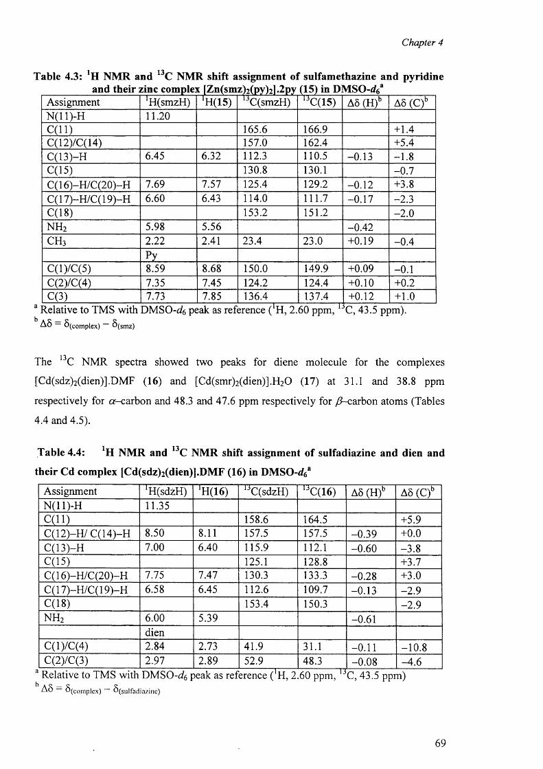
Chapter 4
Table 4.3: *11 NMR and 13C NMR shift assignment of sulfamethazine and pyridine
Assignment ^(sm zH ) AH(15) JC(smzH) 1JC(15) A8 (H)b A8 (C)bN(11)-H 1 1 . 2 0
C (ll) 165.6 166.9 +1.4C(12)/C(14) 157.0 162.4 +5.4C(13)-H 6.45 6.32 112.3 110.5 -0.13 - 1 . 8
C(15) 130.8 130.1 -0 .7C(16)-H/C(20)-H 7.69 7.57 125.4 129.2 - 0 . 1 2 +3.8C( 17)-H/C( 19)-H 6.60 6.43 114.0 111.7 -0.17 -2.3C(18) 153.2 151.2 - 2 . 0
n h 2 5.98 5.56 -0.42c h 3 2 . 2 2 2.41 23.4 23.0 +0.19 -0 .4
PyC(l)/C(5) 8.59 8 . 6 8 150.0 149.9 +0.09 - 0 . 1
C(2)/C(4) 7.35 7.45 124.2 124.4 + 0 . 1 0 + 0 . 2
C(3) 7.73 7.85 136.4 137.4 + 0 . 1 2 + 1 . 0
A8 S (co m p lex ) S (sm z )
The 13C NMR spectra showed two peaks for diene molecule for the complexes
[Cd(sdz)2 (dien)].DMF (16) and [Cd(smr)2 (dien)].H2 0 (17) at 31.1 and 38.8 ppm
respectively for or-carbon and 48.3 and 47.6 ppm respectively for /7-carbon atoms (Tables
4.4 and 4.5).
Table 4.4: NMR and 13C NMR shift assignment of sulfadiazine and dien and
their Cd complex [Cd(sdz)2 (dien)].DMF (16) in DMSO-</6a
Assignment ^ (sd z H ) lH(16) 1JC(sdzH) UC(16) A8 (H)b A8 (C)bN(11)-H 11.35C (ll) 158.6 164.5 +5.9C(12)-H/ C(14)-H 8.50 8 . 1 1 157.5 157.5 -0.39 + 0 . 0
C(13)-H 7.00 6.40 115.9 1 1 2 . 1 -0 .60 -3 .8C(15) 125.1 128.8 +3.7C( 16)-H /C(20)-H 7.75 7.47 130.3 133.3 -0.28 +3.0C(17)-H/C(19)-H 6.58 6.45 1 1 2 . 6 109.7 -0.13 -2 .9C(18) 153.4 150.3 -2 .9n h 2 6 . 0 0 5.39 -0.61
dienC(l)/C(4) 2.84 2.73 41.9 31.1 - 0 . 1 1 - 1 0 . 8
C(2)/C(3) 2.97 2.89 52.9 48.3 -0.08 -4 .6Relative to TMS with DMS0-^6 peak as reference ( ‘H, 2.60 ppm, C, 43.5 ppm)A8 “ S (c o m p le x ) — 6 ( s u |fa d ia z in e )
69
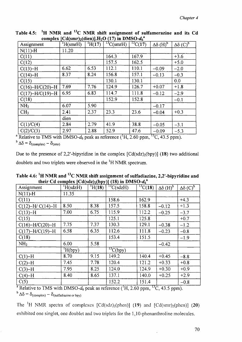
Chapter 4
Table 4.5: !H NMR and 13C NMR shift assignment of sulfamerazine and its Cdcomplex fCd(smr)2(dlien)].H2<3 (17) in DMSO-rf6a
Assignment ^ (sm rH ) ‘H(17) 1JC(smrH) 1JC(17) A8 (H)b A8 (C)bN (ll)-H 1 1 . 2 0
C(l l ) 164.3 167.9 +3.6C(12) 157.5 162.5 +5.0C(13)-H 6.62 6.53 1 1 2 . 1 1 1 0 . 1 -0 .09 - 2 . 0
C(14)-H 8.37 8.24 156.8 157.1 -0.13 -0.3C(15) 130.1 130.1 0 . 0
C( 16)-H /C(20)-H 7.69 7.76 124.9 126.7 +0.07 + 1 . 8
C( 17)-H/C( 19)-H 6.95 6.83 114.7 1 1 1 . 8 - 0 . 1 2 -2 .9C(18) 152.9 152.8 - 0 . 1
n h 2 6.07 5.90 -0.17c h 3 2.41 2.37 23.3 23.6 -0.04 +0.3
dienC(l)/C(4) 2.84 2.79 41.9 38.8 -0.05 -3.1C(2)/C(3) 2.97 2 . 8 8 52.9 47.6 -0.09 -5 .3
a Relative to TMS wit' b A5 — 8 fcr>mnlex> — $
i D M S 0-a?6 peak as reference ( H, 2.60 ppm, C, 43.5 ppm).'( co m p lex ) ° ( s m r )
Due to the presence of 2,2'-bipyridine in the complex [Cd(sdz)2 (bpy)] (18) two additional
doublets and two triplets were observed in the !H NMR spectrum.
Table 4.6: !H NMR and 13C NMR shift assignment of sulfadiazine, 2,2'-bipyridine and
Assignment ^ (sd zH ) ‘H(18) 1JC(sdzH) n 00 A6 (H)b A8 (C)b
N(11)-H 11.35C (ll) 158.6 162.9 +4.3C(12)-H/ C(14)-H 8.50 8.38 157.5 158.8 - 0 . 1 2 +1.3C(13)-H 7.00 6.75 115.9 1 1 2 . 2 -0.25 -3 .7C(15) 125.1 125.8 +0.7C(16)-H/C(20)-H 7.75 7.37 130.3 129.1 -0.38 - 1 . 2
C( 17)-H/C( 19)-H 6.58 6.35 1 1 2 . 6 1 1 1 . 8 -0.23 - 0 . 8
C(18) 153.4 151.5 -1 .9n h 2 6 . 0 0 5.58 -0.42
lH(bpy) ,JC(bpy)C (l)-H 8.70 9.15 149.2 140.4 +0.45 - 8 . 8
C(2)-H 7.45 7.78 120.4 1 2 1 . 2 +0.33 + 0 . 8
C(3)-H 7.95 8.25 124.0 124.9 +0.30 +0.9C(4)-H 8.40 8.65 137.1 140.0 +0.25 +2.9C(5) 152.2 151.4 - 0 . 8
A8 8 (c o m p le x ) ^(sL ilfad iazine o r bpy)
The ]H NM R spectra o f complexes [Cd(sdz)2 (phen)] (19) and [Cd(sm r)2 (phen)] (20)
exhibited one singlet, one doublet and two triplets for the 1,10-phenanthroline molecules.
70
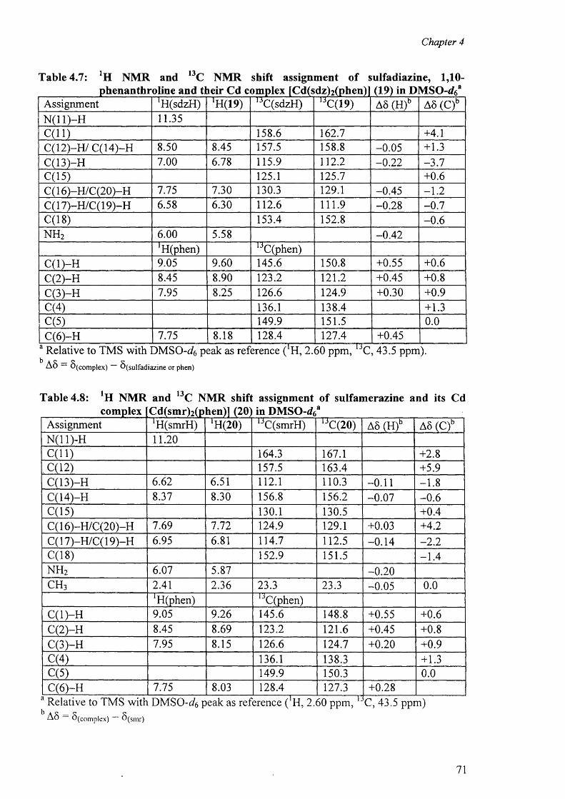
Chapter 4
Table 4.7: NMR and 13C NMR shift assignment of sulfadiazine, 1,10-
Assignment ^ (sd zH ) fH(19) 1 3C(sdzH) 13C(19) AS (H)b AS (C)bN ( ll) -H 11.35C (ll) 158.6 162.7 +4.1C (12)-H /C (14)-H 8.50 8.45 157.5 158.8 -0.05 +1.3C(13)-H 7.00 6.78 115.9 1 1 2 . 2 - 0 . 2 2 -3 .7C(15) 125.1 125.7 + 0 . 6
C( 16)-H /C(20)-H 7.75 7.30 130.3 129.1 -0.45 - 1 . 2
C( 17)-H/C( 19)-H 6.58 6.30 1 1 2 . 6 111.9 -0.28 -0 .7C(18) 153.4 152.8 - 0 . 6
n h 2 6 . 0 0 5.58 -0.42^ (p h en ) 1 3C(phen)
C (l)-H 9.05 9.60 145.6 150.8 +0.55 + 0 . 6
C(2)-H 8.45 8.90 123.2 1 2 1 . 2 +0.45 + 0 . 8
C(3)-H 7.95 8.25 126.6 124.9 +0.30 +0.9C(4) 136.1 138.4 +1.3C(5) 149.9 151.5 0 . 0
C(6 )-H 7.75 8.18 128.4 127.4 +0.45
AS S (co m p lex ) ^ (s u lfa d ia z in e o r phen)
Table 4.8: NMR and 13C NMR shift assignment of sulfamerazine and its Cdcomplex Cd(smr)2(p hen)] (20) in D M S O - r f 6
a
Assignment 'HCsmrH) *H(20) 1 3C(smrH) ljC(20) AS (H)b AS (C)bN(11)-H 1 1 . 2 0
C (ll) 164.3 167.1 +2 . 8
. C( 12) 157.5 163.4 +5.9C(13)-H 6.62 6.51 1 1 2 . 1 110.3 - 0 . 1 1 - 1 . 8
C(14)-H 8.37 8.30 156.8 156.2 -0.07 - 0 . 6
C(15) 130.1 130.5 +0.4C( 16)-H /C(20)-H 7.69 7.72 124.9 129.1 +0.03 +4.2C( 17)-H/C( 19)-H 6.95 6.81 114.7 112.5 -0.14 - 2 . 2
C(18) 152.9 151.5 -1 .4n h 2 6.07 5.87 - 0 . 2 0
c h 3 2.41 2.36 23.3 23.3 -0.05 0 . 0
*H(phen) iJC(phen)C (l)-H 9.05 9.26 145.6 148.8 +0.55 +0 . 6
C(2)-H 8.45 8.69 123.2 1 2 1 . 6 +0.45 +0 . 8
C(3)-H 7.95 8.15 126.6 124.7 +0 . 2 0 +0.9C(4) 136.1 138.3 +1.3C(5) 149.9 150.3 0 . 0
C(6 )-H 7.75 8.03 128.4 127.3 +0.28Relative to TMS with D M SO -^ peak as reference ( H, 2.60 ppm,A S — S (c o m p le x ) — 8 ( s m r)
13C, 43.5 ppm)
71
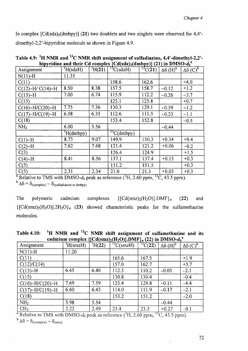
Chapter 4
In complex [Cd(sdz)2 (dmbpy)] (21) two doublets and two singlets were observed for 4,4'-
dimethyl-2,2'-bipyridine molecule as shown in Figure 4.9.
Table 4.9: NMR and 13C NMR shift assignment of sulfadiazine, 4,4'-dimethyI-2,2'-bipyridine and their Cd complex fCd(sdzWdmbpy)l (
Assignment 'H(sdzH) !H(21) C(sdzH) C(21) AS (H)b AS (C)bN(11)-H 11.35C (ll) 158.6 162.6 +4.0C(12)-H/ C(14)-H 8.50 8.38 157.5 158.7 - 0 . 1 2 + 1 . 2
C(13)-H 7.00 6.74 115.9 1 1 2 . 2 -0.26 -3 .7C(15) 125.1 125.8 +0.7C( 16)-H /C(20)-H 7.75 7.36 130.3 129.1 -0.39 - 1 . 2
C(17)-H/C(19)-H 6.58 6.35 1 1 2 . 6 111.5 -0.23 - 1 . 1
C(18) 153.4 152.8 -0.5n h 2 6 . 0 0 5.56 -0.44
*H(dmbpy) 1 3C(dmbpy)C (l)-H 8.73 9.07 149.9 150.3 +0.34 +0.4C(2)-H 7.62 7.68 121.4 1 2 1 . 2 +0.06 - 0 . 2
C(3) 126.4 124.9 +1.5C(4)-H 8.41 8.56 137.1 137.4 +0.15 +0.3C(5) 151.2 151.5 +0.3C(5) 2.31 2.34 2 1 . 0 21.3 +0.03 +0.3
AS S i m p l e x ) b ( Suifa diaz ine or dm bpy)
The polymeric cadmium complexes {[Cd(smz)2 (H2 0 )].DMF}„ (22) and
{[Cd(smz)2 (H2 0 )].2 H 2 0 }„ (23) showed characteristic peaks for the sulfamethazine
molecules.
Table 4.10: NMR and 13C NMR shift assignment of sulfamethazine and its
Assignment *H(smzH) *14(22) 1JC(smzH) 1jC(22) AS (H)b AS (C)bN(11)-H 1 1 . 2 0
C (ll) 165.6 167.5 +1.9C(12)/C(14) 157.0 162.7 +5.7C(13)-H 6.45 6.40 112.3 1 1 0 . 2 -0.05 - 2 . 1
C(15) 130.8 130.4 -0.4C( 16)-H/C(20)-H 7.69 7.59 125.4 129.8 - 0 . 1 1 -4.4C(17)-H/C(19)-H 6.60 6.43 114.0 111.9 -0.17 - 2 . 1
C(18) 153.2 151.2 - 2 . 0
n h 2 5.98 5.54 -0.44c h 3 2 . 2 2 2.49 23.4 23.3 +0.27 - 0 . 1
AS S(compiex) S(smz)
72
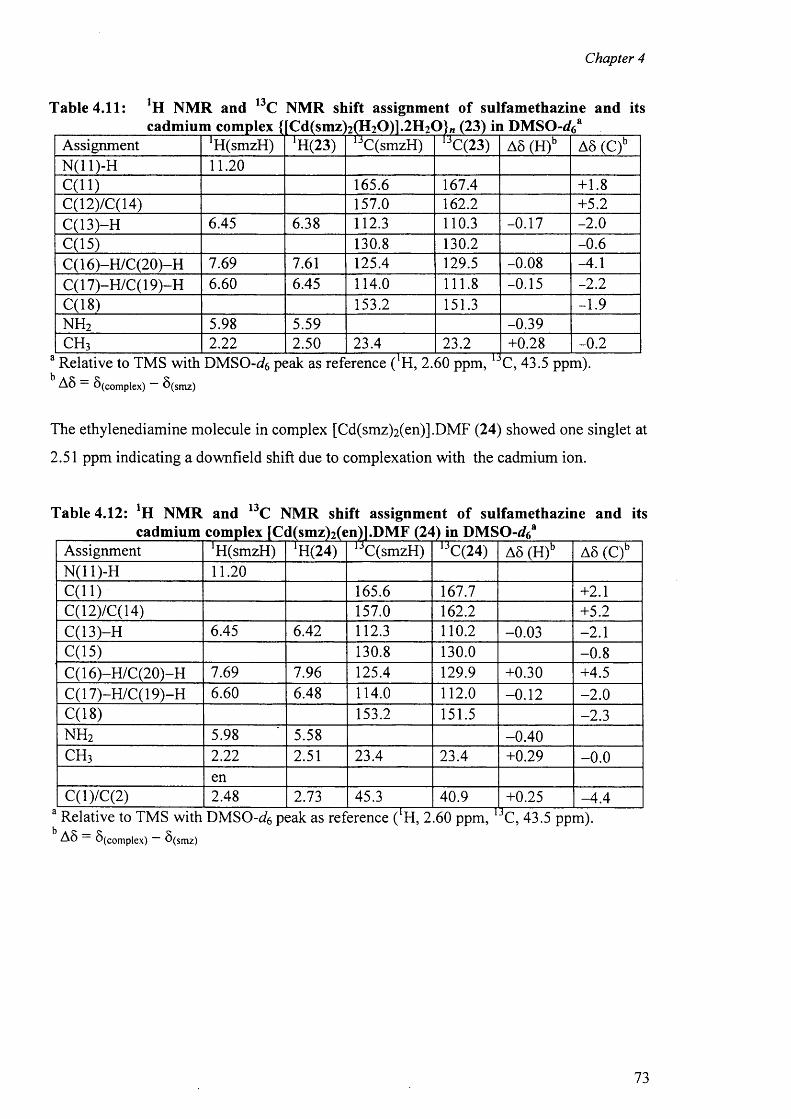
Chapter 4
Table 4.11: !H NMR and 13C NMR shift assignment of sulfamethazine and itscadmium complex {[Cd(smz 2(H20)1.2H2(3}„ (23) in DMSO-J6a
Assignment *H(smzH) *H(23) 1JC(smzH) 1JC(23) A8 (H)b A5 (C)bN (ll)-H 1 1 . 2 0
C (ll) 165.6 167.4 + 1 . 8
C(12)/C(14) 157.0 162.2 +5.2C(13)-H 6.45 6.38 112.3 110.3 -0.17 - 2 . 0
C(15) 130.8 130.2 - 0 . 6
C( 16)-H /C(20)-H 7.69 7.61 125.4 129.5 -0.08 -4.1C( 17)-H/C( 19)-H 6.60 6.45 114.0 1 1 1 . 8 -0.15 - 2 . 2
C(18) 153.2 151.3 -1.9n h 2 5.98 5.59 -0.39c h 3 2 . 2 2 2.50 23.4 23.2 +0.28 - 0 . 2
Relative to TMS with D M S0-g?6 peak as reference ( H, 2.60 ppm,A 8 — 6 ( Com plex) — S ( sm z)
C, 43.5 ppm).
The ethylenediamine molecule in complex [Cd(smz)2 (en)] .DMF (24) showed one singlet at
2.51 ppm indicating a downfield shift due to complexation with the cadmium ion.
Table 4.12: ‘H NMR cadmium <
and 13C NMR shift assignment of sulfamethazine and itsm u c n j a
Assignment 'HCsmzH) 1H(24) ljC(smzH) 1JC(24) A5 (H)b A5 (C)bN(11)-H 1 1 . 2 0
C (ll) 165.6 167.7 +2 . 1
C(12)/C(14) 157.0 162.2 +5.2C(13)-H 6.45 6.42 112.3 1 1 0 . 2 -0.03 - 2 . 1
C(15) 130.8 130.0 - 0 . 8
C( 16)-H /C(20)-H 7.69 7.96 125.4 129.9 +0.30 +4.5C( 17)-H/C( 19)-H 6.60 6.48 114.0 1 1 2 . 0 - 0 . 1 2 - 2 . 0
C(18) 153.2 151.5 -2 .3n h 2 5.98 5.58 -0.40c h 3 2 . 2 2 2.51 23.4 23.4 +0.29 - 0 . 0
enC(l)/C(2) 2.48 2.73 45.3 40.9 +0.25 -4 .4
A8 (complex) d(smz)
73
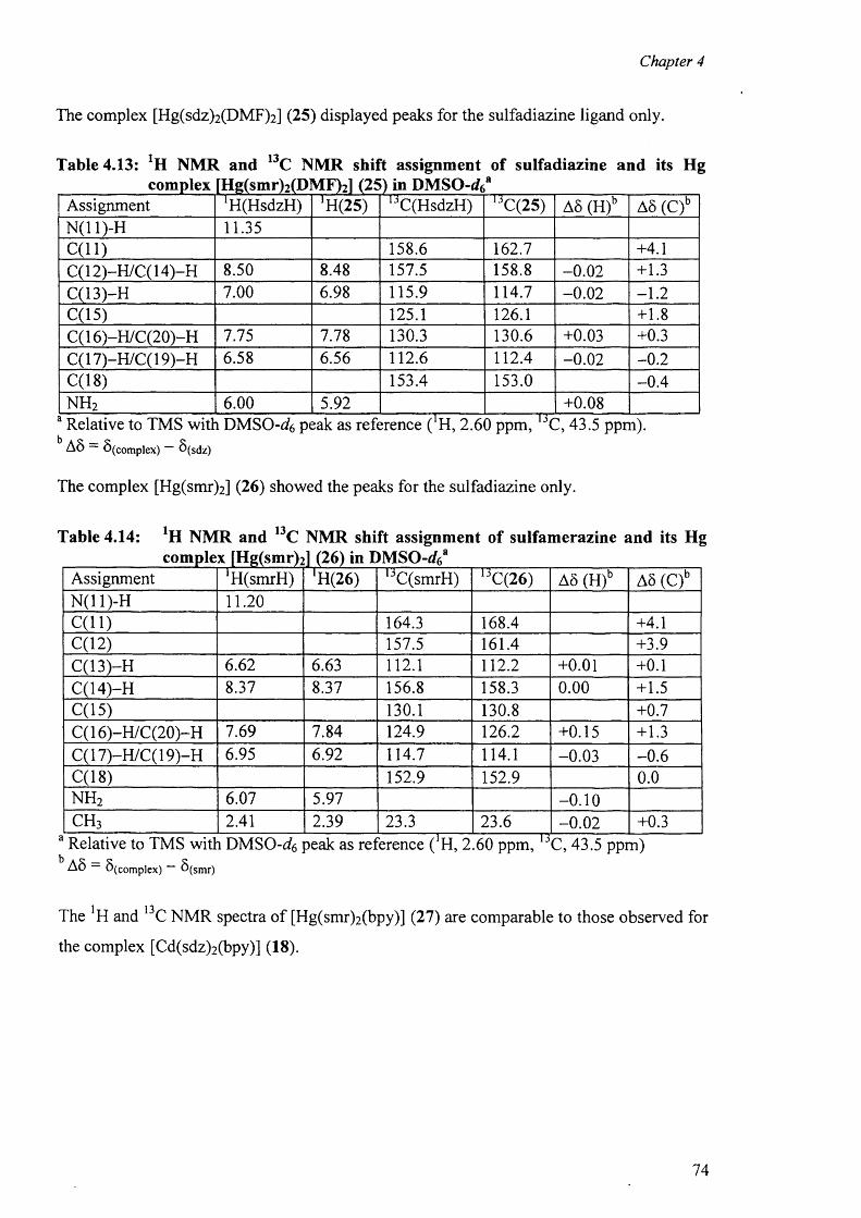
Chapter 4
The complex [Hg(sdz)2 (DMF)2 ] (25) displayed peaks for the sulfadiazine ligand only.
Table 4.13: NMR and 13C NMR shift assignment of sulfadiazine and its Hg
Assignment ^(H sdzH ) *H(25) u C(HsdzH) 1JC(25) A6 (H)b A5 (C)bN(11)-H 11.35C (ll) 158.6 162.7 +4.1C( 12)-H/C( 14)-H 8.50 8.48 157.5 158.8 - 0 . 0 2 +1.3C(13)-H 7.00 6.98 115.9 114.7 - 0 . 0 2 - 1 . 2
C(15) 125.1 126.1 + 1 . 8
C( 16)-H /C(20)-H 7.75 7.78 130.3 130.6 +0.03 +0.3C( 17)-H/C( 19)-H 6.58 6.56 1 1 2 . 6 112.4 - 0 . 0 2 - 0 . 2
C(18) 153.4 153.0 -0 .4n h 2 6 . 0 0 5.92 +0.08Relative to TMS with DMSO - ^ 6 peak as reference ( H, 2.60 ppm,A 5 — S (co m p lex ) — S ( sd z)
C, 43.5 ppm).
The complex [Hg(smr)2 ] (26) showed the peaks for the sulfadiazine only.
Table 4.14: NMR and 13C NMR shift assignment of sulfamerazine and its Hg
Assignment ‘H(smrH) rH(26) 1 3C(smrH) 1JC(26) A5 (H)b A5 (C)bN(11)-H 1 1 . 2 0
C (ll) 164.3 168.4 +4.1C(12) 157.5 161.4 +3.9C(13)-H 6.62 6.63 1 1 2 . 1 1 1 2 . 2 +0 . 0 1 + 0 . 1
C(14)-H 8.37 8.37 156.8 158.3 0 . 0 0 +1.5C(15) 130.1 130.8 +0.7C( 16)-H /C(20)-H 7.69 7.84 124.9 126.2 +0.15 +1.3C(17)-H/C(19)-H 6.95 6.92 114.7 114.1 -0.03 - 0 . 6
C(18) 152.9 152.9 0 . 0
n h 2 6.07 5.97 - 0 . 1 0
c h 3 2.41 2.39 23.3 23.6 - 0 . 0 2 +0.3
A 8 S (co m p lex ) b (s m r)
The and 13C NMR spectra o f [Hg(smr)2 (bpy)] (27) are comparable to those observed for
the complex [Cd(sdz)2 (bpy)] (18).
74
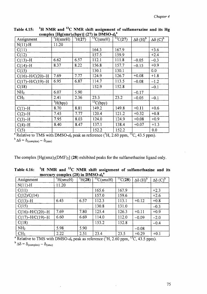
Chapter 4
Table 4.15: *H NMR and 13C NMR shift assignment of sulfamerazine and its Hgcomplex [Hg(smr)2 (bpy)] @7) in DMSO-gfc*
Assignment 'HCsmrH) iH(27) ljC(smrH) 13C(27) AS (H)b AS (C)bN(11)-H 1 1 . 2 0
C (ll) 164.3 167.9 +3.6C(12) 157.5 159.9 +2.4C(13)-H 6.62 6.57 1 1 2 . 1 1 1 1 . 8 -0.05 -0.3C(14)-H 8.37 8 . 2 2 156.8 157.7 -0.15 +0.9C(15) 130.1 130.1 0 . 0
C( 16)-H /C(20)-H 7.69 7.77 124.9 126.7 +0.08 + 1 . 8
C(17)-H/C(19)-H 6.95 6.87 114.7 113.5 -0.08 - 1 . 2
C(18) 152.9 152.8 - 0 . 1
n h 2 6.07 5.90 -0.17c h 3 2.41 2.36 23.3 23.2 -0.05 - 0 . 1
‘HCbpy) UC(bpy)C (l)-H 8.70 8.81 149.2 149.8 + 0 . 1 1 + 0 . 6
C(2)-H 7.45 7.77 120.4 1 2 1 . 2 +0.32 +0 . 8
C(3)-H 7.95 8.03 124.0 124.9 +0.08 +0.9C(4)-H 8.40 8.47 137.1 138.4 +0.07 +1.3C(5) 152.2 152.2 0 . 0
a Relative to TMS wit i DMSO-cfo peak as reJbrence (‘H, 2.60 ppm, 1 3C, 43.5 ppm).AS S ( Com plex) 5 (s m r)
The complex [Hg(smz)2 (DMF)2 ] (28) exhibited peaks for the sulfamethazine ligand only.
Table 4.16: NMR and 13C NMR shift assignment of sulfamethazine and itsmercuryf complex (2£\) in DMSO-rf6a
Assignment ‘HCsmzH) [H(28) 1 3C(smzH) 1JC(28) A6 (H)b AS (C)bN(11)-H 1 1 . 2 0
C (ll) 165.6 167.9 +2.3C(12)/C(14) 157.0 159.6 +2 . 6
C(13)-H 6.45 6.57 112.3 113.1 +0 . 1 2 +0 . 8
C(15) 130.8 131.0 -0.3C( 16)-H /C(20)-H 7.69 7.80 125.4 126.3 + 0 . 1 1 +0.9C( 17)-H/C( 19)-H 6.60 6.69 114.0 1 1 2 . 0 -0 .09 - 2 . 0
C(18) 153.2 152.8 -0 .4n h 2 5.98 5.90 -0.08c h 3 2 . 2 2 2.51 23.4 23.5 +0.29 + 0 . 1
a Relative to TMS with DMSO-cfc peak as reference ( H, 2.60 ppm, C, 43.5 ppm).A S — 5 ( Com plex ) — 8 (s m z )
75
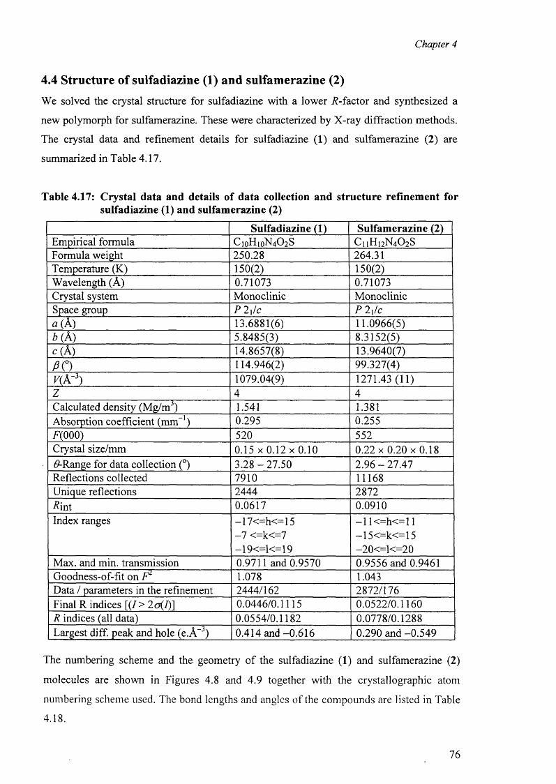
Chapter 4
4.4 Structure of sulfadiazine (1) and sulfamerazine (2)
We solved the crystal structure for sulfadiazine with a lower .R-factor and synthesized a
new polymorph for sulfamerazine. These were characterized by X-ray diffraction methods.
The crystal data and refinement details for sulfadiazine (1) and sulfamerazine (2) are
summarized in Table 4.17.
Table 4.17: Crystal data and details of data collection and structure refinement for sulfadiazine (1) and sulfamerazine (2)
Sulfadiazine (1) Sulfamerazine (2)Empirical formula C,oH1 0N 4 0 2 S C 1 1H,2N4 0 2SFormula weight 250.28 264.31Temperature (K) 150(2) 150(2)Wavelength (A) 0.71073 0.71073Crystal system Monoclinic MonoclinicSpace group P 2\/c P 2\!ca (A) 13.6881(6) 11.0966(5)* (A ) 5.8485(3) 8.3152(5)c ( A) 14.8657(8) 13.9640(7)P O 114.946(2) 99.327(4)K(A“3) 1079.04(9) 1271.43 (11)Z 4 4Calculated density (Mg/m3) 1.541 1.381Absorption coefficient (mm ) 0.295 0.255F(0 0 0 ) 520 552Crystal size/mm 0 .1 5 x 0 .1 2 x 0 .1 0 0 .2 2 x 0 .20x0 .18#-Range for data collection (°) 3.28 -2 7 .5 0 2.96 - 27.47Reflections collected 7910 11168Unique reflections 2444 2872R'mi 0.0617 0.0910Index ranges —17<=h<=15 —1 1 <=h< = 1 1
-7 <=k<=7 —15 <=k<=15—19<=1<= 19 - 2 0 <=1 < = 2 0
Max. and min. transmission 0.9711 and 0.9570 0.9556 and 0.9461Goodness-of-fit on F1 1.078 1.043Data / parameters in the refinement 2444/162 2872/176Final R indices [( /> 2 o(I)\ 0.0446/0.1115 0.0522/0.1160R indices (all data) 0.0554/0.1182 0.0778/0.1288Largest diff. peak and hole (e.A~3) 0.414 and -0.616 0.290 and -0.549
The numbering scheme and the geometry o f the sulfadiazine (1) and sulfamerazine (2)
molecules are shown in Figures 4.8 and 4.9 together with the crystallographic atom
numbering scheme used. The bond lengths and angles of the compounds are listed in Table
4.18.
76
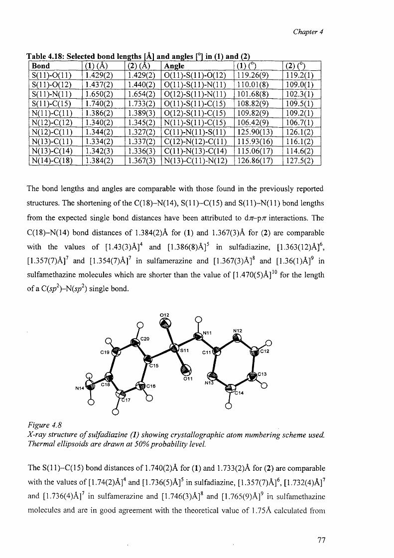
Chapter 4
Table 4.18: Selected bond lengths [A] and angles [°] in (1) and (2)Bond (1) (A) (2) (A) Angle (1) (°) (2) (°)S (ll)-0 (1 1 ) 1.429(2) 1.429(2) 0(11 )-S(l 1)-0(12) 119.26(9) 119.2(1)S(11)-0(12) 1.437(2) 1.440(2) 0(11 )-S(l 1 )-N(l 1) 1 1 0 .0 1 (8 ) 109.0(1)S(11)-N(11) 1.650(2) 1.654(2) 0(12)-S(11)-N(11) 1 0 1 .6 8 (8 ) 102.3(1)S(11 )-C(l 5) 1.740(2) 1.733(2) 0(11 )-S(l 1 )-C(l 5) 108.82(9) 109.5(1)N(1 1 )-C(1 1 ) 1.386(2) 1.389(3) 0(12)-S(11)-C(15) 109.82(9) 109.2(1)N(12)-C(12) 1.340(2) 1.345(2) N(11)-S(11)-C(15) 106.42(9) 106.7(1)N(12)-C(l 1) 1.344(2) 1.327(2) C(11)-N(11)-S(11) 125.90(13) 126.1(2)N(13)-C(l 1) 1.334(2) 1.337(2) C(12)-N(12)-C(l 1) 115.93(16) 116.1(2)N(13)-C(14) 1.342(3) 1.336(3) C(11)-N(13)-C(14) 115.06(17) 114.6(2)N(14)-C(18) 1.384(2) 1.367(3) N(13)-C(l 1)-N(12) 126.86(17) 127.5(2)
The bond lengths and angles are comparable with those found in the previously reported
structures. The shortening of the C(18)-N(14), S(11)—C(15) and S(11)-N (11) bond lengths
from the expected single bond distances have been attributed to d n -p n interactions. The
C(18)-N(14) bond distances o f 1.384(2)A for ( 1 ) and 1.367(3)A for (2 ) are comparable
with the values o f [1.43(3)A ] 4 and [1.386(8)A]5 in sulfadiazine, [1.363(12)A]6,
[1.357(7)A ] 7 and [1.354(7)A ] 7 in sulfamerazine and [1.367(3)A]8 and [1.36(1 )A]9 in
sulfamethazine molecules which are shorter than the value o f [1.470(5)A ]10 for the length
of a C(sp2)-N (sp2) single bond.
0 1 2
N12N11C20
S11 C12C19 C11
C15
,C13011
N13C18 C16N14C14
Figure 4.8X-ray structure o f sulfadiazine (1) showing crystallographic atom numbering scheme used. Thermal ellipsoids are drawn at 50% probability level.
The S(1 1 )—C ( 1 5) bond distances of 1.740(2)A for ( 1 ) and 1.733(2)A for (2 ) are comparable
with the values of [1.74(2)A]4 and [1.736(5)A]5 in sulfadiazine, [1.357(7)A]6, [1.732(4)A]7
and [1.736(4)A]7 in sulfamerazine and [1.746(3)A]8 and [1.765(9)A]9 in sulfamethazine
molecules and are in good agreement with the theoretical value of 1.75A calculated from
77
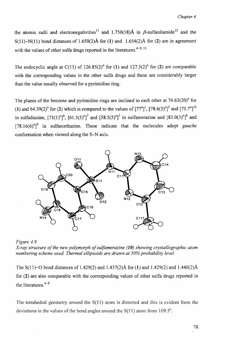
Chapter 4
the atomic radii and electronegativities11 and 1.750(18)A in /2-sulfanilamide12 and the
S(1 1 )-N ( 1 1 ) bond distances of 1.650(2)A for (1) and 1.654(2)A for (2) are in agreement
with the values o f other sulfa drugs reported in the literatures.4-9,13
The endocyclic angle at C ( ll) o f 126.85(2)° for (1) and 127.5(2)° for (2) are comparable
with the corresponding values in the other sulfa drugs and these are considerably larger
than the value usually observed for a pyrimidine ring.
The planes o f the benzene and pyrimidine rings are inclined to each other at 74.63(20)° for
(1) and 64.39(2)° for (2) which is compared to the values o f [770]1, [78.6(3)°]2 and [75.7° ] 13
in sulfadiazine, [71(1)°]6, [61.5(5)°]7 and [58.5(5)°]7 in sulfamerazine and [83.0(3)°]8 and
[78.16(6)°]9 in sulfamethazine. These indicate that the molecules adopt gauche
conformation when viewed along the S-N axis.
N13
0 1 1C14
N11.C20 C11
S11
C19C15 C13
N12012
C12C16
C18
N14 C111C17
Figure 4.9X-ray structure o f the new polymorph o f sulfamerazine (10) showing cryst alio graphic atom numbering scheme used. Thermal ellipsoids are drawn at 50% probability level.
The S( 1 1 )-0 bond distances of 1.429(2) and 1.437(2)A for ( 1 ) and 1.429(2) and 1.440(2)A
for (2 ) are also comparable with the corresponding values of other sulfa drugs reported in
the literatures . 4 - 9
The tetrahedral geometry around the S ( ll) atom is distorted and this is evident from the
deviations in the values o f the bond angles around the S(11) atom from 109.5°.
78
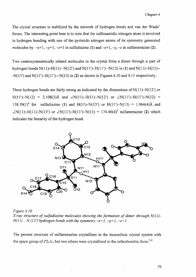
Chapter 4
The crystal structure is stabilized by the network of hydrogen bonds and van der Waals’
forces. The interesting point here is to note that the sulfonamidic nitrogen atom is involved
in hydrogen bonding with one o f the pyrimido nitrogen atoms of its symmetry generated
molecules by -x + 1 , —y+ 1 , -z+ 1 in sulfadiazine (1 ) and -jc+ 1 , - y , - z in sulfamerazine (2 ).
Two centrosymmetrically related molecules in the crystal form a dimer through a pair of
hydrogen bonds N (11)-H (11)-N (12 ') and N(1 T )-H (l l ') -N (1 2 ) in (1) and N (11)-H (11)~
-N(13') and N(1 1')—N(13) in (2) as shown in Figures 4.10 and 4.11 respectively.
These hydrogen bonds are fairly strong as indicated by the dimensions of H(11)-N(12') or
H(1 T)-N (12) = 2.108(3)A and ZN (11)-H (11)-N (12') or ZN(1 l ') -H (ll ') -N (1 2 ) =
158.39(1)° for sulfadiazine (1) and H (ll)-N (1 3 ') or H (ll')-N (1 3 ) = 1.964(4)A and
ZN (11)-H (11)-N (13') or Z N (1 l')-H (l l')-N (13) = 174.46(4)° sulfamerazine (2) which
indicates the linearity of the hydrogen bond.
.C 13
C 1 2C 1 4
N 1 2N 1 3
C 1 1
011 N11H11
C 1 6 S 1 1C 1 7N 1 2 ‘C 1 5
C 1 80 1 2
C 2 0C 1 9
Figure 4.10X-ray structure o f sulfadiazine molecules showing the formation o f dimer through N (ll) - H(11) ...N (12) hydrogen bonds with the symmetry -x+1, -y+1, -z+1.
The present structure of sulfamerazine crystallises in the monoclinic crystal system with
the space group o fP 2\/c , but two others were crystallised in the orthorhombic form . 3 , 4
79
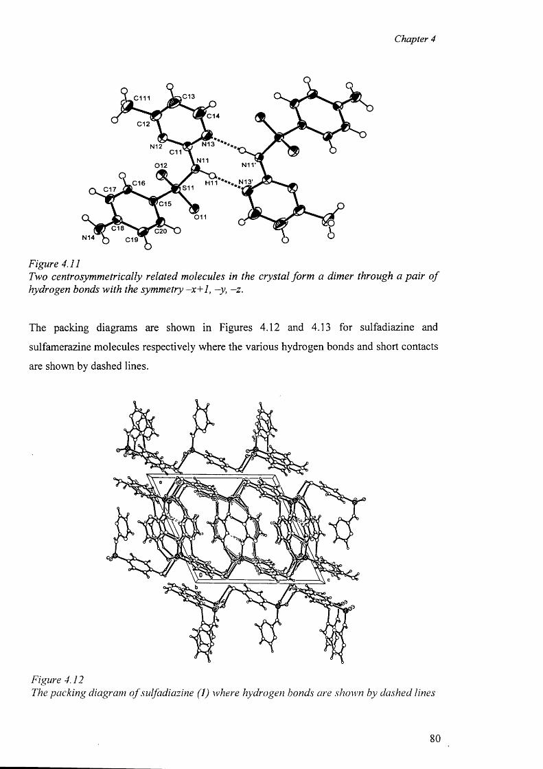
Chapter 4
,C13c m
C14C12
N13N12C11
N11012
N13'H11C16S11C17
C15
0 1 1
C18 C20N14 C19
Figure 4.11Two centrosymmetrically related molecules in the crystal form a dimer through a pair o f hydrogen bonds with the symmetry -x+ 1, -y, -z .
The packing diagrams are shown in Figures 4.12 and 4.13 for sulfadiazine and
sulfamerazine molecules respectively where the various hydrogen bonds and short contacts
are shown by dashed lines.
Figure 4.12The packing diagram o f sulfadiazine (1) where hydrogen bonds are shown by dashed lines
80
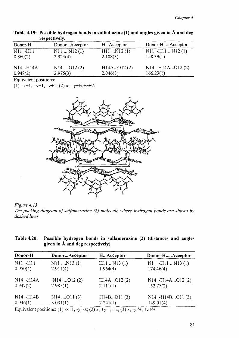
Chapter 4
Table 4.19: Possible hydrogen bonds in sulfadiazine (1) and angles given in A and deg ___________ respectively.__________________________________________________________Donor-H Donor... Acceptor H... Acceptor Donor-H.... AcceptorN i l -H ll N i l ....N12 (1) H ll ...N12 (1) N i l -H ll ...N12 (1)0.860(2) 2.924(4) 2.108(3) 158.39(1)
N14 -H14A N14 ....012(2) H14A...012 (2) N14 -H14A...012 (2)0.948(2) 2.975(3) 2.046(3) 166.23(1)Equivalent positions:( 1 ) -x + 1 , -y + 1 , - z + 1 ; (2 ) x, -y+V^+z+Vi
Figure 4.13The packing diagram o f sulfamerazine (2) molecule where hydrogen bonds are shown by dashed lines.
Table 4.20: Possible hydrogen bonds in sulfamerazine (2) (distances and anglesgiven in A and deg respectively)
Donor-H Donor... Acceptor H...Acceptor Donor-H.....Acceptor
N il -H ll N i l ....N13 (1) H l l ...N13 (1) N i l -H ll ...N13 (1)0.950(4) 2.911(4) 1.964(4) 174.46(4)
N14 -H14A N14 ....012 (2) H14A...012 (2) N14 -H14A...012 (2)0.947(2) 2.985(1) 2 . 1 1 1 ( 1 ) 152.75(2)
N14 -H14B N 14....011 (3) H14B...011 (3) N14 -H 14B...011 (3)0.946(1) 3.091(1) 2.241(1) 149.01(4)Equivalent positions: (1) -x+1, -y, -z; (2) x, +y-l, +z; (3) x, -y-Vi, +z+ 1/ 2
81
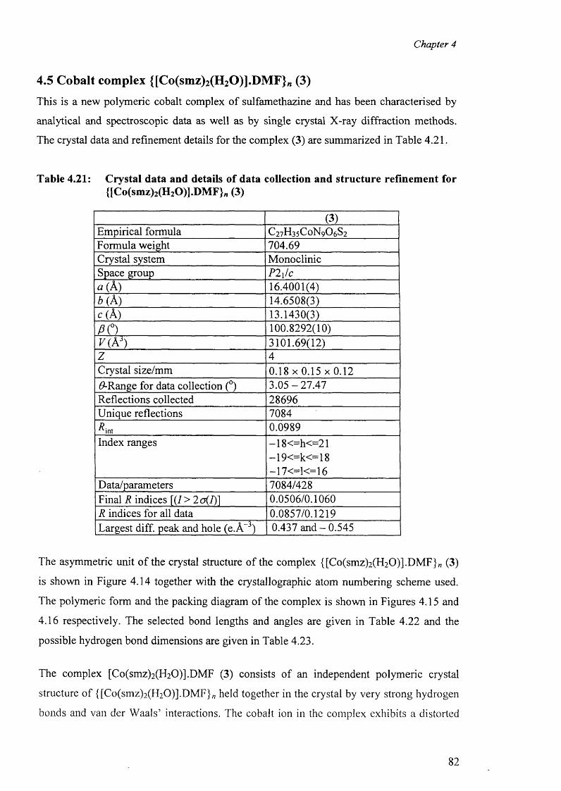
Chapter 4
4.5 Cobalt complex {[Co(smz)2(H20)].DMF}„ (3)
This is a new polymeric cobalt complex o f sulfamethazine and has been characterised by
analytical and spectroscopic data as well as by single crystal X-ray diffraction methods.
The crystal data and refinement details for the complex (3) are summarized in Table 4.21.
Table 4.21: Crystal data and details of data collection and structure refinement for{ICo(smz)2(H20)].DM F}#l (3)
(3)Empirical formula C2 7 H 3 5 C 0 N 9 O6 S2
Formula weight 704.69Crystal system MonoclinicSpace group P2\/ca (A) 16.4001(4)b (A) 14.6508(3)c(A ) 13.1430(3)
P O 100.8292(10)V(Ai) 3101.69(12)z 4Crystal size/mm 0 .1 8 x 0 .1 5 x 0 .1 2#-Range for data collection (°) 3 .0 5 -2 7 .4 7Reflections collected 28696Unique reflections 7084^int 0.0989Index ranges —18<=h<=21
—19<=k<=18 —17<=1<=16
Data/parameters 7084/428Final R indices [(/> 2o(I)] 0.0506/0.1060R indices for all data 0.0857/0.1219Largest diff. peak and hole (e.A-3) 0.437 an d -0 .5 4 5
The asymmetric unit o f the crystal structure o f the complex {[Co(smz)2 (H2 0 )].DMF}„ (3)
is shown in Figure 4.14 together with the crystallographic atom numbering scheme used.
The polymeric form and the packing diagram of the complex is shown in Figures 4.15 and
4.16 respectively. The selected bond lengths and angles are given in Table 4.22 and the
possible hydrogen bond dimensions are given in Table 4.23.
The complex [Co(smz)2 (H2 0 )].DMF (3) consists o f an independent polymeric crystal
structure o f {[Co(smz)2 (H2 0 )].DMF}„ held together in the crystal by very strong hydrogen
bonds and van der Waals’ interactions. The cobalt ion in the complex exhibits a distorted
82
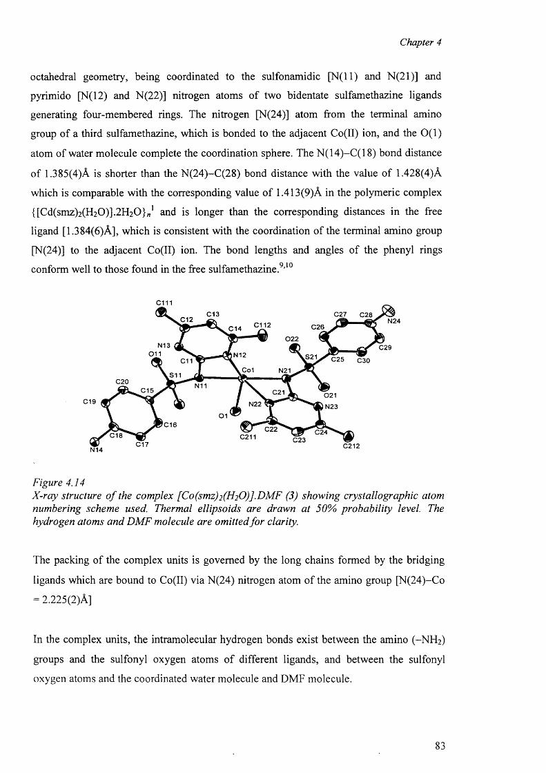
Chapter 4
octahedral geometry, being coordinated to the sulfonamidic [N (ll) and N (2 1 )] and
pyrimido [N(12) and N(22)] nitrogen atoms o f two bidentate sulfamethazine ligands
generating four-membered rings. The nitrogen [N(24)] atom from the terminal amino
group of a third sulfamethazine, which is bonded to the adjacent Co(II) ion, and the 0 (1)
atom of water molecule complete the coordination sphere. The N(14)-C(18) bond distance
of 1.385(4)A is shorter than the N(24)-C(28) bond distance with the value of 1.428(4)A
which is comparable with the corresponding value o f 1.413(9)A in the polymeric complex
{[Cd(smz)2 (H2 0 )].2 H2 0 } „ 1 and is longer than the corresponding distances in the free
ligand [1.384(6)A], which is consistent with the coordination of the terminal amino group
[N(24)J to the adjacent Co(II) ion. The bond lengths and angles of the phenyl rings
conform well to those found in the free sulfamethazine . 9 , 1 0
cmC13 C27 C28C12 N24C112 C26C14
022N13 C29
0 1 1 N12 S21 C25 C30C11Co1 N21
S11C20 N11
C15 C21 021C19 N22 *& r
® r-, C2 2C211
N2301
C16C24C18 C23C17 C212N14
Figure 4.14X-ray structure o f the complex [Co(smz)2(H20)].DMF (3) showing crystallographic atom numbering scheme used. Thermal ellipsoids are drawn at 50% probability level. The hydrogen atoms and DM F molecule are omitted fo r clarity.
The packing of the complex units is governed by the long chains formed by the bridging
ligands which are bound to Co(II) via N(24) nitrogen atom of the amino group [N(24)-Co
= 2.225(2)A]
In the complex units, the intramolecular hydrogen bonds exist between the amino (-N H 2 )
groups and the sulfonyl oxygen atoms o f different ligands, and between the sulfonyl
oxygen atoms and the coordinated water molecule and DMF molecule.
83
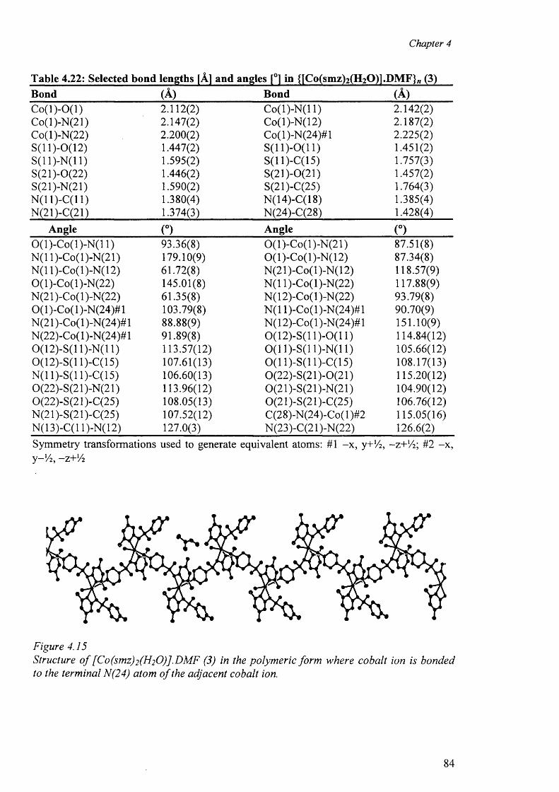
Chapter 4
Table 4.22: Selected bond lengths [A] and angles [°1 in {[Co(smz)2(H2Q)1.DMF}/lBond (A) Bond (A )C o (l)-0 (l) 2 .1 1 2 (2 ) Co(l)-N (l 1) 2.142(2)Co(l)-N(21) 2.147(2) Co(l)-N(12) 2.187(2)Co(l)-N(22) 2 .2 0 0 (2 ) Co(l)-N(24)#l 2.225(2)S(11 )-0 (l 2) 1.447(2) S (ll)-0 (1 1 ) 1.451(2)S(11)-N(11) 1.595(2) S(11 )-C(l 5) 1.757(3)S(21)-0(22) 1.446(2) S(21)-0(21) 1.457(2)S(21)-N(21) 1.590(2) S(21)-C(25) 1.764(3)N (1 1 )-C(1 1 ) 1.380(4) N(14)-C(18) 1.385(4)N(21)-C(21) 1.374(3) N(24)-C(28) 1.428(4)
Angle (°) Angle (°)0 (l)-C o(l)-N (l 1) 93.36(8) 0(l)-C o(l)-N (21) 87.51(8)N(1 l)-Co(l)-N(21) 179.10(9) 0(l)-C o(l)-N (12) 87.34(8)N(1 l)-Co(l)-N(12) 61.72(8) N(21)-Co(l)-N(12) 118.57(9)0(l)-C o(l)-N (22) 145.01(8) N(1 l)-Co(l)-N(22) 117.88(9)N (21 )-Co( 1 )-N(22) 61.35(8) N(12)-Co(l)-N(22) 93.79(8)0(1 )-Co( 1 )-N(24)# 1 103.79(8) N(1 l)-Co(l)-N (24)#l 90.70(9)N (21 )-Co(l )-N(24)#l 88.88(9) N(12)-Co(l)-N(24)#l 151.10(9)N(22)-Co(l)-N(24)#l 91.89(8) 0(12)-S(11)-0(11) 114.84(12)0 (1 2)-S(l 1)-N(11) 113.57(12) 0 (1 1)-S(11)-N(11) 105.66(12)0(12)-S(11 )-C(l 5) 107.61(13) 0 (1 1)-S(11)-C(15) 108.17(13)N (ll)-S (ll)-C (15 ) 106.60(13) 0(22)-S(21)-0(21) 115.20(12)0(22)-S(21)-N(21) 113.96(12) 0(21)-S(21)-N(21) 104.90(12)0(22)-S(21 )-C(25) 108.05(13) 0(21)-S(21)-C(25) 106.76(12)N(21)-S(21)-C(25) 107.52(12) C(28)-N(24)-Co( 1 )#2 115.05(16)N(13)-C(l 1)-N(12) 127.0(3) N(23)-C(21 )-N(22) 126.6(2)Symmetry transformations used to generate equivalent atoms: # 1 -x , y+14, -z+Vr, #2 -x ,y-Vz, -z+ lA
Figure 4.15Structure o f [Co(smz)2(H20)].DMF (3) in the polymeric form where cobalt ion is bonded to the terminal N(24) atom o f the adjacent cobalt ion.
84
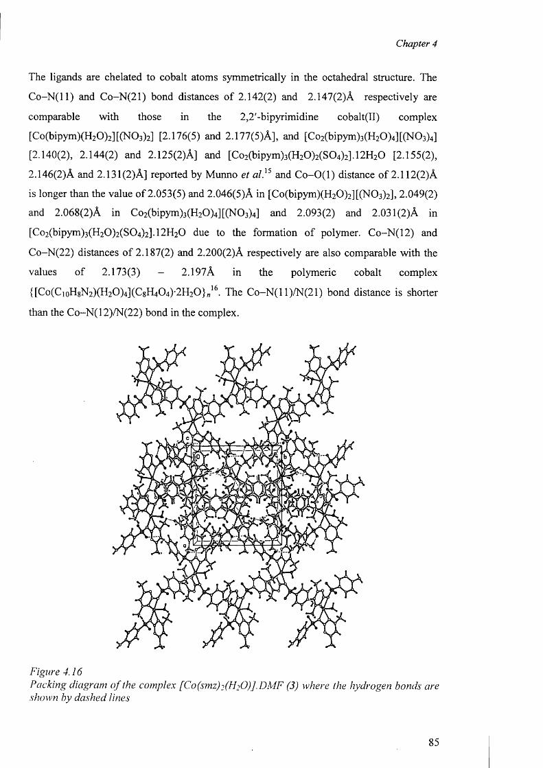
Chapter 4
The ligands are chelated to cobalt atoms symmetrically in the octahedral structure. The
C o -N (ll) and Co-N(21) bond distances o f 2.142(2) and 2.147(2)A respectively are
comparable with those in the 2,2'-bipyrimidine cobalt(II) complex
[Co(bipym)(H2 0 )2 ][(N 0 3 )2 ] [2.176(5) and 2.177(5)A], and [Co2 (bipym)3 (H2 0 )4 ][(N 0 3 )4 ]
[2.140(2), 2.144(2) and 2.125(2)A] and [Co2 (bipym)3 (H2 0 )2 (S0 4 )2 ].1 2 H2 0 [2.155(2),
2.146(2)A and 2.131(2)A] reported by Munno et a l}5 and C o-O (l) distance o f 2 .1 1 2 (2 )A
is longer than the value o f 2.053(5) and 2.046(5)A in [Co(bipym)(H 2 0 )2 ][(N 0 3 )2 ], 2.049(2)
and 2.068(2)A in Co2 (bipym)3 (H2 0 )4 ][(N 0 3 )4 ] and 2.093(2) and 2.031(2)A in
[Co2 (bipym)3 (H2 0 )2 (S0 4 )2 ].1 2 H 2 0 due to the formation of polymer. Co-N(12) and
C o-N (2 2 ) distances o f 2.187(2) and 2.200(2)A respectively are also comparable with the
values of 2.173(3) - 2.197A in the polymeric cobalt complex
{[Co(CioH8N 2 )(H2 0 )4 ](C 8 H4 0 4 )-2 H2 0 }„16. The C o-N (l 1)/N(21) bond distance is shorter
than the Co-N(12)/N(22) bond in the complex.
Figure 4.16Packing diagram o f the complex [Co(smz)2(H20)].DMF (3) where the hydrogen bonds are shown by dashed lines
85
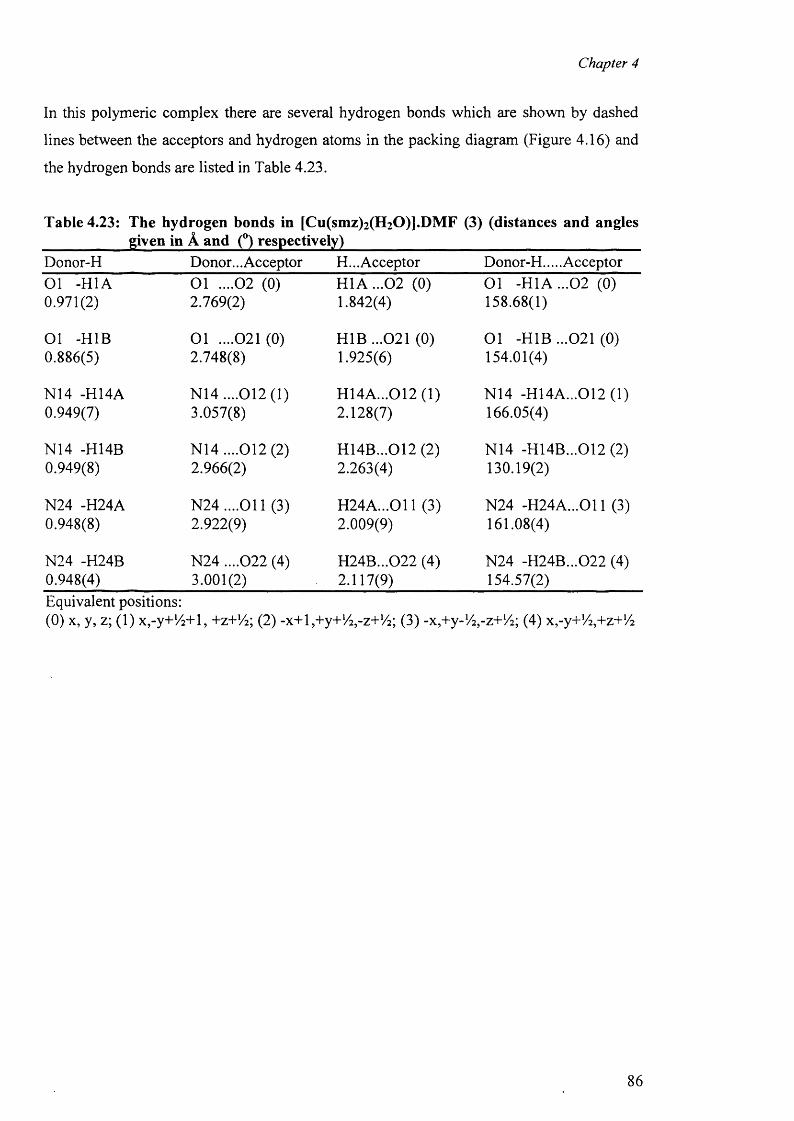
Chapter 4
In this polymeric complex there are several hydrogen bonds which are shown by dashed
lines between the acceptors and hydrogen atoms in the packing diagram (Figure 4.16) and
the hydrogen bonds are listed in Table 4.23.
Table 4.23: The hydrogen bonds in [Cu(smz)2(H20)].DMF (3) (distances and anglesgiven in A and (°) respectively)
Donor-H Donor... Acceptor H... Acceptor Donor-H.... AcceptorO l -H1A O l . . . . 0 2 (0 ) H 1A ...02 (0) O l -H 1A ...02 (0)0.971(2) 2.769(2) 1.842(4) 158.68(1)
Ol -H1B O l . . . . 0 2 1 (0 ) H1B ...021 (0) O l -H1B ...021 (0)0.886(5) 2.748(8) 1.925(6) 154.01(4)
N14 -H14A N14 ....012(1) H14A...012 (1) N14 -H14A...012 (1)0.949(7) 3.057(8) 2.128(7) 166.05(4)
N14 -H14B N14 ....012 (2) H14B...012 (2) N14 -H14B...012 (2)0.949(8) 2.966(2) 2.263(4) 130.19(2)
N24 -H24A N 24....011 (3) H 24A ...011 (3) N24 -H 24A ...011 (3)0.948(8) 2.922(9) 2.009(9) 161.08(4)
N24 -H24B N24 ....022 (4) H24B...022 (4) N24 -H24B...022 (4)0.948(4) 3.001(2) 2.117(9) 154.57(2)Equivalent positions:(0) x, y, z; (1) x,-y+/4+l, +z+!/2; (2) -x+E+y+V^-z+l^; (3) -x,+y-l/2 ,-z+l/ 2; (4) x^y+V^+z+’/a
86
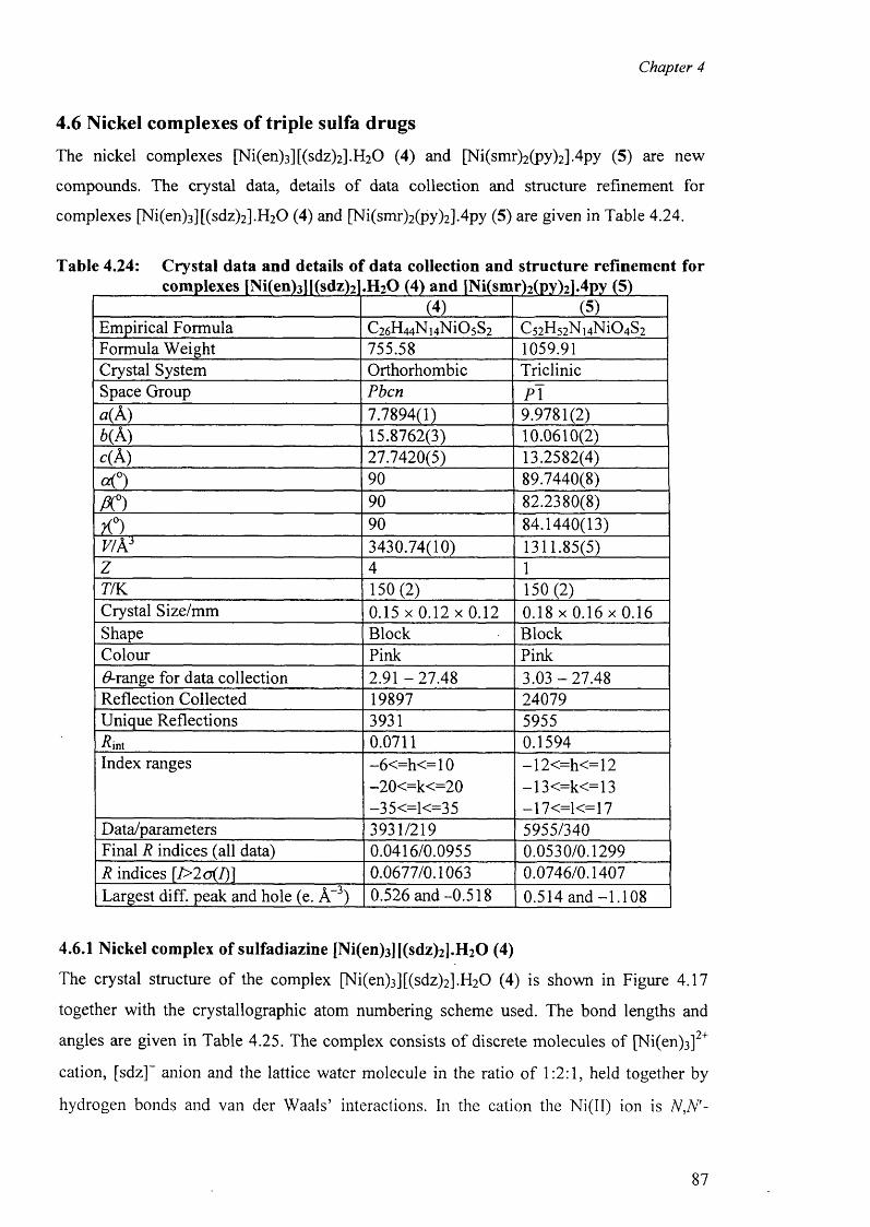
Chapter 4
4.6 Nickel complexes of triple sulfa drugs
The nickel complexes [Ni(en)3 ][(sdz)2 ].H 2 0 (4) and [Ni(smr)2 (py)2 ].4 py (5) are new
compounds. The crystal data, details of data collection and structure refinement for
complexes [Ni(en)3 ][(sdz)2 ].H2 0 (4) and [Ni(smr)2 (py)2 ].4 py (5) are given in Table 4.24.
Table 4.24: Crystal data and details of data collection and structure refinement forcomplexes [Ni(en)3l|(sdz)2].H20 (4) and [Ni(smr)2(py)21.4py (5)
(4) (5)Em pirical Form ula C26H44N14N i05S 2 C 52H52N14N i04S 2Form ula W eight 755.58 1059.91Crystal System O rthorhom bic T riclinicSpace Group Pbcn P \a(A) 7.7894(1) 9.9781(2)6 (A) 15.8762(3) 10.0610(2)c( A) 27.7420(5) 13.2582(4)
O 90 89.7440(8)
PC) 90 82.2380(8)
X°) 90 84.1440(13)V/As 3430.74(10) 1311.85(5)z 4 1
77K 150 (2) 150 (2)Crystal S ize/m m 0 .1 5 x 0 .1 2 x 0 .1 2 0 .1 8 x 0 .1 6 x 0 .1 6Shape B lock B lockColour Pink PinkG range for data co llection 2 .9 1 -2 7 .4 8 3.03 -2 7 .4 8R eflection C ollected 19897 24079U nique R eflection s 3931 5955■ int 0.0711 0.1594Index ranges -6 < = h < = 1 0 —12 < = h < = 12
-2 0 < = k < = 2 0 —13<=k<=13-35<=1<=35 -17<=1<=17
Data/param eters 3931/219 5955/340Final R ind ices (all data) 0.0416/0.0955 0.0530/0.1299R indices |7>2 o (/)] 0.0677/0.1063 0.0746/0.1407Largest diff. peak and h o le (e. A-3) 0.526 and -0.518 0.514 a n d -1.108
4.6.1 Nickel complex of sulfadiazine [Ni(en)3][(sdz)2].H20 (4)
The crystal structure o f the complex [Ni(en)3 ][(sdz)2 ].H 2 0 (4) is shown in Figure 4.17
together with the crystallographic atom numbering scheme used. The bond lengths and
angles are given in Table 4.25. The complex consists o f discrete molecules of [Ni(en)3 ]2+
cation, [sdz]~ anion and the lattice water molecule in the ratio o f 1 :2 :1 , held together by
hydrogen bonds and van der Waals’ interactions. In the cation the Ni(II) ion is N,N'~
87
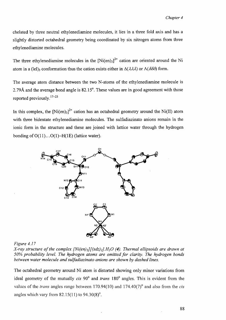
Chapter 4
chelated by three neutral ethylenediamine molecules, it lies in a three fold axis and has a
slightly distorted octahedral geometry being coordinated by six nitrogen atoms from three
ethylenediamine molecules.
The three ethylenediamine molecules in the [Ni(en)3 ]2+ cation are oriented around the Ni
atom in a (lel) 3 conformation thus the cation exists either in A(T2Jl) or A(SSS) form.
The average atom distance between the two N-atoms o f the ethylenediamine molecule is
2.79A and the average bond angle is 82.15°. These values are in good agreement with those1 7—9Treported previously.
In this complex, the [Ni(en)3 ]2+ cation has an octahedral geometry around the Ni(II) atom
with three bidentate ethylenediamine molecules. The sulfadiazinato anions remain in the
ionic form in the structure and these are joined with lattice water through the hydrogen
bonding of 0(11).. .0(1)-H (1E) (lattice water).
0 1C17
£160 1 1
N14 C18 SC15
C20 S11 '012
N11
N12 IC11
N13C12
C13 C14
cr ci
N1
.NilN2 N2'
N3N3'
Figure 4.17X-ray structure o f the complex [Ni(en)3] [(sdz)2] .H2 O (4). Thermal ellipsoids are drawn at 50% probability level. The hydrogen atoms are omitted fo r clarity. The hydrogen bonds between water molecule and sulfadiazinato anions are shown by dashed lines.
The octahedral geometry around Ni atom is distorted showing only minor variations from
ideal geometry o f the mutually cis 90° and trans 180° angles. This is evident from the
values of the trans angles range between 170.94(10) and 174.40(7)° and also from the cis
angles which vary from 82.15(11) to 94.30(8)°.
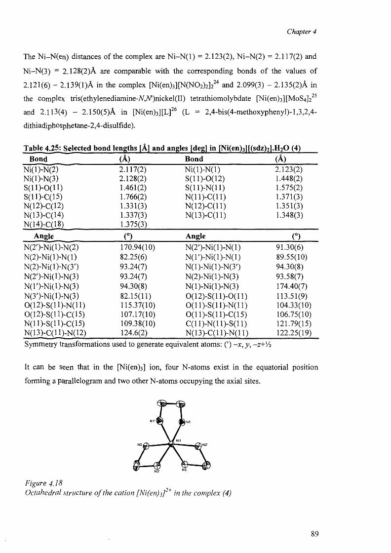
Chapter 4
The Ni-N(en) distances of the complex are N i-N (l) = 2.123(2), N i-N (2) = 2.117(2) and
N i-N (3 ) = 2.128(2)A are comparable with the corresponding bonds o f the values of
2.121(6) - 2.139(1 )A in the complex [Ni(en)3 ][N(N0 2 )2 ]2 2 4 and 2.099(3) - 2.135(2)A in
the complex tris(ethylenediamine-A^,7V’)nickel(II) tetrathiomolybdate [Ni(en)3 ][MoS4 ]2 2 5
and 2.113(4) - 2.150(5)A in [Ni(en)3 ][L ] 2 6 (L = 2,4-bis(4-methoxyphenyl)-l,3,2,4-
dithiadiphosphetane-2,4-disulfide).
Table 4.25: Selected bond lengths [A] and angles [degj in [Ni(en)3 ][(sdz)2 ].H2Q (4)Bond (A) Bond (A)
Ni(l)-N(2) 2.117(2) N i(l)-N (l) 2.123(2)Ni(l)-N(3) 2.128(2) S(11)-0(12) 1.448(2)S (ll)-0 (11) 1.461(2) S(11)-N(11) 1.575(2)S(11)-C(15) 1.766(2) N(11)-C(11) 1.371(3)N(12)-C(12) 1.331(3) N(12)-C(l 1) 1.351(3)N(13)-C(14) 1.337(3) N(13)-C(l 1) 1.348(3)N(14)-C(18) 1.375(3)
Angle (°) Angle oN(2')-Ni(l)-N(2) 170.94(10) N (2')-N i(l)-N (l) 91.30(6)N (2)-N i(l)-N(l) 82.25(6) N (l')-N i(l)-N (l) 89.55(10)N(2)-Ni(l)-N(3') 93.24(7) N (l)-N i(l)-N (3 ') 94.30(8)N(2')-Ni(l)~N(3) 93.24(7) N(2)-Ni(l)-N(3) 93.58(7)N(T)-Ni(l)-N(3) 94.30(8) N (l)-N i(l)-N (3) 174.40(7)N(3')-Ni(l)-N(3) 82.15(11) 0(12)-S(11)-0(11) 113.51(9)0(12)-S(1 l) -N (ll) 115.37(10) 0 (1 1)-S(11)-N(11) 104.33(10)0(12)-S(H)-C(15) 107.17(10) 0 (1 1)-S(11)-C(15) 106.75(10)N(11)-S(H)-C(15) 109.38(10) C(11)-N(11)-S(11) 121.79(15)N(13)-C(H)-N(12) 124.6(2) N(13)-C(l 1)-N(11) 122.25(19)Symmetry transformations used to generate equivalent atoms: (') -x , y, -z+'A
It can be seen that in the [Ni(en)3] ion, four N-atoms exist in the equatorial position
forming a parallelogram and two other N-atoms occupying the axial sites.
N V
Figure 4.18Octahedral structure o f the cation [Ni(en)^]2+ in the complex (4)
89
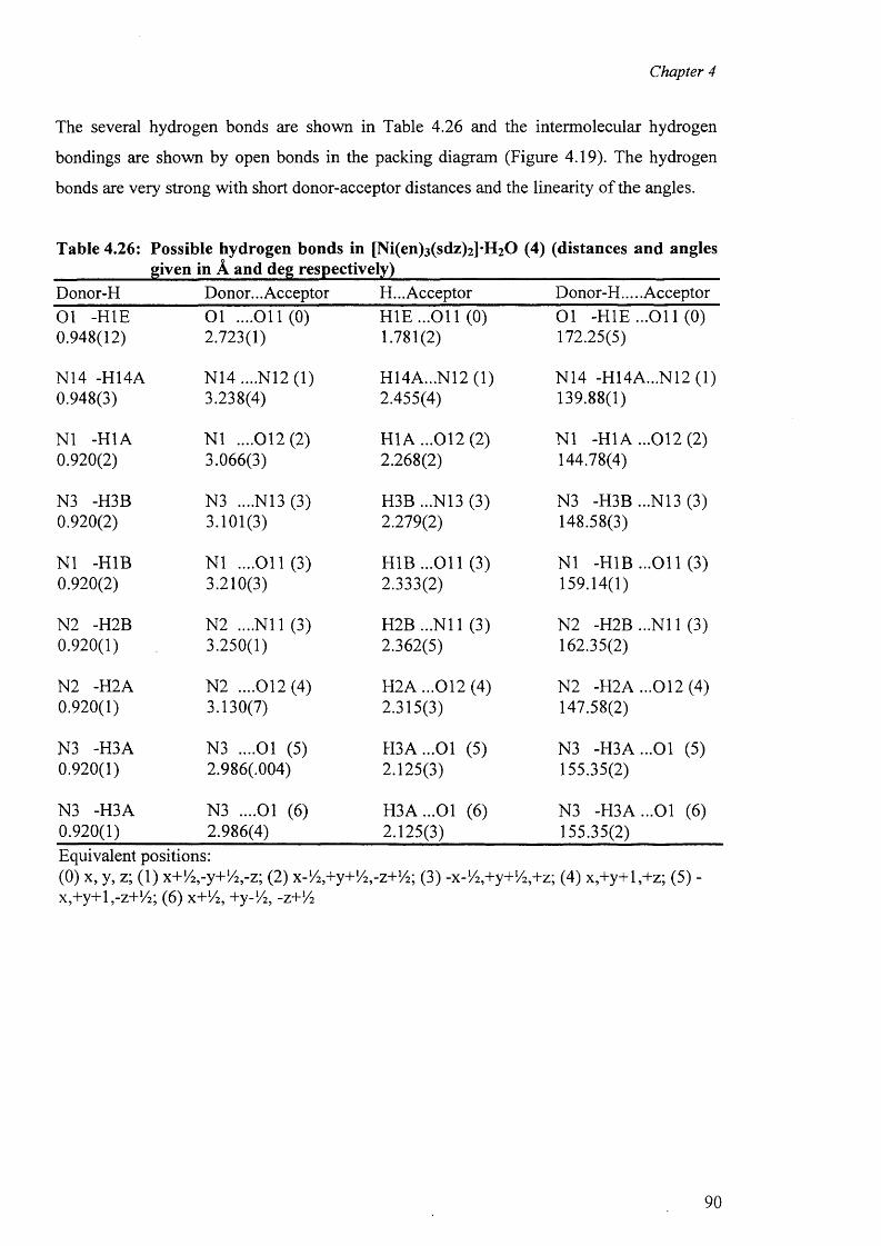
Chapter 4
The several hydrogen bonds are shown in Table 4.26 and the intermolecular hydrogen
bondings are shown by open bonds in the packing diagram (Figure 4.19). The hydrogen
bonds are very strong with short donor-acceptor distances and the linearity of the angles.
Table 4.26: Possible hydrogen bonds in [Ni(en)3(sdz)2]*H20 (4) (distances and anglesgiven in A and deg respectively)
Donor-H Donor... Acceptor H... Acceptor Donor-H.....Acceptor01 -HIE 0.948(12)
O l ....011(0) 2.723(1)
H IE ...O il (0) 1.781(2)
O l -H IE ...O il (0) 172.25(5)
N14 -H14A 0.948(3)
N14 ....N12 (1) 3.238(4)
H14A...N12 (1) 2.455(4)
N14 -H14A...N12 (1) 139.88(1)
NI -H1A 0.920(2)
N I ....012(2) 3.066(3)
H1A ...012 (2) 2.268(2)
N I -H1A ...012 (2) 144.78(4)
N3 -H3B 0.920(2)
N3 ....N13 (3) 3.101(3)
H3B ...N13 (3) 2.279(2)
N3 -H3B ...N13 (3) 148.58(3)
NI -H1B 0.920(2)
NI ....011(3) 3.210(3)
H 1B ...011 (3) 2.333(2)
N I -H 1B ...011 (3) 159.14(1)
N2 -H2B 0.920(1)
N2 ....N il (3) 3.250(1)
H2B ...N il (3) 2.362(5)
N2 -H2B ...N il (3) 162.35(2)
N2 -H2A 0.920(1)
N2 ....012(4) 3.130(7)
H2A ...012 (4) 2.315(3)
N2 -H2A ...012 (4) 147.58(2)
N3 -H3A 0.920(1)
N3 ....Ol (5) 2.986(.004)
H 3A ...01 (5) 2.125(3)
N3 -H 3A ...01 (5) 155.35(2)
N3 -H3A 0.920(1)
N3 ....Ol (6 ) 2.986(4)
H 3A ...01 (6 ) 2.125(3)
N3 -H 3A ...01 (6 ) 155.35(2)
Equivalent positions:(0) x, y, z; (1) x+!/2 ,-y+!/2 ,-z; (2) x-1/ 2 ,+y+1/ 2 ,-z+1/2; (3) -x-V^+y+^+z; (4) x,+y+l,+z; (5) - x,+y+l,-z+!/2; (6 ) x+1/2, +y-lA, -z+Yz
90
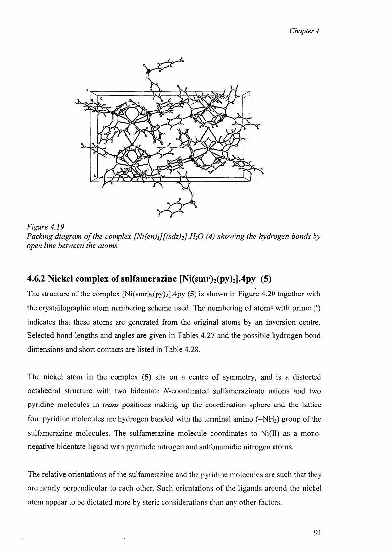
Chapter 4
Figure 4.19Packing diagram o f the complex [Ni(en)s][(sdz)2].H 2 0 (4) showing the hydrogen bonds by open line between the atoms.
4.6.2 Nickel complex of sulfamerazine [Ni(smr)2(py)2].4py (5)
The structure o f the complex [Ni(smr)2(py)2].4py (5) is shown in Figure 4.20 together with
the crystallographic atom numbering scheme used. The numbering o f atoms with prime (')
indicates that these atoms are generated from the original atoms by an inversion centre.
Selected bond lengths and angles are given in Tables 4.27 and the possible hydrogen bond
dimensions and short contacts are listed in Table 4.28.
The nickel atom in the complex (5) sits on a centre o f symmetry, and is a distorted
octahedral structure with two bidentate ^-coordinated sulfamerazinato anions and two
pyridine molecules in trans positions making up the coordination sphere and the lattice
four pyridine molecules are hydrogen bonded with the terminal amino (-N H 2 ) group of the
sulfamerazine molecules. The sulfamerazine molecule coordinates to Ni(II) as a mono
negative bidentate ligand with pyrimido nitrogen and sulfonamidic nitrogen atoms.
The relative orientations o f the sulfamerazine and the pyridine molecules are such that they
are nearly perpendicular to each other. Such orientations o f the ligands around the nickel
atom appear to be dictated more by steric considerations than any other factors.
91
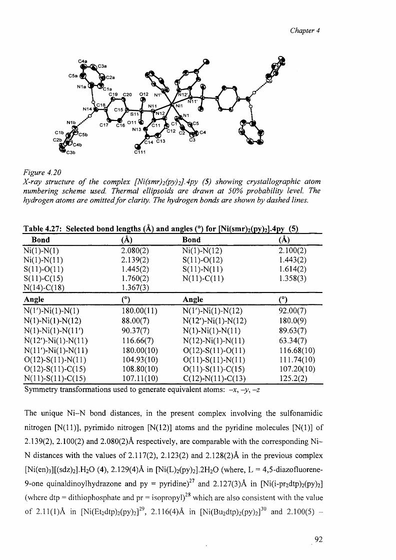
Chapter 4
Figure 4.20X-ray structure o f the complex [Ni(smr)2 (py)2]-4 py (5) showing crystallographic atom numbering scheme used. Thermal ellipsoids are drawn at 50% probability level. The hydrogen atoms are omitted fo r clarity. The hydrogen bonds are shown by dashed lines.
Bond (A) Bond (A)N i(l)-N (l) 2.080(2) N i(l)-N(12) 2.100(2)N i(l)-N (l 1) 2.139(2) S(11)-0(12) 1.443(2)S (ll)-0 (11 ) 1.445(2) S ( ll) -N ( ll) 1.614(2)S(ll)-C(15) 1.760(2) N(11)-C(11) 1.358(3)N(14)-C(18) 1.367(3)Angle (°) Angle (°)N (T)-N i(l)-N (l) 180.00(11) N(T)-Ni(l)-N(12) 92.00(7)N (l)-N i(l)-N(12) 88.00(7) N(12')-Ni(l)-N(12) 180.0(9)N (l)-N i(l)-N (l 1') 90.37(7) N (l)-N i(l)-N (l 1) 89.63(7)N (12 ')-N i(l)-N (ll) 116.66(7) N(12)-Ni(l)-N(l 1) 63.34(7)N (1T )-N i(l)-N (ll) 180.00(10) 0(12)-S(11)-0(11) 116.68(10)0(12)-S(11)-N(11) 104.93(10) 0 (1 1)-S(11)-N(11) 111.74(10)0(12)-S(11)-C(15) 108.80(10) 0 (1 1)-S(11 )-C(l 5) 107.20(10)N(11)-S(11)-C(15) 107.11(10) C(12)-N(l 1)-C(13) 125.2(2)Symmetry transformations used to generate equivalent atoms: -x , -y , - z
The unique N i-N bond distances, in the present complex involving the sulfonamidic
nitrogen [N(l 1)], pyrimido nitrogen [N (l2)] atoms and the pyridine molecules [N (l)] of
2.139(2), 2.100(2) and 2.080(2)A respectively, are comparable with the corresponding N i-
N distances with the values of 2.117(2), 2.123(2) and 2.128(2)A in the previous complex
[Ni(en)3 ][(sdz)2 ].H2 0 (4), 2.129(4)A in [Ni(L)2 (py)2 ].2 H2 0 (where, L = 4,5-diazofluorene-
9-one quinaldinoylhydrazone and py = pyridine)27 and 2.127(3)A in [Ni(i-pr2 dtp)2 (py)2 ]
(where dtp = dithiophosphate and pr = isopropyl)28 which are also consistent with the value
of 2.11(1)A in [Ni(Et2dtp)2(py)2]29, 2.116(4)A in [Ni(Bu2dtp)2(py)2]30 and 2.100(5) -
92
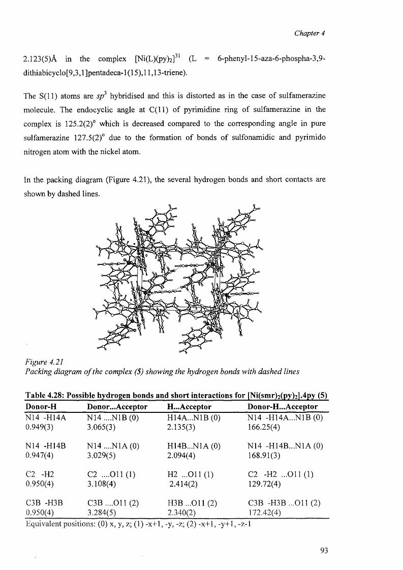
Chapter 4
2.123(5)A in the complex [Ni(L)(py)2]31 (L = 6-phenyl-15-aza-6-phospha-3,9-
dithiabicy clo [9,3,1 Jpentadeca-1 (15), 11,13-triene).
The S ( ll) atoms are sp3 hybridised and this is distorted as in the case of sulfamerazine
molecule. The endocyclic angle at C ( ll) of pyrimidine ring of sulfamerazine in the
complex is 125.2(2)° which is decreased compared to the corresponding angle in pure
sulfamerazine 127.5(2)° due to the formation of bonds of sulfonamidic and pyrimido
nitrogen atom with the nickel atom.
In the packing diagram (Figure 4.21), the several hydrogen bonds and short contacts are
shown by dashed lines.
Figure 4.21Packing diagram o f the complex (5) showing the hydrogen bonds with dashed lines
Table 4.28: Possible hydrogen bonds and short interactions for [Ni(smr)2(py)2l.4py (5)Donor-H Donor...Acceptor H...Acceptor Donor-H... AcceptorN14 -H14A N14 ....NIB (0) H14A...N1B (0) N14 -H14A...N1B (0)0.949(3) 3.065(3) 2.135(3) 166.25(4)
N14 -H14B N14 ....N1A(0) H14B...N1 A (0) N14 -H14B...N1A (0)0.947(4) 3.029(5) 2.094(4) 168.91(3)
C2 -H2 C2 ....O il (1) H2 ...O il (1) C2 -H2 ...O il (1)0.950(4) 3.108(4) 2.414(2) 129.72(4)
C3B -H3B C3B ....O il (2) H3B ...O il (2) C3B -H 3B ...011 (2)0.950(4) 3.284(5) 2.340(2) 172.42(4)Equivalent positions: (0) x, y, z; (1) -x+1, -y, -z; (2) -x+1, -y+1, -z-1
93
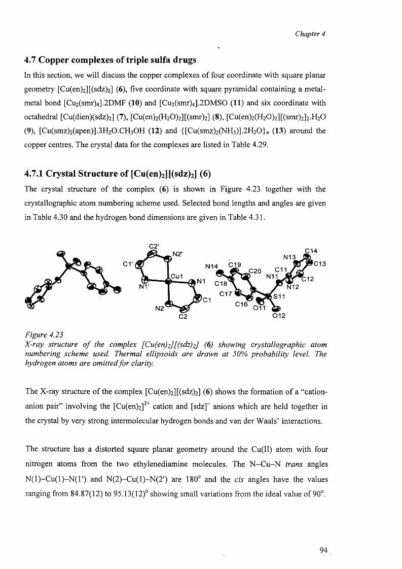
Chapter 4
4.7 Copper complexes of triple sulfa drugs
In this section, we will discuss the copper complexes of four coordinate with square planar
geometry [Cu(en)2 ][(sdz)2 ] (6), five coordinate with square pyramidal containing a metal -
metal bond [Cu2(smr)4].2DMF (10) and [Cu2(smr)4].2DMSO (11) and six coordinate with
octahedral [Cu(dien)(sdz)2] (7), [Cu(en)2(H20)2][(smr)2] (8), [Cu(en)2(H20)2][(smr)2]2.H20
(9), [Cu(smz)2 (apen)].3 H2 0 .CH3 0 H (12) and {[Cu(smz)2(NH3)].2H20}„ (13) around the
copper centres. The crystal data for the complexes are listed in Table 4.29.
4.7.1 Crystal Structure of [Cu(en)2] [(sdz)2] (6)
The crystal structure o f the complex (6) is shown in Figure 4.23 together with the
crystallographic atom numbering scheme used. Selected bond lengths and angles are given
in Table 4.30 and the hydrogen bond dimensions are given in Table 4.31.
C2 012
Figure 4.23X-ray structure o f the complex [Cu(en)2][(sdz)2] (6) showing crystallographic atom numbering scheme used. Thermal ellipsoids are drawn at 50% probability level. The hydrogen atoms are omitted fo r clarity.
The X-ray structure o f the complex [Cu(en)2 ][(sdz)2] (6) shows the formation o f a “cation-
anion pair” involving the [Cu(en)2] cation and [sdz] anions which are held together in
the crystal by very strong intermolecular hydrogen bonds and van der W aals’ interactions.
The structure has a distorted square planar geometry around the Cu(II) atom with four
nitrogen atoms from the two ethylenediamine molecules. The N -C u -N trans angles
N (l)-C u (l)-N (T ) and N (2)-C u(l)-N (2 ') are 180° and the cis angles have the values
ranging from 84.87(12) to 95.13(12)° showing small variations from the ideal value of 90°.
94
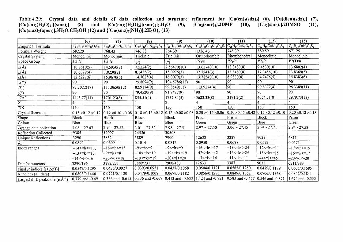
Table 4.29: Crystal data and details of data collection and structure refinement for [Cu(en)2 (sdz)2 ] (6), [Cu(dien)(sdz)2 j (7),[Cu(en)2(H20)2][(smr)2] (8) and [Cu(en)2(H20)2][(smr)2]2.H20 (9), [Cu2(smr)4].2DMF (10), [Cu2(smr)4].2DMSO (11),[Cu(smz)2(apen)].3H2O.CH3OH (12) and {[Cu(smz)2(NH3)].2H20}„ (13)
(6) (7) (8) (9) (10) (11) (12) (13)Empirical Formula ' C24H34CUN12O4S2 C2gH44CuN]404S2 C26H42CuNi206S2 C26H44C11N12O7S2 C5oH5gCu2NjgOjoS4 C26H34CuNg06S4 C33H60CuN12O8S2 C24H33CuN90 6S2Formula Weight 682.29 768.43 746.38 764.39 1326.46 746.39 880.59 671.25Crystal System Monoclinic Monoclinic Triclinic Triclinic Orthorhombic Rhombohedral Monoclinic MonoclinicSpace Group P2x/c P2x/c P\ PI P2xln P2x/n P2xlc P2(l)/n
*(A ) 10.8610(5) 14.5950(3) 7.5224(2) 7.56470(10) 13.63740(10) 18.8480(8) 9.4530(10) 13.6802(4)b( A) 10.6329(4) 7.8230(2) 8.1435(2) 15.0976(3) 32.7241(3) 18.8480(8) 12.3456(10) 13.8369(5)c(A ) 12.5227(6) 15.9670(5) 14.7025(4) 16.0079(3) 13.78540(10) 8.9830(4) 34.7476(5) 15.8383(6)a<°) 90 90 75.8094(9) 104.5786(13) 90 90 90 90
f t 0) 93.3022(17) 111.0650(12) 82.9174(9) 99.8549(11) 113.9274(4) 90 90.8372(4) 96.3389(11)
K°) 90 90 79.4520(9) 91.8427(9) 90 90 90 90V/AJ 1443.77(11) 1701.23(8) 855.51(4) 1737.84(5) 5623.33(8) 3191.2(2) 4054.71(8) 2979.73(18)z 4 2 1 2 4 4 4 4T/K 150 150 150 150 150 150 150 150Crystal Size/mm 0.15 x0.12 x0.12 0.12 xO.10 x0.08 0.18 x0.15 x0.12 0.12 x0.08 xO.08 0.20 x0.15 x0.06 0.50 x0.45 xO.42 0.15 x0.12 xO.10 0.20 x0.18 x0.18Shape Block Block Block Block Prism Prism Block PrismColour Blue Blue Blue Blue Green Green Blue GreenGrange data collection 3.08-27 .47 2.94 - 27.52 3.01-27 .52 2 .98-27.51 2.97 - 27.50 3.06 - 27.45 2.94-27.71 2.94-27.58Reflection Collected 9385 12097 14556 30508Unique Reflections 3290 3882 3889 7900 12633 3387 9033 6811Pint 0.0892 0.0609 0.1014 0.0812 0.0950 0.0698 0.0572 0.0571Index ranges -14<=h<=13, —18<=h<=15 -9<=h<=9 -9<=h<=9 -1 6<=h<=17 -18<=h<=24 —12<=h<—11 —17<=h<=15
— 13<=k<=13 -9<=k<=8 - 10<=1<= 10 —19<=k<=19 -42<=k<=42 -1 6<=k<=24 -15<=k<=15 -16<=k<=17-14<=1<=16 —20<—1<—18 -19<=k<=19 - 20<H<=20 - 17<=1<=14 -1 !<=!<= 11 -44<=1<=45 - 20<=1<=20
Data/parameters 3290/196 3882/231 3889/231 7900/480 12633 3387 9033 6811/383Final R indices [I>2o(I)] 0.0547/0.1295 0.0436/0.0927 0.0393/0.0951 0.0437/0.1068 0.0504/0.1121 0.0565/0.1260 0.0479/0.1179 0.0605/0.1685R indices (all data) 0.0808/0.1446 0.0721/0.1130 0.0479/0.1008 0.0679/0.1182 0.0856/0.1286 0.0849/0.1562 0.0706/0.1368 0.0842/0.1841Largest diff. peak/hole (e .A -3) 0.779 and -0.491 0.366 and -0.615 0.336 and -0.669 0.413 and -0.653 1.424 and -0.721 0.583 and -0.457 0.546 and -0.871 1.674 and -0.535
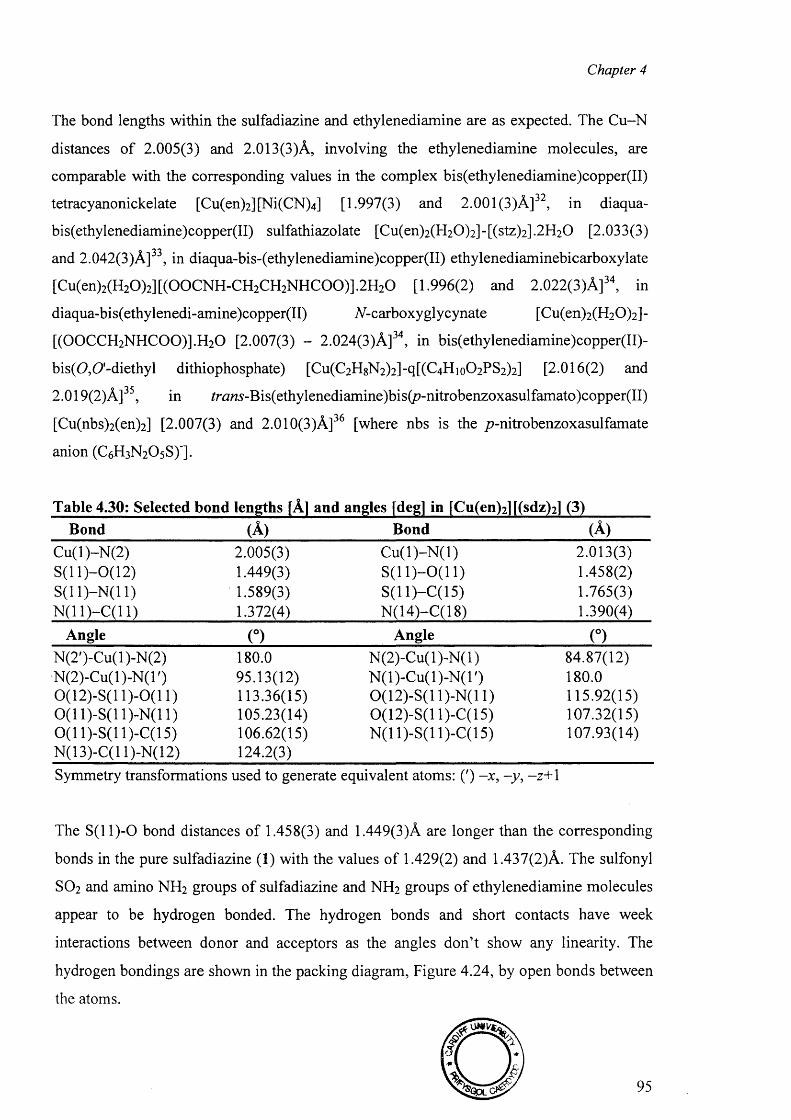
Chapter 4
The bond lengths within the sulfadiazine and ethylenediamine are as expected. The Cu-N
distances o f 2.005(3) and 2.013(3)A, involving the ethylenediamine molecules, are
comparable with the corresponding values in the complex bis(ethylenediamine)copper(II)
tetracyanonickelate [Cu(en)2 ][Ni(CN)4 ] [1.997(3) and 2.001(3)A]32, in diaqua-
bis(ethylenediamine)copper(II) sulfathiazolate [Cu(en)2 (H2 0 )2 ]-[(stz)2 ].2 H 2 0 [2.033(3)
and 2.042(3)A]33, in diaqua-bis-(ethylenediamine)copper(II) ethylenediaminebicarboxylate
[Cu(en)2 (H2 0 )2 ][(0 0 CNH-CH2 CH2NHC 0 0 )].2 H2 0 [1.996(2) and 2.022(3)A]34, in
diaqua-bis(ethylenedi-amine)copper(II) N-carboxy glycynate [Cu(en)2 (H2 0 )2 ] -
[(0 0 CCH2 NHC 0 0 )].H 2 0 [2.007(3) - 2.024(3)A]34, in bis(ethylenediamine)copper(II)-
bis(O,0 '-diethyl dithiophosphate) [Cu(C2H8N 2)2]-q[(C4Hio0 2 PS2 )2 ] [2.016(2) and
2.019(2)A]35, in /r<3 «5 -Bis(ethylenediamine)bis(p-nitrobenzoxasulfamato)copper(II)
[Cu(nbs)2 (en)2 ] [2.007(3) and 2.010(3)A]36 [where nbs is the p-nitrobenzoxasulfamate
anion (C6 H 3 N 2 O5 S)'].
Table 4.30: Selected bond lengths [A] and angles [deg] in [Cu(en)2 l[(sdz)2 ] (3)Bond (A) Bond (A)
Cu(l)-N (2) 2.005(3) C u (l)-N (l) 2.013(3)S ( ll)-0 (1 2 ) 1.449(3) S ( ll) -0 (1 1 ) 1.458(2)S(11)-N(11) 1.589(3) S (ll)-C (1 5 ) 1.765(3)N (11)-C(11) 1.372(4) N(14)-C(18) 1.390(4)
Angle (°) Angle (°)N(2')-Cu(l)-N(2) 180.0 N (2)-Cu(l)-N (l) 84.87(12)N (2)-Cu(l)-N (l') 95.13(12) N (l)-C u(l)-N (T) 180.00(12)-S(11)-0(11) 113.36(15) 0(12)-S(11)-N(11) 115.92(15)0 (1 1)-S(11)-N(11) 105.23(14) 0(12)-S(11 )-C(l 5) 107.32(15)0 (1 1)-S(11)-C(15) 106.62(15) N(11)-S(11)-C(15) 107.93(14)N(13)-C(l 1)-N(12) 124.2(3)Symmetry transformations used to generate equivalent atoms: (') -x , -y , - z + 1
The S ( l l ) - 0 bond distances of 1.458(3) and 1.449(3)A are longer than the corresponding
bonds in the pure sulfadiazine (1 ) with the values of 1.429(2) and 1.437(2)A. The sulfonyl
SO2 and amino NH 2 groups o f sulfadiazine and NH 2 groups o f ethylenediamine molecules
appear to be hydrogen bonded. The hydrogen bonds and short contacts have week
interactions between donor and acceptors as the angles don’t show any linearity. The
hydrogen bondings are shown in the packing diagram, Figure 4.24, by open bonds between
the atoms.
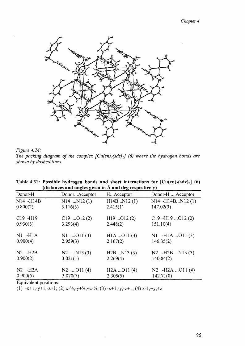
Chapter 4
Figure 4.24:The packing diagram o f the complex [Cu(en)2 (sdz)2] (6 ) where the hydrogen bonds are shown by dashed lines.
Table 4.31: Possible hydrogen bonds and short interactions for [Cu(en)2 (sdz)2 ] (6) ____________(distances and angles given in A and deg respectively)___________________Donor-H Donor... Acceptor H... Acceptor Donor-H.... AcceptorN14 -H14B N14 ....N12 (1) H14B...N12 (1) N14 -H14B...N12 (1)0.800(2) 3.116(3) 2.415(1) 147.02(3)
C19 -H I9 C19 ....012 (2) H19 ...012 (2) C19 -H19 ...012 (2)0.930(3) 3.293(4) 2.448(2) 151.10(4)
NI -H1A NI ....011(3) HI A ...O il (3) NI -HI A ...Ol 1 (3)0.900(4) 2.959(3) 2.167(2) 146.35(2)
N2 -H2B N2 ....N13 (3) H2B ...N13 (3) N2 -H2B ...N13 (3)0.900(2) 3.021(1) 2.269(4) 140.84(2)
N2 -H2A N2 ....O il (4) H2A ...011 (4) N2 -H 2A ...011 (4)0.900(5) 3.070(7) 2.305(5) 142.71(8)Equivalent positions:(1) -x+ l,-y+ l,-z+ l; (2) x-Viry+Viy+z-Vi; (3) -x+l,-y,-z+l; (4) x-l,+y,+z
96
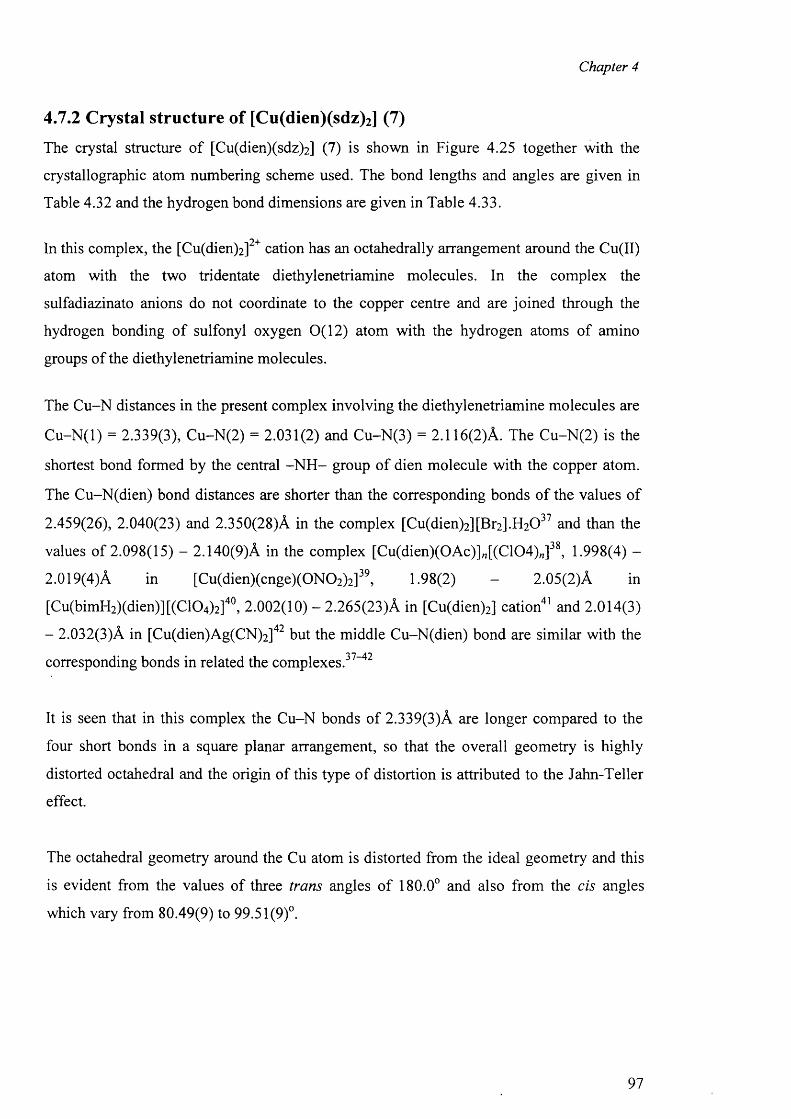
Chapter 4
4.7.2 Crystal structure of [Cu(dien)(sdz)2] (7)
The crystal structure o f [Cu(dien)(sdz)2 ] (7) is shown in Figure 4.25 together with the
crystallographic atom numbering scheme used. The bond lengths and angles are given in
Table 4.32 and the hydrogen bond dimensions are given in Table 4.33.
In this complex, the [Cu(dien)2 ]2+ cation has an octahedrally arrangement around the Cu(II)
atom with the two tridentate diethylenetriamine molecules. In the complex the
sulfadiazinato anions do not coordinate to the copper centre and are joined through the
hydrogen bonding o f sulfonyl oxygen 0(12) atom with the hydrogen atoms of amino
groups o f the diethylenetriamine molecules.
The Cu-N distances in the present complex involving the diethylenetriamine molecules are
C u-N (l) = 2.339(3), Cu-N(2) = 2.031(2) and Cu-N(3) = 2.116(2)A. The Cu-N(2) is the
shortest bond formed by the central -N H - group of dien molecule with the copper atom.
The Cu-N(dien) bond distances are shorter than the corresponding bonds of the values o f
2.459(26), 2.040(23) and 2.350(28)A in the complex [Cu(dien)2 ][Br2 ].H 2 0 37 and than the
values o f 2.098(15) - 2.140(9)A in the complex [Cu(dien)(0Ac)]„[(C104)„]38, 1.998(4) -
2.019(4) A in [Cu(dien)(cnge)(0N02)2]39, 1.98(2) - 2.05(2)A in
[Cu(bimH2)(dien)][(C104)2]40, 2.002(10) - 2.265(23)A in [Cu(dien)2] cation41 and 2.014(3)
- 2.032(3)A in [Cu(dien)Ag(CN)2 ]42 but the middle Cu-N(dien) bond are similar with the
corresponding bonds in related the complexes.37"42
It is seen that in this complex the Cu-N bonds of 2.339(3)A are longer compared to the
four short bonds in a square planar arrangement, so that the overall geometry is highly
distorted octahedral and the origin of this type of distortion is attributed to the Jahn-Teller
effect.
The octahedral geometry around the Cu atom is distorted from the ideal geometry and this
is evident from the values of three trans angles o f 180.0° and also from the cis angles
which vary from 80.49(9) to 99.51(9)°.
97
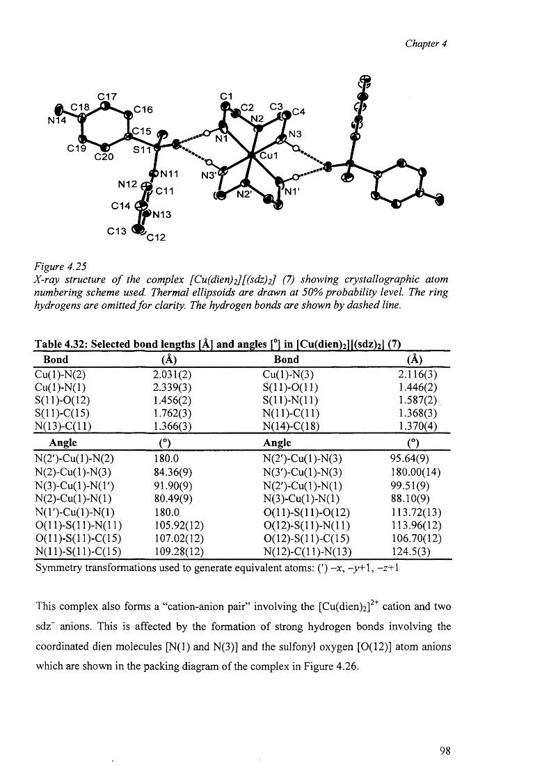
Chapter 4
C1C17F c;Uc4
7 l N 3
C18 C16N14
JC15N1
C19 S11 Cu1C20
N11 N3'N12
C11 N1’N2'C14
N13
C13 C12
Figure 4.25X-ray structure o f the complex [Cu(dien)2][(sdz)2] (7) showing crystallographic atom numbering scheme used. Thermal ellipsoids are drawn at 50% probability level. The ring hydrogens are omitted fo r clarity. The hydrogen bonds are shown by dashed line.
Table 4.32: Selected bond lengths [A] and angles [°] in [Cu(dien)2 l[(sdz)2 ] (7)Bond (A) Bond (A)
Cu(l)-N(2) 2.031(2) Cu(l)-N(3) 2.116(3)C u(l)-N (l) 2.339(3) S (ll)-0 (1 1 ) 1.446(2)S(11 )-0 (l 2) 1.456(2) S(11 )-N(l 1) 1.587(2)S(11)-C(15) 1.762(3) N(11)-C(11) 1.368(3)N(13)-C(l 1) 1.366(3) N(14)-C(18) 1.370(4)
Angle (°) Angle (°)N(2')-Cu(l)-N(2) 180.0 N(2')-Cu(l)-N(3) 95.64(9)N(2)-Cu(l)-N(3) 84.36(9) N(3')-Cu(l)-N(3) 180.00(14)N (3)-Cu(l)-N (l') 91.90(9) N (2')-C u(l)-N (l) 99.51(9)N (2)-Cu(l)-N (l) 80.49(9) N(3)-Cu(l)-N (l) 88.10(9)N (l')-C u(l)-N (l) 180.0 0(11)-S(11)-0(12) 113.72(13)0 (1 1)-S(11)-N(11) 105.92(12) 0(12)-S(11)-N(11) 113.96(12)0 (1 1)-S(11 )-C(l 5) 107.02(12) 0(12)-S(11)-C(15) 106.70(12)N(11)-S(11 )-C(l 5) 109.28(12) N(12)-C(l 1)-N(13) 124.5(3)Symmetry transformations used to generate equivalent atoms: (') -x , -y+ 1, -z+ 1
This complex also forms a “cation-anion pair” involving the [Cu(dien)2 ]2+ cation and two
sdz” anions. This is affected by the formation of strong hydrogen bonds involving the
coordinated dien molecules [N(l) and N(3)] and the sulfonyl oxygen [0(12)] atom anions
which are shown in the packing diagram of the complex in Figure 4.26.
98
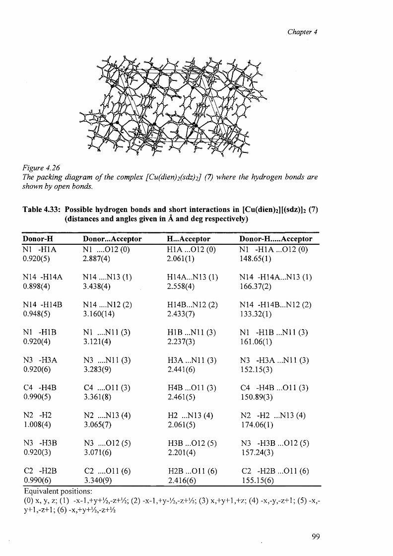
Chapter 4
Figure 4.26The packing diagram o f the complex [Cu(dien)2 (sdz)2] (7) where the hydrogen bonds are shown by open bonds.
Table 4.33: Possible hydrogen bonds and short interactions in [Cu(dien)2 ][(sdz ) ] 2 (7) (distances and angles given in A and deg respectively)
Donor-H Donor...Acceptor H...Acceptor Donor-H.....AcceptorNI -H1A 0.920(5)
N I ....012(0) 2.887(4)
H1A ...012(0) 2.061(1)
NI -H1A ...012 (0) 148.65(1)
N14 -H14A 0.898(4)
N 14....N 13(1)3.438(4)
H14A...N13 (1) 2.558(4)
N14 -H14A...N13 (1) 166.37(2)
N14 -H14B 0.948(5)
N14 ....N12 (2) 3.160(14)
H14B...N12 (2) 2.433(7)
N14 -H14B...N12 (2) 133.32(1)
NI -H1B 0.920(4)
NI ....N il (3) 3.121(4)
H1B...N11 (3) 2.237(3)
NI -H1B ...N il (3) 161.06(1)
N3 -H3A 0.920(6)
N3 ....N il (3) 3.283(9)
H3A...N11 (3) 2.441(6)
N3 -H3A ...N il (3) 152.15(3)
C4 -H4B 0.990(5)
C4 ....O il (3) 3.361(8)
H 4B ...011 (3) 2.461(5)
C4 -H4B ...O il (3) 150.89(3)
N2 -H2 1.008(4)
N2 ....N13 (4) 3.065(7)
H2 ...N13 (4) 2.061(5)
N2 -H2 ...N13 (4) 174.06(1)
N3 -H3B 0.920(3)
N3 ....012(5) 3.071(6)
H3B ...012 (5) 2.201(4)
N3 -H3B ...012(5 ) 157.24(3)
C2 -H2B 0.990(6)
C2 ....O il (6) 3.340(9)
H2B ...O il (6) 2.416(6)
C2 -H2B ...O il (6) 155.15(6)
Equivalent positions:(0) x, y, z; (1) -x -l,+ y + ,/ 2 ,-z+1/2; (2) -x-E + y-^-z+ V E (3) x ,+ y+ l,+ z; (4) -x ,-y ,-z+ l; (5) -x,- y + l,-z+ l; (6 ) -x,+y+V2 ,-z+V2
99
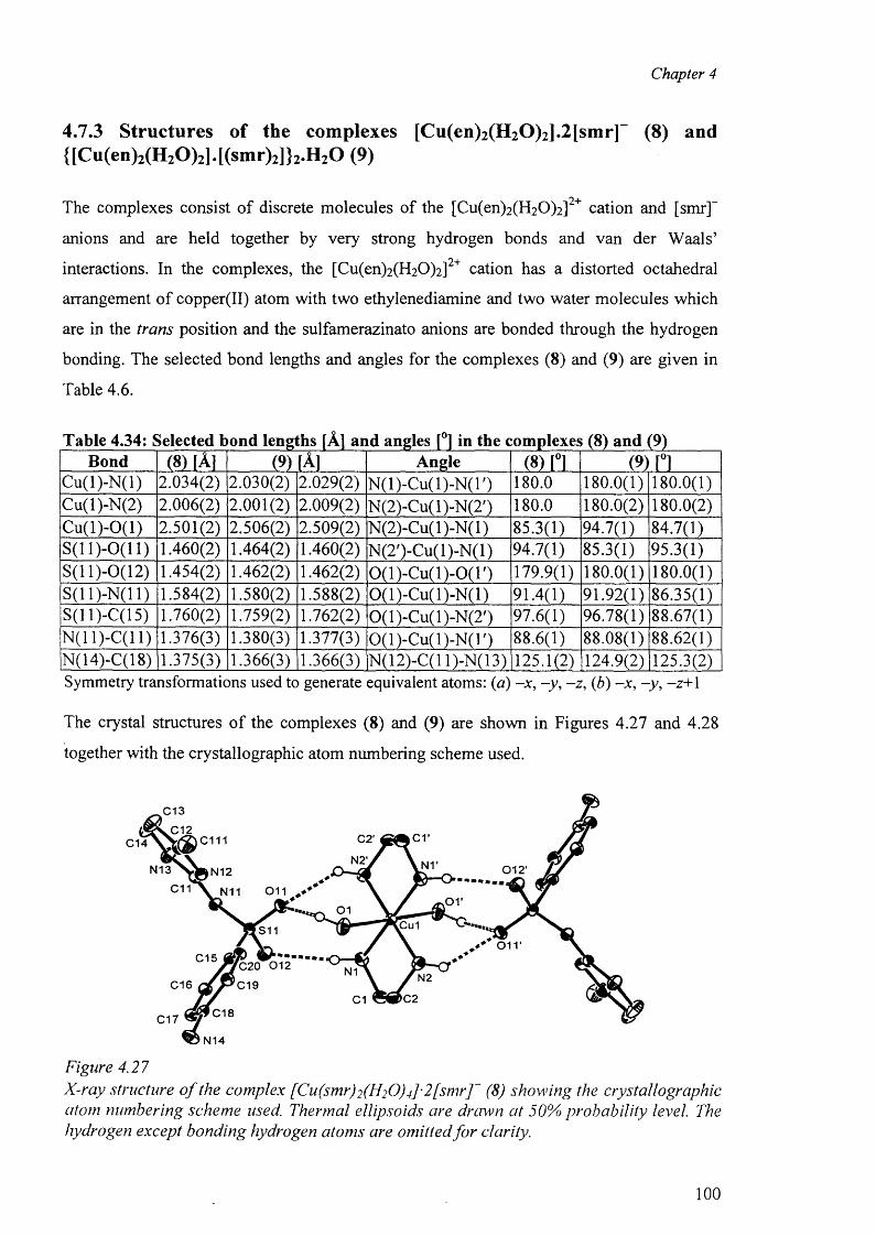
Chapter 4
4.7.3 Structures of the complexes [Cu(en)2(H20)2].2[smr] (8) and{[Cu(en)2(H20)2].[(smr)2]}2.H20 ( 9 )
• 04 - ____________________________________________ _
The complexes consist of discrete molecules o f the [Cu(en)2(H20)2] cation and [smr]
anions and are held together by very strong hydrogen bonds and van der Waals’04-interactions. In the complexes, the [Cu(en)2(H20)2] cation has a distorted octahedral
arrangement o f copper(II) atom with two ethylenediamine and two water molecules which
are in the trans position and the sulfamerazinato anions are bonded through the hydrogen
bonding. The selected bond lengths and angles for the complexes (8) and (9) are given in
Table 4.6.
Table 4.34: Selected bond lengths [A] and angles [°] in the complexes (8) and (9)Bond m M (9) [A] Angle (8) [°1 (9 ) n
C u(l)-N (l) 2.034(2) 2.030(2) 2.029(2) N (l)-C u(l)-N (T) 180.0 180.0(1) 180.0(1)Cu(l)-N(2) 2.006(2) 2.001(2) 2.009(2) N(2)-Cu(l)-N(2') 180.0 180.0(2) 180.0(2)C u(l)-0(1) 2.501(2) 2.506(2) 2.509(2) N (2)-Cu(l)-N(l) 85.3(1) 94.7(1) 84.7(1)S(11)-0(H ) 1.460(2) 1.464(2) 1.460(2) N(2')-Cu(l)-N (l) 94.7(1) 85.3(1) 95.3(1)S (ll)-0 (12 ) 1.454(2) 1.462(2) 1.462(2) 0(1)-C u(l)-0(1 ') 179.9(1) 180.0(1) 180.0(1)S(11 )-N(l 1) 1.584(2) 1.580(2) 1.588(2) 0(1)-C u(l)-N (l) 91.4(1) 91.92(1) 86.35(1)S(ll)-C(15) 1.760(2) 1.759(2) 1.762(2) 0(1)-Cu(l)-N(2') 97.6(1) 96.78(1) 88.67(1)N(11)-C(11) 1.376(3) 1.380(3) 1.377(3) 0(1)-C u(l)-N (l') 88.6(1) 88.08(1) 88.62(1)N(14)-C(18) 1.375(3) 1.366(3) 1.366(3) N(12)-C(l 1)-N(13) 125.1(2) 124.9(2) 125.3(2)Symmetry transformations used to generate equivalent atoms: (a) —jc, -y , -z , (b) -x , -y , - z + \
The crystal structures o f the complexes (8) and (9) are shown in Figures 4.27 and 4.28
together with the crystallographic atom numbering scheme used.
C13
C12 c rC2'c mC14N2'
N13 0 1 2 'N12C11 011N11 .or
01.Cu1S11
o irC15
'C20 0 1 2
C19N1 N2C16
C2C1C18C17
N14
Figure 4.27X-ray structure o f the complex [Cu(smr) 2 (H2 O) 4] ■ 2 [smr]~ (8 ) showing the crystallographic atom numbering scheme used. Thermal ellipsoids are drawn at 50% probability level. The hydrogen except bonding hydrogen atoms are omitted fo r clarity.
100
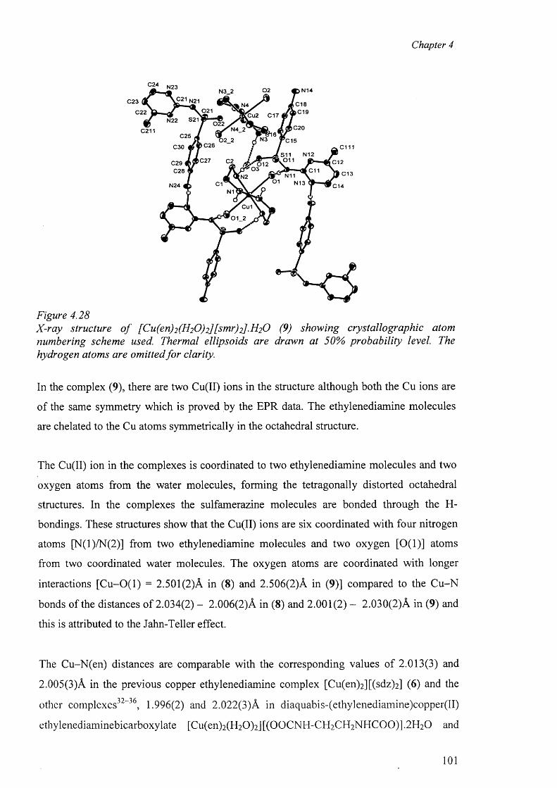
Chapter 4
N23 0 2 N14N3_2
£ 21 N21C 23 C 18)C 1 90 2 1C 22 t u 2 C17
N22 S21 022C20N4_2C211
C25 N302_2 C 15C26C30 c m
N12S11. 0 1 1C27 C12C29 (
C28 C11 C 13N1101 N13C1N24 C14
N1
Cu1
01_2
©---
Figure 4.28X-ray structure o f [Cu(en)2 (H2 0 ) 2][smr)2].H 2 0 (9) showing crystallographic atom numbering scheme used. Thermal ellipsoids are drawn at 50% probability level. The hydrogen atoms are omitted fo r clarity.
In the complex (9), there are two Cu(II) ions in the structure although both the Cu ions are
of the same symmetry which is proved by the EPR data. The ethylenediamine molecules
are chelated to the Cu atoms symmetrically in the octahedral structure.
The Cu(II) ion in the complexes is coordinated to two ethylenediamine molecules and two
oxygen atoms from the water molecules, forming the tetragonally distorted octahedral
structures. In the complexes the sulfamerazine molecules are bonded through the H-
bondings. These structures show that the Cu(II) ions are six coordinated with four nitrogen
atoms [N(l)/N(2)] from two ethylenediamine molecules and two oxygen [0(1)] atoms
from two coordinated water molecules. The oxygen atoms are coordinated with longer
interactions [C u-O (l) = 2.501 (2)A in (8) and 2.506(2)A in (9)] compared to the Cu-N
bonds of the distances o f 2.034(2) - 2.006(2)A in (8) and 2.001(2) - 2.03 0(2) A in (9) and
this is attributed to the Jahn-Teller effect.
The Cu-N(en) distances are comparable with the corresponding values of 2.013(3) and
2.005(3)A in the previous copper ethylenediamine complex [Cu(en)2 ][(sdz)2 ] (6) and the
other complexes32”36, 1.996(2) and 2.022(3)A in diaquabis-(ethylenediamine)copper(II)
ethylenediaminebicarboxylate [Cu(en)2(H20)2] [(OOCNH-CH 2 CH 2 NHCOO)] .2 H2 O and
101
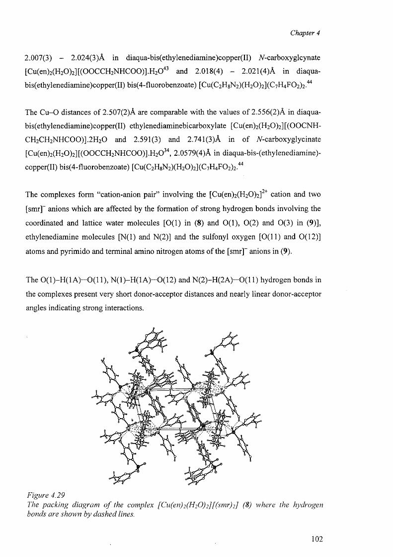
Chapter 4
2.007(3) - 2.024(3)A in diaqua-bis(ethylenediamine)copper(II) TV-carboxyglcynate
[Cu(en)2(H20 ) 2][(OOCCH2NHCOO)].H20 43 and 2.018(4) - 2.021(4)A in diaqua-
bis(ethylenediamine)copper(II) bis(4-fluorobenzoate) [Cu(C2H8N2)(H20 ) 2](C7H4F02)2.44
The C u -0 distances of 2.507(2)A are comparable with the values o f 2.556(2)A in diaqua-
bis(ethylenediamine)copper(II) ethylenediaminebicarboxylate [Cu(en)2(H20 )2] [(OOCNH-
CH2CH2N H C 00)].2H 20 and 2.591(3) and 2.741(3)A in o f A-carboxyglycinate
[Cu(en)2(H20 ) 2][(00C C H 2N H C 00)].H 20 34, 2.0579(4)A in diaqua-bis-(ethylenediamine)-
copper(II) bis(4-fluorobenzoate) [Cu(C2HgN2)(H20 ) 2](C7H4F02)2.44
• • 2 4“ •The complexes form “cation-anion pair” involving the [Cu(en)2(H20 ) 2] cation and two
[smr]- anions which are affected by the formation of strong hydrogen bonds involving the
coordinated and lattice water molecules [0(1) in (8) and 0(1), 0 (2) and 0(3) in (9)],
ethylenediamine molecules [N(l) and N(2)] and the sulfonyl oxygen [0(11) and 0(12)]
atoms and pyrimido and terminal amino nitrogen atoms of the [smr]- anions in (9).
The 0 (1 )-H (1 A )-0 (1 1 ), N (1)-H (1A )-0(12) and N (2 )-H (2 A )-0 (1 1) hydrogen bonds in
the complexes present very short donor-acceptor distances and nearly linear donor-acceptor
angles indicating strong interactions.
Figure 4.29The packing diagram o f the complex [Cu(en)2 (H2 0 ) 2] [(sm r)2] (8) where the hydrogen bonds are shown by dashed lines.
102
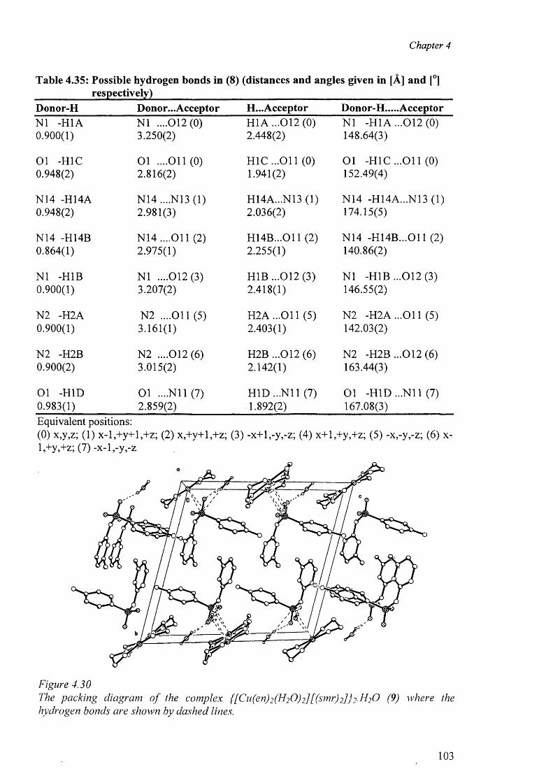
Chapter 4
Table 4.35: Possible hydrogen bonds in (8) (distances and angles given in [A] and [°]respectively)
Donor-H Donor...Acceptor H...Acceptor Donor-H.....AcceptorNI -H1A N I ....012(0) H1A ...012(0) N I -H1A ...012(0)0.900(1) 3.250(2) 2.448(2) 148.64(3)
Ol -H1C O l ....011(0) H 1C ...011 (0) O l -H1C ...O il (0)0.948(2) 2.816(2) 1.941(2) 152.49(4)
N14 -H14A N14 ....N13 (1) H14A...N13 (1) N14 -H14A...N13 (1)0.948(2) 2.981(3) 2.036(2) 174.15(5)
N14 -H14B N 14....011 (2) H 14B...011 (2) N14 -H 14B ...011 (2)0.864(1) 2.975(1) 2.255(1) 140.86(2)
NI -H1B N I ....012(3) H1B ...012 (3) NI -H1B ...012(3 )0.900(1) 3.207(2) 2.418(1) 146.55(2)
N2 -H2A N2 ....O il (5) H 2A ...011 (5) N2 -H2A ...O il (5)0.900(1) 3.161(1) 2.403(1) 142.03(2)
N2 -H2B N2 ....012(6) H2B ...012 (6) N2 -H2B ...012(6 )0.900(2) 3.015(2) 2.142(1) 163.44(3)
Ol -HID Ol ....N il (7) H ID ...N il (7) O l -HID ...N il (7)0.983(1) 2.859(2) 1.892(2) 167.08(3)Equivalent positions:(0) x,y,z; (1) x-l,+y+l,+z; (2) x,+y+l,+z; (3) -x+l,-y,-z; (4) x+l,+y,+z; (5) -x,-y,-z; (6) x- l,+y,+z;(7)-x-l,-y ,-z
Figure 4.30The packing diagram o f the complex {[Cn(en)2 (H2 O)2] [(sm r)2] } 2 - I f O (9) where the hydrogen bonds are shown by dashed lines.
103
Chapter 4
Table 4.36: Possible hydrogen bonds in the complex [Cu(en)2(H20)2][(smr)2].H20 (9)(distances and angles are given in A and deg respectively)
Donor-H Donor...Acceptor H...Acceptor Donor-H.... AcceptorOl -H1A O l ....N il (0) H1A...N11 (0) Ol -HIA ...N il (0)0.894(2) 2.896(4) 2.036(3) 160.87(4)
N2 -H2D N2 ....012(0) H2D ...012(0) N2 -H2D ...012(0)0.920(2) 3.002(3) 2.091(2) 170.56(3)
N3 -H3A N3 ....03 (0) H3A...03 (0) N3 -H3A...03 (0)0.920(1) 2.988(2) 2.080(2) 168.47(2)
N4 -H4B N4 ....021 (0) H4B ...021 (0) N4 -H4B ...021 (0)0.920(1) 3.075(1) 2.195(5) 159.81(2)
Ol -H1B Ol ....011(1) H1B ...O il (1) Ol -H1B ...O il (1)0.919(3) 2.844(4) 2.006(3) 150.91(5)
03 -H3E 03 ....022(1) H3E ...022(1) 03 -H3E ...022(1)0.944(2) 2.847(2) 1.911(1) 171.21(2)
03 -H3F 03 ....012(1) H3F ...012(1) 03 -H3F ...012(1)0.967(2) 2 .888(2) 1.987(1) 154.05(2)
N24 -H24B N24 ....O il (2) H24B...011 (2) N24 -H24B...011 (2)0.880(2) 3.016(2) 2.376(1) 129.84(3)
N24 -H24B N24 ....N12 (2) H24B...N12 (2) N24 -H24B...N12 (2)0.880(2) 3.150(4) 2.371(3) 147.72(3)
N2 -H2C N2 ....O il (2) H2C ...O il (2) N2 -H2C ...O il (2)0.920(2) 3.160(2) 2.343(1) 147.75(2)
N24 -H24A N24 ....N13 (3) H24A...N13 (3) N24 -H24A...N13 (3)0.880(3) 2.969(5) 2.150(3) 154.48(5)
N14 -H14A N14....N23 (4) H14A...N23 (4) N14 -H14A...N23 (4)0.880(3) 2.984(5) 2.149(3) 158.17(5)
N4 -H4A N4 ....022(5) H4A ...022 (5) N4 -H4A ...022 (5)0.920(2) 2.972(2) 2.067(1) 167.17(2)
N14 -H14B N14 ....021 (5) H14B...021 (5) N14 -H14B...021 (5)0.880(2) 2.971(2) 2.292(1) 133.93(3)
02 -H2A 02 ....N21 (5) H2A...N21 (5) 0 2 -H2A...N21 (5)1.022(2) 2.900(4) 1.901(3) 165.06(4)
02 -H2B 02 ....021 (6) H2B ...021 (6) 02 -H2B ...021 (6)0.986(3) 2.924(4) 1.954(3) 167.41(5)Equivalent positions:(0) x,y,z; (1) x-l,+y,+z; (2) -x+l,-y,-z; (3) -x,-y,-z; (4) -x+2,-y,-z+l; (5) -x+l,-y,-z+l; (6) - x,-y,-z+i
104
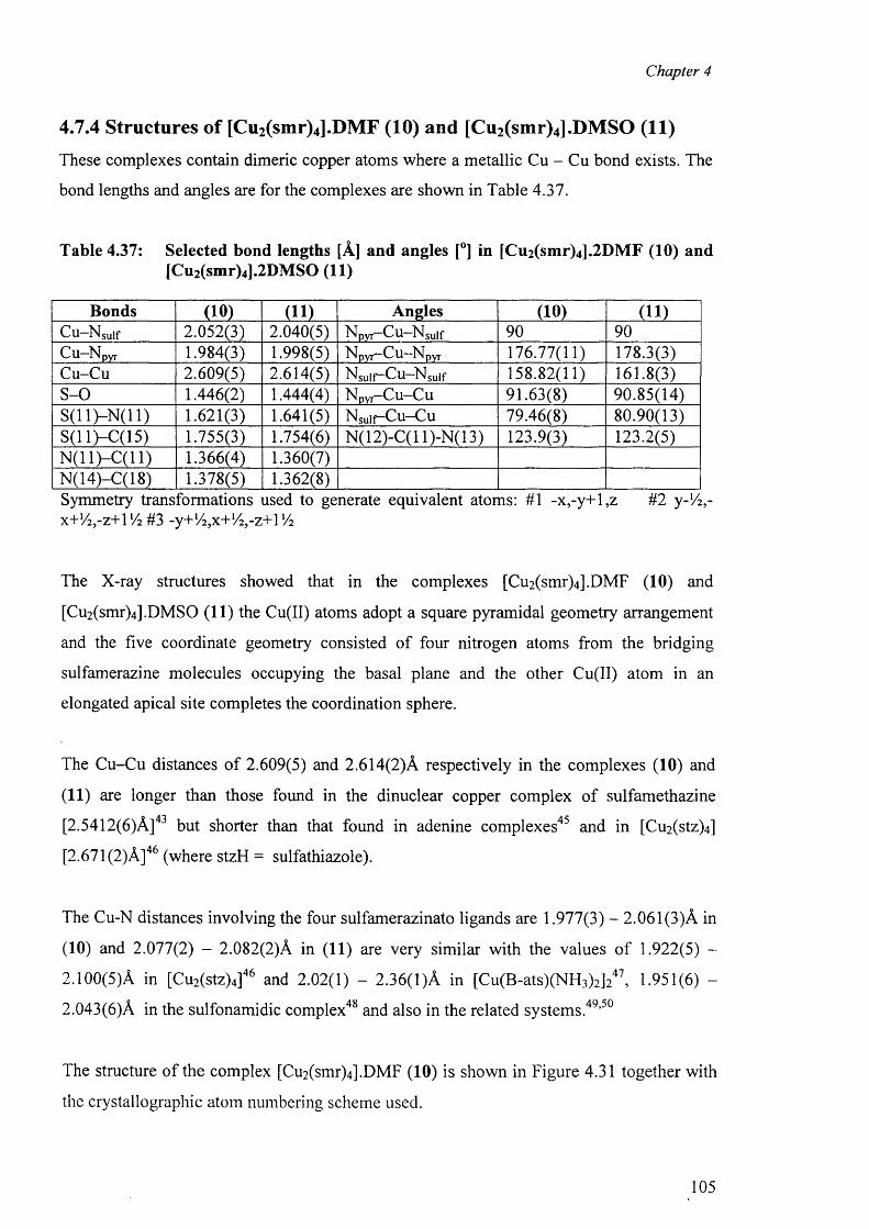
Chapter 4
4.7.4 Structures of [Cu2(smr)4].DMF (10) and [Cu2(smr)4].DMSO (11)
These complexes contain dimeric copper atoms where a metallic Cu - Cu bond exists. The
bond lengths and angles are for the complexes are shown in Table 4.37.
Table 4.37: Selected bond lengths [A] and angles [°] in [Cu2(smr)4].2DMF (10) and[Cu2(smr)4].2DMSO (11)
Bonds (10) (11) Angles (10) (11)Cu—Nsuif 2 . 0 5 2 ( 3 ) 2 . 0 4 0 ( 5 ) Npyr—C u —Nsulf 9 0 9 0C u Npyr 1 . 9 8 4 ( 3 ) 1 . 9 9 8 ( 5 ) N pyr—C u —N pyr 1 7 6 . 7 7 ( 1 1 ) 1 7 8 . 3 ( 3 )
C u - C u 2 . 6 0 9 ( 5 ) 2 . 6 1 4 ( 5 ) Nsulf~Cu—N suif 1 5 8 . 8 2 ( 1 1 ) 1 6 1 . 8 ( 3 )
S - 0 1 . 4 4 6 ( 2 ) 1 . 4 4 4 ( 4 ) N p y r - C u - C u 9 1 . 6 3 ( 8 ) 9 0 . 8 5 ( 1 4 )
S ( 1 1 ) - N ( 1 1) 1 . 6 2 1 ( 3 ) 1 . 6 4 1 ( 5 ) N s u i f - C u - C u 7 9 . 4 6 ( 8 ) 8 0 . 9 0 ( 1 3 )
S ( 1 1)—C ( 1 5) 1 . 7 5 5 ( 3 ) 1 .7 5 4 ( 6 ) N ( 1 2 ) - C ( l 1 ) - N ( 1 3 ) 1 2 3 . 9 ( 3 ) 1 2 3 . 2 ( 5 )
N ( l l ) - C ( l l ) 1 . 3 6 6 ( 4 ) 1 .3 6 0 ( 7 )
N ( 1 4 ) - C ( 1 8 ) 1 . 3 7 8 ( 5 ) 1 .3 6 2 ( 8 )
Symmetry transformations used to generate equivalent atoms: #1 -x,-y+l,z #2 y-Vi,-x + ! /2 ,- z + 1 ! /2 #3 -y+V2,x+l/ 2,-z+\ V2
The X-ray structures showed that in the complexes [Cu2 (smr)4].DMF (10) and
[Cu2(smr)4 ].DMSO (11) the Cu(II) atoms adopt a square pyramidal geometry arrangement
and the five coordinate geometry consisted of four nitrogen atoms from the bridging
sulfamerazine molecules occupying the basal plane and the other Cu(II) atom in an
elongated apical site completes the coordination sphere.
The Cu-Cu distances o f 2.609(5) and 2.614(2)A respectively in the complexes (10) and
(11) are longer than those found in the dinuclear copper complex of sulfamethazine
[2.5412(6)A]43 but shorter than that found in adenine complexes45 and in [Cu2(stz)4]
[2.671 (2)A ]46 (where stzH = sulfathiazole).
The Cu-N distances involving the four sulfamerazinato ligands are 1.977(3) - 2.061(3)A in
(10) and 2.077(2) - 2.082(2)A in (11) are very similar with the values o f 1.922(5) -
2.100(5)A in [Cu2(stz)4]46 and 2.02(1) - 2.36(1)A in [Cu(B-ats)(NH3)2]247, 1.951(6) -
2.043(6)A in the sulfonamidic complex48 and also in the related systems.49,50
The structure o f the complex [Cu2(smr)4].DMF (10) is shown in Figure 4.31 together with
the crystallographic atom numbering scheme used.
105

Chapter 4
Figure 4.31X-ray structure o f the complex [Cu2 (smr)4].DM F (10) showing crystallographic atom numbering scheme used. Thermal ellipsoids are drawn at 50% probability level. The ring hydrogen atoms and DMF molecules are omitted fo r clarity.
Figure 4.32The packing diagram o f the complex [Cu2 (smr)4] .2DMF (10) where the hydrogen bonds are shown by dashed lines.
106
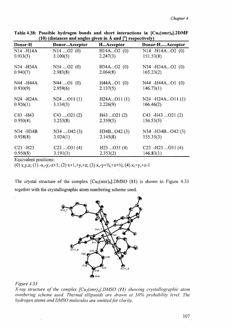
Chapter 4
Table 4.38: Possible hydrogen bonds and short interactions in [Cu2(smr)4].2DMF ___________ (10) (distances and angles given in A and [°] respectively)_______________Donor-H Donor...Acceptor H...Acceptor Donor-H.....AcceptorN14 -H I4A N14 ....02 (0) H14A...02 (0) N14 -H14A...02 (0)0.933(5) 3.100(5) 2.247(3) 151.53(8)
N34 -H34A N34 ....02 (0) H34A...02 (0) N34 -H34A...02 (0)0.940(7) 2.983(8) 2.064(8) 165.23(2)
N44 -H44A N 44....01 (0) H44A...01 (0) N44 -H44A...01 (0)0.930(9) 2.959(6) 2.137(5) 146.73(1)
N24 -H24A N 24....011 (1) H 24A ...011 (1) N24 -H 24A ...011 (1)0.926(1) 3.134(3) 2.226(9) 166.46(2)
C43 -H43 C43 ....021 (2) H43 ...021 (2) C43 -H43 ...021 (2)0.950(4) 3.253(8) 2.359(5) 156.53(5)
N34 -H34B N34 ....042 (3) H34B...042 (3) N34 -H34B...042 (3)0.938(8) 3.024(1) 2.145(8) 155.35(3)
C23 -H23 C23 ....031 (4) H23 ...031 (4) C23 -H23 ...031 (4)0.950(8) 3.191(3) 2.353(2) 146.83(1)Equivalent positions:(0) x,y,z; (1) -x,-y,-z+l; (2) x+l,+y,+z; (3) x 9-y+l/2y+z+l/2; (4) x,+y,+z-l
The crystal structure of the complex [Cu2 (smr)4 ].DMSO (11) is shown in Figure 4.33
together with the crystallographic atom numbering scheme used.
Figure 4.33X-ray structure o f the complex [Cii2 (smr)4] .DMSO (11) showing crystallographic atom numbering scheme used. Thermal ellipsoids are drawn at 50% probability level. The hydrogen atoms and DMSO molecules are omitted fo r clarity.
107
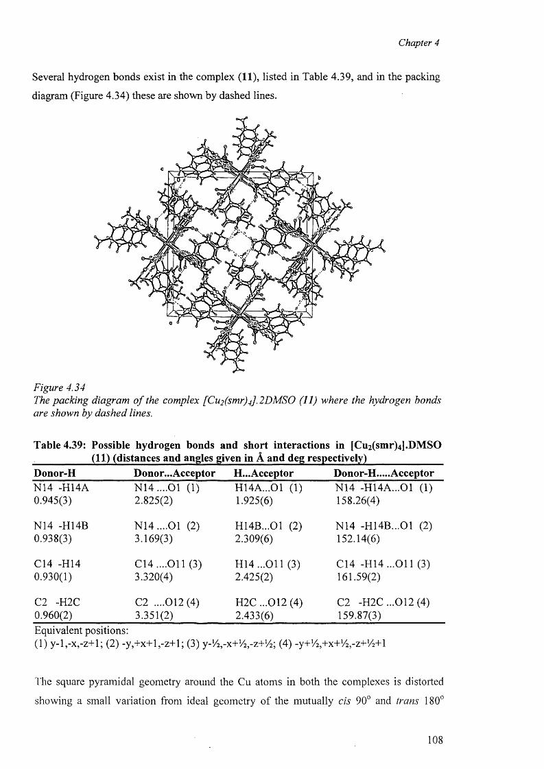
Chapter 4
Several hydrogen bonds exist in the complex (11), listed in Table 4.39, and in the packing
diagram (Figure 4.34) these are shown by dashed lines.
Figure 4.34The packing diagram o f the complex [Cu2 (sm r)J.2 DMSO (11) where the hydrogen bonds are shown by dashed lines.
Table 4.39: Possible hydrogen bonds and short interactions in [Cu2 (smr)4 ].DMSO ___________ (11) (distances and angles given in A and deg respectively)_______________Donor-H Donor...Acceptor H...Acceptor Donor-H.....AcceptorN14 -H I4A N 14....01 (1) H14A...01 (1) N14 -H14A...01 (1)0.945(3) 2.825(2) 1.925(6) 158.26(4)
N14 -H14B N 14....01 (2) H14B...01 (2) N14 -H14B...01 (2)0.938(3) 3.169(3) 2.309(6) 152.14(6)
C14 -H14 C 14....011 (3) H 14...011 (3) C14 -H 14...011 (3)0.930(1) 3.320(4) 2.425(2) 161.59(2)
C2 -H2C C2 ....012(4) H2C ...012 (4) C2 -H2C ...012(4)0.960(2) 3.351(2) 2.433(6) 159.87(3)Equivalent positions:(1) y-l,-x,-z+l; (2) -y,+x+l,-z+ l; (3) y-V^-x+V^-z+Va; (4) -y+V^+x+VVz-HA+l
The square pyramidal geometry around the Cu atoms in both the complexes is distorted
showing a small variation from ideal geometry of the mutually cis 90° and trans 180°
108
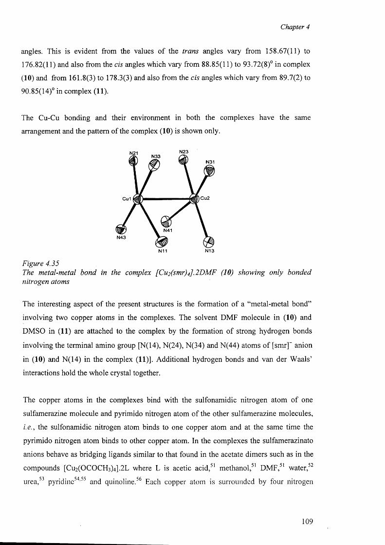
Chapter 4
angles. This is evident from the values o f the trans angles vary from 158.67(11) to
176.82(11) and also from the cis angles which vary from 88.85(11) to 93.72(8)° in complex
(10) and from 161.8(3) to 178.3(3) and also from the cis angles which vary from 89.7(2) to
90.85(14)° in complex (11).
The Cu-Cu bonding and their environment in both the complexes have the same
arrangement and the pattern of the complex (10) is shown only.
N23N21N33
N31
Cu2Cu1
N41N43
N13N11
Figure 4.35The metal-metal bond in the complex [Cu2 (smr)4] .2DMF (10) showing only bonded nitrogen atoms
The interesting aspect of the present structures is the formation of a “metal-metal bond”
involving two copper atoms in the complexes. The solvent DMF molecule in (10) and
DMSO in (11) are attached to the complex by the formation o f strong hydrogen bonds
involving the terminal amino group [N(14), N(24), N(34) and N(44) atoms of [smr]~ anion
in (10) and N(14) in the complex (11)]. Additional hydrogen bonds and van der W aals’
interactions hold the whole crystal together.
The copper atoms in the complexes bind with the sulfonamidic nitrogen atom of one
sulfamerazine molecule and pyrimido nitrogen atom of the other sulfamerazine molecules,
i.e., the sulfonamidic nitrogen atom binds to one copper atom and at the same time the
pyrimido nitrogen atom binds to other copper atom. In the complexes the sulfamerazinato
anions behave as bridging ligands similar to that found in the acetate dimers such as in the
compounds [Cu2(OCOCH3)4].2L where L is acetic acid,51 methanol,51 DMF,51 water,52
urea,53 pyridine54,55 and quinoline.56 Each copper atom is surrounded by four nitrogen
109
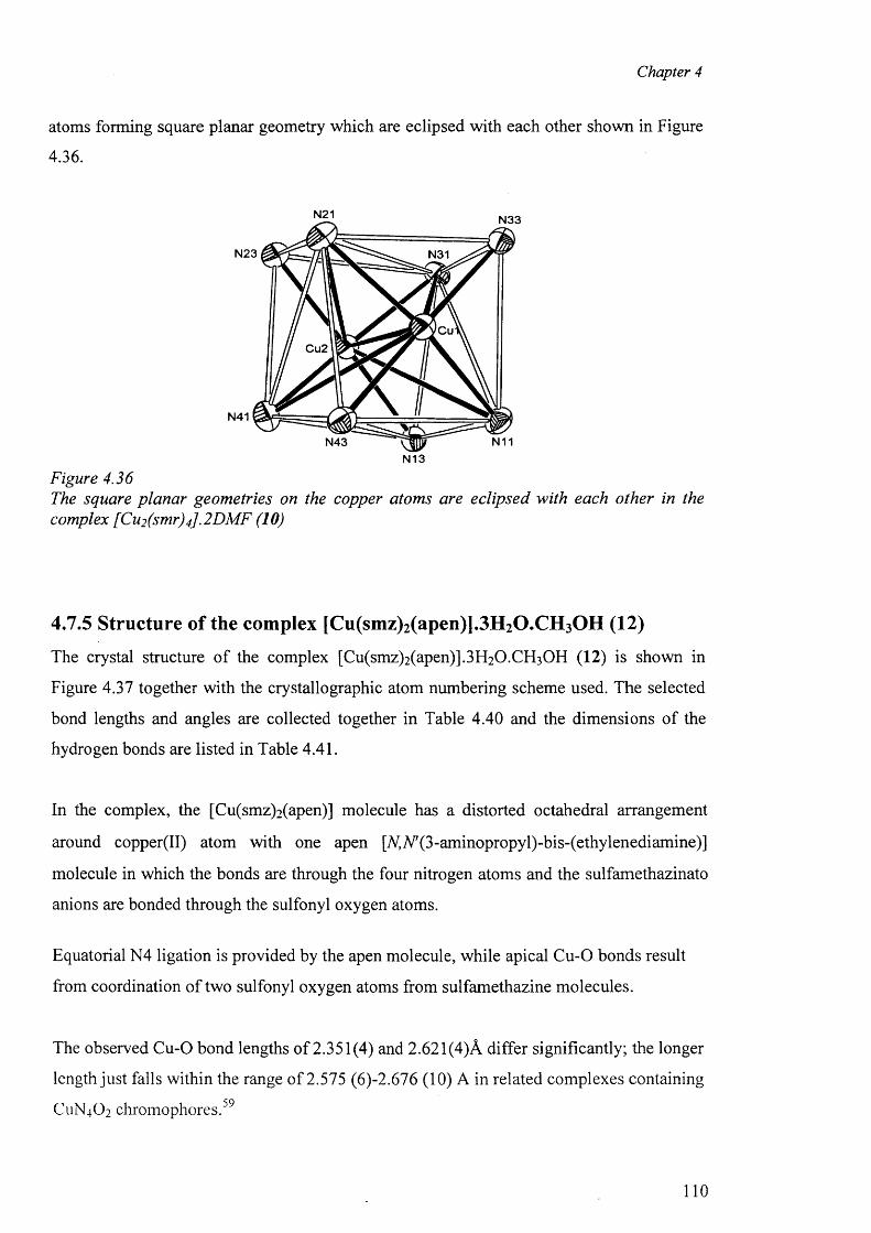
Chapter 4
atoms forming square planar geometry which are eclipsed with each other shown in Figure
4.36.
N21 N33
N23 N31
CuCu2
N41
N11N43N13
Figure 4.36The square planar geometries on the copper atoms are eclipsed with each other in the complex [Cu2 (smr)4].2DM F (10)
4.7.5 Structure of the complex [Cu(smz)2(apen)].3H2O.CH3OH (12)
The crystal structure of the complex [Cu(smz)2 (apen)].3 H 2 0 .CH3 0 H (12) is shown in
Figure 4.37 together with the crystallographic atom numbering scheme used. The selected
bond lengths and angles are collected together in Table 4.40 and the dimensions of the
hydrogen bonds are listed in Table 4.41.
In the complex, the [Cu(smz)2 (apen)] molecule has a distorted octahedral arrangement
around copper(II) atom with one apen [A(A^(3-aminopropyl)-bis-(ethylenediamine)]
molecule in which the bonds are through the four nitrogen atoms and the sulfamethazinato
anions are bonded through the sulfonyl oxygen atoms.
Equatorial N4 ligation is provided by the apen molecule, while apical C u-0 bonds result
from coordination o f two sulfonyl oxygen atoms from sulfamethazine molecules.
The observed C u-0 bond lengths of 2.351(4) and 2.621 (4)A differ significantly; the longer
length just falls within the range of 2.575 (6)-2.676 (10) A in related complexes containing
C11N4O2 chrom ophores.59
110
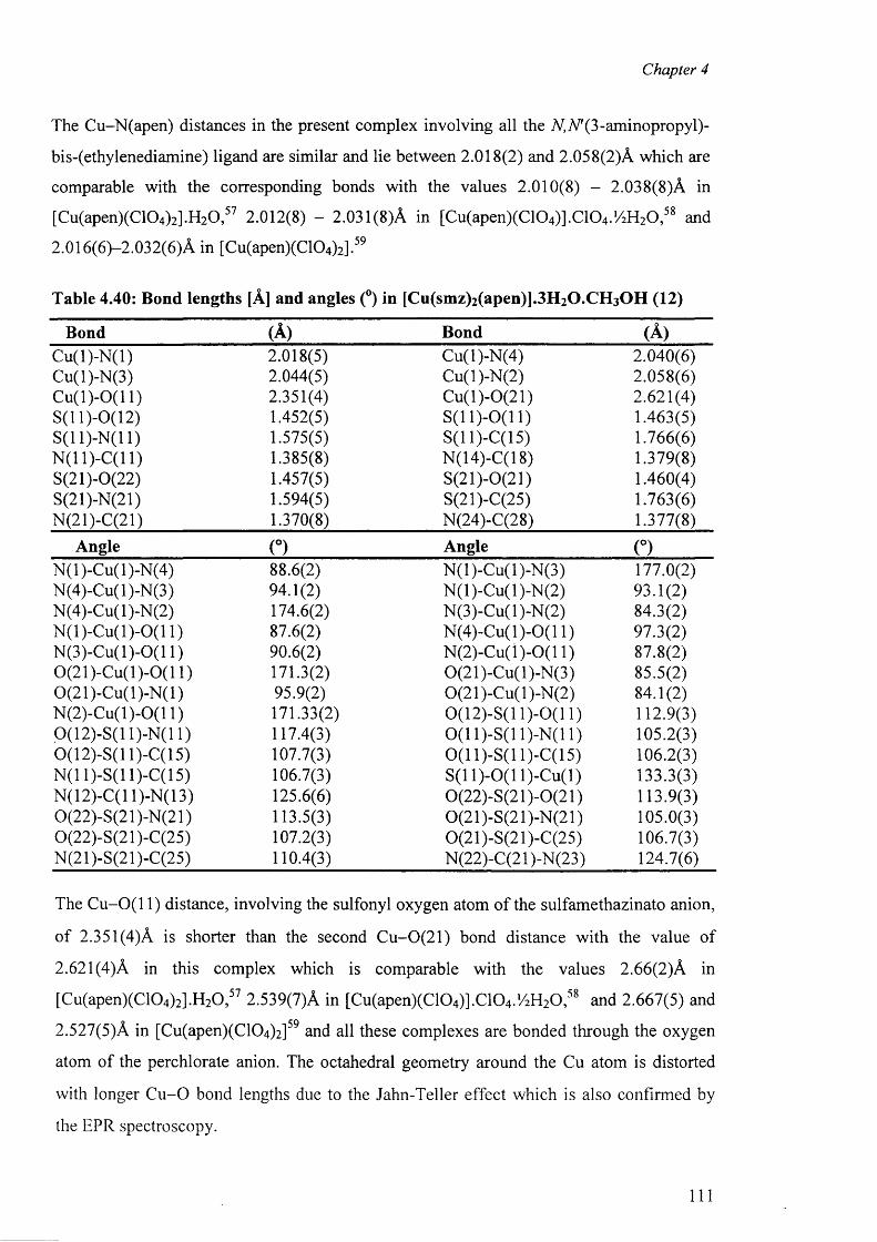
Chapter 4
The Cu-N(apen) distances in the present complex involving all the AfTV’Q-aminopropyl)-
bis-(ethylenediamine) ligand are similar and lie between 2.018(2) and 2.058(2)A which are
comparable with the corresponding bonds with the values 2.010(8) - 2.038(8)A in
[Cu(apen)(C104)2].H20 ,57 2.012(8) - 2.031(8)A in [Cu(apen)(C104)].C104.!/2H20 ,58 and
2.016(6)—2.032(6)A in [Cu(apen)(C104)2].59
Table 4.40: Bond lengths [A] and angles (°) in [Cu(smz)2 (apen)].3 H 2 0 .CH3 0 H (12)
Bond (A) Bond (A )C u(l)-N (l) 2.018(5) Cu(l)-N(4) 2.040(6)Cu(l)-N(3) 2.044(5) Cu(l)-N(2) 2.058(6)C u (l)-0 (11) 2.351(4) Cu(l)-0(21) 2.621(4)S (ll)-0 (12 ) 1.452(5) S (ll)-0 (11 ) 1.463(5)S(11)-N(11) 1.575(5) S(11)-C(15) 1.766(6)N(11)-C(11) 1.385(8) N(14)-C(18) 1.379(8)S(21)-0(22) 1.457(5) S(21)-0(21) 1.460(4)S(21)-N(21) 1.594(5) S(21)-C(25) 1.763(6)N(21)-C(21) 1.370(8) N(24)-C(28) 1.377(8)
Angle (°) Angle (°)N (l)-Cu(l)-N(4) 88.6(2) N (l)-Cu(l)-N (3) 177.0(2)N(4)-Cu(l)-N(3) 94.1(2) N (l)-Cu(l)-N (2) 93.1(2)N(4)-Cu(l)-N(2) 174.6(2) N(3)-Cu(l)-N(2) 84.3(2)N (l)-C u(l)-0 (11) 87.6(2) N (4)-C u(l)-0(11) 97.3(2)N (3)-C u(l)-0(11) 90.6(2) N (2)-C u(l)-0(11) 87.8(2)0(21)-C u(l)-0 (11) 171.3(2) 0(21)-Cu(l)-N(3) 85.5(2)0(21)-C u(l)-N (l) 95.9(2) 0(21)-Cu(l)-N(2) 84.1(2)N (2)-C u(l)-0(11) 171.33(2) 0(12)-S(11)-0(11) 112.9(3)0(12)-S(11)-N(11) 117.4(3) 0 (1 1)-S(11 )-N( 11) 105.2(3)0(12)-S(11)-C(15) 107.7(3) 0 (1 1)-S(11 )-C(l 5) 106.2(3)N(11)-S(11)-C(15) 106.7(3) S(11)-0(1 l)-C u(l) 133.3(3)N(12)-C(l 1)-N(13) 125.6(6) 0(22)-S(21)-0(21) 113.9(3)0(22)-S(21)-N(21) 113.5(3) 0(21)-S(21)-N(21) 105.0(3)0(22)-S(21)-C(25) 107.2(3) 0(21)-S(21)-C(25) 106.7(3)N(21)-S(21)-C(25) 110.4(3) N(22)-C(21)-N(23) 124.7(6)
The O u-O (l 1) distance, involving the sulfonyl oxygen atom of the sulfamethazinato anion,
o f 2.351(4)A is shorter than the second C u-0(21) bond distance with the value of
2.621(4)A in this complex which is comparable with the values 2.66(2)A in
[Cu(apen)(C104)2].H20,57 2.539(7)A in [Cu(apen)(C104)].C104.‘/2H20 ,5S and 2.667(5) and
2.527(5)A in [Cu(apen)(C1 0 4)2 ]59 and all these complexes are bonded through the oxygen
atom of the perchlorate anion. The octahedral geometry around the Cu atom is distorted
with longer C u -0 bond lengths due to the Jahn-Teller effect which is also confirmed by
the EPR spectroscopy.
I l l

Chapter 4
c m N24
C27C13 C6C7C12 C28C112
C26N12 C5 C29N3C8C14
N13 C11 C25 C30N11N4 C4
Cu10 1 2 S11 N2 022011
N21 N22C21C15 C16 ,C3N1
C20 N23C17 C22 C211
C2C1C19 C24 C23C18
C212N14
Figure 4.37X-ray structure o f [Cu(smz)2 (apen)]. 3 H2 O. CH3 OH (12) showing crystallographic atom numbering scheme used. Thermal ellipsoids are drawn at 50% probability level. The ring hydrogens are omitted fo r clarity.
The complex forms several hydrogen bonds involving the coordinated N,N*(3-
aminopropyl)-bis-(ethylenediamine) molecule and sulfamethazine molecule which are
listed in Table 4.41.
0
Figure 4.38The packing diagram o f the complex [Cu(smz)2 (apen)].3 H 2 O.CH3 OH (12) where the hydrogen bonds are shown by dashed lines.
112
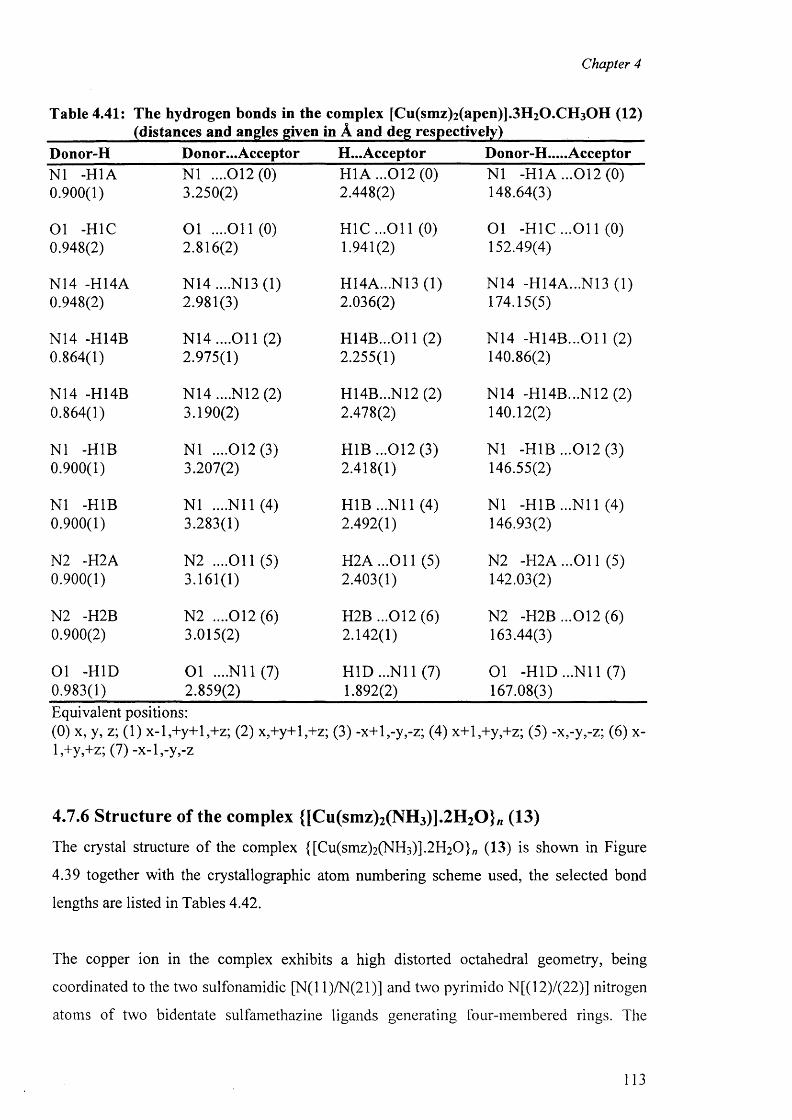
Chapter 4
Table 4.41: The hydrogen bonds in the complex [Cu(smz)2 (apen)].3 H 2 0 .CH3 0 H (12)(distances and angles given in A and deg respectively)
Donor-H Donor...Acceptor H...Acceptor Donor-H.....AcceptorN1 -H1A N1 ....012(0) H1A ...012(0) N1 -HI A . ..012 (0 )0.900(1) 3.250(2) 2.448(2) 148.64(3)
01 -H1C O l ....011(0) H 1C ...011 (0) O l -H1C ...O il (0)0.948(2) 2.816(2) 1.941(2) 152.49(4)
N14 -H14A N 14....N 13(1) H14A...N13 (1) N14 -H14A...N13 (1)0.948(2) 2.981(3) 2.036(2) 174.15(5)
N14 -H14B N 14....011 (2) H 14B...011 (2) N14 -H 14B ...011 (2)0.864(1) 2.975(1) 2.255(1) 140.86(2)
N14 -H14B N14 ....N12 (2) H14B...N12 (2) N14 -H14B...N12 (2)0.864(1) 3.190(2) 2.478(2) 140.12(2)
N1 -H1B N1 ....012(3) H1B ...012 (3) N1 -H1B ...012 (3)0.900(1) 3.207(2) 2.418(1) 146.55(2)
N1 -H1B N1 ....N il (4) H1B ...N il (4) N1 -H1B ...N il (4)0.900(1) 3.283(1) 2.492(1) 146.93(2)
N2 -H2A N2 ....O il (5) H 2A ...011 (5) N2 -H 2A ...011 (5)0.900(1) 3.161(1) 2.403(1) 142.03(2)
N2 -H2B N2 ....012(6) H2B ...012(6) N2 -H2B ...012 (6)0.900(2) 3.015(2) 2.142(1) 163.44(3)
01 -HID O l ....N il (7) H ID ...N il (7) O l -HID ...N il (7)0.983(1) 2.859(2) 1.892(2) 167.08(3)Equivalent positions:(0) x, y, z; (1) x-l,+y+l,+z; (2) x,+y+l,+z; (3) -x+l,-y,-z; (4) x+l,+y,+z; (5) -x,-y,-z; (6) x- l,+y,+z; (7) -x-l,-y,-z
4.7.6 Structure of the complex {[Cu(smz)2(NH3)].2H20 } /I (13)
The crystal structure of the complex {[Cu(smz)2(NH3)].2H20}„ (13) is shown in Figure
4.39 together with the crystallographic atom numbering scheme used, the selected bond
lengths are listed in Tables 4.42.
The copper ion in the complex exhibits a high distorted octahedral geometry, being
coordinated to the two sulfonamidic [N(l 1)/N(21)] and two pyrimido N[(12)/(22)] nitrogen
atoms of two bidentate sulfamethazine ligands generating four-membered rings. The
113
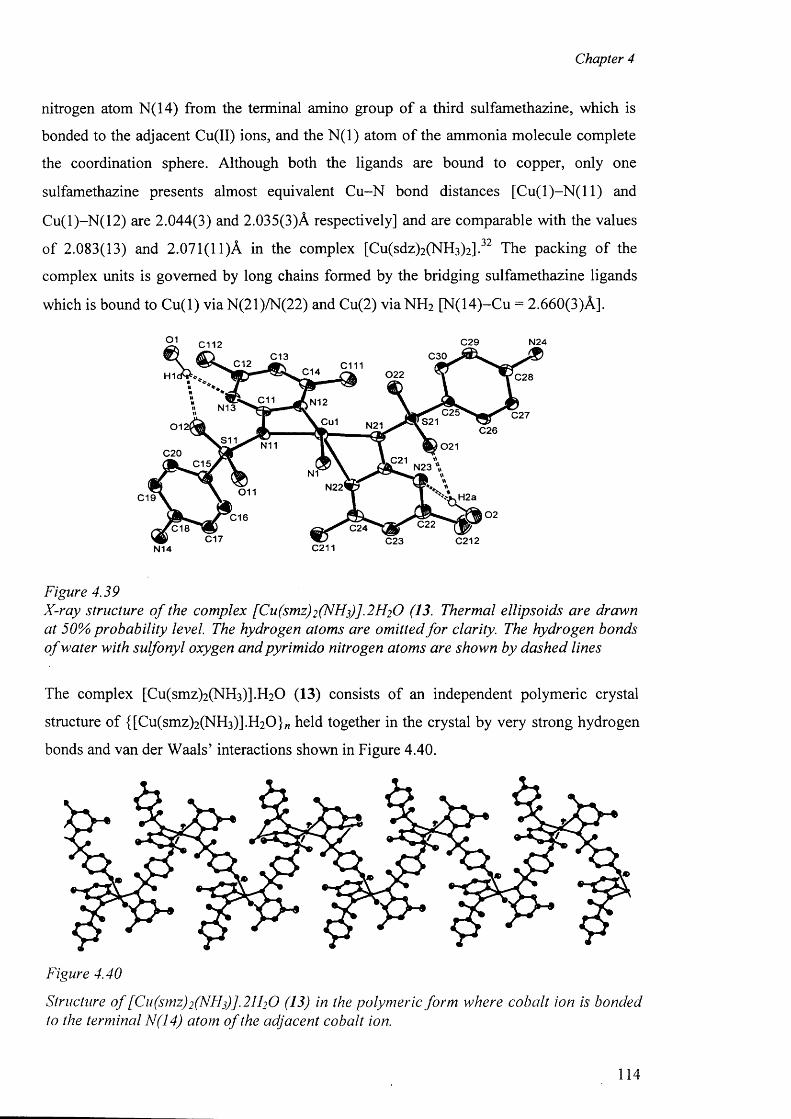
Chapter 4
nitrogen atom N(14) from the terminal amino group o f a third sulfamethazine, which is
bonded to the adjacent Cu(II) ions, and the N (l) atom of the ammonia molecule complete
the coordination sphere. Although both the ligands are bound to copper, only one
sulfamethazine presents almost equivalent C u-N bond distances [C u (l) -N (ll) and
Cu(l)-N (12) are 2.044(3) and 2.035(3)A respectively] and are comparable with the values
o f 2.083(13) and 2.071(11)A in the complex [Cu(sdz)2 (NH 3 )2 ].32 The packing of the
complex units is governed by long chains formed by the bridging sulfamethazine ligands
which is bound to C u(l) viaN(21)/N(22) and Cu(2) v iaN H 2 [N(14)-Cu = 2.660(3)A].
Figure 4.39X-ray structure o f the complex [Cu(smz)2 (N H f ]. 2 H 2 O (13. Thermal ellipsoids are drawn at 50% probability level. The hydrogen atoms are omitted fo r clarity. The hydrogen bonds o f water with sulfonyl oxygen and pyrimido nitrogen atoms are shown by dashed lines
The complex [Cu(smz)2(NH3)].H20 (13) consists of an independent polymeric crystal
structure o f {[Cu(smz)2 (NH3 )] .H2 O } „ held together in the crystal by very strong hydrogen
bonds and van der W aals’ interactions shown in Figure 4.40.
Figure 4.40
Structure o f [Cu(smz)2 (NHs)].2 H2 0 (13) in the polymeric form where cobalt ion is bonded to the terminal N(14) atom o f the adjacent cobalt ion.
N14 C211
114
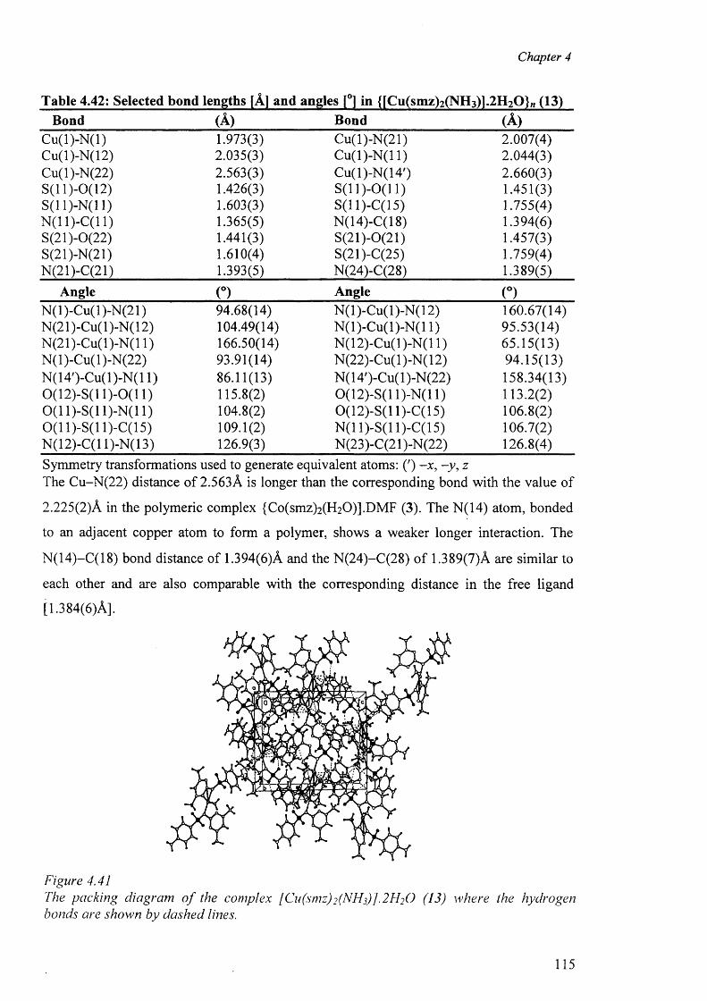
Chapter 4
Table 4.42: Selected bond lengths [A] and angles [°] in {[Cu(smz)2(NH3)].2H2Q}/i (13)Bond (A) Bond (A )
Cu(l)-N (l) 1.973(3) Cu(l)-N(21) 2.007(4)Cu(l)-N(12) 2.035(3) C u(l)-N (l 1) 2.044(3)Cu(l)-N(22) 2.563(3) Cu(l)-N(14') 2.660(3)S(11 )-0 (l 2) 1.426(3) S (ll)-0 (1 1 ) 1.451(3)S(11)-N(11) 1.603(3) S(11 )-C(l 5) 1.755(4)N(11)-C(11) 1.365(5) N(14)-C(18) 1.394(6)S(21)-0(22) 1.441(3) S(21)-0(21) 1.457(3)S(21)-N(21) 1.610(4) S(21)-C(25) 1.759(4)N(21)-C(21) 1.393(5) N(24)-C(28) 1.389(5)
Angle (°) Angle (°)N (l)-Cu(l)-N(21) 94.68(14) N (l)-Cu(l)-N (12) 160.67(14)N(21)-Cu(l)-N(12) 104.49(14) N (l)-C u(l)-N (l 1) 95.53(14)N(21)-Cu(l)-N(l 1) 166.50(14) N(12)-Cu(l)-N(l 1) 65.15(13)N(l)-Cu(l)-N(22) 93.91(14) N(22)-Cu(l)-N(12) 94.15(13)N(14')-Cu(l)-N(l 1) 86.11(13) N(14')-Cu(l)-N(22) 158.34(13)0(12)-S(11)-0(11) 115.8(2) 0(12)-S(11)-N(11) 113.2(2)0 (1 1)-S(11)-N(11) 104.8(2) 0(12)-S(11)-C(15) 106.8(2)0 (1 1)-S(11)-C(15) 109.1(2) N(11)-S(11 )-C(l 5) 106.7(2)N(12)-C(l 1)-N(13) 126.9(3) N(23)-C(21)-N(22) 126.8(4)Symmetry transformations used to generate equivalent atoms: (') -x , -y , zThe Cu-N(22) distance of 2.563A is longer than the corresponding bond with the value of
2.225(2)A in the polymeric complex {Co(smz)2(H20)].DMF (3). The N(14) atom, bonded
to an adjacent copper atom to form a polymer, shows a weaker longer interaction. The
N(14)-C(18) bond distance of 1.394(6)A and the N(24)-C(28) o f 1.389(7)A are similar to
each other and are also comparable with the corresponding distance in the free ligand
[1.384(6)A].
Figure 4.41The packing diagram o f the complex [ Cu(smz)2 (NHf]. 21FO (13) where the hydrogen bonds are shown by dashed lines.
115
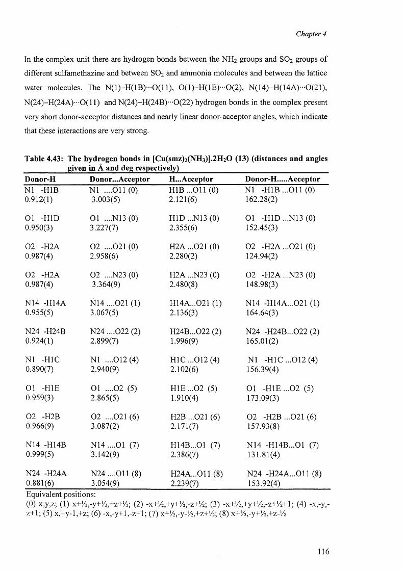
Chapter 4
In the complex unit there are hydrogen bonds between the NH 2 groups and SO2 groups of
different sulfamethazine and between SO2 and ammonia molecules and between the lattice
water molecules. The N (1)-H (1B )-0(11), 0 ( l ) -H ( lE ) -0 (2 ) , N (14)-H (14A )-0(21),
N(24)-H(24A)—0(11) and N(24)-H(24B)~-0(22) hydrogen bonds in the complex present
very short donor-acceptor distances and nearly linear donor-acceptor angles, which indicate
that these interactions are very strong.
Table 4.43: The hydrogen bonds in [Cu(smz)2(NH3)].2H20 (13) (distances and anglesgiven in A and deg respectively)
Donor-H Donor... Acceptor H...Acceptor Donor-H.... AcceptorN1 -H1B 0.912(1)
N1 ....011(0) 3.003(5)
H 1B...011 (0) 2.121(6)
N1 -H1B .. 162.28(2)
.O il (0)
01 -HID 0.950(3)
Ol ....N13 (0) 3.227(7)
HID ...N13 (0) 2.355(6)
O l -H ID .. 152.45(3)
.N13 (0)
02 -H2A 0.987(4)
0 2 ....021 (0) 2.958(6)
H2A ...021 (0) 2.280(2)
0 2 -H2A .. 124.94(2)
.021 (0)
02 -H2A 0.987(4)
0 2 ....N23 (0) 3.364(9)
H2A ...N23 (0) 2.480(8)
0 2 -H2A .. 148.98(3)
.N23 (0)
N14 -H14A 0.955(5)
N 14....021 (1) 3.067(5)
H14A...021 (1) 2.136(3)
N14 -H14A...021 (1) 164.64(3)
N24 -H24B 0.924(1)
N24 ....022 (2) 2.899(7)
H24B...022 (2) 1.996(9)
N24 -H24B...022 (2) 165.01(2)
N1 -H1C 0.890(7)
N1 ....012(4) 2.940(9)
H1C ...012 (4) 2.102(6)
N1 -H1C . 156.39(4)
..0 1 2 (4 )
Ol -HIE 0.959(3)
O l ....02 (5) 2.865(5)
H IE ...02 (5) 1.910(4)
O l -H IE .. 173.09(3)
.02 (5)
02 -H2B 0.966(9)
0 2 ....021 (6) 3.087(2)
H2B ...021 (6) 2.171(7)
0 2 -H2B . 157.93(8)
..021 (6)
N14 -H14B 0.999(5)
N 14....01 (7) 3.142(9)
H14B...01 (7) 2.386(7)
N14 -H14B...01 (7) 131.81(4)
N24 -H24A 0.881(6)
N 24....011 (8) 3.054(9)
H24A...011 (8) 2.239(7)
N24 -H 24A ...011 (8) 153.92(4)
Equivalent positions:(0) x,y,z; (1) x+1/2,-y+,A,+z+!4; (2) -x+1/2,+y+!/2,-z+1/2; (3) -x+V^+y+'/^-z+Va+l; (4) -x,-y,- z+1; (5) x,+y-l,+z; (6) -x,-y+l,-z+l; (7) x+'/Vy-V^+z+l^; (8) x+W y+V ^+z-1/-*
116
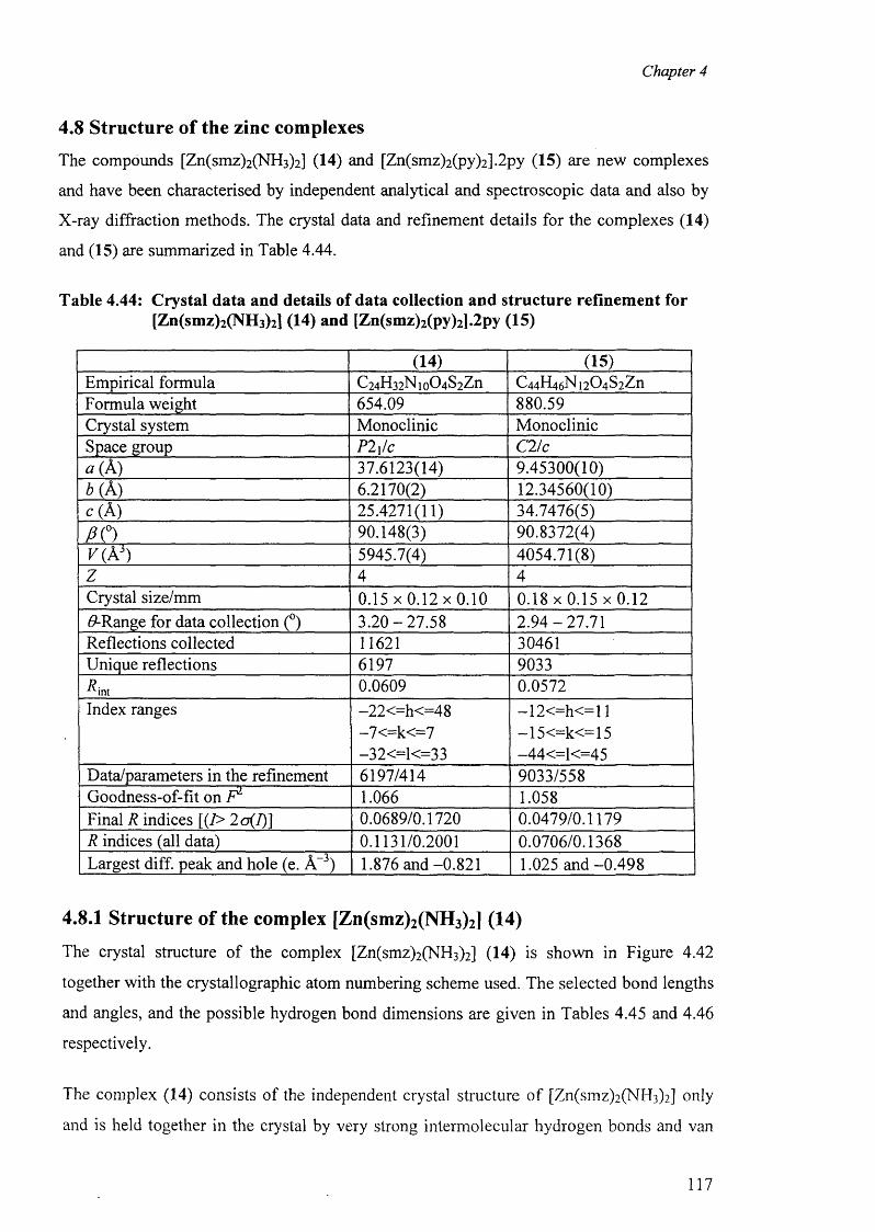
Chapter 4
4.8 Structure of the zinc complexes
The compounds [Zn(smz)2 (NH3 )2 ] (14) and [Zn(smz)2 (py)2 ].2 py (15) are new complexes
and have been characterised by independent analytical and spectroscopic data and also by
X-ray diffraction methods. The crystal data and refinement details for the complexes (14)
and (15) are summarized in Table 4.44.
Table 4.44: Crystal data and details of data collection and structure refinement for [Zn(smz)2(NH3)2] (14) and [Zn(smz)2(py)2].2py (15)
(14) (15)Empirical formula C24H32Nio04S2Zn C4 4 H4 6 N i2 0 4 S2ZnFormula weight 654.09 880.59Crystal system Monoclinic MonoclinicSpace group P2i/c C2/c« (A) 37.6123(14) 9.45300(10)b( A) 6.2170(2) 12.34560(10)c(A ) 25.4271(11) 34.7476(5)
f iO 90.148(3) 90.8372(4)v ( V ) 5945.7(4) 4054.71(8)z 4 4Crystal size/mm 0 .1 5 x 0 .1 2 x 0 .1 0 0 .1 8 x 0 .1 5 x 0 .1 2#-Range for data collection (°) 3 .2 0 -2 7 .5 8 2 .9 4 -27 .71Reflections collected 11621 30461Unique reflections 6197 9033Rmi 0.0609 0.0572Index ranges -22<=h<=48 —1 2 <=h< = 1 1
-7<=k<=7 —15<=k<=15-32<=1<=33 -44<=1<=45
Data/parameters in the refinement 6197/414 9033/558Goodness-of-fit on F 1 1.066 1.058Final R indices [(7> 2o(I)] 0.0689/0.1720 0.0479/0.1179R indices (all data) 0.1131/0.2001 0.0706/0.1368Largest diff. peak and hole (e. A-3) 1.876 and -0.821 1.025 and -0.498
4.8.1 Structure of the complex [Zn(smz)2(NH3)2] (14)
The crystal structure of the complex [Zn(smz)2 (NH3)2 ] (14) is shown in Figure 4.42
together with the crystallographic atom numbering scheme used. The selected bond lengths
and angles, and the possible hydrogen bond dimensions are given in Tables 4.45 and 4.46
respectively.
The complex (14) consists of the independent crystal structure o f [Zn(smz)2 (NH3)2 ] only
and is held together in the crystal by very strong intermolecular hydrogen bonds and van
117
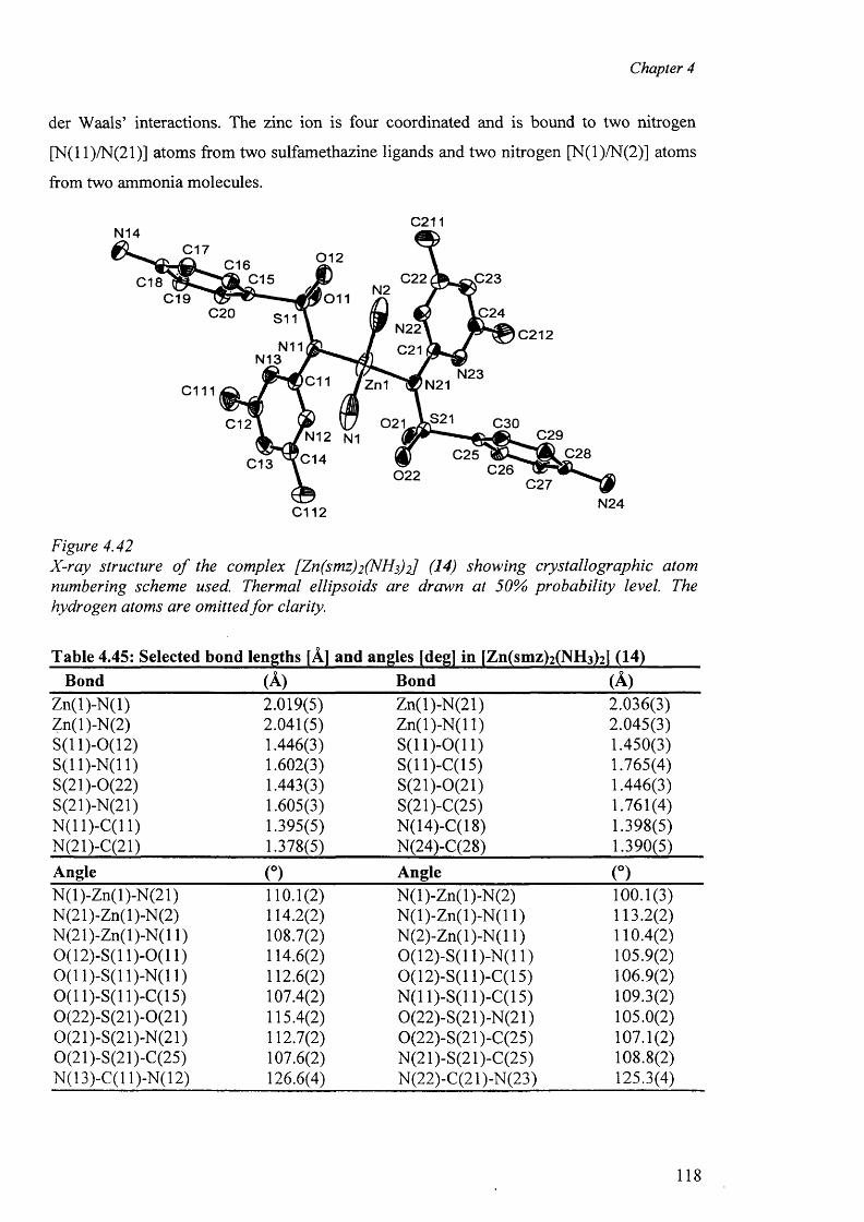
Chapter 4
der Waals’ interactions. The zinc ion is four coordinated and is bound to two nitrogen
[N(l 1 )/N(21)] atoms from two sulfamethazine ligands and two nitrogen [N(l)/N(2)] atoms
from two ammonia molecules.
C2 11N 1 4
C 1 7 012C 1 6C 2 2 0 2 3C 1 5C 1 8 N 2
C 1 9 0110 2 4020 S 11
N 2 2
C21C212
N11N 1 3
N 2 3C11 Zn1 N21cm0 3 0C 1 2
0 2 9' N 1 2 N1
0 1 4 0 2 8C 2 5C 1 3 C 2 6022
C 2 7N 2 40112
Figure 4.42X-ray structure o f the complex [Zn(smz)2 (NHs)2] (14) showing crystallographic atom numbering scheme used. Thermal ellipsoids are drawn at 50% probability level. The hydrogen atoms are omitted fo r clarity.
Table 4.45: Selected bond lengths [A] and angles [deg] in [Zn(smz)2 (NH3 )2 ] (14)Bond (A) Bond (A)
Zn(l)-N (l) 2.019(5) Zn(l)-N(21) 2.036(3)Zn(l)-N(2) 2.041(5) Zn(l)-N (l 1) 2.045(3)S(11)-0(12) 1.446(3) S (ll)-0 (11 ) 1.450(3)S ( ll) -N (ll) 1.602(3) S(11)-C(15) 1.765(4)S(21)-0(22) 1.443(3) S(21)-0(21) 1.446(3)S(21)-N(21) 1.605(3) S(21)-C(25) 1.761(4)N(11)-C(11) 1.395(5) N(14)-C(18) 1.398(5)N(21)-C(21) 1.378(5) N(24)-C(28) 1.390(5)Angle (°) Angle (°)N(l)-Zn(l)-N(21) 110.1(2) N (l)-Zn(l)-N (2) 100.1(3)N(21)-Zn(l)-N(2) 114.2(2) N (l)-Z n(l)-N (l 1) 113.2(2)N(21)-Zn(l)-N(l 1) 108.7(2) N(2)-Zn(l)-N(l 1) 110.4(2)0(12)-S(11)-0(11) 114.6(2) 0(12)-S(11)-N(11) 105.9(2)0(11 )-S(l 1)-N(11) 112.6(2) 0(12)-S(11)-C(15) 106.9(2)0 (1 1)-S(11)-C(15) 107.4(2) N(11)-S(11)-C(15) 109.3(2)0(22)-S(21)-0(21) 115.4(2) 0(22)-S(21 )-N(21) 105.0(2)0(21)-S(21)-N(21) 112.7(2) 0(22)-S(21 )-C(25) 107.1(2)0(21)-S(21)-C(25) 107.6(2) N(21)-S(21)-C(25) 108.8(2)N( 13)-C( 11 )-N( 12) 126.6(4) N(22)-C(21 )-N(23) 125.3(4)
118
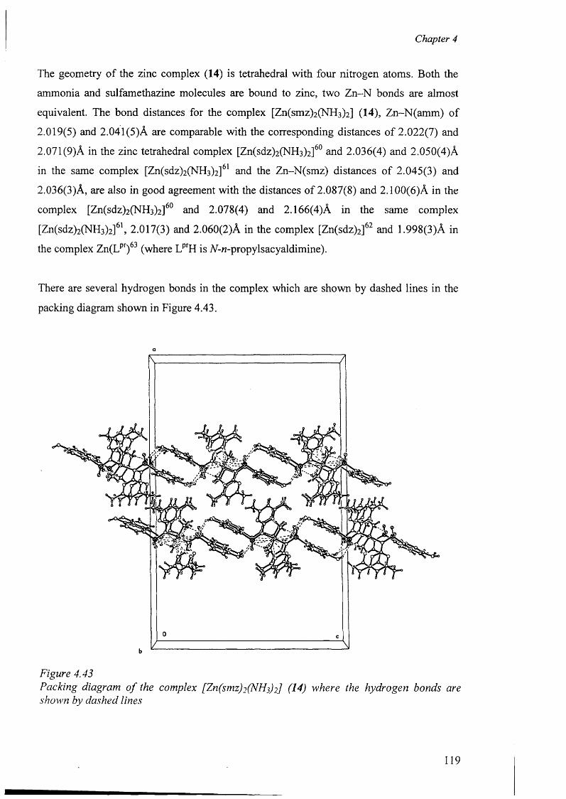
Chapter 4
The geometry o f the zinc complex (14) is tetrahedral with four nitrogen atoms. Both the
ammonia and sulfamethazine molecules are bound to zinc, two Zn-N bonds are almost
equivalent. The bond distances for the complex [Zn(smz)2 (NH3 )2 ] (14), Zn-N(amm) of
2.019(5) and 2.041(5)A are comparable with the corresponding distances o f 2.022(7) and
2.071 (9)A in the zinc tetrahedral complex [Zn(sdz)2 (NH3 )2]60 and 2.036(4) and 2.050(4)A
in the same complex [Zn(sdz)2 (NH3 )2 ]61 and the Zn-N(smz) distances of 2.045(3) and
2.036(3)A, are also in good agreement with the distances of 2.087(8) and 2.100(6)A in the
complex [Zn(sdz)2 (NH3 )2 ]60 and 2.078(4) and 2.166(4)A in the same complex
[Zn(sdz)2(NH3)2]61, 2.017(3) and 2.060(2)A in the complex [Zn(sdz)2]62 and 1.998(3)A in
the complex Zn(Lpr)63 (where LprH is 77-tt-propylsacyaldimine).
There are several hydrogen bonds in the complex which are shown by dashed lines in the
packing diagram shown in Figure 4.43.
Figure 4.43Packing diagram o f the complex [Zn(sm z)i(NH f2] (14) where the hydrogen bonds are shown by dashed lines
119
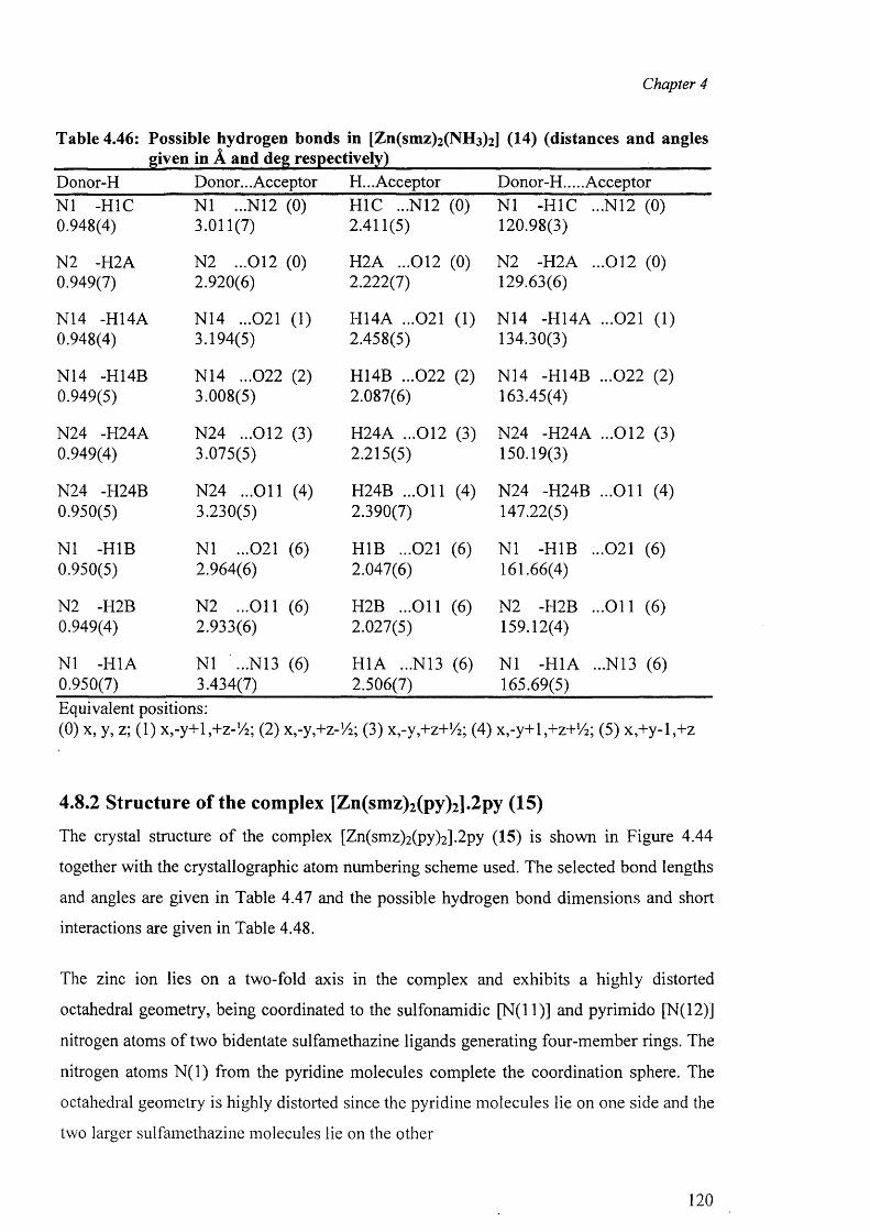
Chapter 4
Table 4.46: Possible hydrogen bonds in [Zn(smz)2 (NH3 )2 ] (14) (distances and angles ___________ given in A and deg respectively)________________________________Donor-H Donor... Acceptor H... Acceptor Donor-H.... AcceptorN1 -H1C N1 ...N12 (0) H1C ...N12 (0) N1 -H1C ...N12 (0)0.948(4) 3.011(7) 2.411(5) 120.98(3)
N2 -H2A N2 ...012 (0) H2A ...012 (0) N2 -H2A ...012 (0)0.949(7) 2.920(6) 2.222(7) 129.63(6)
N14 -H14A N14 ...021 (1) H14A ...021 (1) N14 -H14A ...021 (1)0.948(4) 3.194(5) 2.458(5) 134.30(3)
N14 -H14B N14 ...022 (2) H14B ...022 (2) N14 -H14B ...022 (2)0.949(5) 3.008(5) 2.087(6) 163.45(4)
N24 -H24A N24 ...012 (3) H24A ...012 (3) N24 -H24A ...012 (3)0.949(4) 3.075(5) 2.215(5) 150.19(3)
N24 -H24B N24 ...O il (4) H24B ...O il (4) N24 -H24B ...O il (4)0.950(5) 3.230(5) 2.390(7) 147.22(5)
N1 -H1B N1 ...021 (6) H1B ...021 (6) N1 -H1B ...021 (6)0.950(5) 2.964(6) 2.047(6) 161.66(4)
N2 -H2B N2 ...O il (6) H2B ...O il (6) N2 -H2B ...O il (6)0.949(4) 2.933(6) 2.027(5) 159.12(4)
N1 -H1A N1 ...N13 (6) H1A ...N13 (6) N1 -H1A ...N13 (6)0.950(7) 3.434(7) 2.506(7) 165.69(5)Equivalent positions:(0) x, y, z; (1) x,-y+l,+z-!/2; (2) x.-y^z-Vi; (3) x,-y,+z+72; (4) x,-y+l,+z+72; (5) x,+y-l,+z
4.8.2 Structure of the complex [Zn(smz)2(py)2].2py (15)
The crystal structure of the complex [Zn(smz)2(py)2].2py (15) is shown in Figure 4.44
together with the crystallographic atom numbering scheme used. The selected bond lengths
and angles are given in Table 4.47 and the possible hydrogen bond dimensions and short
interactions are given in Table 4.48.
The zinc ion lies on a two-fold axis in the complex and exhibits a highly distorted
octahedral geometry, being coordinated to the sulfonamidic [N (ll)] and pyrimido [N(l 2)]
nitrogen atoms of two bidentate sulfamethazine ligands generating four-member rings. The
nitrogen atoms N (l) from the pyridine molecules complete the coordination sphere. The
octahedral geometry is highly distorted since the pyridine molecules lie on one side and the
two larger sulfamethazine molecules lie on the other
120
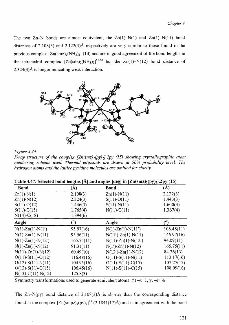
Chapter 4
The two Zn-N bonds are almost equivalent, the Z n (l)-N (l) and Z n ( l) -N (ll) bond
distances o f 2.108(3) and 2.122(3)A respectively are very similar to those found in the
previous complex [Zn(smz)2(NH3)2 ] (14) and are in good agreement of the bond lengths in
the tetrahedral complex [Zn(sdz)2 (NH3 )2]61,62 but the Zn(l)-N (12) bond distance of
2.324(3)A is longer indicating weak interaction.
C2
C1C3C17N14 C16 C4'0 1 2
N1
C1 C5
N11C15ZnfC19
C20 0 1 1 |
C11N1
C1
C13
C112
Figure 4.44X-ray structure o f the complex [Zn(smz)2 (py)2]-2py (15) showing crystallographic atom numbering scheme used. Thermal ellipsoids are drawn at 50% probability level. The hydrogen atoms and the lattice pyridine molecules are omitted fo r clarity.
Table 4.47: Selected bond lengths [A] and angles [deg] in [Zn(smz)2(py)21.2py (15)Bond (A) Bond (A)
Zn(l)-N (l) 2.108(3) Z n(l)-N (ll) 2.122(3)Zn(l)-N(12) 2.324(3) S (ll)-0 (11 ) 1.443(3)S(11)-0(12) 1.446(3) S ( ll) -N ( ll) 1.600(3)S(11)-C(15) 1.765(4) N (ll) -C (ll) 1.367(4)N(14)-C(18) 1.394(6)Angle (°) Angle (°)N (l)-Zn(l)-N (T) 95.97(16) N (l)-Zn(l)-N (l T) 106.48(11)N (l)-Zn(l)-N (l 1) 95.56(11) N(1 T)-Zn(l)-N (l 1) 146.97(16)N(l)-Zn(l)-N (12') 165.75(11) N(11)-Zn(l)-N(12') 94.09(11)N(l)-Zn(l)-N(12) 91.31(11) N (l')-Zn(l)-N (12) 165.75(11)N(11)-Zn(l)-N(12) 60.49(10) N(12')-Zn(l)-N(12) 84.36(13)0 (1 1)-S(11)-0(12) 116.48(16) 0 (1 1)-S(11)-N(11) 113.17(16)0(12)-S(11)-N(11) 104.95(16) 0 (1 1)-S(11)-C(15) 107.27(17)0(12)-S(11)-C(15) 106.45(16) N (ll) -S (l 1)-C(15) 108.09(16)N(13)-C(l 1)-N(12) 125.8(3)Symmetry transformations used to generate equivalent atoms: (') -x+1, y, -z+'A
The Zn-N(py) bond distance of 2.108(3)A is shorter than the corresponding distance
found in the complex [Zn(smpz)2(py)2 ]43 (2.1841(17)A) and is in agreement with the bond
121
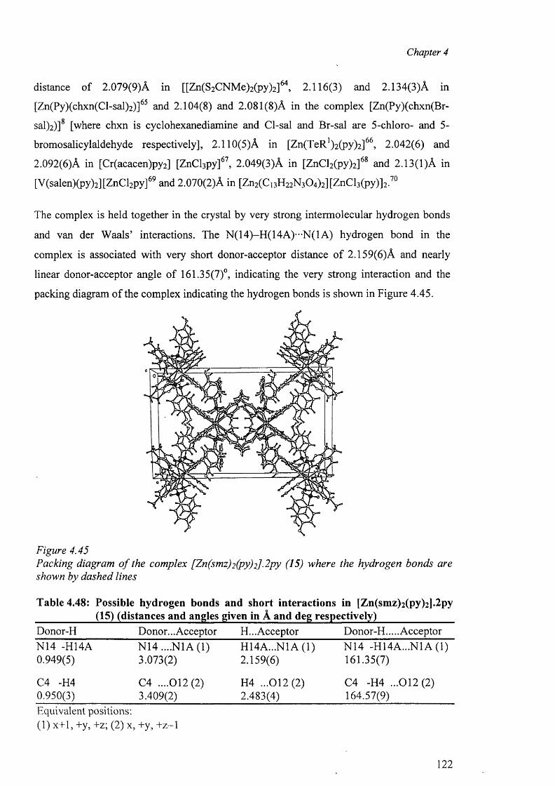
Chapter 4
distance o f 2.079(9)A in [[Zn(S2CNMe)2(py)2 ]64> 2.116(3) and 2.134(3)A in
[Zn(Py)(chxn(Cl-sal)2)]65 and 2.104(8) and 2.081(8)A in the complex [Zn(Py)(chxn(Br-
sal)2)] [where chxn is cyclohexanediamine and Cl-sal and Br-sal are 5-chloro- and 5-
bromosalicylaldehyde respectively], 2.110(5)A in [Zn(TeR')2(py)2]66, 2.042(6) and
2.092(6)A in [Cr(acacen)py2] [ZnCl3py]67, 2.049(3)A in [ZnCl2(py)2]68 and 2.13(1)A in
[V(salen)(py)2][ZnCl2py]69 and 2.070(2)A in [Zn2 (Ci3 H2 2 N 3 0 4 )2 ][ZnCl3 (py ) ] 2 . 7 0
The complex is held together in the crystal by very strong intermolecular hydrogen bonds
and van der Waals’ interactions. The N(14)-H(14A)-”N(1A) hydrogen bond in the
complex is associated with very short donor-acceptor distance o f 2.159(6)A and nearly
linear donor-acceptor angle of 161.35(7)°, indicating the very strong interaction and the
packing diagram o f the complex indicating the hydrogen bonds is shown in Figure 4.45.
Figure 4.45Packing diagram o f the complex [Zn(smz)2 (py)2]-2 py (15) where the hydrogen bonds are shown by dashed lines
Table 4.48: Possible hydrogen bonds and short interactions in [Zn(smz)2(py)2]»2py ___________ (15) (distances and angles given in A and deg respectively)_______________Donor-H Donor... Acceptor H... Acceptor Donor-H.... AcceptorN14 -H14A N14 ....N1A (1) H14A...N1A (1) N14 -H14A...N1A (1)0.949(5) 3.073(2) 2.159(6) 161.35(7)
C4 -H4 C4 ....012(2) H4 ...012(2) C4 -H4 ...012(2)0.950(3) 3.409(2) 2.483(4) 164.57(9)Equivalent positions:(1) x+1, +y, +z; (2) x, +y, + z - l
122
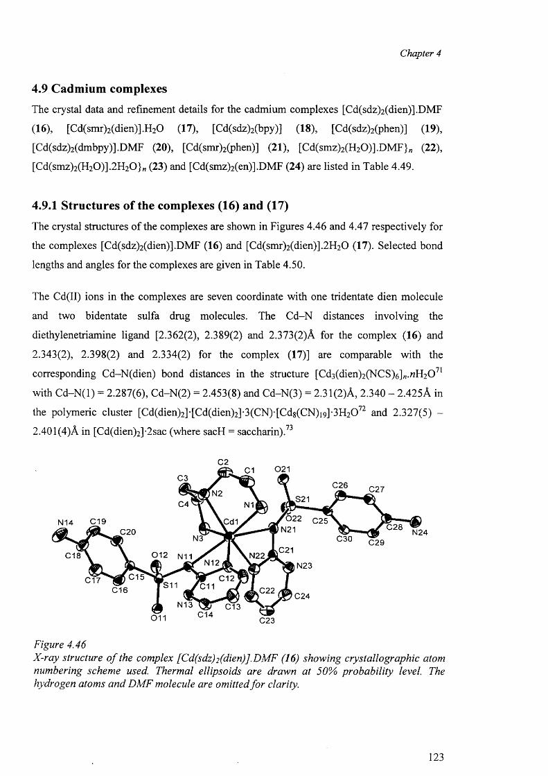
Chapter 4
4.9 Cadmium complexes
The crystal data and refinement details for the cadmium complexes [Cd(sdz)2 (dien)].DMF
(16), [Cd(smr)2(dien)].H20 (17), [Cd(sdz)2(bpy)] (18), [Cd(sdz)2(phen)] (19),
[Cd(sdz)2(dmbpy)].DMF (20), [Cd(smr)2(phen)] (21), [Cd(smz)2(H20)].DMF}„ (22),
[Cd(smz)2(H20)].2H 20}„ (23) and [Cd(smz)2(en)].DMF (24) are listed in Table 4.49.
4.9.1 Structures of the complexes (16) and (17)
The crystal structures of the complexes are shown in Figures 4.46 and 4.47 respectively for
the complexes [Cd(sdz)2(dien)].DMF (16) and [Cd(smr)2(dien)].2H20 (17). Selected bond
lengths and angles for the complexes are given in Table 4.50.
The Cd(II) ions in the complexes are seven coordinate with one tridentate dien molecule
and two bidentate sulfa drug molecules. The Cd-N distances involving the
diethylenetriamine ligand [2.362(2), 2.389(2) and 2.373(2)A for the complex (16) and
2.343(2), 2.398(2) and 2.334(2) for the complex (17)] are comparable with the
corresponding Cd-N(dien) bond distances in the structure [Cd3(dien)2(NCS)6]„.«H20 71
with C d-N (l) = 2.287(6), Cd-N(2) = 2.453(8) and Cd-N(3) = 2.31(2)A, 2.340 - 2.425A in
the polymeric cluster [Cd(dien)2]-[Cd(dien)2]-3(CN)-[Cd8(CN)i9]-3H20 72 and 2.327(5) -
2.401(4)A in [Cd(dien)2]-2sac (where sacH = saccharin).73
C2021C1
C3C26 C27N2
S21C4 N1
C 19 lC cHN14 C 28C20 N24N3 C30 C29
C21C18 N22
N12 N23C15 C12C17 S11 C11C16 C22
C24N13 C13
C14011 C23
Figure 4.46X-ray structure o f the complex [Cd(sdz)2 (dien)].DMF (16) showing crystallographic atom numbering scheme used. Thermal ellipsoids are drawn at 50% probability level. The hydrogen atoms and DMF molecule are omitted fo r clarity.
123
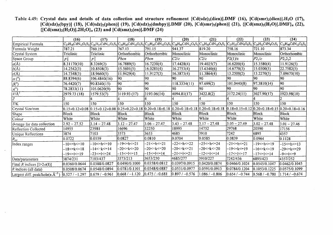
Table 4.49: Crystal data and details of data collection and structure refinement [Cd(sdz)2 (dien)].DMF (16), (Cd(smr)2(dien)].H20 (17), [Cd(sdz)2(bpy)] (18), [Cd(sdz)2(phen)] (19), [Cd(sdz)2(dmbpy)].DMF (20), [Cd(smr)2(phen)] (21), ([Cd(smz)2(H20)].DM F}rt (22), {[Cd(smz)2(H20)].2H 20 } w (23) and [Cd(smz)2(en)].DMF (24)
(16) (17) (18) (19) (20) (21) (22) (23) (24)Empirical Formula C27H38CdN,20 5S2 C26H37CdN i j 0 3S2 C3oH26CdN100 4S2C32H26CdN io04S2 C38H44CdN120 6S2C34H30CdN io04S2 C27H35CdN90 6S2 C24H32CdN80 7S2 C32H48CdN120 6S2Formula Weight 787.21 760.19 767.13 791.15 941.37 819.20 758.16 721.10 873.34Crystal System Triclinic Triclinic Orthorhombic Orthorhombic Monoclinic Monoclinic Monoclinic Monoclinic OrthorhombicSpace Group P\ P\ Pbcn Pbcn C2/c C2/c P2(l)/c P2x/c P2{1{2«(A) 8.31170(10) 8.3269(2) 16.7889(5) 16.7230(4) 17.4428(4) 19.4025(7) 16.6200(4) 15.3580(4) 11.9126(5)b( A) 14.2542(3) 10.8508(3) 15.5601(5) 16.0281(4) 16.2753(4) 15.6340(6) 14.6770(3) 15.0300(5) 22.7582(5)c(A) 14.7348(3) 18.9660(5) 11.9429(4) 11.9127(3) 16.3873(4) 11.3864(4) 13.2500(2) 13.2270(5) 7.09670(10)a<°) 88.8394(6) 106.4840(16) 90 90 90 90 90 90 90PC) 76.4420(7) 90.8340(15) 90 90 118.3334(11) 97.699(2) 101.0440(8) 97.3810(14) 90
X°) 78.2833(11) 105.0620(9) 90 90 90 90 90 90 90v/A3 2979.73 (18) 1579.53(7) 3119.93 (17) 3193.06(14) 4094.81(17) 3422.8(2) 3172.24(11) 3027.90(17) 1923.98(10)z 2 4 4 4 4 4 4 4 2TfK 150 150 150 150 150 150 150 150 150Crystal Size/mm 0.15x0.12x0.08 0.15x0.12x0.08 0.25x0.22x0.18 0.20x0.18x0.18 0.20x0.18x0.18 0.20x0.18x0.18 0.18x0.15x0.12 0.20x0.18x0.15 0.20x0.18x0.16Shape Block Block Block Block Block Block Block Block BlockColour White White White White White White White White WhiteGrange for data collection 2 .92-27 .52 3 .14-27 .48 3 .12-27.47 3 .06-27 .47 3.43 -27 .48 3.17-27 .48 3.05 -27 .49 3.02-27.48 3.01-27.46Reflection Collected 14955 23981 16696 32250 18995 14752 29768 20590 17156Unique Reflections 3874 7103 3573 3653 4685 3910 7242 6895 4357int 0.0722 0.0559 0.0827 0.0810 0.0704 0.0585 0.0839 0.0964 0.1128
Index ranges -1 0<=h<=10 -10<=h<=10 -19<=h<=21 -2 1 <=h<=21 -22<=h<=22 -25<=h<=24 -21<=h<=21 -19<=h<=19 -15<=h<=13-18<=k<=18 -14<=k<=14 -20<=k<=20 -20<=k<=20 -20<=k<=21 -20<=k<=20 -1 9<=k<=19 -16<=k<=19 -29<=k<=29—19<=1<= 19 -23<=I<=24 — 15<=1<= 15 — 15<=1<= 14 -21 <=1<=21 -12<=1<=14 -1 7<=1<= 17 —17<=1<= 14 -9<=I<=9
Data/parameters 3874/231 7103/437 3573/213 3653/230 4685/277 3910/227 7242/436 6895/423 4357/252Final R indices [I>2o(I)] 0.0360/0.0644 0.0388/0.0827 0.0490/0.1000 0.0358/0.0812 0.0397/0.0915 0.0420/0.0874 0.0466/0.1024 0.0545/0.1047 0.0462/0.1045R indices (all data) 0.0508/0.0674 0.0548/0.0894 0.0781/0.1101 0.0548/0.0887 0.0531/0.0977 0.0591/0.0913 0.0784/0.1204 0.1053/0.1225 0.0575/0.1099Largest diff. peak/hole(e.A-3) 0.527/-1.297 0.679/-0.961 0.668/ - l . 120 0.475/-0.681 0.897/-0.576 1.086/-0.806 0.654/-0.744 0.568/-0.780 1.714,/-0.674
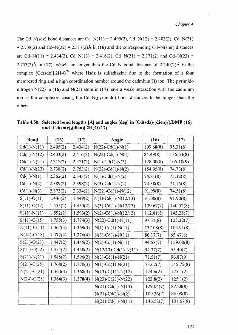
Chapter 4
The Cd-N(sdz) bond distances are C d -N (ll) = 2.495(2), Cd-N(12) = 2.403(2), Cd-N(21)
= 2.738(2) and Cd-N(22) = 2.317(2)A in (16) and the corresponding Cd-N(smr) distances
are C d -N (ll) = 2.434(2), Cd-N(13) = 2.416(2), Cd-N(21) = 2.371(2) and Cd-N(23) =
2.753(2)A in (17), which are longer than the C d-N bond distance of 2.240(2)A in the
complex [Cd(sdz)].2H2074 where Hsdz is sulfadiazine due to the formation o f a four
membered ring and a high coordination number around the cadmium(II) ion. The pyrimido
nitrogen N(22) in (16) and N(23) atom in (17) have a weak interaction with the cadmium
ion in the complexes casing the Cd-N(pyrimido) bond distances to be longer than the
others.
Table 4.50: Selected bond lengths [A] and angles [deg] in [Cd(sdz)2 (dien)2 ].DMF (16) and [Cd(smr)2(dien)].2H20 (17)
Bond (16) (17) Angle (16) (17)
C d(l)-N (ll) 2.495(2) 2.434(2) N(22)-Cd(l)-N(l) 109.68(8) 95.31(8)
Cd(l)-N(12) 2.403(2) 2.416(2) N(22)-Cd(l)-N(3) 84.49(8) 136.64(8)
Cd(l)-N(21) 2.317(2) 2.371(2) N(l)-Cd(l)-N(3) 128.00(8) 105.10(9)
Cd(l)-N(22) 2.738(2) 2.753(2) N(22)-Cd(l)-N(2) 154.95(8) 74.73(8)
Cd(l)-N (l) 2.362(2) 2.343(2) N (l)-Cd(l)-N(2) 74.81(8) 75.32(8)
Cd(l)-N(2) 2.389(2) 2.398(2) N(3)-Cd(l)-N(2) 74.38(8) 74.16(8)
Cd(l)-N(3) 2.373(2) 2.334(2) N(22)-Cd( 1 )-N( 12) 91.99(8) 74.51(8)
S (ll)-0 (11) 1.446(2) 1.449(2) N(l)-Cd(l)-N(12/13) 91.00(8) 91.90(8)S(11)-0(12) 1.455(2) 1.450(2) N(3)-Cd(l)-N(12/13) 139.67(7) 140.52(8)
S ( ll) -N ( ll) 1.592(2) 1.593(2) N(2)-Cd(l)-N(12/13) 112.81(8) 145.28(7)S(11)-C(15) 1.755(3) 1.774(3) N (22)-Cd(l)-N (ll) 97.31(8) 125.32(7)
N(11)-C(11) 1.367(3) 1.369(3) N (l)-C d(l)-N (l 1) 137.08(8) 105.91(8)N(14)-C(18) 1.372(4) 1.376(4) N(3)-Cd(l)-N(l 1) 86.17(7) 85.47(8)
S(21)-0(21) 1.447(2) 1.445(2) N(2)-Cd(l)-N(l 1) 94.58(7) 159.00(8)
S(21)-0(22) 1.454(2) 1.450(2) N(12/13)-Cd(l)-N(l 1) 54.37(7) 55.40(7)
S(21)-N(21) 1.588(2) 1.596(2) N(3)-Cd(l)-N(21) 78.51(7) 96.87(9)
S(21)-C(25) 1.768(2) 1.770(3) N(l)-Cd(l)-N(21) 73.62(7) 145.75(8)N(21)-C(21) 1.360(3) 1.368(3) N(13)-C(l 1)-N(12) 124.6(2) 125.1(2)
N(24)-C(28) 1.364(3) 1.378(4) N(23)-C(21 )-N(22) 123.8(2) 125.1(2)
N(21)-Cd(l)-N(13) 129.04(7) 87.28(8)
N(21)-Cd(l)-N(2) 109.36(7) 86.09(8)
N (21)-C d(l)-N (ll) 146.52(7) 101.67(8)
124
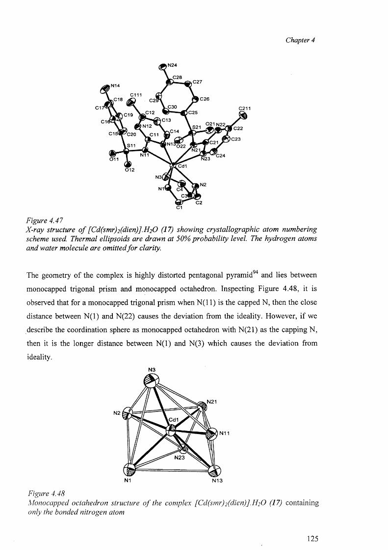
Chapter 4
Figure 4.47X-ray structure o f [Cd(smr)2 (dien)].H2 O (17) showing crystallographic atom numbering scheme used. Thermal ellipsoids are drawn at 50% probability level. The hydrogen atoms and water molecule are omitted fo r clarity.
The geometry of the complex is highly distorted pentagonal pyramid94 and lies between
monocapped trigonal prism and monocapped octahedron. Inspecting Figure 4.48, it is
observed that for a monocapped trigonal prism when N (11) is the capped N, then the close
distance between N (l) and N(22) causes the deviation from the ideality. However, if we
describe the coordination sphere as monocapped octahedron with N(21) as the capping N,
then it is the longer distance between N (l) and N(3) which causes the deviation from
ideality.
N3
N1 N13
Figure 4.48Monocapped octahedron structure o f the complex [Cd(smr)2 (dien)J.H2 0 (17) containing only the bonded nitrogen atom
125
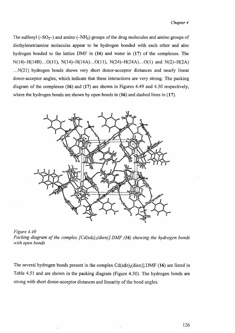
Chapter 4
The sulfonyl (-SO 2- ) and amino (-NH 2 ) groups o f the drug molecules and amino groups of
diethylenetriamine molecules appear to be hydrogen bonded with each other and also
hydrogen bonded to the lattice DMF in (16) and water in (17) o f the complexes. The
N (14)-H (14B )...0(11), N(14)-H(14A)...0(11), N (24)-H (24A )...0(1) and N(2)-H(2A)
...N(21) hydrogen bonds shows very short donor-acceptor distances and nearly linear
donor-acceptor angles, which indicate that these interactions are very strong. The packing
diagram of the complexes (16) and (17) are shown in Figures 4.49 and 4.50 respectively,
where the hydrogen bonds are shown by open bonds in (16) and dashed lines in (17).
Figure 4.49Packing diagram o f the complex [Cd(sdz)2 (dien)].DMF (16) showing the hydrogen bonds with open bonds
The several hydrogen bonds present in the complex Cd(sdz)2 (dien)].DMF (16) are listed in
Table 4.51 and are shown in the packing diagram (Figure 4.50). The hydrogen bonds are
strong with short donor-acceptor distances and linearity o f the bond angles.
126
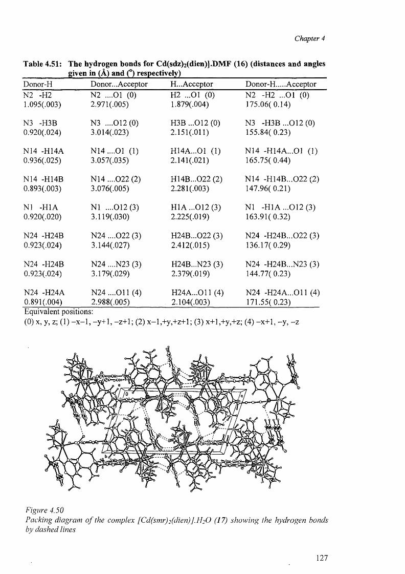
Chapter 4
Table 4.51: The hydrogen bonds for Cd(sdz)2 (dien)].DMF (16) (distances and anglesgiven in (A) and (°) respectively)
Donor-H Donor... Acceptor H... Acceptor Donor-H.... AcceptorN2 -H2 N2 ....01 (0) H2 ...01 (0) N2 -H2 ...01 (0)1.095(.003) 2.971(.005) 1.879(.004) 175.06(0.14)
N3 -H3B N3 ....012(0) H3B ...012 (0) N3 -H3B ...012(0)0.920(.024) 3.014(.023) 2.151(.011) 155.84( 0.23)
N14 -H14A N 14....01 (1) H14A...01 (1) N14 -H14A...01 (1)0.936(.025) 3.057(.035) 2.141(.021) 165.75(0.44)
N14 -H14B N14 ....022 (2) H14B...022 (2) N14 -H14B...022 (2)0.893(.003) 3.076(.005) 2.281(.003) 147.96(0.21)
N1 -H1A N1 ....012(3) H1A ...012 (3) N1 -H1A ...012 (3)0.920(.020) 3.119(.030) 2.225(.019) 163.91(0.32)
N24 -H24B N24 ....022 (3) H24B...022 (3) N24 -H24B...022 (3)0.923(.024) 3.144(.027) 2.412(.015) 136.17( 0.29)
N24 -H24B N24 ....N23 (3) H24B...N23 (3) N24 -H24B...N23 (3)0.923(.024) 3.179(.029) 2.379(.019) 144.77( 0.23)
N24 -H24A N24 ....O il (4) H 24A ...011 (4) N24 -H 24A ...011 (4)0.891(.004) 2.988(.005) . 2.104(.003) 171.55( 0.23)Equivalent positions:(0) x, y, z; (1) -x -1 , -y+1, -z+1; (2) x -l,+ y ,+ z+ l; (3) x+l,+y,+z; (4) -x+1, -y , - z
Figure 4.50Packing diagram o f the complex [Cd(smr)2 (dien)].H 2 0 (17) showing the hydrogen bonds by dashed lines
127
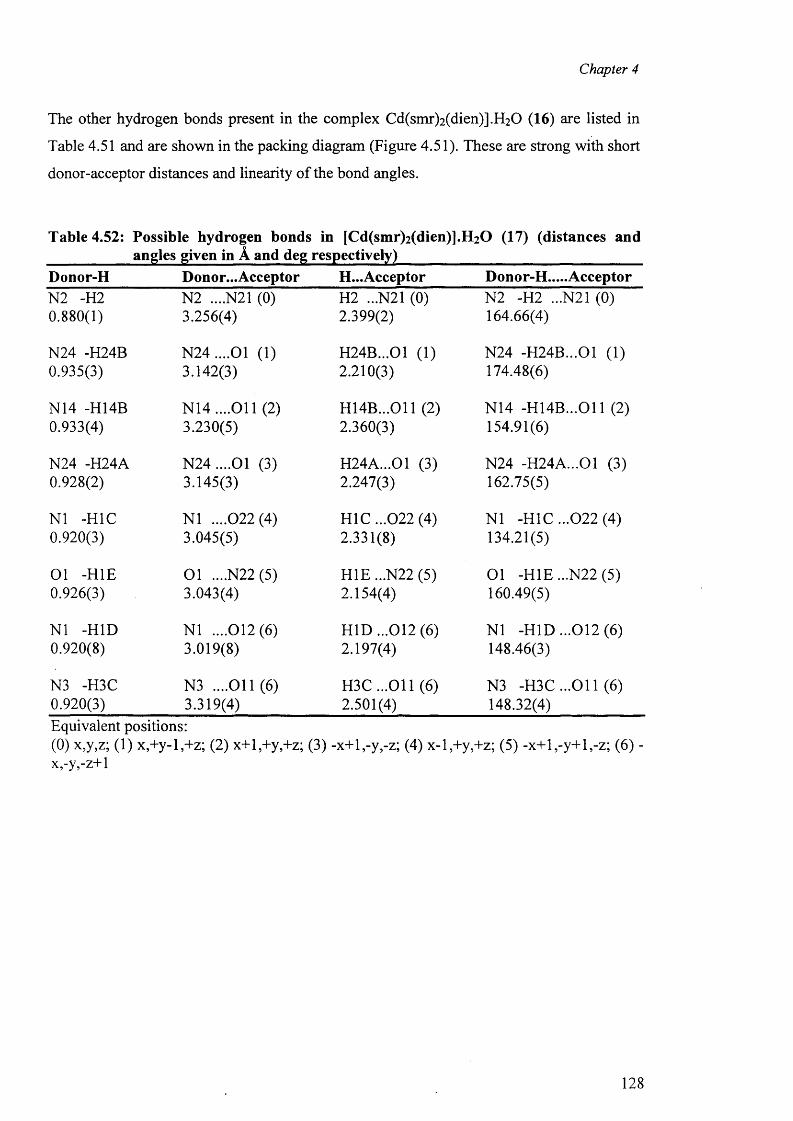
Chapter 4
The other hydrogen bonds present in the complex Cd(smr)2(dien)].H20 (16) are listed in
Table 4.51 and are shown in the packing diagram (Figure 4.51). These are strong with short
donor-acceptor distances and linearity o f the bond angles.
Table 4.52: Possible hydrogen bonds in [Cd(smr)2(dien)].H20 (17) (distances andangles given in A and deg respectively)
Donor-H Donor... Acceptor H...Acceptor Donor-H.....AcceptorN2 -H2 N2 ....N21 (0) H2 ...N21 (0) N2 -H2 ...N21 (0)0.880(1) 3.256(4) 2.399(2) 164.66(4)
N24 -H24B N 24....01 (1) H24B...01 (1) N24 -H24B...01 (1)0.935(3) 3.142(3) 2.210(3) 174.48(6)
N14 -H14B N 14....011 (2) H14B...011 (2) N14 -H 14B ...011 (2)0.933(4) 3.230(5) 2.360(3) 154.91(6)
N24 -H24A N 24....01 (3) H24A...01 (3) N24 -H24A...01 (3)0.928(2) 3.145(3) 2.247(3) 162.75(5)
N1 -H1C N1 ....022(4) H 1C ...022 (4) N1 -H1C ...022 (4)0.920(3) 3.045(5) 2.331(8) 134.21(5)
Ol -HIE O l ....N22 (5) H1E...N22 (5) Ol -HIE ...N22 (5)0.926(3) 3.043(4) 2.154(4) 160.49(5)
N1 -HID N1 ....012(6) H ID ...012 (6) N1 -HID ...012 (6)0.920(8) 3.019(8) 2.197(4) 148.46(3)
N3 -H3C N3 ....011(6) H 3C...011 (6) N3 -H 3C ...011 (6)0.920(3) 3.319(4) 2.501(4) 148.32(4)Equivalent positions:(0) x,y,z; (1) x,+y-l,+z; (2) x+l,+y,+z; (3) -x+l,-y,-z; (4) x-l,+y,+z; (5) -x+ l,-y+l,-z; (6) - x,-y,-z+l
128
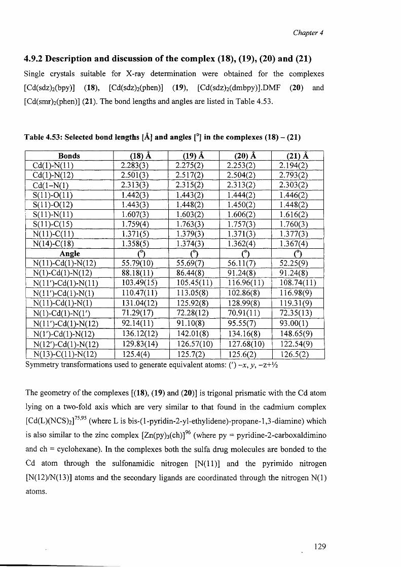
Chapter 4
4.9.2 Description and discussion of the complex (18), (19), (20) and (21)
Single crystals suitable for X-ray determination were obtained for the complexes
[Cd(sdz)2(bpy)] (18), [Cd(sdz)2(phen)] (19), [Cd(sdz)2(dmbpy)].DMF (20) and
[Cd(smr)2(phen)] (21). The bond lengths and angles are listed in Table 4.53.
Table 4.53: Selected bond lengths [A] and angles [°] in the complexes (18) - (21)
Bonds (18) A (19) A (20) A (21) ACd(l)-N(l 1) 2.283(3) 2.275(2) 2.253(2) 2.194(2)Cd(l)-N(12) 2.501(3) 2.517(2) 2.504(2) 2.793(2)C d(l-N (l) 2.313(3) 2.315(2) 2.313(2) 2.303(2)S (ll)-0 (11) 1.442(3) 1.443(2) 1.444(2) 1.446(2)S (ll)-0 (12) 1.443(3) 1.448(2) 1.450(2) 1.448(2)S (ll)-N (l 1) 1.607(3) 1.603(2) 1.606(2) 1.616(2)S(ll)-C(15) 1.759(4) 1.763(3) 1.757(3) 1.760(3)N(11)-C(11) 1.371(5) 1.379(3) 1.371(3) 1.377(3)N(14)-C(18) 1.358(5) 1.374(3) 1.362(4) 1.367(4)
Angle 0 (°) (°) (°)N(11)-Cd(l)-N(12) 55.79(10) 55.69(7) 56.11(7) 52.25(9)N(l)-Cd(l)-N(12) 88.18(11) 86.44(8) 91.24(8) 91.24(8)N(1 T )-C d(l)-N (ll) 103.49(15) 105.45(11) 116.96(11) 108.74(11)N(1 T)-Cd(l)-N(l) 110.47(11) 113.05(8) 102.86(8) 116.98(9)N(11)-Cd(l)-N(l) 131.04(12) 125.92(8) 128.99(8) 119.31(9)N (l)-C d(l)-N (l') 71.29(17) 72.28(12) 70.91(11) 72.35(13)N(1 l')-Cd(l)-N(12) 92.14(11) 91.10(8) 95.55(7) 93.00(1)
, N(T)-Cd(l)-N(12) 136.12(12) 142.01(8) 134.16(8) 148.65(9)N(12')-Cd(l)-N(12) 129.83(14) 126.57(10) 127.68(10) 122.54(9)N(13)-C(l 1)-N(12) 125.4(4) 125.7(2) 125.6(2) 126.5(2)
Symmetry transformations used to generate equivalent atoms: (') -x , y , -Z+V 2
The geometry of the complexes [(18), (19) and (20)] is trigonal prismatic with the Cd atom
lying on a two-fold axis which are very similar to that found in the cadmium complex
[Cd(L)(NCS)2]75,95 (where L is bis-(l-pyridin-2-yl-ethylidene)-propane-1,3-diamine) which
is also similar to the zinc complex [Zn(py)3 (ch)]% (where py = pyridine-2-carboxaldimino
and ch = cyclohexane). In the complexes both the sulfa drug molecules are bonded to the
Cd atom through the sulfonamidic nitrogen [N (ll)] and the pyrimido nitrogen
[N(12)/N(13)] atoms and the secondary ligands are coordinated through the nitrogen N (l)
atoms.
129
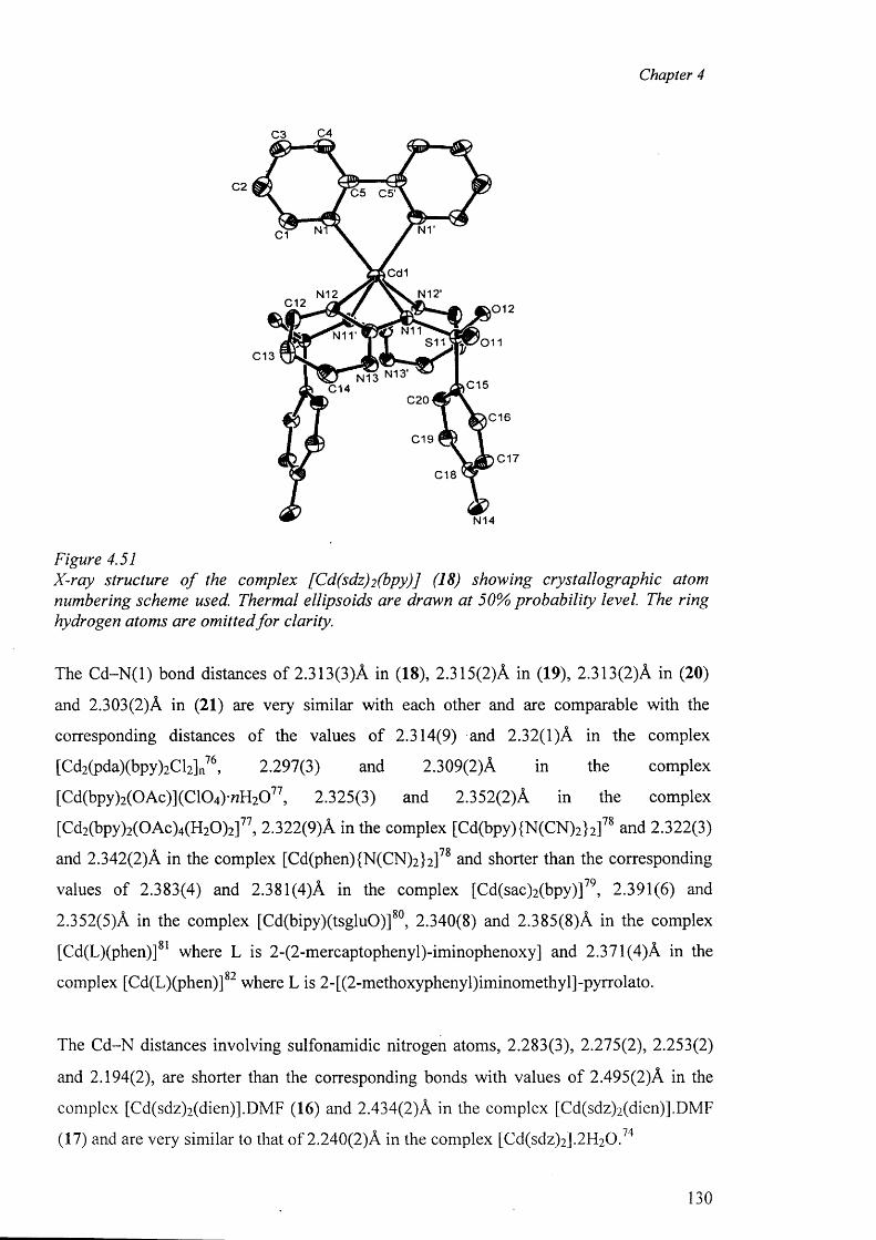
Chapter 4
C4C3
C2 C5 C5'
C1
Cd1
N12'N12C12 ,0 1 2
N11S11
C13
C15C14C20
C16
C19C17
C18
N14
Figure 4.51X-ray structure o f the complex [Cd(sdz)2 (bpy)] (18) showing crystallographic atom numbering scheme used. Thermal ellipsoids are drawn at 50% probability level. The ring hydrogen atoms are omitted fo r clarity.
The C d-N (l) bond distances of 2.313(3)A in (18), 2.315(2)A in (19), 2.313(2)A in (20)
and 2.303(2)A in (21) are very similar with each other and are comparable with the
corresponding distances of the values of 2.314(9) and 2.32(1)A in the complex
[Cd2(pda)(bpy)2Cl2]n76, 2.297(3) and 2.309(2)A in the complex
[Cd(bpy)2(0Ac)](C104)-«H20 77, 2.325(3) and 2.352(2)A in the complex
[Cd2(bpy)2( 0 Ac)4(H20 ) 2]11, 2.322(9)A in the complex [Cd(bpy){N(CN)2}2]78 and 2.322(3)
and 2.342(2)A in the complex [Cd(phen){N(CN)2}2]78 and shorter than the corresponding
values of 2.383(4) and 2.381(4)A in the complex [Cd(sac)2(bpy)]79, 2.391(6) and
2.352(5)A in the complex [Cd(bipy)(tsgluO)]80, 2.340(8) and 2.385(8)A in the complex
[Cd(L)(phen)]81 where L is 2-(2-mercaptophenyl)-iminophenoxy] and 2.371 (4)A in the
complex [Cd(L)(phen)] where L is 2-[(2-methoxyphenyl)iminomethyl]-pyrrolato.
The Cd-N distances involving sulfonamidic nitrogen atoms, 2.283(3), 2.275(2), 2.253(2)
and 2.194(2), are shorter than the corresponding bonds with values of 2.495(2)A in the
complex [Cd(sdz)2(dien)].DMF (16) and 2.434(2)A in the complex [Cd(sdz)2(dien)].DMF
(17) and are very similar to that of 2.240(2)A in the complex [Cd(sdz)2].2H20 .74
130
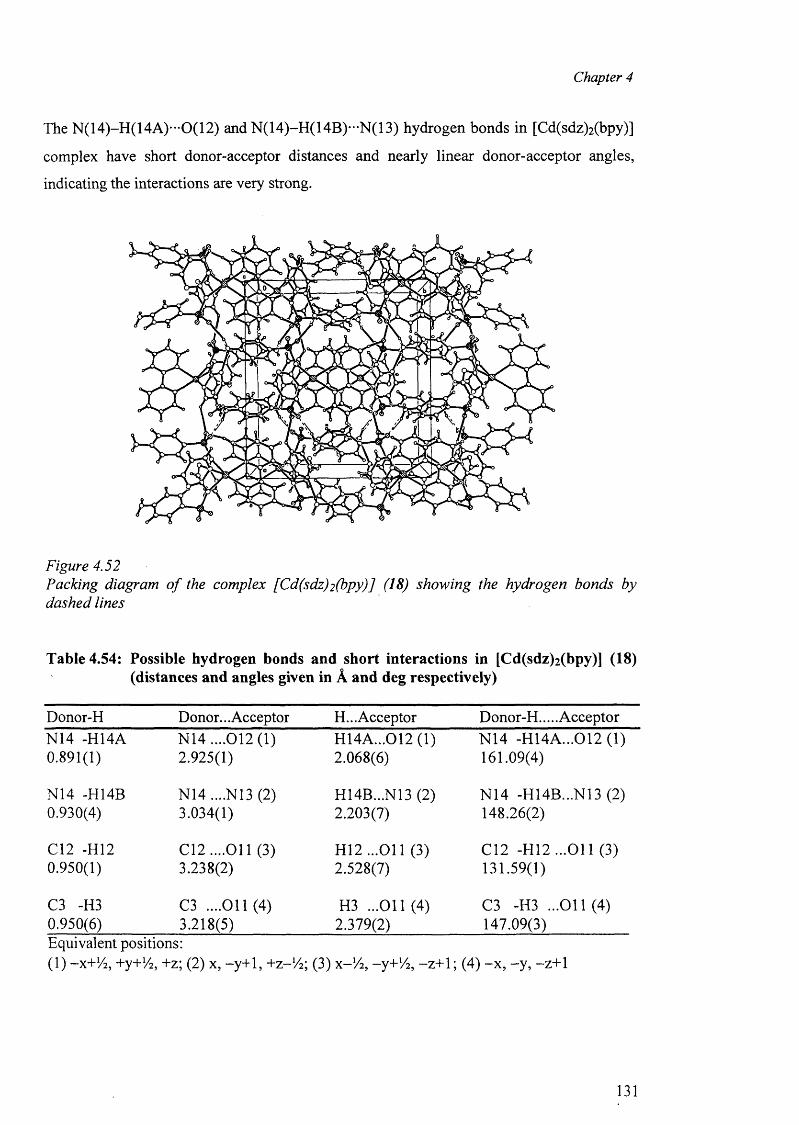
Chapter 4
The N(14)-H(14A)—0(12) and N(14)-H(14B)*-N(13) hydrogen bonds in [Cd(sdz)2 (bpy)]
complex have short donor-acceptor distances and nearly linear donor-acceptor angles,
indicating the interactions are very strong.
Figure 4.52Packing diagram o f the complex [Cd(sdz)2 (bpy)] (18) showing the hydrogen bonds by dashed lines
Table 4.54: Possible hydrogen bonds and short interactions in [Cd(sdz)2 (bpy)] (18) (distances and angles given in A and deg respectively)
Donor-H Donor... Acceptor H... Acceptor Donor-H.... AcceptorN14 -H14A N14 ....012(1) H14A...012 (1) N14 -H14A...012 (1)0.891(1) 2.925(1) 2.068(6) 161.09(4)
N14 -H14B N14 ....N13 (2) H14B...N13 (2) N14 -H14B...N13 (2)0.930(4) 3.034(1) 2.203(7) 148.26(2)
C12 -H12 C12....011 (3) H12 ...O il (3) C12 -H12 ...O il (3)0.950(1) 3.238(2) 2.528(7) 131.59(1)
C3 -H3 C3 ....011(4) H3 ...011(4) C3 -H3 ...011(4)0.950(6) 3.218(5) 2.379(2) 147.09(3)Equivalent positions:(1) -X+V2 , +y+1/ 2 , +z; (2) x, -y+1, +Z-V2 ; (3) X-V2 , -y+'A, -z+ 1; (4) - x , -y , -z+1
131
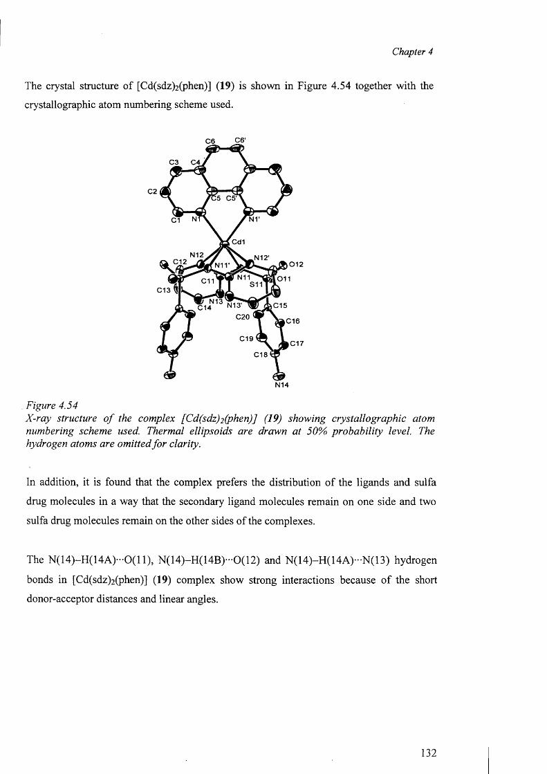
Chapter 4
The crystal structure of [Cd(sdz)2 (phen)] (19) is shown in Figure 4.54 together with the
crystallographic atom numbering scheme used.
C6 C6'
C3 C4
C25 C5‘
C1
Cd1
N12 N 1 2 'C12 0 1 2N11'
011C13
C15C14C20 C16
C19C17
C18
N14
Figure 4.54X-ray structure o f the complex [Cd(sdz)2 (phen)J (19) showing crystallographic atom numbering scheme used. Thermal ellipsoids are drawn at 50% probability level. The hydrogen atoms are omitted fo r clarity.
In addition, it is found that the complex prefers the distribution o f the ligands and sulfa
drug molecules in a way that the secondary ligand molecules remain on one side and two
sulfa drug molecules remain on the other sides of the complexes.
The N (14)-H (14A )-0(11), N (14)-H (14B )-0(12) and N (14)-H (14A )-N (13) hydrogen
bonds in [Cd(sdz)2 (phen)] (19) complex show strong interactions because o f the short
donor-acceptor distances and linear angles.
132
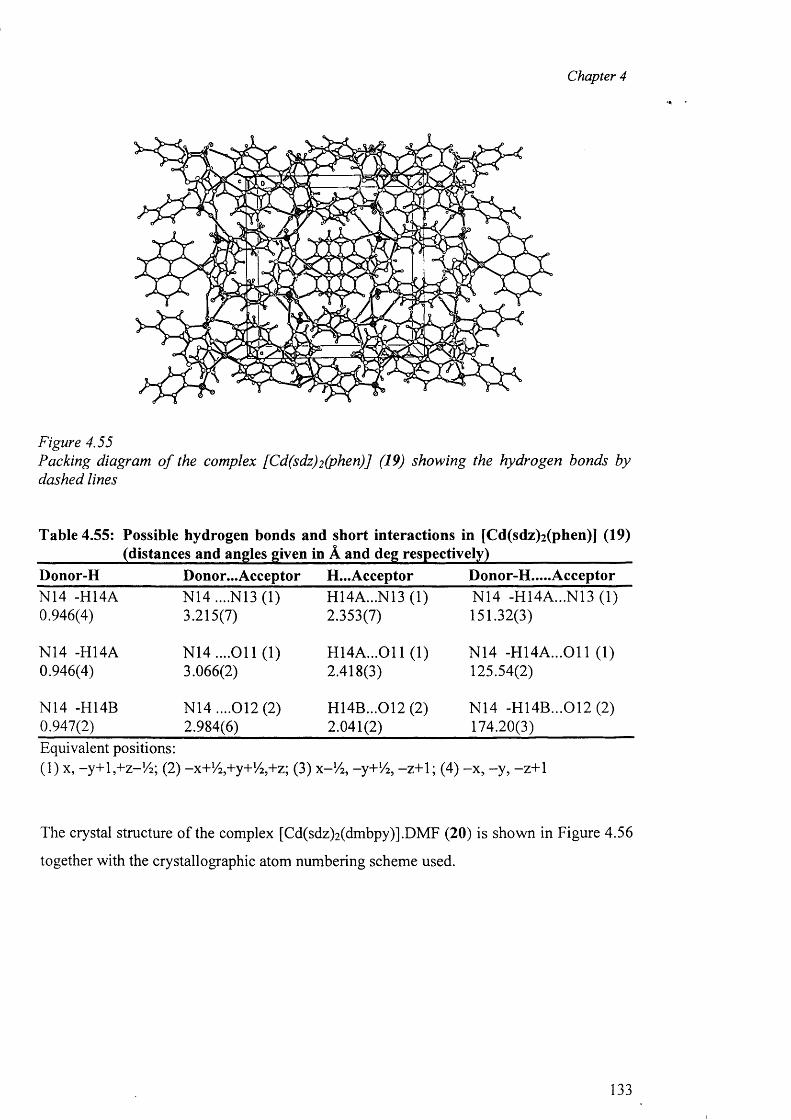
Chapter 4
Figure 4.55Packing diagram o f the complex [Cd(sdz)2 (phen)] (19) showing the hydrogen bonds by dashed lines
Table 4.55: Possible hydrogen bonds and short interactions in [Cd(sdz)2 (phen)] (19) ___________ (distances and angles given in A and deg respectively)___________________Donor-H Donor... Acceptor H...Acceptor Donor-H.....AcceptorN14 -H14A N14 ....N13 (1) H14A...N13 (1) N14 -H14A...N13 (1)0.946(4) 3.215(7) 2.353(7) 151.32(3)
N14 -H14A N 14....011 (1) H14A ...011 (1) N14 -H 14A ...011 (1)0.946(4) 3.066(2) 2.418(3) 125.54(2)
N14 -H14B N14 ....012 (2) H14B...012 (2) N14 -H14B...012 (2)0.947(2) 2.984(6) 2.041(2) 174.20(3)Equivalent positions: (1) x, -y+ l,+ z-!4 ; (2)' -x+V^+y+V^+z; (3) x - !/ 2 , -y+Vz, -z+1; (4) -x , -y , -z+1
The crystal structure of the complex [Cd(sdz)2 (dmbpy)].DMF (20) is shown in Figure 4.56
together with the crystallographic atom numbering scheme used.
133
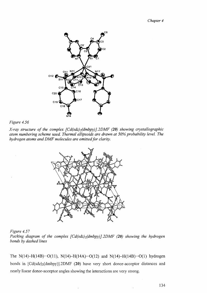
Chapter 4
N14
Figure 4.56
X-ray structure o f the complex [Cd(sdz)2 (dmbpy)].2 DMF (20) showing crystallographic atom numbering scheme used. Thermal ellipsoids are drawn at 50% probability level. The hydrogen atoms and DM F molecules are omitted fo r clarity.
Figure 4.57Packing diagram o f the complex [Cd(sdz)2 (dmbpy)].2 DMF (20) showing the hydrogen bonds by dashed lines
The N (14)-H (14B )-0(11), N (14)-H (14A )-0(12) and N (14 )-H (14B )-0 (1 ) hydrogen
bonds in [Cd(sdz)2(dmbpy)].2DMF (20) have very short donor-acceptor distances and
nearly linear donor-acceptor angles showing the interactions are very strong.
134
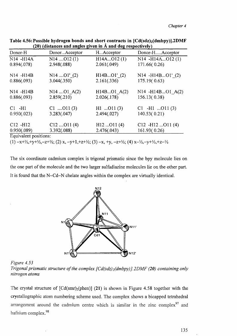
Chapter 4
Table 4.56: Possible hydrogen bonds and short contracts in [Cd(sdz)2(dmbpy)].2DMF __________ (20) (distances and angles given in A and deg respectively)_________ ______Donor-H Donor... Acceptor H... Acceptor Donor-H.... AcceptorN14 -H14A N14 ....012(1) H14A...012 (1) N14 -H14A...012 (1)0.894(.078) 2.948(.088) 2.061 (.049) 171.66( 0.26)
N14 -H14B N 14....01 ' (2) H14B...01' (2) N14 -H14B...01' (2)0.886(.093) 3.044(.350) 2.161 (.336) 175.19( 0.63)
N14 -H14B N 14....01 A(2) H14B...01 A(2) N14 -H14B...01 A(2)0.886(.093) 2.859(.210) 2.026(.178) 156.13( 0.38)
Cl -HI Cl ....011(3) HI ...011(3) Cl -HI ...0 1 1 (3 )0.950(.023) 3.283(.047) 2.494(.027) 140.53(0.21)
C12 -H I2 C12....011 (4) H 12...011 (4) C12 -H12 ...O il (4)0.950(.089) 3.392(.088) 2.476(.043) 161.93(0.26)Equivalent positions:(1) -x+V2 ,+y+1/ 2 ,-z+ 1/ 2 ; (2) x, -y+lj+z+Vi; (3) -x , +y, -z+!/2; (4) x-V ^-y+V ^+z-1/^
The six coordinate cadmium complex is trigonal prismatic since the bpy molecule lies on
the one part o f the molecule and the two larger sulfadiazine molecules lie on the other part.
It is found that the N -C d-N chelate angles within the complex are virtually identical.
N12
N11
N1N i r
Cd1
N12'
Figure 4.53Trigonal prismatic structure o f the complex [Cd(sdz)2 (dmbpy)].2 DMF (20) containing only nitrogen atoms
The crystal structure o f [Cd(smr)2 (phen)] (21) is shown in Figure 4.58 together with the
crystallographic atom numbering scheme used. The complex shows a bicapped tetrahedral
arrangement around the cadmium centre which is similar in the zinc complex97 and
hafnium complex.98
135
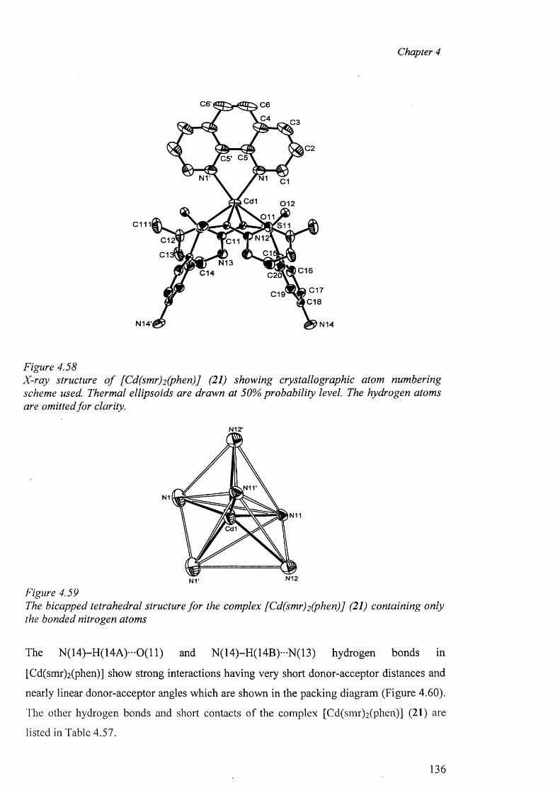
Chapter 4
C6' C6
C4 C3
C20 5 ' C5
N1 C l
Cd1 0 1 2
011C111I S11
N12C12 C11C1C1
N13C16C14 C20'
i C17 C18
C19
N14'i N14
Figure 4.58X-ray structure o f [Cd(smr)2 (phen)] (21) showing crystallographic atom numbering scheme used. Thermal ellipsoids are drawn at 50% probability level. The hydrogen atoms are omitted fo r clarity.
N12'
Figure 4.59The bicapped tetrahedral structure fo r the complex [Cd(smr)2 (phen)J (21) containing only the bonded nitrogen atoms
The N(14)-H(14A)*"0(11) and N (14)-H (14B)-N (13) hydrogen bonds in
[Cd(smr)2 (phen)] show strong interactions having very short donor-acceptor distances and
nearly linear donor-acceptor angles which are shown in the packing diagram (Figure 4.60).
The other hydrogen bonds and short contacts of the complex [Cd(smr)2 (phen)] (21) are
listed in Table 4.57.
136
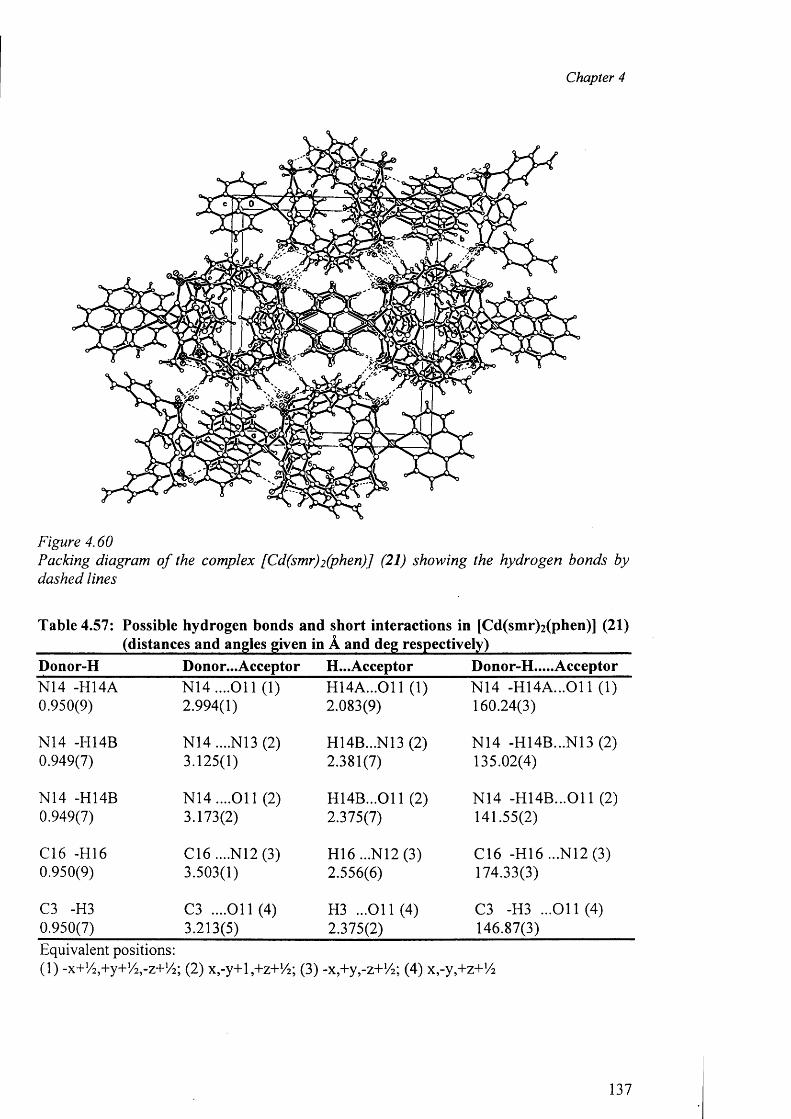
Chapter 4
Figure 4.60Packing diagram o f the complex [Cd(smr)2 (phen)] (21) showing the hydrogen bonds by dashed lines
Table 4.57: Possible hydrogen bonds and short interactions in [Cd(smr)2 (phen)] (21) ___________ (distances and angles given in A and deg respectively)___________________Donor-H Donor...Acceptor H...Acceptor Donor-H.....AcceptorN14 -H14A N 14....011 (1) H14A ...011 (1) N14 -H 14A ...011 (1)0.950(9) 2.994(1) 2.083(9) 160.24(3)
N14 -H14B N14 ....N13 (2) H14B...N13 (2) N14 -H14B...N13 (2)0.949(7) 3.125(1) 2.381(7) 135.02(4)
N14 -H14B N 14....011 (2) H14B...011 (2) N14 -H 14B ...011 (2)0.949(7) 3.173(2) 2.375(7) 141.55(2)
C16 -H16 C16 ....N12 (3) H16 ...N12 (3) C16 -H16 ...N12 (3)0.950(9) 3.503(1) 2.556(6) 174.33(3)
C3 -H3 C3 ....011(4) H3 ...O il (4) C3 -H3 ...011(4)0.950(7) 3.213(5) 2.375(2) 146.87(3)Equivalent positions:(1) -x+!/2 ,+y+!/2 ,-z+1/ 2 ; (2) x,-y+l,+z+/4; (3) -x,+y,-z+!/2 ; (4) x-y^+z+Vi
137
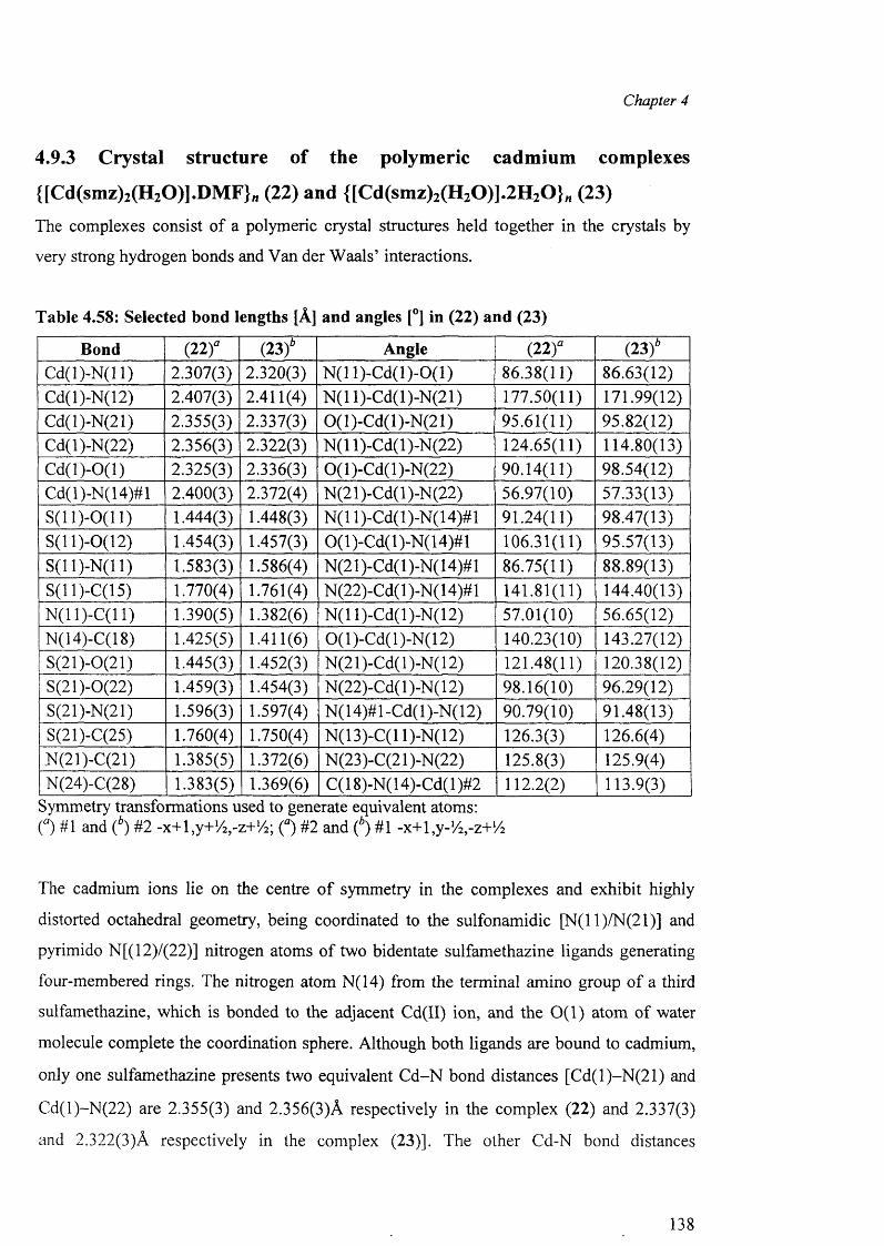
Chapter 4
4.9.3 Crystal structure of the polymeric cadmium complexes
{[Cd(smz)2(H20)].DM F}n (22) and {[Cd(smz)2(H20)].2H 20 } n (23)
The complexes consist of a polymeric crystal structures held together in the crystals by
very strong hydrogen bonds and Van der Waals’ interactions.
Table 4.58: Selected bond lengths [A] and angles [°] in (22) and (23)
Bond (2 2 )a (23)* Angle (22)° (23)*Cd(l)-N(l 1) 2.307(3) 2.320(3) N(11)-Cd(l)-0(1) 86.38(11) 86.63(12)Cd(l)-N(12) 2.407(3) 2.411(4) N(11)-Cd(l)-N(21) 177.50(11) 171.99(12)Cd(l)-N(21) 2.355(3) 2.337(3) 0(1)-Cd(l)-N(21) 95.61(11) 95.82(12)Cd(l)-N(22) 2.356(3) 2.322(3) N (ll)-C d(l)-N (22) 124.65(11) 114.80(13)C d(l)-0(1) 2.325(3) 2.336(3) 0(1)-Cd(l)-N(22) 90.14(11) 98.54(12)Cd(l)-N(14)#l 2.400(3) 2.372(4) N (21 )-Cd( 1 )-N (22) 56.97(10) 57.33(13)S (ll)-0 (11) 1.444(3) 1.448(3) N( 11 )-Cd( 1 )-N( 14)# 1 91.24(11) 98.47(13)S(11)-0(12) 1.454(3) 1.457(3) 0(1)-Cd(l)-N(14)#l 106.31(11) 95.57(13)S(11)-N(11) 1.583(3) 1.586(4) N (21 )-Cd( 1 )-N( 14)# 1 86.75(11) 88.89(13)S(11 )-C(l 5) 1.770(4) 1.761(4) N(22)-Cd(l)-N(14)#l 141.81(11) 144.40(13)N(11)-C(11) 1.390(5) 1.382(6) N(11)-Cd(l)-N(12) 57.01(10) 56.65(12)N(14)-C(18) 1.425(5) 1.411(6) 0(1)-Cd(l)-N(12) 140.23(10) 143.27(12)S(21)-0(21) 1.445(3) 1.452(3) N(21)-Cd(l)-N(12) 121.48(11) 120.38(12)S(21)-0(22) 1.459(3) 1.454(3) N(22)-Cd( 1 )-N( 12) 98.16(10) 96.29(12)S(21)-N(21) 1.596(3) 1.597(4) N(14)#l-Cd(l)-N(12) 90.79(10) 91.48(13)S(21)-C(25) 1.760(4) 1.750(4) N(13)-C(l 1)-N(12) 126.3(3) 126.6(4),N(21)-C(21) 1.385(5) 1.372(6) N(23)-C(21 )-N(22) 125.8(3) 125.9(4)N(24)-C(28) 1.383(5) 1.369(6) C( 18)-N( 14)-Cd( 1 )#2 112.2(2) 113.9(3)
Symmetry transformations used to generate equivalent atoms:O #1 and (b) #2 -x+ l,y+1/ 2 ,-z+1/2; O # 2 and (4) #1 -x+l.y-K.-z+'A
The cadmium ions lie on the centre o f symmetry in the complexes and exhibit highly
distorted octahedral geometry, being coordinated to the sulfonamidic [N(l 1)/N(21)] and
pyrimido N[(12)/(22)] nitrogen atoms of two bidentate sulfamethazine ligands generating
four-membered rings. The nitrogen atom N(14) from the terminal amino group o f a third
sulfamethazine, which is bonded to the adjacent Cd(II) ion, and the 0 (1 ) atom of water
molecule complete the coordination sphere. Although both ligands are bound to cadmium,
only one sulfamethazine presents two equivalent C d-N bond distances [C d(l)-N (21) and
Cd(l)-N (22) are 2.355(3) and 2.356(3)A respectively in the complex (22) and 2.337(3)
and 2.322(3)A respectively in the complex (23)]. The other Cd-N bond distances
138
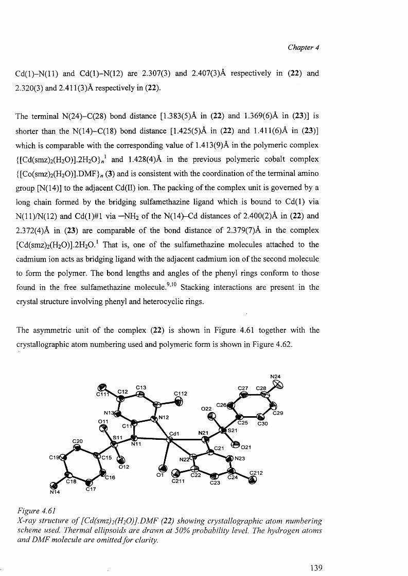
Chapter 4
C d (l)-N (ll) and Cd(l)-N (12) are 2.307(3) and 2.407(3)A respectively in (22) and
2.320(3) and 2.411(3)A respectively in (22).
The terminal N(24)-C(28) bond distance [1.383(5)A in (22) and 1.369(6)A in (23)] is
shorter than the N(14)-C(18) bond distance [1.425(5)A in (22) and 1.411(6)A in (23)]
which is comparable with the corresponding value of 1.413(9)A in the polymeric complex
{[Cd(smz)2 (H2 0 )].2 H2 0 }„1 and 1.428(4)A in the previous polymeric cobalt complex
{[Co(smz)2(H20)].DMF}„ (3) and is consistent with the coordination of the terminal amino
group [N(14)] to the adjacent Cd(II) ion. The packing of the complex unit is governed by a
long chain formed by the bridging sulfamethazine ligand which is bound to C d(l) via
N(11)/N(12) and C d(l)# l via —NH2 of the N(14)-Cd distances of 2.400(2)A in (22) and
2.372(4)A in (23) are comparable o f the bond distance of 2.379(7)A in the complex
[Cd(smz)2(H20)].2H20.1 That is, one o f the sulfamethazine molecules attached to the
cadmium ion acts as bridging ligand with the adjacent cadmium ion of the second molecule
to form the polymer. The bond lengths and angles of the phenyl rings conform to those
found in the free sulfamethazine molecule.9,10 Stacking interactions are present in the
crystal structure involving phenyl and heterocyclic rings.
The asymmetric unit o f the complex (22) is shown in Figure 4.61 together with the
crystallographic atom numbering used and polymeric form is shown in Figure 4.62.
N24
C13 C27 C28C12 C112C11
C26|022
N1 C29N12
011 C25 C30C1S21N21Cd1
S11C20 N11 021C21
C1 C15 N23N2:012
C21201 C22 C240 1 6C211C18 C23
C17N14
Figure 4.61X-ray structure o f [Cd(smz)2(H20)].DMF (22) showing crystallographic atom numbering scheme used. Thermal ellipsoids are drawn at 50% probability level. The hydrogen atoms and DMF molecule are omitted fo r clarity.
139
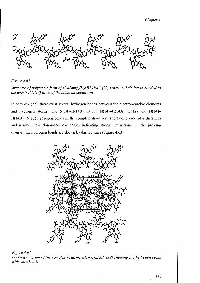
Chapter 4
Figure 4.62
Structure o f polymeric form o f [Cd(smz)2 (H2 0 )].DM F (22) where cobalt ion is bonded to the terminal N(14) atom o f the adjacent cobalt ion.
In complex (22), there exist several hydrogen bonds between the electronegative elements
and hydrogen atoms. The N (14)-H (14B )-0(11), N (14)-H (14A )-0(12) and N (14)-
H(14B)—N(13) hydrogen bonds in the complex show very short donor-acceptor distances
and nearly linear donor-acceptor angles indicating strong interactions. In the packing
diagram the hydrogen bonds are shown by dashed lines (Figure 4.63).
Figure 4.63Packing diagram o f the complex [Cd(smz)2 (H2 0 )].D M F (22) showing the hydrogen bonds with open bonds
140
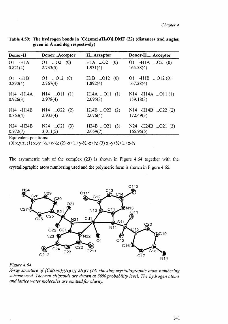
Chapter 4
Table 4.59: The hydrogen bonds in [Cd(smz)2(H20)].DMF (22) (distances and angles given in A and deg respectively)
Donor-H Donor...Acceptor H...Acceptor Donor-H.....Acceptor
01 -H1A 01 ...02 (0) H1A ...02 (0) 01 -H1A ...02 (0)0.821(4) 2.733(5) 1.931(4) 165.58(4)
01 -H1B O l ...012 (0) H1B ...012 (0) 01 -H1B ...012(0 )0.890(4) 2.767(4) 1.892(4) 167.28(4)
N14 -H14A N14 ...O il (1) H14A ...O il (1) N14 -H14A ...O il (1)0.926(3) 2.978(4) 2.095(3) 159.18(3)
N14 -H14B N14 ...022 (2) H14B ...022 (2) N14 -H14B ...022 (2)0.863(4) 2.933(4) 2.076(4) 172.49(3)
N24 -H24B N24 ...021 (3) H24B ...021 (3) N24 -H24B ...021 (3)0.972(7) 3.011(5) 2.059(7) 165.95(5)Equivalent positions:(0) x,y,z; (1) Xj-y+V^+z-1/^; (2) -x+l,+y-1>4,-z+1/2; 0 ) x -y+ l/2+\,+z-V2
The asymmetric unit o f the complex (23) is shown in Figure 4.64 together with the
crystallographic atom numbering used and the polymeric form is shown in Figure 4.65.
C 1 1 2N 2 4 C 1 3
C 1 1 1C 2 9 C 1 4C 2 8 C 1 2C 3 0
021N 1 3C 2 7 C 1 1N 1 2S 2 1 011
C 2 5C 2 6
S 1 1C 2 0
N1 1 0 1 50 2 2 C 2 1
N 2 3 « r, 0 1 9
N 2 20 1 201
C 1 6C22C 2 4 C 2 1 1 C 1 8C 2 3C 2 1 2 C 1 7
N 1 4
Figure 4.64X-ray structure o f [Cd(smz)2 (H2 0 )] .2 H2 O (23) showing crystallographic atom numbering scheme used. Thermal ellipsoids are drawn at 50% probability level. The hydrogen atoms and lattice water molecules are omitted fo r clarity.
141
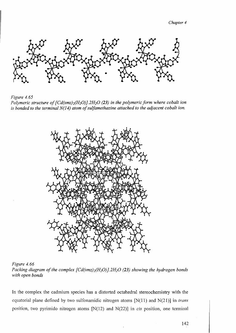
Chapter 4
Figure 4.65Polymeric structure o f [Cd(smz)2 (H2 0 )]. 2 H2 O (23) in the polymeric form where cobalt ion is bonded to the terminal N(14) atom o f sulfamethazine attached to the adjacent cobalt ion.
Figure 4.66Packing diagram o f the complex [Cd(smz)2 (H2 0 )] .2 H2 0 (23) showing the hydrogen bonds with open bonds
In the complex the cadmium species has a distorted octahedral stereochemistry with the
equatorial plane defined by two sulfonamidic nitrogen atoms [N (ll) and N(21)] in trans
position, two pyrimido nitrogen atoms [N(12) and N(22)] in cis position, one terminal
142
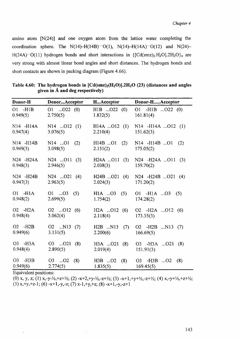
Chapter 4
amino atom [N(24)] and one oxygen atom from the lattice water completing the
coordination sphere. The N(14)-H(14B)""0(1), N(14)-H(14A)' 0(12) and N (24)-
H(24A) 0(11) hydrogen bonds and short interactions in {[Cd(smz)2.H20].2H20}„ are
very strong with almost linear bond angles and short distances. The hydrogen bonds and
short contacts are shown in packing diagram (Figure 4.66).
Table 4.60: The hydrogen bonds in [Cd(smz)2(H20)].2H20 (23) (distances and angles given in A and deg respectively)
Donor-H Donor...Acceptor H...Acceptor Donor-H.....Acceptor
01 -H1B 0.949(5)
01 ...022 2.750(5)
(0) H1B ...022 1.832(5)
(0) 01 -H1B ...022 161.81(4)
(0)
N14 -H14A 0.947(4)
N14 ...012 3.076(5)
(1) H14A ...012 2.210(4)
(1) N14 -H14A ...012 151.62(3)
( 1)
N14 -H14B 0.949(3)
N14 ...01 3.098(5)
(2) H14B ...01 2.151(2)
(2) N14 -H14B ...01 175.05(2)
(2)
N24 -H24A 0.948(3)
N24 ...O il 2.946(5)
(3) H24A ...O il 2.038(3)
(3) N24 -H24A ...O il 159.70(2)
(3)
N24 -H24B 0.947(3)
N24 ...021 2.963(5)
(4) H24B ...021 2.024(3)
(4) N24 -H24B ...021 171.20(2)
(4)
01 -H1A 0.948(2)
01 ...032.699(5)
(5) H1A ...03 1.754(2)
(5) 01 -H1A ...03 174.28(2)
(5)
0 2 -H2A 0.948(4)
0 2 ...012 3.062(4)
(6) H2A ...012 2.118(4)
(6) 0 2 -H2A ...012 173.35(3)
(6)
0 2 -H2B 0.949(6)
0 2 ...N13 3.131(5)
(7) H2B ...N13 2.200(6)
(7) 0 2 -H2B ...N13 166.69(5)
(7)
03 -H3A 0.948(4)
03 ...021 2.890(5)
(8) H3A ...021 2.019(4)
(8) 03 -H3A ...021 151.91(3)
(8)
03 -H3B 0.949(6)
03 ...02 2.774(5)
(8) H3B ...02 1.835(5)
(8) 03 -H3B ...02 169.45(5)
(8)
Equivalent positions:(0) x, y, z; (1) x,-y-!/2,+z+!4; (2) -x+2,+y-1/2,-z+1/2; (3) -x+l,+y+1/2 ,-z+1/ 2; (4) Xj-y+V^+z+Vz; (5) x,+y,+z-l; (6) -x+l,-y,-z; (7) x-l,+y,+z; (8) -x+l,-y,-z+l
143
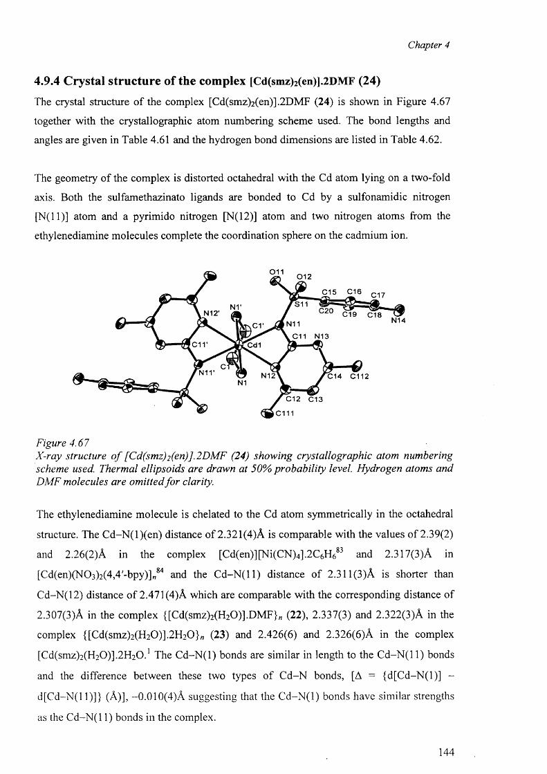
Chapter 4
4.9.4 Crystal structure of the complex [Cd(smz)2(en)].2DMF (24)
The crystal structure o f the complex [Cd(smz)2(en)].2DMF (24) is shown in Figure 4.67
together with the crystallographic atom numbering scheme used. The bond lengths and
angles are given in Table 4.61 and the hydrogen bond dimensions are listed in Table 4.62.
The geometry o f the complex is distorted octahedral with the Cd atom lying on a two-fold
axis. Both the sulfamethazinato ligands are bonded to Cd by a sulfonamidic nitrogen
[N (ll)] atom and a pyrimido nitrogen [N(12)] atom and two nitrogen atoms from the
ethylenediamine molecules complete the coordination sphere on the cadmium ion.
011 01 2
C15 C16 C1?
S11C20 C19 C18N12'
N14N11C1'C11 N13
d r Cd1
N12 C14 C112N1
C12 C13
c m
Figure 4.67X-ray structure o f [Cd(smz)2(en)].2DMF (24) showing crystallographic atom numbering scheme used. Thermal ellipsoids are drawn at 50% probability level. Hydrogen atoms and DMF molecules are omitted fo r clarity.
The ethylenediamine molecule is chelated to the Cd atom symmetrically in the octahedral
structure. The C d-N (l)(en) distance o f 2.321(4)A is comparable with the values o f 2.39(2)
and 2.26(2)A in the complex [Cd(en)][Ni(CN)4 ].2 C6 H 6 83 and 2.317(3)A in
[Cd(en)(N0 3 )2(4,4'-bpy)]„84 and the C d -N (ll) distance o f 2.311 (3)A is shorter than
Cd-N(12) distance o f 2.471 (4)A which are comparable with the corresponding distance o f
2.307(3)A in the complex {[Cd(smz)2 (H2 0 )].DMF}„ (22), 2.337(3) and 2.322(3)A in the
complex {[Cd(smz)2 (H2 0 )].2 H2 0 }„ (23) and 2.426(6) and 2.326(6)A in the complex
[Cd(smz)2(H20)].2H 20.1 The C d-N (l) bonds are similar in length to the C d-N (l 1) bonds
and the difference between these two types o f C d-N bonds, [A = (d[C d-N (l)] -
d [C d-N (ll)]} (A)], -0.010(4)A suggesting that the C d -N (l) bonds have similar strengths
as the C d-N (l 1) bonds in the complex.
144
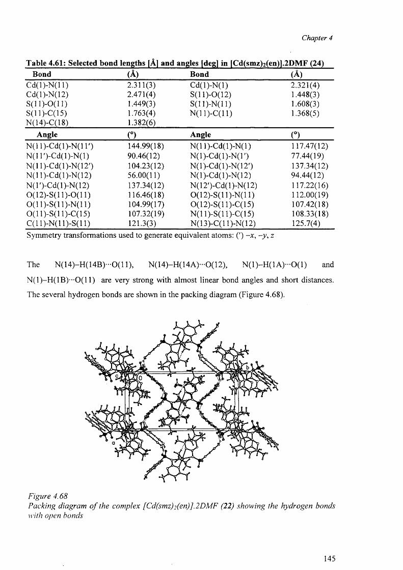
Chapter 4
Table 4.61: Selected bond lengths |A] and angles [deg] in [Cd(smz)2(en)].2DMF (24)Bond (A) Bond (A )
C d(l)-N (l 1) 2.311(3) C d(l)-N (l) 2.321(4)Cd(l)-N(12) 2.471(4) S(11)-0(12) 1.448(3)S(11)-0(11) 1.449(3) S(11)-N(11) 1.608(3)S(ll)-C (15) 1.763(4) N(11)-C(11) 1.368(5)N(14)-C(18) 1.382(6)
Angle (°) Angle (°)N (11)-Cd(l)-N (l T) 144.99(18) N (11)-Cd(l)-N (l) 117.47(12)N (ll ')-C d (l)-N (l) 90.46(12) N (l)-C d(l)-N (T ) 77.44(19)N (ll)-C d(l)-N (12 ') 104.23(12) N (l)-C d(l)-N (12') 137.34(12)N (11 )-Cd( 1 )-N( 12) 56.00(11) N (l)-C d(l)-N (12) 94.44(12)N (l')-C d(l)-N (12) 137.34(12) N(12')-Cd(l)-N(12) 117.22(16)0(12)-S(11)-0(11) 116.46(18) 0(12)-S(11)-N(11) 112.00(19)0 ( 1 1)-S(11)-N(11) 104.99(17) 0(12)-S(11)-C(15) 107.42(18)0(11 )-S( 11 )-C( 15) 107.32(19) N(11)-S(11)-C(15) 108.33(18)C( 11 )-N( 11 )-S(l 1) 121.3(3) N(13)-C(l 1)-N(12) 125.7(4)Symmetry transformations used to generate equivalent atoms: (') -x , -y , z
The N (1 4 )-H (1 4 B )-0 (1 1), N (14)-H (14A )-0 (12), N (l) -H (l A )-O (l) and
N (1)-H (1B )--0(11) are very strong with almost linear bond angles and short distances.
The several hydrogen bonds are shown in the packing diagram (Figure 4.68).
Figure 4.68Packing diagram o f the complex [Cd(smz)2 (en)].2DMF (22) showing the hydrogen bonds with open bonds
145
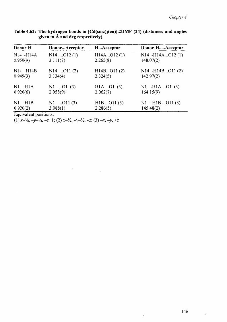
Chapter 4
Table 4.62: The hydrogen bonds in [Cd(smz)2(en)].2DMF (24) (distances and angles given in A and deg respectively)
Donor-H Donor... Acceptor H...Acceptor Donor-H.....Acceptor
N14 -H14A N14 ....012(1) H 14A...012 (1) N14 -H14A...012 (1)0.950(9) 3.111(7) 2.265(8) 148.07(2)
N14 -H14B N14 ....O il (2) H 14B ...011 (2) N14 -H 14B ...011 (2)0.949(3) 3.134(4) 2.324(5) 142.97(2)
N1 -H1A N1 ....01 (3) H 1A ...01 (3) N1 -H 1A ...01 (3)0.920(6) 2.958(9) 2.062(7) 164.15(9)
N1 -H1B N1 ....011(3 ) H1B ...O il (3) N1 -H 1B ...011 (3)0.920(2) 3.088(1) 2.286(5) 145.48(2)Equivalent positions:(1) x-Vi, y V2 , -z+ 1 ; (2) x - lA, -y - 'A , -z ; (3) -x , -y , +z
146
Chapter 4
4.10 Mercury complexes of triple sulfa drugs (TSD)
The diversity o f the coordination numbers for the mercury complexes can be found in the
literature. In monoorganomercury(II) compounds, the primary bonds leave the Hg atom
with enough residual acidity for it to be able to reach a coordination number of six when
the donor atoms forming the secondary bonds are small. There are only a small number ofo r
Hg complexes with coordination number two. On the other hand, Hg(II), has an
extremely high affinity for thiol-containing compounds, and forms 1:2 metahligand linearo /
complexes with them.
The tetrahedral geometry was postulated in the mercury(II) complexes o f the type HgX2 L2
(where L = l,3-imidazole-2-thione, 1-methyl-1,3-imidazole-2-thione; X = Cl~, Br”) on the0 7
basis of IR and NM R data. In the Hg(II) complex with 2-(cr-hydroxybenzyl)thiamine, the
metal coordination unit consists o f two distorted tetrahedra sharing two vertexes, and the* • Q Ohigh basicity o f the N o f the pyrimidine ring allows the coordination with the metal. An
example o f octahedral local geometry is the complex o f Hg(II) with the bidentate ligandRQlactobionic acid (L): [HgL2].2H20. With respect to Hg(II)-sulfa drug complexes, both
1 'J
geometries (linear arrangement and tetrahedral ) were founded by X-ray crystal structure.
In this section, we will discuss the mercury complexes o f sulfa drugs with two
(sulfamerazine complex), four (sulfadiazine and sulfamethazine) and six (sulfamerazine
with 2,2'-bipyridine) coordination geometry.
4.10.1 Crystal structure of the complexes (25), (26), (27) and (28)
Single crystals suitable for X-ray determination were obtained for the complexes and
characterised by independent analytical and spectroscopic data. We also determined the
modes o f coordination and molecular geometry and compared these with other related
species. The crystal data and refinement details for the complexes are listed in Table 4.63.
The crystal structures o f the complexes [Hg(sdz)2 (DMF)2 ] (25), [Hg(smr)2 ] (26),
[Hg(smr)2 (bpy)] (27) and [Hg(smz)2 (DMF)2 ] (28) are shown in Figures 4.69, 4.71, 4.73
and 4.75 respectively together with the crystallographic atom numbering scheme used. The
selected bond lengths and angles are collected together in Table 4.64.
147
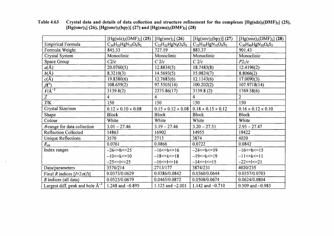
Table 4.63 Crystal data and details of data collection and structure refinement for the complexes [Hg(sdz)2 (DMF)2 ] (25), [Hg(smr)2] (26), [Hg(smr)2(bpy)] (27) and [Hg(smz)2(DMF)2] (28)
[Hg(sdz)2(DMF)2] (25) [Hg(smr)2] (26) [Hg(smr)2(bpy)] (27) [Hg(smz)2(DMF)2] (28)Empirical Formula C26H32HgN i0O6S2 C22H22HgN804S2 C32H3oHgN,o04S2 C3oH4oHgNio06S2Formula Weight 845.33 727.19 883.37 901.43Crystal System Monoclinic Monoclinic Monoclinic MonoclinicSpace Group C2/c C 2/c C 2/c P2\!ca{ A) 20.0760(5) 12.8834(5) 18.7483(8) 12.4196(2)b( A) 8.3210(3) 14.5693(5) 15.0824(7) 8.8066(2)c(A ) 19.8380(6) 12.7683(6) 12.1143(6) 17.0090(3)A °) 108.659(2) 97.5505(14) 100.202(2) 107.9718(14)v/Ar3 3139.8(2) 2375.86(17) 3139.8(2) 1769.58(6)z 4 4 4 277K 150 150 150 150Crystal Size/mm 0 .1 2x0 .10x0 .08 0 .15x0 .12x0 .08 0 .1 8x0 .15x0 .12 0 .16x0 .12x0 .10Shape Block Block Block BlockColour White White White WhiteGrange for data collection 3 .01-27 .46 3.19-27 .46 3.20-27.51 2.93 - 27.47Reflection Collected 14863 16902 14955 19422Unique Reflections 3570 2713 3874 4020int 0.0761 0.0866 0.0722 0.0842
Index ranges -26<=h<=25 -16<=h<=16 —24<=h<=19 -16<=h<=15—10<=k<=10 -18<=k<=18 — 19<=k<=19 -11<=k<=11—25<=1<=25 —16<=1<= 16 -14<=1<=15 -22<=1<=21
Data/parameters 3570/214 2713/177 3874/231 4020/235Final R indices [7>2o(/)] 0.0373/0.0629 0.0386/0.0842 0.0360/0.0644 0.0357/0.0703R indices (all data) 0.0523/0.0679 0.0465/0.0872 0.0508/0.0674 0.0624/0.0804Largest diff. peak and hole A-3 1.248 and -0.895 1.125 and -2.001 1.142 and -0.710 0.509 and -0.983
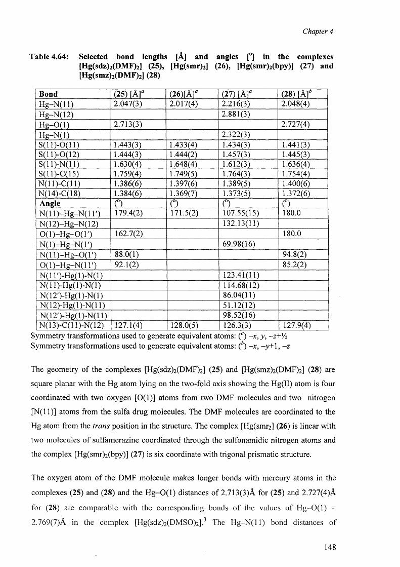
Chapter 4
Table 4.64: Selected bond lengths [A] and angles [°] in the complexes[Hg(sdz)2(DMF)2] (25), [Hg(smr)2] (26), [Hg(smr)2(bpy)] (27) and [Hg(smz)2(DMF)2] (28)
Bond (25) [A r (26)[A f (27) [A f (28) [ A fH g -N (ll) 2.047(3) 2.017(4) 2.216(3) 2.048(4)Hg-N(12) 2.881(3)H g-O (l) 2.713(3) 2.727(4)H g-N (l) 2.322(3)S(1 l) -O (ll) 1.443(3) 1.433(4) 1.434(3) 1.441(3)S (ll)-0 (1 2 ) 1.444(3) 1.444(2) 1.457(3) 1.445(3)S(11)-N(11) 1.630(4) 1.648(4) 1.612(3) 1.636(4)S( 11 )-C(l 5) 1.759(4) 1.749(5) 1.764(3) 1.754(4)N(11)-C(11) 1.386(6) 1.397(6) 1.389(5) 1.400(6)N(14)-C(18) 1.384(6) 1.369(7) 1.373(5) 1.372(6)Angle (°) (°) (°) (°)N (ll) -H g -N (1 T ) 179.4(2) 171.5(2) 107.55(15) 180.0N (12)-H g-N (12) 132.13(11)0 (1 )-H g -0 (T ) 162.7(2) 180.0N (l)-H g -N (T ) 69.98(16)N(1 l)-H g -O (T ) 88.0(1) 94.8(2)0 (1 ) -H g -N ( ir ) 92.1(2) 85.2(2)N (1T )-H g(l)-N (l) 123.41(11)N (11)-H g(l)-N (l) 114.68(12)N (12 ')-H g(l)-N (l) 86.04(11)N (12)-H g(l)-N (ll) 51.12(12)N (12')-H g(l)-N (l 1) 98.52(16)N(13)-C(l 1)-N(12) 127.1(4) 128.0(5) 126.3(3) 127.9(4)
Symmetry transformations used to generate equivalent atoms: (fl) - x ,y , -z+V.■> Symmetry transformations used to generate equivalent atoms: (b) -x , -y+ 1, -z
The geometry o f the complexes [Hg(sdz)2(DMF)2] (25) and [Hg(smz)2(DMF)2 ] (28) are
square planar with the Hg atom lying on the two-fold axis showing the Hg(II) atom is four
coordinated with two oxygen [0(1)] atoms from two DMF molecules and two nitrogen
[N( 11)] atoms from the sulfa drug molecules. The DMF molecules are coordinated to the
Hg atom from the trans position in the structure. The complex [Hg(smr2] (26) is linear with
two molecules o f sulfamerazine coordinated through the sulfonamidic nitrogen atoms and
the complex [Hg(smr)2(bpy)] (27) is six coordinate with trigonal prismatic structure.
The oxygen atom of the DMF molecule makes longer bonds with mercury atoms in the
complexes (25) and (28) and the H g -O (l) distances o f 2.713(3)A for (25) and 2.727(4)A
for (28) are comparable with the corresponding bonds of the values of H g-O (l) =
2.769(7)A in the complex [Hg(sdz)2(DMSO)2].3 The H g -N (ll) bond distances of
148
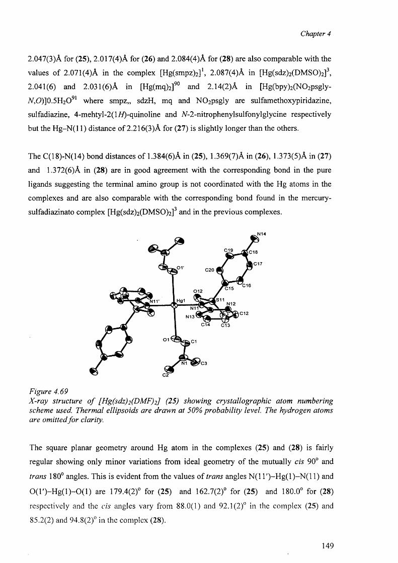
Chapter 4
2.047(3)A for (25), 2.017(4)A for (26) and 2.084(4)A for (28) are also comparable with the
values o f 2.071(4)A in the complex [Hg(smpz)2 ]1, 2.087(4)A in [Hg(sdz)2 (DMSO)2 ]3,
2.041(6) and 2.031(6) A in [Hg(mq)2 ]90 and 2.14(2)A in [Hg(bpy)2 (N 0 2 psgly-
7V,O)]0.5H2O91 where smpz,, sdzH, mq and N02psgly are sulfamethoxypiridazine,
sulfadiazine, 4-m ehtyl-2(l//)-quinoline and N-2-nitrophenylsulfonylglycine respectively
but the H g-N (l 1) distance o f 2.216(3)A for (27) is slightly longer than the others.
The C(18)-N(14) bond distances of 1.384(6)A in (25), 1.369(7)A in (26), 1.373(5)A in (27)
and 1.372(6)A in (28) are in good agreement with the corresponding bond in the pure
ligands suggesting the terminal amino group is not coordinated with the Hg atoms in the
complexes and are also comparable with the corresponding bond found in the mercury-
sulfadiazinato complex [Hg(sdz)2 (DMSO)2 ] and in the previous complexes.
N14
C19 C18
C170 1 C20
C16C150 1 2
,S11Hgi N12N11
C12N13
C14 C13
01 C1
N1 C3
C2
Figure 4.69X-ray structure o f [Hg(sdz)2 (DMF)2] (25) showing crystallographic atom numbering scheme used. Thermal ellipsoids are drawn at 50% probability level. The hydrogen atoms are omitted fo r clarity.
The square planar geometry around Hg atom in the complexes (25) and (28) is fairly
regular showing only minor variations from ideal geometry o f the mutually cis 90° and
trans 180° angles. This is evident from the values o f trans angles N(1 T )-H g (l)-N (l 1) and
0 (T )-H g ( l) -0 (1 ) are 179.4(2)° for (25) and 162.7(2)° for (25) and 180.0° for (28)
respectively and the cis angles vary from 88.0(1) and 92.1(2)° in the complex (25) and
85.2(2) and 94.8(2)° in the complex (28).
149
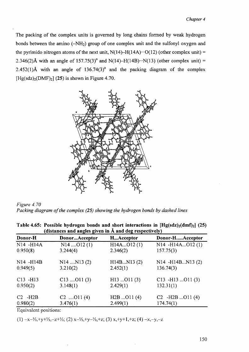
Chapter 4
The packing o f the complex units is governed by long chains formed by weak hydrogen
bonds between the amino (-N H 2 ) group o f one complex unit and the sulfonyl oxygen and
the pyrimido nitrogen atoms o f the next unit, N(14)-H(14A)—0(12) (other complex unit) =
2.346(2)A with an angle o f 157.75(3)° and N(14)-H(14B)--N(13) (other complex unit) =
2.452(1)A with an angle o f 136.74(3)° and the packing diagram of the complex
[Hg(sdz)2 (DMF)2 ] (25) is shown in Figure 4.70.
Figure 4.70Packing diagram o f the complex (25) showing the hydrogen bonds by dashed lines
Table 4.65: Possible hydrogen bonds and short interactions in [Hg(sdz)2 (dmf)2 ] (25) ____________(distances and angles given in A and deg respectively)___________________Donor-H Donor... Acceptor H...Acceptor Donor-H.....AcceptorN14 -H14A N14 ....012(1) H14A...012 (1) N14 -H14A...0 1 2 (1 )0.950(8) 3.244(4) 2.346(2) 157.75(3)
N14 -H14B N 14....N 13(2) H14B...N13 (2) N14 -H14B...N13 (2)0.949(5) 3.210(2) 2.452(1) 136.74(3)
C13 -H13 C13 ....O il (3) H13 ...O il (3) C13 -H I3 ...O il (3)0.950(2) 3.148(1) 2.429(1) 132.31(1)
C2 -H2B C2 ....O il (4) H 2B ...011 (4) C2 -H2B ...O il (4)0.980(2) 3.476(1) 2.499(1) 174.74(1)Equivalent positions:
(1) - x - '/2 ,+y+ !/2 ,-z + lA ; (2) x-'/^+y-'/^+z; (3) x,+y+l,+z; (4) - x - y , - z
150
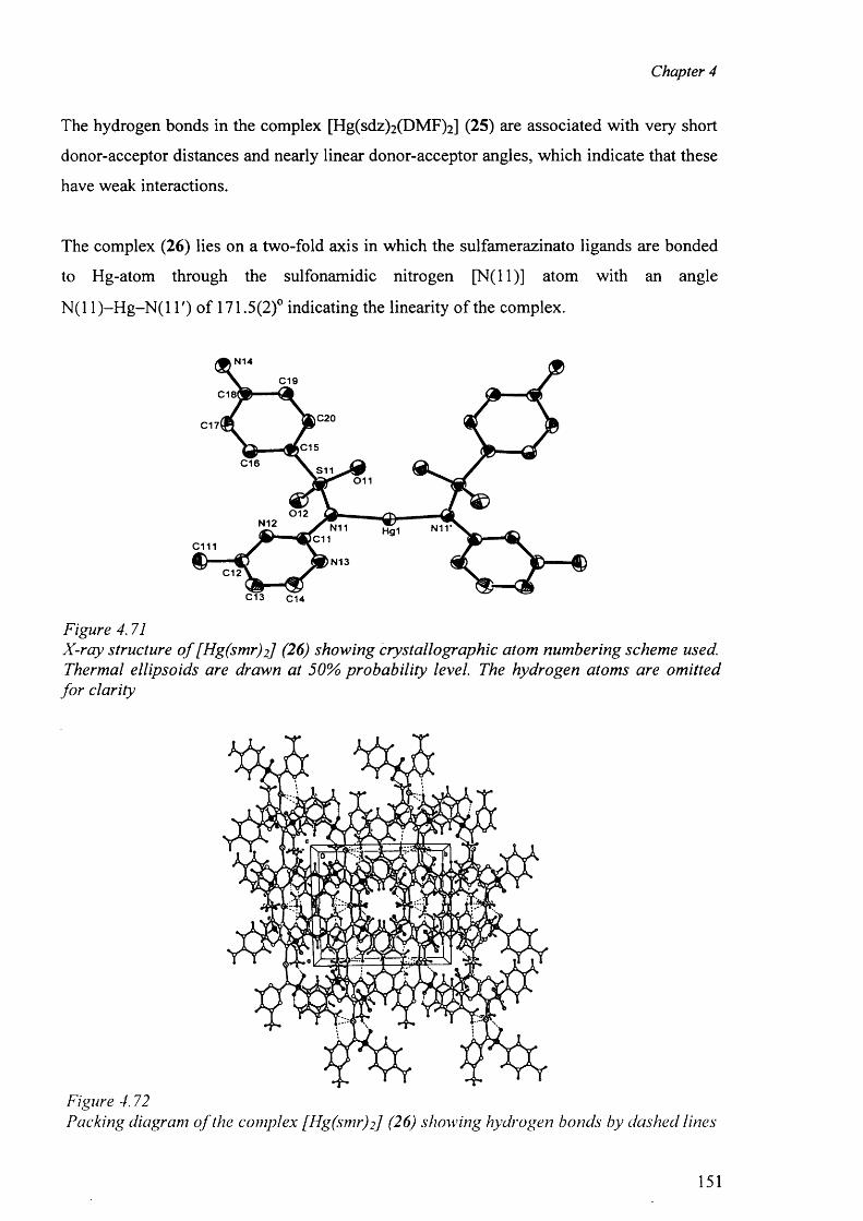
Chapter 4
The hydrogen bonds in the complex [Hg(sdz)2 (DMF)2 ] (25) are associated with very short
donor-acceptor distances and nearly linear donor-acceptor angles, which indicate that these
have weak interactions.
The complex (26) lies on a two-fold axis in which the sulfamerazinato ligands are bonded
to Hg-atom through the sulfonamidic nitrogen [N (ll)] atom with an angle
N (11)-H g-N (l T) o f 171.5(2)° indicating the linearity o f the complex.
N14
C19C 18I
C20C17l
|C15C16 S11
01 1
0 1 2N12 N11 Hg1
C11c mN13
C12
C13 C14
Figure 4.71X-ray structure o f [Hg(smr)2] (26) showing crystallographic atom numbering scheme used. Thermal ellipsoids are drawn at 50% probability level. The hydrogen atoms are omitted
fo r clarity
Figure 4.72Packing diagram o f the complex [Hg(smr)2] (26) showing hydrogen bonds by dashed lines
151
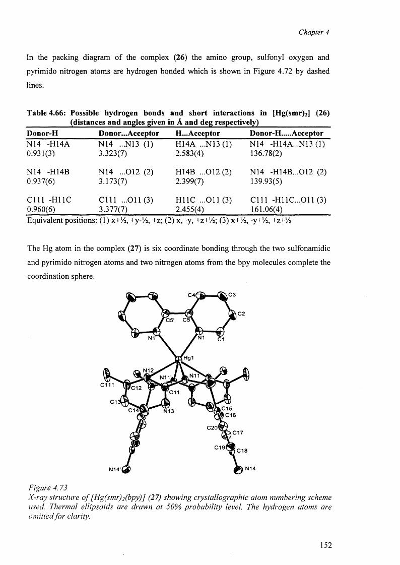
Chapter 4
In the packing diagram of the complex (26) the amino group, sulfonyl oxygen and
pyrimido nitrogen atoms are hydrogen bonded which is shown in Figure 4.72 by dashed
lines.
Table 4.66: Possible hydrogen bonds and short interactions in [Hg(smr)2 ] (26) ____________(distances and angles given in A and deg respectively)___________________Donor-H Donor...Acceptor H...Acceptor Donor-H.....AcceptorN14 -H14A N14 ...N13 (1) H14A ...N13 (1) N14 -H14A...N13 (1)0.931(3) 3.323(7) 2.583(4) 136.78(2)
N14 -H14B N14 ...012 (2) H14B ...0 1 2 (2 ) N14 -H14B...012 (2)0.937(6) 3.173(7) 2.399(7) 139.93(5)
C l l l -HI 1C C l l l ...O il (3) H11C ...O il (3) C l l l -HI 1C...011 (3)0.960(6) 3.377(7) 2.455(4) 161.06(4)Equivalent positions: (1) x+lA, +y-1A, +z; (2) x, -y, +z+lA; (3) x+!4, -y+!4, +z +lA
The Hg atom in the complex (27) is six coordinate bonding through the two sulfonamidic
and pyrimido nitrogen atoms and two nitrogen atoms from the bpy molecules complete the
coordination sphere.
C3C4l
C2C5' C5
N1N1 C1
Hg1
N12N11N11V
c m C12C11
C1C1 N13
C20C17
C19 C18
N14N14'
Figure 4.73X-ray structure o f [Hg(smr)2 (bpy)] (27) showing crystallographic atom numbering scheme used. Thermal ellipsoids are drawn at 50% probability level. The hydrogen atoms are omitted fo r clarity.
152
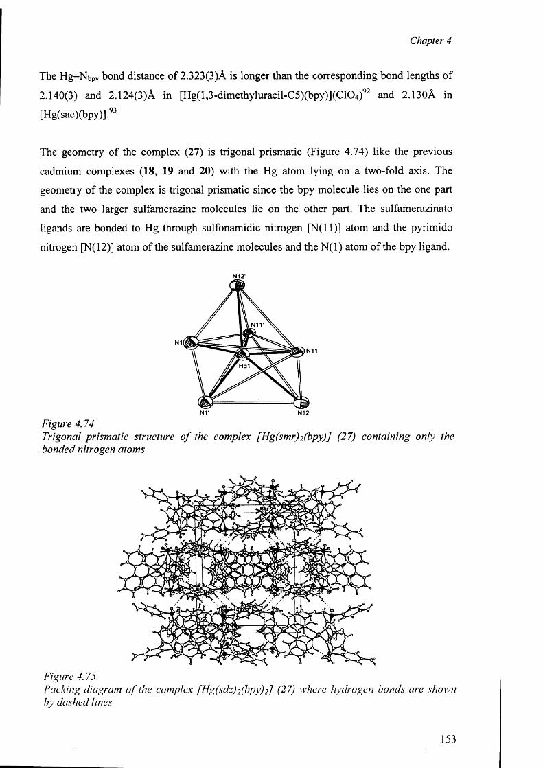
Chapter 4
The Hg-Nbpy bond distance o f 2.323(3)A is longer than the corresponding bond lengths of
2.140(3) and 2.124(3)A in [H g(l,3 -dimethyluracil-C 5 )(bpy)](C 1 0 4 ) 9 2 and 2.130A in
[Hg(sac)(bpy)].93
The geometry o f the complex (27) is trigonal prismatic (Figure 4.74) like the previous
cadmium complexes (18, 19 and 20) with the Hg atom lying on a two-fold axis. The
geometry of the complex is trigonal prismatic since the bpy molecule lies on the one part
and the two larger sulfamerazine molecules lie on the other part. The sulfamerazinato
ligands are bonded to Hg through sulfonamidic nitrogen [N (ll)] atom and the pyrimido
nitrogen [N(l 2)] atom o f the sulfamerazine molecules and the N (l) atom of the bpy ligand.
N12'
NT N12
Figure 4.74Trigonal prismatic structure o f the complex [Hg(smr)2 (bpy)J (27) containing only the bonded nitrogen atoms
Figure 4.75Packing diagram o f the complex [Hg(sdz)2 (bpy)2] (27) where hydrogen bonds are shown by dashed lines
153
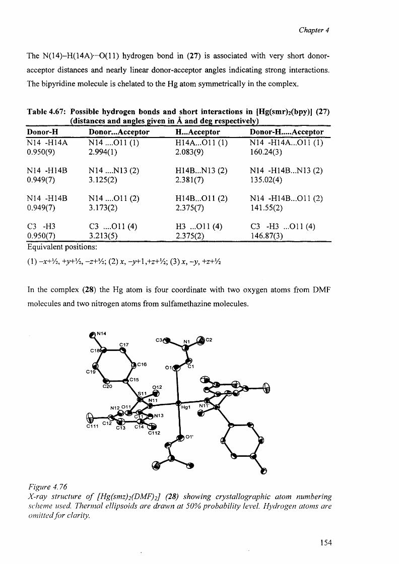
Chapter 4
The N(14)-H(14A)**-0(11) hydrogen bond in (27) is associated with very short donor-
acceptor distances and nearly linear donor-acceptor angles indicating strong interactions.
The bipyridine molecule is chelated to the Hg atom symmetrically in the complex.
Table 4.67: Possible hydrogen bonds and short interactions in [Hg(smr)2 (bpy)] (27) ____________(distances and angles given in A and deg respectively)___________________Donor-H Donor...Acceptor H...Acceptor Donor-H.....AcceptorN14 -H14A N 14....011 (1) H 14A ...011 (1) N14 -H 14A ...011 (1)0.950(9) 2.994(1) 2.083(9) 160.24(3)
N14 -H14B N14 ....N13 (2) H14B...N13 (2) N14 -H14B...N13 (2)0.949(7) 3.125(2) 2.381(7) 135.02(4)
N14 -H14B N 14....011 (2) H 14B ...011 (2) N14 -H 14B ...011 (2)0.949(7) 3.173(2) 2.375(7) 141.55(2)
C3 -H3 C3 ....011(4) H3 ...O il (4) C3 -H3 ...011(4 )0.950(7) 3.213(5) 2.375(2) 146.87(3)Equivalent positions:
(1) -X+V2, +y+V:2, -Z+V2; (2) x, -y+\,+z+l/2; (3) jc, -y , +z+V2
In the complex (28) the Hg atom is four coordinate with two oxygen atoms from DMF
molecules and two nitrogen atoms from sulfamethazine molecules.
N14C2C3| N1C17
C1
C16 C101C19
C15C20 012
N11 A , — # Hg1 0 N 1 3
C112j t.S11
N11'
C12c m C14C13
Figure 4.76X-ray structure o f [Hg(smz)2 (DMF)2] (28) showing crystallographic atom numbering scheme used. Thermal ellipsoids are drawn at 50% probability level. Hydrogen atoms are omitted fo r clarity.
154
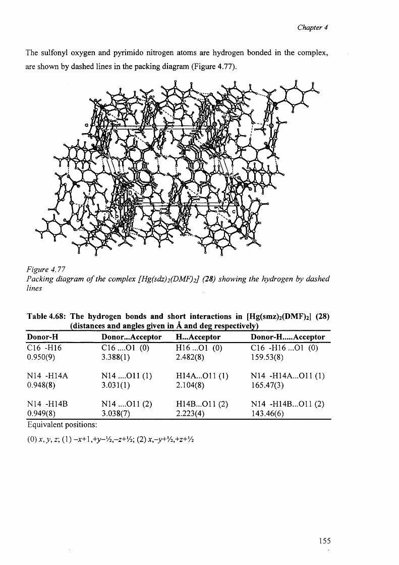
Chapter 4
The sulfonyl oxygen and pyrimido nitrogen atoms are hydrogen bonded in the complex,
are shown by dashed lines in the packing diagram (Figure 4.77).
#
Figure 4.77Packing diagram o f the complex [Hg(sdz)2 (DMF)2] (28) showing the hydrogen by dashed lines
Table 4.68: The hydrogen bonds and short interactions in [Hg(smz)2 (DMF)2 ] (28) ____________(distances and angles given in A and deg respectively)___________________Donor-H Donor...Acceptor H...Acceptor Donor-H.....AcceptorC16 -H16 C 16....01 (0) H 16...01 (0) C16 -H 16...01 (0)0.950(9) 3.388(1) 2.482(8) 159.53(8)
N14 -H14A N 14....011 (1) H 14A ...011 (1) N14 -H 14A ...011 (1)0.948(8) 3.031(1) 2.104(8) 165.47(3)
N14 -H14B N 14....011 (2) H 14B ...011 (2) N14 -H 14B ...011 (2)0.949(8) 3.038(7) 2.223(4) 143.46(6)Equivalent positions:
( 0 ) x, v,z; (1) -x + 1 , + y - l/ 2 - z + l/ 2; ( 2 ) x - y + '^ + z + 'A
155
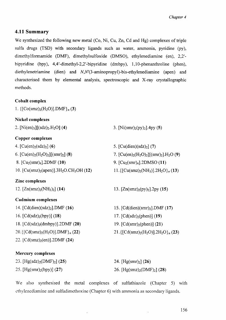
Chapter 4
4.11 Summary
We synthesized the following new metal (Co, Ni, Cu, Zn, Cd and Hg) complexes o f triple
sulfa drugs (TSD) with secondary ligands such as water, ammonia, pyridine (py),
dimethylformamide (DMF), dimethylsulfoxide (DMSO), ethylenediamine (en), 2,2'-
bipyridine (bpy), 4,4'-dimethyl-2,2'-bipyridine (dmbpy), 1,10-phenanthroline (phen),
diethylenetriamine (dien) and ^^(S-am inopropy^-bis-ethylenediam ine (apen) and
characterised them by elemental analysis, spectroscopic and X-ray crystallographic
methods.
Cobalt complex
1. {[Co(smz)2 (H 2 0)].DM F}n (3)
Nickel complexes
2. [Ni(en)3 ][(sdz)2 .H2 0 ] (4) 3. [Ni(smr)2 (py)2 ].4 py (5)
Copper complexes
4. [Cu(en)2 (sdz)2] (6 )
6 . [Cu(en)2 (H2 0 ) 2 ][(smr)2] (8 )
8 . [Cu2 (smr)4 ].2DMF (10)
10. [Cu(smz)2 (apen)].3H 2 O.CH3OH (12)
5. [Cu(dien)(sdz)2] (7)
7. [Cu(en)2 (H2 0 ) 2 ][(smr)2 ].H20 (9)
9. [Cu2 (smr)4 ].2DMSO (11)
ll.{[C u(sm z)2(NH3)].2H20}„(13)
Zinc complexes
12. [Zn(smz)2 (NH3)2] (14) 13. [Zn(smz)2 (py)2 ].2py (15)
Cadmium complexes
14. [Cd(dien)(sdz)2].DMF (16)
16. [Cd(sdz)2 (bpy)] (18)
18. [Cd(sdz)2 (dmbpy)].2DMF (20)
20.{[Cd(smz)2 (H2 O)].DMF}„ (22)
22. [Cd(smz)2 (en)].2DMF (24)
15. [Cd(dien)(smr)2].DMF (17)
17. [Cd(sdz)2 (phen)] (19)
19. [Cd(smr)2 (phen)] (21)
21. {[Cd(smz)2 (H 2 0)] .2H20 }„ (23)
Mercury complexes
23. [Hg(sdz)2 (DMF)2] (25)
25. [Hg(smr)2 (bpy)] (27)
24. [Hg(smr)2] (26)
26. [Hg(smz)2 (DMF)2] (28)
We also synthesised the metal complexes o f sulfathiazole (Chapter 5) with
ethylenediamine and sulfadimethoxine (Chapter 6 ) with ammonia as secondary ligands.
Chapter 4
Metal complexes o f sulfathiazole
1. [C 0 C I4] [(H2 Stz)2 ].CH 3 COOH (30); 2. [Cu(en)2 (H2 0 )2 ][stz)2 ].2 H 2 0 (31)
Zinc complex of sulfadimethoxine
1. [Zn(sdm)2 (NH3 )2 ].2 H 2 0 (33)
4.11.1 Type of coordination around the metal centres
The metal atoms in the complexes studied display different coordination numbers. The
most common coordination number found is six with the metal having octahedral,
{[Co(smz)2 (H2 0)] .DMF} „ (3), [Ni(en)3 ][(sdz)2 .H2 0 ] (4), [Ni(smr)2 (py)2 ].4py (5),
[Cu(dien)(sdz)2] (7), [Cu(en)2 (H2 0 )2 ][(smr)2 ] (8 ), [Cu(en)2 (H2 0 )2 ][(smr)2 ].H 2 0 (9)
[Cu(smz)2 (apen)].3H 2 O.CH3OH ( 1 2 ), {[Cu(smz)2 (NH 3 )].2H 2 0}„ (13), [Zn(smz)2 (NH3)2]
(14), [Zn(smz)2 (py)2 ].2py (15), {[Cd(smz)2 (H 2 0)].DM F}„ (22), {[Cd(smz)2 (H2 0 )]H2 0 }„
(23), [Cd(smz)2 (en)].2 DMF (24) and [Cu(en)2 (H2 0 )2 ][stz)2 ].2 H 2 0 (31); trigonal prismatic,
[Cd(sdz)2 (bpy)] (18), [Cd(sdz)2 (phen)] (19), [Cd(sdz)2 (dmbpy)].2DMF (20),
[Hg(smr)2 (bpy)] (27) and [Zn(sdm)2 (NH 3 )2 ].2 H 2 0 (33); and bicapped tetrahedral
geometry, [Cd(smr)2 (phen)] (21). The two copper complexes [Cu2 (smr)4 ].2 DMF (10) and
[Cu2 (smr)4 ].2 DMSO (11) are five coordinate with one metal-metal bond. The complexes
[Cu(en)2 (sdz)2] (6 ), [Hg(sdz)2 (DMF)2] (25), [Hg(smz)2 (DMF)2] (28), [CoCl4 ][(smz)2]-
CH3COOH (30) show a coordination number o f four (square planar) and the only mercury
complex o f sulfamerazine [Hg(smr)2 ] (26) shows a coordination number o f two with linear
arrangement. The complexes [Cd(dien)(sdz)2 ].DMF (16) and [Cd(dien)(smr)2 ].DMF (17)
have seven coordination number with distorted pentagonal bipyramidal geometry. The
geometries are often associated with distortions from ideal geometries both in bond lengths
and in angles. These aspects have already been discussed and explained when describing
each individual structure in the previous chapters. However it needs to be emphasised here
that the distortions are in most cases due to electronic effects, steric factors and also, in
most cases, due to the intra- and/or inter-molecular hydrogen bonding effects.
4.11.2 Status of TSD anion as ligand
It has been mentioned earlier (Chapter 2) that TSD ions have six possible donor sites for
potential binding with a metal ion. These six sites are the sulfonamidic nitrogen, two
pyrimido nitrogen, two sulfonyl oxygen and amino nitrogen atoms. Since the neutral TSD
molecules contain a hydrogen atom on the sulfonamidic nitrogen atom, after deprotonation
157

Chapter 4
the negative charge is located (entirely or at least most o f it) on the nitrogen atom of the
TSD anion. Therefore, the N atom with a negative charge should be the most preferred site
for bonding with a metal ion. In fact, this mode o f coordination o f the TSD anion has been
found as the most common in the complexes where most o f the sulfa drug molecules are
directly bonded with the metals. Thus N-coordinated complexes are listed in Table 4.68. In
one unusual case, the sulfamethazinato ion is bonded to the metal centre, not through the
sulfonamidic nitrogen atom, but through the sulfonyl oxygen atom ( 1 2 ).
Table 4.68: Comparison of the M -N (ll) and M -N (12) bond lengths
ComplexesBonds
M -N (ll) M -N(12){[Co(smz)2 (H 2 0)].D M F}„ (3) 2.142(2) 2.187(2)[Ni(smr)2 (py)2 ].4py (5) 2.139(2) 2 . 1 0 0 (2 )[Cu2 (smr)4] .2DMF (10) 2.057(3) 1.992(3)[Cu2 (smr)4 ].2DMSO (11) 2.040(5) 1.998(5){[Cu(smz)2 (NH 3 )].2H2 0 } „ (13) 2.044(3) 2.035(3)[Zn(smz)2 (NH3)2] (14) 2.040(3) 2.844(3)[Zn(smz)2 (py)2] .2py (15) 2.223(3) 2.323(3)[Cd(dien)(sdz)2] .DMF (16) 2.495(2) 2.403(2)[Cd(dien)(smr)2].DMF (17) 2.434(2) 2.416(2)fCd(sdz)2 (bpy)l (18) 2.283(3) 2.501(3)[Cd(sdz)2 (phen)] (19) 2.275(2) 2.517(2)[Cd(sdz)2 (dmbpy)].2DMF (20) 2.253(2) 2.504(2)[Cd(smr)2 (phen)] (21) 2.193(2) 2.793(2){[Cd(smz)2 (H2 0)].DMF}„ (22) 2.307(3) 2.407(3){[Cd(smz)2 (H2 0)].H 2 0}„ (23) 2.337(3) 2.322(3)[Cd(smz)2 (en)].2DMF (24) 2.311(3) 2.471(4)[Hg(sdz)2 (DMF)2] (25) 2.047(3)[Hg(smr)2] (26) 2.017(4)fHg(smr)2 (bpy)] (27) 2.216(3) 2.881(3)[Hg(smz)2 (DMF)2] (28) 2.048(4)
In some cases, the nickel and copper complexes [(4), (6 ), (7), (8 ), (9), (30) and (31)
complexes], the sulfa drug molecules are not coordinated with the metal ions because a
stronger ligand such as ethylenediamine and/or diethylenetriamine are attached to the
metal centres, and they remain outside the coordination sphere and are attached to the
cationic species through very strong hydrogen bonds. In complex (30), the cobalt atom is
surrounded by four chloride ions forming the negatively charged anion and the two
molecules o f positively charged sulfathiazole cations remain outside the coordination
spheres. These complexes are typical examples for the formation of “cation-anion” pairs.
158
Chapter 4
Thus our studies clearly indicate that (i) the sulfa drug anions are not directly coordinated
with the metals in some cases, and (ii) when it is coordinated to a metal, it commonly
involves the sulfonamidic and pyrimido nitrogen atoms. Involvement o f the sulfonyl
oxygen atom is also possible and in fact, it is observed in one case o f the copper complex
of sulfamethazine (12), and in the polymeric complexes o f sulfamethazine, the terminal
amino group takes part in the coordination to the metal centres.
Another aspect that was stated earlier but should also be pointed out here is that the sulfa
drug anions may act as a monodentate, bidentate or tridentate chelating as well as bridging
ligands, as observed in polymeric complex o f sulfadiazine [Zn(sdz)2 ]1 and
[Cd(sdz)2].2H202 and in the complex o f sulfamethazine [Cd(smz)2(H20)].2H20.3 In our
present complexes, however, the ligands are also monodentate, bidentate or tridentate in
some cases.
The question that may arise in this context is: why do the sulfa drug molecules show
different coordination modes in different complexes. The explanation is not simple, but it
would obviously depend on the competition between different possible coordinating
species in the reaction media as well as their relative basicities. Ligands such as H 2O, N H 3,
pyridine, etc., are well known Lewis bases, and can bind with metal ions due to the ease
with which these species can donate their electrons to the metal ions, which act as Lewis
acids. Since in our complexes sulfa drug molecules are found to coordinate with the metal
centres along with H 2 O, pyridine, ethylenediamine, bipyridine, phenanthroline, we believe
that sulfa drug molecules are also relatively strong ligands and can compete with the above
mentioned secondary ligands for bonding with the metal ions. The relative basicities at the
sulfonyl oxygen atoms o f the sulfa drug molecules would be much less compared with that
of the sulfonamidic and pyrimido nitrogen atoms. So coordination involving sulfonyl
oxygen atoms is rather rare, and such modes o f coordination would be guided not by the
electron donating capacity alone, but by other factors such as hydrogen bonding and/or
steric reasons.
4.11.3 Role of hydrogen bonds in the formation of crystal architecture
Hydrogen bonds between a proton and corresponding donor and acceptor atoms are
probably the most important non-covalent interactions in organic and metal complexes.
159
Chapter 4
Their roles in determining molecular geometry, conformations, and crystal architecture and
the overall stability have long been emphasised and recognised.
In the complexes studied in this work, both intra- and inter-molecular/ionic hydrogen
bonds and short van der Waals’ interactions have been noted and listed in selected Tables
for the individual complexes. These hydrogen bonds are o f the O -H —O/N type and in the
case of diamine/triamine complexes of the N -H —O type. They play important roles in the
distribution of ligands around the metal centres and their relative orientations. The sulfa
drug anions that remain outside the coordination sphere are held in the crystal due to the
extensive hydrogen bonding network. This determines the overall packing arrangement in
the unit cell. The truth o f this statement lies in the fact that for ALL the complex species
containing water molecules either coordinated and/or in the lattice, hydrogen bonds are
always present and their dimensions indicate that they are fairly strong hydrogen bonds
based on normal criteria [0 (-H )—0/N = 3.1, H---0/N = 2.0 A]. The complexes that do not
contain any water molecules do not have such strong hydrogen bonds, but these complexes
also contain weaker interactions of the type C -H —O and van der W aals’ interactions, and
these are the forces that hold individual molecules together in the crystal.
4.11.4 Conclusion
The work in this thesis is not complete as far as the global aim is concerned, but
nonetheless, it embraces the structural studies of the metal complexes o f sulfa drugs. The
metal complexes o f sulfa drugs, which have been studied, are Co, Ni, Cu, Zn, Cd and Hg
with sulfadiazine, sulfamerazine, sulfamethazine, sulfathiazole and sulfadimethoxine. It
has been shown that when sulfa drugs are bonded to the metal atom, it is usually through
the sulfonamidic and pyrimido nitrogen atoms. In most o f the complexes the bonds through
the sulfonamidic nitrogen atom is shorter than that o f the pyrimido nitrogen atom.
4.11.5 Further works
As the sulfa drugs are active antibacterial compounds and some metal complexes are still
used as topical medicine for bum treatment, the compounds, which we have synthesized in
the laboratory, may also have some biological activity and antibacterial activity. In
addition there may be more complexes of sulfa drugs with metal ions and secondary
ligands. Their biological activity may be investigated in future.
160
Chapter 4
References
1. A. Garcia-Raso, J.J. Fiol, S. Rigo, A. Lopez-Lopez, E. Molins, E. Espinosa, E. Borras, G. Alzuet, Joaquin Borras and A. Castineiras; Polyhedron; 2 0 0 0 ,19, 991
2. J. Casanova, G. Aluzet, S. Ferrer, J. Borras, S. Garcia-Granda and E. Perez-Carreno; J. Inorg. Biochem., 1993, 51, 689
3. A. Garcia-Raso, J. J. Fiol, G. Martorell, A. Lopez-Zafra and M. Quiros; Polyhedron, 1997, 16,613
4. J. R. Pilbrow; “Transition Ion Electron Paramagnetic Resonance”, Oxford Science Publications, 1990, 20
5. H. S. Shin, G. S. Ihn, H. S. Kim and C. H. Koo; J. Korean Chem. Soc., 1974,18, 329
6. M. K. kokila, Puttaraja, M. V. Kulkami and S. Thampi; Acta Cryst., 1995, C51, 333
7. K. R. Acharya; K.N. Kuchela and G. Kartha; J. Cryst. Spectros. Res., 1982 ,12, 369
8. M. R. Caira and R. Mohamed; Acta Cryst., 1992, B48, 492
9. A. K. Basak, S. K. Mazumdar and S. Chaudhuri; Acta Crsyt., 1983, C39, 492
10. R. K. Tiwari, M. Haridas and T. P. Sing; Acta Cryst., 1984, C40, 655
11. A. Camerman; Canadian J. Chem., 1970, 48, 179
12. M. R. Truter; J. Chem. Soc., 1962, 3400
13. A. M. O ’Connel and E. N. Maslen; Acta Cryst., 1967, 22, 134
14. V. V. Joshi, R. K. Tiwari, T. C. Patel and T. P. Singh; Indian J. Physics, 1983, 57A, 79
15. G. D. Munno, T. Poerio , M. Julve, F. Lloret and G. Viau; New J. Chem., 1998, 3, 299
16. C. -Z . Xie, B.-F. Zhang, X. -Q . Wang, G. -Q . Shen and D.-Z. Shen; Acta Cryst., 2004, E60, m l703
17. M. U. Haque, C. N. Caughlan and K. Emerson; Inorg. Chem., 1970, 9, 2421
18. R. E. Cramer and J. T. Hunke; Inorg. Chem., 1978 ,17, 365
19. C. L. Raston, A. H. White and A. C. Willis; Aust. J. Chem., 1978, 31, 415
20. J. D. Korp, I. Bernal, R. A. Palmer and J. C. Robinson; Acta Cryst., 1980, B36, 560
21. J. G. H. D. Preez, H. E. Rohwer, B. J. van Brecht and M. R. Caira; J. Chem. Soc., Dalton Trans., 1984, 975
22. R. E. Cramer, W. van Doone and J. T. Hunke; Inorg. Chem., 1976, 15, 529
161
Chapter 4
23. T. Mizuta, H. Yoneda and Y. Kushi; Inorg. Chim. Acta, 1987, 132, 11
24. G. V. Romanenco, V. L. Varand, N. V. Podberezskaya, D. Y. Naumov, S. A. Gromilov and S. V. Larionov; J. Struct. Chem., 2001, 42, 1036
25. J. Ellermeier, C. Nather and Wolfgang Bensch; Acta Cryst., 1999, C55, 501
26. H. -L . Liu, H. -Y . Mao, C. Xu, H. -Y . Zhang, H. -W . Hou, Q. -A . Wu, Y. Zhu, B. -X . Ye and L. -J . Yuan; Polyhedron, 2004, 23, 1799
27. C. Xu, H. -Y . Mao, H. -Y . Zhang, Q. -A . Wu, H. -L . Liu, H. -W . Hou, Y. Zhu andG. Li; J. Mol. Struct., 2003, 658, 59
28. W. Lei, Q. Hao, L. Lu, Z. Yang and X. Wang; J. Chem. Cryst., 2004, 34, 387
29. S. Ooi, and Q. Fernando; Inorg. Chem., 1976, 6, 1558
30. S. X. Liu, C. C. Lin, Z. Xu, Y. P. Yu and X. Z. You; China J. Org. Chem., 1987, 5, 369
31. L. Escriche, J. A. Munoz, J. Rosell, R. Kivekas, R. Sillanpaa, and J. Casabo; Inorg. Chem., 1998, 37, 4807
32. J. Lokaj, K. Gyerova, A. Sopkova, J. Sivy, V. Kettman and V. Vrabel; Acta Cryst., 1991, C47, 2447
33. J. R. Anacona, N. Ramos, G. D. D. Delgado and E. M. Roque; J. Coord. Chem., 2002,55,901
34. L. A. Kovbasyuk, I. O. Frisky, V. N. Kokozay and T. S. Iskenderov; Polyhedron, 1997,16, 1723
35. H. -K . Fun, Q. -L . Hao, J. Wu, X. -J . Yang, L. Lu, X. Wang, S. Chantrapromma, I. A. Razak and A. Usman; Acta Cryst., 2002, C58, m87
36. C. Kazak, V. T. Yilmaz and T. K. Yazicilar; Acta Cryst., 2004, E60, m593
37. F. S. Stephens; J. Chem. Soc., 1969, (A), 2233
38. B. -H . Ye, L. -N . Ji, F. Xue and T. C. W. Mak; Polyhedron, 1998 ,17, 2687
39. M. J. Begley, P. Hubberstey and J. Stroud; Polyhedron, 1997,16, 805
40. S. -J . Liu and C. -C . Su; Polyhedron, 1996,15, 1141
41. F. S. Stephens; J. Chem. Soc., A, 1969, 833
42. J. Cemak, J. Chomic and W. Massa; Acta Cryst., 2002, C58, m490
43. L. Gutierrez, G. Alzuet, J. Borras, A. Castineiras; A. Rodriguez-Fortea and E. Ruiz; Inorg. Chem., 2001, 40, 3089
44. Z. -D . Liu, M. -Y . Tan and H. -L . Zhu; Acta Cryst., 2004, E60, m l081
162
Chapter 4
45. D. Sonnenfroh and R. W. Kerilick; Inorg. Chem., 1980,19, 1259
46. J. Casanova, G. Alzuet, J. LaTorre and J. Borras; Inorg. Chem., 1997, 36, 2052
47. J. C. Pedregosa, J. Casanova, G. Alzuet, J. Borras, S. Garda-Granda, M. R. Diaz andA. Gutierrez-Rodriguez; Inorg. Chim. Acta., 1995, 232, 117
48. A. Nanthakumar, J. Miura, S. Diltz, C. -K . Lee, G. Aguirre, F. Ortega, J.W. Ziller and P. J. Walsh; Inorg. Chem., 1999, 38, 3010
49. J. Casanova, G. Alzuet, J. Borra's, J. LaTorre, M. Sanau', and S. Garda-Granda; J. Inorg. Biochem., 1995, 60, 219
50. A. Pezeshk, and V. Pezeshk; J. Inorg. Biochem., 1990, 38, 185
51. V. M. Rao, D. N. Sathaynarayana and H. Manohar; J. Chem. Soc., Dalton Trans., 1983,2167
52. P. de Meester, S. R. Fletcher and A. C. Skapski; J. Chem. Soc., Dalton Trans., 1975, 2575
53. Y. A. Simonov, V. I. Ivanov, T. I. Malinovskii and L. I. Landa; Izv. Akad. Nauk. Mold. SSR, Ser. Fiz-Tekh. Mat. N auk, 1976, 3, 33
54. G. A. Barclay and C. H. L. Kennard; J. Chem. Soc., 1961, 5244
55. F. Hanic, D. Stempelova and K. Hanikova; Acta Cryst., 1964 ,17, 633
56. Y. A. Simonov, V. I. Ivanov, A. V. Ablov, L. N. Milkova and T. I. Malinovskii; Zh.Strukt. Khim., 1976 ,17, 516
57. C. -H . Kwak, J. Jeong and J. Kim; Inorg. Chem. Comm., 2001, 4, 264
58. T. -Y . Lee, T. -J . Lee, C. -Y . Hong, M. -Y . Hsieh, D. -T . Wu and C. S. Chung;Acta Cryst., 1986, C42, 1316 and the references therein.
59. T. G. M. Fawcett, S. M. Rudich, B. H. Toby, R. A. Lanlancette, J. A. Potenza and H. J. Schugar; Inorg. Chem., 1980,19, 940
60. C.J. Brown, D.S. Cook and L. Sengier; Acta Cryst., 1987, C43, 2332
61. N.C. Baenziger; Acta Cryst., 1983, C39, 1620
62. R. -X . Yuan, R. -G . Xiong, Z. -F . Chen, P. Zhang, H. -X . Ju, Z. Dai, Z. -J . Guo, H.2K. Fun and X. -Z . You; J. Chem. Soc. Dalton Trans., 2001, 774
63. M. A. Torzilli, S. Colquhoun, D. Doucet and R. H. Beer; Polyhedron, 2002, 21, 697
64. K. A. Fraser and M. M. Harding; Acta Cryst., 1967, 22, 75
65. E. Szlyk, A. Wojtczak, A. Surdykowski and M. Gozdzikiewicz; Inorg. Chim. Acta,2005, 358, 467
163
Chapter 4
66. M. Bochmann, G. C. Bwembya, A. K. Powell and X. Song; Polyhedron, 1995, 14, 3495
67. P. J. Toscano, P. T. DiMauro, S. Geremia, L. Randaccio and E. Zangrando; Inorg. Chim. Acta , 1994, 217, 195
68. W. L. Steifen and G.J. Palenik; Acta Cryst., 1976, B32, 298
69. M. Mazzanti, S. Gambarotta, C. Floriani, A. Chiesi-Villa and C. Guastini; Inorg. Chem., 1986, 25, 2308
70. P. G. Jones, I. Dix, A. J. Kirbyc and C. Beckmann; Acta Cryst., 2003, E59, ml71
71. A. Mondal, G. Mostafa, A. Ghosh, I. R. Laskar and N. R. Chaudhuri; J. Chem. Soc.,Dalton Trans., 1999, 9
72. H. Kurihara, S. Nishikiori and T. Iwamoto; Chem. Lett., 1997, 61
73. C. A. Johns and K. M. A. Malik; Polyhedron, 2002, 21, 395
74. L. Menabue and M. Saladini; J. Inorg. Biochem., 1993, 49, 201
75. S. Baneijee, A. Ghosh, B. Wu, P. -G . Lassahan and C. Janiak; Polyhedron, 2005, 2 4 ,593
76. Y. -F . Zhou, R. -H . Wang, B. -L . Wu, R. Cao and M. -C . Hong; J. Mol. Struct., 2004, 697, 73
77. B. -H . Ye, X. -M . Chen, F. Xue, L. -N . Ji and T. C. W. Mak; Inorg. Chim. Acta, 2000, 299, 1
78. J. -H . Luo, M. -C . Hong, R. Cao, Y. -C . Liang, Y. -J . Zhao, R. -H . Wang and J. -B. Weng; Polyhedron, 2002, 21, 893
79. C. A. Johns, G. M. G. Hossain, K. M. A. Malik, S. Z. Haider and U. K. R. Romman; Polyhedron, 2001, 20, 721
80. A. B. Corradi, G. Lusvardi, L. Menabue, M. Saladini and P. Sgarabotto; Polyhedron, 1999, 18, 1975
81. E. Labisbal, J. Romero, J. A. Garcia-Vasquez, C. Gomez, A. Sousa, R. Pritchard andC. A. McAuliffe; Polyhedron, 1994, 13, 1735
82. J. A. Castro, J. Romero, J. A. Garcia-Vazquez, A. Sousa, E. E. Castellano and J. Zukerman-Schpector; Polyhedron, 1993,12, 31
83. T. Miyoshi, T. Iwamoto and Y. Sasaki; Inorg. Chim. Acta, 1972, 6, 59
84. L. Li, B. Chen, Y. Song, G. Li, H. Hou, Y. Fan and L. Mi; Inorg. Chim. Acta, 2003,344, 95
85. J. S. Casas, M. S. Garcia-Tasende and J. Sordo; Coord. Chem. Rev., 1999, 193-195, 283
164
Chapter 4
86. K. K. Divine, F. Ayala-Fierro, D. S. Barber and D. E. Carter; J. Toxicol. Environ. Health, 1999, A57, 489
87. Z. Popovic, D. Matkovic-Calogovic, Z. Soldin, G. Pavlovic, N. Davidovic and D. Vikic-Topic; Inorg. Chim. Acta , 1999, 294, 35
88. N-H. Hu, T. Norifusa and K. Aoki; Polyhedron, 1999,18, 2987
89. B. Gyurcsik and L. Nagy; Coord. Chem. Rev., 2000, 203, 81
90. D. M. L. Goodgame, S. P. W. Hill and D. J. Williams; Polyhedron, 1992,11, 1507
91. M. Saladini, L. Menabue, E. Ferrari and D. Iacopino; J. Chem. Soc., Dalton Trans., 2001, 1513
92. F. Zamora, M. Sabat and B. Lippert; Inorg. Chim. Acta, 1998, 282, 237
93. A. Hergold-Brundic, B. kamenar and G. Jovanovski; Acta Cryst., 1989, C45, 556
94. J. -L . Tian, M. -J . Xie, S. -P . Yan, D. -Z . Liao, Z. -H . Jiang and P. Cheng; Acta Cryst., 2004, E60, m l689 and the references therein.
95. S. Gao, J. -W . Liu, L. -H . Huo and H. Zhao; Acta Cryst., 2004, E60, m l728
96. W.O. Gillum, J.C. Huffman, W.E. Streib, R. A. D. Wentworth; J. Chem. Soc.,Chem. Commun., 1969, 843
97. G. Rajsekhar, C. P. Rao, K. Nattinen and K. Rissanen; Inorg. Chem. Comm., 2003,6, 1156
98. M. D. Fryzuk, A. Carter, S. J. Rettig; Organometallies', 1992, 11, 469
165
STUDIES OF METAL COMPLEXES OF SULFA THIAZOLE WITH
SECONDAR Y LIGANDS
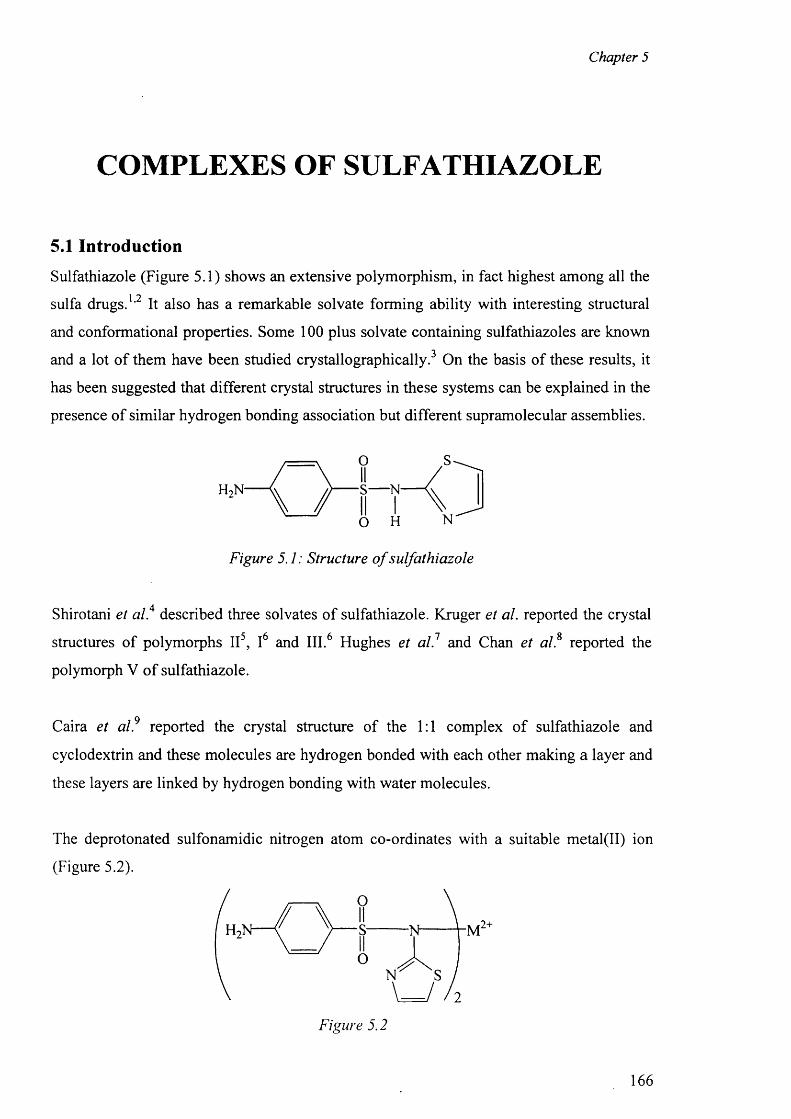
Chapter 5
COMPLEXES OF SULFATHIAZOLE
5.1 Introduction
Sulfathiazole (Figure 5.1) shows an extensive polymorphism, in fact highest among all the1 9sulfa drugs. ’ It also has a remarkable solvate forming ability with interesting structural
and conformational properties. Some 100 plus solvate containing sulfathiazoles are known
and a lot o f them have been studied crystallographically. On the basis of these results, it
has been suggested that different crystal structures in these systems can be explained in the
presence o f similar hydrogen bonding association but different supramolecular assemblies.
Figure 5.1: Structure o f sulfathiazole
Shirotani et a l 4 described three solvates o f sulfathiazole. Kruger et al. reported the crystal
structures of polymorphs II5, I6 and III.6 Hughes et al.1 and Chan et al.8 reported the
polymorph V o f sulfathiazole.
Caira et al.9 reported the crystal structure of the 1:1 complex o f sulfathiazole and
cyclodextrin and these molecules are hydrogen bonded with each other making a layer and
these layers are linked by hydrogen bonding with water molecules.
The deprotonated sulfonamidic nitrogen atom co-ordinates with a suitable metal(II) ion
(Figure 5.2).
Figure 5.2
166
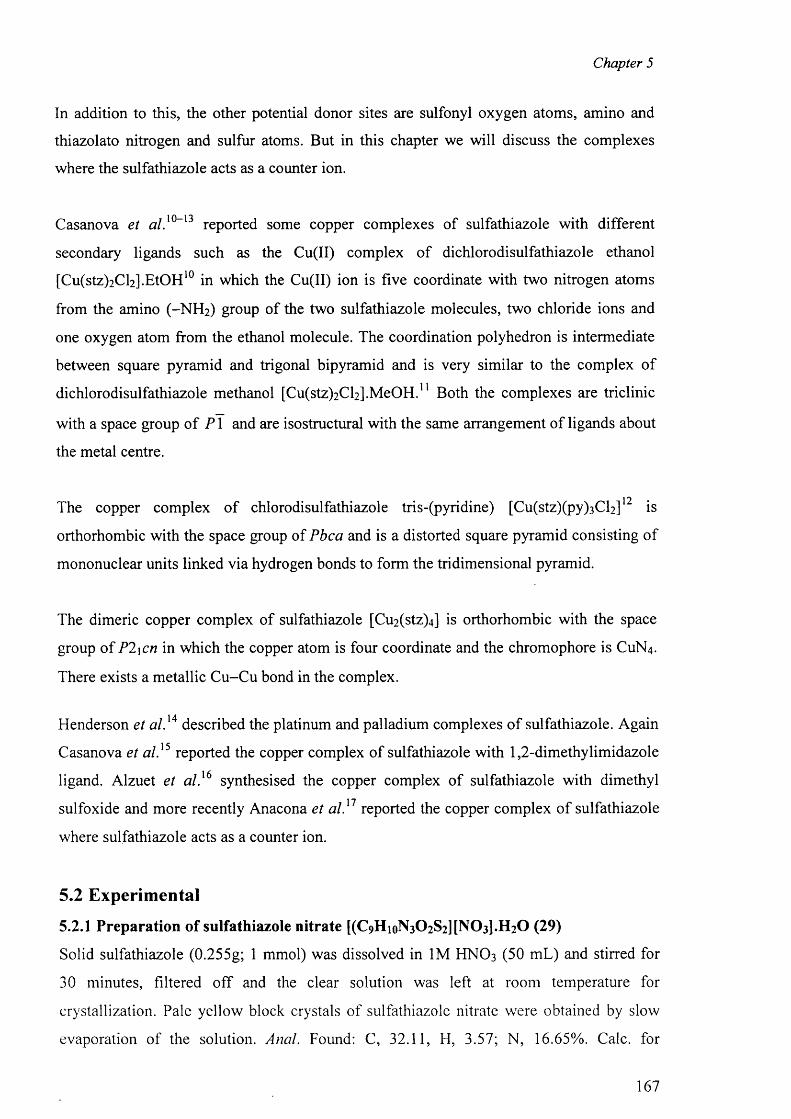
Chapter 5
In addition to this, the other potential donor sites are sulfonyl oxygen atoms, amino and
thiazolato nitrogen and sulfur atoms. But in this chapter we will discuss the complexes
where the sulfathiazole acts as a counter ion.
Casanova et a / . 1 0 - 1 3 reported some copper complexes of sulfathiazole with different
secondary ligands such as the Cu(II) complex o f dichlorodisulfathiazole ethanol
[Cu(stz)2 Cl2 ].EtOH 1 0 in which the Cu(II) ion is five coordinate with two nitrogen atoms
from the amino (-N H 2 ) group of the two sulfathiazole molecules, two chloride ions and
one oxygen atom from the ethanol molecule. The coordination polyhedron is intermediate
between square pyramid and trigonal bipyramid and is very similar to the complex of
dichlorodisulfathiazole methanol [C u^ tz^C y .M eO H . 11 Both the complexes are triclinic
with a space group o f P 1 and are isostructural with the same arrangement o f ligands about
the metal centre.
1 n #The copper complex o f chlorodisulfathiazole tris-(pyridine) [Cu(stz)(py)3 Cl2 ] is
orthorhombic with the space group of Pbca and is a distorted square pyramid consisting o f
mononuclear units linked via hydrogen bonds to form the tridimensional pyramid.
The dimeric copper complex of sulfathiazole [Cu2 (stz)4 ] is orthorhombic with the space
group o f P2\cn in which the copper atom is four coordinate and the chromophore is CUN4 .
There exists a metallic C u-C u bond in the complex.
Henderson et al . 14 described the platinum and palladium complexes o f sulfathiazole. Again
Casanova et a l} 5 reported the copper complex o f sulfathiazole with 1,2-dimethylimidazole
ligand. Alzuet et a l} 6 synthesised the copper complex of sulfathiazole with dimethyl
sulfoxide and more recently Anacona et a l} 1 reported the copper complex o f sulfathiazole
where sulfathiazole acts as a counter ion.
5.2 Experimental
5.2.1 Preparation of sulfathiazole nitrate [(C9 HioN3 0 2 S2 ][N0 3 ].H 2 0 (29)
Solid sulfathiazole (0.255g; 1 mmol) was dissolved in 1M HNO 3 (50 mL) and stirred for
30 minutes, filtered off and the clear solution was left at room temperature for
crystallization. Pale yellow block crystals o f sulfathiazole nitrate were obtained by slow
evaporation of the solution. Anal. Found: C, 32.11, H, 3.57; N, 16.65%. Calc, for
167
Chapter 5
C9 Hi2N 4 0 6 S2 : C, 32.16; H, 3.55; N, 16.63%. IR: (KBr disc): c m '1, 3510(b), 3410(s),
3375(m), 1621 (s), 1535(vs), 1329(vs), 1269(vs), 1143(m), 933(m), 689(w), 627(vw).
5.2.2 Preparation of cobalt complex of sulfathiazole [CoCl4 ][(stz)2 ].CH 3 COOH (30)
Solid sulfathiazole (0.565g, 2 mmol) was dissolved in hot methanol and methanolic
solution o f C0 CI2 .6 H2 O (0.23 8 g, 1 mmol) was added with constant stirring. The purple
precipitate was filtered off and dried over silica gel. It was dissolved in water in presence
of acetic acid. After three days green crystals were obtained. Anal. Found: C, 31.03, H,
3.10; N, 10.86%. Calc, for C2 0 H 2 4 CI4 C0 N 6 O6 S4 : C, 31.10; H, 3.15; N, 10.75%. IR: (KBr
disc): c m '1, 3513(b), 3426(s), 3383(m), 1625(s), 1537(vs), 1331(vs), 1267(vs), 1141(m),
933(m), 689(w), 625(vw).
5.2.3 Preparation of the complex [Cu(en)2(H20)2][(stz)2].2H20 (31)
Sodium salt o f sulfathiazole (Nastz) (2 mmol) was dissolved in hot methanol and to this
methanolic solution o f CUCI2 .2 H2 O (1 mmol) was added with constant stirring. The pale
blue precipitate was filtered off and dried over silica gel. It was again dissolved in 1:10
ethylenediamine:water solution and stirred for 30 min. The solution was filtered off and the
filtrate was kept for crystallisation. After seven days purple crystals were obtained, filtered
and dried over silica gel. The compound is soluble in water, DMF and DMSO. Anal.
Found: C, 35.54, H, 5.23; N, 18.31%. Calc, for C2 2 H4 oCuN1 0 0 8 S4 : C, 35.48; H, 5.27; N,
18.35%. IR: (KBr disc): cm-1, 3512(b), 3436(s), 3390(m), 1623(s), 1537(vs), 1328(vs),
1270(vs), 1141(m), 931(m), 687(w), 625(vw).
5.3 Results and discussion for the complexes (29), (30) and (31)
5.3.1 IR Spectra
The bands at 3510, 3513 and 3512 cm - 1 are assigned to O -H of water molecules in (29)
and (30) and acetic acid in (31) respectively. The bands at 3410 and 3375 cm - 1 for (29),
3426 and 3383 cm - 1 for (31) are assigned to as{N -H ) and 5 y(N-H) vibrations o f the -N H 2
group which are shifted with respect to those o f the free sulfathiazole (3349 and 3319
cm-1). The bands due to ay(S0 2 ) at 1328 - 1331 cm - 1 and 5 y(S0 2 ) at 1141 - 1137 cm - 1
remain unchanged. There is no change o f the bands at 1537, 933 and 625 - 687 cm - 1
assigned to the characteristic thiazole ring, (S-N ) and (C-S) vibrations respectively.
168
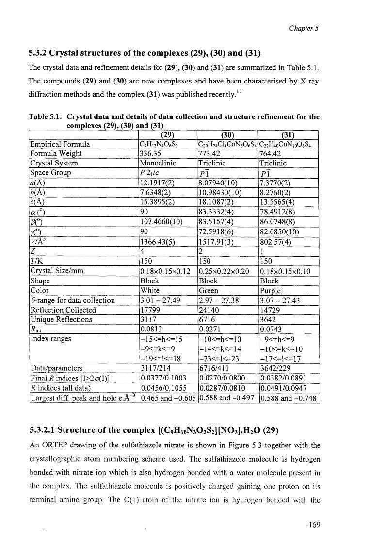
Chapter 5
5.3.2 Crystal structures of the complexes (29), (30) and (31)
The crystal data and refinement details for (29), (30) and (31) are summarized in Table 5.1.
The compounds (29) and (30) are new complexes and have been characterised by X-ray
diffraction methods and the complex (31) was published recently . 17
Table 5.1: Crystal data and details of data collection and structure refinement for thecomplexes (29), (30) and (31)
(29) (30) (31)Empirical Formula C9 H12N4 O6 S2 C2 0H2 4CI4 C0 N6O6 S4 C2 2H4 0 CUN10O8 S4
Formula Weight 336.35 773.42 764.42Crystal System Monoclinic Triclinic TriclinicSpace Group P 2\lc P \ P \a(A ) 12.1917(2) 8.07940(10) 7.3770(2)b( A) 7.6348(2) 10.98430(10) 8.2760(2)c(A ) 15.3895(2) 18.1087(2) 13.5565(4)a (° ) 90 83.3332(4) 78.4912(8)
A 0) 107.4660(10) 83.5157(4) 86.0748(8)
K°) 90 72.5918(6) 82.0850(10)v/ks 1366.43(5) 1517.91(3) 802.57(4)z 4 2 1
77K 150 150 150Crystal Size/mm 0.18x0.15x0.12 0.25x0.22x0.20 0.18x0.15x0.10Shape Block Block BlockColor White Green Purple(9-range for data collection 3.01 -2 7 .4 9 2 .9 7 -2 7 .3 8 3 .0 7 -2 7 .4 3Reflection Collected 17799 24140 14729Unique Reflections 3117 6716 3642Rint 0.0813 0.0271 0.0743Index ranges —15 <=h<=15 —1 0 <=h< = 1 0 -9<=h<=9
-9<=k<=9 —14<=k<=14 - 1 0 <=k< = 1 0
-19<=1<=18 —2 3 <=1<=2 3 —17<=1<=17Data/parameters 3117/214 6716/411 3642/229Final R indices [I>2a(I)] 0.0377/0.1003 0.0270/0.0800 0.0382/0.0891R indices (all data) 0.0456/0.1055 0.0287/0.0810 0.0491/0.0947Largest diff. peak and hole e.A - 3 0.465 and -0.605 0.588 and -0.497 0.588 and -0.748
5.3.2.1 Structure of the complex [(C9HioN3 0 2S2][N0 3 ].H20 (29)
An ORTEP drawing o f the sulfathiazole nitrate is shown in Figure 5.3 together with the
crystallographic atom numbering scheme used. The sulfathiazole molecule is hydrogen
bonded with nitrate ion which is also hydrogen bonded with a water molecule present in
the complex. The sulfathiazole molecule is positively charged gaining one proton on its
terminal amino group. The 0(1) atom of the nitrate ion is hydrogen bonded with the
169
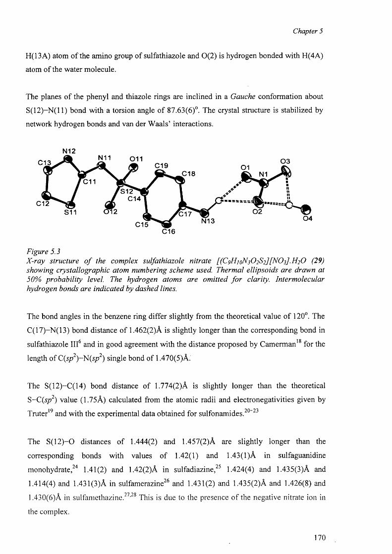
Chapter 5
H(13A) atom of the amino group of sulfathiazole and 0(2) is hydrogen bonded with H(4A)
atom o f the water molecule.
The planes o f the phenyl and thiazole rings are inclined in a Gauche conformation about
S (1 2 )-N (ll) bond with a torsion angle o f 87.63(6)°. The crystal structure is stabilized by
network hydrogen bonds and van der W aals’ interactions.
N12N11 011 0 3C13 C19 01
C18 N1C11
S12C14
C12
0 402S11 0 1 7
N13C15C16
Figure 5.3X-ray structure o f the complex sulfathiazole nitrate [(C 9H 1 qN3O2S2][NOz].H 2 0 (29) showing crystallographic atom numbering scheme used. Thermal ellipsoids are drawn at 50% probability level. The hydrogen atoms are omitted fo r clarity. Intermolecular hydrogen bonds are indicated by dashed lines.
The bond angles in the benzene ring differ slightly from the theoretical value o f 120°. The
C(17)-N(13) bond distance of 1.462(2)A is slightly longer than the corresponding bond in6 18 sulfathiazole III and in good agreement with the distance proposed by Camerman for the
length of C(sp2)~N(sp2) single bond of 1.470(5)A.
The S(12)-C(14) bond distance of 1.774(2)A is slightly longer than the theoretical
S-C(s/?2) value (1.75A) calculated from the atomic radii and electronegativities given by10 • 9 0 — 9 ^Truter and with the experimental data obtained for sulfonamides.
The S (1 2 )-0 distances o f 1.444(2) and 1.457(2)A are slightly longer than the
corresponding bonds with values o f 1.42(1) and 1.43(1)A in sulfaguanidine
monohydrate , 2 4 1.41(2) and 1.42(2)A in sulfadiazine , 2 5 1.424(4) and 1.435(3)A and
1.414(4) and 1.431 (3)A in sulfamerazine2 6 and 1.431(2) and 1.435(2)A and 1.426(8) and
1.430(6)A in sulfamethazine . 2 7 , 2 8 This is due to the presence o f the negative nitrate ion in
the complex.
170
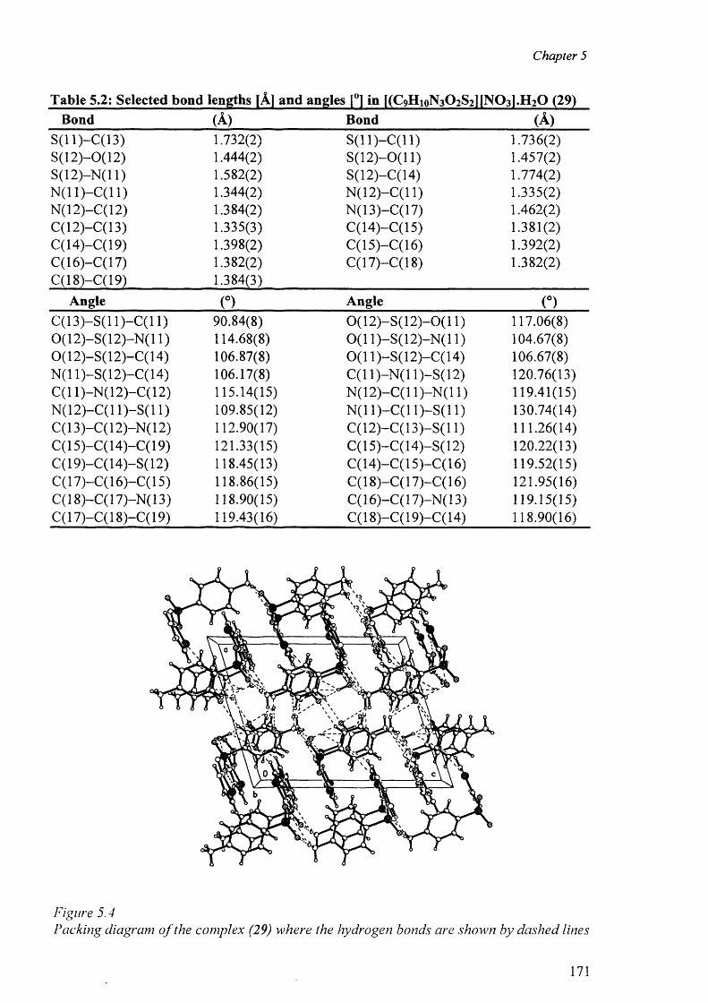
Chapter 5
Table 5.2: Selected bond lengths [A] and angles |°1 in KC^ipNaC^lfNOal.EbO (29)Bond (A) Bond (A )
S(11)-C(13) 1.732(2) S ( l l ) - C ( l l ) 1.736(2)S(12)—0(12) 1.444(2) S (1 2 )-0 (l 1) 1.457(2)S(12)-N (l 1) 1.582(2) S(12)-C(14) 1.774(2)N (11)—C(11) 1.344(2) N (12)-C (l 1) 1.335(2)N(12)-C(12) 1.384(2) N(13)-C(17) 1.462(2)C(12)-C(13) 1.335(3) C(14)-C(15) 1.381(2)C( 14)—C( 19) 1.398(2) C(15)-C(16) 1.392(2)C(16)-C(17) 1.382(2) C(17)-C(18) 1.382(2)C(18)-C(19) 1.384(3)
Angle (°) Angle (°)C (13)-S(l 1)-C (11) 90.84(8) 0 (1 2 )-S (1 2 )-0 (1 1) 117.06(8)0 ( 1 2 )—S(1 2 )—N ( 1 1 ) 114.68(8) 0 ( 1 1 )—S( 1 2 )—N( 1 1 ) 104.67(8)0 ( 12)-S ( 12)-C ( 14) 106.87(8) 0 (1 1)-S(12)-C(14) 106.67(8)N( 11 )-S ( 12)-C ( 14) 106.17(8) C ( ll) -N ( ll) -S (1 2 ) 120.76(13)C(11)—N (12)—C( 12) 115.14(15) N (12)-C (l 1)-N (11) 119.41(15)N (12)-C (l 1 )-S (11) 109.85(12) N (11)-C (11)-S (11) 130.74(14)C(13)-C(12)-N(12) 112.90(17) C (12)-C (13)-S(l 1) 111.26(14)C(15)-C( 14)-C ( 19) 121.33(15) C(15)-C(14)-S(12) 120.22(13)C(19)-C(14)-S(12) 118.45(13) C (14)-C (l 5 )-C (l 6 ) 119.52(15)C( 17)-C ( 16)-C ( 15) 118.86(15) C( 18)-C ( 17)-C ( 16) 121.95(16)C(18)-C(17)-N(13) 118.90(15) C( 16)-C ( 17)-N ( 13) 119.15(15)C( 17)-C( 18)-C( 19) 119.43(16) C( 18)-C ( 19)-C ( 14) 118.90(16)
Figure 5.4Packing diagram o f the complex (29) where the hydrogen bonds are shown by dashed lines
171
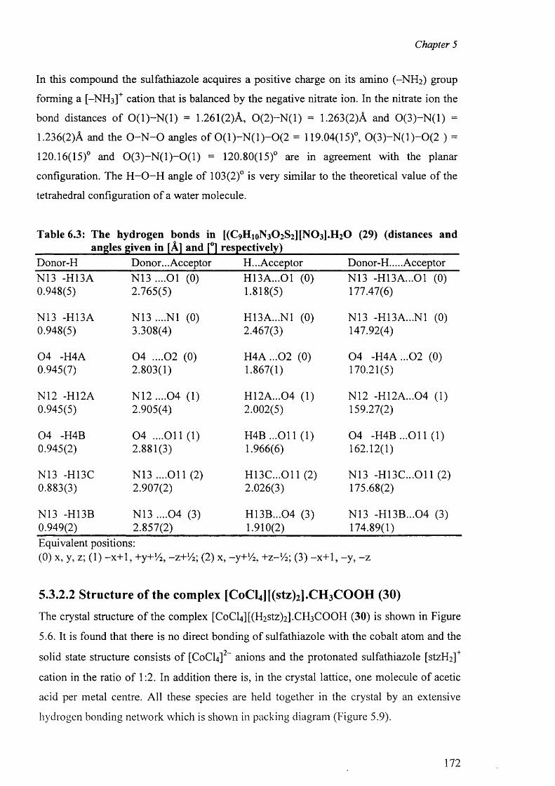
Chapter 5
In this compound the sulfathiazole acquires a positive charge on its amino (-N H 2 ) group
forming a [-NH 3 ]+ cation that is balanced by the negative nitrate ion. In the nitrate ion the
bond distances o f 0 ( 1 )—N (l) = 1.261(2)A, 0 (2 ) -N ( l) = 1.263(2)A and 0 (3 ) -N (l) =
1.236(2)A and the O -N -O angles o f 0 ( l ) - N ( l ) - 0 ( 2 = 119.04(15)°, 0 (3 ) -N ( l) -0 (2 ) =
120.16(15)° and 0 ( 3 ) - N ( l ) - 0 ( l ) = 120.80(15)° are in agreement with the planar
configuration. The H -O -H angle of 103(2)° is very similar to the theoretical value o f the
tetrahedral configuration o f a water molecule.
Table 6.3: The hydrogen bonds in [(C9 HioN3 C>2 S2 ][N 0 3 ].H 2 0 (29) (distances andangles given in [A] and [°] respectively)
Donor-H Donor... Acceptor H... Acceptor Donor-H.... AcceptorN13 -H13A N13 ....01 (0) H13A...01 (0) N13 -H13A...01 (0)0.948(5) 2.765(5) 1.818(5) 177.47(6)
N13 -H I3A N13 ....Nl (0) H13A...N1 (0) N13 -H13A...N1 (0)0.948(5) 3.308(4) 2.467(3) 147.92(4)
0 4 -H4A 0 4 ....02 (0) H 4A ...02 (0) 0 4 -H 4A ...02 (0)0.945(7) 2.803(1) 1.867(1) 170.21(5)
N12 -H12A N 1 2 ....0 4 (1) H12A...04 (1) N12 -H12A...04 (1)0.945(5) 2.905(4) 2.002(5) 159.27(2)
0 4 -H4B 0 4 ....O il (1) H 4B ...011 (1) 0 4 -H4B ...O il (1)0.945(2) 2.881(3) 1.966(6) 162.12(1)
N13 -H13C N13 ....O il (2) H 13C ...011 (2) N13 -H 13C ...011 (2)0.883(3) 2.907(2) 2.026(3) 175.68(2)
N13 -H13B N13 ....04 (3) H13B...04 (3) N13 -H 13B...04 (3)0.949(2) 2.857(2) 1.910(2) 174.89(1)Equivalent positions:(0 ) x, y, z; ( 1 ) —x+ 1 , +y+!/2, -z+'A; (2 ) x, -y+ lA, + z -lA; (3) - x + 1 , -y , - z
§.3.2.2 Structure of the complex [CoCl4][(stz)2].CH3COOH (30)
The crystal structure o f the complex [CoCl4 ][(H 2 Stz)2 ].CH 3 COOH (30) is shown in Figure
5.6. It is found that there is no direct bonding o f sulfathiazole with the cobalt atom and the
solid state structure consists o f [C0 CI4 ]2- anions and the protonated sulfathiazole [stzH2 ]+
cation in the ratio o f 1:2. In addition there is, in the crystal lattice, one molecule o f acetic
acid per metal centre. All these species are held together in the crystal by an extensive
hydrogen bonding network which is shown in packing diagram (Figure 5.9).
172
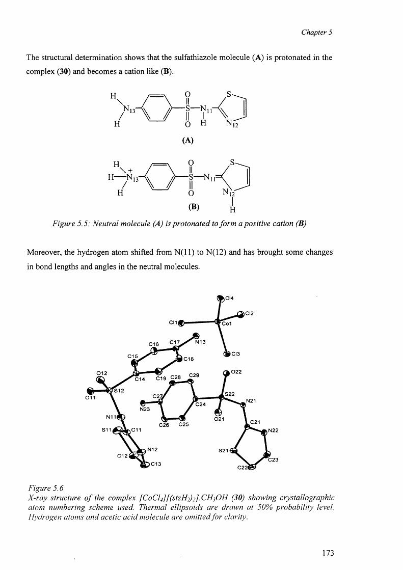
Chapter 5
The structural determination shows that the sulfathiazole molecule (A) is protonated in the
complex (30) and becomes a cation like (B).
H\
H
O
/"A /HrK.o H
(A)
O
H O N 12"
(®) H
Figure 5.5: Neutral molecule (A) is protonated to form a positive cation (B)
Moreover, the hydrogen atom shifted from N ( l l ) to N (1 2 ) and has brought some changes
in bond lengths and angles in the neutral molecules.
CI4
CI2
Co1
N13C17C16
CI3C15 C18
02 20 1 2 C19 C28 C29C14
S12 S22C27i0 1 1 N21C24N23
N11 021 C2 1C26 C25S11 C 1 1 N22
N12 S21C12
C23C13 C22<
Figure 5 . 6
X-ray structure o f the complex [C o C lf [ (stzFl2) 2]■ CH3 OH (30) showing crystallographic atom numbering scheme used. Thermal ellipsoids are drawn at 50% probability level. Hydrogen atoms and acetic acid molecule are omitted fo r clarity.
173
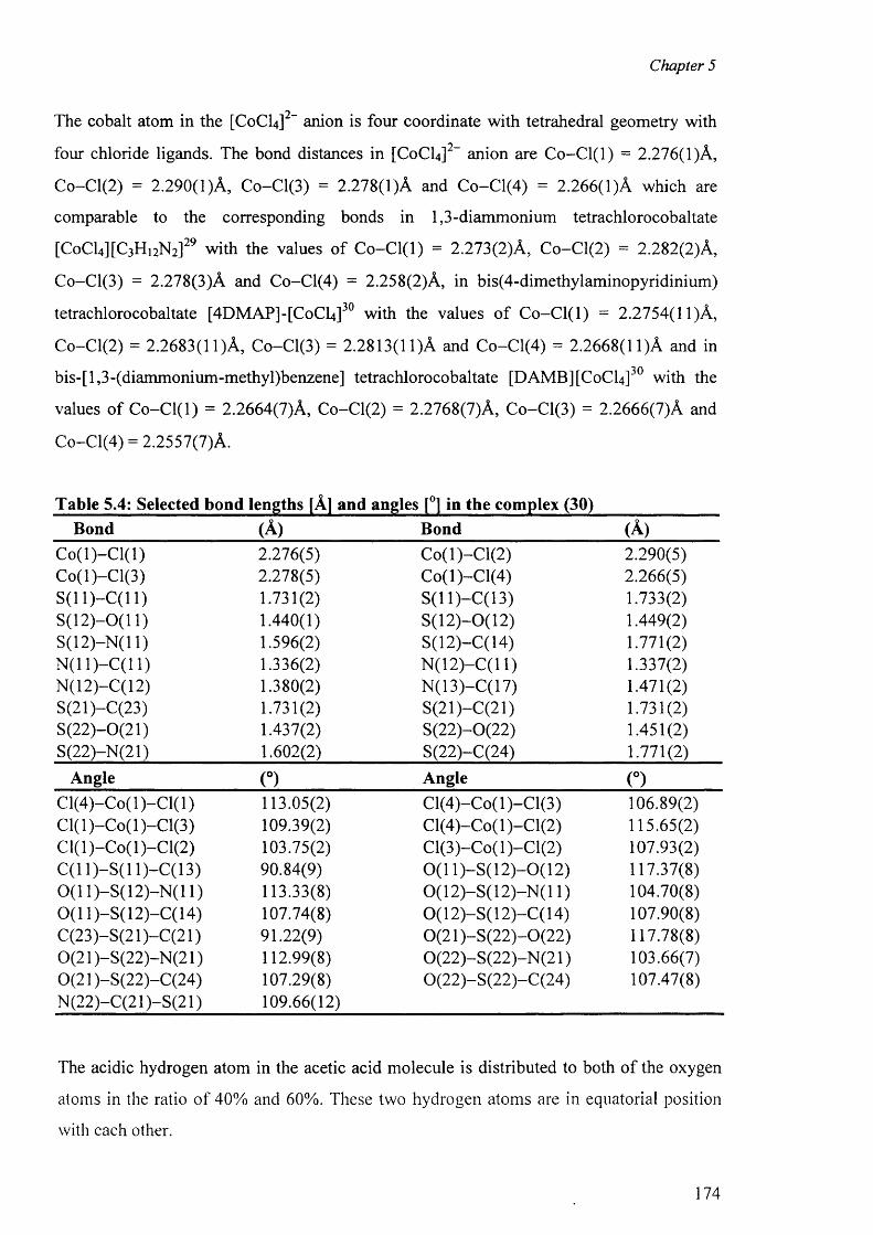
Chapter 5
The cobalt atom in the [C0 CI4 ] anion is four coordinate with tetrahedral geometry with
four chloride ligands. The bond distances in [CoCLJ2- anion are C o-C l(l) = 2.276(1)A,
C o-C l(2 ) = 2.290(1 )A , Co-Cl(3) = 2.278(1)A and C o-C l(4) = 2.266(1)A which are
comparable to the corresponding bonds in 1,3-diammonium tetrachlorocobaltate
[C0 CI4 HC3 H 12N 2 ] 2 9 with the values o f C o -C l(l) = 2.273(2)A, C o-C l(2 ) = 2.282(2)A,
Co-Cl(3) = 2.278(3)A and Co-Cl(4) = 2.258(2)A, in bis(4-dimethylaminopyridinium)
tetrachlorocobaltate [4DMAP]-[CoCU ] 3 0 with the values o f C o -C l(l) = 2.2754(11)A,
Co-Cl(2) = 2.2683(11)A , Co-Cl(3) = 2.2813(11)A and C o-C l(4) = 2.2668(11)A and in
bis-[l,3-(diammonium-methyl)benzene] tetrachlorocobaltate [DAMB][CoCl4 ] with the
values o f C o-C l(l) = 2.2664(7)A, Co-C l(2 ) = 2.2768(7)A, Co-Cl(3) = 2.2666(7)A and
Co-Cl(4) = 2.2557(7)A.
Table 5.4: Selected bond lengths [A] and angles [°] in the complex (30)Bond (A) Bond (A)
C o (l)-C l(l) 2.276(5) C o(l)-C l(2) 2.290(5)Co(l)-C l(3) 2.278(5) C o(l)-C l(4) 2.266(5)S ( l l ) - C ( l l ) 1.731(2) S(11)—C( 13) 1.733(2)S(12)—0(11) 1.440(1) S(12)-0(12) 1.449(2)S(12)-N (l 1) 1.596(2) S(12)-C(14) 1.771(2)N (11)—C(11) 1.336(2) N (12)-C (l 1) 1.337(2)N(12)-C(12) 1.380(2) N(13)-C(17) 1.471(2)S(21)-C(23) 1.731(2) S(21)-C(21) 1.731(2)S(22)-0(21) 1.437(2) S(22)-0(22) 1.451(2)S(22)-N(21) 1.602(2) S(22)-C(24) 1.771(2)
Angle (°) Angle (°)C l(4 )-C o(l)-C l(l) 113.05(2) C l(4)-C o(l)-C l(3) 106.89(2)C l(l)-C o(l)-C l(3 ) 109.39(2) Cl(4)-Co( 1 )-C l(2) 115.65(2)C l(l)-C o(l)-C l(2 ) 103.75(2) Cl(3)-Co( 1 )-C l(2) 107.93(2)C(11)—S( 11)—C(13) 90.84(9) 0 (1 1)-S (12)-0(12) 117.37(8)0 (1 1)-S(12)-N (11) 113.33(8) 0 ( 12)-S ( 12)-N (11) 104.70(8)0 (1 1)-S(12)-C(14) 107.74(8) 0(12)-S (12)-C (14) 107.90(8)C (23)-S(21 )-C (2 1) 91.22(9) 0(21 )-S (2 2 )-0 (2 2 ) 117.78(8)0(21 )-S (22)-N (21) 112.99(8) 0 (22 )-S (22 )-N (21) 103.66(7)0(21 )-S(22)-C(24) 107.29(8) 0(22)-S (22)-C (24) 107.47(8)N (22)-C (21 )-S (2 1) 109.66(12)
The acidic hydrogen atom in the acetic acid molecule is distributed to both o f the oxygen
atoms in the ratio o f 40% and 60%. These two hydrogen atoms are in equatorial position
with each other.
174
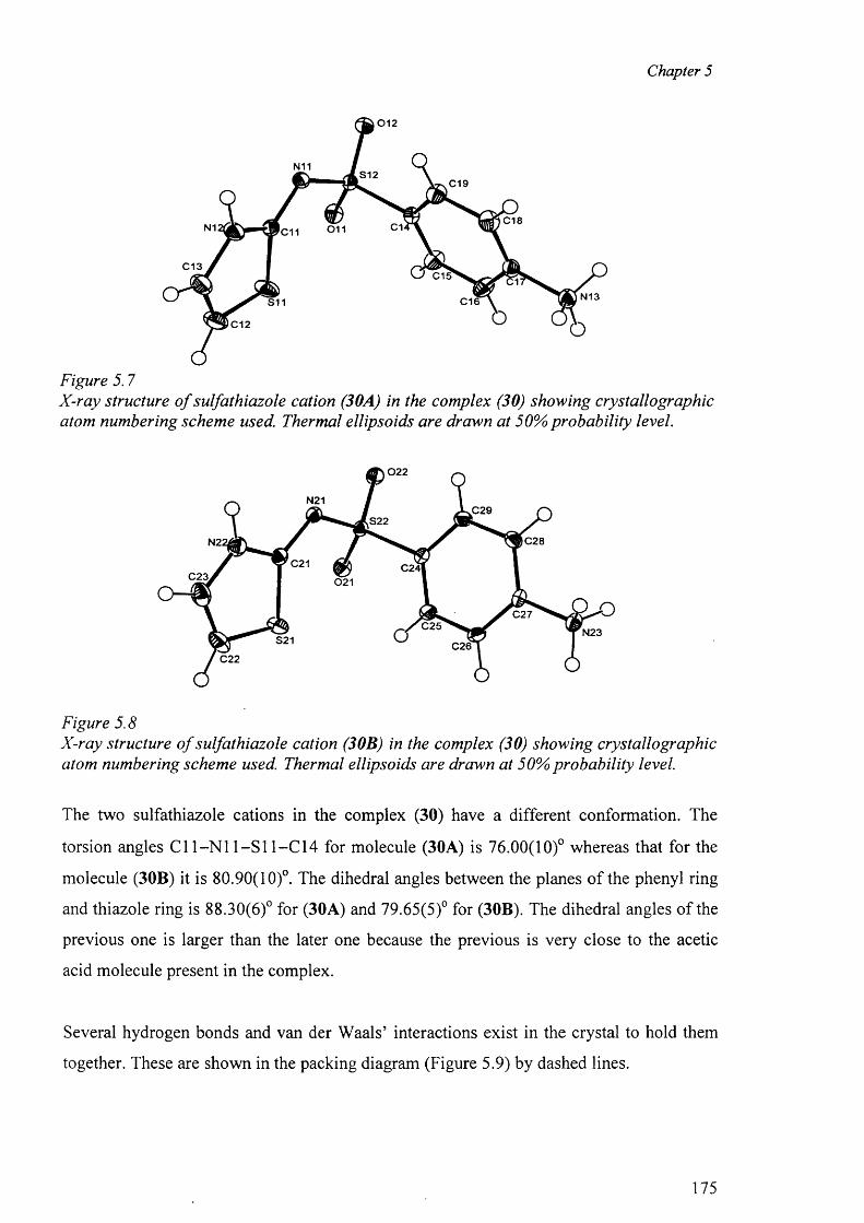
Chapter 5
0 1 2
N11S12
C19
C18C1N1 0 1 1'C11
C13C15
N13C16S11
C12
Figure 5.7X-ray structure o f sulfathiazole cation (30A) in the complex (30) showing crystallographic atom numbering scheme used. Thermal ellipsoids are drawn at 50% probability level.
0 2 2
N21kC29
,S22
C28N2;
C21 C24lC23. 021
C27C25 N23
S21 C26C22
Figure 5.8X-ray structure o f sulfathiazole cation (30B) in the complex (30) showing crystallographic atom numbering scheme used. Thermal ellipsoids are drawn at 50% probability level.
The two sulfathiazole cations in the complex (30) have a different conformation. The
torsion angles Cl 1 -N 1 1 —S 11-C14 for molecule (30A) is 76.00(10)° whereas that for the
molecule (30B) it is 80.90(10)°. The dihedral angles between the planes o f the phenyl ring
and thiazole ring is 88.30(6)° for (30A) and 79.65(5)° for (30B). The dihedral angles o f the
previous one is larger than the later one because the previous is very close to the acetic
acid molecule present in the complex.
Several hydrogen bonds and van der Waals’ interactions exist in the crystal to hold them
together. These are shown in the packing diagram (Figure 5.9) by dashed lines.
175
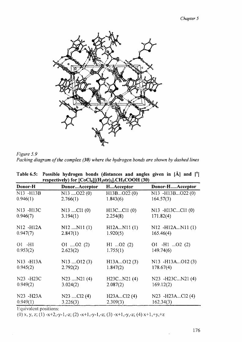
Chapter 5
Figure 5.9Packing diagram o f the complex (30) where the hydrogen bonds are shown by dashed lines
Table 6.5: Possible hydrogen bonds (distances and angles given in [A] and [°]respectively) for [CoCl4 |[(H 2 Stz)2 l.CH 3 COOH (30)
Donor-H Donor... Acceptor H...Acceptor Donor-H.....AcceptorN13 -H13B N13 ....022 (0) H13B...022 (0) N13 -H13B...0 2 2 (0 )0.946(1) 2.766(1) 1.843(6) 164.57(3)
N13 -H13C N13 ....Cll (0) H13C...C11 (0) N13 -H13C...C11 (0)0.946(7) 3.194(1) 2.254(8) 171.82(4)
N12 -H12A N12....N11 (1) H12A...N11 (1) N12 -H12A...N11 (1)0.947(7) 2.847(1) 1.920(5) 165.46(4)
01 -HI 0 1 . . . . 0 2 (2 ) HI ...02 (2) 01 -HI ...02 (2)0.953(2) 2.623(2) 1.755(1) 149.74(6)
N13 -H13A N13 ....012(3) H13A...012 (3) N13 -H13A...012 (3)0.945(2) 2.792(2) 1.847(2) 178.67(4)
N23 -H23C N23 ....N21 (4) H23C...N21 (4) N23 -H23C...N21 (4)0.949(2) 3.024(2) 2.087(2) 169.12(2)
N23 -H23A N23 ....C12 (4) H23A...C12 (4) N23 -H23A...C12 (4)0.949(1) 3.226(3) 2.309(3) 162.34(3)Equivalent positions:(0) x, y, z; (1) -x+2,-y-l,-z; (2) -x+l,-y-l,-z; (3) -x+l,-y,-z; (4) x+l,+y,+z
176
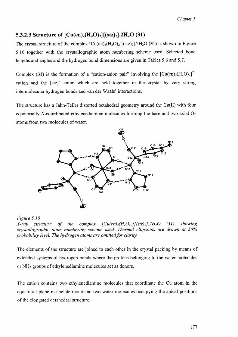
Chapter 5
5.3.2.3 Structure of [Cu(en)2(H20)2][(stz)2].2H20 (31)
The crystal structure o f the complex [Cu(en)2 (H 2 0 )2 ][(stz)2 ].2 H2 0 (31) is shown in Figure
5.10 together with the crystallographic atom numbering scheme used. Selected bond
lengths and angles and the hydrogen bond dimensions are given in Tables 5.6 and 5.7.
2+Complex (31) is the formation of a “cation-anion pair” involving the [Cu(en)2 (H2 0 )2 ]
cation and the [stz]~ anion which are held together in the crystal by very strong
intermolecular hydrogen bonds and van der W aals’ interactions.
The structure has a Jahn-Teller distorted octahedral geometry around the Cu(II) with four
equatorially TV-coordinated ethylenediamine molecules forming the base and two axial O-
atoms from two molecules o f water.02
N13C18 C17C2
C1 011 C19
C16N2N1 S12
0 1 2
Cu1 N110 1.011
o r S11
N12
c r C13 C12C2‘
Figure 5.10X-ray structure o f the complex [Cu(en)2(H20)2][(stz)2].2H20 (31) showingcrystallographic atom numbering scheme used. Thermal ellipsoids are drawn at 50% probability level. The hydrogen atoms are omitted fo r clarity.
The elements o f the structure are joined to each other in the crystal packing by means of
extended systems of hydrogen bonds where the protons belonging to the water molecules
or NH 2 groups o f ethylenediamine molecules act as donors.
The cation contains two ethylenediamine molecules that coordinate the Cu atom in the
equatorial plane in chelate mode and two water molecules occupying the apical positions
o f the elongated octahedral structure.
177
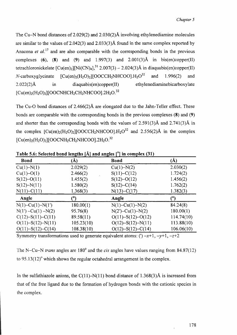
Chapter 5
The C u-N bond distances of 2.029(2) and 2.030(2)A involving ethylenediamine molecules
are similar to the values o f 2.042(3) and 2.033(3)A found in the same complex reported by
Anacona et a l} 1 and are also comparable with the corresponding bonds in the previous
complexes (6 ), (8 ) and (9) and 1.997(3) and 2.00 1 (3)A in bis(en)copper(II)
tetrachloronickelate [Cu(en)2 ][Ni(CN ) 4 ] , 31 2.007(3) - 2.024(3)A in diaquabis(en)copper(II)
A-carboxyglycinate [Cu(en)2 (H 2 0 )2 ][0 0 CCH 2NHC 0 0 ].H 2 0 3 2 and 1.996(2) and
2 .0 2 2 (2 ) A in diaquabis(en)copper(II) ethylenediaminebicarboxylate
[Cu(en)2 (H2 0 ) 2] [OOCNHCH2 CH2 NHCOO] .2H2 0 . 3 2
The Cu-0 bond distances o f 2.466(2)A are elongated due to the Jahn-Teller effect. These
bonds are comparable with the corresponding bonds in the previous complexes (8 ) and (9)
and shorter than the corresponding bonds with the values o f 2.591(3)A and 2.741(3)A in
the complex [Cu(en)2 (H 2 0 )2 ][0 0 CCH2 NHC 0 0 ].H2 0 3 2 and 2.556(2)A in the complex
[Cu(en)2 (H2 0 )2] [OOCNH2 CH2 NHCOO] .2H2 0 . 3 2
Table 5.6: Selected bond lengths [A] and angles [°] in complex (31)Bond (A) Bond (A)
C u (l)-N (l) 2.029(2) C u(l)-N (2) 2.030(2)C u (l)-0 (1 ) 2.466(2) S(11)-C(12) 1.724(2)S (12 )-0 (l 1) 1.455(2) S(12)-0(12) 1.456(2)S(12)-N (l 1) 1.580(2) S(12)-C(14) 1.762(2)N (11)-C(11) 1.368(3) N(13)-C(17) 1.382(3)
Angle (°) Angle (°)N (l)-C u (l)-N (T ) 180.00(1) N (l)-C u (l)-N (2 ) 84.24(8)N(T) -C u (l) -N (2) 95.76(8) N (2')-Cu( 1 )-N(2) 180.00(1)C(12)-S(l 1)-C (11) 89.58(11) 0 ( 1 1)-S(12)-0(12) 114.74(10)0 (1 1)-S(12)-N (11) 105.23(10) 0 (12)-S (12)-N (11) 113.88(10)0 (1 1)-S(12)-C(14) 108.38(10) 0 (1 2)-S (l 2 )-C (l 4) 106.06(10)Symmetry transformations used to generate equivalent atoms: (') -x+1, -y + 1, -z+2
The N -C u -N trans angles are 180° and the cis angles have values ranging from 84.87(12)
to 95.13(12)° which shows the regular octahedral arrangement in the complex.
In the sulfathiazole anions, the C ( 1 1 )—N ( 1 1 ) bond distance o f 1.368(3)A is increased from
that o f the free ligand due to the formation o f hydrogen bonds with the cationic species in
the complex.
178
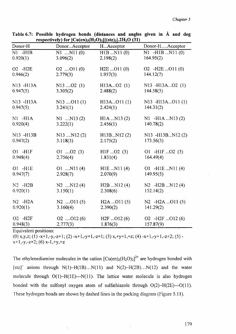
Chapter 5
Table 6.7: Possible hydrogen bonds (distances and angles given in A and degrespectively) for [Cu(en)2(H2Q)2lf(stz)2l.2H2Q (31)
Donor-H Donor... Acceptor H... Acceptor Donor-H.... AcceptorN1 -H1B N1 ....N il (0) H1B ...N il (0) N1 -H1B ...N il (0)0.920(1) 3.096(2) 2.198(2) 164.95(2)
0 2 -H2E 0 2 ....O il (0) H 2E ...011 (0) 0 2 -H 2E...011 (0)0.946(2) 2.779(3) 1.957(3) 144.12(7)
N13 -H13A N13 ....02 (1) H13A...02 (1) N13 -H13A...02 (1)0.947(5) 3.305(2) 2.488(2) 144.58(3)
N13 -H I3A N13 ....O il (1) H 13A ...011 (1) N13 -H 13A ...011 (1)0.947(5) 3.241(1) 2.424(1) 144.31(2)
N1 -H1A N1 ....N13 (2) H1A ...N13 (2) N1 -H1A...N13 (2)0.920(4) 3.222(1) 2.456(1) 140.78(2)
N13 -H13B N13 ....N12 (2) H13B...N12 (2) N13 -H13B...N12 (2)0.947(2) 3.118(3) 2.175(2) 173.56(3)
01 -H1F 01 ....02 (3) H 1F ...02 (3) 01 -H1F ...02 (3)0.948(4) 2.756(4) 1.831(4) 164.49(4)
01 -HIE 01 ....N il (4) H IE ...N il (4) 01 -H IE ...N il (4)0.947(7) 2.928(7) 2.070(9) 149.95(5)
N2 -H2B N2 ....N12 (4) H2B ...N12 (4) N2 -H2B ...N12 (4)0.920(1) 3.150(1) 2.308(6) 152.14(2)
N2 -H2A N2 ....O il (5) H 2A ...011 (5) N2 -H 2A ...O H (5)0.920(1) 3.160(4) 2.390(2) 141.29(2)
0 2 -H2F 0 2 ....0 1 2 (6 ) H2F ...012 (6 ) 0 2 -H2F ...012 (6 )0.948(3) 2.777(3) 1.876(3) 157.87(9)Equivalent positions:(0) x,y,z; (1) -x+ l,-y ,-z+ l; (2) -x+ l,-y+ l,-z+ l; (3) x,+y+l,+z; (4) -x+l,-y+l,-z+2; (5) - x+l,-y,-z+ 2 ; (6 ) x-l,+y,+z
The ethylenediamine molecules in the cation [Cu(en)2 (H2 0 )2 ] are hydrogen bonded with
[stz]~ anions through N (1)-H (1B )...N (11) and N (2)-H (2B)...N (12) and the water
molecule through 0(1)-H (1E )—N (ll) . The lattice water molecule is also hydrogen
bonded with the sulfonyl oxygen atom of sulfathiazole through 0(2)-H (2E )—0(11).
These hydrogen bonds are shown by dashed lines in the packing diagram (Figure 5.11).
179
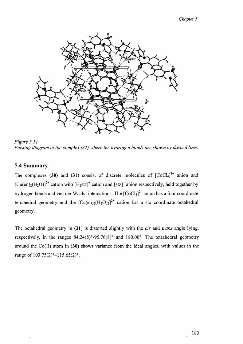
Chapter 5
Figure 5.11Packing diagram o f the complex (31) where the hydrogen bonds are shown by dashed lines
5.4 Summary9 —
The complexes (30) and (31) consist o f discrete molecules o f [C0 CI4 ] anion and
[Cu(en)2 (H2 0 )]2+ cation with [H2 Stz]+ cation and [stz]~ anion respectively, held together by9—hydrogen bonds and van der Waals’ interactions. The [C0 CI4 ] anion has a four coordinate
94-tetrahedral geometry and the [Cu(en)2 (H2 0 )2 ] cation has a six coordinate octahedral
geometry.
The octahedral geometry in (31) is distorted slightly with the cis and trans angle lying,
respectively, in the ranges 84.24(8)°-95.76(8)° and 180.00°. The tetrahedral geometry
around the Co(II) atom in (30) shows variance from the ideal angles, with values in the
range of 103.75(2)°-! 15.65(2)°.
180
Chapter 5
References
1. M. Kuhnert-Brandstaetter; Thermomicroscopy in the Analysis o f Pharmaceuticals, Pergamon Press, Oxford, 1974
2. S. Yang and J. K. Guillory; J. Pharm. Sci., 1972, 61, 26
3. A. L. Bingham, D. S. Hughes, M. B. Hursthouse, R. W. Lancaster, S. Tavener and T. L. Threlfall; Chem. Commun., 2001, 7, 603
4. K. -I. Shirotani, E. Suzuki and K. Sekiguchi; Chem. Pharm. Bull., 1983, 31, 2085
5. G.J. Kruger and G. Gafner; Acta Cryst., 1971, B27, 326
6 . G.J. Kruger and G. Gaftier; Acta Cryst., 1972, B28, 272
7. D. S. Hughes, M. B. Hursthouse, T. Threlfall and S. Tavener; Acta Cryst., 1999, C55,1831
8 . F. C. Chan, J. Anwar, R. Cemik, P. Barnes and R. M. Wilson; J. Appl. Cryst., 1999, 3 2 ,436
9. M. R. Caira, V. J. Griffith, L. R. Nassimbeni, R. Luigi and B. V. Oudtshoom; J. Inclus. Phen. Mole. Recog. Chem., 1994, 17, 187
10. J. Casanova, G. Aluzet, J. Borras, J. Timoneda, S. Garcia-Granda and I. Candano- Gonzalez; J. Inorg. Biochem., 1994, 56, 65
11. J. Casanova, G. Aluzet, S. Ferrer, J. Borras, S. Garcia-Granda and E. Perez-Carreno; J. Inorg. Biochem., 1993, 51, 689
12. J. Casanova, G. Aluzet, J. Borras, J. Latorre, M. Sanau and S. Garcia-Granda; J. Inorg. Biochem., 1995, 60, 219
13. J. Casanova, G. Aluzet, J. Latorre, M. Sanau and J. Borras; Inorg. Chem., 1997, 36, 2052
14. W. Henderson, L.J. McCaffrey, M.B. Dinger and B.K. Nicholson; Polyhedron, 1998, 17,3137
15. J. Casanova, G. Aluzet, J. Borras and O. Carugo; J. Chem. Soc. Dalton Trans., 1996, 2239
16. G. ALzuet, J. A. Real, R. Santiago-Garcia and S. Garcia-Granda; Inorg. Chem., 2001, 4 0 ,2420
17. J.R. Anacona, N. Ramos, G.D.D. Delgado and E. M. Roque; J. Coord. Chem., 2002, 55, 901
18. A. Camerman; Canad. J. Chem., 1970, 48, 179
19. M. R. Truler; J. Chem. Soc., 1962, 3400
181
Chapter 5
20. B. H. O ’Conner B.H and E. N. Maslen; Acta Cryst., 1965, 18, 363
21. B. H. O ’Conner and E. N. Maslen; Acta Cryst., 1967, 22, 134
22. M. Alkaume and J. Decap; Acta Cryst., 1965,18, 731
23. M. Alkaume and J. Decap; Acta Cryst., 1965,19, 394
24. C. H. Koo, H. S. Kim, W. C. Shin and C. H. Choe; J. Korean Chem. Soc., 1974 ,18, 97
25. H. S. Shin, G. S. Ihn, H. S. Kim and C. H. Koo; J. Korean Chem. Soc., 1974 ,18, 329
26. M. R. Caira and R. Mohamed; Acta Cryst., 1992, B48, 492
27. A. K. Basak, S. K. Mazumdar and S. Chaudhuri; Acta Crsyt., 1983, C39, 492
28. R. K. Tiwari, M. Haridas and T. P. Singh; Acta Crsyt., 1984, C40, 655
29. G. Ning, L. Yong_hua, Z. Guang-Fu and X. Shi-Quan; Acta Crsyt., 1992, C48, 542
32. S. Haddad, A. Vij and R. D. Willett; J. Chem.Crsyt., 2003, 33, 245
31. J. Lokaj, K. Gyerova, A. Sopkova, J. Sivy, V. Kettman and V. Vrabel; Acta Cryst., 1991, C47, 2447
32. L. A. Kovbasyuk, I. O. Fritsky, V. N. Kokozay and T. S. Iskenderov; Polyhedron, 1997,16, 1723 and the references therein.
182
SYNTHESIS AND STRUCTURAL STUDIES OF ZINC COMPLEX OF
SULFADIMETHOXINE WITHAMMONIA
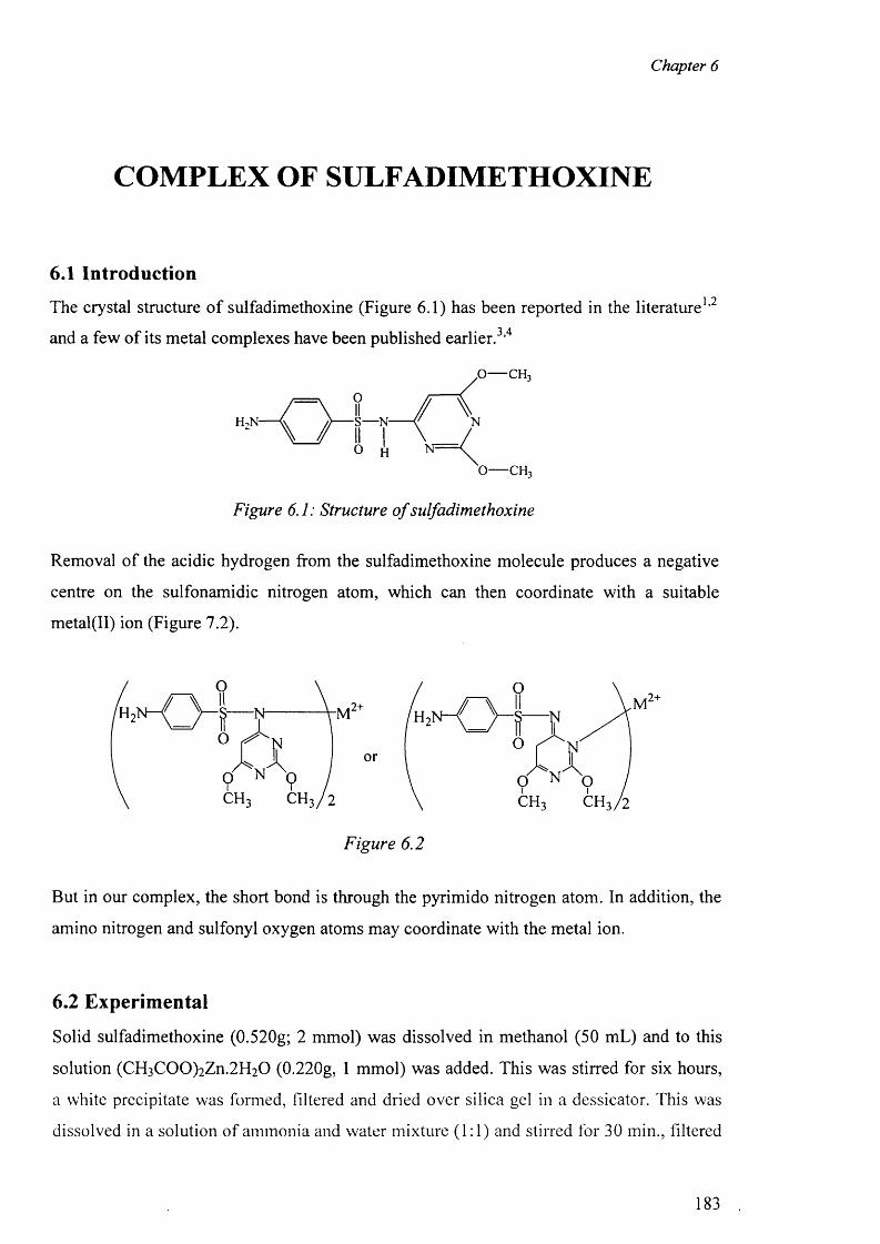
Chapter 6
COMPLEX OF SULFADIMETHOXINE
6.1 Introduction
The crystal structure o f sulfadimethoxine (Figure 6.1) has been reported in the literature1 , 2
and a few o f its metal complexes have been published earlier . 3 , 4
.o — c h 3
o — c h 3
Figure 6.1: Structure o f sulfadimethoxine
Removal o f the acidic hydrogen from the sulfadimethoxine molecule produces a negative
centre on the sulfonamidic nitrogen atom, which can then coordinate with a suitable
metal(II) ion (Figure 7.2).
But in our complex, the short bond is through the pyrimido nitrogen atom. In addition, the
amino nitrogen and sulfonyl oxygen atoms may coordinate with the metal ion.
6.2 Experimental
Solid sulfadimethoxine (0.520g; 2 mmol) was dissolved in methanol (50 mL) and to this
solution (CH3 C 0 0 )2 Zn.2 H 2 0 (0.220g, 1 mmol) was added. This was stirred for six hours,
a white precipitate was formed, filtered and dried over silica gel in a dessicator. This was
dissolved in a solution of ammonia and water mixture ( 1 : 1 ) and stirred for 30 min., filtered
Figure 6.2
183
Chapter 6
and the filtrate was kept for crystallisation. After a week, two types o f crystals were
obtained, one was block and the other was needle like crystals. The block type crystal was
the ligand molecule sulfadimethoxine (32) and the needle like crystal was the zinc complex
[Zn(sdm)2 (NH 3 )2 ].2H20 (33). IR for (32): (KBr disc), cm-1, 3456(s), 3125(s), 2956(sh),
1625(vs), 1598, 1570, 1507(vs), 1465(sh), 1408, 1351, 1306(m), 1259(vs), 1190(sh),
1155(vs), 1125(s), 1093(s), 1005(s), 968(vs), 881(vw), 834, 805, 589, 535(vs). 'H NMR
(DMSO-<4): 6 11.18s [1H, N (11),] 6.11 [2H, NH2], 5.97 [1H, C(14)], 6.63d [2H,
C(17)/C(19), J = 8 .6 ], 7.60d [2H, C(16)/C(20), J = 8 .6 ], 3.79s [C(l 11)] and 3.78s [C(l 12)],
13C NMR (DMSO-^k): 5 160.1 [C (ll)], 164.3 [C(12)], 171.6 [C(13)], 84.2 [C(14)], 124.1
[C (l5)], 129.3 [C(16)/C(20)], 112.4 [C(17)/C(19)], 153.3 [C(18)], 54.4 [C ( ll l) ] and 53.6
[C(l 12)]. IR fo r (33): (KBr disc), cm-1, 3461(s), 1629(vs), 1599, 1582, 1501(vs), 1460(sh),
1404, 1373, 1224(vs), 1152(vs), 1122(s), 1096(s), 1014(s), 876(vw), 830, 805, 702(vs),
574, 554(vs). lU NM R (DMSO-</6): 5 5.78 [2 H, NH2], 5.71 [1H, C(14)], 6.57d [2 H,
C(17)/C(19), J = 10.6], 7.45d [2H, C(16)/C(20), J = 8 .6 ], 3.71s [C ( ll l) ] and 3.54
[C(l 12)], 13C NMR (DMSO-^6): 6 163.6 [C(l 1)], 166.4 [C(12)], 171.6 [C(13)], 84.6
[C(14)], 128.3 [C (l5)], 128.9 [C(16)/C(20)], 112.3 [C (l7 )/C (l9)], 151.4 [C(18)], 53.8
[C ( ll l) ] and 53.3 [C(l 12)].
6.3 Results and discussion for the compounds (32) and (33)
6.3.1 IR Spectra
The absorption bands at 3461 cm - 1 suggest the presence o f water molecules and NH 2
groups in the complex. This peak is very broad and split because o f the involvement o f
water molecules and NH 2 group in hydrogen bonding to various extents.
The peak at 3125 cm - 1 for N -H of the sulfonamidic group is not present in the IR spectra
o f the complex, and this confirms the deprotonation o f the - S 0 2 N H - moiety. The
characteristic v(C -C ) (ring) and v(C -N ) absorptions in the IR spectra appear at 1570 cm ' 1
and 1351 cm ' 1 respectively in the sulfadimethoxine molecule. These peaks are observed at
1582 cm ' 1 and 1373 cm"1, respectively, in the complex.
The scissoring vibrations for the -N H 2 group appear at 1629 and the peaks at 1570-1598
cm ' 1 are due to the phenyl rings. The peaks at 1306 and 1259 cm ' 1 are assigned to c«(S02),
and that at 1125 cm ' 1 to 5,y(S 0 2). The (CC) absorption band overlaps with the tfs(S02)
184
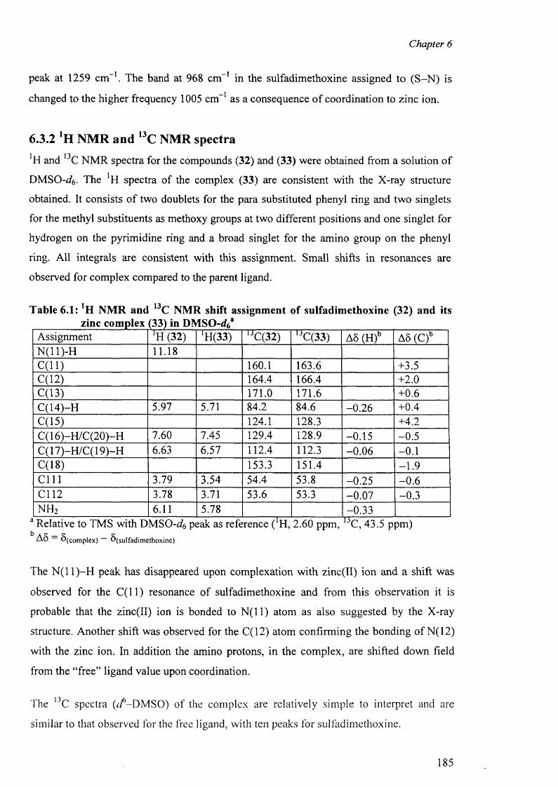
Chapter 6
peak at 1259 cm l. The band at 968 cm 1 in the sulfadimethoxine assigned to (S -N ) is
changed to the higher frequency 1005 cm - 1 as a consequence o f coordination to zinc ion.
6.3.2 *H NMR and l3C NMR spectra1 13H and C NMR spectra for the compounds (32) and (33) were obtained from a solution o f
DMSO-^6 - The !H spectra of the complex (33) are consistent with the X-ray structure
obtained. It consists o f two doublets for the para substituted phenyl ring and two singlets
for the methyl substituents as methoxy groups at two different positions and one singlet for
hydrogen on the pyrimidine ring and a broad singlet for the amino group on the phenyl
ring. All integrals are consistent with this assignment. Small shifts in resonances are
observed for complex compared to the parent ligand.
Table 6.1: NMR and 13C NMR shift assignment of sulfadimethoxine (32) and itszinc complex (33) in DMSO-dk*
Assignment *H (32) *H(33) 13C(32) ljC(33) A8 (H)b A5 (C)bN(11)-H 11.18C (ll) 160.1 163.6 +3.5C(12) 164.4 166.4 + 2 . 0
C(13) 171.0 171.6 + 0 . 6
C(14)-H 5.97 5.71 84.2 84.6 -0 .26 +0.4C(15) 124.1 128.3 +4.2C( 16)-H /C (20)-H 7.60 7.45 129.4 128.9 -0.15 -0 .5C(17)-H/C(19)-H 6.63 6.57 112.4 112.3 -0 .06 - 0 . 1
C(18) 153.3 151.4 -1 .9c m 3.79 3.54 54.4 53.8 -0.25 - 0 . 6
Cl 12 3.78 3.71 53.6 53.3 -0 .07 -0 .3n h 2 6 . 1 1 5.78 -0.33
a Relative to TMS with D M SO -^ peak as reference ( H, 2.60 ppm, C, 43.5 ppm)A 8 8 ( Complex) ^ (su lfa d im e th o x in e )
The N ( l l ) -H peak has disappeared upon complexation with zinc(II) ion and a shift was
observed for the C ( l l ) resonance of sulfadimethoxine and from this observation it is
probable that the zinc(II) ion is bonded to N ( l l ) atom as also suggested by the X-ray
structure. Another shift was observed for the C(12) atom confirming the bonding o f N(12)
with the zinc ion. In addition the amino protons, in the complex, are shifted down field
from the “free” ligand value upon coordination.
The 13C spectra (</-DM SO) of the complex are relatively simple to interpret and are
similar to that observed for the free ligand, with ten peaks for sulfadimethoxine.
185
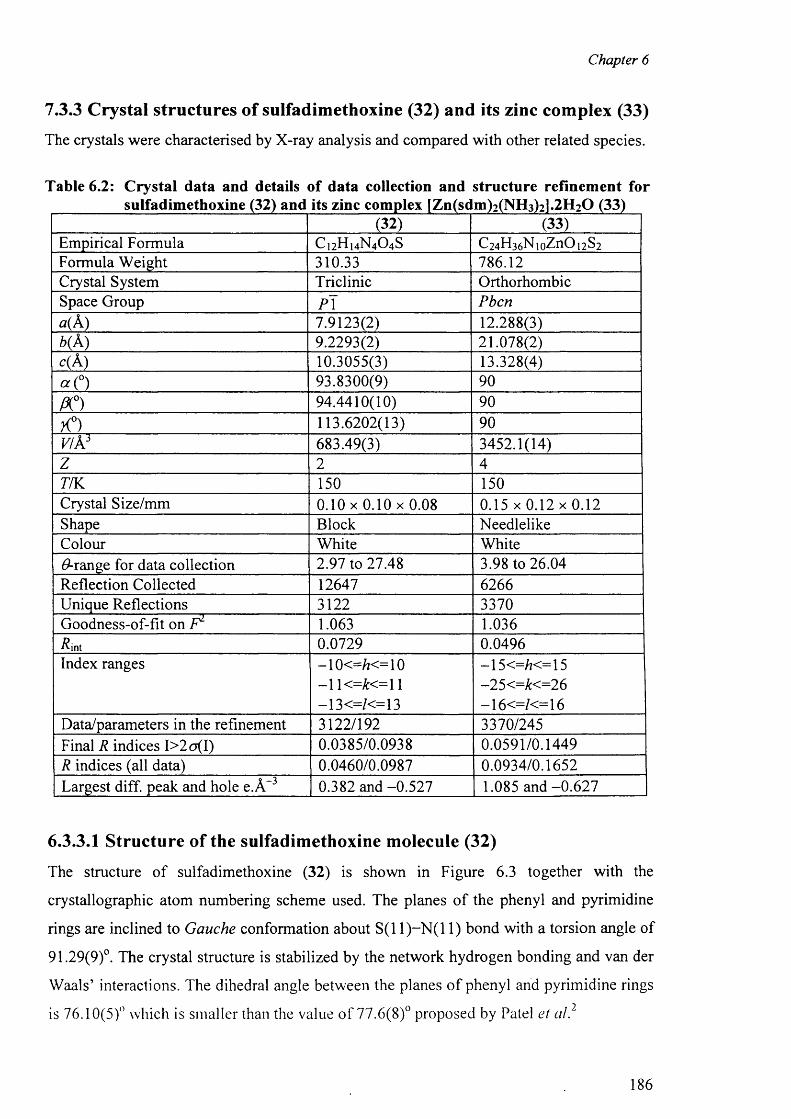
Chapter 6
7.3.3 Crystal structures of sulfadimethoxine (32) and its zinc complex (33)
The crystals were characterised by X-ray analysis and compared with other related species.
Table 6.2: Crystal data and details o f data collection and structure refinement forsulfadimethoxine (32) and its zinc complex [Zn(sdm)2(NH3)2].2H20 (33)
(32) (33)Empirical Formula C 1 2H 1 4N 4 O4 S C2 4H3 6N1oZnOi2S2Formula Weight 310.33 786.12Crystal System Triclinic OrthorhombicSpace Group P \ Pbcna( A) 7.9123(2) 12.288(3)6(A) 9.2293(2) 21.078(2)c (k ) 10.3055(3) 13.328(4)« (°) 93.8300(9) 90
A °) 94.4410(10) 90
X°) 113.6202(13) 90VI k 3 683.49(3) 3452.1(14)z 2 477K 150 150Crystal Size/mm 0 .1 0 x 0 .1 0 x 0 .0 8 0 .1 5 x 0 .1 2 x 0 .1 2Shape Block NeedlelikeColour White WhiteGrange for data collection 2.97 to 27.48 3.98 to 26.04Reflection Collected 12647 6266Unique Reflections 3122 3370Goodness-of-fit on F2 1.063 1.036Rint 0.0729 0.0496Index ranges - 1 0 < = £ < = 1 0 —15 <=/?<= 15
- 1 1 < = £ < = 1 1 -25<=£<=26—13<=/<=l 3 —16<=/<=l 6
Data/parameters in the refinement 3122/192 3370/245Final R indices I>2o(I) 0.0385/0.0938 0.0591/0.1449R indices (all data) 0.0460/0.0987 0.0934/0.1652Largest diff. peak and hole e.A -3 0.382 and -0.527 1.085 and -0 .627
6.3.3.1 Structure of the sulfadimethoxine molecule (32)
The structure o f sulfadimethoxine (32) is shown in Figure 6.3 together with the
crystallographic atom numbering scheme used. The planes o f the phenyl and pyrimidine
rings are inclined to Gauche conformation about S (11)-N (11) bond with a torsion angle of
91.29(9)°. The crystal structure is stabilized by the network hydrogen bonding and van der
Waals’ interactions. The dihedral angle between the planes o f phenyl and pyrimidine rings
is 76.10(5)° which is smaller than the value o f 77.6(8)° proposed by Patel et al.2
186
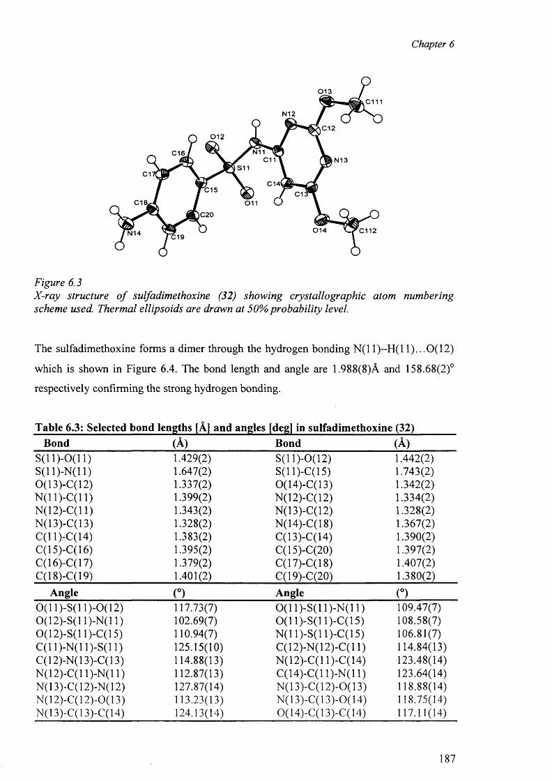
Chapter 6
0 1 3c m
N12
C120 1 2
N11C16C11 N13
S11C1
C14lC15 C1
C18. 0 1 1
|C20
0 1 4 C112N14 C19
Figure 6.3X-ray structure o f sulfadimethoxine (32) showing crystallographic atom numbering scheme used. Thermal ellipsoids are drawn at 50% probability level.
The sulfadimethoxine forms a dimer through the hydrogen bonding N (11)-H (11)...0 (12)
which is shown in Figure 6.4. The bond length and angle are 1.988(8)A and 158.68(2)°
respectively confirming the strong hydrogen bonding.
Table 6.3: Selected bond lengths [A] and angles [deg] in sulfadimethoxine (32)Bond (A) Bond (A)
S (ll)-0 (1 1 ) 1.429(2) S(11)-0(12) 1.442(2)S(11)-N(11) 1.647(2) S(11 )-C( 15) 1.743(2)0(13)-C(12) 1.337(2) 0(14)-C(13) 1.342(2)N(11)-C(11) 1.399(2) N(12)-C(12) 1.334(2)N(12)-C(l 1) 1.343(2) N(13)-C(12) 1.328(2)N(13)-C(13) 1.328(2) N(14)-C(18) 1.367(2)C(11)-C(14) 1.383(2) C(13)-C(14) 1.390(2)C(15)-C(16) 1.395(2) C(15)-C(20) 1.397(2)C(16)-C(17) 1.379(2) C(17)-C(18) 1.407(2)C(18)-C(19) 1.401(2) C(19)-C(20) 1.380(2)
Angle (°) Angle (°)0 (1 1)-S(11 )-0 ( 12) 117.73(7) 0 ( 1 1)-S(11)-N(11) 109.47(7)0(12)-S(11)-N(11) 102.69(7) 0 ( 1 1)-S(11)-C(15) 108.58(7)0(12)-S(11)-C(15) 110.94(7) N(11)-S(11)-C(15) 106.81(7)C(11)-N(11)-S(11) 125.15(10) C(12)-N(12)-C(l 1) 114.84(13)C(12)-N(13)-C(13) 114.88(13) N(12)-C(l 1)-C(14) 123.48(14)N(12)-C(l 1)-N(11) 112.87(13) C(14)-C(l 1)-N(11) 123.64(14)N(13)-C(12)-N(12) 127.87(14) N (13)-C(12)-0(13) 118.88(14)N(12)-C(12)-0(13) 113.23(13) N(13)-C(13)-0(14) 118.75(14)N(13)-C(13)-C(14) 124.13(14) 0(14)-C(13)-C(14) 117.11(14)
187
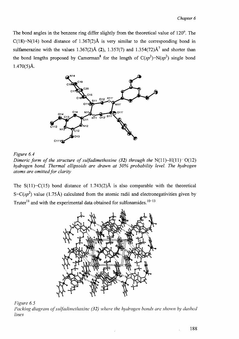
Chapter 6
The bond angles in the benzene ring differ slightly from the theoretical value o f 120°. The
C(18)-N(14) bond distance of 1.367(2)A is very similar to the corresponding bond in
sulfamerazine with the values 1.367(2)A (2), 1.357(7) and 1.354(72)A7 and shorter than8 2 2 the bond lengths proposed by Camerman for the length o f C(sp ) -N (sp ) single bond
1.470(5)A.
N 14
,C19C1
C 2 0C1
C 15C16* 0 1 2S11
011I N11'
C 1 4 N1 o i r0 1 4
H11 0 1 2 1 S 1 1 'C13C11
N12C112N1 C12
0 1 3
C111|
Figure 6.4Dimeric form o f the structure o f sulfadimethoxine (32) through the N ( l l ) - H ( l l ) 0(12) hydrogen bond. Thermal ellipsoids are drawn at 50% probability level. The hydrogen atoms are omitted fo r clarity
The S( 11)—C(15) bond distance of 1.743(2)A is also comparable with the theoretical
S-C (sp 2) value (1.75A) calculated from the atomic radii and electronegativities given by
Truter11 and with the experimental data obtained for sulfonamides . 1 0 - 1 3
Figure 6.5Packing diagram o f sulfadimethoxine (32) where the hydrogen bonds are shown by dashed lines
188
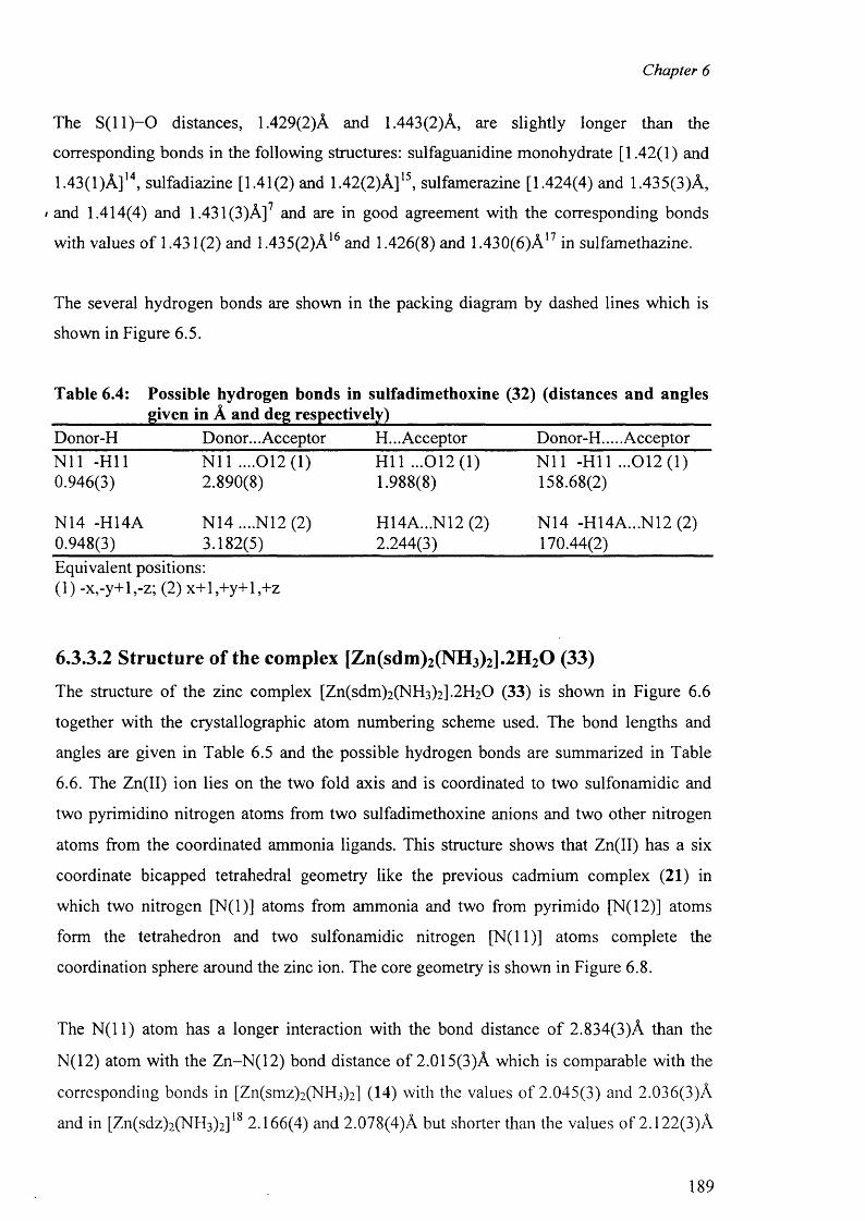
Chapter 6
The S ( l l ) - 0 distances, 1.429(2)A and 1.443 (2)A, are slightly longer than the
corresponding bonds in the following structures: sulfaguanidine monohydrate [1.42(1) and
1.43(1 )A ]14, sulfadiazine [1.41(2) and 1.42(2)A]15, sulfamerazine [1.424(4) and 1.435(3)A,
• and 1.414(4) and 1.431(3)A]7 and are in good agreement with the corresponding bonds
with values o f 1.431(2) and 1.435(2)A16 and 1.426(8) and 1.430(6)A17 in sulfamethazine.
The several hydrogen bonds are shown in the packing diagram by dashed lines which is
shown in Figure 6.5.
Table 6.4: Possible hydrogen bonds in sulfadimethoxine (32) (distances and angles____________given in A and deg respectively)________________________________________Donor-H Donor... Acceptor H... Acceptor Donor-H.....AcceptorN i l -HI 1 N i l ....012(1) H ll ...012 (1 ) N i l -H ll ...0 1 2 (1 )0.946(3) 2.890(8) 1.988(8) 158.68(2)
N14 -H14A N14 ....N12 (2) H14A...N12 (2) N14 -H14A...N12 (2)0.948(3) 3.182(5) 2.244(3) 170.44(2)Equivalent positions:( 1 ) -x,-y+l,-z; (2 ) x+ l,+ y+ l,+ z
6.3.3.2 Structure of the complex [Zn(sdm)2(NH3)2].2H20 (33)
The structure o f the zinc complex [Zn(sdm)2 (NH 3 )2 ].2 H2 0 (33) is shown in Figure 6 . 6
together with the crystallographic atom numbering scheme used. The bond lengths and
angles are given in Table 6.5 and the possible hydrogen bonds are summarized in Table
6 .6 . The Zn(II) ion lies on the two fold axis and is coordinated to two sulfonamidic and
two pyrimidino nitrogen atoms from two sulfadimethoxine anions and two other nitrogen
atoms from the coordinated ammonia ligands. This structure shows that Zn(II) has a six
coordinate bicapped tetrahedral geometry like the previous cadmium complex (21) in
which two nitrogen [N (l)] atoms from ammonia and two from pyrimido [N(12)] atoms
form the tetrahedron and two sulfonamidic nitrogen [N( 11)] atoms complete the
coordination sphere around the zinc ion. The core geometry is shown in Figure 6 .8 .
The N ( l l ) atom has a longer interaction with the bond distance o f 2.834(3)A than the
N (1 2 ) atom with the Z n -N ( 1 2 ) bond distance of 2.015(3)A which is comparable with the
corresponding bonds in [Zn(smz)2(NH 3 )2] (14) with the values o f 2.045(3) and 2.036(3)A
and in [Zn(sdz)2(NH 3 )2 ] 18 2.166(4) and 2.078(4)A but shorter than the values o f 2.122(3)A
189
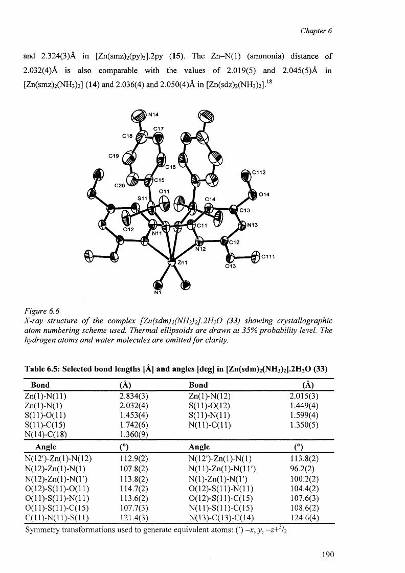
Chapter 6
and 2.324(3)A in [Zn(smz)2 (py)2 ].2 py (15). The Z n -N (l) (ammonia) distance o f
2.032(4)A is also comparable with the values o f 2.019(5) and 2.045(5)A in
[Zn(smz)2 (NH 3 )2] (14) and 2.036(4) and 2.050(4)A in [Zn(sdz)2 (NH 3 )2 ] . 18
N1
Figure 6 . 6
X-ray structure o f the complex [Zn(sdm)2 (N 1 3) 2]. 2 H 2 O (33) showing crystallographic atom numbering scheme used. Thermal ellipsoids are drawn at 35% probability level. The hydrogen atoms and water molecules are omitted fo r clarity.
Table 6.5: Selected bond lengths [A] and angles [deg] in [Zn(sdm)2 (NH 3 )2 ].2 H 2 0 (33)
Bond (A) Bond (A)Zn(l)-N (l 1) 2.834(3) Zn(l)-N(12) 2.015(3)Z n(l)-N (l) 2.032(4) S(11)-0(12) 1.449(4)S (ll)-0 (1 1 ) 1.453(4) S(1 1 )-N(1 1 ) 1.599(4)S(ll)-C (15) 1.742(6) N(11)-C(11) 1.350(5)N(14)-C(18) 1.360(9)
Angle (°) Angle (°)N(12')-Zn(l)-N(12) 112.9(2) N (12 ')-Z n(l)-N (l) 113.8(2)N (12)-Zn(l)-N (l) 107.8(2) N (11)-Zn(l)-N (l T) 96.2(2)N (12)-Zn(l)-N(T) 113.8(2) N (l)-Z n(l)-N (T ) 1 0 0 .2 (2 )0 (1 2)-S(l 1 )-0 ( l 1) 114.7(2) 0(12)-S(11)-N (11) 104.4(2)0 (1 1)-S(11)-N(11) 113.6(2) 0(12)-S(11)-C(15) 107.6(3)0 (1 1)-S(11 )-C(l 5) 107.7(3) N(11)-S(11 )-C(l 5) 108.6(2)C(11)-N(11)-S(11) 121.4(3) N(13)-C(13)-C(14) 124.6(4)Symmetry transformations used to generate equivalent atoms: (') -x , y, —z+ / 2
190
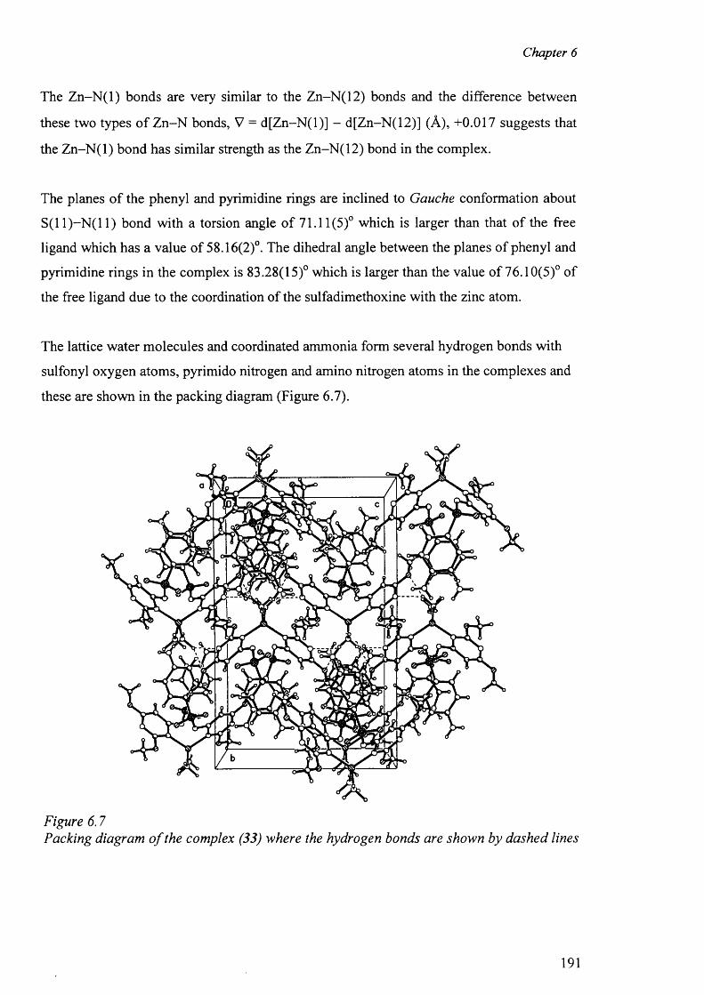
Chapter 6
The Z n -N (l) bonds are very similar to the Zn-N(12) bonds and the difference between
these two types o f Z n-N bonds, V = d[Z n-N (l)] - d[Zn-N(12)] (A), +0.017 suggests that
the Z n -N (l) bond has similar strength as the Zn-N(12) bond in the complex.
The planes o f the phenyl and pyrimidine rings are inclined to Gauche conformation about
S(11)—N (11) bond with a torsion angle o f 71.11(5)° which is larger than that of the free
ligand which has a value of 58.16(2)°. The dihedral angle between the planes of phenyl and
pyrimidine rings in the complex is 83.28(15)° which is larger than the value o f 76.10(5)° of
the free ligand due to the coordination of the sulfadimethoxine with the zinc atom.
The lattice water molecules and coordinated ammonia form several hydrogen bonds with
sulfonyl oxygen atoms, pyrimido nitrogen and amino nitrogen atoms in the complexes and
these are shown in the packing diagram (Figure 6.7).
Figure 6.7Packing diagram o f the complex (33) where the hydrogen bonds are shown by dashed lines
191
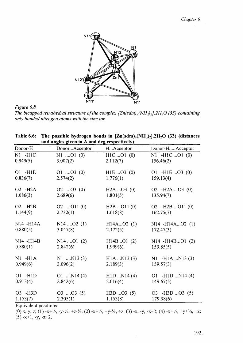
Chapter 6
Figure 6.8The bicapped tetrahedral structure o f the complex [Zn(sdm)2 (NH3) 2] .2 H 2 O (33) containing only bonded nitrogen atoms with the zinc ion
Table 6.6: The possible hydrogen bonds in [Zn(sdm)2(NH3)2].2H20 (33) (distancesand angles given in A and deg respectively)
Donor-H Donor... Acceptor H... Acceptor Donor-H.....AcceptorN1 -H1C 0.949(5)
N1 ....01 (0) 3.007(2)
H 1C ...01 (0) 2.112(7)
N1 -H 1C ...01 (0) 156.46(2)
01 -H IE 0.836(7)
01 ....03 (0) 2.574(2)
H IE ...03 (0) 1.776(1)
01 -H IE ...03 (0) 159.13(4)
0 2 -H2A 1.086(3)
0 2 ....03 (0) 2.689(6)
H 2A ...03 (0) 1.801(5)
0 2 -H 2A ...03 (0) 135.94(7)
0 2 -H2B 1.144(9)
0 2 ....O il (0) 2.732(1)
H2B ...O il (0) 1.618(8)
0 2 -H2B ...O il (0) 162.75(7)
N14 -H14A 0.880(5)
N 1 4 ....0 2 (1) 3.047(8)
H14A...02 (1) 2.172(5)
N14 -H14A...02 (1) 172.47(3)
N14 -H14B 0.880(1)
N 14....01 (2) 2.842(6)
H14B...01 (2) 1.999(6)
N14 -H14B...01 (2) 159.85(5)
N1 -H1A 0.949(6)
N1 ....N13 (3) 3.096(2)
H1A ...N13 (3) 2.189(3)
N1 -H1A...N13 (3) 159.57(3)
01 -HID 0.913(4)
01 ....N14 (4) 2.842(6)
H ID ...N14 (4) 2.016(4)
01 -HID ...N14 (4) 149.67(5)
03 -H3D 1.153(7)
0 3 ....03 (5) 2.305(1)
H 3D ...03 (5) 1.153(8)
03 -H 3D ...03 (5) 179.98(6)
Equivalent positions:(0) x, y, z; ( 1 ) -x+V2, -y-'/2, +z-1/2; (2) -x+Yi, +y-1/ 2 , +z; (3) -x, -y, -z+2; (4) -x+'/2, +y+!/2, +z; (5) -x+1, -y, -z+2.
192
Chapter 6
References
1. P. Narula; M. Haridas; T. P. Singh; Indian J. Phys; 1987, 61A, 132
2. U. H. Patel, B. H. Patel, and B. N. Patel, Cryst. Res. Tech.; 2001, 36, 1445
3. Y. A. Simonov, M. D. Mazus, I. A. Popa, V. N. Shafranskii and T. I. Malonovskii,Zh. Strukt. Khim; 1990,31, 77
4. M. H. Torre, G. Facchin, E. Kremer, E. Castellano, O. E. Piro and E. J. Baran; J. Inorg. Biochem ., 2003, 94, 200.
5. G. M. Sheldrick; Acta Cryst., 1990, A46, 467
6 . G. M. Sheldrick; SHELXL-97; Program fo r Crystal structure refinement, University o f Gottingen, Germany, 1996
7. M. R. Caira, R. Mohamed; Acta Cryst., 1992, B48, 492.
8 . A. Camerman; Canad. J. Chem., 1970, 48, 179
9. M. R. Truter; J. Chem. Soc., 1962, 3400
10. B. H. O ’Conner and E. N. Maslen; Acta Cryst., 1965,18, 363
11. B.H. O ’Conner and E. N. Maslen; Acta Cryst., 1967, 22, 134
12. M. Alkaume and J. Decap; Acta Cryst., 1965A, 18, 731
13. M. Alkaume and J. Decap; Acta Cryst., 1965B, 19, 394
14. C. H. Koo, H. S.Kim, W. C. Shin and C. H. Choe; J. Kor. Chem. Soc., 1974, 18, 97
15. H. S. Shin, G. S Ihn, H. S. Kim and C. H. Koo; J. Korean Chem. Soc., 1974, 18, 329
16. A. K. Basak, S.K. Mazumdar and S. Chaudhuri; Acta Crsyt., 1983, C39, 492
17. R.K. Tiwari, M. Haridas and T. P. Singh; Acta Crsyt., 1984, C40, 655
18. N. C. Baenziger, S. L. Modak and C. L. Fox Jr., Acta Cryst., 1983, C39, 1620
193


![Syntheses, Characterization and biological Activity of 4 ... … · Syntheses, Characterization and biological Activity of 4-{[(Z)-(2-methoxyphenyl) methylidene] amino} ... All complexes](https://img.pdfslide.us/doc/110x75/5ecce93a63134f68651a6edb/syntheses-characterization-and-biological-activity-of-4-syntheses-characterization.jpg)