Embed Size (px)
Citation preview
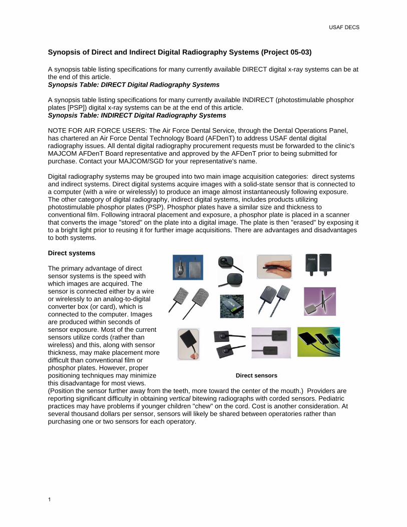
Synopsis of Direct and Indirect Digital Radiography Systems (Project 05-03) A synopsis table listing specifications for many currently available DIRECT digital x-ray systems can be at the end of this article. Synopsis Table: DIRECT Digital Radiography Systems A synopsis table listing specifications for many currently available INDIRECT (photostimulable phosphor plates [PSP]) digital x-ray systems can be at the end of this article. Synopsis Table: INDIRECT Digital Radiography Systems NOTE FOR AIR FORCE USERS: The Air Force Dental Service, through the Dental Operations Panel, has chartered an Air Force Dental Technology Board (AFDenT) to address USAF dental digital radiography issues. All dental digital radiography procurement requests must be forwarded to the clinic's MAJCOM AFDenT Board representative and approved by the AFDenT prior to being submitted for purchase. Contact your MAJCOM/SGD for your representative's name. Digital radiography systems may be grouped into two main image acquisition categories: direct systems and indirect systems. Direct digital systems acquire images with a solid-state sensor that is connected to a computer (with a wire or wirelessly) to produce an image almost instantaneously following exposure. The other category of digital radiography, indirect digital systems, includes products utilizing photostimulable phosphor plates (PSP). Phosphor plates have a similar size and thickness to conventional film. Following intraoral placement and exposure, a phosphor plate is placed in a scanner that converts the image "stored" on the plate into a digital image. The plate is then "erased" by exposing it to a bright light prior to reusing it for further image acquisitions. There are advantages and disadvantages to both systems. Direct systems The primary advantage of direct sensor systems is the speed with which images are acquired. The sensor is connected either by a wire or wirelessly to an analog-to-digital converter box (or card), which is connected to the computer. Images are produced within seconds of sensor exposure. Most of the current sensors utilize cords (rather than wireless) and this, along with sensor thickness, may make placement more difficult than conventional film or phosphor plates. However, proper positioning techniques may minimize this disadvantage for most views. (Position the sensor further away from the teeth, more toward the center of the mouth.) Providers are reporting significant difficulty in obtaining vertical bitewing radiographs with corded sensors. Pediatric practices may have problems if younger children "chew" on the cord. Cost is another consideration. At several thousand dollars per sensor, sensors will likely be shared between operatories rather than purchasing one or two sensors for each operatory.
Direct sensors
USAF DECS
1
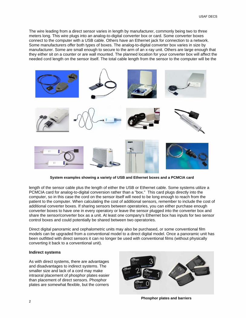
Phosphor plates and barriers
The wire leading from a direct sensor varies in length by manufacturer, commonly being two to three meters long. This wire plugs into an analog-to-digital converter box or card. Some converter boxes connect to the computer with a USB cable. Others have an Ethernet jack for connection to a network. Some manufacturers offer both types of boxes. The analog-to-digital converter box varies in size by manufacturer. Some are small enough to secure to the arm of an x-ray unit. Others are large enough that they either sit on a counter or are wall mounted. The planned location for your converter box will affect the needed cord length on the sensor itself. The total cable length from the sensor to the computer will be the
length of the sensor cable plus the length of either the USB or Ethernet cable. Some systems utilize a PCMCIA card for analog-to-digital conversion rather than a "box." This card plugs directly into the computer, so in this case the cord on the sensor itself will need to be long enough to reach from the patient to the computer. When calculating the cost of additional sensors, remember to include the cost of additional converter boxes. If sharing sensors between operatories, you can either purchase enough converter boxes to have one in every operatory or leave the sensor plugged into the converter box and share the sensor/converter box as a unit. At least one company's Ethernet box has inputs for two sensor control boxes and could potentially be shared between two operatories. Direct digital panoramic and cephalometric units may also be purchased, or some conventional film models can be upgraded from a conventional model to a direct digital model. Once a panoramic unit has been outfitted with direct sensors it can no longer be used with conventional films (without physically converting it back to a conventional unit). Indirect systems As with direct systems, there are advantages and disadvantages to indirect systems. The smaller size and lack of a cord may make intraoral placement of phosphor plates easier than placement of direct sensors. Phosphor plates are somewhat flexible, but the corners
System examples showing a variety of USB and Ethernet boxes and a PCMCIA card
USAF DECS
2

cannot be bent (as is sometimes done with film) without damaging the plates. Phosphor plates can potentially be reused hundreds of times, but are susceptible to scratching which will shorten their useful life. Phosphor plates are light sensitive and exposure to ambient light must be minimized during the time period between removal from their protective cover and placement into the scanner. The length of time that plates are exposed to ambient light during this transfer process will determine the level of allowable ambient light at the scanner location. Scanners in which plates are loaded directly into a slot can generally be used in areas of higher ambient light compared to systems in which the plates are loaded on drums prior to placement in the scanner.
The primary disadvantage of phosphor plate systems involves the time required to scan and erase the plates. Following exposure, plates must be removed from their contaminated barrier pouches, run through the scanner, "erased" with bright light, and repackaged in clean barrier pouches prior to using again. For plate erasure, some scanners incorporate an "erase" cycle within the scanner itself. With other units the plates are moved to a separate plate eraser following the scanning process. It is less expensive to purchase enough phosphor plates to place in every operatory compared to purchasing enough direct sensors for every operatory. The cost for an intraoral phosphor
plate is less than twenty-five dollars compared to several thousand dollars for each direct sensor. Most existing panoramic and cephalometric units do not require expensive upgrades for use with phosphor plates. Phosphor plates are simply placed in cassettes similar to film, except that no intensifying screens are used. Therefore, the same panoramic or cephalometric unit can be used to expose either conventional film or phosphor plates. Phosphor plate systems require purchase of a scanner. Large clinics may want to purchase two or more scanners to distribute throughout the clinic. Imaging area dimensions
The imaging area of direct sensors and phosphor plates listed in the synopsis tables can be compared to the imaging area of conventional dental film. Listed here are conventional film sizes for comparison purposes. Imaging software
Prior to the development of DICOM (Digital Imaging and Communications in Medicine) standards, most manufacturers utilized proprietary image file formats that were not compatible with other manufacturers' systems. However, more and more dental imaging systems are becoming DICOM compliant. DICOM compliant systems can share image files. This makes it easier to transfer saved images from one manufacturer's system to another. While this improves the transfer of saved images, most manufacturers' imaging software still will only acquire images using that same manufacturer's sensors. In other words, with most systems, if a large clinic happened to own ten of company A's direct sensors, company B's direct pano unit, and was considering the purchase of two new sensors from company C, they could not use company A's imaging software to directly acquire images from company B's pano and company C's sensors. However, there are some "open platform" imaging software programs that can acquire digital
Phosphor plate scanners
Phosphor plate eraser
Size 0: 22 x 35 mm Size 1: 24 x 40 mm Size 2: 31 x 41 mm Size 3: 27 x 54 mm Size 4: 57 x 76 mm
USAF DECS
3
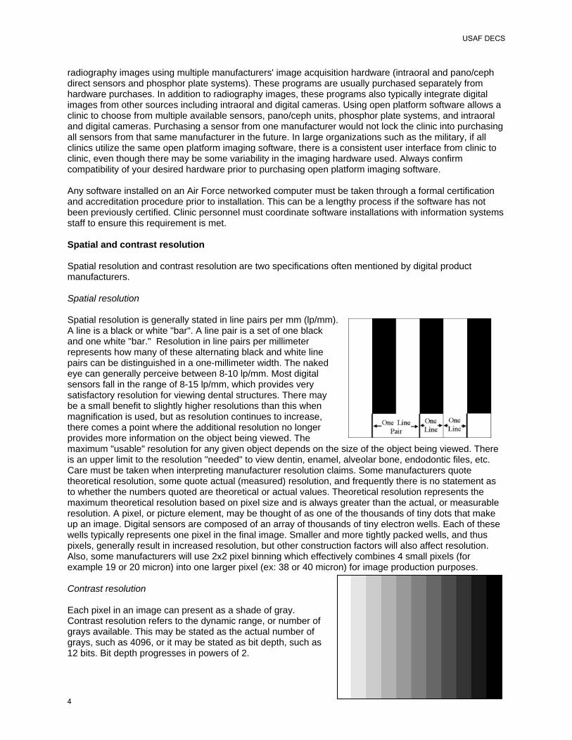
radiography images using multiple manufacturers' image acquisition hardware (intraoral and pano/ceph direct sensors and phosphor plate systems). These programs are usually purchased separately from hardware purchases. In addition to radiography images, these programs also typically integrate digital images from other sources including intraoral and digital cameras. Using open platform software allows a clinic to choose from multiple available sensors, pano/ceph units, phosphor plate systems, and intraoral and digital cameras. Purchasing a sensor from one manufacturer would not lock the clinic into purchasing all sensors from that same manufacturer in the future. In large organizations such as the military, if all clinics utilize the same open platform imaging software, there is a consistent user interface from clinic to clinic, even though there may be some variability in the imaging hardware used. Always confirm compatibility of your desired hardware prior to purchasing open platform imaging software. Any software installed on an Air Force networked computer must be taken through a formal certification and accreditation procedure prior to installation. This can be a lengthy process if the software has not been previously certified. Clinic personnel must coordinate software installations with information systems staff to ensure this requirement is met. Spatial and contrast resolution Spatial resolution and contrast resolution are two specifications often mentioned by digital product manufacturers. Spatial resolution Spatial resolution is generally stated in line pairs per mm (lp/mm). A line is a black or white "bar". A line pair is a set of one black and one white "bar." Resolution in line pairs per millimeter represents how many of these alternating black and white line pairs can be distinguished in a one-millimeter width. The naked eye can generally perceive between 8-10 lp/mm. Most digital sensors fall in the range of 8-15 lp/mm, which provides very satisfactory resolution for viewing dental structures. There may be a small benefit to slightly higher resolutions than this when magnification is used, but as resolution continues to increase, there comes a point where the additional resolution no longer provides more information on the object being viewed. The maximum "usable" resolution for any given object depends on the size of the object being viewed. There is an upper limit to the resolution "needed" to view dentin, enamel, alveolar bone, endodontic files, etc. Care must be taken when interpreting manufacturer resolution claims. Some manufacturers quote theoretical resolution, some quote actual (measured) resolution, and frequently there is no statement as to whether the numbers quoted are theoretical or actual values. Theoretical resolution represents the maximum theoretical resolution based on pixel size and is always greater than the actual, or measurable resolution. A pixel, or picture element, may be thought of as one of the thousands of tiny dots that make up an image. Digital sensors are composed of an array of thousands of tiny electron wells. Each of these wells typically represents one pixel in the final image. Smaller and more tightly packed wells, and thus pixels, generally result in increased resolution, but other construction factors will also affect resolution. Also, some manufacturers will use 2x2 pixel binning which effectively combines 4 small pixels (for example 19 or 20 micron) into one larger pixel (ex: 38 or 40 micron) for image production purposes. Contrast resolution Each pixel in an image can present as a shade of gray. Contrast resolution refers to the dynamic range, or number of grays available. This may be stated as the actual number of grays, such as 4096, or it may be stated as bit depth, such as 12 bits. Bit depth progresses in powers of 2.
USAF DECS
4

1 bit 21 = 2 shades of gray (black and white) 2 bits 22 = 2x2 = 4 shades of gray 3 bits 23 = 2x2x2 = 8 shades of gray 8 bits 28 = 2x2x2x2x2x2x2x2 = 256 shades of gray 12 bits 212 = 2x2x2x2x2x2x2x2x2x2x2x2 = 4096 shades of gray 16 bits 216 = 2x2x2x2x2x2x2x2x2x2x2x2x2x2x2x2 = 65,536 shades of gray Many sensors capture 12 bit images (4096 grays), some sensors capture 16 bit images (65,536 grays). Most computer monitors can only display 8 bit grayscale images (256 grays) and the human eye can typically only differentiate between 32-64 grays. If an image contains data for additional grays above what the monitor can display, the additional data may be of some benefit if the imaging software is capable of a digital processing method called windowing. Windowing is the process of selecting a certain segment of the total range of grays captured, then displaying that segment on the monitor over the full grayscale range from white to black. Windowing gives the ability to focus in on specific segments of the large number of grays captured by 12 and 16 bit sensors. While this ability may provide some advantages, current research has not supported the concept that increasingly greater bit depths improve diagnostic ability. 8 bit data provides very satisfactory images and diminishing returns are obtained from bit depths greater than this. Infection Control Issues The USAF Guidelines for Infection Control in Dentistry and the CDC's Guidelines for Infection Control in Dental Health-Care Settings - 2003 offer similar guidance regarding the use of digital sensors. Quoting from the USAF Guidelines, sensors are categorized as semi-critical items and they: "…should be cleaned and ideally should be heat-sterilized or high-level disinfected between patients. However, these items vary by manufacturer or type of device in their ability to be sterilized or high-level disinfected. The following apply for digital radiography sensors: a. Use FDA-cleared barriers. b. To minimize the potential for device-associated infections, after removing the barrier, clean and disinfect using an EPA-registered hospital disinfectant with an intermediate-level activity after each patient." Because sensors and associated components vary by manufacturer and are expensive, manufacturers should be consulted regarding specific disinfection products and procedures. Some manufacturers recommend against using certain chemicals on their sensors. Also, some manufacturers allow immersion of their sensors while others do not. Other considerations Other considerations when selecting a digital radiography system include company service/support, system cost, length of warranty, and ease of use/capability of imaging software. Compatibility with open platform imaging software will be a consideration for those clinics utilizing these software packages. Adequate cable length and sensor size/shape is another consideration. Sensors with rounded edges may be more comfortable. Sensor wires exiting from the back of the sensor may make for easier placement in some situations compared to configurations where the wire exits from the end of the sensor. Sensor thickness disadvantages may be minimized when sensors are positioned properly (more toward the center of the mouth). Multiple considerations come into play when selecting a digital radiography package and there are advantages and disadvantages to all systems.
USAF DECS
5

Direct Dental Digital Radiography Systems (CCD, CMOS)
Model
Dent-X EVA
DEXIS Digital X-ray System
GE Healthcare Sigma
Company Dent-X Corporation USA 250 Clearbrook Road Elmsford, NY 10523 (800) 225-1702
Dexis 2550 Northwinds Parkway Suite 100 Alpharetta, GA 30004 (888) 883-3947
GE Healthcare 300 West Edgerton Avenue Milwaukee, WI 53207-6025 (800) 558-6120
Web link www.dent-x.com www.dexray.com www.gehealthcare.com
Gov't point of contact
Adam Rabinovitch (914) 592-6100 ext 210 [email protected]
Kim Mercer (888) 883-3947 ext 253 [email protected]
Mike Null (414) 747-6352 [email protected]
Components in package1
Sensor - choice of size 1 or 2 USB box Imaging software (Site license) Positioning kit
Sensor - universal size PCMCIA capture card Imaging software (Single user license) Positioning kit
Sensor - see pkgs below USB box Imaging software (5 user licenses) Positioning kit
Cost of package
Retail/gov't $8,200/$6,295
Retail/gov't $11,000/$9,000
Retail/gov't Size #1: $10,375/$6,336 Size #2: $11,800/$7,207
Cost of additional sensors
Retail/gov't (Includes ADC3 which is attached to sensor cord) Size 1: $7,500/$6095 Size 2: $7,500/$6095
Retail/gov't (Includes ADC3 capture card) Universal size: $9,000/$7,000
Retail/gov't (Sensor only) Size 1: $7,272/$4,442 Size 2: $8,587/$5,245
Other equipment costs
Retail/gov't USB docking station: $300/$200
Retail/gov't USB adaptor for capture card: $499/$499
Retail/gov't USB box (houses ADC3): $2680/$1637
Sensor warranty2 3 years 1 year 2 years
Sensor type CMOS CCD CCD
Sensor external dimensions
Size 1: 25.6 x 38.4 x 4.8 mm Size 2: 30.8 x 44.1 x 4.8 mm
Universal: 29.3 x 38.7 x 9.0 mm
Size 1: 24 x 36 x 6.8 mm Size 2: 30 x 40 x 6.8 mm
Sensor imaging area dimensions
Size 1: 20 x 30 mm Size 2: 25.8 x 36 mm Universal: 25.6 x 32 mm Size 1: 20 x 32 mm
Size 2: 26 x 34 mm
Pixel size 30 micron 40 micron 19.5 micron
Dynamic range 12 bit 12 bit 12 bit
Sensor cable length 2 meter 2.44 meter with 1.22 meter
extension available 3 meters
Computer interface options
USB PCMCIA card USB adaptor for PCMCIA card available
USB
Manufacturers provided data for table 1Primary items in package are listed. Other items such as cables and hygienic covers may be included. Check with manufacturer for software licensing details. Other package configurations may be available from manufacturer. 2Check with manufacturer for warranty details. 3ADC = Analog-to-digital converter.
USAF DECS
6

Model
Gendex GX-S Visualix HDI
Kodak RVG 6000
Lightyear
Company Gendex Dental Systems 340 E. Main Street Lake Zurich, IL 60047 (888) 275-5286
Kodak Dental Systems 1765 The Exchange Atlanta, GA 30339 1-800-944-6365
Lightyear Technology, Inc. 85-C Mill Street, Suite 100 Roswell, GA 30075 (866) 946-2431
Web link www.gendex.com www.kodak.com/go/dental www.lightyeartechnology.com
Gov't point of contact
Call Gendex to obtain regional rep: (888) 275-5286
William Altvater 800-262-8144 x7277 [email protected]
George Hummert (866) 946-2431 Ext. 107 ghummert@ lightyeartechnology.com
Components in package1
Sensor - size #2 USB box Imaging software (Site license) Positioning kit
Sensor - see pkgs below Imaging software (Site license) Positioning kit
Sensor - size #2 USB box Imaging software (Single user license) Positioning kit
Cost of package
Retail/gov't $10,050/$6,166
Retail/gov't Size 1: $12,895/$7,737 Size 2: 13,985/$8,337
Retail/gov't $11,995/$8,996
Cost of additional sensors
Retail/gov't (Sensor only) Size 1: $6,690/$3,206 Size 2: $7,775/$3,991
Retail/gov't (Includes ADC3 which is attached to sensor cord) Size 1: $9,250/$5,550 Size 2: $9,995/$5,997
Retail/gov't (Sensor only) Size 0: $5,995/$4,496 Size 1: $5,995/$4,496 Size 2: $6,995/$5,246
Other equipment costs
Retail/gov't USB box (houses ADC3): $2,165/$1,297
Retail/gov't Separate USB box not required. Optional USB hub: $100/$60
Retail/gov't USB box (houses ADC3): $1,495/$1,121
Sensor warranty2 2 years 2 years 5 years
Sensor type CCD CMOS CCD
Sensor external dimensions
Size 1: 25 x 39.5 x 5.7 mm Size 2: 32.5 x 42.5 x 5.6 mm
Size 1: 40 mm x 26 mm x 8 mm Size 2: 45 mm x 31 mm x 8 mm
Size 0: 26.4 x 32.5 x 3.2 mm Size 1: 24.7 x 37.8 x 3.2 mm Size 2: 31.8 x 43.0 x 3.2 mm
Sensor imaging area dimensions
Size 1: 20 x 30 mm Size 2: 28 x 36 mm
Size 1: 22 x 30 mm Size 2: 27 x 36 mm
Size 0: 21.6 x 26.8 mm Size 1: 20.6 x 32.7 mm Size 2: 26.6 x 36.8 mm
Pixel size 22 micron 18.5 micron _ _ _ _ _ _
Dynamic range 12 bit 12 bit 12 bit
Sensor cable length 3 meters 2.5 meter 3 meter
Computer interface options
USB USB USB
Manufacturers provided data for table 1Primary items in package are listed. Other items such as cables and hygienic covers may be included. Check with manufacturer for software licensing details. Other package configurations may be available from manufacturer. 2Check with manufacturer for warranty details. 3ADC = Analog-to-digital converter.
USAF DECS
7
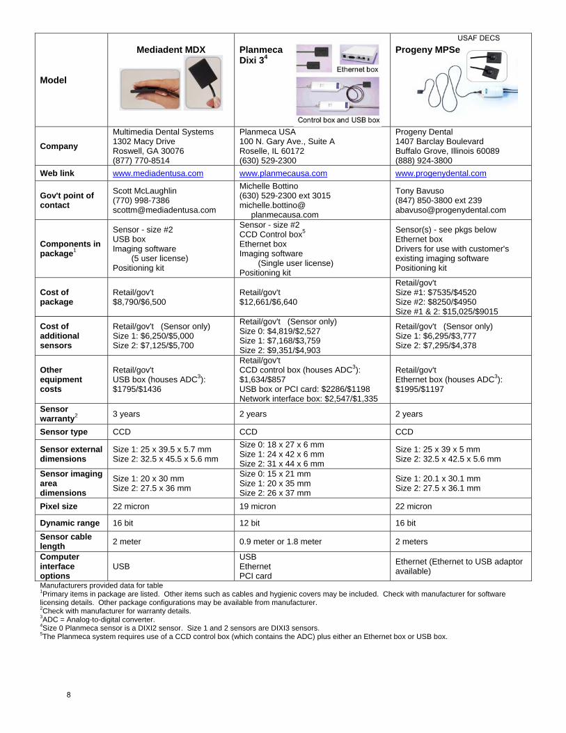
Model
Mediadent MDX
Planmeca Dixi 34
Progeny MPSe
Company Multimedia Dental Systems 1302 Macy Drive Roswell, GA 30076 (877) 770-8514
Planmeca USA 100 N. Gary Ave., Suite A Roselle, IL 60172 (630) 529-2300
Progeny Dental 1407 Barclay Boulevard Buffalo Grove, Illinois 60089 (888) 924-3800
Web link www.mediadentusa.com www.planmecausa.com www.progenydental.com
Gov't point of contact
Scott McLaughlin (770) 998-7386 [email protected]
Michelle Bottino (630) 529-2300 ext 3015 michelle.bottino@ planmecausa.com
Tony Bavuso (847) 850-3800 ext 239 [email protected]
Components in package1
Sensor - size #2 USB box Imaging software (5 user license) Positioning kit
Sensor - size #2 CCD Control box5
Ethernet box Imaging software (Single user license) Positioning kit
Sensor(s) - see pkgs below Ethernet box Drivers for use with customer's existing imaging software Positioning kit
Cost of package
Retail/gov't $8,790/$6,500
Retail/gov't $12,661/$6,640
Retail/gov't Size #1: $7535/$4520 Size #2: $8250/$4950 Size #1 & 2: $15,025/$9015
Cost of additional sensors
Retail/gov't (Sensor only) Size 1: $6,250/$5,000 Size 2: $7,125/$5,700
Retail/gov't (Sensor only) Size 0: $4,819/$2,527 Size 1: $7,168/$3,759 Size 2: $9,351/$4,903
Retail/gov't (Sensor only) Size 1: $6,295/$3,777 Size 2: $7,295/$4,378
Other equipment costs
Retail/gov't USB box (houses ADC3): $1795/$1436
Retail/gov't CCD control box (houses ADC3): $1,634/$857 USB box or PCI card: $2286/$1198 Network interface box: $2,547/$1,335
Retail/gov't Ethernet box (houses ADC3): $1995/$1197
Sensor warranty2 3 years 2 years 2 years
Sensor type CCD CCD CCD
Sensor external dimensions
Size 1: 25 x 39.5 x 5.7 mm Size 2: 32.5 x 45.5 x 5.6 mm
Size 0: 18 x 27 x 6 mm Size 1: 24 x 42 x 6 mm Size 2: 31 x 44 x 6 mm
Size 1: 25 x 39 x 5 mm Size 2: 32.5 x 42.5 x 5.6 mm
Sensor imaging area dimensions
Size 1: 20 x 30 mm Size 2: 27.5 x 36 mm
Size 0: 15 x 21 mm Size 1: 20 x 35 mm Size 2: 26 x 37 mm
Size 1: 20.1 x 30.1 mm Size 2: 27.5 x 36.1 mm
Pixel size 22 micron 19 micron 22 micron
Dynamic range 16 bit 12 bit 16 bit Sensor cable length 2 meter 0.9 meter or 1.8 meter 2 meters
Computer interface options
USB USB Ethernet PCI card
Ethernet (Ethernet to USB adaptor available)
Manufacturers provided data for table 1Primary items in package are listed. Other items such as cables and hygienic covers may be included. Check with manufacturer for software licensing details. Other package configurations may be available from manufacturer. 2Check with manufacturer for warranty details. 3ADC = Analog-to-digital converter. 4Size 0 Planmeca sensor is a DIXI2 sensor. Size 1 and 2 sensors are DIXI3 sensors. 5The Planmeca system requires use of a CCD control box (which contains the ADC) plus either an Ethernet box or USB box.
USAF DECS
8

Model
Schick CDR
Schick CDR Wireless
Sirona SIDEXIS
Company Schick Technologies, Inc. 30-00 47th Avenue Long Island City, NY 11101 (718) 937-5765
Schick Technologies, Inc. 30-00 47th Avenue Long Island City, NY 11101 (718) 937-5765
Sirona Dental Systems LLC 4835 Sirona Drive, Suite 100 Charlotte, NC 28273 (800) 659-5977
Web link www.schicktech.com www.schicktech.com www.sirona.com
Gov't point of contact
Manny Pena (877) 724-4251 [email protected]
Manny Pena (877) 724-4251 [email protected]
Patricia Czaplinsky (800) 659-5977 ext 117 [email protected]
Components in package1
Sensor - size #2 USB box Imaging software (Single user license) Positioning kit
Sensor - #2 wireless Wireless receiver Imaging Software (Single user license) Positioning Kit
Sensor(s) - see pkgs below USB box Imaging software (Site license) Positioning kit
Cost of package
Retail/gov't $12,037/$6,418
Retail/gov't $17,766/$9,105
Retail/gov't Size #1: $10,920/$6,047 Size #2: $11,750/$6,509 Size #1 & 2: $18,200 /$10,050
Cost of additional sensors
Retail/gov't (Sensor only) Size 0: $4,658/$2,638 Size 1: $7,108/$3,894 Size 2: $8,335/$4,523
Retail/gov't (Sensor only) Size 1: $10,663/$5,465 Size 2: $12,500/$6,407
Retail/gov't (Sensor only) Size 1: $6,759/$3,866 Size 2: $7,570/$4,342
Other equipment costs
Retail/gov't USB box (houses ADC3): $1595/$817
Retail/gov't Wireless receiver (houses ADC3): $3432/$1759
Retail/gov't USB box (houses ADC3): $2,080/$1,151 Ethernet box (houses ADC3): $2,599/$1,439
Sensor warranty2 1 year 1 year 2 years
Sensor type CMOS CMOS CCD Sensor external dimensions (mm x mm x mm)
Size 0: 22 x 31 x 5 mm Size 1: 24 x 37 x 5 mm Size 2: 30 x 43 x 5 mm
Size 1: 24 x 37 x 5 mm Size 2: 30 x 43 x 5 mm
Size 1 = 24 x 35.8 x 4.0 mm Size 2 = 30.1 x 40.2 x 5.0 mm
Sensor imaging area dimensions (mm x mm)
Size 0: 18 x 24 mm Size 1: 20 x 30 mm Size 2: 25.6 x 36 mm
Size 1: 20 x 30 mm Size 2: 25.6 x 36 mm
Size 1 = 20 x 30 mm Size 2 = 26 x 34 mm
Pixel size 40 micron 40 micron 19.5 micron Dynamic range 12 bit 12 bit 12 bit
Sensor cable length 2 meter NA 3 meters
Computer interface options
USB USB USB Ethernet
Manufacturers provided data for table 1Primary items in package are listed. Other items such as cables and hygienic covers may be included. Check with manufacturer for software licensing details. Other package configurations may be available from manufacturer. 2Check with manufacturer for warranty details. 3ADC = Analog-to-digital converter.
USAF DECS
9

Indirect Dental Digital Radiography Systems (Phosphor Plates)
Model
Air Techniques ScanX
Air Techniques ScanX Intraoral
Gendex DenOptix
Company
Air Techniques 70 Cantiague Rock Road P.O. Box 870 Hicksville, NY 11802 Ph: (800) 247-8324
Air Techniques 70 Cantiague Rock Road P.O. Box 870 Hicksville, NY 11802 Ph: (800) 247-8324
Gendex Dental Systems 340 E. Main Street Lake Zurich, IL 60047 (888) 275-5286
Web link www.airtechniques.com www.airtechniques.com www.gendex.comGov't point of contact
Eugene Heil (423) 753-9909 [email protected]
Eugene Heil (423) 753-9909 [email protected]
Call Gendex to obtain regional rep: (888) 275-5286
Components in basic package1
Scanner 4 each size 2 PSP guides 1 each size 0, 1, & 3 PSP guides 20 size 2 PSPs (imaging plates) 1 plate transfer box
Scanner 4 each size 2 PSP guides 1 each size 0, 1, & 3 PSP guides 20 size 2 PSPs (imaging plates) 1 plate transfer box
Scanner Phosphor plate carousel(s) Imaging software (Site license) Imaging plates (varies by pkg)
Cost of basic package
Retail/gov't $19,995/$12,597
Retail/gov't $11,495/$7,047
Retail/gov't Intraoral: $14,111/$8,018 Intraoral & pano: $20,497/$11,624 Intraoral, pano, ceph: $22,145/call
Unit accepts listed plate sizes Cost of additional plates
Retail/gov't Size 0: $22.00/$16.80 Size 1: $22.00/$16.80 Size 2: $22.00/$16.80 Size 3: $22.00/$16.80 Size 4: $60.00/$45.83 5"x12" pano: $775/$514 6"x12" pano: $795/$527 8"x10" ceph: $850/$564 Plate eraser $875/$536
Retail/gov't Size 0: $22.00/$16.80 Size 1: $22.00/$16.80 Size 2: $22.00/$16.80 Size 3: $22.00/$16.80 Size 4: $60.00/$45.83 Plate eraser $875/$536
Retail/gov't Size 0: $21.00/$17.00 Size 1: $21.50/17.43 Size 2: $22.50/16.13 Size 3: $27.50/43.35 Size 4: $65.00/51.85 5"x12" pano: $875/543 6"x12" pano: $885/575 8"x10" ceph: $970/596
Scanner warranty2 2 years 2 years 2 years
Intraoral plate imaging area dimensions
Size 0: 22mm x 35mm Size 1: 24mm x 40mm Size 2: 31mm x 41mm Size 3: 27mm x 54mm
Size 0: 22mm x 35mm Size 1: 24mm x 40mm Size 2: 31mm x 41mm Size 3: 27mm x 54mm
Size 0: 22mm x 35mm Size 1: 24mm x 40mm Size 2: 31mm x 41mm Size 3: 27mm x 54mm
Scan time from plate insertion to image display
Intraoral: 17 sec for 1st plate, then 4 sec each additional plate at standard resolution Pano: 25 sec at std resolution
Intraoral: 17 sec for 1st plate, then 4 sec each additional plate at standard resolution
Intraoral: 72seconds at standard resolution Pano: 180seconds at standard resolution
Method to erase plates Separate plate eraser Separate plate eraser Separate plate eraser
Ambient lighting recommendations at scanner location
Less than 400 lux; designed for use in normal office lighting; avoid direct sunlight.
Less than 400 lux; designed for use in normal office lighting; avoid direct sunlight.
Low light (10-20 lux)
Scanner dimensions(HxWxD) 24" x 14" x 14" 15.5" x 15" x 15" 15.5" x 19.4" x 10.1"
Scanner electrical requirements 100-240V, 50/60 Hz 100-240V, 50/60 Hz 100-240V, 50/60 Hz
Scanner interface with computer USB USB USB
Manufacturers provided data in table
1Primary items in package are listed. Other items such as cables and hygienic covers may be included. Check with manufacturer for software licensing details. 2Check with manufacturer for warranty details.
USAF DECS
10
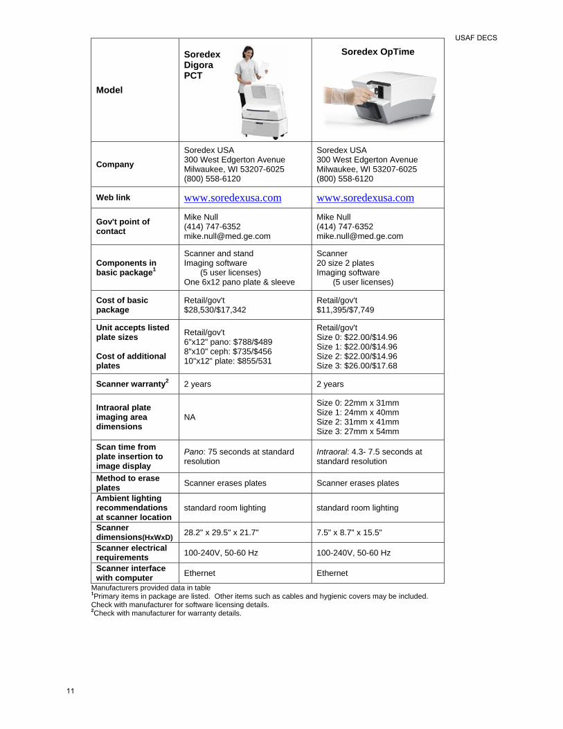
Model
Soredex Digora PCT
Soredex OpTime
Company Soredex USA 300 West Edgerton Avenue Milwaukee, WI 53207-6025 (800) 558-6120
Soredex USA 300 West Edgerton Avenue Milwaukee, WI 53207-6025 (800) 558-6120
Web link www.soredexusa.com www.soredexusa.com
Gov't point of contact
Mike Null (414) 747-6352 [email protected]
Mike Null (414) 747-6352 [email protected]
Components in basic package1
Scanner and stand Imaging software (5 user licenses) One 6x12 pano plate & sleeve
Scanner 20 size 2 plates Imaging software (5 user licenses)
Cost of basic package
Retail/gov't $28,530/$17,342
Retail/gov't $11,395/$7,749
Unit accepts listed plate sizes Cost of additional plates
Retail/gov't 6"x12" pano: $788/$489 8"x10" ceph: $735/$456 10"x12" plate: $855/531
Retail/gov't Size 0: $22.00/$14.96 Size 1: $22.00/$14.96 Size 2: $22.00/$14.96 Size 3: $26.00/$17.68
Scanner warranty2 2 years 2 years
Intraoral plate imaging area dimensions
NA
Size 0: 22mm x 31mm Size 1: 24mm x 40mm Size 2: 31mm x 41mm Size 3: 27mm x 54mm
Scan time from plate insertion to image display
Pano: 75 seconds at standard resolution
Intraoral: 4.3- 7.5 seconds at standard resolution
Method to erase plates Scanner erases plates Scanner erases plates
Ambient lighting recommendations at scanner location
standard room lighting standard room lighting
Scanner dimensions(HxWxD) 28.2" x 29.5" x 21.7" 7.5" x 8.7" x 15.5"
Scanner electrical requirements 100-240V, 50-60 Hz 100-240V, 50-60 Hz
Scanner interface with computer Ethernet Ethernet
Manufacturers provided data in table 1Primary items in package are listed. Other items such as cables and hygienic covers may be included. Check with manufacturer for software licensing details. 2Check with manufacturer for warranty details.
USAF DECS
11