Embed Size (px)
Citation preview

Can Respir J Vol 10 No 1 January/February 2003 39
Synchronous lung and liver metastases from medullarythyroid carcinoma
H Yanardag MD1, C Tetikkurt MD2, S Tetikkurt MD3
1Department of Internal Medicine and 2Deparment of Pulmonary Diseases, Cerrahpasa Faculty of Medicine, and 3Department of Pathology,Taksim State Hospital, Istanbul, Turkey
Correspondence and reprints: Dr Cuneyt Tetikkurt, Tanzimat sok. Serkan Apt No 8/16 Caddebostan 81060, Istanbul, Turkey. Telephone 90-216-359-60-35, fax 90-212-529-40-18, e-mail [email protected]
H Yanardag, C Tetikkurt, S Tetikkurt. Synchronous lung andliver metastases from medullary thyroid carcinoma. Can Respir J2003;10(1):39-41.
Metastatic disease is one of the most common causes of calcified nod-ules in the lung or liver. The incidence of calcified metastasis mainlyto the lung and liver is high at the initial presentation in patientswith medullary thyriod carcinoma. Synchronous calcified metastasisin the lung and liver is reported for the first time. The diagnosis ofmedullary thyroid carcinoma may be evident from the synchronouspresence of miliary calcified nodules in two different sites if they areassociated with high concentrations of serum markers.
Key Words: Calcified metastasis; Medullary thyroid carcinoma;Metastasis; Synchronous metastases
Des métastases pulmonaires et hépatiques syn-chrones causées par un carcinome médullairethyroïdien
RÉSUMÉ : La maladie métastatique est l’une des principales causes denodules calcifiés dans le poumon ou le foie. En cas de carcinome médul-laire thyroïdien, les calcifications métastatiques peuvent s’observer dansle poumon ou le foie. On rend compte pour la première fois d’une métas-tase calcifiée à la fois dans le poumon et le foie. Le diagnostic de carci-nome médullaire thyroïdien peut être mis en évidence en raison de laprésence synchrone de nodules calcifiés miliaires dans deux foyers dis-tincts, s’ils s’associent à de fortes concentrations de marqueurs sériques.
Alarge variety of neoplasms can produce calcified metas-tases. Calcified nodules seen on chest or abdominal radi-
ographs may resemble tuberculosis or other granulomatousdiseases. In the case of medullary thyroid carcinoma, metastat-ic calcifications may be found in the lung or liver (1,2). A rea-sonable suspicion of malignancy is necessary for theidentification of calcified metastases from medullary thyroidcarcinoma because the primary tumour may be clinicallyoccult.
We report a sporadic case of medullary thyroid carcinomawith synchronous calcified miliary lung and liver metastases. Toour knowledge, this is the first report in the literature citing syn-chronous calcified metastasis in medullary thyroid carcinoma.
CASE PRESENTATIONA 25-year-old man was admitted for anorexia, fatigue and diar-rhea lasting four months. His personal and family historieswere excellent. Physical examination was normal with a bloodpressure of 120/80 mmHg. A complete blood count and bloodchemistry were within the normal ranges. The serum calciumlevel was 2.2 mmol/L and the phosphorus level was 1.1 mmol/L.The erythrocyte sedimentation rate was 42 mm/h and a tuber-culin test was negative. Recently performed intravenous pyel-ography radiographs for the right flank pain to investigatenephrolithiasis showed multiple calcific nodules in the liver(Figure 1). Chest x-ray showed miliary calcific opacities in themiddle zones of both lungs and there was no calcification in
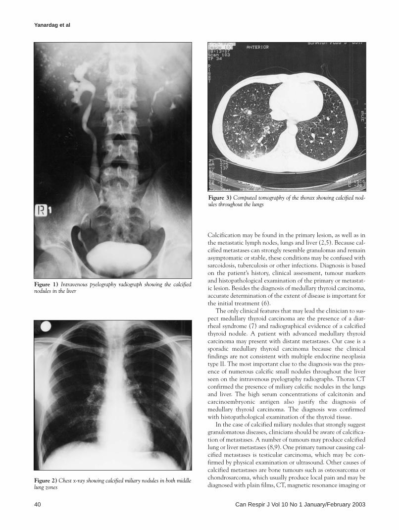
the neck region (Figure 2). Computed tomography (CT) of thethorax revealed numerous calcified small nodules 2 to 4 mm indiameter throughout the lungs (Figure 3). CT did not showcalcification in the thyroid gland. The calcified nodules in theliver measured between 2 and 8 mm. Abdominal CT did notreveal an adrenal mass compatible with pheochromocytoma.Stool examination, colonoscopy, fine needle aspiration biopsyof the liver and fibreoptic bronchoscopic examination werenegative. Transbronchial biopsy specimens were not diagnostic.No acid-fast bacilli were seen on stained bronchial washings.Mycobacterial and fungal cultures of the bronchial lavage werenegative. Laparoscopic liver biopsy revealed neuroendocrinecarcinoma metastasis compatible with medullary thyroid carci-noma. Thyroid scintigraphy showed diffuse hyperplasia andultrasonography showed a 7×8 mm nodule in the left lobe ofthe thyroid gland. Histopathological examination of the resect-ed thyroid tissue revealed medullary thyroid carcinoma. Serumcalcitonin and carcinoembryonic antigen were measured to be1270 ng/L and 1000 µg/L, respectively. The patient was referredto the oncology department for further treatment.
DISCUSSIONMedullary thyroid carcinoma originating in the thyroid C cellsaccounts for 5% to 10% of all thyroid malignancies (3). Theincidence of distant metastases in medullary thyroid carcinomais high, mainly to the lung and liver. The distant metastasesmay appear at the time of initial presentation (4).
©2003 Pulsus Group Inc. All rights reserved
CASE REPORT
Yanardag.qxd 1/31/2003 2:49 PM Page 39
Calcification may be found in the primary lesion, as well as inthe metastatic lymph nodes, lungs and liver (2,5). Because cal-cified metastases can strongly resemble granulomas and remainasymptomatic or stable, these conditions may be confused withsarcoidosis, tuberculosis or other infections. Diagnosis is basedon the patient’s history, clinical assessment, tumour markersand histopathological examination of the primary or metastat-ic lesion. Besides the diagnosis of medullary thyroid carcinoma,accurate determination of the extent of disease is important forthe initial treatment (6).
The only clinical features that may lead the clinician to sus-pect medullary thyroid carcinoma are the presence of a diar-rheal syndrome (7) and radiographical evidence of a calcifiedthyroid nodule. A patient with advanced medullary thyroidcarcinoma may present with distant metastases. Our case is asporadic medullary thyroid carcinoma because the clinicalfindings are not consistent with multiple endocrine neoplasiatype II. The most important clue to the diagnosis was the pres-ence of numerous calcific small nodules throughout the liverseen on the intravenous pyelography radiographs. Thorax CTconfirmed the presence of miliary calcific nodules in the lungsand liver. The high serum concentrations of calcitonin andcarcinoembryonic antigen also justify the diagnosis ofmedullary thyroid carcinoma. The diagnosis was confirmedwith histopathological examination of the thyroid tissue.
In the case of calcified miliary nodules that strongly suggestgranulomatous diseases, clinicians should be aware of calcifica-tion of metastases. A number of tumours may produce calcifiedlung or liver metastases (8,9). One primary tumour causing cal-cified metastases is testicular carcinoma, which may be con-firmed by physical examination or ultrasound. Other causes ofcalcified metastases are bone tumours such as osteosarcoma orchondrosarcoma, which usually produce local pain and may bediagnosed with plain films, CT, magnetic resonance imaging or
Yanardag et al
Can Respir J Vol 10 No 1 January/February 200340
Figure 1) Intravenous pyelography radiograph showing the calcifiednodules in the liver
Figure 2) Chest x-ray showing calcified miliary nodules in both middlelung zones
Figure 3) Computed tomography of the thorax showing calcified nod-ules throughout the lungs
Yanardag.qxd 1/31/2003 2:49 PM Page 40

radionuclide bone scan. Breast and gastrointestinal tumoursmay also metastasize as calcified nodules in the lung or liver.They usually present with local findings and are less likely topresent as an occult primary tumour (10).
Metastatic disease is one of the most common causes of cal-cified nodules in the lung or liver. Because the metastases aredetected after the primary lesion, the presumptive diagnosis ofmetastases from the known primary lesion is a secure diagnosis.
If there is no history of a previous tumour, then the presence ofcalcified nodules in the lung or liver is a diagnostic challengefor the clinician. Our case is remarkable for identifying thesynchronous calcified metastasis in the lung and liver for thefirst time in the literature. The diagnosis of medullary thyroidcarcinoma may be evident from the synchronous presence ofmiliary calcified nodules in two different sites if they are asso-ciated with high concentrations of serum markers.
Synchronous metastases from medullary thyroid carcinoma
Can Respir J Vol 10 No 1 January/February 2003 41
REFERENCES1. Maile CW, Rodan BA, Godwin JD, Chen JT, Ravin CE.
Calcification in pulmonary metastases. Br J Radiol 1982;55:108-13.2. McDonnell CH 3rd, Fishman EK, Zerhouni EA. CT demonstration
of calcified liver metastases in medullary thyroid carcinoma.J Comput Assist Tomogr 1986;10:976-8.
3. Giuffrida D, Gharib H. Current diagnosis and management ofmedullary thyroid carcinoma. Ann Oncol 1998;9:695-701.
4. Shaha AR, Ferlito A, Rinaldo A. Distant metastases from thyroidand parathyroid cancer. ORL J Otorhinolaryngol Relat Spec2001;63:243-9.
5. Chariot P, Feliz A, Monet I. Miliary opacities diagnosed as lungmetastases of a thyroid carcinoma after 13 years of stability. Chest1993;104:981-2.
6. Gorman B, Charboneau JW, James EM, et al. Medullary thyroid carcinoma: role of high-resolution US. Radiology1987;162:147-50.
7. Cox TM, Fagan EA, Hillyard CJ, Allison DJ, Chadwick VS. Role ofcalcitonin in diarrhea associated with medullary carcinoma of thethyroid. Gut 1979;20:629-33.
8. Dahnert W. Disorders of liver, biliary tract, pancreas and spleen.In: Dahnert W, ed. Radiology Review Manual. Baltimore: Williams& Wilkins, 1999:547-614.
9. Ferrozzi F, Rossi A. X-ray computed tomographic aspects of calcifyingmetastases. Apropos of 40 cases. J Radiol 1991;72:305-12.
10. Reed JC. Multiple nodules and masses. In: Reed JC, ed. ChestRadiology. St Louis: Mosby, 1997:355-69.
Yanardag.qxd 1/31/2003 2:49 PM Page 41

Submit your manuscripts athttp://www.hindawi.com
Stem CellsInternational
Hindawi Publishing Corporationhttp://www.hindawi.com Volume 2014
Hindawi Publishing Corporationhttp://www.hindawi.com Volume 2014
MEDIATORSINFLAMMATION
of
Hindawi Publishing Corporationhttp://www.hindawi.com Volume 2014
Behavioural Neurology
EndocrinologyInternational Journal of
Hindawi Publishing Corporationhttp://www.hindawi.com Volume 2014
Hindawi Publishing Corporationhttp://www.hindawi.com Volume 2014
Disease Markers
Hindawi Publishing Corporationhttp://www.hindawi.com Volume 2014
BioMed Research International
OncologyJournal of
Hindawi Publishing Corporationhttp://www.hindawi.com Volume 2014
Hindawi Publishing Corporationhttp://www.hindawi.com Volume 2014
Oxidative Medicine and Cellular Longevity
Hindawi Publishing Corporationhttp://www.hindawi.com Volume 2014
PPAR Research
The Scientific World JournalHindawi Publishing Corporation http://www.hindawi.com Volume 2014
Immunology ResearchHindawi Publishing Corporationhttp://www.hindawi.com Volume 2014
Journal of
ObesityJournal of
Hindawi Publishing Corporationhttp://www.hindawi.com Volume 2014
Hindawi Publishing Corporationhttp://www.hindawi.com Volume 2014
Computational and Mathematical Methods in Medicine
OphthalmologyJournal of
Hindawi Publishing Corporationhttp://www.hindawi.com Volume 2014
Diabetes ResearchJournal of
Hindawi Publishing Corporationhttp://www.hindawi.com Volume 2014
Hindawi Publishing Corporationhttp://www.hindawi.com Volume 2014
Research and TreatmentAIDS
Hindawi Publishing Corporationhttp://www.hindawi.com Volume 2014
Gastroenterology Research and Practice
Hindawi Publishing Corporationhttp://www.hindawi.com Volume 2014
Parkinson’s Disease
Evidence-Based Complementary and Alternative Medicine
Volume 2014Hindawi Publishing Corporationhttp://www.hindawi.com