Embed Size (px)
Citation preview
SynapsesA. Neuromuscular Junction (typical ACh synapse)
1. arrival of action potential at terminal bulb triggers opening of voltage-gated Ca++ channels
QuickTime™ and aTIFF (Uncompressed) decompressor
are needed to see this picture.
Synapses2. Ca++ influx phosphorylation
a. vesicle liberated from presynaptic actin networkb. vesicle binds to presynaptic membrane
QuickTime™ and aTIFF (Uncompressed) decompressor
are needed to see this picture.
Synapsesexamples of phosphorylated proteins: N-ethylmaleimide-sensitive fusion protein (NSF)
QuickTime™ and aTIFF (Uncompressed) decompressor
are needed to see this picture.
Synapsessoluble NSF attachment proteins (SNAPs)
QuickTime™ and aTIFF (Uncompressed) decompressor
are needed to see this picture.
SNAP receptors (SNAREs)
Synapsesvesicle recycled via endocytosis
QuickTime™ and aTIFF (Uncompressed) decompressor
are needed to see this picture.
Synapsessome neurotransmitter leaks out of cleft
QuickTime™ and aTIFF (Uncompressed) decompressor
are needed to see this picture.
some inactivated (acetylcholinesterase)
Neuromuscular Junctionremainder binds to postsynaptic receptor on motor end plate
QuickTime™ and aTIFF (Uncompressed) decompressor
are needed to see this picture.
each vesicle contains enough ACh to trigger miniature end-plate potential (mepp)
Neuromuscular Junctionmepps can sum to generate end-plate potential (epp)
QuickTime™ and aTIFF (Uncompressed) decompressor
are needed to see this picture.
epps can sum to threshold to generate action potential adjacent to the motor end plate
Neuromuscular Junction DisordersCurare
binds to AChRparalysis
QuickTime™ and aTIFF (Uncompressed) decompressor
are needed to see this picture.
Neuromuscular Junction DisordersBotulinum toxin
prevents release of AChparalysiscleaves SNARE proteins
QuickTime™ and aTIFF (Uncompressed) decompressor
are needed to see this picture.
Neuromuscular Junction DisordersMyasthenia gravis
antibody generated against AChRweakness worsens progressively
QuickTime™ and aTIFF (Uncompressed) decompressor
are needed to see this picture.
Neuromuscular Junction DisordersNeuromyotonia
antibody generated against presynaptic K+ channelsaxon terminal constantly depolarized (cramping)
QuickTime™ and aTIFF (Uncompressed) decompressor
are needed to see this picture.
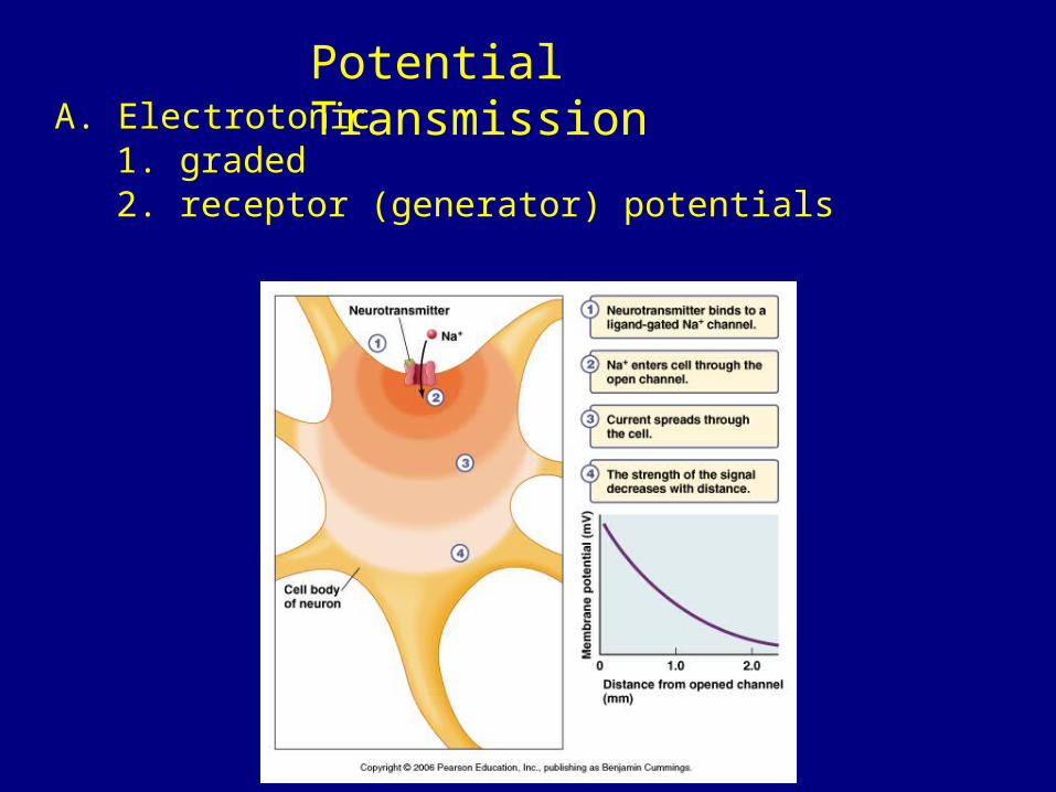
Potential TransmissionA. Electrotonic
1. graded2. receptor (generator) potentials
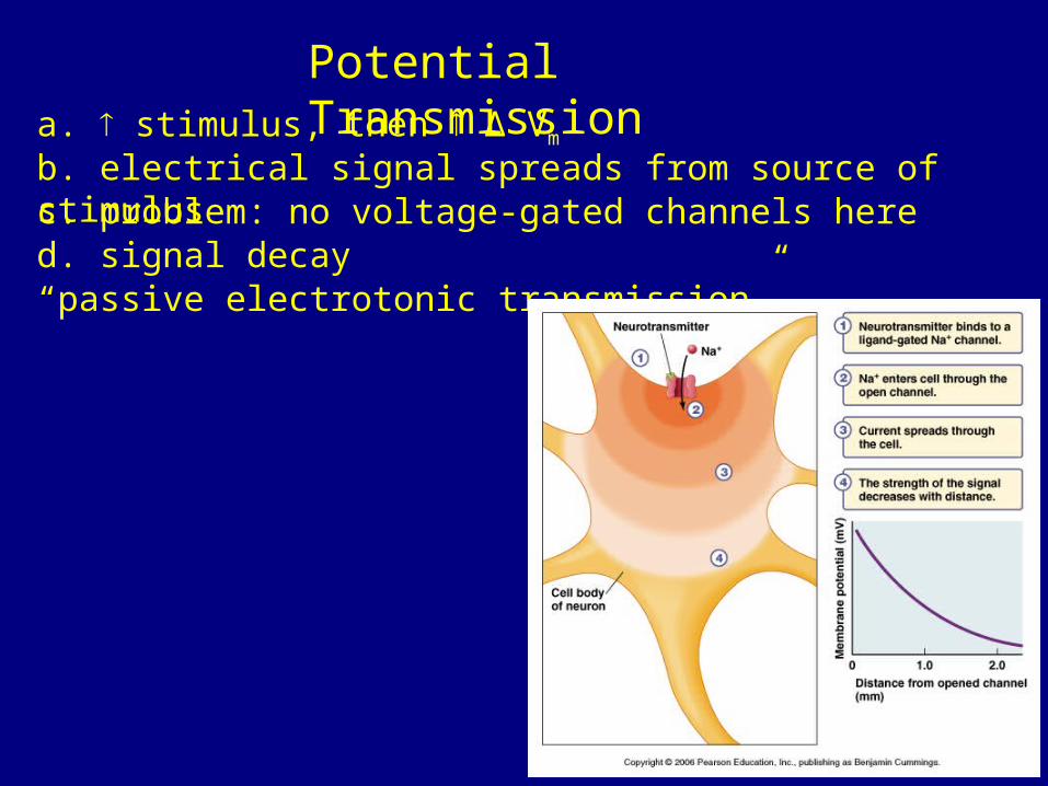
Potential Transmissiona. stimulus, then ∆ Vm
b. electrical signal spreads from source of stimulusc. problem: no voltage-gated channels hered. signal decay“passive electrotonic transmission”
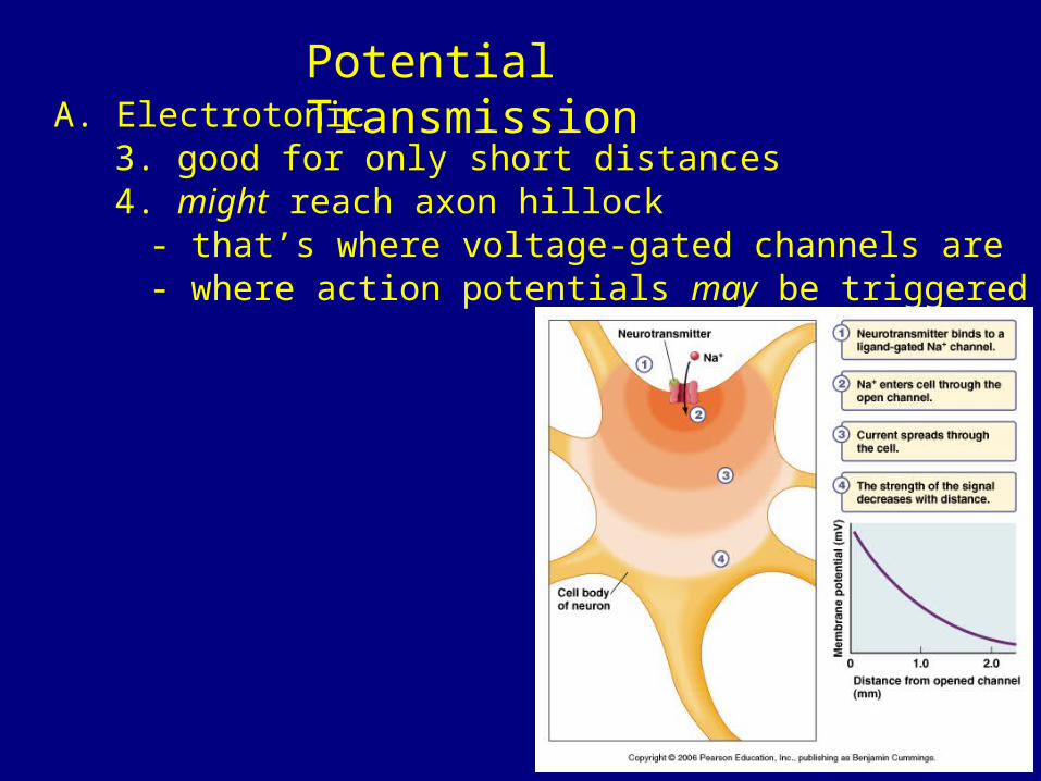
Potential TransmissionA. Electrotonic
3. good for only short distances4. might reach axon hillock
- that’s where voltage-gated channels are- where action potentials may be triggered
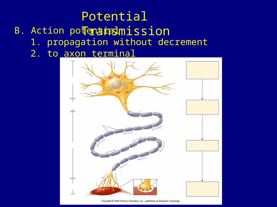
Potential TransmissionB. Action potential
1. propagation without decrement2. to axon terminal
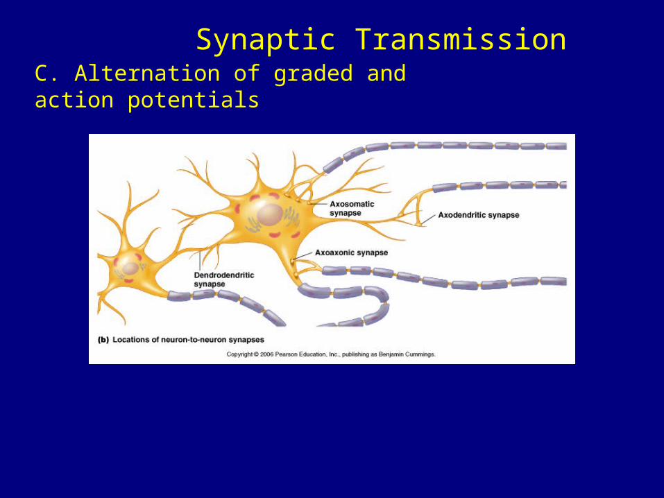
Synaptic TransmissionC. Alternation of graded and action potentials
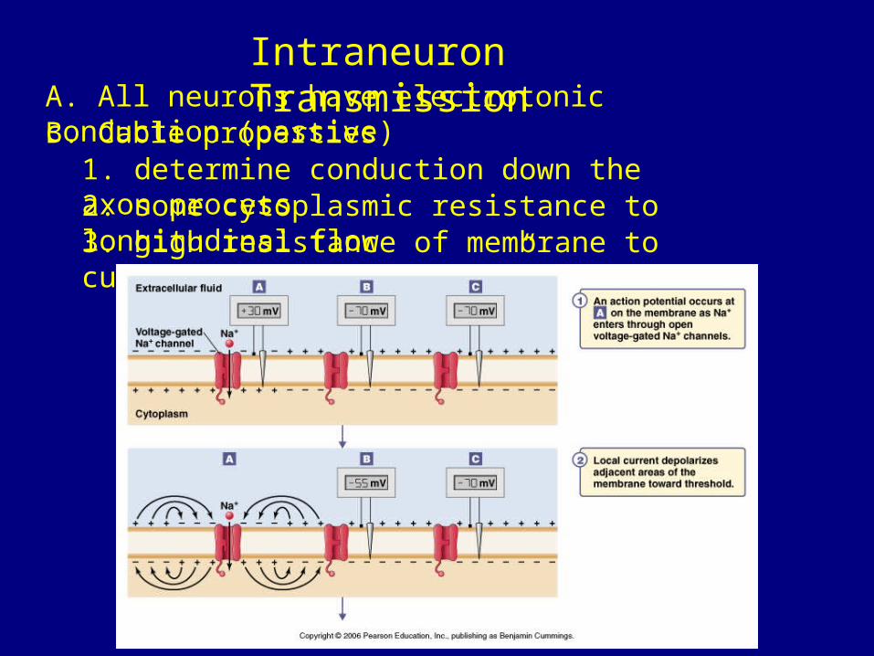
Intraneuron TransmissionA. All neurons have electrotonic conduction (passive)B. Cable properties
1. determine conduction down the axon process2. some cytoplasmic resistance to longitudinal flow3. high resistance of membrane to current
“but membrane is leaky”
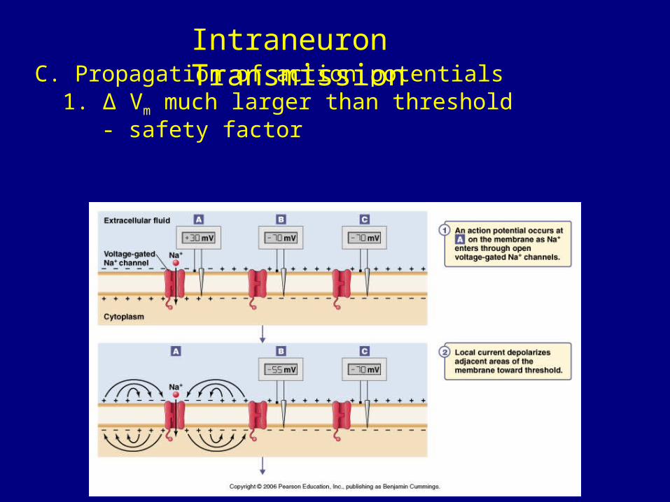
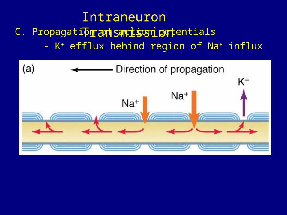
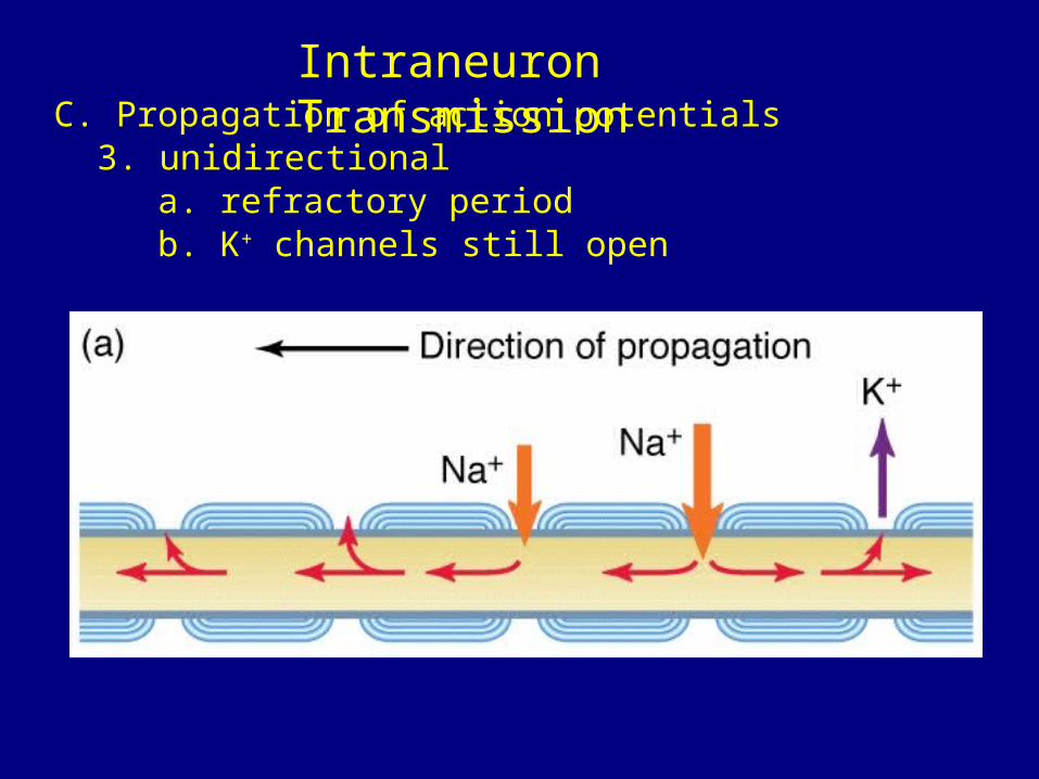
Intraneuron TransmissionC. Propagation of action potentials
1. ∆ Vm much larger than threshold- safety factor
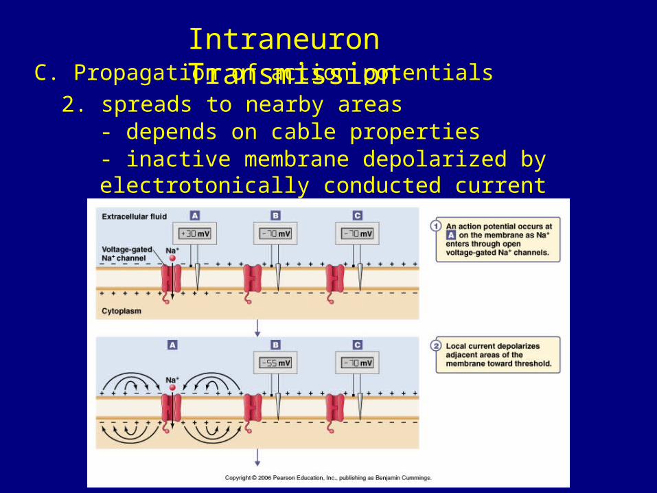
Intraneuron TransmissionC. Propagation of action potentials
2. spreads to nearby areas- depends on cable properties- inactive membrane depolarized by electrotonically conducted current
Intraneuron TransmissionC. Propagation of action potentials
- K+ efflux behind region of Na+ influx
Intraneuron TransmissionC. Propagation of action potentials
3. unidirectionala. refractory periodb. K+ channels still open
Intraneuron TransmissionC. Propagation of action potentials
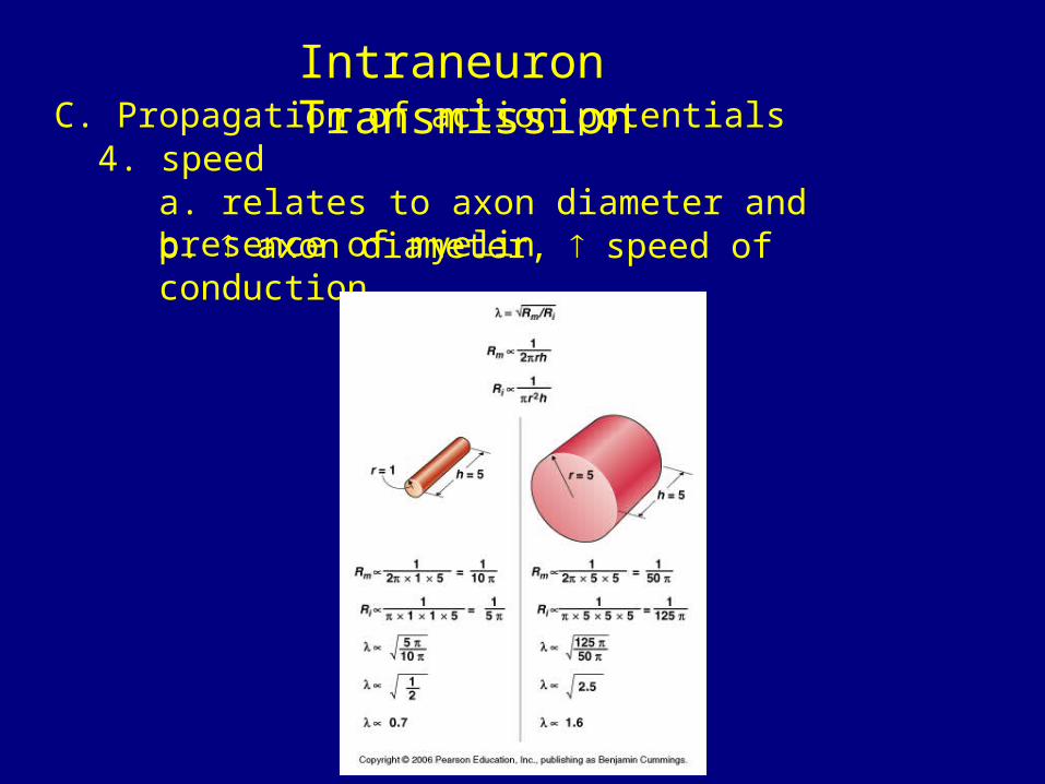
4. speeda. relates to axon diameter and presence of myelinb. axon diameter, speed of conduction
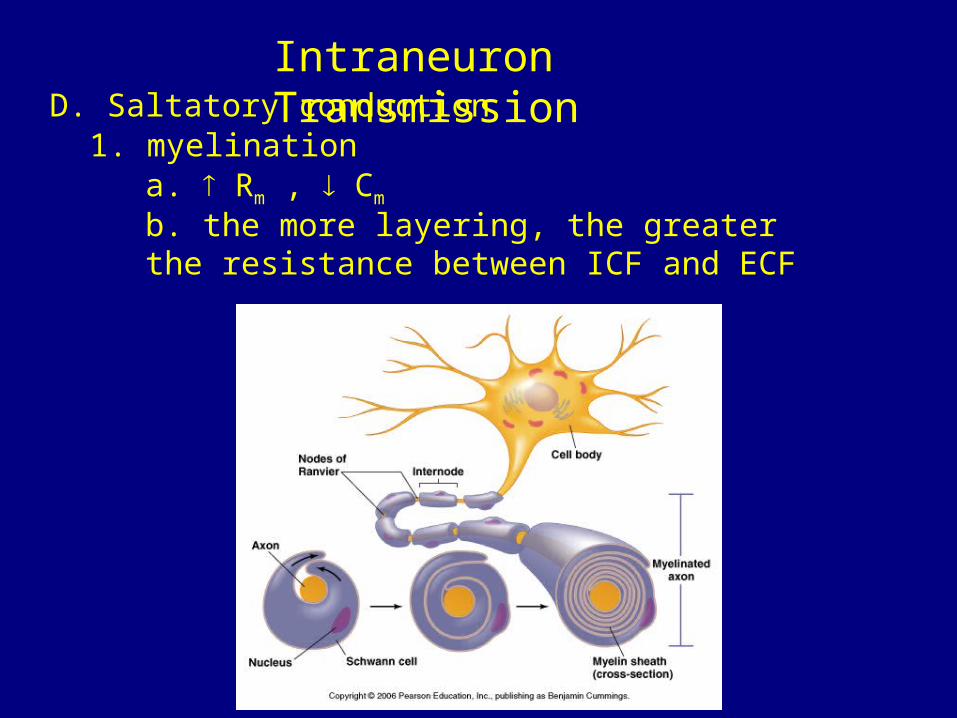
Intraneuron TransmissionD. Saltatory conduction
1. myelinationa. Rm , Cm
b. the more layering, the greater the resistance between ICF and ECF
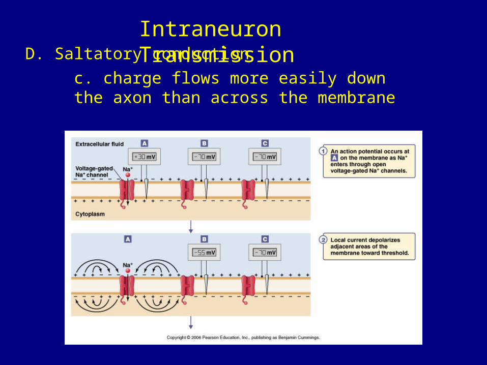
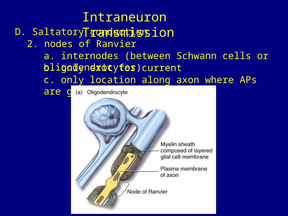
Intraneuron TransmissionD. Saltatory conduction
c. charge flows more easily down the axon than across the membrane
Intraneuron TransmissionD. Saltatory conduction
2. nodes of Ranviera. internodes (between Schwann cells or oligodendrocytes)b. only exit for currentc. only location along axon where APs are generated
Neuronal Integration
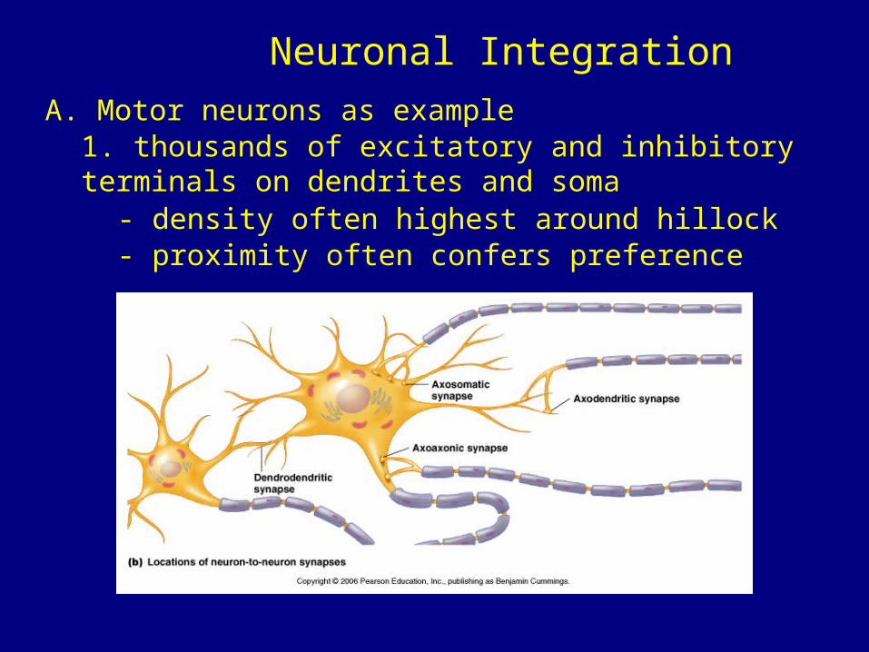
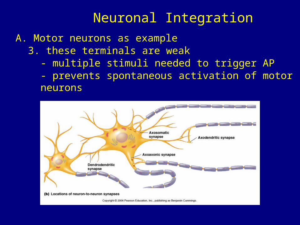
A. Motor neurons as example1. thousands of excitatory and inhibitory terminals on dendrites and soma
- density often highest around hillock- proximity often confers preference
Neuronal Integration
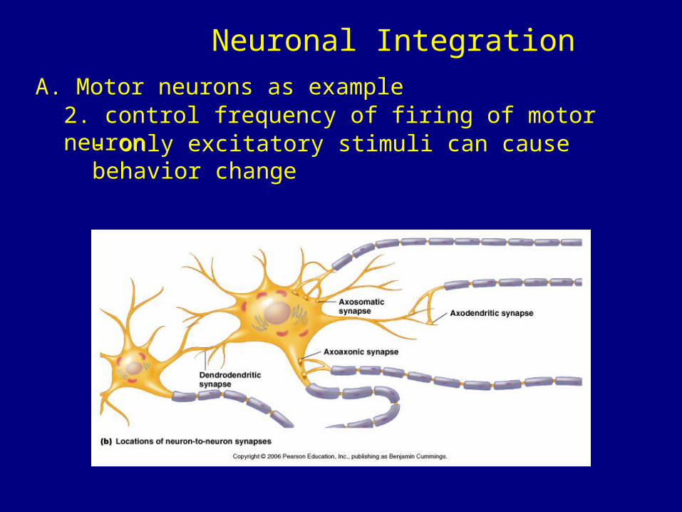
A. Motor neurons as example2. control frequency of firing of motor neuron
- only excitatory stimuli can cause behavior change
Neuronal Integration
A. Motor neurons as example3. these terminals are weak
- multiple stimuli needed to trigger AP- prevents spontaneous activation of motor neurons
Neuronal Integration
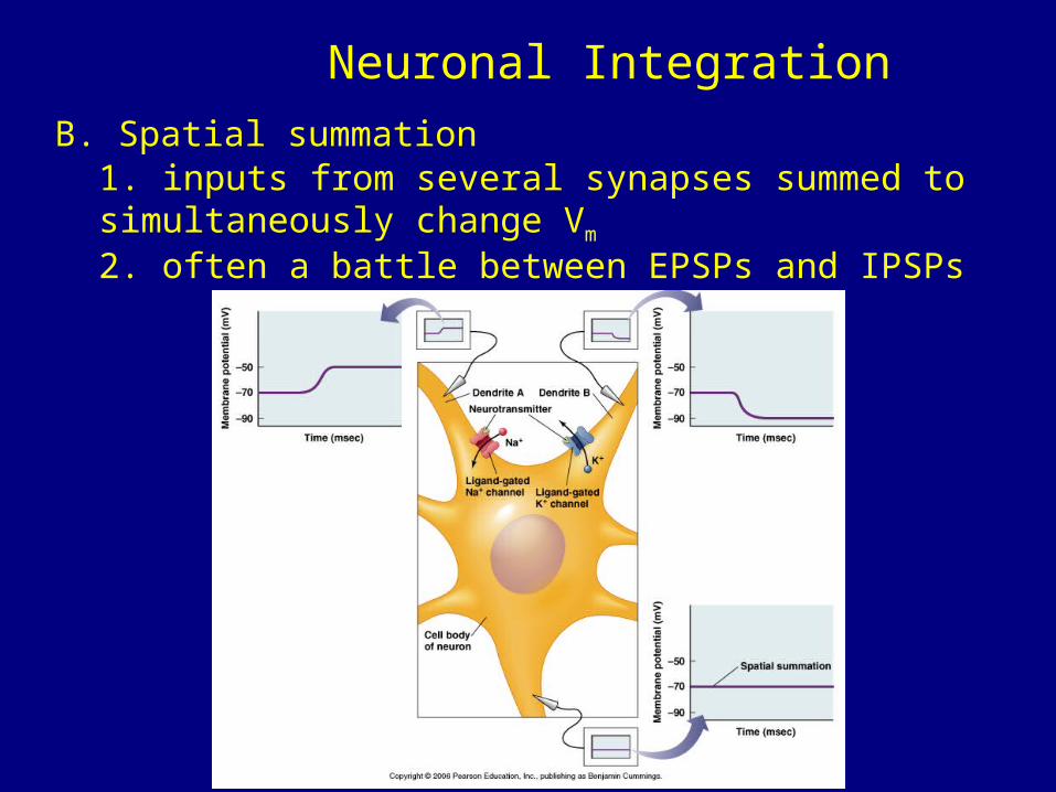
B. Spatial summation1. inputs from several synapses summed to simultaneously change Vm
2. often a battle between EPSPs and IPSPs
Neuronal Integration
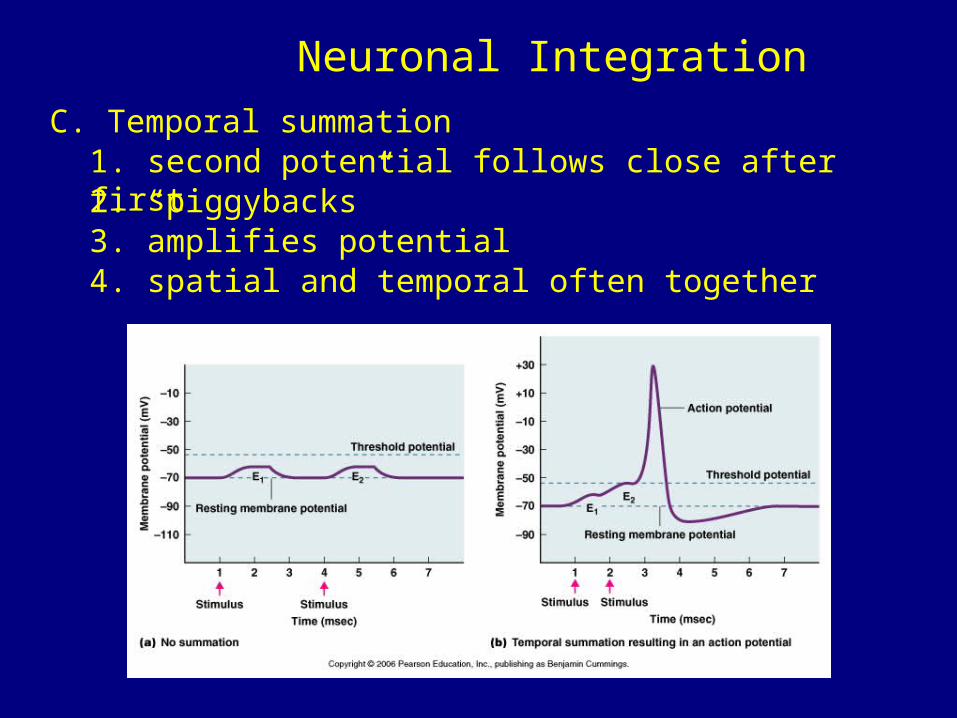
C. Temporal summation1. second potential follows close after first2. “piggybacks”3. amplifies potential4. spatial and temporal often together
![SciHub - WordPress.com · 11/09/2016 · leads to an overabundance of acetylcholine at the neuronal synapses and the neuromuscular junction [12,13]. After ... hub.cc](https://img.pdfslide.us/doc/110x75/5ad692f27f8b9aff228e79bc/scihub-to-an-overabundance-of-acetylcholine-at-the-neuronal-synapses-and-the-neuromuscular.jpg)