Embed Size (px)
Citation preview
RESEARCH ARTICLE Open Access
TDP-43 promotes the formation ofneuromuscular synapses through theregulation of Disc-large expression inDrosophila skeletal musclesNina Strah1†, Giulia Romano1†, Clelia Introna1, Raffaella Klima1, Marta Marzullo2,3, Laura Ciapponi3,Aram Megighian4, Monica Nizzardo5 and Fabian Feiguin1*
Abstract
Background: The ribonuclear protein TDP-43 has been implicated in the pathophysiology of amyotrophic lateralsclerosis (ALS), with genetic mutations being linked to the neurological symptoms of the disease. Though alterations inthe intracellular distribution of TDP-43 have been observed in skeletal muscles of patients suffering from ALS, it is notclear whether such modifications play an active role in the disease or merely represent an expression of musclehomeostatic mechanisms. Also, the molecular and metabolic pathways regulated by TDP-43 in the skeletal muscleremain largely unknown. Here, we analyze the function of TBPH, the Drosophila melanogaster ortholog of TDP-43, inskeletal muscles.
Results: We modulated the activity of TDP-43 in Drosophila muscles by means of RNA interference and observedthat it is required to promote the formation and growth of neuromuscular synapses. TDP-43 regulated theexpression levels of Disc-large (Dlg), and restoring Dlg expression either in skeletal muscles or in motoneuronswas sufficient to suppress the locomotive and synaptic defects of TDP-43-null flies. These results were validatedby the observation of a decrease in Dlg levels in human neuroblastoma cells and iPSC-differentiated motoneuronsderived from ALS patients, suggesting similar mechanisms may potentially be involved in the pathophysiology of thedisease.
Conclusions: Our results help to unveil the physiological role of TDP-43 in skeletal muscles as well as the mechanismsresponsible for the autonomous and non-autonomous behavior of this protein concerning the organization ofneuromuscular synapses.
Keywords: TDP-43, Skeletal muscles, Dlg, Neuromuscular junctions, ALS
© The Author(s). 2020 Open Access This article is licensed under a Creative Commons Attribution 4.0 International License,which permits use, sharing, adaptation, distribution and reproduction in any medium or format, as long as you giveappropriate credit to the original author(s) and the source, provide a link to the Creative Commons licence, and indicate ifchanges were made. The images or other third party material in this article are included in the article's Creative Commonslicence, unless indicated otherwise in a credit line to the material. If material is not included in the article's Creative Commonslicence and your intended use is not permitted by statutory regulation or exceeds the permitted use, you will need to obtainpermission directly from the copyright holder. To view a copy of this licence, visit http://creativecommons.org/licenses/by/4.0/.The Creative Commons Public Domain Dedication waiver (http://creativecommons.org/publicdomain/zero/1.0/) applies to thedata made available in this article, unless otherwise stated in a credit line to the data.
* Correspondence: [email protected]†Nina Strah and Giulia Romano contributed equally to this work.1International Centre for Genetic Engineering and Biotechnology, Padriciano99, 34149 Trieste, ItalyFull list of author information is available at the end of the article
Strah et al. BMC Biology (2020) 18:34 https://doi.org/10.1186/s12915-020-00767-7
BackgroundAmyotrophic lateral sclerosis (ALS) is a devastating dis-ease characterized by the progressive denervation ofskeletal muscles followed by motoneuron degenerationand loss. Its pathology is related to biochemical defectsand genetic mutations in the ribonuclear protein (RNP)TDP-43 which have been detected in the great majorityof patients and are linked to the neurological symptomsof the disease [1, 2]. Histological studies of the nervoussystem of affected individuals have revealed the presenceof protein inclusions consisting of misfolded, abnormallyphosphorylated TDP-43 [3–5]. The pathological modifi-cations affect the subcellular distribution of TDP-43,which is found throughout the cytoplasm instead of itsnormal nuclear localization. Even though the TDP-43 al-terations described above were mostly identified in theupper and lower motoneurons, additional brain regionsand cell types were recently implicated in the pathologyof ALS. A critical role for glia and immune cells hasbeen recognized in the progression of the disease whichhas been confirmed in different animal models [6–10].In this respect, we have reported that the endogenousTDP-43 (TBPH) is localized in glial cells in Drosophilamelanogaster, where it prevents motoneuron degener-ation. This supports the idea that ALS may have a non-neuronal origin [11]. In consonance with this hypothesis,pathological modifications of TDP-43 have been foundin the skeletal muscles of patients with different neuro-muscular diseases such as inclusion body myositis (IBM)and sporadic, familial forms of ALS [12]. Likewise, over-expression of TDP-43 leads to age-related muscle weak-ness and degeneration in mice [13], zebrafish [14], andDrosophila [15, 16], suggesting that the regulation ofTDP-43 function could play an important role in musclephysiology. A requirement for TDP-43 has also been re-ported for the in vitro differentiation of C2C12 myo-blasts as well as for the regeneration of tibial musclesin vivo [17]. Notwithstanding these findings, the mole-cules and metabolic pathways regulated by TDP-43 inskeletal muscles remain largely unknown. It is notknown either whether primary defects of TDP-43 activ-ity at the muscle level could somehow trigger the degen-erative course of ALS nor which could be its potentialconsequences regarding the formation and maintenanceof neuromuscular synapses. In order to address thesequestions, we have analyzed the endogenous function ofTBPH, the TDP-43 ortholog gene, in skeletal muscles ofthe fruit fly Drosophila melanogaster.
MethodsFly strainsThe following genotypes were used:w[1118] - w[1118]; TBPHΔ23/Cyo-GFP [18] - w[1118];
TBPHΔ142/Cyo-GFP [18] - w[1118]; Mef2-GAL4/TM3-Sb -
w[1118]; MHC-GAL4/TM3-Sb - w[1118]; UAS-mCD8::GFP/Cyo - w[1118]; UAS-TBPH/Cyo - w[1118]; UAS-TBPHF/L/TM3-Sb - w[1118]; UAS-hTDP/TM3-Sb -w[1118]; UAS-DLG(egfp)/UAS-DLG(egfp) - w[1118]; UAS-TBPH RNAi (#ID38377) [18] - w[1118]; UAS-GFP RNAi(#9330) - w[1118]; UAS-GFP RNAi (#9331) - w[1118],Dicer(X).
Larval movementWandering 3rd instar larvae (about 96 h old) werepicked from tubes and washed in a drop of deminera-lized water. If necessary, they were selected against dif-ferent markers such as tubby or Cyo-GFP and placedinto 6-cm-diameter dishes, filled with 0.7% agar. A singlelarva at the time was transferred into a 10-cm-diameterdish, filled with 0.7% agar. After 30 s of adaption period,the number of peristaltic waves were counted for aperiod of 2 min. Tested larvae were subsequently trans-ferred to a fresh fly tube to check them, both for hatch-ing (after 4 days) and for correct genotype selection.Generally, 20–25 larvae per genotype were tested.
Survival rateOne- to 2-day-old adult flies were collected from the flytube of the experimental cross in a 1:1 proportion of fe-male and male and transferred to a fresh fly tube andstored in the incubator under controlled conditions(suitable temperature and humidity, 12 h light and 12 hnight). Every second day, flies were transferred into afresh fly tube without anesthesia and the number ofdeaths was scored. Approximately 200 flies per genotypewere tested.
Climbing assayOne- to 2-day-old adult flies were collected from the flytube of the experimental cross in a 1:1 proportion of fe-male and male and transferred into a fresh fly tube andmaintained in an incubator as previously described. Theday of the setting of the experiment was counted as day0. Flies were tested on days 4, 7, 14, and 21. A 50-mlglass cylinder was divided into three parts, as bottom,middle, and top (5 cm each part). Flies were carefullyflipped into the cylinder from the fly tube without anyanesthesia and gently dropped to the bottom. After 30 sof adaptation period, flies were dropped again onto thebottom of the cylinder, and after the time interval of 15s, the numbers of flies present in each part of the cylin-der were scored. For each genotype, 3 trials per tubewere done and the average of the scored fly numberswas considered as the final score. A minimum of 200flies was tested for each genotype.
Strah et al. BMC Biology (2020) 18:34 Page 2 of 13

Walking assayYoung flies 2–3 days old were tested for walking ability.A 145-mm dish was used. The bottom surface was di-vided in a grid of 1 cm × 1 cm squares to facilitate themeasuring of the distance walked by flies. The fly with-out any anesthesia was placed in the middle of the dish,and after 30 s of adaptation to the environment, the dis-tance walked by the fly was recorded for 30 s, countingthe number of squares. A minimum of 50 flies were in-dividually tested for each genotype.
ImmunohistochemistryWandering 3rd instar larvae were picked from the fly tube,in a drop of demineralized water, selected for the genotype,and maintained during the time of dissection in a 6-cm-diameter dish filled with 0.7% agarose dissolved in water. In-dividually picked larva was dissected on Sylgard plates, inDissection Solution (128mM NaCl, 2mM KCl, 4mMMgCl2, 0.1mM CaCl2, 35.5mM sucrose, and 5mM Hepes(pH 7.2)). Larvae were pinned at both ends with minute pins(Austerlic Isect Pins 0.1mm diameter, Fine Science Tools,Germany) and opened on the dorsal site with Spring scissors(Fine Science Tools, Germany). Once larva was opened, in-ternal organs were removed and the interior was extensivelywashed with Dissection Solution leaving the muscle wallopened, pinned flat on the surface. The subsequent step wasa fixation, generally done with 4% PFA in PBS for 20min;however, in the case of glutamate receptors staining a metha-nol fixation of 5min at − 20 °C was performed. Fixation solu-tion was removed with 3 washes in PBS-T (PBS 1×supplemented with 0.1% (v/v) Tween20) for 5min each.After a blocking step of 30min in blocking solution (5%NGS (normal goat serum (#S-1000 Chemicon) in PBS-Tbuffer), larvae were incubated overnight at 4 °C in primaryantibodies diluted in blocking solution. The day after, pri-mary antibody was removed with three washes of 10mineach with PBS-T and a further blocking step of 30min wasperformed before secondary antibody addition. All secondaryantibodies were diluted in blocking solution. An incubationwas 2 h long, carried out at the room temperature. Excess ofantibody was removed by 3 subsequent washes of 20mineach in PBS-T. Finally, dissected-stained larvae were incu-bated overnight at 4 °C in Slowfade®Gold antifade (#S36936Life Technologies) reagent, before being mounted on a glassslide: anti-HRP (#323-005-021, lot:104838, Jackson 1:150),anti-GFP (#A11122, lot:1789911, Life Technologies 1:200),anti-GluRIIA 8B4D2c (DSHB 1:15), anti-Dlg 4F3c (DSHB 1:250), anti-Futsch 22C10s (DSHB 1:50), Alexa-Fluor® 488(mouse #A11001 or rabbit #A11008 1:500), and Alexa-Fluor®555 (mouse #A21422 or rabbit #A21428 1:500).
Acquisition and quantification of confocal imagesIn each experiment, the genotypes of interest were proc-essed simultaneously, and the images were acquired
using the same settings. Images of muscles 6 and 7 onsecond abdominal segment were acquired using theLSM Zeiss Software on a Zeiss 510Meta confocal micro-scope (63 × oil lens) and then analyzed using ImageJ(Wayne Rasband, NIH). For the quantification of pre-and postsynaptic markers, samples were double labeledwith anti-HRP and the marker of interest: the ratio be-tween the mean intensity of the marker and the HRPwas calculated for each bouton of the terminal [18, 19].
Quantification of boutonsBoutons were stained with an anti-HRP antibody. Theshape of boutons was evaluated as regular if they wereround and with a smooth surface, with an equal diam-eter on both axes. On the other hand, boutons wereconsidered as irregular if the shape was not round, themembrane was wrinkled, and the diameter of one axewas different compared to the other one [19].
Electrophysiology on NMJ of the third instar larvapreparationLarval body wall preparations were dissected out inCa2+-free HL3 solution from third instar larvae pinnedon Sylgard-coated petri dishes. The central nervous sys-tem was excised by cutting segmental nerve roots. Afterreplacing Ca2+-free HL3 solution with Ca2+ 1 mMHL3, postsynaptic potentials at neuromuscular junctionof fiber 6/7 of abdominal segments A3/A4 were intracel-lularly recorded, at room temperature in current-clampcondition, using an intracellular microelectrode (tipdiameter 0.5 μm, 15MΩ resistance). The recorded signalwas amplified by a current-clamp amplifier (SEC 05,NPI, Tamm, Germany), digitized at 10-kHz samplingrate using an A/D interface (National Instruments, Aus-tin, TX, USA) and fed to a computer for display andstorage using an appropriate software (Win EDR, Strath-clyde University, Glasgow, UK).Fibers with a resting membrane potential below − 60
mV were considered for the experiment. In these fibers,membrane potential was set at − 70mV throughout theexperiment by injecting current through the intracellularelectrode. Evoked postsynaptic potentials (EPSPs or exci-tatory junctional potentials or EJPs) were recorded bystimulating at 0.1 Hz (pulse duration 0.4ms; 1.5 thresholdvoltage) the segmental nerve using a suction electrode (tipdiameter ~ 10 μm) connected to a stimulator (S88, Grass,Pleasanton, CA, USA) through a stimulus isolation unit(SIU5, Grass, Pleasanton, CA, USA). Intracellular record-ings were analyzed offline using pClamp software(pClamp, Axon, Sunnyvale, CA, USA). Statistical compari-sons and graphs were made using Graphpad software(Graphpad, La Jolla, CA, USA) or MATLAB (Matworks,Natick, MA, USA).
Strah et al. BMC Biology (2020) 18:34 Page 3 of 13
ImmunoprecipitationProtein G magnetic beads (#10003D Invitrogen) werewashed two times with PBS + 0.02% Tween and coatedwith anti-FLAG M2 monoclonal antibody (#F3165, Lot:SLBQ7119V, Sigma). Thoraces or heads of adult flies werecut and stored in lysis buffer containing 20mM Hepes,150mM NaCl, 0.5 mM EDTA, 10% Glycerol, 0.1% TritonX-100, 1mM DTT, and protease inhibitor (#04 693 159001 Roche). Samples were homogenized with a Douncehomogenizer, and major debris were removed by centrifu-gation step of 5min at 0.4g at 8 °C. The pretreated beadsand tissue extracts were mixed and incubated for 30minat 4 °C. After this binding step, beads were washed fivetimes with washing buffer (20mM Hepes, 150mM NaCl,0.5 mM EDTA, 10% glycerol, 0.1% Triton X-100, 1 mMDTT, protease inhibitor, 0.2% DOC, 0.5M Urea) usingDynaMagTM-Spin (#123.20D Invitrogen). RNA tran-scripts bound by Flag-tagged TBPH were extracted treat-ing the beads with Trizol (#15596026 Ambion), and RNAwas precipitated with isopropanol adding glycogen(#R0551 Thermo Scientific). Retro-transcription was per-formed with Superscript III First-Strand Synthesis(#18080-093 Invitrogen) and oligo-dT and subjected toreal-time PCR with gene-specific primers, whose se-quences are listed below.
Target fw primer rv primer
Rpl11 5′-CCATCGGTATCTATGGTCTGGA-3′
5′-CATCGTATTTCTGCTGGAACCA-3′
Syntaxin 5′-TGTTCACGCAGGGCATCATC-3′
5′-GCCGTCTGCACATAGTCCATAG-3′
Hdac-6 5′-CGAGCGGCTGAAGGAGAC-3′
5′-ACCAGATGGTCCACCAATTCG-3′
Dlg 5′-ACTGGGCTTCTCAATTGCCG-3′
5′-CCAGTTCGTGCGTTACGTTC-3′
In order to calculate the enrichment fold, initially, alldata were normalized to the respective inputs. Thesignal was represented by how many more fold increasewas measured compared to the control signal. Theenrichment was calculated according to the 2-ΔΔCt
method.The results were derived from three independent
immunoprecipitation experiments [19, 20].
Protein extractionTo collect adult heads, flies were flash-frozen in liquidnitrogen for 10 s and immediately vortexed to easily de-tach heads from bodies. Heads were subsequently trans-ferred into Lysis buffer (150 mM Tris, 5 mM EDTA, 10%glycerol, 5 mM EGTA, 50mM NaF, 4M urea, 5 mMDTT, and protease inhibitors (#04 693 159 001 Roche)).After a squeezing step, performed both, manually and
mechanically, the homogenized samples were gotten ridof major debris by centrifugation at 0.5×g for 6 min on4 °C. The protein concentration of the collected super-natant was quantified with Quant-iT™ Protein Assay Kit(#Q33212 Invitrogen), following the supplier protocol.Transfected neuroblastoma cell line SH-SY-5Y was re-
suspended in iced RIPA buffer added of protease inhibi-tors (#04693159001 Roche) and subjected to sonication(Biorupture sonication system, Diagenode).Lysates were quantified (BCA Protein kit #23225
Thermo Scientific), following the supplier protocol.
RNA extraction and qRT-PCRRNA was extracted from Drosophila adult heads, 1 dayaged and sex-matched, of both wildtype and TBPH-nullalleles (tbphD23 and tbphD142) and from human MN differ-entiated cells with RNeasy Microarray tissue kit (QIAGEN#73304) and treated with Turbo DNA-free kit (Ambion#AM1907).Retro-transcription was performed with Superscript III
First-Strand Synthesis (#18080-093 Invitrogen) usingoligo-dT with the exception of the analysis of the Dros-ophila intronic region for which random hexamers havebeen used. Real-time PCR was carried out with theprimers listed below using Platinum Syber Green(#11733-038 Thermo Fisher) on a Bio-Rad CFX96 qPCRSystem, and minus-retro control has been performed.The primer sets used are listed in the below table; Sdhaand GAPDH genes have been used as Drosophila andhuman reference, respectively.
Target fw primer rv primer
Drosophila
d-DLG exon1–4
5′-CTGTGCCAAACGCATGTCC-3′
5′-TTCACAGCTCAATGGCTCCT-3′
d-DLGintron 1–4
5′-TCTGCGACCCAGTTTTCCAC-3′
5′-TCGCCCGTGGTTGTAATTGT-3′
Sdha 5′-CATGCTGCTGTGTTCCGCGA-3′
5′-ACCATCCAGGGGCTTGCTGA-3′
Human
h-DLG 5′-GTTGCACAATATCGACCTGAAGA-3′
5′-GGGATCGCTTCTGGCTAGTTC-3′
GAPDH 5′-CTGGGCTACACTGAGCACC-3′
5′-AAGTGGTCGTTGAGGGCAATG-3′
SDS-PAGEProtein samples were diluted in 1x Laemmli buffer(composition of 5x: 0.3 M Tris-HCl pH 6.8, 50% glycerol,10% SDS, 25% β-mercaptoethanol, 0.05% bromophenolblue) to reach the same concentration among all andthen boiled at 95 °C for 5 min. Afterwards, they wereloaded on a polyacrylamide gel.
Strah et al. BMC Biology (2020) 18:34 Page 4 of 13

The loaded gel was placed into a chamber with 1×running buffer (10× running buffer: 30.28 g Tris, 114.13g glycine, 10 g SDS in 1 l water). The conditions set were25mA per gel.
Western blotWhen proteins were separated by the electrophoresis, theywere transferred to a nitrocellulose membrane Amersham™Protran™ 0.2 μm NC (Life Science). The western blotsandwich was put into the chamber, filled with transferbuffer 1× containing 20% methanol (transfer buffer 10×: 30 gTris, 144 g glycine in 1 l water). The transfer lasted 1 h at350mA. The membrane was incubated with a solution of5% milk in 1× TBS 0.01% Tween (TBS-T) for 30min at aroom temperature on a shaker (TBS buffer 10×: 24.2 g Tris,80 g NaCl in 1 l water, pH 7.6). After blocking, themembrane was set into dilution of primary antibody withTBS-T with 5% milk. It was placed at 4 °C overnight. Whenthe incubation with primary antibody was over, five washeswith TBS-T followed, 5min each. Next, the membrane wasincubated with the secondary antibody diluted in TBS-T with5% milk for 1 h at room temperature. The protein detectionwas performed with SuperSignal®West Femto MaximumSensitivity Substrate (#TB260893 Thermo Fisher Scientific).Primary antibodies include anti-Dlg 4F3c (DSHB 1:10000),anti-TBPH (home-made 1:4000), anti-tubulin (#CP06, Lot:2681308, Calbiochem 1:4000), anti-TDP-43 (#12892-1-AP,Lot:00016371, Proteintech 1:4000), anti-Dlg 2D11 (#SC9961,Lot:G2617, Santa Cruz 1:1000), and anti-GAPDH(#SC25778, Lot:G1008, Santa Cruz 1:2000). Secondary anti-bodies include anti-mouse-HRP (#31430 Thermo Fisher Sci-entific 1:30000(flies), 1:10000(cells)) and anti-rabbit-HRP(#31460 Thermo Fisher Scientific 1:10000).
Cell culture and RNA interferenceSH-SY-5Y neuroblastoma cell line was cultured instandard conditions in DMEM-Glutamax (#31966-021,Thermo Fisher Scientific) supplemented with 10% fetalbovine serum and 1 × antibiotic-antimycotic solution(#A5955; Sigma). RNA interference of TDP-43 wasachieved using HiPerfect Transfection Reagent (#301705,Qiagen) and siRNA specific for human TDP43 (5′-gcaaagccaagaugagccu-3′); as control, siRNA for luciferasewas used (5′-uaaggcuaugaagagauac-3′; Sigma). Immedi-ately before transfection, 2–4 × 10⁵ cells were seeded in 6-well plates in 1.4 ml of medium containing 10% fetalserum. A volume of 3 μl of each siRNA (40 μM solution inwater) was added to 91 μl of Opti-MEM I reduced serummedium (#51985-026, Thermo Fisher Scientific) and incu-bated 5min at room temperature, and subsequently, 6 μlof HiPerfect Transfection Reagent was added. The silen-cing procedure was performed again after 24 and 48 h.
Human iPSC culture and MN differentiationHuman iPSC culture and MN differentiation were alreadydescribed in [21]. All the studies performed with humansamples were in compliance with the Code of Ethics ofthe World Medical association (Declaration of Helsinki)and with the national legislation and institutionalguidelines. Briefly, fibroblasts from dermal biopsies(Eurobiobank) from ALS patients (TDP-43 mutations #1:G294V; #2:G378S) and controls were reprogrammed intoiPSCs with CytoTune-iPS 2.0 Sendai reprogramming Kit(#A16517, Thermo Fisher) and differentiated into MNswith the multistep protocol described by Ng [22].
Statistical analysisAll statistical analysis was performed with Prism(GraphPad, USA) version 5.1. One-way ANOVA withBonferroni correction and t test with Mann-Whitneycorrection were applied as statistical test. In all fig-ures, all the values were presented as the mean andthe standard error of the mean (SEM). Statistical sig-nificance was portrayed as *p < 0.05, **p < 0.01, ***p <0.001, and ****p < 0.0001.
ResultsThe suppression of TBPH in muscles affects Drosophilalocomotion and life spanWe have previously described that TBPH is expressed inDrosophila muscles, being present in myocytes from larvalstages until adulthood [16]. To analyze its role in thesetissues, we suppressed TBPH expression by means ofRNA interference (RNAi). Two different GAL4 lines wereutilized: Mhc (myosin heavy chain)-GAL4, withpredominant expression in larval muscles, and Mef2-GAL4, expressed during muscle development and inadulthood [23]. The expression of anti-TBPH RNAi (TBi)in either Mhc-GAL4 or Mef2-GAL4 significantly affectedthe locomotor capacities of flies at both larval and adultstages (Fig. 1a, b; Additional file 1 Fig.S1). Moreover, thelife span of Mef2-GAL4 flies expressing TBi was stronglycompromised compared with that of Mef2-GAL4 controlflies expressing GFP-RNAi (GFPi) (Fig. 1c). It could alsobe inferred that a reduction of one copy of the endogen-ous TBPH gene due to the expression of TBi results in anexacerbation of locomotive defects (Mhc-GAL4, TBPH−/+,TBi vs Mhc-GAL4, TBPH−/+, GFP-RNAi). This was an in-dication that these phenotypes are gene dose-sensitive andrather specific (Fig. 1a). At the cellular level, the expres-sion of TBi strongly affected the organization of neuro-muscular synapses, as revealed by the localizationof postsynaptic proteins in muscle cell membranes(Fig. 1d–g). More specifically, we detected strongly re-duced levels of the postsynaptic protein Disc-large (Dlg)in TBPH-RNAi-treated muscles compared with controlsand observed the presence of numerous gaps of a
Strah et al. BMC Biology (2020) 18:34 Page 5 of 13
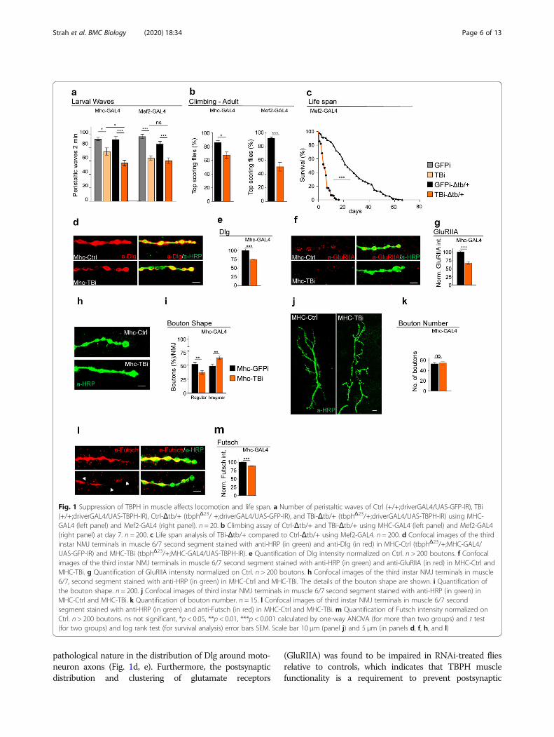
pathological nature in the distribution of Dlg around moto-neuron axons (Fig. 1d, e). Furthermore, the postsynapticdistribution and clustering of glutamate receptors
(GluRIIA) was found to be impaired in RNAi-treated fliesrelative to controls, which indicates that TBPH musclefunctionality is a requirement to prevent postsynaptic
Fig. 1 Suppression of TBPH in muscle affects locomotion and life span. a Number of peristaltic waves of Ctrl (+/+;driverGAL4/UAS-GFP-IR), TBi(+/+;driverGAL4/UAS-TBPH-IR), Ctrl-Δtb/+ (tbphΔ23/ +;driverGAL4/UAS-GFP-IR), and TBi-Δtb/+ (tbphΔ23/+;driverGAL4/UAS-TBPH-IR) using MHC-GAL4 (left panel) and Mef2-GAL4 (right panel). n = 20. b Climbing assay of Ctrl-Δtb/+ and TBi-Δtb/+ using MHC-GAL4 (left panel) and Mef2-GAL4(right panel) at day 7. n = 200. c Life span analysis of TBi-Δtb/+ compared to Ctrl-Δtb/+ using Mef2-GAL4. n = 200. d Confocal images of the thirdinstar NMJ terminals in muscle 6/7 second segment stained with anti-HRP (in green) and anti-Dlg (in red) in MHC-Ctrl (tbphΔ23/+;MHC-GAL4/UAS-GFP-IR) and MHC-TBi (tbphΔ23/+;MHC-GAL4/UAS-TBPH-IR). e Quantification of Dlg intensity normalized on Ctrl. n > 200 boutons. f Confocalimages of the third instar NMJ terminals in muscle 6/7 second segment stained with anti-HRP (in green) and anti-GluRIIA (in red) in MHC-Ctrl andMHC-TBi. g Quantification of GluRIIA intensity normalized on Ctrl. n > 200 boutons. h Confocal images of the third instar NMJ terminals in muscle6/7, second segment stained with anti-HRP (in green) in MHC-Ctrl and MHC-TBi. The details of the bouton shape are shown. i Quantification ofthe bouton shape. n = 200. j Confocal images of third instar NMJ terminals in muscle 6/7 second segment stained with anti-HRP (in green) inMHC-Ctrl and MHC-TBi. k Quantification of bouton number. n = 15. l Confocal images of third instar NMJ terminals in muscle 6/7 secondsegment stained with anti-HRP (in green) and anti-Futsch (in red) in MHC-Ctrl and MHC-TBi. m Quantification of Futsch intensity normalized onCtrl. n > 200 boutons. ns not significant, *p < 0.05, **p < 0.01, ***p < 0.001 calculated by one-way ANOVA (for more than two groups) and t test(for two groups) and log rank test (for survival analysis) error bars SEM. Scale bar 10 μm (panel j) and 5 μm (in panels d, f, h, and l)
Strah et al. BMC Biology (2020) 18:34 Page 6 of 13
membrane disorganization (Fig. 1f, g). The downregulationof TBPH in Drosophila muscles also provoked non-autonomous alterations in the structure of presynaptic ter-minals, which showed a loss of the characteristics roundand smooth shape of the synaptic boutons (Fig. 1h, i) with-out affecting their total number (Fig. 1j, k). At a molecularlevel, these morphological alterations were associated witha significative reduction in the levels of the presynapticmicrotubule binding protein futsch, homolog to the humanprotein MAP1B, involved in the organization of the synap-tic microtubule cytoskeleton (Fig. 1l, m).
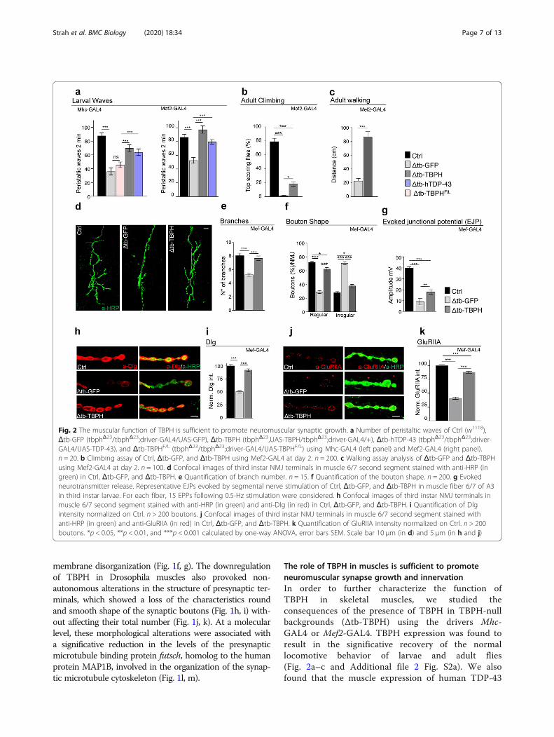
The role of TBPH in muscles is sufficient to promoteneuromuscular synapse growth and innervationIn order to further characterize the function ofTBPH in skeletal muscles, we studied theconsequences of the presence of TBPH in TBPH-nullbackgrounds (Δtb-TBPH) using the drivers Mhc-GAL4 or Mef2-GAL4. TBPH expression was found toresult in the significative recovery of the normallocomotive behavior of larvae and adult flies(Fig. 2a–c and Additional file 2 Fig. S2a). We alsofound that the muscle expression of human TDP-43
Fig. 2 The muscular function of TBPH is sufficient to promote neuromuscular synaptic growth. a Number of peristaltic waves of Ctrl (w1118),Δtb-GFP (tbphΔ23/tbphΔ23;driver-GAL4/UAS-GFP), Δtb-TBPH (tbphΔ23,UAS-TBPH/tbphΔ23;driver-GAL4/+), Δtb-hTDP-43 (tbphΔ23/tbphΔ23;driver-GAL4/UAS-TDP-43), and Δtb-TBPHF/L (tbphΔ23/tbphΔ23;driver-GAL4/UAS-TBPHF/L) using Mhc-GAL4 (left panel) and Mef2-GAL4 (right panel).n = 20. b Climbing assay of Ctrl, Δtb-GFP, and Δtb-TBPH using Mef2-GAL4 at day 2. n = 200. c Walking assay analysis of Δtb-GFP and Δtb-TBPHusing Mef2-GAL4 at day 2. n = 100. d Confocal images of third instar NMJ terminals in muscle 6/7 second segment stained with anti-HRP (ingreen) in Ctrl, Δtb-GFP, and Δtb-TBPH. e Quantification of branch number. n = 15. f Quantification of the bouton shape. n = 200. g Evokedneurotransmitter release. Representative EJPs evoked by segmental nerve stimulation of Ctrl, Δtb-GFP, and Δtb-TBPH in muscle fiber 6/7 of A3in third instar larvae. For each fiber, 15 EPPs following 0.5-Hz stimulation were considered. h Confocal images of third instar NMJ terminals inmuscle 6/7 second segment stained with anti-HRP (in green) and anti-Dlg (in red) in Ctrl, Δtb-GFP, and Δtb-TBPH. i Quantification of Dlgintensity normalized on Ctrl. n > 200 boutons. j Confocal images of third instar NMJ terminals in muscle 6/7 second segment stained withanti-HRP (in green) and anti-GluRIIA (in red) in Ctrl, Δtb-GFP, and Δtb-TBPH. k Quantification of GluRIIA intensity normalized on Ctrl. n > 200boutons. *p < 0.05, **p < 0.01, and ***p < 0.001 calculated by one-way ANOVA, error bars SEM. Scale bar 10 μm (in d) and 5 μm (in h and j)
Strah et al. BMC Biology (2020) 18:34 Page 7 of 13

in a TBPH-null background (Δtb-hTDP-43) similarlyled to a motility recovery, suggesting that the role ofTDP-43/TBPH is conserved in skeletal muscles(Fig. 2a and Additional file 2 Fig. S2b). On thecontrary, the expression of a RNA-binding-deficientisoform of TBPH (TBPHF/L) was not able to revertTBPH-minus phenotypes, demonstrating that RNAbinding is essential for TDP-43/TBPH functionalityin these tissues (Fig. 2a) [19]. The TBPH-induced re-covery of muscle function stimulated the growth of
motoneuron axons as well as the formation ofterminal branches and new synaptic boutons inTBPH-minus flies (Fig. 2d–f and Additional file 2Fig. S2 c and d). Interestingly, the non-autonomousrescue of presynaptic terminals was followed by thereestablishment of evoked junction potentials (EJPs)between motoneurons and their underlying muscles,suggesting the recovery of synaptic transmission inTBPH-expressing muscles compared with controls(Fig. 2g).
Fig. 3 TBPH in the muscle promotes synaptic growth through the regulation of Dlg levels. a qRT-PCR analysis of mRNAs immunoprecipitated byFlag-tagged TBPH (UAS-TBPH/+;Mef2-GAL4/+, IP-TBPH) and its mutant variants TBPHF/L (+/+;UAS-TBPH F/L/Mef2-GAL4, IP-TBPHF/L) in adult thoraxes.The dlg enrichment folds was referred to rpl-11 (negative control); hdac6 has been used as the positive control. n = 3 (biological replicates). Individualdata values are provided in Additional file 4. Individual Data Values.xls. b Number of peristaltic waves of Ctrl (w1118), Δtb-GFP (tbphΔ23/tbphΔ23;Mef2-GAL4/UAS-GFP), and Δtb-Dlg (tbphΔ23,UAS-Dlg/tbphΔ23;Mef2-GAL4/+). n = 20. c Climbing assay of Ctrl, Δtb-GFP, and Δtb-Dlg using Mef2-GAL4 at day2. n = 200. d Evoked neurotransmitter release. Representative EJPs evoked by segmental nerve stimulation of Ctrl, Δtb-GFP, and Δtb-Dlg in musclefiber 6/7 of A3 in third instar larvae. For each fiber, 15 EPPs following 0.5-Hz stimulation were considered. e Confocal images of third instar NMJterminals in muscle 6/7 second segment stained with anti-HRP (in green) in Ctrl, Δtb-GFP, and Δtb-Dlg. f Confocal images of third instar NMJ terminalboutons in muscle 6/7 second segment stained with anti-HRP (in green) in Ctrl, Δtb-GFP, and Δtb-Dlg. g Quantification of branch number. n = 15.h Quantification of the bouton shape. n = 200. i Confocal images of third instar NMJ terminals in muscle 6/7 second segment stained with anti-HRP (ingreen) and anti-GluRIIA (in red) in Ctrl, Δtb-GFP, and Δtb-Dlg. j Quantification of GluRIIA intensity normalized on Ctrl. n > 200 boutons. **p < 0.01 and***p < 0.001 calculated by one-way ANOVA, error bars SEM. Scale bar 10 μm (in e) and 5 μm (in f and i)
Strah et al. BMC Biology (2020) 18:34 Page 8 of 13
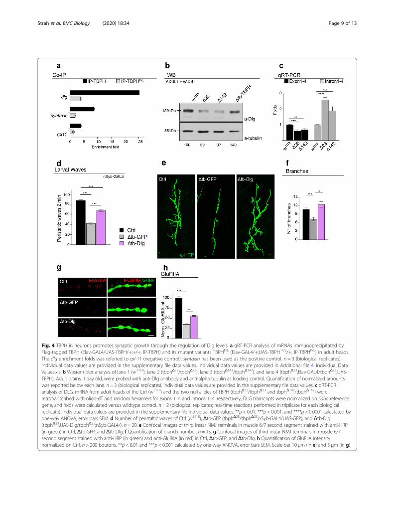
Fig. 4 TBPH in neurons promotes synaptic growth through the regulation of Dlg levels. a qRT-PCR analysis of mRNAs immunoprecipitated byFlag-tagged TBPH (Elav-GAL4/UAS-TBPH/+;+/+, IP-TBPH) and its mutant variants TBPHF/L (Elav-GAL4/+;UAS-TBPH F/L/+, IP-TBPHF/L) in adult heads.The dlg enrichment folds was referred to rpl-11 (negative control); syntaxin has been used as the positive control. n = 3 (biological replicates).Individual data values are provided in the supplementary file data values. Individual data values are provided in Additional file 4. Individual DataValues.xls. b Western blot analysis of lane 1 (w1118), lane 2 (tbphΔ23/tbphΔ23), lane 3 (tbphΔ142/tbphΔ142), and lane 4 (tbphΔ23,Elav-GAL4/tbphΔ23,UAS-TBPH). Adult brains, 1 day old, were probed with anti-Dlg antibody and anti-alpha-tubulin as loading control. Quantification of normalized amountswas reported below each lane. n = 3 (biological replicates). Individual data values are provided in the supplementary file data values. c qRT-PCRanalysis of DLG. mRNA from adult heads of the Ctrl (w1118) and the two null alleles of TBPH (tbphΔ23/tbphΔ23 and tbphΔ142/tbphΔ142) wereretrotranscribed with oligo-dT and random hexamers for exons 1–4 and introns 1–4, respectively; DLG transcripts were normalized on Sdha referencegene, and folds were calculated versus wildtype control. n = 2 (biological replicates; real-time reactions performed in triplicate for each biologicalreplicate). Individual data values are provided in the supplementary file individual data values. **p < 0.01, ***p < 0.001, and ****p < 0.0001 calculated byone-way ANOVA, error bars SEM. d Number of peristaltic waves of Ctrl (w1118), Δtb-GFP (tbphΔ23/tbphΔ23;nSyb-GAL4/UAS-GFP), and Δtb-Dlg(tbphΔ23,UAS-Dlg/tbphΔ23;nSyb-GAL4/). n = 20. e Confocal images of third instar NMJ terminals in muscle 6/7 second segment stained with anti-HRP(in green) in Ctrl, Δtb-GFP, and Δtb-Dlg. f Quantification of branch number. n = 15. g Confocal images of third instar NMJ terminals in muscle 6/7second segment stained with anti-HRP (in green) and anti-GluRIIA (in red) in Ctrl, Δtb-GFP, and Δtb-Dlg. h Quantification of GluRIIA intensitynormalized on Ctrl. n > 200 boutons. **p < 0.01 and ***p < 0.001 calculated by one-way ANOVA, error bars SEM. Scale bar 10 μm (in e) and 5 μm (in g)
Strah et al. BMC Biology (2020) 18:34 Page 9 of 13
TBPH promotes the assembly of neuromuscular synapsesby regulating muscle and neuronal levels of DlgThe expression of TBPH in the muscles of TBPH-nullflies also resulted in an almost complete restoration ofthe cytoplasmic levels and postsynaptic distribution ofDlg around presynaptic terminals (Fig. 2h, i and Add-itional file 2 Fig. S2e and f). This was complemented bya significative reorganization of glutamate receptors inwell-defined clusters at the postsynaptic membrane level(Fig. 2j, k and Additional file 2 Fig. S2g and h). Regard-ing the molecular basis of the relationship betweenTBPH and Dlg, the fact that several putative TBPH bind-ing sites are present along the mRNA sequence of Dlg issuggestive of physical interaction points (Additional file 3:Table 1 and 2 and [24–26]). To test this possibility, weperformed RIP assays employing a tagged version ofTBPH expressed in vivo in Drosophila muscles. We ob-served that TBPH, but not TBPHF/L, was able to pulldown Dlg mRNA (Fig. 3a). In addition, the ectopic expres-sion of Dlg in Drosophila muscles via Mef2-GAL4 wasable to restore motility and climbing in TBPH-null flies(Fig. 3b, c) and to re-establish evoked junction potentials(EJPs) between motoneurons and their underlying muscles(Fig. 3d). Moreover, the postsynaptic expression of Dlgwas observed to promote the non-autonomous growth ofmotoneuron axons (Fig. 3e–h) as well as the recovery ofthe postsynaptic organization of glutamate receptors atthe neuromuscular membranes (Fig. 3i, j), indicating thatDlg is as a bona fide mediator of signaling pathways re-lated to TBPH in Drosophila muscles. Dlg expression wasin fact detected in motoneuron axons, which is consistent
with a possible role of TBPH in the regulation of Dlg ex-pression in neuronal tissues. In support of this idea, RIPassays showed that TBPH binds Dlg mRNA in Drosophilabrains upon TBPH expression through the pan-neuronaldriver elav-GAL4 (Fig. 4a). Moreover, we found that theprotein levels of Dlg appeared downregulated in adultheads of Drosophila TBPHΔ23 and TBPHΔ142 homozygousalleles compared to wildtype controls and, importantly,the decreased Dlg protein levels were restored by the ex-pression of endogenous TBPH demonstrating a degree ofspecificity in the results observed (Fig. 4b). Regarding themolecular mechanisms behind Dlg protein defects, wequantified by qRT-PCR the primary and mature tran-scripts of Dlg mRNA. For these experiments, we designeda set of primers able to amplify the initial coding exons 1and 4 present in the most conserved and longest isoformsof Dlg mature mRNA. To detect the primary transcriptsof Dlg mRNA, we selected primers directed to the intronicregion between the exons described above. As a result, wefound that Dlg mature mRNA was downregulated inTBPH-null alleles (Fig. 4c, left graph). On the contrary,the primary transcripts of Dlg appeared upregulated inmutant flies compared to wildtype controls (Fig. 4c, rightgraph) suggesting that TBPH function might be requiredto regulate the splicing dynamics of Dlg mRNA in vivo.At a functional level, we determined that the
presynaptic expression of Dlg in TBPH-minus flies pro-motes the recovery of the locomotive problems charac-teristic of TBPH-null larvae (Fig. 4d) and stimulates thepresynaptic growth of motoneuron axons and the forma-tion of new terminal branches and synaptic boutons
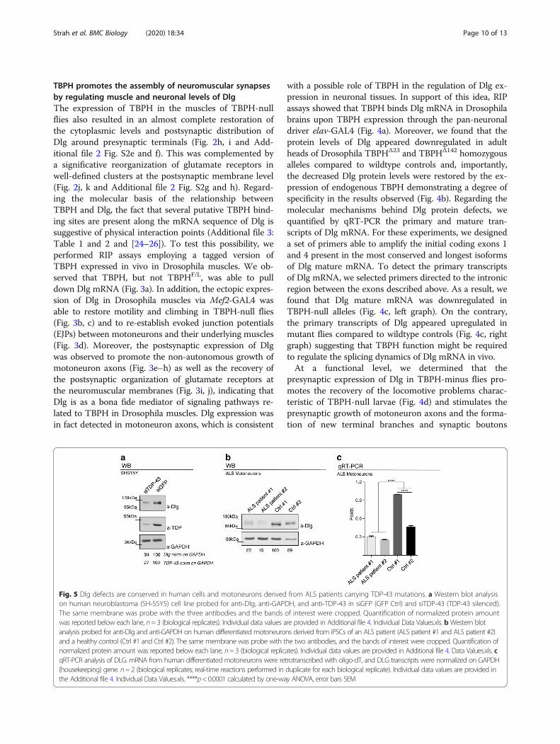
Fig. 5 Dlg defects are conserved in human cells and motoneurons derived from ALS patients carrying TDP-43 mutations. a Western blot analysison human neuroblastoma (SH-S5Y5) cell line probed for anti-Dlg, anti-GAPDH, and anti-TDP-43 in siGFP (GFP Ctrl) and siTDP-43 (TDP-43 silenced).The same membrane was probe with the three antibodies and the bands of interest were cropped. Quantification of normalized protein amountwas reported below each lane, n=3 (biological replicates). Individual data values are provided in Additional file 4. Individual Data Values.xls. bWestern blotanalysis probed for anti-Dlg and anti-GAPDH on human differentiated motoneurons derived from iPSCs of an ALS patient (ALS patient #1 and ALS patient #2)and a healthy control (Ctrl #1 and Ctrl #2). The same membrane was probe with the two antibodies, and the bands of interest were cropped. Quantification ofnormalized protein amount was reported below each lane, n=3 (biological replicates). Individual data values are provided in Additional file 4. Data Values.xls. cqRT-PCR analysis of DLG. mRNA from human differentiated motoneurons were retrotranscribed with oligo-dT, and DLG transcripts were normalized on GAPDH(housekeeping) gene. n=2 (biological replicates; real-time reactions performed in duplicate for each biological replicate). Individual data values are provided inthe Additional file 4. Individual Data Values.xls. ****p<0.0001 calculated by one-way ANOVA, error bars SEM
Strah et al. BMC Biology (2020) 18:34 Page 10 of 13
(Fig. 4e, f). Additionally, the neuronal expression of Dlginduced the non-autonomous clustering of glutamate re-ceptors in postsynaptic membranes of TBPH-minusNMJs (Fig. 4g, h), demonstrating that TBPH regulatesthe pre- and postsynaptic levels of Dlg and is required inmuscles and neurons to promote the formation ofNMJs.
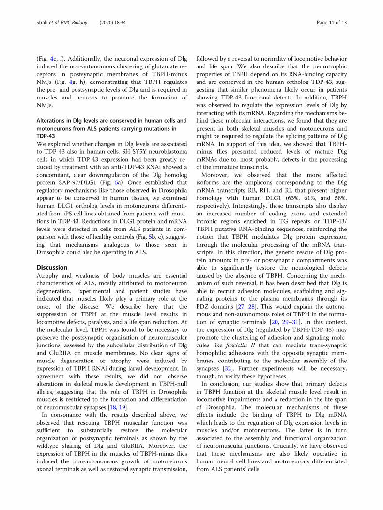
Alterations in Dlg levels are conserved in human cells andmotoneurons from ALS patients carrying mutations inTDP-43We explored whether changes in Dlg levels are associatedto TDP-43 also in human cells. SH-SY5Y neuroblastomacells in which TDP-43 expression had been greatly re-duced by treatment with an anti-TDP-43 RNAi showed aconcomitant, clear downregulation of the Dlg homologprotein SAP-97/DLG1 (Fig. 5a). Once established thatregulatory mechanisms like those observed in Drosophilaappear to be conserved in human tissues, we examinedhuman DLG1 ortholog levels in motoneurons differenti-ated from iPS cell lines obtained from patients with muta-tions in TDP-43. Reductions in DLG1 protein and mRNAlevels were detected in cells from ALS patients in com-parison with those of healthy controls (Fig. 5b, c), suggest-ing that mechanisms analogous to those seen inDrosophila could also be operating in ALS.
DiscussionAtrophy and weakness of body muscles are essentialcharacteristics of ALS, mostly attributed to motoneurondegeneration. Experimental and patient studies haveindicated that muscles likely play a primary role at theonset of the disease. We describe here that thesuppression of TBPH at the muscle level results inlocomotive defects, paralysis, and a life span reduction. Atthe molecular level, TBPH was found to be necessary topreserve the postsynaptic organization of neuromuscularjunctions, assessed by the subcellular distribution of Dlgand GluRIIA on muscle membranes. No clear signs ofmuscle degeneration or atrophy were induced byexpression of TBPH RNAi during larval development. Inagreement with these results, we did not observealterations in skeletal muscle development in TBPH-nullalleles, suggesting that the role of TBPH in Drosophilamuscles is restricted to the formation and differentiationof neuromuscular synapses [18, 19].In consonance with the results described above, we
observed that rescuing TBPH muscular function wassufficient to substantially restore the molecularorganization of postsynaptic terminals as shown by thewildtype sharing of Dlg and GluRIIA. Moreover, theexpression of TBPH in the muscles of TBPH-minus fliesinduced the non-autonomous growth of motoneuronsaxonal terminals as well as restored synaptic transmission,
followed by a reversal to normality of locomotive behaviorand life span. We also describe that the neurotrophicproperties of TBPH depend on its RNA-binding capacityand are conserved in the human ortholog TDP-43, sug-gesting that similar phenomena likely occur in patientsshowing TDP-43 functional defects. In addition, TBPHwas observed to regulate the expression levels of Dlg byinteracting with its mRNA. Regarding the mechanisms be-hind these molecular interactions, we found that they arepresent in both skeletal muscles and motoneurons andmight be required to regulate the splicing patterns of DlgmRNA. In support of this idea, we showed that TBPH-minus flies presented reduced levels of mature DlgmRNAs due to, most probably, defects in the processingof the immature transcripts.Moreover, we observed that the more affected
isoforms are the amplicons corresponding to the DlgmRNA transcripts RB, RH, and RL that present higherhomology with human DLG1 (63%, 61%, and 58%,respectively). Interestingly, these transcripts also displayan increased number of coding exons and extendedintronic regions enriched in TG repeats or TDP-43/TBPH putative RNA-binding sequences, reinforcing thenotion that TBPH modulates Dlg protein expressionthrough the molecular processing of the mRNA tran-scripts. In this direction, the genetic rescue of Dlg pro-tein amounts in pre- or postsynaptic compartments wasable to significantly restore the neurological defectscaused by the absence of TBPH. Concerning the mech-anism of such reversal, it has been described that Dlg isable to recruit adhesion molecules, scaffolding and sig-naling proteins to the plasma membranes through itsPDZ domains [27, 28]. This would explain the autono-mous and non-autonomous roles of TBPH in the forma-tion of synaptic terminals [20, 29–31]. In this context,the expression of Dlg (regulated by TBPH/TDP-43) maypromote the clustering of adhesion and signaling mole-cules like fasciclin II that can mediate trans-synaptichomophilic adhesions with the opposite synaptic mem-branes, contributing to the molecular assembly of thesynapses [32]. Further experiments will be necessary,though, to verify these hypotheses.In conclusion, our studies show that primary defects
in TBPH function at the skeletal muscle level result inlocomotive impairments and a reduction in the life spanof Drosophila. The molecular mechanisms of theseeffects include the binding of TBPH to Dlg mRNAwhich leads to the regulation of Dlg expression levels inmuscles and/or motoneurons. The latter is in turnassociated to the assembly and functional organizationof neuromuscular junctions. Crucially, we have observedthat these mechanisms are also likely operative inhuman neural cell lines and motoneurons differentiatedfrom ALS patients’ cells.
Strah et al. BMC Biology (2020) 18:34 Page 11 of 13
Supplementary informationSupplementary information accompanies this paper at https://doi.org/10.1186/s12915-020-00767-7.
Additional file 1: Figure S1. Control of TBPH silencing. Western blotanalysis on larval carcasses probed for anti-TBPH and anti-tubulin intbphΔ23/+;Mef2-GAL4/UAS-GFP-IR and tbphΔ23/+;Mef2-GAL4/UAS-TBPH-IR. The same membrane was probed with the two antibodies and thebands of interest were cropped. n = 3 (biological replicates). Individualdata values are provided in the Additional file 4. Individual DataValues.xls.
Additional file 2: Figure S2. a. Western blot analysis on larval carcassesprobed for anti-TBPH and anti-tubulin in Ctrl (w1118), tbphΔ23/tbphΔ23,tbphΔ23,UAS-TBPH/tbphΔ23;Mef2-GAL4/+, tbphΔ23/tbphΔ23;Mef2-GAL4/UAS-TBPHF/L. The same membrane was probed with two antibodies andthe bands of interest were cropped. n = 3 (biological replicates). b. West-ern blot analysis on larval carcasses probed for anti-TDP and anti-tubulinin Ctrl (w1118) and tbphΔ23/tbphΔ23;Mef2-GAL4/UAS-TDP-43 The samemembrane was probed with two antibodies and the bands of interestwere cropped. n = 3 (biological replicates). c. Quantification of branchesnumber in Ctrl, Δtb-GFP and Δtb-TBPH. n = 15. d. Quantification of bou-tons shape in Ctrl, Δtb-GFP and Δtb-TBPH. n = 200. e. Confocal images ofthird instar NMJ terminals in muscle 6/7 second segment stained withanti-HRP (in green) and anti-Dlg (in red) in Ctrl (w1118), Δtb-GFP (tbphΔ23/tbphΔ23;Mhc-GAL4/UAS-GFP), Δtb-TBPH (tbphΔ23,UAS-TBPH/tbphΔ23;Mhc-GAL4/+). f. Quantification of Dlg intensity normalized on Ctrl. n > 200boutons. g. Confocal images of third instar NMJ terminals in muscle 6/7second segment stained with anti-HRP (in green) and anti-GluRIIA (in red)in Ctrl, Δtb-GFP and Δtb-TBPH. h. Quantification of GluRIIA intensity nor-malized on Ctrl. n > 200 boutons.
Additional file 3: Table 1. TG repeats distribution in drosophila DLG1gene. TG repeats distribution in drosophila DLG1 gene. In first column TGlength is reported, second and third columns report chromosomalcoordinates for chromosome X, while fourth column reports exonic orintronic location. Table 2. TG repeats distribution in human DLG1 gene.TG repeats distribution in human DLG1 gene. In first column TG length isreported, second and third columns report chromosomal coordinates forchromosome 3, while fourth column reports exonic or intronic location.
Additional file 4. Individual data values. Individual data values for: Fig.3: pannel a; Fig. 4: pannels a,b,c; Fig. 5: pannels a,b,c; Additional file 1 Fig.S1.
AcknowledgementsWe thank professors Rodolfo Garcia for critical comments and proofreadingthe manuscript and Chun-Fang Wu for the DLG transgenic flies. The Bloom-ington Stock Center and Developmental Studies Hybridoma Bank for stocksand reagents.
Authors’ contributionsNS, GR, CI, RK, MM, and LC performed the experiments and discussed thedata. AM performed the electrophysiology analysis, and MN provided thehuman iPS cells and the differentiated motoneurons. FF supervised the work,discussed the results, and wrote the manuscript. The authors read andapproved the final manuscript.
FundingThe present work was supported by ARISLA (CHRONOS) and BENEFICENTIAStiftung.
Availability of data and materialsAll data generated or analyzed during this study are included in thispublished article and its additional information files.
Ethics approval and consent to participateNot applicable
Consent for publicationNot applicable
Competing interestsThe authors declare that they have no competing interests.
Author details1International Centre for Genetic Engineering and Biotechnology, Padriciano99, 34149 Trieste, Italy. 2Istituto di Biologia e Patologia Molecolari del CNR,Sapienza Università di Roma, Rome, Italy. 3Dipartimento di Biologia eBiotecnologie “C. Darwin”, Sapienza Università di Roma, Rome, Italy.4Department of Biomedical Sciences, University of Padova, via Marzolo 3,35131 Padua, Italy. 5Department of Pathophysiology and Transplantation(DePT), Dino Ferrari Centre, University of Milan, Neuroscience Section, IRCCSFoundation Ca’ Granda Ospedale Maggiore Policlinico, Via Francesco Sforza35, 20122 Milan, Italy.
Received: 25 July 2019 Accepted: 10 March 2020
References1. Bruijn LI, Miller TM, Cleveland DW. Unraveling the mechanisms involved in
motor neuron degeneration in ALS. Annu Rev Neurosci. 2004;27:723–49.2. Sreedharan J, Blair IP, Tripathi VB, Hu X, Vance C, Rogelj B, et al. TDP-43
mutations in familial and sporadic amyotrophic lateral sclerosis. Science.2008;319:1668–72.
3. Arai T, Hasegawa M, Akiyama H, Ikeda K, Nonaka T, Mori H, et al. TDP-43 is acomponent of ubiquitin-positive tau-negative inclusions in frontotemporallobar degeneration and amyotrophic lateral sclerosis. Biochem Biophys ResCommun. 2006;351:602–11.
4. Geser F, Brandmeir NJ, Kwong LK, Martinez-Lage M, Elman L, McCluskey L,et al. Evidence of multisystem disorder in whole-brain map of pathologicalTDP-43 in amyotrophic lateral sclerosis. Arch Neurol. 2008;65:636–41.
5. Neumann M, Sampathu DM, Kwong LK, Truax AC, Micsenyi MC, Chou TT,et al. Ubiquitinated TDP-43 in frontotemporal lobar degeneration andamyotrophic lateral sclerosis. Science. 2006;314:130–3.
6. Boillée S, Vande Velde C, Cleveland DW. ALS: a disease of motor neuronsand their nonneuronal neighbors. Neuron. 2006;52:39–59.
7. Brettschneider J, Libon DJ, Toledo JB, Xie SX, McCluskey L, Elman L, et al.Microglial activation and TDP-43 pathology correlate with executivedysfunction in amyotrophic lateral sclerosis. Acta Neuropathol (Berl). 2012;123:395–407.
8. Diaper DC, Adachi Y, Lazarou L, Greenstein M, Simoes FA, Domenico AD,et al. Drosophila TDP-43 dysfunction in glia and muscle cells causecytological and behavioural phenotypes that characterize ALS and FTLD.Hum Mol Genet. 2013. https://doi.org/10.1093/hmg/ddt243.
9. Ince PG, Highley JR, Kirby J, Wharton SB, Takahashi H, Strong MJ, et al.Molecular pathology and genetic advances in amyotrophic lateral sclerosis:an emerging molecular pathway and the significance of glial pathology.Acta Neuropathol (Berl). 2011;122:657–71.
10. Tong J, Huang C, Bi F, Wu Q, Huang B, Liu X, et al. Expression of ALS-linkedTDP-43 mutant in astrocytes causes non-cell-autonomous motor neurondeath in rats. EMBO J. 2013;32:1917–26.
11. Romano G, Appocher C, Scorzeto M, Klima R, Baralle FE, Megighian A, et al.Glial TDP-43 regulates axon wrapping, GluRIIA clustering and fly motility byautonomous and non-autonomous mechanisms. Hum Mol Genet. 2015;24:6134–45.
12. Cykowski MD, Powell SZ, Appel JW, Arumanayagam AS, Rivera AL, Appel SH.Phosphorylated TDP-43 (pTDP-43) aggregates in the axial skeletal muscle ofpatients with sporadic and familial amyotrophic lateral sclerosis. ActaNeuropathol Commun. 2018;6:28.
13. Kraemer BC, Schuck T, Wheeler JM, Robinson LC, Trojanowski JQ, Lee VMY,et al. Loss of murine TDP-43 disrupts motor function and plays an essentialrole in embryogenesis. Acta Neuropathol (Berl). 2010;119:409–19.
14. Schmid B, Hruscha A, Hogl S, Banzhaf-Strathmann J, Strecker K, van der ZeeJ, et al. Loss of ALS-associated TDP-43 in zebrafish causes muscledegeneration, vascular dysfunction, and reduced motor neuron axonoutgrowth. Proc Natl Acad Sci U S A. 2013;110:4986–91.
15. Diaper DC, Adachi Y, Sutcliffe B, Humphrey DM, Elliott CJH, Stepto A, et al.Loss and gain of Drosophila TDP-43 impair synaptic efficacy and motorcontrol leading to age-related neurodegeneration by loss-of-functionphenotypes. Hum Mol Genet. 2013;22:1539–57.
16. Llamusi B, Bargiela A, Fernandez-Costa JM, Garcia-Lopez A, Klima R, FeiguinF, et al. Muscleblind, BSF and TBPH are mislocalized in the muscle
Strah et al. BMC Biology (2020) 18:34 Page 12 of 13
sarcomere of a Drosophila myotonic dystrophy model. Dis Model Mech.2013;6:184–96.
17. Vogler TO, Wheeler JR, Nguyen ED, Hughes MP, Britson KA, Lester E, et al.TDP-43 and RNA form amyloid-like myo-granules in regenerating muscle.Nature. 2018;563:508–13.
18. Feiguin F, Godena VK, Romano G, D’Ambrogio A, Klima R, Baralle FE.Depletion of TDP-43 affects Drosophila motoneurons terminal synapsis andlocomotive behavior. FEBS Lett. 2009;583:1586–92.
19. Godena VK, Romano G, Romano M, Appocher C, Klima R, Buratti E, et al.TDP-43 regulates Drosophila neuromuscular junctions growth bymodulating Futsch/MAP1B levels and synaptic microtubules organization.PLoS One. 2011;6:e17808.
20. Romano G, Klima R, Buratti E, Verstreken P, Baralle FE, Feiguin F.Chronological requirements of TDP-43 function in synaptic organizationand locomotive control. Neurobiol Dis. 2014;71:95–109.
21. Romano G, Holodkov N, Klima R, Grilli F, Guarnaccia C, Nizzardo M, et al.Downregulation of glutamic acid decarboxylase in Drosophila TDP-43-nullbrains provokes paralysis by affecting the organization of theneuromuscular synapses. Sci Rep. 2018;8:1809.
22. Ng S-Y, Soh BS, Rodriguez-Muela N, Hendrickson DG, Price F, Rinn JL, et al.Genome-wide RNA-Seq of human motor neurons implicates selective ERstress activation in spinal muscular atrophy. Cell Stem Cell. 2015;17:569–84.
23. Bour BA, O’Brien MA, Lockwood WL, Goldstein ES, Bodmer R, Taghert PH,et al. Drosophila MEF2, a transcription factor that is essential formyogenesis. Genes Dev. 1995;9:730–41.
24. Buratti E, Baralle FE. Characterization and functional implications of the RNAbinding properties of nuclear factor TDP-43, a novel splicing regulator ofCFTR exon 9. J Biol Chem. 2001;276:36337–43.
25. Buratti E, Dörk T, Zuccato E, Pagani F, Romano M, Baralle FE. Nuclear factorTDP-43 and SR proteins promote in vitro and in vivo CFTR exon 9 skipping.EMBO J. 2001;20:1774–84.
26. Tollervey JR, Curk T, Rogelj B, Briese M, Cereda M, Kayikci M, et al.Characterizing the RNA targets and position-dependent splicing regulationby TDP-43. Nat Neurosci. 2011;14:452–8.
27. Thomas U, Kim E, Kuhlendahl S, Koh YH, Gundelfinger ED, Sheng M, et al.Synaptic clustering of the cell adhesion molecule fasciclin II by discs-large andits role in the regulation of presynaptic structure. Neuron. 1997;19:787–99.
28. Zito K, Fetter RD, Goodman CS, Isacoff EY. Synaptic clustering of Fascilin IIand Shaker: essential targeting sequences and role of Dlg. Neuron. 1997;19:1007–16.
29. Budnik V, Koh YH, Guan B, Hartmann B, Hough C, Woods D, et al.Regulation of synapse structure and function by the Drosophila tumorsuppressor gene dlg. Neuron. 1996;17:627–40.
30. Miskiewicz K, Jose LE, Yeshaw WM, Valadas JS, Swerts J, Munck S, et al.HDAC6 is a Bruchpilot deacetylase that facilitates neurotransmitter release.Cell Rep. 2014;8:94–102.
31. Chang J-C, Hazelett DJ, Stewart JA, Morton DB. Motor neuron expression ofthe voltage-gated calcium channel cacophony restores locomotion defectsin a Drosophila, TDP-43 loss of function model of ALS. Brain Res. 2014;1584:39–51.
32. Kohsaka H, Takasu E, Nose A. In vivo induction of postsynaptic molecularassembly by the cell adhesion molecule Fasciclin2. J Cell Biol. 2007;179:1289–300.
Publisher’s NoteSpringer Nature remains neutral with regard to jurisdictional claims inpublished maps and institutional affiliations.
Strah et al. BMC Biology (2020) 18:34 Page 13 of 13


![SciHub - WordPress.com · 11/09/2016 · leads to an overabundance of acetylcholine at the neuronal synapses and the neuromuscular junction [12,13]. After ... hub.cc](https://img.pdfslide.us/doc/110x75/5ad692f27f8b9aff228e79bc/scihub-to-an-overabundance-of-acetylcholine-at-the-neuronal-synapses-and-the-neuromuscular.jpg)