Embed Size (px)
Citation preview

swedish dental journal vol. 25 issue 1 2001 1
contentscontents
A retrospective long term study of teeth restored with prefabricated carbon fifi ber reinforced epoxy resin posts
Segerström, Astbäck, Ekstrand
11
Impaired positioning of the gape Impaired positioning of the gape in whiplash-associated disordersin whiplash-associated disorders
Zafar, Nordh, Eriksson
99
Analysis of the interleukin-1 and Analysis of the interleukin-1 and interleukin-6 polymorphisms in interleukin-6 polymorphisms in patients with chronic periodontitis. patients with chronic periodontitis. A pilot studyA pilot study
Jansson, Lyssenko, Gustavsson, Hamberg, Söderfeldt, Groop, Bratthall
1717
Periodontal conditions in a Swedish Periodontal conditions in a Swedish city population of adolescents: city population of adolescents: A cross-sectional studyA cross-sectional study
Abrahamsson, Koch, Norderyd, Romao, Wennström
2525
A 20-year study of dentists’ and dental A 20-year study of dentists’ and dental hygienists’ assessment of dental hygienists’ assessment of dental caries lesions in bite-wing radiographscaries lesions in bite-wing radiographs
Gabre, Birring, Gahnberg
3535
SwedishSwedishDental JournalDental Journal
A 20-year study of dentists’ and dental hygienists’ assessment of dental caries lesions in bite-wing radiographs page 35page 35
Scientific Journal of The Swedish Dental AssociationScientific Journal of The Swedish Dental Association
No. No. 1/061/06Vol. 30 Pages 1-45Vol. 30 Pages 1-45

Instructions to authorsSwedishDental JournalScientifi c journalof the Swedish Dental Associationand the Swedish Dental Societyissn: 0347-9994
Editor-in-chiefProfessor Göran Koch, Jönköping
Associate EditorsProfessor Gunnar Dahlén, GöteborgProfessor Björn Klinge, StockholmProfessor Ulf Lerner, UmeåProfessor Lars Matsson, Malmö
Advisory Editorial BoardAssoc. prof. Michael Ahlqvist, StockholmOdont. dr. Annika Bäcker, GöteborgAssoc. prof. Dan Ericson, MalmöAssoc. prof. Malin Ernberg, StockholmAssoc. prof. Anders Gustafsson, StockholmProfessor Eva Hellsing, StockholmProfessor Anders Hugoson, JönköpingProfessor Ingegerd Johansson, UmeåProfessor Åke Larsson, MalmöAssoc. prof. Tomas Magnusson, JönköpingProfessor Margareta Molin, UmeåAssoc. prof. Peter Nilsson, JönköpingProfessor Arne Petersson, MalmöOdont. dr. Karin Sjögren, GöteborgProfessor Björn Söderfäldt, MalmöProfessor Svante Twetman, UmeåProfessor Jan van Dijken, UmeåOdont. dr. Ulf Örtengren, Göteborg
ProductionEwa Knutsson, Tfn +46 (0)8 666 15 [email protected]
Editorial addressSwedish Dental JournalOdontologiska InstitutionenBox 1030, SE-551 11 Jönköping, SwedenTfn: +46 (0)36 32 46 04Fax: +46 (0)36 71 22 35
Subscription/business addressSwedish Dental JournalBox 1217, SE-111 82 Stockholm, SwedenTfn: +46 (0)8 666 15 00Fax: +46 (0)8 662 58 42e-mail: [email protected]: Skandinaviska Enskilda BankenBankgiro: 404-4699 Postgiro: 45 86 34-3
SubscriptionsSweden: sek 850 Others: sek 1 100(Supplements are not included.)For subscriptions delivered to adresses within the European Union. Please notice: If you have a VAT registration number you must provide this. Otherwise, please add your local VAT to the above price in SEK.
Printing offi ceEdita Västra Aros AB
IntroductionSwedish Dental Journal, the scientifi c jour-nal of The Swedish Dental Association and the Swedish Dental Society, is published 4 times a year to promote practice, education and research within odontology. Manu-scripts containing original research areaccepted for consideration if neither thearticle nor any part of its essential substan-ce has been or will be published elsewhere. Reviews (after consultations with the edi-tors), Case Reports and Short Communi-cations will also be considered for publica-tion. All manuscript will be exposed to a referee process.
The ManuscriptThree complete copies of the manuscript should be sent to the Editor-in-chief Pro-fessor Göran Koch at the Editorial add-ress (see beside). The paper should be in English using English spelling, be typed double-spaced with one-inch margins. The format of the manuscript should be arranged as follows:
Title Page, Abstract, Sammanfattning (in Swedish), Introduction, Materialand Methods, Results, Discussion,Acknowledgements, References,Figure Legends and Tables.
The letter attached to the manuscript should be signed by all the authors. When the paper has been accepted for publication the author will be asked to supply an upda-ted fi nal manuscript on disk together with two complete manuscripts.
The Title Page should contain in the following order: A concise and covering title, authors’ full names (without titles), affi liation(s) of the author(s) including city and country, Key-words (according to In-dex Medicus and not more than 5), Running title and name and contact information of the corresponding author.
The Abstract should be short and concise and not exceeding 300 words. The Swedish Sammanfattning can be somewhat more extensive.
ReferencesIn the reference list the references should be arranged in alphabetical order and numbe-red consecutively by Arabic numerals. Indi-cate references in the running text by using the Arabic numeral within brackets.
Abbreviations should follow ”List of Journals indexed in Index Medicus”. (http://www.nlm.nih.gov). Examples of references are presented below.
Article:Helm S, Seidler B. Timing of permanent tooth emergence in Danish children. Com-munity Dent Oral Epidemiol 1974; 2:122–9
Book: Andreasen JO, Petersen JK, Laskin DM, eds. Textbook and color atlas of tooth im-pactions. Copenhagen: Munksgaard, 1997
Illustrations should be numbered in se-quence with Arabic numerals. Legends to all the illustrations should be on a separate sheet. Author’s name and fi gure number should be written on the back of each il-lustration. Cost for coulor fi gures are ge-nerally covered by the author. Each Table should be written on a separate sheet. They should be numbered with Arabic numerals and each should have a heading.
Reprints are not generally available. The main author will receive 20 numbers of the issue where the paper is published. A pdf of the article is also sent to the main author.
Galley proof will be sent to the author and should be returned to the Editor without delay.
Page charge will be due if the article is longer than 6 printed pages. For excessof pages the charge is 1 000 sek per page.
Supplements can be arranged, the full cost beeing paid by the author. Contact the Edi-tor.

long term study of epoxy reisin posts
swedish dental journal vol. 30 issue 1 2006 1
A retrospective long term study of teeth restored with prefabricated carbon fi ber reinforced epoxy resin postssusanna segerström1, johnny astbäck1, and karl ekstrand1
Abstract • The Composipost® endodontic post, made of stretched aligned carbon fi bers em-bedded in an epoxy-resin matrix, has since the beginning of the nineties been widely used for the restoration of endodontically treated teeth.
The aim of this retrospective study was to evaluate the treatment outcome of the Composipost system up to 7 years.
In a study published 1998, 236 patients treated during 1992-93 by seven Swedish general dental private practitioners were studied. Five of the former seven private practitionersí consented to participate in this follow up of that study. Thus the ma-terial was reduced to 138 patients. Thirty-nine of these were excluded due to insuffi -cient data. For the remaining 99 patients, data were collected from dental records. All patients were offered a clinical examination but only 25 accepted. Data were collected from dental records for the remaining 74 patients. The mean follow up time was 6,7 years with a range from 1 month to 10 years (median 7.6 years, SD 2.5 years), (fi ve teeth were extracted during the previous study). The outcome was considered suc-cessful if the post and core was in situ and showed no clinical or radiographic signs of technical failures.
Sixty-four teeth (65%) restored with the Composipost system were successful after a mean time of 6.7 years. Thirty-two teeth were extracted due to fractures, periapical lesions and periodontitis. Dislodgment of post was observed in three cases.
In conclusion, within the limitations of this study, after a mean time of 6.7 years, the Composipost restored teeth had shorter survival times than those of previously documented cast posts.
Key words Cementation, composite resins, failure, post-and-core technique
swed dent j 2006; 30: 1–8 • segerström, astbäch, ekstrand
1 Department of Prosthetic Dentistry, Uppsala Sweden
segerstöm, astbäck, ekstrand
2 swedish dental journal vol. 30 issue 1 2006
En retrospectiv långtidsstudie av tänder restaurerade med prefabricerade kolfi berförstärkta epoxystiftsusanna segerström, johnny astbäck och karl ekstrand
Sammanfattning
• En retrospektiv långtidsstudie av tänder restaurerade med prefabricerade kolfi berför-stärkta epoxy stift. Kolfi berförstärkta stift (Composipost®) tillverkade i kolfi ber inbädda-de i epoxy resin matrix har sedan början på 1990 talet använts för att restaurera rotfyllda tänder. Målsättningen med denna undersökning var att uvärdera behandlingsresultatet efter en uppföljningstid på cirka 7 år.
I en studie som publicerades 1998 hade 236 patienter behandlats med Composipost®stift mellan åren 1992-1993 av sju svenska privatpraktiserande tandläkare. Av dessa sju kunde fem delta i en ny utvidgad uppföljning. Materialet var emellertid reducerat till 138 patienter. Ytterligare trettionio patienter exkluderades pga otillräckliga journal- och statusuppgifter. För de resterande 99 patienterna samlades data in från journalanteck-ningar. Alla patienter erbjöds en klinisk undersökning med radiologisk dokumentation men endast 25 accepterade erbjudandet. För de resterande 74 patienterna samlades resultat in via journal- och daganteckningar. Den genomsnittliga uppföljningstiden var 6,7 år med en spridning från 1 månad till 10 år. För att erhålla ett lyckat resultat måste Composipost® stiftet sitta kvar i tanden och inte uppvisa några kliniska eller radiologiska defekter.
Sextiofyra tänder (65%) restaurerade med Composipost® var fortfarande i funktion efter en medeluppföljningstid på 6,7 år. Trettiotvå tänder hade extraherats pga olika orsa-ker som frakturer, periapikala läsioner och parodontit. Lossnande stift kunde observeras i tre fall.
Av resultaten framgår, med reservation för materialets storlek och bortfall av antalet patienter, att efter en uppföljningstid på nästan 7 år, uppvisar Composipost®försedda tänder sämre lyckandefrekvens jämfört med konventionellt framställda stift av metall.
swed dent j 2006; 30: 1–8 • segerström, astbäck, ekstrand
long term study of epoxy reisin posts
swedish dental journal vol. 30 issue 1 2006 3
IntroductionIn case of extensive loss of tooth structure a restora-tion of the tooth with a complete crown for rehabi-litation is needed. Posts-and-cores are not necessary when a minimal loss of tooth structure exists (30). If a large portion of the clinical crown has been lost it is often impossible to achieve suffi cient anchorage for a conventionally cemented restoration in the remaining dentin and a root-canal-retained resto-ration is required. This may be the case when ho-rizontal loss of the clinical crown has occurred and little ferrule can be created in the remaining tooth structure (34).Posts-and-cores have been proposed for the stabi-lization of weakened endodontically treated teeth. There are confl icting reports on the ability of metal posts to reinforce endodontically treated teeth (17). Several studies determine that intracoronal reinfor-cement does not strengthen the tooth nor does it increase the resistance to fracture (1, 35). Teeth wit-hout post-and-core foundations test stronger when compressive load is applied compared to teeth with post-and-core (33). Preserving tooth structure is one of the most important factors in avoiding technical complications with intracoronally reinforced teeth (27, 28, 36). There is little difference between the wide range of post designs and systems when a com-plete crown restoration is performed since fracture resistance may be more dependent on the amount of remaining dentin ferruled by the crown restoration (24, 34).
Alternatives to cast posts and cores have been de-veloped. The use of prefabricated posts and custom-made build-ups has become increasingly popular (29). Composipost®, a non-metallic post system, was introduced in 1990 for fabrication of dental posts (7). This system allows more dentin of the tooth to be preserved. It is based on the carbon-fi ber reinforcement principle. The cylindrical Composi-post® is made of 64% equally stretched and aligned carbon-fi bers (8 µm in diameter), solidly attached to a special matrix of epoxy-resin (36%). The car-bon fi bers impart high strength to the posts. (Com-posipost®, Recherches Techniques Dentaires, RTD, Meylan, France).
Composiposts are passive and designed to be used with a bonding technique. Endodontic drills of mat-ching diameter, resin luting cement and resin com-posite core material complement the system. The recommended core material is Resilient composite, a Bis-GMA resin fi lled with short glass fi bers (RTD, Meylan, France).
The fabrication of the carbon fi ber reinforced epoxy resin post is, according to the manufacturer, less expensive and time-consuming than fabrication of conventional post-and cores (7). In Sweden, bet-ween 20-25% of general practitioners use the Com-posipost system and 68% use composite materials for core build-up (8).
Epoxy-resin materials degrade when in contact with water (9). The Composipost® is a combined material of hydrophobic fi bers and an epoxy resin matrix that absorbs water and it is evident that me-chanical properties change when in an aqueous en-vironment (5). The fl exural values of the Composi-post® decrease signifi cantly after water storage and after thermocycling (5, 37). The effect of thermal cycling makes the carbon post more susceptible to fracture and unable to withstand additional load cycling. The breakdown is possibly related to de-gradation of the polymer holding the fi bers to-gether (6). A change of the mechanical properties may have an impact on the function time of the post.
Fiber-reinforced polymeric composites are also susceptible to microbial degradation, since fungi can utilize the resins or fi ber chemical sizing as carbon and energy sources (16). This might also affect the longterm outcome of the post restored tooth.
Conventional cast post-and-core have a failure rate of 1.6% per year after 6 years (2). Parallel and tapered cast posts evaluated after 4-5 years have a 92% success rate for the parallel and 85% for the ta-pered posts (38). A retrospective study indicates that the Composipost® system is superior to the con-ventional cast post and core system after 4 years of clinical service, with success rates of 95% versus 84% respectively (11). Another study evaluates three dif-ferent fi ber posts after clinical service ranging from 1-6 years and it is concluded that the fi ber posts in combination with bonding/luting materials can be routinely used (12). Such success rates and recom-mendations are; however, contradicted(23,33) in a prospective trial where more failures are seen in the carbon fi ber reinforced group than among the pre-fabricated conventional posts (23).
The mode of failure with a fi ber reinforced post is described to be more favourable and with lower failure rates than with metallic posts (21, 22). This is contradicted by others claiming that the use of carbon fi ber reinforced composite posts does not change the fracture resistance nor the failure mode when compared to the use of metallic posts (32).
The purpose of this study was to conduct a retro-

segerstöm, astbäck, ekstrand
4 swedish dental journal vol. 30 issue 1 2006
spective clinical and radiographic evaluation of the Composipost® post-and-core up to seven years in service. The hypothesis was that the survival time for the conventional cast post is longer than that of the carbon fi ber-reinforced post.
Material and MethodsThe material and methods of this study is based on a clinical retrospective study by Fredriksson et al (13). Seven dentists were randomly selected to contribute data from patients treated with the Composipost system until July 1993. A total of 236 patients were selected for evaluation. The list of participating pri-vate practitioners from this study was kindly given to us by the authors. The seven practitioners were contacted. Initially all of them accepted to partici-pate in this follow up study. In two cases the dental clinics had new owners. A few weeks later a confi r-mation letter along with further information and patient instruction was sent. In one case the dentist had changed his mind and did no longer want to participate (90 patients). In the other case, the list of patients could not match the fi les from the original study (8 patients). The patients of these two clinics were therefore excluded. Five of the former seven dentists were thereby willing to take part in this fol-low up study. The patients were introduced to this study by a letter, approved by the Uppsala university ethical committee (Dnr 01-479) and asked to par-ticipate in a clinical and radiographic examination. The practical arrangements were admin-istered by each dentist.
The number of available patients was 138. Thirty-nine patients were excluded due to insuffi cient data. The age of the 99 remaining patients ranged from 36 to 90 years (mean 62 years). Of these 99 patients (38 men and 61 women) 25 patients agreed to participate in a clinical and radiographic examination. Each one of the 99 patients contributed with one Composi-post® restored tooth each in the study. As the pa-tients had previously been included in an individual recall program, data could be obtained from records for the remaining 74 patients who were unable or unwilling to participate in the clinical follow up.
The clinical examinations and the collection of data from dental records were carried out indepen-dently by two calibrated observers.
The posts were luted with the chemically cured composite resin cement recommended by the ma-nufacturer. The core build up material was also used according to the manufacturers recom-mendation. The luting cement used for the crowns was in most
cases zinc phosphate cement, but was not always re-gistered in the fi les.
Periodontal conditions were assessed by recor-ding the plaque index (Silness & Löe), the gingival index (Löe & Silness), the bleeding index (Lenox & Kopczyk) and measuring 4 point pocket depth. Den-tal examination also included diagnosing whether caries was present or not. An intraoral radiograph was taken of each Composipost treated tooth and the contra-lateral. In all cases the contra-lateral too-th was considered as a control tooth.
The collected data from the dental records were based on the latest appointment when visiting the dentist. In 93% of the cases radiographs were availa-ble and investigated.
The outcome was considered successful if the post-and-core was in situ and showed no clinical or radiographic signs of technical failures, loss of reten-tion, root fracture or post fracture.
ResultsThe results of the measurements were analysed by t-tests with a signifi cance level < 0.05.
The frequency of types of teeth treated is shown in Table 1.
The duration of service of the Composipost® restored teeth varied from 1 month to 10 years (Fig 1). The mean service time was 6.7 years (median 7.6 ye-ars, SD 2.5 years). Only twenty-fi ve patients could be clinically examined. These patients had the longest follow up times (8.4 years). The vast majority of col-lected data came from dental records (74 patients). The latest information regarding the Composipost restored tooth was from a prior date, consequently the follow up times for these patients were shorter (6.1 years).
The clinical examination was performed on 25 te-eth. The condition of soft tissues surrounding the post restored and contra-lateral teeth was similar. Plaque Index, Gingival Index and Bleeding index did not differ between the teeth restored with Com-posipost and the control teeth. Fifty percent of the
• Table 1 Distribution of position of 99 Composipost® treated teeth.The number of teeth is given.
Central Lateral Incisors incisors Canines Premolars Molar TotalMaxillae 7 7 5 23 12 54Mandible 1 0 4 21 19 45

long term study of epoxy reisin posts
swedish dental journal vol. 30 issue 1 2006 5
sites (surfaces) exhibited bleeding on probing and there were no differences between test and control teeth. The mean pocket depths of the post-retained teeth and the contra-lateral teeth did not differ from eachother; 3.8 mm (SD ± 1.9) and 3.3 (SD ± 1.3) re-spectively. However, the evaluation and assessment of the periodontal condition was only based on 25 subjects.
Dental caries was detected in nine of the post trea-ted teeth. Decayed teeth were not considered as fai-lures since the posts and cores were still in situ. Dis-lodgment of the posts was observed in 3 teeth after
• Figure 1. Distribution of Composipost® restored teeth in relation to years of service
1.5, 1.9 and 4.8 years and thus considered failures at the time of debonding. The recemented posts were thereafter in function for 1, 2 and 3 years respectively before a second dislodgement occurred. Thirty-two teeth had been extracted and the reasons for extrac-tion were; in 14 cases fracture, in 10 cases periapical lesions and in 5 cases periodontitis. Three cases were not accounted for. The mean functional time for failures was 4.8 years, with a range of 1 month to 10 years (Fig 2).
None of the radiographs taken of the examined patients showed any evidence of periapical destruc-
• Figure 2. Reasons for failures of Composipost® treated teeth. The number of teeth is given.
Num
ber
of p
ost r
esto
red
teet
hN
umbe
r of
pos
t res
tore
d te
eth

segerstöm, astbäck, ekstrand
6 swedish dental journal vol. 30 issue 1 2006
tion. Periodontal bone height was not measured on the radiographs because of the low number of exa-mined teeth.
The fi nal restorations of the treated teeth were in most cases metal ceramic crowns (86%) and the re-maining teeth were restored with composite materi-al. No correlation between failure and type of resto-ration could be noted. Of the opposing occluding teeth; 90% had fi xed restorations and 8% occluded with unrestored teeth. Two percent lacked occluding teeth. No signs of variations in failure versus posi-tion in the arch was evident.
DiscussionTechnical complications with posts-and-cores are not unusual. Different types of technical complica-tions can occur, such as loss of retention, root frac-ture, root perforation and fracture of the post. The most common complication is loss of retention (2, 31, 36, 38). Success rates for direct posts-and-cores (carbon-fi ber-reinforced posts not included) range from 68% after 10 years to 92% after 8 years (25, 29).
In a prospective study of carbon-fi ber reinforced epoxy-resin posts in endodontically treated teeth co-vered with metal ceramic crowns teeth are followed for 0.6-3.8 years (average 2.3 years) and the survival rate is 89,6% (15). When a comparison of survival rates for a 6-year period is made in a systematic re-view article, the meta-analytic comparison of cast and direct post restorations indicates that neither treatment modality is superior (20). The survival rate for cast posts-and-cores range from 87,2% (2) to 88,1% (29) compared to direct cores with a survival rate of 86,4% (25).
The size of this patient material was small. Un-fortunately only approximately 25% of the patients were willing to participate in a clinical and radio-graphic examination. However, out of the evaluated 99 teeth, 64 teeth (65%) restored with the Composi-post® system were still in function after a mean time of 6.7 years. Teeth restored with the Composipost®system had shorter survival times than those showed for cast posts or direct core build-ups, compared to results in studies mentioned above. When relating the results to the three-year follow up by Fredriksson et. al (13), a difference in results was evident. The rea-son for this was a longer service period. Long term clinical observations are emphasized in the littera-ture (18).
The material was from multiple general practice settings without standardization of clinical proto-
cols and this naturally infl uences the reliability of the sample. Thirty-nine patients were excluded due to insuffi cient data. During a period of ten years, the longest recorded follow up time, many changes took place that made it impossible to collect suffi cient and reliable data. For instance, dental offi ces were computerized and this meant that some of the ear-lier dental records were lost or inadeqate.
Some variations in failure rates were seen between the different dental practitioners. One explanation for this could be that the Composipost® system is a technically sensitive method. Another explanation may be that the selection criteria for the teeth to be restored could have varied between the dental prac-titioners. Possibly the selection criteria in general for all Composipost® treated teeth were expanded in trying a new and promising method at the time.
It is also important to emphasize that the manu-facturer had made changes to both the cement and the core material. This could be relevant for a more positive or negative outcome than shown in this study.
Debonding of the post and core from the tooth and a leachable restoration might be caused by poly-merization shrinkage (4). Microleakage can also be caused by thermal stresses caused by food induced temperature changes (39). The moisture within the tooth may also be of importance. The moisture con-tent of dentin is reported to 14% (19). Since some of this moisture is not coupled to the calcifi ed matrix it may have an effect on the cement and core. Only long term studies can prove if this has any clinical relevance.
Previous studies suggest that gutta-percha does not offer an effective barrier to leakage when expo-sed to the oral environment (26). Since microlea-kage most likely occurs, regardless of type of post-and-core restoration, it is of utmost importance to apply a layer of material that expands on hardening and prevents leakage to the apical portion of the tooth.
A recently published review article points out that the literature indicates that preservation of tooth structure is necessary and that posts should not be used with the intention of reinforcing the tooth. Furthermore a consideration of functional and pa-rafunctional forces must be undertaken before rest-oring the tooth as these will infl uence the prognosis (10). The importance of avoiding nonaxial forces is emphasized. In a comparison of post-and-core de-sign and the direction of functional load it is clai-med that the direction of the load has a greater effect

long term study of epoxy reisin posts
swedish dental journal vol. 30 issue 1 2006 7
than the post-and-core design on maximum stress and displacement (40).
The amount of remaining dentin is defi nitely an important factor for the longevity of post-and-core restored teeth (3, 24). Unfortunately the dentin fer-rule of the post-and-core restored teeth was not a known parameter in the present study and therefore no conclusions can be made regarding this aspect. If an insuffi cient ferrule is present it may be appro-priate to consider either a crown lengthening pro-cedure or an orthodontic extrusion (14). If neither of the suggested procedures can be performed, an extraction therapy may be considered.
How durable are fi ber-reinforced post and core structures? Can they be recommended for routine use as an alternative to individual cast posts? Since long term follow up results still are scarce, caution should still be recommended. Taking into conside-ration that a fi ber reinforced polymer may be more sensitive to effects produced with time in compari-son to cast posts, the fi ber post may create a higher risk. In addition, the technique is quite sensitive as well. There are many individual parameters to con-sider before deciding on what type of post to use. Individual decision making must be made regar-ding: occlusion, type of tooth/teeth to be restored, amount of remaining dentin, expected and desired duration of treatment, patient´s age, cooperation and economic situation. When post and core is necessary, for a limited duration in time, the fi ber reinforced post is a possibility. However, when ex-tensive fi xed prosthodontics are planned and/or long duration of therapy is desired, the traditional post and core technique (cast post) should be the fi rst choice.
Within the limitations of this retrospective study the following conclusions could be drawn:1. Survival time of Composipost restored teeth were shorter than those of previously documented cast posts.2. Since reasons for failure of a post-and-core resto-ration are dependent on a variety of different fac-tors, some unknown; no defi nite conclusions can be drawn regarding the reasons for failures from this study.3. It is essential that longterm in vivo prospective studies are made so that evaluation of new systems continously can take place.
AcknowledgementsThe authors would like to thank the participating dentists for their decision to contribute in this study.
This study was in part supported by Uppsala County Council and Praktikertjänst AB.
References1. Assif D, Bitenski A, Pilo R, Oren E. Effect of post
design on resistance to fracture of endodontically treated teeth with complete crowns. J Prosthet Dent 1993;69:36-40.
2. Bergman B, Lundquist P, Sjögren U, Sundquist G. Restorative and endodontic results after treatment with cast posts and cores. J Prosthet Dent 1989;61:10-5.
3. Creugers NH, Mentink AG, Fokkinga WA, Kreulen CM. 5-year follow-up of a prospective clinical study on various types of core restorations. Int J Prosthodont 2005;18:34-9.
4. Davidson CL, Feilzer AJ. Polymerization shrinkage and polymerization shrinkage stress in polymer-based restoratives. J Dent 1997;25:435-40.
5. Drummond JL. In vitro evaluation of endodontic posts. Am J Dent 2000;13:5B-8B.
6. Drummond JL, Toepke TRS, King TJ. Thermal and cyclic loading o endodontic posts. Eur J Oral Sci 1999;107:220-4.
7. Duret B, Reynaud M, Duret F. New concept of coronoradicular reconstruction: the Composipost (1). Chir Dent Fr 1990;60:131-41.
8. Eckerbom M, Magnusson T. Restoring endodontically treated teeth: a survey of current opinions among board-certifi ed prosthodontists and general dental practitioners in Sweden. Int J Prosthodont 2001;14:245-9.
9. Ekstrand K, Ruyter IE, Wellendorf H. Carbon/graphite fi ber reinforced poly(methyl methacrylate): properties under dry and wet conditions. J Biomed Mater Res 1987;21:1065-80.
10. Fernandes AS, Dessai GS. Factors affecting the fracture resistance of post-core reconstructed teeth; a review. Int J Prosthodont 2001;14:355-63.
11. Ferrari M, Vichi A, Garcia-Godoy F. Clinical evaluation of fi ber-reinforced epoxy resin posts and cast post and cores. Am J Dent 2000;13:15B-18B.
12. Ferrari M, Vichi A, Mannocci F, Mason PN. Retrospective study of the clinical performance of fi ber posts. Am J Dent 2000;13:9B-13B.
13. Fredriksson M, Astbäck J, Pamenius M, Arvidson K. A retrospective study of 236 patients with teeth restored by carbon fi ber-reinforced epoxy resin posts. J Prosthet Dent 1998;80:151-7.
14. Freeman MA, Nicholls JI, Kydd WL, Harrington GW. Leakage associated with load fatigue-induced preliminary failure of full crowns placed over three different post and core systems. J Endod 1998;24:26-32.
15. Glazer B. Restoration of endodontically treated teeth with carbon fi bre posts—a prospective study. J Can Dent Assoc 2000;66:613-8.
16. Gu JD, Lu C, Mitchell R Thorp K, Crasto A. Fungal degradation of fi ber-reinforced composite materials. Mater Perform 1997;36:37-42.
17 Guzy GE, Nicholls JI. In vitro comparison of intact endodontically treated teeth with and without endo-post reinforcement. J Prosthet Dent 1979;42:39-44.
18 Hedlund SO, Johansson NG, Sjögren G. A retrospective study of prefabricated carbon fi bre root canal posts. J Oral Rehabil 2003;30(10):1036-40.
19. Helfer AR, Melnick S, Schilder H. Determination of the

segerstöm, astbäck, ekstrand
8 swedish dental journal vol. 30 issue 1 2006
moisture content of vital and pulpless teeth. Oral Surg Oral Med Oral Pathol 1972;34:661-70.
20. Heydecke G, Peters MC. The restoration of endodontically treated, single rooted teeth with cast or direct posts and cores: a systematic review. J Prosthet Dent 2002;87:380-6.
21. Isidor, F, Ödman P, Brondum K. Intermittent loading of teeth restored using prefabricated carbon fi ber posts. Int J Prosthodont 1996;9:131-6.
22. King PA, Setchell DJ. An in vitro evaluation of a prototype CFRC prefabricated post developed for the restoration of pulpless teeth. J Oral Rehabil 1990;17:599-609.
23. King PA, Setchell DJ, Rees JS. Clinical evaluation of a carbon fi bre reinforced carbon endodontic post. J Oral Rehabil 2003;30:785-9.
24. Libman WJ, Nicholls JI. Load fatigue of teeth restored with cast posts and cores and complete crowns. Int J Prosthodont 1995;8:155-61.
25. Linde LA. The use of composites as core material in root-fi lled teeth. II. Clinical investigation. Swed Dent J 1984;8:209-16.
26. Magura ME, Kafrawy AH, Brown CE, Newton CW. Human saliva coronal microleakage in obturated root canals: an in vitro study. J Endod 1991;17:324-31.
27. Marshak BL, Helft H, Filo R. Factors mitigating against the use of dowels in endodontically treated teeth. Quintessence Int 1988;19:417-21.
28. Mattison GD, von Fraunhofer JA. Angulation loading effects on cast-gold endodontic posts: a photoelastic stress analysis. J Prosthet Dent 1983;49:636-8.
29. Mentink AG, Meeuwissen R, Kayser AF, Mulder J. Survival rate and failure characteristics of the all metal post and core restoration. J Oral Rehabil 1993;20:455-61.
30. Robbins JW. Restoration of the endodontically treated tooth. Dent Clin North Am 2002;46:367-84.
31. Randow K, Glanz PO, Zoger B. Technical failures and some related clinical complications in extensive fi xed prosthodontics. An epidemiological study of long-term clinical quality. Acta Odontol Scand 1986;44:241-55.
32. Raygot CG, Chai J, Jameson L. Fracture resistance and primary failure mode of endodontically treated teeth restored with a carbon fi ber-reinforced resin post system in vitro. Int J Prosthodont 2001;14:141-5.
33. Sidoli GE, King PA, Setchell DJ. An in vitro evaluation of a carbon fi ber based post and core system. J Prosthet Dent 1997;78:5-9.
34. Sorensen JA, Engelman MJ. Ferrule design and fracture resistance of endodontically treated teeth. J Prosthet Dent 1990;63:529-36.
35. Sorensen JA, Martinoff T. Clinically signifi cant factors in dowel design. J Prosthet Dent 1984;52:28-35.
36. Turner CH. The utilization of roots to carry post-retained crowns. J Oral Rehabil 1982;9:193-202.
37. Torbjörner A, Karlsson S, Syverud M, Hensten-Pettersen A. Carbon fi ber reinforced root canal posts. Mechanical and cytotoxic properties. Eur J Oral Sci 1996;104:605-11.
38. Torbjörner A, Karlsson S, Ödman PA. Survival rate and failure characteristics for two post designs. J Prosthet Dent 1995;73:439-44.
39. Yang H-S, Lang LA, Guckes AD, Felton DA. The effect of thermal change on various dowel-and-core restorative materials. J Prosthet Dent 2001;86:74-80.
40. Yang HS, Lang LA, Molina A, Felton DA. The effects of
dowel design and load direction on dowel-and-core restorations. J Prosthet Dent 2001;85:558-67.
Address:Dr Susanna SegerströmDepartment of Prosthetic Dentistry,PO Box 602SE-751 25 Uppsala; SwedenE-mail: [email protected]

impaired gape positioning in wad
swedish dental journal vol. 30 issue 1 2006 9
Impaired positioning of the gape in whiplash-associated disordershamayun zafar 1, 3, erik nordh 2, and per-olof eriksson 1, 3
Abstract •We have previously introduced a new concept for natural jaw function suggesting that “functional jaw movements” are the result of coordinated jaw and neck muscle activation, leading to simultaneous movements in the temporomandibular, atlanto-occipital and cervical spine joints. Thus, jaw function requires a healthy state of both the jaw and the neck motor systems.
The aim of this study was to examine the positioning of the gape in space during maximal jaw opening at fast and slow speed in healthy as well as whiplash-associa-ted disorders (WAD) individuals.
A wireless optoelectronic technique for three-dimensional movement recording was used. Subjects were seated in an upright position, with back support up to the mid-scapular level without headrest. The position of the gape in space was defi ned as the vertical midpoint position of the gape at maximal jaw opening (MP). In healthy, the MP generally coincided with the reference position at the start of jaw opening. In the WAD group, the MP was signifi cantly lower than the reference position. No sex or speed related differences were found.
The results suggest that both the width and orientation of the gape in space relies on coordinated jaw and neck muscle activation and mandibular and head-neck mo-vements. This study also suggests an association between neck pain and dysfunction following trauma, and reduced width and impaired positioning of the gape in space. Finally, the MP seems to be a useful marker in evaluation of the functional state of the jaw-neck motor system.
Key words Human, gape-positioning, head, neck, mandible, movements, whiplash
swed dent j 2006; 30: 9–15 • zafar, nordh, eriksson
1Department of Odontology, Clinical Oral Physiology, Umeå University, S-901 87 Umeå, Sweden2Department of Clinical Neurophysiology, Umeå University Hospital3Centre for Musculoskeletal Research, Gävle University, Sweden

zafar, nordh, eriksson
10 swedish dental journal vol. 30 issue 1 2006
Försämrad förmåga att positionera gapet efter whiplashskadahamayun zafar, erik nordh och per-olof eriksson
Sammanfattning
• Vi har tidigare introducerat ett nytt koncept för naturlig käkfunktion vilket innebär att ändsmålsenliga käkaktiviteter, som att äta, gäspa, tala, kräver koordinerad rekrytering av såväl käkmuskler som nackmuskler och samtidiga rörelser i käkleden, atlanto-occipi-talleden och halskotpelaren. Käkfunktion kräver således hälsa i såväl käksystemet som nacksystemet.
Syftet med denna studie var att undersöka gapets, munöppningens, position i rymden vid maximal gapning hos friska personer och hos individer som råkat ut för en nack-skada, ”Whiplash Associated Disorders” (WAD). Såväl snabba som långsamma gapnings-rörelser registrerades med teknik för optoelektronisk trådlös rörelsemätning. Personerna satt i upprätt ställning med stöd för ryggen men utan nackstöd. Gapets position i rym-den defi nierades som mittpunkten för den maximala munöppningen (MP). För gruppen friska personer sammanföll MP med referenspositionen vid starten för gapning. I WAD-gruppen däremot var MP signifi cant lägre än referenspositionen. Inga skillnader notera-des med avseende på gapningshastighet eller kön. En slutsats är att gapets amplitud och position i rymden beror på koordinerade mandibel och huvud-nackrörelser och att det fi nns ett samband mellan whiplashskada, WAD, och försämrad förmåga att positionera gapet. Mittpunkten för gapets position i rymden, MP, är en användbar markör vid be-dömning av käk-nacksystemets funktion.
swed dent j 2006; 30: 9–15 • zafar, nordh, eriksson

impaired gape positioning in wad
swedish dental journal vol. 30 issue 1 2006 11
IntroductionAnatomical and experimental investigations in animal and man suggest a close functional linkage between the jaw-face and head-neck regions (c.f. 1, 4, 9, 12, 14 ). From recent fi ndings in man, we have introduced a new concept for natural jaw function, i.e. ”functional jaw movements” are the result of coordinated activation of jaw as well as neck muscles, leading to simultaneous movements in the temporomandibular, atlanto-occipital and cervical spine joints (5, 11, 23). It has also been sug-gested that the mandibular and head-neck move-ments are executed by neural commands, which are common in origin (5, 23) and that these con-comitant mandibular and head-neck movements are invari-ant in nature (24). Furthermore, ultra-sono-graphic observations of fetuses have demon-strated concomitant mandibular and head-neck move-ments during fetal yawning (16, 18, 19). Ta-ken together, these data suggest not only a strong functional coupling between the jaw and the neck sensory motor systems during natural jaw func-tion, but also that this functional coupling is es-tablished early during development and is innate (23). We therefore propose that, by defi nition, na-tural jaw function is in fact integrated jaw and neck function. One parameter for judging the functio-nal state of the jaw-neck motor system is the po-sitioning of the gape in space, a crucial ability for any jaw action which relies on movements of both the mandible and the head-neck. Given that three joint systems are involved in natural jaw function, it is reasonable to suggest that disease or injury in any of the joints would derange natural jaw beha-viour. This hypothesis has recently been tested by examining jaw behaviour in individuals suffering from post-traumatic pain and dysfunction in the neck, i.e. Whiplash Associated Disorders (WAD) (c.f. 20). Compared to healthy, the WAD individu-als showed smaller amplitudes, slower speed and a deranged coordination between the mandibular and the head-neck movements during jaw activities (6, 10, 21).
Since both the jaw and the neck motor systems are involved in natural jaw function, it can be assu-med that neck pain and dysfunction with reduced movements will compromise the optimal positio-ning of the gape. The present study further tested the hypothesis of a functional linkage between the human temporomandibular and craniocervical regions during natural jaw function. The specifi c aim was to examine the positioning of the gape in
space during maximal jaw opening at fast and slow speed in WAD as well as in healthy individuals.
Materials and MethodsTwenty-six individuals with WAD, twenty-one fema-les (aged 27-57 years, median 34 years) and fi ve ma-les (aged 28-50 years, median 28 years), and fi fteen healthy subjects, nine males (M) and six females (F) (aged 22-45 years; median 24 years) were examined. All subjects gave their informed consent according to the World Medical Association’s Declaration of Helsinki. The investigation was approved by the Ethics committee of Umeå University.
The WAD individuals suffered from chronic pain and dysfunction in the neck following motor vehicle accidents (14 F, 4 M), fall (5 F, 1 M) or other trauma (2 F). Routine medical examination had not shown any skeletal damage after trauma. All WAD individu-als were consecutive patients referred to the depart-ment of Clinical Oral Physiology, Umeå University Hospital, for assessment and management of pain and dysfunction in the jaw-face, which had develo-ped following the accident. The duration between the accident and the examination for jaw-face pain and dysfunction was 1 to 9 years (median 4 years). One of the authors (P-OE) documented jaw-face pain and dysfunction by clinical examination (c.f. 17), and the fi ndings were summarised by Helkimo´s anamnestic (Ai) and clinical (Di) dysfunction indici (8). In these indici, Ai 0, Ai I and Ai II denote absence of symptoms, mild symptoms and severe symptoms, respectively, and Di 0, Di I, Di II and Di III denote absence of clinical signs, mild, moderate and severe dysfunction, respectively. Ai was II for all patients and the median for Di was III. The jaw-face pain and dysfunction was of muscular origin. All patients were tender to palpation in neck muscles. Pain in-tensity was documented for the jaw-face and neck by means of a visual analogue scale, VAS, labeled from 0 (indicating no pain) to 10 (indicating worst pain imaginable). Pain intensity was rated for ”present pain”, ”least pain” and ”worst pain”. On average, jaw-face pain was rated 5 (SD 3), 2 (2) and 8 (3) and neck pain 6 (2), 3 (2) and 9 (1), respectively.
Movements of the mandible and the head-neck were simultaneously recorded using a wireless op-toelectronic technique for 3D movement recording (13, 22). The participants were sitting upright wit-hout head-neck support, as previously described (4, 22). They were instructed to perform ten fast and ten slow maximal jaw opening-closing movements. In addition, for two of the WAD individuals, one female
zafar, nordh, eriksson
12 swedish dental journal vol. 30 issue 1 2006
and one male, the recording was repeated following treatment of jaw-neck dysfunction for a period of 9 and 11 months, respectively. The treatment was aimed at regaining jaw function by “reprogram-ming” the integrated jaw-neck motor behaviour (7), and included patient education, specifi c exercises for jaw-neck coordination and modulation of bio-mechanical load and sensory input to the jaw-neck neuromuscular systems by an intra oral appliance attached to the teeth of the upper jaw, and for 24 hours use.
The kinematic analyses were based on the recor-dings of the movements of the head-neck (Head), the mandible in space, i.e. the combined move-ments of the mandible and the head-neck (Man-dible-S), and the mandible in relation to the head (Mandible-H) calculated after 3D compensation for the head-neck movements (22). All movements started and ended with the teeth in light contact, i.e. in the intercuspal jaw position (IP). The ana-lyses of the Head and the Mandible-S movements were performed for the period between the start and the end of the Mandible-H movement. The start of the Mandible-H movement was defi ned as the position at which the mandible began the downward movement for jaw opening from IP, the end as the position at which the mandible had
completed the upward movement for jaw closing to reach IP. Maximal jaw opening was defi ned as the most inferior Mandible-H position.
The position of the vertical midpoint of the gape in space (MP) during the complete jaw opening-closing cycle was calculated according to the for-mula: (y
Head + y
Mandible-S) / 2
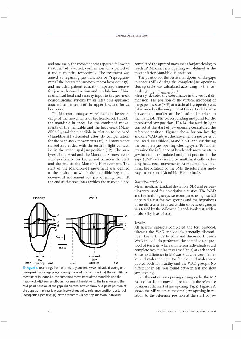
where y denotes the coordinates in the vertical di-mension. The position of the vertical midpoint of the gape in space (MP) at maximal jaw opening was determined as the midpoint of the vertical distance between the marker on the head and marker on the mandible. The corresponding midpoint for the intercuspal jaw position (IP), i.e. the teeth in light contact at the start of jaw opening constituted the reference position. Figure 1 shows for one healthy and one WAD subject the movement trajectories of the Head, Mandible-S, Mandible-H and MP during the complete jaw opening-closing cycle. To further examine the infl uence of head-neck movements in jaw function, a simulated midpoint position of the gape (SMP) was created by mathematically exclu-ding head-neck movements. At maximal jaw ope-ning, the location of the SMP therefore was mid-way the maximal Mandible-H amplitude.
Statistical analysisMean, median, standard deviation (SD) and percen-tiles were used for descriptive statistics. The WAD and the healthy groups were compared using two tail unpaired t-test for two groups and the hypothesis of no difference in speed within or between groups was tested by the Wilcoxon Signed-Rank test, with a probability level of 0.05.
ResultsAll healthy subjects completed the test protocol, whereas the WAD individuals generally disconti-nued the task due to pain and discomfort. Seven WAD individuals performed the complete test pro-tocol of ten tests, whereas nineteen individuals could complete two to nine tests (median 5) at each speed. Since no difference in MP was found between fema-les and males the data for females and males were pooled both for healthy and the WAD groups. No difference in MP was found between fast and slow jaw opening.
For the entire jaw opening closing cycle, the MP was not static but moved in relation to the reference position at the start of jaw opening (Fig.1). Figure 2 A shows the MP values at maximal jaw opening in re-lation to the reference position at the start of jaw
• Figure 1. Recordings from one healthy and one WAD individual during one jaw opening-closing cycle, showing traces of the head-neck (a), the mandibular movement in space, i.e. the combined movement of the mandible and the head-neck (d), the mandibular movement in relation to the head (e), and the Mid-point position of the gape (b). Vertical arrows show Mid-point position of the gape at maximal jaw opening with regard to reference position at start of jaw opening (see text) (c). Note differences in healthy and WAD individual.

impaired gape positioning in wad
swedish dental journal vol. 30 issue 1 2006 13
• Figure 2. The Mid-point position of the gape at maximal jaw opening (MP). The zero-line corresponds to reference position at start of jaw opening (see text). A. Box and whisker plots (10th, 25th, 50th, 75th and 90th percentiles) show results of healthy (unfi lled, n = 15) and WAD individuals (fi lled, n = 26). Differences between healthy and WAD groups are marked. B. Mean and 95 % confi dence interval values of MP for two WAD individuals during pre- (initial) and post- (follow up) treatment recordings. The duration between the pre- (5 fast, 5 slow, n = 10) and post- (10 fast, 10 slow, n = 20) treatment recordings was 9 months for female and 11 months for male individual. Note “normalization” of MP following treatment.
opening, for fast and slow speed, in healthy and WAD groups. In healthy, there was no signifi cant difference between the MP value at maximal jaw opening and the reference position at the start of jaw opening. For fast speed, the MP at maximal jaw opening was 1 mm below the reference posi-tion at the start of jaw opening, and for slow speed 1 mm above the reference position at the start of jaw opening (median values). In contrast, in the WAD group there was a signifi cant difference between the MP at maximal jaw opening and the reference po-sition at the start of jaw opening. For both slow and fast speed the MP was 7 mm below the reference position at the start of jaw opening.
In healthy, the SMP was 22 mm (SD 7) and 23 mm (SD 7) below the reference position at the start of the mandibular movement for fast and slow speed,respectively. The corresponding values for the WAD group were 19 mm (SD 5) and 18 mm (SD 5).
In healthy, the vertical movement amplitudes for the Mandible-H were 48 mm (SD 6) and 47 mm (SD 5) for fast and slow speed, respectively. The cor-responding Head amplitudes were 23 mm (SD 9) and 26 mm (SD 13). In the WAD group, the ampli-tudes for the Mandible-H were 37 mm (SD 10) and
36 mm (SD 9) for fast and slow speed, respectively, and the corresponding Head amplitudes were 12 (SD 8) mm and 13 (SD 9) mm. In healthy, the relative change in MP in relation to the reference position at the start of jaw opening was 2% and 0.5 % of the maximal Mandible-H amplitudes, for fast and slow movements, respectively. In the WAD group, the corresponding values were 18% and 19%, respecti-vely. For the two WAD individuals who received tre-atment, the post-treatment recordings showed a sig-nifi cant upward shift of the MP to levels comparable with those of the healthy subjects (Fig. 2 B).
DiscussionIf a jaw opening movement was performed only in the temporomandibular joint, i.e. without head-neck extension, the MP would be shifted downwards, along with the downward movement of the mandi-ble. Furthermore, the magnitude of this downward shift of the MP would be half that of the mandibular movement in relation to the head, i.e. half that of the maximal amplitude of Mandible-H. However, in the healthy subjects we found that the MP at maximal jaw opening generally coincided with the reference position at the start of jaw opening. This suggests

zafar, nordh, eriksson
14 swedish dental journal vol. 30 issue 1 2006
that the orientation of the gape in space is achie-ved not only by movements in the temporomandi-bular joint but also involve head-neck movements. Such an interpretation is corroborated by the pre-sent fi ndings in the WAD group, where the MP at maximal jaw opening was signifi cantly lower than the reference position at the start of jaw opening. Notably, in the WAD group the magnitude of this downward shift of the MP was nearly twenty per cent of that of the Mandible-H. This observation can be explained by the relatively small head-neck extension during jaw opening found in the WAD group. Furthermore, the fi nding that the gape was positioned “too low” during jaw opening in the WAD individuals, supports our previous proposal of an association between neck injury/dysfunction and deranged jaw-neck motor behaviour (6, 7, 10, 21).
The infl uence of head-neck extension in the orientation of the gape was also demonstrated by the analysis of the MP during the entire jaw ope-ning-closing cycle. In healthy, the MP started to shift downwards with the start of the jaw opening mo-vement, indicating that the relative acceleration was faster for the mandible than for the head. However, at maximal jaw opening, the MP generally coinci-ded with the reference position at the start of the jaw opening. The latter fi nding refl ects a relative increase in acceleration of the head extension during the late phase of jaw opening. Also in the WAD group, the MP started to shift downwards with the start of the jaw opening movement, but in contrast to what was found in healthy, it remained in a downward posi-tion during the rest of the jaw opening-closing cy-cle. Again the difference between healthy and WAD individuals seems to be associated with the smaller head-neck extension in the WAD individuals.
We have previously shown that the trajectory of the MP during the complete jaw opening-closing cycle has a high spatiotemporal consistency both in healthy (24) and in WAD individuals (6), suggesting that even if the integrative jaw and head-neck beha-viour is disturbed in response to neck dysfunction, it can still be performed in an invariant manner. The present results support our previous observations (6). The fi nding of obvious differences in motor be-haviour between healthy and WAD individuals in this study and previously (6, 10, 21), indicate that the jaw motor system in WAD individuals has adapted to new neural settings and motor synergies to per-form conceptually similar jaw tasks.
Positioning of the gape in space is performed
without visual guidance and therefore probably with signifi cant aid of proprioceptive information from muscles and joints. As judged from the com-plex nature of muscle spindle structure in both jaw (2, 3) and neck (15) muscles, the proprioceptive me-chanisms behind jaw–neck motor control appear to be advanced, and probably apt for fi ne control in complex tasks such as feeding, yawning and speech. The present data of a disturbed ability in the WAD group to correctly position the gape in space may refl ect an altered propriocetive input or central processing during jaw action.
Finely tuned head-neck movements during jaw function are probably associated with two main goals. First, free head-neck extension has biome-chanical advantages by enabling optimal space for movements of the mandible during jaw opening, thus gaining maximal freedom for execution of the compound gaping movement (4). The opposite, a reduced head-neck extension ability, would limit the space for mandibular movements, due to imping-ement of the mandible with suprahyoid and air-way structures (4). Second, as demonstrated in this study, the orientation of the gape in space involves both mandibular and head-neck movements. Thus, both jaw opening and the positioning of the gape in space seem to be governed by neural commands simultaneously activating jaw and neck neuromus-cular synergies. Without such fi nely tuned neural control, jaw function would be disturbed.
In this report, two examples were included to il-lustrate the possible clinical use of evaluating gape position in analyses and documentation of post treatment changes in jaw function. The fi nding of a post-treatment normalization of the gape posi-tion is notable and adds to previous observations suggesting an important role of neck function in jaw motor control. Thus, in response to treatment, there was an upward shift of the MP at maximal jaw opening, to a level comparable with that of the heal-thy subjects. Moreover, this post-treatment change in MP was found to be associated with an increase in the head-neck extension amplitude during jaw opening. The faulty position of the gape in the WAD group could be related to change in proprio-ceptive ability, and the post-treatment observations seem to mirror an improvement of proprioceptive function and central processing of neuromuscular commands. Besides giving support for the notion of a functional linkage between the jaw and the neck motor systems, the result also points to a new ap-proach for rehabilitation and improvement of neck

impaired gape positioning in wad
swedish dental journal vol. 30 issue 1 2006 15
mobility in WAD. This matter with some exception (7), has not been addressed previously. However, further studies are needed and ongoing in our la-boratory.
In conclusion, the results suggest that both the width and orientation of the gape in space relies on coordinated mandibular and head-neck movements, and that there is an association between neck pain and dysfunction following trauma and reduced width and impaired positioning of the gape in space. In this context, the mid-point of the gape in space seems to be a useful marker in evaluation of the functional state of the jaw-neck motor system. Finally, the role of neck function in jaw activities should be taken into account in research and clinical management.
AcknowledgementsThe skilful technical assistance of Mr. Jan Öberg, and the programming assistance of Mr. Mattias Backén is gratefully acknowledged. Supported by the Umeå University, the Västerbotten Public Dental Health Service, and the Swedish Dental Society.
References1. Dessem D, Luo P. Jaw-muscle spindle afferent feedback
to the cervical spinal cord in the rat. Exp Brain Res 1999;128:451-59.
2. Eriksson P-O, Thornell L-E Relation to extrafusal fi bre-type composition in muscle spindle structure and location in the human masseter muscle. Arch Oral Biol. 1987;32:483-91.
3. Eriksson P-O, Butler-Browne GS, Thornell LE. Immunohistochemical characterization of human masseter muscle spindles. Muscle Nerve 1994;17:31-41.
4. Eriksson P-O, Zafar H, Nordh E. Concomitant mandibular and head-neck movements during jaw opening-closing in man. J Oral Rehabil 1998;25:859-70.
5. Eriksson P-O, Häggman-Henrikson B, Nordh E,, Zafar H. Co-ordinated mandibular and head-neck movements during rhythmic jaw activities in man. J Dent Res 2000;79:1378-84.
6. Eriksson P-O, Zafar H, Häggman-Henrikson B. Deranged jaw-neck motor control in whiplash associated disorders. Eur J Oral Sci 2004;112:25-32
7. Eriksson P-O, Zafar H. Musculoskeletal disorders in the jaw-face and neck. In: Conn’s Current Therapy. Rakel RE, Bope ET, editors. Philadelphia; WB Saunders, 2005. p. 1128-33
8. Helkimo M. Studies on function and dysfunction of the masticatory system. 3. Analyses of anamnestic and clinical recordings of dysfunction with the aid of indices. Sven Tandlak Tidskr 1974;67:165-81.
9. Hellström F, Thunberg J, Bergenheim M, Sjolander P, Pedersen J, Johansson H. Elevated intramuscular concentration of bradykinin in jaw muscle increases the fusimotor drive to neck muscles in the cat. J Dent Res 2000; 79:1815-22.
10. Häggman-Henrikson B, Zafar H, Eriksson P-O. Disturbed jaw behaviour in whiplash associated disorders during rhythmic movements. J Dent Res 2002;81:747-51.
11. Häggman-Henrikson B, Eriksson P-O. Head movements during chewing: Relation to size and texture of bolus. J Dent Res 2004;83:864-8.
12. Igarashi N, Yamamura K, Yamada Y, Kohno S. Head movements and neck muscle activities associated with the jaw movement during mastication in the rabbit. Brain Res 2000;871:151-5.
13. Josefsson T, Nordh N, Eriksson P-O. A fl exible high-precision video system for digital recording of motor acts through light-weight refl ex markers. Comput Methods Programs Biomed 1996;49:119-29.
14. Kohno S, Matsuyama T, Medina RU, Arai Y. Functional-rhythmical coupling of head and mandibular movements. J Oral Rehabil 2001;28:161-7.
15. Liu JX, Thornell LE, Pedrosa-Domellof F. Muscle spindles in the deep muscles of the human neck: a morphological and immunocytochemical study. J Histochem Cytochem. 2003;5:175-86.
16. Masuzaki H, Masuzaki M, Ishimaru T. Color Doppler imaging of fetal yawning. Ultrasound Obstet Gynecol. 1996;8:355-6.
17. Okesson P. Orofacial pain. Guidelines for assessment, diagnosis and management. Quintessence, 1996; pp 19-52.
18. Petrikovsky B, Kaplan G, Holsten N. Fetal yawning activity in normal and high-risk fetuses: a preliminary observation. Ultrasound Obstet Gynecol 1999;13:127-30.
19. Sepulveda W, Mangiamarchi M. Fetal yawning. Ultrasound Obstet Gynecol. 1995;5:57-9.
20. Spitzer WO, Skovron ML, Salmi LR, Cassidy JD, Duranceau J, Suissa S et al. Scientifi c monograph of the Quebec Task Force on Whiplash-Associated Disorders: redefi ning ”whiplash” and its management. Spine 1995;20 (8 Suppl):1S-73S.
21. Zafar H. Integrated jaw and neck function in man. Studies of mandibular and head-neck movements during jaw opening-closing tasks. (Doctoral thesis) Swed Dent J 2000;Suppl 143. pp 1-41.
22. Zafar H, Eriksson P-O, Nordh E, Häggman-Henrikson B. Wireless optoelectronic recordings of mandibular and associated head-neck movements in man: a methodological study. J Oral Rehabil 2000;27: 227-38.
23. Zafar H, Nordh E, Eriksson P-O. Temporal coordination between mandibular and head-neck movements during jaw opening-closing tasks in man. Arch Oral Biol 2000;45:675-82.
24. Zafar H, Nordh E, Eriksson P-O. Spatiotemporal consistency of human mandibular and head-neck movement trajectories during jaw opening-closing tasks. Exp Brain Res 2002;146:70-6.
Address:Dr Hamayun ZafarDept. Clinical Oral PhysiologyUmeå UniversitySE-901 87 Umeå, SwedenTel: (+46) - 90 785 62 46Fax: (+46) - 90 13 25 78E-mail: [email protected]
24
supplements to swedish dental journal
•••The supplementscan be ordered from Swedish Dental Journal, Box 1217,111 82 Stockholm, Sweden.Subscriptionof the supplementscan be arranged.
81. Temporomandibular joint dysfunction and systemic joint laxity.Lilian Westling (1992)
82. Calcium tranport in dentinogenesis. An experimental study in the rat incisorodontoblast. Ted Lundgren (1992)
83. Cancelled.84. Aspects of bone healing and bone substitute incorporation.
An experimental study in rabbit skull bone defects. Sten Isaksson (1992) 85. Release of mercury vapour from dental amalgam. An in vivo and in vitro study.
Anders Berglund (1992) 86. A cross-cultural study of occlusal tooth wear.
Anders Johansson (1992) 87. Acupuncture in the treatment of patients with craniomandibular disorders.
Comparative, longitudinal and methodological studies. Thomas List (1992)
88. Post-treatment effects of the herbst appliance. A radiographic, clinicaland biometric investigation. Ken Hansson (1992)
89. Infl uence of age and salivary secretion rate and oral sugar clearance.Jan Coby Hase (1993)
90. Actinobacillus actinomycetemcomitans and lokalized juvenil periodontitis.Lars Christersson (1993)
91. Speech and other oral function.Sture Lundqvist (1993)
92. Human and experimental osteoarthrosis of the temporomandibular joint.Susanna Axelsson (1993)
93. Prevalence and technical standard of endodontic treatment in a Swedish population. Mats Eckerbom (1993)
94. Oral health in groups of refugees in Sweden. Mikael Zimmerman (1993)
95. Tinnitus and carniomandibular disorders. Barbara Rubinstein (1993)
96. Clinical aspects of restorative treatment in the primary dentition. Mirja Varpio (1993)
97. Objective evaluation of mouth dryness. A methodological study.Vincent Henricsson (1994)
98. Characterization of human oro-facial and masticatory muscles with respectto fi bre types, myosins ans capillaries. Per Stål (1994)
99. Orthodontic magnets.Lars Bondemark (1994)
100. Ectopic eruption of the maxillary fi rst permanent molar. Krister Bjerklin (1994)
101. Dental enamel in relation to ionized calcium and parathyroid hormone.Lotta Ranggård (1994)
102. Studies of maxillary overdentures on osseointegrated implants.Jan-Ivan Smedberg (1995)
supplement SDj 24supplement SDj 24 05-12-06 08.39.2105-12-06 08.39.21
400 SEK
400 SEK
400 SEK
400 SEK
400 SEK
400 SEK
400 SEK
400 SEK
400 SEK
400 SEK
400 SEK
400 SEK
400 SEK
400 SEK
400 SEk
400 SEK
400 SEK
400 SEK
400 SEK
400 SEK
400 SEK

il-1 and il-6 in chronic periodontitis
swedish dental journal vol. 30 issue 1 2006 17
Analysis of the interleukin-1 and interleukin-6 polymorphisms in patients with chronic periodontitis. A pilot study henrik jansson1, valeriya lyssenko2, åsa gustavsson1, kristina hamberg1, björn söderfeldt3, leif groop2 and gunilla bratthall1
• The aim of this study was to analyse whether the interleukin-1 (IL-1) and IL-6 gene polymorphisms were associated with the susceptibility of chronic periodontitis.
Genomic DNA was obtained from 20 patients with chronic periodontitis and 31 periodontally healthy subjects. All subjects were of North European heritage. The test subjects were kept in a maintenance program after periodontal treatment but yet showing signs of recurrent disease. Genotyping of the IL-1α [+4845C>T], IL-1β [-3954C>T] and IL-6 [-174G>C] polymorphisms was carried out using an allelic discrimi-nation Assay-by-Design method on ABI PRISM 7900 Sequence Detection System. All genotypes were analyzed using the GeneMapper 2.0 software.
A similar distribution of Single Nucleotide Polymorphism (SNP) was seen in both groups. Analysis by logistic regression including gender, IL -1α [+4845C>T], IL -1β [-3954C>T], IL -6 [-174G>C] genotypes, the composite IL -1 genotype, the combination of the composite IL -1 genotype and the IL -6 -174G>C genotype and adjusting for smoking did not result in any statistically signifi cant difference.
SNPs in IL-1α [+4845C>T], IL-1β [-3954C>T] and IL-6 [-174G>C] do not seem to in-crease the susceptibility to chronic periodontitis in this group of subjects.
Key words Genotype, interleukin-1, interleukin-6, periodontal diseases, polymorphism.
swed dent j 2006; 30: 17–23 • jansson et al
1 Department of Periodontology, Centre for Oral Health Sciences, Malmö University.2 Department of Clinical Sciences, Diabetes & Endocrinology, Wallenberg laboratory, Malmö University Hospital (UMAS), Lund University.3 Department of Dental Public Health, Centre for Oral Health Sciences, Malmö University.

jansson et al
18 swedish dental journal vol. 30 issue 1 2006
Analys av interleukin-1 och interleukin-6polymorfi sm hos patienter med kronisk parodontithenrik jansson, valeriya lyssenko, åsa gustavsson, kristina hamberg, björn söderfeldt, leif groop och gunilla bratthall
Sammanfattning
• Målet med den här studien var att undersöka huruvida IL-1 och IL-6 polymorfi sm är relaterat till ökad känslighet för kronisk parodontit.
Blodprov togs på 20 patienter med kronisk parodontit och 31 parodontalt friska in-divider. DNA extraherades från blodproverna. Samtliga individer var av nordeuropeiskt ursprung. Parodontitpatienterna ingick i ett parodontalt stödbehandlingsprogram efter avslutad parodontal behandling på specialistkliniken för Parodontologi i Malmö, Folktandvården Skåne, men trots detta uppvisade patienterna tecken på förnyad paro-dontal sjukdom. Genotypning av IL-1α [+4845C>T], IL-1β [-3954C>T] och IL-6 [-174G>C] polymorfi smerna utfördes med allelic discrimination Assay-by-Design på en ABI PRISM 7900 sekvens detektionssystem. Alla genotyper analyserades med mjukvaroprogrammet GeneMapper 2.0.
Fördelning av genetiska variationer var likvärdig i de båda undersökta grupperna. Logistisk regressions analys avseende kön, IL-1α +4845C>T, IL-1β -3954C>T, IL-6 -174G>C genotyperna, en kombination av den sällsynta genotypen för IL-1α och IL-1β, samt kombi-nation av IL-1 och IL-6 -174G>C genotyperna, justerat för rökning, resulterade inte i någon statistisk signifi kant skillnad.
Genetiska variationer vid IL-1α [+4845C>T], IL-1β [-3954C>T] och IL-6 [-174G>C] verkar inte öka mottagligheten för kronisk parodontit i den här gruppen av individer.
swed dent j 2006; 30: 17–23 • jansson et al

il-1 and il-6 in chronic periodontitis
swedish dental journal vol. 30 issue 1 2006 19
IntroductionPeriodontitis is a chronic infl ammatory disease ini-tiated by specifi c bacteria, predominantly anaero-bes (10). The immuno-infl ammatory responses activated by bacteria lead to destruction of colla-gen and bone supporting the teeth (21). Although bacteria appear to be essential for disease initiation, there are individual differences in disease progres-sion due to modifying host factors such as diabetes, smoking and genetics (9, 20). The body of accu-mulating evidences has shown that the pro-infl am-matory cytokines such as tumour necrosis factor-alpha (TNF-α) and IL-1 are important mediators in a number of chronic infl ammatory disorders (2, 6). IL-1 in particular is associated with periodontal disease because of its role as potent inducer of bone resorption (16) and increased levels have been de-monstrated in both gingival crevicular fl uid (GCF) (29) and gingival tissues (19) in patients with adult periodontitis. Variations in the immune system may be explained by genetic diversity. One geno-type on the IL-1B gene is associated with increased IL-1β production (26). There are indications that some polymorphisms in the IL-1 gene cluster may be associated with periodontal disease, but no con-sistent results have been obtained. It has also been reported that variations in cytokine expression as a response to noxious stimuli appear to be under ge-netic control (25) and that carriers of allele 2 of the IL-1β +3953 polymorphism have increased the IL-1β production (26). Kornman et al. (14) have found a strong association between the severity of perio-dontitis and composite genotype in a Caucasian po-pulation with north European heritage. The compo-site genotype associated with periodontitis comprised allele 2 of IL-1α -889 polymorphism plus allele 2 of the IL-1β +3953 polymorphism. IL-6 is also a pro-infl am-matory cytokine, and the -174G>C polymorphism in the promoter region of the IL-6 gene has been as-sociated with rheumatoid arthritis (24). IL-6 has also been detected in GCF, where a correlation between IL-6 concentration and the level of clinical disease (7), and increased levels of IL-6 in infl amed gingival tis-sue (30).
There are confl icting results whether IL-6 (-174) single nucleotide polymorphism (SNP) is associated with periodontal disease.
The aim of the present study was to investigate, whether common variants in the IL-1α [+4845C>T], IL-1β [-3954C>T] and IL-6 [-174G>C] genes are associated with increased susceptibility to chronic periodontitis.
Material and MethodsSubjectsTwo groups of Caucasian subjects were included. One group with chronic periodontitis (periodon-tally diseased [PD+] group) and one group wit-hout showing any sites of >5 mm probing depth and normal radiographic bone levels, i.e. a distance of <3 mm between the CEJ and bone crest of the proximal tooth sites (periodontally healthy [PD-] group). The PD+ group consisted of 20 patients (9 females and 11 males, 48-70 years of age; mean 57.9 ± SD 6.2) recruited from the Specialist clinic for Periodontology in Malmö, Sweden. These subjects were kept in a maintenance program after perio-dontal treatment but yet showing signs of recurrent disease. Details of sample selection, inclusion and exclusion criteria have been described previously (13). The PD- group consisted of 31 subjects (16 females and 15 males, 46-69 years of age; mean 55.0 ± SD 7.4), Test and control individuals were asked about smoking habits with smoking defi ned as >10 cigarettes/day.
The periodontal examination was performed by two calibrated examiners (HJ and ÅG) and has been described in detail previously (13).
Blood sampling and extraction of DNAFive ml of peripheral human blood was obtained from all subjects by standard venipuncture using ethylenediaminetetraacetic acid (EDTA)-tubes. The blood samples were stored at +4oC overnight and then centrifuged at +40C at 3000 rpm for 15 minutes. The cellular component was separated and stored at -180C until further analysis. The DNA was obtained using a modifi cation of a standard method (32).
GenotypingThe laboratory analysis assembled a composite genotype for each study member for the IL-1α+4845C>T and IL-1β -3954C>T polymorphisms, which lie within the IL-1 gene cluster on chromoso-me 2q13. To determine the genotypes of these SNPs, polymerase chain reaction (PCR), with fl uorogenic probes (Taqman® MGB probe) (4) was used, which is more reliable and faster than the conventionally PCR-restriction fragment length polymorphism (RFLP) assay (27). The same technique was used analysing SNP at IL-6 -174G>C.
Genotyping of the IL-1α +4845 (National Cen-ter for Biotechnology Information (NCBI) acces-sion number rs17561), IL-1β -3954 (NCBI accession number rs1143634) and IL-6 -174 (NCBI accession

jansson et al
20 swedish dental journal vol. 30 issue 1 2006
number rs1800795) polymorphisms was genoty-ped using allelic discrimination in the ABI PRISM 7900 Sequence Detection System (Applied Biosys-tems, Foster City, CA) in a 5-µl reaction according to the manufacturerís instructions. Primers and probes were designed using Assays-by-Design (Applied Bio-systems, Foster City, CA, USA) and were as follows:IL-1α +4845; forward primer, 5í-TCTGCACTTGTGATCATGGTTTTAGA- 3í; reverse primer 5í- CATTGGCTCGAATTATACTTTGATTGAGG-3í; probe: VIC- CTAGGTCAGCACCTTT and FAM- CCTAGGTCATCACCTTT;IL-1β -3954; forward primer 5í- ACCTAAACAACATGTGCTCCACA-3í; reverse primer 5í- ATCGTGCACATAAGCCTCGTTA-3í; probe: VIC- CATGTGTCGAAGAAGA and FAM-CATGTGTCAAAGAAGA;IL-6 -174G>C; forward primer 5í-GACGACCTAAGCTGCACTTTTCñ3í; reverse primer 5í-GGGCTGATTGGAAACCTTATTAAGATTGñ3í; probe: VIC- CCTTTAGCATCGCAAGAC and FAM-CTTTAGCATGGCAAGAC and probes.A composite genotype was defi ned as at least one allele 2 present at each locus (IL-1α +4845 and IL-1β-3954), according to Kornman et al. (14).
Statistical AnalysesFisher´s exact test was used to test for signifi cance of differences in genotype and allele frequencies in PD+ and PD- subjects. Adjustments for gender and smoking were carried out by logistic regression analysis, where goodness of fi t was judged by clas-sifi cation plots and calculation of model chi-square. Two-sided P Vales (p-values) <0.05 were considered statistically signifi cant. All analyses were done using Statistical Package for Social Sciences (SPSS) for Windows, version 13.0 (http://www.spss.com).
Ethical requirementsThe Medical Ethics Committee of Lund University, Lund, Sweden approved the study in accordance with the Helsinki Declaration. All patients gave their signed, informed consent prior to inclusion in the project.
ResultsThe allele and genotype frequencies of the IL-1α+4845C>T, IL-1β -3954C>T and IL-6 -174G>C po-lymorphisms are summarized in Table 1. The IL-1α+4845C>T, IL-1β -3954C>T and IL-6 -174G>C poly-morphisms were common in all subjects (frequency of 75% for the IL-1α +4845C allele, 78% for the IL-1β -3954C and 66% for the IL-6 -174G in this group individuals). There were no signifi cant differences
between the PD- and PD+ groups according to al-lele or genotype frequency.
Analysis by logistic regression including gender, IL-1α +4845C>T genotype, IL-1β -3954C>T geno-type, IL-6 -174G>C genotype, the composite IL-1 genotype, the combination of the composite IL-1 genotype and the IL-6 -174G>C genotype and ad-justing for smoking did not result in any statistically signifi cant difference. Nor was the model as a whole signifi cant.
DiscussionThe results of the present study did not show any statistically signifi cant difference of the composite IL-1 genotype alone, IL-6 -174G>C gene polymorp-hism alone or the combination of the composite IL-1 and IL-6 -174G>C gene polymorphisms in patients with chronic periodontitis. This is to our knowledge the fi rst study analysing the combina-tion of the composite IL-1 and IL-6 -174G>C gene polymorphisms in smoking patients with chronic periodontitis.
Our result regarding the composite IL-1 geno-type is in contrast to Meisel et al. (18) and McDevitt et al. (17). Meisel et al. (18) reported an increased risk of periodontal disease in a group of genotype-positive smokers. McDevitt et al. (17) performed a case-control study, comprising 44 subjects with moderate to severe periodontal disease and 46 sub-jects with healthy periodontal tissues or mild pe-riodontal disease, in either non-smokers or former smokers. They reported a 41% prevalence of the positive composite genotype in the test group and 28% in the control group with no statistically signi-fi cant difference. When using a multivariate logis-tic regression analyses, adjusting for confounders such as age and past smoking history McDewitt et al. (17) showed a correlation between the rare IL-1 genotype and periodontal disease.
Irrespective of smoking, our result is in accor-dance with Papapanou et al. (22), however. They investigated the prevalence of the composite ge-notype in 132 patients with periodontitis and 73 periodontally healthy individuals and found an occurrence rate of 42, 9% of the composite IL-1 genotype. There was no statistically signifi cant dif-ference between test and control subjects. Contrary to our result Kornman et al. (14), Gore et al. (8) and Laine et al. (15) have shown an association between combinations of two single nucleotide polymorp-hisms (SNP) in the IL-1α and IL-1β genes and hig-her prevalence of severe periodontal disease.

il-1 and il-6 in chronic periodontitis
swedish dental journal vol. 30 issue 1 2006 21
• Table 1: Allele and genotype frequencies of the studied polymorphisms, and the frequency of the composite IL-1 genotype (IL-AB) and combination of the composite IL-1 genotype and the rare IL-6 -174 genotype (IL-AB6) in subjects with [PD+] or without[PD-] chronic periodontitis.
Allele/Genotype % (n) p-value
IL-1α +4845C>T All (n = 51) PD- (n = 31) PD+ (n = 20)T 25 (26) 24 (15) 27,5 (11)C 75 (76) 76 (47) 73,5 (29) NS
TT 4 (2) 3 (1) 5 (1)CT 43 (22) 42 (13) 45 (9)CC 53 (27) 55 (17) 50 (10) NS
IL-1β -3954C>T All (n = 51) PD- (n = 31) PD+ (n = 20)T 22 (22) 21 (13) 22,5 (9)C 78 (80) 79 (49) 77,5 (31) NS
TT 2 (1) 3 (1) 0 (0)CT 39 (20) 36 (11) 45 (9)CC 59 (30) 61 (19) 55 (11) NS
IL-6 -174G>C All (n = 51) PD- (n = 31) PD+ (n = 19)C 34 (35) 29 (18) 45 (17)G 66 (67) 71 (44) 55 (21) NS
CC 20 (10) 13 (4) 32 (6)GC 29 (15) 32 (10) 26 (5)GG 51 (26) 55 (17) 42 (8) NS
IL-1AB All (n = 51) PD- (n = 31) PD+ (n = 20)Negative 67 (34) 71 (22) 60 (12)Positive 33 (17) 29 (9) 40 (8) NS
IL-AB6 All (n = 50) PD- (n = 31) PD+ (n = 19)Negative 82 (41) 84 (26) 79 (15)Positive 18 (9) 16 (5) 21 (4) NS
Cullinan et al. (5) investigated the relationship between IL-1 genotype and periodontitis in a pro-spective longitudinal study in an adult population of essentially European heritage. They found a con-sistent trend for IL-1 genotype positive subjects to experience attachment loss when compared with IL-1 genotype negative subjects. They concluded that an interaction of the IL-1 positive genotype with age, smoking and P. gingivalis could be a contributory risk factor for periodontal disease progression in that population.
In contrast to Cullinan et al. (5), Cattbriga et al. (3) reported that there were no statistically signifi -cant differences related to IL-1 genotype in tooth loss after 10 years in a non-smoking, well-main-tained periodontal population. On an individual level, however, the IL-1 genotype in combination with the initial bone level was considered to be of value for predicting future bone level variation.
A pronounced difference in the prevalence of the composite genotype was reported in a study com-prising a Chinese population (1). The prevalence of the composite IL-1 genotype was 2.3%, which is dramatically lower than what is reported for Cau-casians. No conclusions could be drawn concer-ning the relation between IL-1 polymorphism and periodontal disease. Neither could this association be found in African-Americans with localized juve-nile periodontitis (33) and in European Caucasians with generalized Early Onset Periodontitis (23). It is conceivable that the frequency of genetic alleles varies between different ethnical populations and forms of periodontal disease.
No association between IL-6 polymorphism and chronic periodontitis was found in the present study. This might be due to the fact that IL-6 is a pleiotropic cytokine, with both infl ammatory and anti-infl ammatory properties. The -174G>C poly-

jansson et al
22 swedish dental journal vol. 30 issue 1 2006
morphism in the promoter region of the IL-6 gene has been associated with rheumatoid arthritis (24) and several features of the metabolic syndrome in Caucasians (11). Trevilatto et al (31) found a re-lationship between IL-6 -174G>C polymorphism and chronic periodontitis. No association, however, was found between chronic periodontitis and IL-6 -174G>C polymorphism by Holla et al. (12).
All test patients were selected from a large group of patients enrolled in a maintenance care program at a specialist clinic of periodontology but yet sho-wing signs of recurrent disease. With this in mind and the special clinical features of the recruited test group, it was diffi cult to fi nd a control group (pe-riodontally healthy smokers) at the same age and gender. A correlation between chronic periodontitis and the positive genotype in smokers cannot be ex-cluded, however.
Estimation of genetic predisposition and smo-king represents a couple of several dimensions of patient-based risk for disease progression. Further research is needed to identify other periodontal can-didate genes or combinations of genes involved in the immuno-infl ammatory response in periodontal disease.
In conclusion, SNPs in IL-1α [+4845C>T], IL-1β[-3954C>T] and IL-6 [-174G>C] do not seem to increase the susceptibility to chronic periodontitis in this group of subjects.
AcknowledgementsWe would like to thank Professor Kenneth S. Korn-man for his assistance with primer and probe design. DDS Christina Diogo-Löfgren for assisting with pa-tient selection. Dr. Harvest Gu for helpful discus-sion.
The Swedish Dental Society and The Swedish Patent and Revenue Research Fund for Preventive Odontology, Sweden supported the study.
References1. Armitage GC, Wu Y, Wang HY, Sorrell J, di Giovine FS,
Duff GW. Low prevalence of a periodontitis-associated interleukin-1 composite genotype in individuals of Chinese heritage. J Periodontol 2000; 71: 164-71.
2. Birkedal-Hansen H. Role of cytokines and infl ammatory mediators in tissue destruction. J Periodontal Res 1993; 28: 500-10.
3. Cattabriga M, Rotundo R, Muzzi L et al. Retrospective evaluation of the infl uence of the interleukin-1 genotype on radiographic bone levels in treated periodontal patients over 10 years. J Periodontol 2001; 72: 767-73.
4. di Giovine FS, Camp NJ, Cox A, Chaudhary AG, Sorrell JA,
Crane A, Duff GW. Detection and population analysis of IL-1 and TNF gene polymorphisms. In: Balkwill F ed. Cytokine Moleculare Biology: A Practical approach. 3rd ed. Oxford: Oxford University Press, 2000: 21-46.
5. Cullinan MP, Westerman B, Hamlet SM, Palmer LE, Faddy MJ, Lang NP, Seymour GJ. A longitudinal study of interleukin-1 gene polymorphisms and periodontal disease in a general adult population. J Clin Periodontol 2001; 28: 1137-44.
6. Dinarello CA. Biologic basis for interleukin-1 in disease. Blood 1996; 87: 2095-147.
7. Geivelis M, Turner DW, Pederson ED, Lamberts BL. Measurements of interleukin-6 in gingival crevicular fl uid from adults with destructive periodontal disease. J Periodontol. 1993; 64: 980-3.
8. Gore EA, Sanders JJ, Pandey JP, Palesch Y, Galbraith GM. Interleukin-1beta+3953 allele 2: association with disease status in adult periodontitis. J Clin Periodontol 1998; 25: 781-5.
9. Grossi SG, Zambon JJ, Ho AW, Koch G, Dunford RG, Machtei EE, Norderyd OM, Genco RJ. Assessment of risk for periodontal disease. I. Risk indicators for attachment loss. J Periodontol 1994; 65: 260-7.
10. Haffajee AD, Socransky SS. Microbial etiological agents of destructive periodontal disease Periodontol 2000 1994; 5: 78-111.
11. Hamid YA, Rose CS, Urhammer SA, Glümer C, Nolsoe R, Kristiansen OP, Mandrup-Poulsen T, Borch-Johnsen K, Jorgensen T, Hansen T, Pedersen O. Variations of the interleukin-6 promoter are associated with features of the metabolic syndrome in Caucasian Danes. Diabetologia 2005; 48: 251-60.
12. Holla LI, Fassmann A, Stejskalova A, Znojil V, Vanek J, Vacha J. Analysis of the interleukin-6 gene promoter polymorphisms in Czech patients with chronic periodontitis. J Periodontol 2004; 75: 30-6.
13. Jansson H, Bratthall G, Söderholm G. Clinical outcome observed in subjects with recurrent periodontal disease following local treatment with 25% metronidazole gel. J Periodontol 2003; 74: 372-7.
14. Kornman KS, Crane A, Wang HY, di Giovlne FS, Newman MG, Pirk FW, Wilson Jr. TG, Higginbottom FL, Duff GW. The interleukin-1 genotype as a severity factor in adult periodontal disease. J Clin Periodontol 1997a; 24: 72-7.
15. Laine ML, Farre MA, Gonzalez G, van Dijk LJ, Ham AJ, Winkel EG, Crusius JB, Vandenbroucke JP, van Winkelhoff AJ, Pena AS. Polymorphisms of the interleukin-1 gene family, oral microbial pathogens, and smoking in adult periodontitis. J Dent Res 2001; 80: 1695-9.
16. Masada MP, Persson R, Kenney JS, Lee SW, Page RC, Allison AC. Measurement of interleukin-1 alpha and -1 beta in gingival crevicular fl uid: implications for the pathogenesis of periodontal disease. J Periodontal Res 1990; 25: 156-63.
17. McDevitt MJ, Wang HY, Knobelman C, Newman MG, di Giovine FS, Timms J, Duff GW, Kornman KS. Interleukin-1 genetic association with periodontitis in clinical practice. J Periodontol 2000; 71: 156-63.
18. Meisel P, Siegemund A, Grimm R, Herrmann FH, John U, Schwahn C, Kocher T. The interleukin-1 polymorphism smoking and the risk of periodontal disease in the population based SHIP study. J Dent Res 2003; 82: 189-93.
19. Molvig J, Baek L, Christensen P, Manogue KR, Vlassara
il-1 and il-6 in chronic periodontitis
swedish dental journal vol. 30 issue 1 2006 23
H, Platz P, Nielsen LS, Svejgaard A, Nerup J. Endotoxin-stimulated human monocyte secretion of interleukin 1, tumour necrosis factor alpha, and prostaglandin E2 shows stable interindividual differences. Scand J Immunol 1988; 27: 705-16.
20. Offenbacher S. Periodontal diseases: pathogenesis. Ann Periodontol 1996; 1: 821-78.
21. Page RC. The role of infl ammatory mediators in the pathogenesis of periodontal disease. J Periodontal Res 1991; 26: 230-42.
22. Papapanou PN, Neiderud AM, Sandros J, Dahlen G. Interleukin-1 gene polymorphism and periodontal status. A case-control study. J Clin Periodontol 2001; 28: 389-96.
23. Parkhill JM, Hennig BJ, Chapple IL, Heasman PA, Taylor JJ. Association of interleukin-1 gene polymorphisms with early-onset periodontitis. J Clin Periodontol 2000; 27: 682-9.
24. Pawlik A, Wrzesniewska J, Florczak M, Gawronska.Szklarz B, Herczynska M. IL-6 promoter polymorphism in patients with rheumatoid arthritis. Scand J Rheumatol. 2005; 34: 109-13.
25. Pociot F, Molvig J, Wogensen L, Worsaae H, Dalboge H, Baek L, Nerup J. A tumour necrosis factor beta gene polymorphism in relation to monokine secretion and insulin-dependent diabetes mellitus. Scand J Immunol 1991; 33: 37-49.
26. Pociot F, Molvig J, Wogensen L, Worsaae H, Nerup J. A TaqI polymorphism in the human interleukin-1 beta (IL-1 beta) gene correlates with IL-1 beta secretion in vitro. Eur J Clin Invest 1992; 22: 396-402.
27. Schmitt C, Humeny A, Becker CM, Brune K, Pahl A. Polymorphisms of TLR4: rapid genotyping and reduced response to lipopolysaccharide of TLR4 mutant alleles. Clin Chem. 2002; 48: 1661-7.
28. Socransky SS, Haffajee AD, Smith C, Duff GW. Microbiological parameters associated with IL-1 gene polymorphisms in periodontitis patients. J Clin Periodontol 2000; 27: 810-8.
29. Stashenko P, Fujiyoshi P, Obernesser MS, Prostak L, Haffajee AD, Socransky SS. Levels of interleukin 1 beta in tissue from sites of active periodontal disease. J Clin Periodontol 1991; 18: 548-54.
30. Takahashi K, Takashiba S, Nagai A, Takigawa M, Myoukai E, Kurihara H, Murayama Y. Assessment of interleukin-6 in the pathogenesis of periodontal disease. J Periodontol. 1994; 65: 147-53.
31. Trevilatto PC, Scarel-Caminaga RM, de Brito Jr RB, de Souza AP, Line SRP. Polymorphism at position 174 of IL-6 gene is associated with susceptibility to chronic periodontitis in a Caucasian Brazilian population. J Clin Periodontol. 2003; 30: 438-42.
32. Vandenplas S, Wiid I, Grobler-Rabie A, Brebner M, Ricketts M, Wallis G, Bester A, Boyd C, Mathew C. Blot hybridisation analysis of genomic DNA. J Med Genet 1984; 21: 164-72.
33. Walker SJ, Van Dyke TE, Rich S, Kornman KS, di Giovine FS, Hart TC. Genetic polymorphisms of the IL-1alpha and IL-1beta genes in African-American LJP patients and an African-American control population. J Periodontol 2000; 71: 723-8.
Address:Dr Henrik JanssonDepartment of PeriodontologyCentre for Oral Health SciencesSE-205 06 Malmö, SwedenE-mail: [email protected]

103. Dental fear and behavior management problems in children. Gunilla Klingberg (1995)
104. Accuracy and precision in some dental radiographic methods. Leif Kullman (1995)
105. Tooth movements and root resorptions with special referenceto magnitude and duration of orthodontic forces. Py Owman-Moll (1995)
106. On oral health in infants and toddlers.Lill-Kari Wendt (1995)
107. Periodontal disease in adult insulin-dependent diabetics. Helene Thorstensson (1995)
108. Implants for single tooth replacement.Bernt Andersson (1995)
109. Juvenile chronic arthistis. Dentofacial morphology, growth, mandibular functionand orthodontic treatment. Heidrun Kjellberg (1995)
110. Toothpaste technique.Karin Sjögren (1995)
111. Dental ceramics and ceramic restorations. An in vitro and in vivo study .Göran Sjögren (1996)
112. Radiography of the mandible prior to endosseous implant treatment. Christina Lindh (1996)
113. Temporomandibular joint disc position in the sagital and coronal plane– a macroscopic and radiological study. Jonas Liedberg (1996)
114. Dentists’ judgement in the removal of asympthomatic molars.Kerstin Knutsson (1996)
115. Dentin formation after corticosteroid treatment.Karin Näsström (1996)
116. Xerostomia: Prevalence and Pharmacotherapy. Tommy Nederfors (1996)
117. Orthodontic care in Sweden.Kurt Bergström (1996)
118. Digital radiographic imaging of periapical bone tissue. Boel Kullendorff K (1996)
119. Panoramic radiography in dental diagnostics. Björn Molander K (1996)
120. Degenerative changes of the human temporomandibular joint.Lennart Flygare (1997)
121. Teratological studies on craniofacial malformations. Catharina Jacobsson (1997)
122. Children’s dental health in Europe. An epidemiological investigationof 5- and 12-year-old children from eight EU countries. Ann-Kristin Bolin (1997)
123. On surgical intervention in the temporomandibular joint. Göran Widmark (1997)
supplements to swedish dental journal
The supplementscan be ordered from Swedish Dental Journal, Box 1217,111 82 Stockholm, Sweden.Subscriptionof the supplementscan be arranged.
400 SEK
400 SEK
400 SEK
400 SEK
400 SEK
400 SEK
400 SEK
400 SEK
400 SEK
400 SEK
400 SEK
400 SEK
400 SEK
400 SEK
400 SEK
400 SEK
400 SEK
400 SEK
400 SEK
400 SEK
400 SEK
periodontal conditions in swedish adolescents
swedish dental journal vol. 30 issue 1 2006 25
Periodontal conditions in a Swedish city population of adolescents:A cross-sectional studykajsa h. abrahamsson1, 2, gunilla koch1, ola norderyd3, cristina romao1, and jan l wennström1
Abstract • The aim of this epidemiological survey was to analyze the periodontal conditions of 19-year old individuals in an urban area of Sweden, with special reference to gen-der and socioeconomic factors.
A randomized sample of 272 individuals living in Göteborg, Sweden, was clinically examined with regard to oral hygiene, gingivitis, periodontal pockets, probing attach-ment loss (PAL) and gingival recession. Bitewing radiographs were used for assess-ments of alveolar bone level (ABL) and dental calculus. A questionnaire-based inter-view regarding oral hygiene habits was included. Data were analyzed with regard to differences between gender and socioeconomic grouping.
The subjects showed a mean plaque score of 59% and a gingivitis score of 44%. 70% of the adolescents had a plaque score of ≥50%, whereas corresponding fi gure for gingivitis was 37%. 27% of the subjects had at least one tooth with gingival recession. The mean prevalence of sites with probing depth of ≥6 mm was 0.5, and the preva-lence of PAL ≥2 mm was 0.7. A radiographic bone level of ≥2 mm was observed at on average 0.8 teeth per subject. Females had signifi cantly less plaque and gingivitis than males and signifi cantly higher number of teeth with gingival recession. There were no clinically signifi cant differences in periodontal conditions between socioeco-nomic groups.
In conclusion, the survey revealed higher prevalence of plaque and gingivitis among male than female adolescents but no differences between socioeconomic groups.
Key words Gingivitis, periodontal disease, oral epidemiology, gender, socio-economical.
swed dent j 2006; 30: 25–34 • abrahamsson et al
1 Department of Periodontology, Institute of Odontology, The Sahlgrenska Academy at Göteborg University, Sweden2 Vårdal Institute, Göteborg University, Sweden3 Institute for Postgraduate Dental Education, Jönköping, Sweden

abrahamsson et al
26 swedish dental journal vol. 30 issue 1 2006
En epidemiologisk tvärsnittstudie avseende parodontal hälsa bland 19-åringar i Göteborgkajsa h. abrahamsson, gunilla koch, ola norderyd, cristina romao och jan l wennström1
Sammanfattning
• Syftet med studien var att undersöka parodontalt hälsostatus bland 19-åringar i Gö-teborg, med fokus på eventuella skillnader i relation till kön och socioekonomiskt status. Ett randomiserat urval om 272 individer deltog i studien. Den kliniska undersökningen omfattade registrering av plaque, gingivit, fi cksonderingsdjup och parodontal fästeför-lust samt buckal gingival retraktion. Den röntgenologiska undersökningen omfattade 4 bite-wings som analyserades avseende alveolär benhöjd och förekomst av tandsten. Vidare användes ett strukturerat intervjuformulär rörande munhygienvanor. För socio-ekonomisk gruppering av individerna användes det index som låg till grund för Folktand-vårdens resursfördelning till klinikerna.
Deltagarna uppvisade i medeltal ett plackindex på 59% och ett gingivalt blödningsin-dex på 44%. Sjuttio procent av ungdomarna hade ett plackindex ≥ 50% och 37% uppvi-sade gingivalt blödningsindex av motsvarande storleksordning. Tjugosju procent hade minst en tand med buckal gingival retraktion. Frekvensen av fi ckor med ett sonderings-djup ≥6mm var i genomsnitt 0,5 och med en sonderbar fästeförlust av ≥2mm 0,7 per in-divid. Alveolar benförlust >2mm noterades vid 0,8 tänder/individ. Flickor hade signifi kant mindre plaque och gingivit men signifi kant mer gingivala retraktioner än pojkar. Inga signifi kanta skillnader i parodontalt status noterades med avseende på socioekonomisk gruppering.
Sammanfattningsvis påvisade studien högre plaque- och gingivitförekomst hos pojkar jämfört med fl ickor men att det inte förelåg några skillnader i parodontalt status med avseende på socioekonomisk gruppering.
swed dent j 2006; 30: 25–34 • abrahamsson et al
periodontal conditions in swedish adolescents
swedish dental journal vol. 30 issue 1 2006 27
IntroductionInfection control through adequate oral hygiene measures is crucial for the prevention of periodon-tal diseases. In case of improper oral hygiene, build up of dental biofi lms (dental plaque) will trigger the development of gingivitis (3, 11, 12, 15), which in turn by time may progress into destructive pe-riodontal disease (14, 19, 23). For several decades, the issue of prevention has been in the focus of oral health promotion programs for children and adolescents provided by the organized community dental care service in Sweden. However, whether such oral health promotion programs have resul-ted in long-term changes in oral health behavi-ours and oral conditions among adolescents is not well documented. In a recent systematic review on oral health education interventions in various age groups (27), it was concluded that in the majority of included studies improved oral hygiene and gin-gival conditions were achieved in the short-term, but that the clinical and public health signifi cance of these changes is questionable because of limi-ted long-term data. Though cost-effectiveness data from long-term randomized controlled trials of oral health education interventions are required, also data on oral conditions in adolescents/young adults generated from epidemiological studies of populations subjected to organized preventive me-asures may provide pertinent information.
The Public Dental Service in Sweden provides regular dental care without cost for the individual up to 20 years of age, and in most communities preventive programs for children and adolescents were launched already in the 1970s. In a series of cross-sectional studies performed in 1973, 1983 and 1993 of the population living in the community of Jönköping, Sweden, Hugoson et al. (9) evaluated changes in oral health conditions of 20-year old individuals for description of the effectiveness of population-based strategies for prevention used in the Public Dental Service of the community. The data revealed a decrease in mean plaque and gingivitis scores between 1973 and 1983, but a rel-apse in 1993 to fi gures comparable to those in 1973. Thus, while the prevalence of sites with gingivitis decreased during the fi rst 10-year period from an average of 35% to 17%, the corresponding fi gure was 32% at the 20-year survey. In 1993, 36% of the individuals showed a plaque score exceeding 50%, while 30% of the individuals had a gingivitis score of corresponding magnitude. It was speculated that reasons for the impairment of oral hygiene
and gingival conditions during the latter part of the observation period might be related to a reduction of the resources for the community dental service, but also that the group of young individuals mig-ht have become more diffi cult to motivate to oral health care (17).
The present epidemiological survey was per-formed to generate information on periodontal conditions of adolescents as background data for a research project focusing on various behaviou-ral aspects of young individuals in relation to oral health issues. The specifi c aim of this report was to analyze the periodontal conditions of 19-year old individuals living in the community of Göteborg, Sweden, with special reference to gender and socio-economic factors.
Material and methodsSubjectsThe survey was performed in year 2000 and com-prised a computer-based random selection of 10% of all individuals born in 1981 (19 years of age) and living in the community of Göteborg, Sweden. To the 472 randomly selected individuals a letter of invitation was mailed with information about the purpose and content of the study. The individuals who were willing to participate in the study were scheduled for an appointment at their regular den-tal clinic for a structured interview and a clinical and radiographic examination. The adolescents who did not respond to the invitation were contac-ted a second time. The Ethics Committee of Göte-borg University reviewed and approved the study protocol and all participants provided informed consents.
Socio-economic groupingA socio-economic (SE) characterization of the po-pulation sample was performed according to an index used by the Public Service in the commu-nity. Briefl y, the SE index for a specifi c region of the community is determined based on the % of individuals in the age 18-64 years (i) having a native country other than the Scandinavian countries, (ii) receiving social allowance, (iii) being unemployed, and (iv) having a low education level (elementary school only), in relation to corresponding fi gures for the total population in the community. Based on the calculated SE-index for the various regions of geographic location of the community dental clinics, 3 socio-economic groups were defi ned; SE-1 (SE-index <9.5%; 9 clinics), SE-2 (SE-index

abrahamsson et al
28 swedish dental journal vol. 30 issue 1 2006
9.8-15.5%; 9 clinics) and SE-3 (SE-index >19.2%; 7 clinics). Hence, the adolescents were socio-eco-nomically classifi ed according to the SE-grouping of the community dental clinic at which they were listed as patients.
QuestionnaireA questionnaire-based interview was performed to obtain information about oral hygiene habits (tooth brushing frequency and interdental cleaning).
Clinical assessmentsTwo specially trained and calibrated dental hygie-nists performed the clinical examinations. Besides the number of teeth (3rd molars excluded), the fol-lowing variables were included in the clinical exa-mination:• Oral hygiene status (Plaque score) - assessed as presence/absence of visible plaque on 4 surfaces (mesial, buccal, distal, lingual) of the 6 Ramfjord index teeth (21).• Gingivitis – defi ned as presence of bleeding following probing of the sulcus area (13) and registered at 6 sites (mesio-buccal, mid-buccal, disto-buccal, disto-lingual, mid-lingual and mesio-lingual) of all teeth.• Probing pocket depth (PPD) - measured with a standard periodontal probe (UNC 15 probe) to the closest higher mm. The measurements were performed at 6 sites of all teeth and only measures exceeding 3 mm were recorded.• Probing attachment loss (PAL) - probing pocket depth assessed from the cement-enamel junction (CEJ) at 6 sites of all teeth.* Gingival recession - defi ned as location of the gin-gival margin apical to the CEJ and scored in mm for facial tooth sites.
Radiographic assessmentsFour bitewing radiographs of the premolar and molar regions were obtained using a standardized parallel technique. In the radiographs, the alveolar bone level (ABL) was assessed by measuring the distance in mm from the CEJ to the alveolar bone crest, i.e. the point at which the periodontal liga-ment space was considered to have a normal width (2). The measurements were made by the use of a magnifying lens (7x) to the nearest lower 0.5 mm at all mesial and distal tooth surfaces reproduced in the bitewing radiographs. A site was considered ”non-readable” if the alveolar bone crest or the CEJ could not be defi ned. In addition, dental calculus
was scored dichotomously as present/absent for each posterior jaw quadrant. A jaw quadrant was scored positive for calculus if at least 2 tooth sur-faces demonstrated presence of radiographically detectable calculus. One examiner performed all radiographic assessments.
The intra-examiner reproducibility of alveolar bone level measurements was determined by repe-ated assessments of 10 randomly selected subjects (a total of 299 sites) with a one-week interval. Re-plicate pairs of measurements showed a mean dif-ference of 0.4 mm (S.D. 0.11). Of the total number of measurements, 96.4% were reproduced within a difference of ±0.5 mm. The error of the met-hod corresponded to 6% of the variance for the mean alveolar bone level in the entire population sample.
Data analysisThe highest value with respect to probing measure-ments (pocket depth, attachment loss and gingi-vitis) at mesio-buccal/mesio-lingual and disto-buccal/disto-lingual tooth sites, respectively, was selected to represent the proximal site. The outcome data were expressed as mean values and standard errors (S.E.), median values (range), and as absolute and % frequencies based on the indi-vidual as the unit for the analysis. Differences in proportions of individuals with regard to various characteristics were statistically tested by the use of χ2-analysis. Student´s t-test and Mann-Whitney U-test were used for analysis of continuous vari-ables. For statistical analysis of differences between SE-groups ANOVA and the Scheffe´s posthoc test were applied. Finally, multiple logistic regression analysis was used in order to explore associations between various subject and clinical characteris-tics. The dependent variables evaluated were pla-que (≥50%), gingivitis (≥50%), probing pocket
depth ≥4mm (≥10 sites), and gingival recession (≥1 sites). All data analyses were processed by the use of the Statistical Products Service Solutions - SPSS (26) and with a p-value of <0.05 as the level of statistical signifi cance.
ResultsOut of the 472 randomly selected subjects, 272 (58%) attended the examination. Fifty-two % of the re-spondents were females. Based on the socio-econo-mic index used, 95 individuals (35%) were classifi ed as belonging to the SE-1 group, 108 (40%) to the SE-2 and 69 (25%) to the SE-3 group.

periodontal conditions in swedish adolescents
swedish dental journal vol. 30 issue 1 2006 29
Respondents versus non-respondents (Table 1) Reasons for not participating in the study (n=200) were (i) no time/not interested (44%), (ii) moved from the area (30%), deceased (0.5%) and (iv) unk-nown (25.5%).
No statistically signifi cant differences with re-gard to SE-grouping or native country, i.e. Scan-dinavia or another country, were observed bet-ween respondents and non-respondents. However, the proportion of males was signifi cantly greater among the non-respondents than the respondents (p= 0.004).
Recent bitewing radiographs could be retrieved from the dental clinics for 133 of the non-respon-dents for analysis of alveolar bone levels (ABL). The mean number of tooth sites with an ABL of >2 mm was for the non-respondents 0.6 (S.E. 0.1; range 0-8) compared to 0.8 (0.1; range 0-13) for the respondents (p>0.05).
Oral hygiene habitsA majority (83%) of the respondents brushed their teeth at least twice a day, whereas about 1% indi-cated tooth brushing less than once/day. A tooth-brushing frequency of ≥2 times/day was more common among women than men (90% versus 75%; p=0.002) and among adolescents in the SE-1 compared to the SE-3 group (p=0.007) (SE-1 92%; SE-2 83%; SE-3 70%).
Interdental oral hygiene means (dental fl oss) were used daily by 3% of the subjects and 47% re-ported a sporadic use. Females were more frequent
• Table 1. Comparisons between respondents (n=272) and non-respondents (n=200) with regard to gender, SE-index, native country and ABL (alveolar bone level).Variable Respondents Non-respondents p-value* n % n %Males 130 48 122 61Females 142 52 78 39 0.004SE- indexSE-1 95 35 56 28SE-2 108 40 74 37SE-3 69 25 70 35 NS Native countryScandinavia 222 82 127 75Other countries 50 18 43 25 NS Mean (S.E.) Range Mean (S.E.)φ Range p-value**ABL (no. ofsites >2 mm) 0.8 (0.1) 0-13 0.6 (0.1) 0-8 NS* χ2 test **Mann-Whitney U-test; φ n=133
• Figure 1. Cumulative percent of individuals in relation to plaque score (%; each mark mayrepresent several cases).
users than males (59% versus 40%; p=0.003), while the SE-groups did not signifi cantly differ regarding interdental tooth cleaning habits.
Clinical assessmentsNumber of teethSeventy-nine % of the individuals had all 28 te-eth, 12% missed 1-2 teeth and 8% missed 3-4 teeth. Missing teeth were predominantly premolars (5%). One individual with the diagnosis hereditary aplasia had only 10 teeth. There were no signifi cant differen-ces between genders or between the three SE-groups with regard to number of missing teeth.
Oral hygiene status (Table 2 and Fig. 1)The mean % of tooth surfaces harbouring visible
Cum
. % o
f sub
ject
s

abrahamsson et al
30 swedish dental journal vol. 30 issue 1 2006
plaque was for the entire sample 59%, with a higher score for proximal (79%) than buccal (24%) tooth surfaces. Seventy % of the examined individuals had
a plaque score of ≥50%, whereas a score of ≤20% was found in only 7% of the subjects. Males had a signifi cantly higher mean plaque score than fema-les (63% versus 55%; p=0.002). No signifi cant dif-ferences were observed in oral hygiene conditions between the three SE-groups.
Gingivitis (Table 2 and Fig. 2)The mean % of gingival sites showing gingivitis (bleeding following superfi cial probing) was 44%. Gingivitis was more prevalent for proximal (68%) than buccal sites (17%). Thirty-seven % of the in-dividuals showed a full-mouth gingivitis score of ≥50%, and only 9% had a score of <20%. Men had a signifi cantly higher % of sites with gingivitis than women (48% versus 41%; p=0.006). No signifi cant differences were observed in prevalence of gingivitis between the three SE-groups.
Probing pocket depths (Table 3 and Fig. 3)The mean number of sites with a PPD of ≥4mm was 12.5, out of which 96% were located proximally. 9% of the adolescents showed no such sites, while 63% had >5 sites and 12% had >25 sites. A lower prevalen-ce of sites with PPD ≥4 mm was found for subjects in the SE-2 group (8 sites) compared to the SE-1 (15 sites) and SE-3 groups (14 sites) (p=0.001).
The overall prevalence of sites with PPD ≥6 mm was low (mean value 0.5 sites/individual). Sites with this magnitude of PPD were found in 19% of the subjects and predominantly at the distal aspect of the second molars. The proportion of subjects har-bouring sites with PPD ≥6 mm was lower for the SE-2 group (10%) compared to the SE-1 (28%, p=0.004) and the SE-3 group (20%). There was no statistically signifi cant difference between genders in the preva-lence of deepened pockets.
Probing attachment loss (Table 3 and Fig. 4)The mean number of sites with a probing attach-ment loss (PAL) of ≥2 mm was 0.7 /individual (SE-1 1.0; SE-2 0.4; SE-3 0.8). About 22% of the individu-als presented with 1-3 sites with a PAL of ≥2 mm. The highest frequency of such sites (38 sites) was re-corded for a girl in the SE-3 group (recently moved to Sweden and never received any dental care). No statistically signifi cant differences regarding the pre-valence of sites with probing attachment loss were found between genders or between the SE-groups.
• Table 2. Plaque and gingivitis scores. Mean percentage (S.E.), median value and range of sites with presence of visible plaque and gingivitis.Variable Mean (S.E.) Median Range Plaque score (%) All surfaces 58.7 (1.4) 62.5 4.2 - 100 Buccal 24.3 (1.5) 16.7 0 - 100 Proximal 79.3 (1.5) 91.7 0 - 100 Gingivitis score (%) All surfaces 44.4 (1.1) 42.0 3.6 - 92.9 Buccal 17.0 (1.0) 10.7 0 - 82.1 Proximal 67.7 (1.4) 70.9 7.1 - 100
• Figure. 2. Cumulative percent of individuals in relation to gingivitis score (%).
• Table 3. Probing pocket depths (PPD), probing attachment loss ≥2 mm (PAL), gingival recession and alveolar bone level (ABL). Mean number of sites (S.E.), median value and range.
Variable Mean (S.E.) Median RangePPD ≥4 mm All surfaces 12.5 (0.8) 9 0 - 100 Proximal 12.0 (0.8) 8 0 - 86
PPD ≥6 mm All surfaces 0.5 (0.1) 0 0 - 35 Proximal 0.5 (0.1) 0 0 - 35
PAL ≥2 mm All surfaces 0.7 (0.2) 0 0 - 31 Proximal 0.3 (0.1) 0 0 - 38Gingival recession 0.7 (0.7) 0 0 - 11 (Facial sites)ABL >2 mm 0.8 (0.1) 0 0 - 13
Cum
. % o
f sub
ject
s
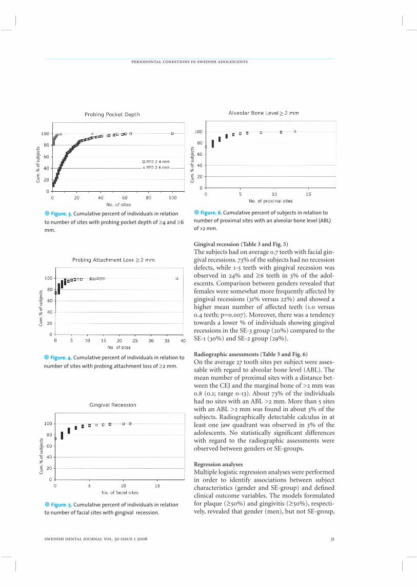
periodontal conditions in swedish adolescents
swedish dental journal vol. 30 issue 1 2006 31
• Figure. 6. Cumulative percent of subjects in relation to number of proximal sites with an alveolar bone level (ABL) of >2 mm.
• Figure. 3. Cumulative percent of individuals in relation to number of sites with probing pocket depth of ≥4 and ≥6 mm.
• Figure. 4. Cumulative percent of individuals in relation to number of sites with probing attachment loss of ≥2 mm.
• Figure. 5. Cumulative percent of individuals in relation to number of facial sites with gingival recession.
Gingival recession (Table 3 and Fig. 5)The subjects had on average 0.7 teeth with facial gin-gival recessions. 73% of the subjects had no recession defects, while 1-5 teeth with gingival recession was observed in 24% and ≥6 teeth in 3% of the adol-escents. Comparison between genders revealed that females were somewhat more frequently affected by gingival recessions (31% versus 22%) and showed a higher mean number of affected teeth (1.0 versus 0.4 teeth; p=0.007). Moreover, there was a tendency towards a lower % of individuals showing gingival recessions in the SE-3 group (20%) compared to the SE-1 (30%) and SE-2 group (29%).
Radiographic assessments (Table 3 and Fig. 6)On the average 27 tooth sites per subject were asses-sable with regard to alveolar bone level (ABL). The mean number of proximal sites with a distance bet-ween the CEJ and the marginal bone of >2 mm was 0.8 (0.1; range 0-13). About 73% of the individuals had no sites with an ABL >2 mm. More than 5 sites with an ABL >2 mm was found in about 3% of the subjects. Radiographically detectable calculus in at least one jaw quadrant was observed in 3% of the adolescents. No statistically signifi cant differences with regard to the radiographic assessments were observed between genders or SE-groups.
Regression analysesMultiple logistic regression analyses were performed in order to identify associations between subject characteristics (gender and SE-group) and defi ned clinical outcome variables. The models formulated for plaque (≥50%) and gingivitis (≥50%), respecti-vely, revealed that gender (men), but not SE-group,
Cum
. % o
f sub
ject
s
Cum
. % o
f sub
ject
s
Cum
. % o
f sub
ject
sCu
m. %
of s
ubje
cts
≥
≥
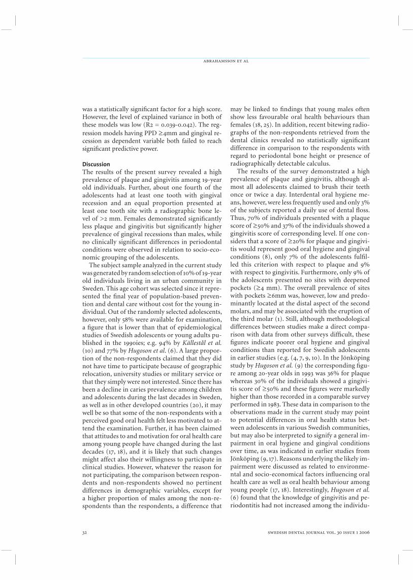
abrahamsson et al
32 swedish dental journal vol. 30 issue 1 2006
was a statistically signifi cant factor for a high score. However, the level of explained variance in both of these models was low (R2 = 0.039-0.042). The reg-
ression models having PPD ≥4mm and gingival re-cession as dependent variable both failed to reach signifi cant predictive power.
DiscussionThe results of the present survey revealed a high prevalence of plaque and gingivitis among 19-year old individuals. Further, about one fourth of the adolescents had at least one tooth with gingival recession and an equal proportion presented at least one tooth site with a radiographic bone le-vel of >2 mm. Females demonstrated signifi cantly less plaque and gingivitis but signifi cantly higher prevalence of gingival recessions than males, while no clinically signifi cant differences in periodontal conditions were observed in relation to socio-eco-nomic grouping of the adolescents.
The subject sample analyzed in the current study was generated by random selection of 10% of 19-year old individuals living in an urban community in Sweden. This age cohort was selected since it repre-sented the fi nal year of population-based preven-tion and dental care without cost for the young in-dividual. Out of the randomly selected adolescents, however, only 58% were available for examination, a fi gure that is lower than that of epidemiological studies of Swedish adolescents or young adults pu-blished in the 1990ies; e.g. 94% by Källestål et al. (10) and 77% by Hugoson et al. (6). A large propor-tion of the non-respondents claimed that they did not have time to participate because of geographic relocation, university studies or military service or that they simply were not interested. Since there has been a decline in caries prevalence among children and adolescents during the last decades in Sweden, as well as in other developed countries (20), it may well be so that some of the non-respondents with a perceived good oral health felt less motivated to at-tend the examination. Further, it has been claimed that attitudes to and motivation for oral health care among young people have changed during the last decades (17, 18), and it is likely that such changes might affect also their willingness to participate in clinical studies. However, whatever the reason for not participating, the comparison between respon-dents and non-respondents showed no pertinent differences in demographic variables, except for a higher proportion of males among the non-re-spondents than the respondents, a difference that
may be linked to fi ndings that young males often show less favourable oral health behaviours than females (18, 25). In addition, recent bitewing radio-graphs of the non-respondents retrieved from the dental clinics revealed no statistically signifi cant difference in comparison to the respondents with regard to periodontal bone height or presence of radiographically detectable calculus.
The results of the survey demonstrated a high prevalence of plaque and gingivitis, although al-most all adolescents claimed to brush their teeth once or twice a day. Interdental oral hygiene me-ans, however, were less frequently used and only 3% of the subjects reported a daily use of dental fl oss. Thus, 70% of individuals presented with a plaque score of ≥50% and 37% of the individuals showed a gingivitis score of corresponding level. If one con-siders that a score of ≥20% for plaque and gingivi-tis would represent good oral hygiene and gingival conditions (8), only 7% of the adolescents fulfi l-led this criterion with respect to plaque and 9% with respect to gingivitis. Furthermore, only 9% of the adolescents presented no sites with deepened pockets (≥4 mm). The overall prevalence of sites with pockets ≥6mm was, however, low and predo-minantly located at the distal aspect of the second molars, and may be associated with the eruption of the third molar (1). Still, although methodological differences between studies make a direct compa-rison with data from other surveys diffi cult, these fi gures indicate poorer oral hygiene and gingival conditions than reported for Swedish adolescents in earlier studies (e.g. (4, 7, 9, 10). In the Jönköping study by Hugoson et al. (9) the corresponding fi gu-re among 20-year olds in 1993 was 36% for plaque whereas 30% of the individuals showed a gingivi-tis score of ≥50% and these fi gures were markedly higher than those recorded in a comparable survey performed in 1983. These data in comparison to the observations made in the current study may point to potential differences in oral health status bet-ween adolescents in various Swedish communities, but may also be interpreted to signify a general im-pairment in oral hygiene and gingival conditions over time, as was indicated in earlier studies from Jönköping (9, 17). Reasons underlying the likely im-pairment were discussed as related to environme-ntal and socio-economical factors infl uencing oral health care as well as oral health behaviour among young people (17, 18). Interestingly, Hugoson et al. (6) found that the knowledge of gingivitis and pe-riodontitis had not increased among the individu-

periodontal conditions in swedish adolescents
swedish dental journal vol. 30 issue 1 2006 33
als over a 20-year period between 1973-1993 despite extensive information being presented at schools and dental clinics. Mayer et al. (16) evaluated the long-term effect upon oral health knowledge and reported behaviour of preventive programs given to 13-16 years old Brazilian schoolchildren. The aut-hors found signifi cantly improved knowledge and reported behaviour at the termination of the 3-year programs, but at a follow-up evaluation 5 years la-ter the effect of improved knowledge on oral health behaviour was not evident. Independents of know-ledge and participation in preventive programs, the participants showed generally more favourable oral health behaviours, which suggest that other fac-tors than knowledge are important for behaviour (16). Hence, the benefi cial effect of population-ba-sed prevention measures on long-term changes in oral health behaviours and oral conditions among adolescents may be questioned. For successful oral health education interventions it is important to consider the views and demands of the young indi-viduals (18, 22) and, consequently, future studies on prevention ought to be focused also on behaviour aspects among these individuals.
The current data showed that female adolescents had signifi cantly less plaque and gingivitis but more gingival recessions than males. Furthermore, a tooth-brushing frequency of ≥2 times/day was signifi cantly more common among females than males. In a longitudinal study of Swedish teena-gers, Crossner at al. (4) demonstrated differences in periodontal health conditions between genders (more favourable conditions in girls), and that the differences increased with age and became clinical signifi cant at the age of 19-year. Hugoson et al. (9) found that gender was a strong explanatory vari-able for gingivitis among 20-year olds, and signi-fi cantly more females than males had low gingivi-tis scores. On the contrary, in an epidemiological study among 16- and 18-year old individuals, Käl-lestål et al. (10) found no signifi cant difference in periodontal status or presence of plaque and calcu-lus between genders, but that girls brushed their te-eth and used fl ossing signifi cantly more often than boys. Taken together these data demonstrate more favourable oral health behaviour of young females than males, which will be manifested as better oral health conditions at an age of about 20 years.
No clinically signifi cant differences in periodon-tal conditions were found with regard to socio-eco-nomical grouping. However, there was a signifi cant difference between the SE-groups 1 and 3 with re-
gard to self reported frequency of tooth-brushing ≥2 times/day (SE-1 92% and SE-3 70%). Hjern et al. (5) investigated social inequality with regard to oral health and the use of dental care in a popula-tion-based Swedish sample of 25-64 years old. The authors found that the infl uence from socio-eco-nomical factors, i.e. low education, having no cash margin and being born outside Sweden, on self-perceived oral health and the use of dental service was most marked in older (45-65 years) adults, but signifi cant in young adults as well. However, the re-sults from the current study among 19-year old in-dividuals, subjected to organized dental care by the community, failed to lend support to the opinion that socio-economical factors infl uence periodon-tal health conditions. This fi nding should however be interpreted with caution since the proportion of non-respondents in the group with the poorest so-cio-economical conditions (SE-3) was about 50% and irregular dental care habits or dental care av-oidance among these individuals may be high, as indicated by others (24).
To conclude, the results from the present study demonstrated poor oral hygiene and gingival con-ditions among Swedish adolescents, despite the ex-posure to oral health promotion programs. Hence, future studies ought to be directed towards an in-creased understanding of the infl uence of various psychosocial and behavioural factors on oral health conditions in young individuals in order to enhan-ce cost-effectiveness of prevention programs.
Acknowledgements This study received support from The Research Foundation for Västra Götaland, and from Public Dental Service, Göteborg, Sweden.
References1. Ainamo J, Nordblad A, Kallio P. Use of the CPITN
in populations under 20 years of age. Int Dent J 1984;34(4):285-91.
2. Björn H, Halling A, Thyberg H. Radiographic assessment of marginal bone loss. Odontol Revy 1969;20(2):165-79.
3. Brecx MC, Frohlicher I, Gehr P, Lang NP. Stereological observations on long-term experimental gingivitis in man. J Clin Periodontol 1988;15(10):621-7.
4. Crossner CG, Unell L. A longitudinal study of dental health and treatment need in Swedish teenagers. Community Dent Oral Epidemiol 1986;14(1):10-4.
5. Hjern A, Grindefjord M, Sundberg H, Rosen M. Social inequality in oral health and use of dental care in Sweden. Community Dent Oral Epidemiol 2001;29(3):167-74.
6. Hugoson A, Koch G, Bergendal T, Hallonsten AL, Slotte C, Thorstensson B, et al. Oral health of individuals aged 3-

abrahamsson et al
34 swedish dental journal vol. 30 issue 1 2006
80 years in Jönköping, Sweden in 1973, 1983, and 1993. I. Review of fi ndings on dental care habits and knowledge of oral health. Swed Dent J 1995;19(6):225-41.
7. Hugoson A, Koch G, Bergendal T, Hallonsten AL, Slotte C, Thorstensson B, et al. Oral health of individuals aged 3-80 years in Jönköping, Sweden in 1973, 1983, and 1993. II. Review of clinical and radiographic fi ndings. Swed Dent J 1995;19(6):243-60.
8. Hugoson A, Norderyd O, Slotte C, Thorstensson H. Distribution of periodontal disease in a Swedish adult population 1973, 1983 and 1993. J Clin Periodontol 1998;25(7):542-8.
9. Hugoson A, Norderyd O, Slotte C, Thorstensson H. Oral hygiene and gingivitis in a Swedish adult population 1973, 1983 and 1993. J Clin Periodontol 1998;25(10):807-12.
10. Källestål C, Matsson L, Holm AK. Periodontal conditions in a group of Swedish adolescents. (I). A descriptive epidemiologic study. J Clin Periodontol 1990;17(9):601-8.
11. Lang NP, Cumming BR, Löe H. Toothbrushing frequency as it relates to plaque development and gingival health. J Periodontol 1973;44(7):396-405.
12. Lang NP, Sander L, Barlow A, Brennan K, White DJ, Bacca L, et al. Experimental gingivitis studies: effects of triclosan and triclosan-containing dentifrices on dental plaque and gingivitis in three-week randomized controlled clinical trials. J Clin Dent 2002;13(4):158-66.
13. Löe H. The Gingival Index, the Plaque Index and the Retention Index Systems. J Periodontol 1967;38(6):Suppl:610-6.
14. Löe H, Morrison E. Periodontal health and disease in young people: screening for priority care. Int Dent J 1986;36(3):162-7.
15. Löe H, Theilade E, Jensen SB. Experimental Gingivitis in Man. J Periodontol 1965;36:177-87.
16. Mayer MP, de Paiva Buischi Y, de Oliveira LB, Gjermo O. Long-term effect of an oral hygiene training program on knowledge and reported behavior. Oral Health Prev Dent 2003;1(1):37-43.
17. Norderyd O. Risk for periodontal disease in a Swedish adult population. Cross-sectional and longitudinal studies over two decades. Thesis. Lund University, Malmö, Sweden 1998.
18. Ostberg AL. On self-perceived oral health in Swedish adolescents. Thesis. Malmö University, Sweden 2002.
19. Page RC, Kornman KS. The pathogenesis of human periodontitis: an introduction. Periodontol 2000 1997;14:9-11.
20. Petersson GH, Bratthall D. The caries decline: a review of reviews. Eur J Oral Sci 1996;104(4 ( Pt 2)):436-43.
21. Ramfjord SP. The Periodontal Disease Index (PDI). J Periodontol 1967;38(6):Suppl:602-10.
22. Rolandsson M. Snuff use and oral health among young ice-hockey players: Implications for oral health promotion. Thesis. Göteborg University, Sweden 2005.
23. Schätzle M, Löe H, Burgin W, Anerud A, Boysen H, Lang NP. Clinical course of chronic periodontitis. I. Role of gingivitis. J Clin Periodontol 2003;30(10):887-901.
24. Skaret E, Raadal M, Kvale G, Berg E. Factors related to missed and cancelled dental appointments among adolescents in Norway. Eur J Oral Sci 2000;108(3):175-83.
25. Skaret E, Raadal M, Kvale G, Berg E. Gender-based differences in factors related to non-utilization of
dental care in young Norwegians. A longitudinal study. Eur J Oral Sci 2003;111(5):377-82.
26. SPSS. Advanced Statistics 11.0. In. Chicago, Illinois: SPSS Inc.; 2004.
27. Watt RG, Marinho VC. Does oral health promotion improve oral hygiene and gingival health? Periodontol 2000 2005;37:35-47.
Address:Dr Kajsa H. AbrahamssonDepartment of Periodontology, Institute of Odontology, The Sahlgrenska Academy at GöteborgUniversity, Box 450, SE-405 30 Göteborg, Sweden.E-mail: [email protected].

assessment of dental caries lesions over 20 years
swedish dental journal vol. 30 issue 1 2006 35
A 20-year study of dentists’ and dental hygienists’ assessment of dental caries lesions in bite-wing radiographspia gabre1, eva birring2, and lars gahnberg1
Abstract • Epidemiological data reveal that the prevalence of dental caries in western countries has decreased in recent decades. The aim of this study was to investigate how dentists and dental hygienists assess dental caries lesions in bite-wing radiographs between 1983 and 2003.All dentists and dental hygienists in Public Dental Health in Uppsala County were offered to take part in the study. The participants assessed manifest and initial caries lesions in eight bite-wing radiographs from three patients individually. An X-ray viewer and binoculars were used. The assessments were repeated in the same radiographs every fi ve years, a total of fi ve times, between 1983 and 2003. In the different test occasions 80-103 dentists and 11-48 dental hygienists participated.
The registration of dental caries changed between 1983 and 2003. The number of manifest lesions registered by dentists decreased between 1983 and 1988, but were stable after 1988. Dental hygienists showed no changes in the registration of manifest lesions during the study. Initial lesions registered by dentists and dental hygienists increased between 1988 and 1998. Assessments of initial caries lesions displayed a wider range than manifest lesions. Increasing age and more years in the profession resulted in fewer registered initial caries lesions. Dental hygienists had a tendency to register less caries than dentists.
In conclusion, the result of the study indicate that inclusion of initial caries lesions in epidemiological reports should lead to a reduction in reliability. The changes in assessments of manifest caries lesions that took place in the 1980s should be considered when epidemiological data are evaluated.
Key words Dental caries, initial lesions, manifest lesions, bite-wing radiographs, repeated cross-sectional study
swed dent j 2006; 30: 35–42 • gabre, birring, gahnberg
1Department of Preventive Dentistry, Public Dental Health Services, Uppsala County Council, Sweden; 2Department of Surgical Sciences, Oral and Maxillofacial Surgery, University Hospital, Uppsala, Sweden

gabre, birring, gahnberg
36 swedish dental journal vol. 30 issue 1 2006
Tandläkares och tandhygienisters bedömning av karies på bite-wing röntgenbilder mellan 1983 och 2003pia gabre, eva birring och lars gahnberg
Sammanfattning
• Epidemiologiska data visar att förekomsten av karies hos befolkning i västvärlden har minskat under de senaste decennierna. Målet med denna studie var att undersöka hur tandläkare och tandhygienister diagnostiserade karies under 20-årsperioden 1983-2003.
Alla tandläkare och tandhygienister anställda i Folktandvården, Uppsala län, erbjöds att deltaga i en upprepad tvärsnittsstudie. Röntgenbilder av typen bite-wing från tre patienter bedömdes med avseende på manifest och initial karies. Bedömningen gjordes av varje tandläkare och tandhygienist individuellt och anonymt. Som hjälpmedel använ-des ett ljusskåp och kikare. Samma röntgenbilder granskades var femte år mellan 1983 och 2003, sammanlagt fem gånger. Vid de olika bedömningstillfällena deltog 80-103 tandläkare och 11-48 tandhygienister.
Bedömningen av karies förändrades mellan 1983 och 2003. Tandläkarnas registrering av antalet manifesta läsioner minskade mellan 1983 och 1988, men har varit stabil efter 1988. Tandhygienisterna förändrade inte sin bedömning av manifest karies under den studerade perioden. Mellan åren 1988 och 1998 ökade både tandläkarnas och tandhy-gienisternas registrering av initial karies och bedömningen av initial karies uppvisade en större spridning än manifest karies. Ökad ålder samt fl er år i yrket resulterade i färre registreringar av initial karies. Tandhygienister hade en tendens att registrera färre kari-esläsioner än tandläkarna.
Slutsatsen av studien är att den förändrade bedömningen av manifest karies som ägde rum under 1980-talet bör tas med i beräkningen då epidemiologiska data skall värderas. Om även initialkaries inkluderas i epidemiologiska data minskar rapporternas reliabilitet.
swed dent j 2006; 30: 35–42 • gabre, birring, gahnberg
assessment of dental caries lesions over 20 years
swedish dental journal vol. 30 issue 1 2006 37
IntroductionData from the WHO show that the prevalence of dental caries in western countries has decreased (18). National epidemiological data show the same trend in the Swedish population. The percentage of 12-year-old children with no experience of caries in-creased from 22% in 1985 to 57% in 2002. During the same period, the percentage of 19-year-old adol-escents with no experience of approximal caries le-sions increased from 36 to 59 % (17). The national epidemiological data in Sweden are based on clinical examinations and, when approximal surfaces can-not be inspected, bite-wing radiographs are recom-mended. Compared with solely clinical information, bite-wing radiographs increase the fi ndings of ap-proximal caries lesions many times over (15). In ad-dition, the presence of bite-wing radiographs plays a major role in caries risk assessment (10) and in the dentists’ restorative decisions for approximal tooth surfaces (8).
Caries diagnoses in radiographs show variations between examiners. Even when observers evaluate the same radiographs, the assessment varies con-siderably (7,11,16). Swedish dental hygienists have been shown to have the same accuracy in diagnosing dental caries lesions as dentists. The inter-examina-tion variation is wide in both groups (14).
A large majority of Swedish dentists state that they would not automatically restore approximal caries lesions, unless the radiographic appearance shows obvious progression in the outer 1/3 to 1/2 of the dentine (9). In recent decades, new methods of caries management have been adopted, in which the biological rather than the technical approach is a priority (2). In Norway, a shift in operative treat-ment criteria among dentists has taken place during the last decades (5,19). It is not known whether this
change in caries management has infl uenced den-tists’ and dental hygienists’ way of diagnosing caries. This knowledge would be useful in the evaluation of oral health in the population.
The aim of the present study was to investigate how dentists and dental hygienists assessed dental caries lesions in bite-wing radiographs between 1983 and 2003.
Material and MethodsIn the Public Dental Health in Uppsala County, Swe-den, all dentists and dental hygienists were offered to participate in a test in which dental caries lesions were diagnosed on radiographs. The test was repea-ted every fi ve year between 1983 and 2003. A repeated cross-sectional study design was used. Dental hygie-nists were included in the study from 1988. Dentists therefore participated in the study fi ve times and dental hygienists four times. The dentists and dental hygienists were informed verbally and through writ-
InstructionsInstructionsInstructionsPatient 1 (radiographs 1a and 1b):In the protocol there are 7 beforehand noted caries lesions. Mark the lesions that, in your opinion, have progressed.
Patient 2 and 3 (radiographs 2 and 3):Register initial and manifest caries lesions.Initial caries= lesion only in enamelManifest caries=lesion involving both enamel and dentin.Only primary and approximal lesions should be registered.
• Figure. 1. Instructions given to dentists and dental hygienists before assessment of radiographs.
• Figure. 2. Patient 1: assessment of progression of caries. Radiographs 1 b were taken one year after radiographs 1 a.Patients 2 and 3: registration of initial and manifest caries lesions.

gabre, birring, gahnberg
38 swedish dental journal vol. 30 issue 1 2006
ten instructions before the assessments were made (Fig. 1). Using an X-ray viewer and binoculars (x2 magnifi cation), the participants individually obser-ved a set of radiographs and noted the result in a report. The reports were fi lled in anonymously.Two bite-wing radiographs from each of two pa-tients (no 2 and 3) were observed on all fi ve occa-sions. From 1988, the progression of dental caries from four bite-wing radiographs taken on two oc-casions in a third patient (no 1) was estimated (Fig. 2). Thus the same radiographs from three patients were observed throughout the study. The criteria for the chosen radiographs were as follows: 1) the air space should be really black2) a clear difference in density (grey scale) between enamel and dentine3) no overlapping of adjoining crown surfaces4) in the progression case: no observable difference in the density of the radiographs from the two occasions
The purpose of the criteria was to fi nd optimal radiographs for the diagnostic procedure of den-tal caries, i.e. a well-exposed radiograph, suffi cient contrast in the image and the ability to detect caries
• Table 1. The number of participating dentists and dental hygienists, gender, age, years in the profession, participants in proportion to the total number of employees and participating in earlier tests (PET). Dental hygienists did not participate in 1983.
1983 1988 1993 1998 2003Dentists, no of participants 93 89 98 80 103Participation rate 94 % 82 % 87 % 76 % 86 %Age: All, mean (range) -1 -1 41.5 (26-65) 43.4 (25-61) 45.5 (25-64) Men -1 -1 43.0 (29-65) 45.3 (28-61) 48.4 (25-62) Women -1 -1 40.9 (26-60) 41.9 (26-60) 42.8 (24-64)Years in profession: All, mean (range) -1 -1 13.8 (1-39) 16.1 (1-37) 18.7 (1-40) Men -1 -1 13.0 (1-39) 16.9 (2-37) 19.3 (1-38) Women -1 -1 14.6 (1-36) 15.6 (1-33) 16.6 (1-40)Gender, % women -1 -1 52 % 61 % 68 %PET - 54 % 63 % 62 % 51 %
Dental hygienists, no of participants 11 17 20 48Participation rate 52 % 65 % 56 % 86 %Age: All, mean (range) -1 43.1 (28-60) 41.7 (22-63) 45.0 (27-61) Men -1 28 30 38 Women -1 44.1 (28-60) 42.3 (22-63) 45.2 (27-61)Years in profession: All, mean (range) -1 7.5 (1-16) 10.7 (1-30) 10.1 (1-26) Men -1 5 3 15 Women -1 7.6 (1-16) 11.1 (1-30) 9.9 (1-26)Gender, % women -1 94 % 95 % 98 %PET - 47 % 83 % 58 %1 Age, years in profession and gender were not registered in 1983 and 1988
in the contact area (6). The selection of radiographs was made by a dentist specialized in dentomaxillo-facial radiology. The fi lm Ultraspeed from Kodak (Eastman Kodak Company, Rochester, New York, USA) was used and the radiographs were exposed on a copyfi lm (Agfa Curix Duplicating 100 NIF, Agfa-Gevaert NV, Mortsel, Belgium), multiplied in a copying machine (Agfa Curix Dupli, Agfa-Gevaert NV, Mortsel, Belgium) and mounted in templates .
All the dentists and dental hygienists present at work on the day the test was performed participa-ted in the study. On the different test occasions, 76-94% of the employed dentists and 50-86% of the dental hygienists took part in the investigation. Re-asons for not participating were illness, part-time work and leave of absence. The mean age, years in the profession and gender are presented in Table 1.
Statistical analysisDifferences between means in two groups were ana-lysed by the unpaired t-test when the sample size was large and the distribution was symmetric. Due to small sample sizes, the Mann-Whitney U-test was

assessment of dental caries lesions over 20 years
swedish dental journal vol. 30 issue 1 2006 39
• Table 2. Dental caries lesions registered by dentists and dental hygienists in 1983, 1988, 1993, 1998 and 2003. Patient No 1 Patient No 2 Patient No 3 Progression Initial Manifest Initial ManifestDentists1983: Mean (±SD) — 8.4 (±2.4) 7.1 (±2.2) 5.3 (±2.1) 5.4 (±1.8) Range — 3-15 2-12 0-11 2-101988:Mean (±SD) 3.0 (±1.7) 8.5 (±2.0) 5.33 (±1.9) 5.7 (±1.7) 4.65 (±1.6) Range 0-7 2-14 2-11 1-10 2-101993:Mean (±SD) 3.51 (±1.4) 10.12 (±2.4) 5.6 (±1.3) 6.44 (±1.9) 4.9 (±1.6) Range 0-7 0-14 3-10 3-10 2-91998:Mean (±SD) 3.6 (±1.5) 9.8 (±2.2) 5.5 (±1.7) 6.4 (±1.9) 4.8 (±1.8) Range 1-7 4-14 0-9 2-11 1-102003:Mean (±SD 3.1 (±1.5) 9.5 (±2.2) 5.2 (±1.3) 6.4 (±2.0) 4.6 (±1.7) Range 0-6 5-14 2-10 1-12 2-9
Dental hygienists 1988:Mean (±SD) 3.0 (±1.5) 7.1 (±1.9) 5.3 (±1.7) 4.9 (±1.4) 4.9 (±0.9) Range 1-5 4-10 3-9 3-7 4-71993:Mean (±SD) 3.2 (±1.4) 7.9 (±2.5) 3.9 (±2.4) 5.2 (±2.3) 4.0 (±1.8) Range 1-6 5-13 0-9 0-8 2-81998:Mean (±SD) 3.2 (±1.2) 9.06 (±3.1) 5.4 (±1.2) 6.87 (±2.1) 4.2 (±1.6) Range 1-6 0-15 4-9 0-10 1-72003:Mean (±SD) 3.4 (±1.4) 9.3 (±2.8) 4.8 (±1.3) 6.8 (±2.1) 4.4 (±1.8) Range 1-6 0-13 2-9 2-11 1-81 Un-paired t-test: 1993 vs. 1988 p<0.052Un-paired t-test: 1993 vs. 1988 p<0.00013 Un-paired t-test: 1988 vs. 1983 p<0.00014 Un-paired t-test: 1993 vs. 1988 p<0.01
5 Un-paired t-test: 1988 vs. 1983 p<0.016 Mann Whitney U-test: 1998 vs. 1993 p<0.057 Mann Whitney U-test: 1998 vs. 1993 p<0.05
• Figure. 3. Mean values for decayed surfaces assessed by dentists in patients 2 and 3 between 1983 and 2003. * Signifi cant differences in the assessment of manifest lesions between 1983 and 1988 in both patients. Initial caries showed signifi cant differences between 1988 and 1993 in both patients. Manifest Initial
used among dental hygienists in 1988-1998. The re-lationship between gender, age and years in the pro-fession and caries assessments was studied using a multiple linear regression test. A 5 % level of signi-fi cance was used.
ResultsBetween 1993 and 2003, the mean age of the dentists increased by 4 years and the experience of profession by 5 years. The mean age of the dental hygienists sho-wed small changes, but years in profession increased by 3 years during the studied period. The percentage of women among the dentists increased from 52 to 68%. Women had a lower age in all of the investigated occasions and by every registration the difference in mean age between men and women increased. With the exception of one man all participating dental hy-gienists were women (Table 1).
The dentists’ assessment of caries lesions chan-ged during the period. Fewer surfaces with mani-fest caries lesions were registered in 1988 and later compared with the registration in 1983. A smaller number of initial lesions were registered in 1983 and

gabre, birring, gahnberg
40 swedish dental journal vol. 30 issue 1 2006
• Figure 5. Distribution of registered surfaces of dental caries in patient 2 in 2003. * Statistically signifi cant differences dentists vs. dental hygienists p=0.0476 (unpaired t-test). Dentists, manifest caries Dental hygienists, manifest caries Dentists, initial caries Dental hygienists, initial caries
• Figure. 4. Mean values for decayed surfaces assessed by dental hygienists in patients 2 and 3 between 1988 and 2003. * Signifi cant differences in the assessment of initial caries between 1993 and 1998 in both patients. Manifest Initial_ _ _ _ _
_ _ _ _ _ _
_ _ _ _ _ _
1988 compared with 1993 and the later registrations (Table 2, Fig. 3). In patient 1, where the progres-sion of caries was to be assessed, dentists registered more surfaces in 1993 and 1998 compared with 1988 (p<0.05, Table 2). The assessment of manifest caries lesions among the dental hygienists did not change between 1988 and 2003. The registration of initial ca-ries lesions did, however, increase between 1993 and 1998 (Table 2, Fig 4).
On three occasions, dentists found more manifest caries lesions compared with dental hygienists (pa-tient 2 in 1993 and 2003, p<0.01 and p<0.05; patient 3 1993, p<0.05). Moreover, in the assessment of initial caries lesions, dentists had a higher registration on three occasions (patient 2 in 1988 and 1993, p<0.05; patient 3 in 1993, p<0.05; Table 2, Fig. 5).
The range of registered decayed surfaces was lar-
ge in both professions (Table 2, Fig 5). However, a clear majority of dentists and dental hygienists were within ±1 lesion compared to the mean value in their assessment, as illustrated in Fig. 5. Assessments of initial caries lesions displayed a wider range than manifest lesions. No differences could be found bet-ween dentists and dental hygienists.
There was a relationship between the dentists’ age and the number of registered initial caries lesions for both assessed patients in 1993, 1998 and 2003 (p-values <0.05-<0.0001, r2 =0.042-0.193). The older the dentist, the fewer decayed surfaces were found. A similar relationship was observed between years in profession and initial caries lesions. A multiple linear regression test showed that age and years in profession were dependent variables. The number of registered manifest caries lesions showed no rela-

assessment of dental caries lesions over 20 years
swedish dental journal vol. 30 issue 1 2006 41
tionship with age or years in the profession. Among dental hygienists, age and years in the profession did not infl uence the assessment of initial and manifest caries lesions.
Differences between genders could only be obser-ved among dentists in 2003. In 2003 women found more initial caries lesions in both patients (p<0.05 and p<0.01). The statistical analysis showed that the difference due to gender could not be explained by the differences in mean age between men and wo-men. Previous participation in the test showed no correlation with the result.
DiscussionThis study shows that the assessment of dental ca-ries lesions changed during the period 1983 to 2003. The changes relating to manifest caries took place in 1983-1988. No changes in the assessment of initial ca-ries could be noted after 1993. Epidemiological data from Uppsala County, as well as the whole of Sweden and other parts of the industrialized world, report a decreasing prevalence of dental caries in the 3-19 age group during the period studied. The result of this study indicates that, during the 1980s, the improve-ment in dental health could be partly explained by a change in the evaluation of caries lesions among dentists. From 1988 and onwards, an infl uence of this kind is less likely.
When dentists and dental hygienists were compa-red, some differences in caries assessment could be seen. There was a tendency for dental hygienists to register fewer caries lesions than dentists, especially when assessing initial caries. However, in 1988, 1993 and 1998, the number of participating dental hygie-nists was small. In recent years in Sweden, dental hy-gienists are involved to a greater extent in the exami-nation and confi rmation of diagnoses in children’s dental care. The increasing experience in this area may be one explanation of the fact that dental hygie-nists registered more initial caries lesions the longer the study continued.
The wide range of registered caries lesions may be a problem in clinical work and epidemiological research. Efforts have been made to obtain greater agreement between observers in the process of caries diagnosis. In an extended calibration program in-cluding six days of training, the observers were able to achieve a high level of calibration (13). When den-tal students used a computer-assisted learning cali-bration program for caries diagnosis on bite-wing radiographs, the performance improved (12). Howe-ver, several studies have reported that the benefi ts of
examiner training are limited (3, 16). Caries diagno-sis includes the ability to detect the presence of caries (sensitivity) but also the ability to report the absence of caries (specifi city). Dental students had an ability to detect caries that was similar to that of general dental practitioners, but the students reported more false positive lesions (11). The present study only in-vestigated diagnoses of dental caries lesions. It is not known whether an evaluation of the chosen therapy would have revealed the same wide range. Despite the wide range, the majority of dentists and dental hygienists displayed an ability to assess the number of caries lesions with a difference of ±1 lesion com-pared with the mean value. From the angle of caries examination, both professions may therefore be re-garded as having the equivalent skills.
A majority of the subjects had participated in more than one assessment of radiographs. Eigh-teen of the dentists had participated in all fi ve as-sessments. It is possible that a learning effect could have infl uenced the results. However, no differences between subjects who had participated earlier and those who had not could be seen. The anonymity makes longitudinal analysis impossible and also ob-structs paired statistical tests. It can not be excluded that additional differences could have been detected with tests for dependent samples. However, in this study tests for independent samples is a conserva-tive way to treat data with small risks to report false signifi cance.
A factor infl uencing assessment of caries lesions in radiographs is the radiographic quality. The ra-diographs in this study were selected by using clear including criteria, but an objective, quantitative control was not possible. The copying of the x-rays was highly standardized. Minor variations in radio-graphs, however, cannot be completely excluded. Since the same set of radiographic copies were used throughout the study, the possible infl uence of such a variation on our data was kept constant.
Increasing age and more years in the profession resulted in fewer registered initial caries lesions. Wo-men showed a lower mean age compared to men in all studied test occasions. Even when the lower age where taken into consideration, women registered more initial caries lesions than men. The reason for this is not known and requires further investigation. The assessment of initial caries lesions displayed a wider range than that of manifest lesions. Registra-tions of manifest caries lesions were therefore gene-rally more reliable compared with initial caries, a result in line with the fi ndings of Espelid and Tveit
gabre, birring, gahnberg
42 swedish dental journal vol. 30 issue 1 2006
(4). Epidemiological data relating to dental caries in Sweden are based on manifest caries and the varia-tion in assessments of initial caries lesions does not therefore infl uence the epidemiological data. Our data indicate that the inclusion of initial caries le-sions should lead to a reduction in reliability in epi-demiological reports. However, to evaluate the caries situation of the individual, it is important to include all the stages of clinical caries (1).
In conclusion, the dentists’ assessment of dental caries changed between 1983 and 1993, but during the past decade the assessments were stable. Dental hygienists had a tendency to register less caries than dentists. Both groups showed a wide, yet equivalent range of registered dental caries. The result of this study should be considered when epidemiological reports of dental caries are evaluated.
AcknowledgementsThe authors gratefully acknowledge the County Council of Uppsala, Sweden, for fi nancial support. We also thank Drs. Eva Asp, Dan Björnestål, Inger Ganea, Gunilla Högfors, Kjell Kullander and Gun-nar Sörlin for help with the planning and practical work in this study and Lars Berglund for support with statistical analysis.
References1. Amarante E, Raadal M, Espelid I. Impact of diagnostic
criteria on the prevalence of dental caries in Norwegian children aged 5, 12 and 18 years. Community Dent Oral Epidemiol 1998;26:87-94
2. Burke FJ, Wilson NH. When is caries caries, and what should we do about it? Quintessence Int 1998; 29:668-72
3. Downer MC, Kay EJ. Restorative treatment decisions from bitewing radiographs- performance of dental epidemiologists and general dental practitioners. Community Dent Oral Epidemiol 1996; 24:101-5
4. Espelid I, Tveit AB. Diagnostic quality and observer variation in radiographic diagnosis of approximal caries. Acta Odontol Scand 1986; 44:39-46
5. Gimmestad AL, Holst D, Fylkesnes K. Changes in restorative caries treatment in 15-year-olds in Oslo, Norway, 1979-1996. Community Dent Oral Epidemiol 2003; 31:246-51
6. Gröndahl H-G. The value of the radiological examination in caries diagnosis, page 376. In: Thylstrup A, Fejerskov O, editors: Textbook of Clinical Cariology.
Copenhagen: Munksgaard, 19947. Leijon G, Markén KE. Roentgenological diagnosis of
proximal caries. Deviations between observers and comparison between the recordings from periapical and bite-wing roentgenograms. Acta Odontol Scand 1968; 26:35-61
8. Lewis DW, Kay EJ, Main PA, Pharoah MG, Csima A. Dentists’ variability in restorative decisions, microscopic and radiographic caries depth. Community Dent Oral Epidemiol 1996; 24: 106-11
9. Mejare I, Sundberg H, Espelid I, Tveit B. Caries assessment and restorative treatment thresholds reported by Swedish dentists. Acta Odontol Scand 1999; 57: 149-54
10. Mejare I, Stenlund H, Zelezny-Holmlund C. Caries incidence and lesion progression from adolescence to young adulthood: a prospective 15-year cohort study in Sweden. Caries Res 2004; 38:130-41
11. Mileman PA, van den Hout WB. Comparing the accuracy of Dutch dentists and dental students in the radiographic diagnosis of dentinal caries. Dentomaxillofac Radiol 2002; 31:7-14
12. Mileman PA, van den Hout WB, Sanderink GC. Randomized controlled trial of a computer-assisted learning program to improve caries detection from bitewing radiographs. Dentomaxillofac Radiol 2003; 32:116-23
13. Morris AL, Bentley JM, Vito AA. Training and calibrating evaluators for a national private dental offi ce assessment program. J Public Health Dent 1987; 47:165-71
14. Ohrn K, Crossner CG, Borgesson I, Taube A. Accuracy of dental hygienists in diagnosing dental decay. Community Dent Oral Epidemiol 1996; 24:182-6
15. Poorterman JH, Aartman IH, Kieft JA, Kalsbeck H. Value of bite-wing radiographs in a clinical epidemiological study and their effect on the DMFS index. Caries Res 2000; 34:159-63
16. Poorterman JH, Kieft JA, Eijkman MA. Differences in the assessment of restorative dental care. Ned Tijdschr Tandheelkd 2002;109:355-7
17. The National Board of Health and Welfare. Tandhälsan hos barn och ungdomar 1985-2002 (In
Swedish). Socialstyrelsen 2003. ISBN:91-7201-790-218. The WHO Oral Health Report 2003. Continuous
improvement of oral health in the 21st century - the approach of the WHO Global Oral Health Programme. World Health Organization, Geneva, Switzerland
19. Tveit AB, Espelid I, Skodje F. Restorative treatment decisions on approximal caries in Norway. Int Dent J 1999; 49:165-72
Address: Dr Pia Gabre, Department of Preventive Dentistry, Ulleråkersv 21, SE-750 17 Uppsala, SwedenE-mail: [email protected]

124. Studies of occlusal adjustment therapy in patients with craniomandibulardisorders. Danila N. Vallon (1997)
125. Pulp survival and hard tissue formation subsequent to dental trauma. Agneta Robertson (1997)
126. Studies on fl uoridated toothpicks. Hossein Kashani (1997) 127. Survival of onlay bone grafts. A study in the adult rat.
Monica Gordh (1997) 128. On the prosthodontic patient. An investigation of factors including patient
expectations and satisfaction with extensive prosthodontic care. Ulf Hakestam (1998)
129. Growth factors and bone regeneration – implications of barrier membranes. Göran Zellin (1998)
130. Aspects of maxillary sinus reconstruction with endosseous implants.John Eric Blomqvist (1998)
131. Treatment of temporomandibular disorders of arthrogeneous origin. EwaCarin Ekberg (1998)
132. Risk for periodontal disease in a Swedish adult population. Cross-sectional and longitudinal studies over two decades.Ola Norderyd (1998)
133. On the reporting of dental health, time for dental care, and the treatmentpanorama. Yngve Swedberg (1999)
134. Temporomandibular disorders and mandibular function in relation toClass II malocclusion and othodontic treatment. Thor Henrikson (1999)
135. On oral disease, illness and impairment among 50-year olds in twoSwedish counties. Lennart Unell (1999)
136. Cancelled.137. Prosthodontics and the general dentist. Mats Kronström (1999) 138. Conventional spiral and low dose computed mandibular tomography
in dental implant planning. Annika Ekestubbe (1999) 139. Some characteristics of solid-state and photo-stimulable phosphor detectors
for intra-oral radiography. Eva Borg (1999) 140. On dental trauma in children and adolescents. Ulf Glendor (2000) 141. On composite resin materials. Ulf Örtengren (2000) 142. Studies on oral health in mentally retarded adults. Pia Gabre (2000) 143. Integrated jaw and neck function in man. Hamayun Zafar (2000) 144. Endodontic retreatment. Aspects of decision making and clinical outcome.
Thomas Kvist (2000) 145. Adult patients with treated complete cleft lip and palate:
Methodological and clinical studies. Agneta Marcusson (2001) 146. Verbal communication in prosthetic dentistry.
Katarina Sondell (2001) 147. Frequency of radiographic caries examinations and development
of dental caries. Agneta Lith (2001)
supplements to swedish dental journal
The supplementscan be ordered from Swedish Dental Journal, Box 1217,111 82 Stockholm, Sweden.Subscriptionof the supplementscan be arranged.
400 SEK
400 SEK400 SEK
400 SEK
400 SEK
400 SEK
400 SEK
400 SEK
400 SEK
400 SEK
400 SEK
400 SEK
400 SEK
400 SEK
400 SEK400 SEK400 SEK400 SEK400 SEK
400 SEK
400 SEK
400 SEK
400 SEK

148. Mutans streptococci – in families and on tooth sites. Ing-Mari Redmo Emanuelsson (2001)
149. Enamel hypomineralization in permanent fi rst molars. A clinical,histomorpholgical and biochemical study. Birgitta Jälevik (2001)
150. Effects of masticatory muscle function and bite-raising on mandibularmorphology in the growing rat. Andrea Bresin (2001)
151. Masticatory muscle function and transverse dentofacial growth. Christos Katsaros (2001)
152. On the absorption behaviour of saliva and purifi ed salivary proteinsat solid/liquid interfaces. Liselotte Lindh (2002)
153. Benzodiazepine sedation in paediatric dentistry. Boel Jensen (2002)
154. Odontoblast phosphate and calcium transport in dentinogenesis. Patrik Lundquist (2002)
155. On self-perceived oral health in Swedish adolescents. Anna-Lena Östberg (2002)
156. On dental erosion and associated factors. Ann-Katrin Johansson (2002) 157. Secular changes in tooth size and dental arch dimensions in the mixed
dentition. Rune Lindsten (2002) 158. Assessing caries risk – using the Cariogram Model
Gunnel Hänsel Petersson (2003) 159. Diagnostic accurancy of tuned aperture computed tomography (TACT®)
Madhu K. Nair (2003) 160. Bonding of resin to dentin. Interactions between materials, substrate
and operators. Thomas Jacobsen (2003) 161. Autogenous free tooth transplantation by a two-stage operation technique.
Gunnar Nethander (2003) 162. Oral health among the elderly in Norway Birgitte Moesgaard Henriksen (2003) 163. A mandibular protruding device in obstructive sleep apnea and snoring.
Anette Fransson (2003) 164. Temporomandibular disorders in adolescents.
Kerstin Wahlund (2003) 165. Craniofacial growth related to masticatory muscle function in the ferret.
Tailun He (2004) 166. HLA, mutans streptococci and salivary IgA – is there a relation?
Marie Louise Lundin Wallengren (2004) 167. The miswak (chewing stick) and oral health. Studies on oral hygiene practices
of urban Saudi Arabians. Meshari Al-Otaibi (2004) 168. Sleep apnoea in patients with stable congestive heart failure.
27
The supplementscan be ordered from Swedish Dental Journal, Box 1217,111 82 Stockholm, Sweden.Subscriptionof the supplementscan be arranged.
supplements to swedish dental journal
400 SEK
400 SEK
400 SEK
400 SEK
400 SEK
400 SEK
400 SEK
400 SEK400 SEK
400 SEK
400 SEK
400 SEK
400 SEK
400 SEK400 SEK
400 SEK
400 SEK
400 SEK
400 SEK
400 SEK
400 SEKMahmoud Eskafi (2005)

supplements to swedish dental journal
169 On titanium frameworks and alternative impression techniquesin implant dentistryAnders Örtorp (2005)
170 Variability of the cranial and dental phenotype in Williams syndromeStefan Axelsson (2005)
171 Acute infl ammation in peritoneal dialysis: experimental studies in rats Characterization of regulatory mechanisms
Farhan Bazargani (2005) 172 The effect of low level laser irradiation on implant-tissue interaction
Maawan Khadra (2005) 173 All-ceramic fi xed partial dentures Per Vult von Steyern (2005)174 Smoking and vertical periodontal bone loss Mustafa Baljon (2005) 175 Mandibular Third Molar Removal
Rolf Liedholm (2005) 176 Tobacco smoking and periodontal health in a Saudi Arabian
population. Suzan Natto (2005) 177 Mandibular alveolar bone mass, structure and thickness
in relation to skeletal bone density in dentate women Grethe Jonasson (2005)
178 On caries prevalence and school-based fl uoride programmes in Swedish adolescentUlla Moberg Sköld (2005)
179 Risk factors for oral and oropharyngeal squamous cell carcinoma Kerstin Rosenquist (2005)
The supplementscan be ordered from Swedish Dental Journal, Box 1217,111 82 Stockholm, Sweden.Subscriptionof the supplementscan be arranged.
400 SEK
400 SEK
400 SEK
400 SEK400 SEK
400 SEK
400 SEK
400 SEK
400 SEK
400 SEK
400 SEK

2 swedish dental journal vol. 25 issue 1 2001
Swedish Dental JournalSwedish Dental JournalSwedish Dental Journal is the scientifiSwedish Dental Journal is the scientifi c journal of c journal of
The Swedish Dental Association and of The Swedish Dental Society The Swedish Dental Association and of The Swedish Dental Society
Pos t t idn ing Pos t t idn ing BB