Embed Size (px)
Citation preview
S W A N S O Nf l e x i b l eF I N G E R J O I N T I M P L A N T
surgica l technique
surgical technique presented by
ALFRED B. SWANSON, MD, FACS,GRAND RAPIDS, MICHIGAN.
S W A N S O N F L E X I B L E F I N G E R J O I N T I M P L A N T
o n e
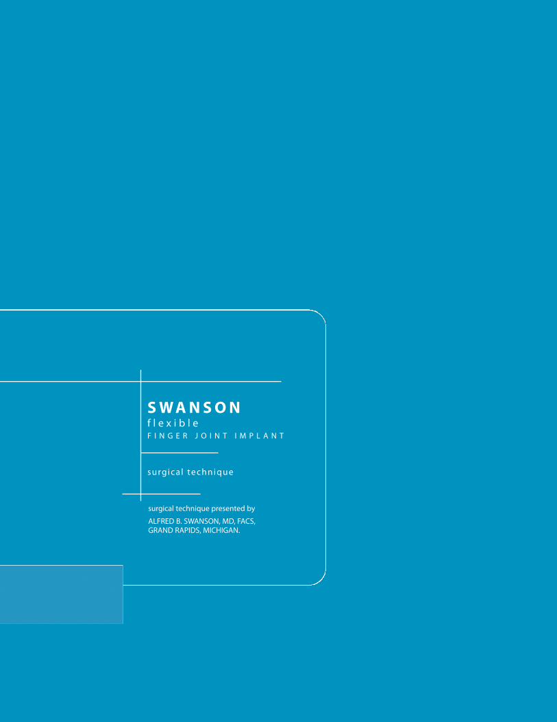
Fitting of the grommet requires a precise press-fit. It must be accurately
centered, otherwise it may impinge the cortex in the intramedullary
canal on one side and could cause bone resorption. Unless the
shoulders of the grommet can be fitted into metaphyseal bone, rotation
of the grommet could occur. In certain cases of severe
metacarpophalangeal joint dislocation, additional bone must be
removed to obtain joint reduction and the implant may have to be used
without the grommet.
The potential for complications or adverse reactions with any implant can
be minimized by following the instructions for use provided in product
literature.
It is the responsibility of each surgeon using implants to consider the
clinical and medical status of each patient and to be knowledgeable
about all aspects of implant procedure and the potential complications
that may occur. The benefits derived from implant surgery may not
meet the patient's expectations or may deteriorate with time,
necessitating revision surgery to replace the implant or to carry out
alternative procedures. Revision surgeries with implants are common.
The patient's mental status must also be considered. Willingness
and/or ability to follow postoperative instructions may also impact the
surgical outcome. Surgeons must balance many considerations to
achieve the best result in individual patients.
IF EXCESSIVE LOADING CANNOT BE PREVENTED, AN IMPLANT
SHOULD NOT BE USED.
SWANSONflexible finger JOINT IMPLANT as described by Alfred B. Swanson, M.D.
specific PRECAUTIONS
general PRECAUTIONS
S W A N S O N F L E X I B L E F I N G E R J O I N T I M P L A N T
t w o
One of the goals of implant surgery is to minimize production of wear
particles. It can never be eliminated because all moving parts e.g.,
implants which articulate against bone wear to some degree. In an
implant arthroplasty, clinically significant wear can result from normal
biomechanical forces. Abnormal or excessive force will further increase
clinically significant wear.
Abnormal force loading may be caused by:
· Uncorrected instability
· Oversized implant
· Inadequate soft tissue support
· Implant malposition
· Excessive motion
· Uncorrected or recurrent deformity
· Patient misuse or overactivity
· Intraoperative fixation
Some preventive measures to consider to minimize the potential for
complications:
· Follow guidelines for indications and contraindications
provided above
· Identify prior pathology
· Stabilize collapse deformities
· Bone graft pre-existing cysts
· Use a properly sized implant
· Avoid K-wires and sutures through the implant
If complications develop, possible corrective procedures include:
· Implant removal
· Synovectomy
· Bone grafting of cysts
· Limited intercarpal fusion
· Replacement of the implant
· Removal of the implant with fusion of the joint
Clinical results depend on surgeon and technique, preoperative and post-
operative care, the implant, patient pathology and daily activity. It is
important that surgeons obtain appropriate informed consent and
discuss the potential for complications with each patient prior to
surgery. This may include a review of alternative, non-implant
procedures such as soft tissue reconstruction or arthrodesis.
S W A N S O N F L E X I B L E F I N G E R J O I N T I M P L A N T
t h r e e
POTENTIAL COMPLICATIONS AND ADVERSE REACTIONSIn any surgical procedure, the potential for complications exists. The
risks and complications with the Swanson Finger Joint Implant and the
Swanson Finger Joint Grommet include:
· Infection or painful, swollen or inflamed implant site
· Fracture of the grommet, the implant stem and/or the hinge
· Loosening or dislocation of the prosthesis requiring revision
surgery
· Bone restoration or over-production
· Allergic reaction(s) to prosthesis material(s)
· Untoward histological responses possibly involving
macrophages and/or fibroblasts
· Migration of particle wear debris possibly resulting in a bodily
response
Some degree of particle formation is inevitable with all implants including
those made of silicone elastomer. The amount will vary with factors
such as patient activity, metacarpal stability or instability post-
implantation, implant position and the amount of soft tissue support.
The patient's biological response to these particles is variable, but can
include local synovitis and bone lysis in contiguous bones. Another
potential concern with silicone implants arises from case reports in the
literature suggesting an association between silicone implants and
immunological abnormalities and autoimmune rheumatic disorders,
although these reports have been contradicted and the association has
not been proven conclusively.
RISK/BENEFIT DECISION BY SURGEONThe judgement by a surgeon to implant silicone elastomer implants is a
complicated risk/benefit decision which must take into account the
patient's needs and desire in addition to the surgeon's knowledge of
expected results and complications as well as therapeutic alternatives.
Wright Medical Technology, Inc. can provide a bibliography of articles
on the use and complications of silicone elastomer implants to any
physician.
S W A N S O N F L E X I B L E F I N G E R J O I N T I M P L A N T
f o u r
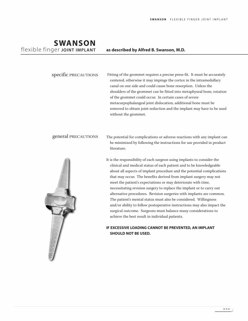
FLEXIBLE IMPLANT
The Swanson Finger Joint Implant* is a flexible intramedullary-stemmed,
one-piece implant developed as an adjunct to resection arthroplasty to
help restore function to hands disabled by rheumatoid, degenerative or
traumatic arthritis. It is made from silicone elastomer, a material that
is highly resistant to flexion-fatigue induced flaw-propagation.
The midsection of the load-distributing flexible hinge has been
designed to help maintain proper joint space and alignment with good
lateral stability and minimal flexion-extension restriction. The implant
is not fixed to bone and becomes stabilized by the encapsulation
process. It acts as a dynamic spacer, internal mold and flexible hinge.
The Swanson Finger Joint Implant is available in 11 sizes to adequately
meet various anatomical requirements. A color-coded sizing set
(supplied non-sterile and not suitable for implantation) is available for
proper size determination during surgery.
GROMMETS
The Swanson Finger Joint Grommets** are thin bone liners designed for
use at the metacarpophalangeal joint level to protect the flexible
implant midsection from the shearing forces of sharp bone edges. The
press-fit encircling grommet is fabricated from unalloyed titanium and
its shape conforms to the contours of the implant midsection and stem
junctions.
The use of grommets to enhance implant durability is especially indicated
in severe cases of rheumatoid arthritis where irregular and sharp bony
edges can initiate tears in the implant midsection.
Grommets are available in 7 sizes corresponding to sizes 3 to 9 of the
Swanson Finger Joint Implant. The outer surface of each grommet is
marked with a numeral, indicating the size of finger joint implant the
grommet fits, as well as the letters "P" or "D", indicating whether it is a
proximal or distal grommet.
device DESCRIPTIONS
*U.S. Patent No. 3,875,594f
**U.S. Patent Nos. 4,158,893; 4,198,713
S W A N S O N F L E X I B L E F I N G E R J O I N T I M P L A N T
f i v e
FLEXIBLE IMPLANT
The Swanson Finger Joint implant can be used with resection arthroplasty
of the metacarpophalangeal (MP), proximal interphalangal (PIP) and
distal interphalangeal (DIP) joints. Insertion of the finger joint implant
at two consecutive joint levels is not recommended (e.g., MP and PIP
joint levels).
The flexible implant resection arthroplasty method is based on the
following concept: "Joint Resection + Implant + Encapsulation =
Functional Joint."
The implant acts as a dynamic spacer to maintain internal alignment and
spacing of the reconstructed joint and as an internal mold that
supports the capsuloligamentous system developing around the
implant while early guided motion is started. The implant becomes
stabilized by this "encapsulation process," and no permanent fixation is
required. Joint stability is achieved from reconstruction of the
ligamentous and musculotendinous systems.
Because the implant is not fixed to bone, the compressive loading forces
are effectively distributed to the resected end of the bone and cortical
shaft. This encourages favorable bone remodeling processes as
evidenced by maintenance of bone length, preserved shape of the
amputated bone end, cortical thickening, and new bone formation next
to the implant midsection and intramedullary stems.
The slight movement of the implant stems allows distribution of forces
over a broader section and allows the flexible hinge to find a better
position with respect to the axis of rotation of the joint. Thus the
implant life is increased and the bone is less likely to react at the
implant interface when the forces are within its strain tolerance. The
low modulus implant is softer than bone and has force-dampening
characteristics that further protect bone and cortical shaft.
Because bone removal is minimal and implants are not attached to bone,
revision procedures to remove or replace an implant, or to reinforce,
release, or realign capsuloligamentous structures around the implant
are easily performed.
rationale
S W A N S O N F L E X I B L E F I N G E R J O I N T I M P L A N T
s i x
GROMMETS
The unattached, press-fit grommets effectively protect the implant
midsection by lessening abrasion, wear and cutting by bone. Because
the grommet is not attached to bone, compressive loading forces are
transmitted to the resected bone-end and a favorable bone response
develops at the grommet/bone interface.
Use of encircling grommets does not alter the function of the implant,
patient indications and contraindications, nor reduce the need for
careful attention to the arthroplasty technique.
SPECIFIC ADVANTAGES OF THE IMPLANTS· Both elastomer and unalloyed titanium implants have an extensive
clinical history of biocompatibility.
· The Swanson Finger Joint Implant and Swanson Finger Joint
Grommets have been sterilized.
· Anatomical sizing (length, height, width) is available in eleven sizes to
meet various operative requirements.
· Swanson Finger Joint Grommets are durable and abrasion-resistant to
protect the implant from sharp bone edges.
· Pliable medical grade silicone elastomer with low elastic modulus
(softer than bone) dampens forceloading and minimizes potential for
necrosis or bone resorption. Cortical bone density typically increases
postoperatively. These benefits are retained with grommet-modified
implants.
· Neither flexible implants nor grommets require fixation to bone.
Intramedullary gliding decreases stress to bone and implant; it allows
implant to locate axis of rotation of the joint.
· Both the flexible implant and grommets are visible on x-rays.
· Design characteristics of load-distributing flexible hinges include:
intramedullary-stemmed, flexible one-piece hinge-like construction
of homogeneous material with stiffness/flexibility balance of implant
material, and proper compression-tension force distribution in
midsection.
treatment CONSIDERATIONS
S W A N S O N F L E X I B L E F I N G E R J O I N T I M P L A N T
s e v e n
· Improves range of motion (especially extension)
· Adequate lateral stability
· Good pain relief
· Maintains joint space and alignment
· Orients and supports joint encapsulation
· Favorable bone remodeling at implant interface; Protects
against bone resorption and stimulates new bone production
· Makes results more predictable, reproducible, and durable
· Early postoperative motion
· Facilitates postoperative rehabilitation
· Essentially salvageable procedure
Any joint implant arthroplasty requires consideration of the following
general indications:
· Good condition of the patient
· Good neurovascular status
· Adequate skin coverage
· Possibility of a functional musculotendinous system
· Adequate bone stock to receive implant
· Availability of postoperative therapy
· Cooperative patient
· Infection
· Physiologically or psychologically inadequate patient
· Inadequate skin, bone or neurovascular status
· Irreparable tendon system
· Possibility for conservative treatment
· Growing patients with open epiphyses
· Patients with high levels of activity
Each patient must be evaluated by the surgeon to determine the
risk/benefit relationship.
clinical ADVANTAGES
general INDICATIONS
general CONTRAINDICATIONS
S W A N S O N F L E X I B L E F I N G E R J O I N T I M P L A N T
e i g h t
GROMMETS
To prevent cutting and abrasion of the flexible implant by sharp bone edges in
metacarpophalangeal joint reconstruction.
Grommets should not be used if:
1. A perfect press-fit cannot be obtained into metaphyseal bone (resection done at
diaphyseal level).
2. During surgery, there appears to be a tendency for dorsal protrusion. This can occur at the
level of the index and little fingers.
3. There are severe bone absorption problems as seen in arthritis mutilans. These cases are
best treated with joint fusion or with silicone implant arthroplasty without the use of
grommets.
4. Bone destruction does not allow a good fit and occasionally in the 5th MP joint.
5. Grommets are not recommended for use in the proximal interphalangeal joint.
Wright Medical Technology, Inc. does not recommend a particular surgical technique when
using the implant. Proper surgical techniques are necessarily the responsibility of the
medical profession. Each surgeon must evaluate the appropriateness of the surgical
technique used based on personal medical training and experience. A description of the
procedure used by Alfred B. Swanson, M.D.*, follows:
SURGICAL STAGING
Excessive manual labor and awkward weight bearing on hand(s) such as occasionally occurs
in some crutch walkers should be avoided after upper extremity reconstruction. If crutches
are absolutely necessary, platform crutches should be used. Lower extremity reconstructive
surgery should be carried out first if feasible. Multiple reconstructive procedures must be
appropriately staged. In metacarpophalangeal joint disabilities with severe wrist
involvement, the wrist should be treated first. Tendon repair and synovectomy of tendon
sheaths should be done 6 to 8 weeks before joint reconstruction in the rheumatoid hand.
However, if the extensor tendons are ruptured and the metacarpophalangeal joints are
dislocated, arthroplasty of the metacarpophalangeal joints is done before the wrist and
tendon reconstruction. In swan-neck deformity, surgery of the metacarpophalangeal and
proximal interphalangeal joints is done at the same stage. However, in boutonniere
deformity, it may be preferable to reconstruct the proximal interphalangeal joint before the
metacarpophalangeal joint. Based on a long-term experience, a system for classification of
treatment for combined involvement of the metacarpophalangeal and interphalangeal joints
has been devised by Swanson | TABLES I, II, III.
surgicalPROCEDURE
* A. B. Swanson, M.D., F.A.C.S. Director of Orthopaedic Training Program, Grand Rapids Hospitals,
Chief of Orthopaedic Research and Hand Surgery Fellowship, Blodgett Memorial Hospital,
Grand Rapids, Michigan, Professor of Surgery, Michigan State University.

S W A N S O N F L E X I B L E F I N G E R J O I N T I M P L A N T
n i n e
TABLE I - Swan-Neck Deformity of PIP Joint without Involvement of MP Joint
1. Initial deformity of PIP Joint a. Local injections (corticosteroids or other agents)b. Flexor-tendon synovectomy with or without tenodesis of flexor digitorum superficialis tendonc. Intrinsic tendon release with or without flexor-tendon synovectomy
2. Flexible deformity of PIP Joint a. Dermadesis at PIP jointb. Relocation of lateral tendons with or without elongation of central tendonc. Tenodesis of flexor digitorum superficialis tendon with flexor synovectomy
3. Rigid, subluxated or dislocated PIP a. Resection of joint with relocation of lateral tendons and implant arthroplasty (rarely)joint b. Resection of joint and fusion
4. Treatment for DIP jont, when a. Temporary pinning in neutral position, if passively correctablerequired in any of these conditions b. Fusion, if severely damaged or flexed
Swan-Neck Deformity of PIP Joint with Involvement of MP Joint
Treatment of MP Joint Treatment of PIP Joint
1. Flexible deformity of PIP joint a. Synovectomy with proximal release of a. Manipulation with temporary Kirschner-wireintrinsic tendons fixation in flexion if required
b. Relocation of subluxated joint with proximal b1. Manipulation with temporary Kirschner-wirerelease of intrinsic tendons fixation in flexion if required
b2. Dermadesisc. Joint resection with proximal realease of c1. Manipulation to 50o of flexion with temporary
intrinsic tendons and implant arthroplasty Kirschner-wire fixationc2. Relocation of lateral tendons with or without
elongation of central tendon
2. Rigid, subluxated or dislocated a. Joint resection with implant arthroplasty a. Relocation of lateral tendons with elongationPIP joint of central tendon
b. Joint resection with fusionc. Joint resection with implant arthroplasty
(rarely)
3. Treatment of DIP joint, if required a. Temporary pinning in neutral position, if passively correctablein any of these conditions b. Fusion, if flexion deformity is severe or articular damage exists
TABLE II - Boutonniere Deformity of PIP Joint
1. Initial deformity of PIP joint a. Local injections (corticosteroids or other agents) or splintingb. Synovectomy
2. Flexible deformity of PIP joint a. Recontruction of central tendonb. Elongation of lateral tendonsc. Combination of procedures a & b, with or without synovectomy
3. Rigid, deformity of PIP joint a. Joint release with reconstruction of central tendon and elongation of lateral tendonswithout bone erosion b. Joint resection with reconstruction of tendons, with or without implant arthroplasty
c. Distal release of lateral tendonswith bone erosion a. Joint resection with reconstruction of central tendon and lateral tendons, with implant
arthroplastyb. Joint resection and fusion
4. Subluxated or dislocated PIP joint a. Joint resection with reconstruction of central tendon and lateral tendons, with implant arthroplastyb. Joint resection and fusion
5. Treatment of DIP joint, if required a. Distal release of lateral tendonsin any of these conditions b. Fusion
TABLE II - Boutonniere Deformity of PIP Joint
1. Nondislocated PIP joint a. Joint resection with implant arthroplasty
2. Subluxated or dislocated PIP joint a. Joint resection with implant arthroplastyb. Joint resection with fusion
3. Treatment of DIP joint, if required a. Fusion, if neededeither of these conditions
TREATMENT OF INTERPHALANGEAL DISABILITIESWITH OR WITHOUT METACARPOPHALANGEAL JOINT INVOLVEMENT
S W A N S O N F L E X I B L E F I N G E R J O I N T I M P L A N T
t e n
Several reconstructive procedures can be performed during one operative
session. Two surgical teams can be working on the upper and lower
extremities at the same time. An operation on an extremity should not
exceed two hours; a stellate ganglion block is recommended if the
tourniquet time should exceed 1 ½ hours. Pre-cooling of the arm with
ice packs are recommended by Tajima can prolong the surgical time
available.
METACARPOPHALANGEAL JOINT IMPLANT ARTHROPLASTYPainful rheumatoid or post-traumatic disabilities with:
1. Fixed or stiff MP joints.
2. X-ray evidence of joint destruction or subluxation
3. Ulnar drift, noncorrectable by surgery of soft tissues alone.
4. Contracted intrinsic and extrinsic musculature and ligament system
5. Associated stiff interphalangeal joints.
INCISION AND EXPOSURE
A transverse skin incision is made on the dorsum of the hand over the
necks of the metacarpals. The dissection is carried down through
subcutaneous tissue to expose the extensor tendons. The dorsal veins,
which lie between the metacarpal heads are carefully released by blunt
longitudinal dissection and are retracted laterally. The extensor hood is
exposed to the base of the proximal phalanx. Its radial portion is
usually stretched out and the extensor tendon dislocated ulnarward. In
the index finger, the incision is made between the extensor digitorum
communis and the indicis proprius tendons. In the middle and ring
fingers, a longitudinal incision is made in the extensor hood parallel to
the extensor tendon on its ulnar aspect. In the little finger, the
approach is made between the extensor communis and proprius
tendons. The hood fibers and capsule are carefully dissected from the
underlying synovium and retracted to the radial side. The joint is
exposed and the head of the metacarpal is identified.
RESECTION OF METACARPAL HEAD
The neck of the metacarpal is exposed subperiosteally and cleanly
transected with an air drill or motor saw, leaving part of the
metaphyseal flare. Care should be taken to avoid splintering the bone.
The head of the metacarpal is grasped and removed along with the
hypertrophied synovial material. A pituitary rongeur has been found to
be useful to remove further involved synovia of the joint cavity and
surrounding tissues.
clinical INDICATIONS
S W A N S O N F L E X I B L E F I N G E R J O I N T I M P L A N T
e l e v e n
SOFT TISSUE RELEASE
A comprehensive soft tissue release procedure must be done at this stage
to allow the base of the proximal phalanx to be loose enough to be
displaced dorsally above the metacarpal. The ulnar collateral ligament
is released from its phalangeal insertion in all fingers; if severely
contracted, it can be excised along with a palmar plate when necessary.
At the level of all fingers, the radial collateral ligament insertions are
preserved whenever possible. If it is necessary to detach this ligament,
it should be reattached to the metacarpal or the base of the proximal
phalanx. The important repair technique of this ligament will be
described later.
The ulnar intrinsic tendon is identified, pulled up into the wound with a
blunt hook, and sectioned at the myotendinous junction if tight.
However, the ulnar intrinsic of the index (first volar interosseous)
normally applies a supinatory force to this digit and should be
preserved to help avoid a postoperative pronation deformity tendency.
In some patients who have demonstrated evidence of a flexor synovitis,
the flexor sheath can be incised longitudinally in its dorsal aspect. The
long flexor tendons can be identified and pulled up gently into the
wound with a blunt hook. The degree of involvement of the flexor
tendons can be evaluated. In some cases, a partial synovectomy and
tendon sheath release or an injection of corticosteroids is done through
this incision.
The tendon of the abductor digiti minimi is exposed on the ulnar aspect
of the fifth metacarpophalangeal joint, pulled into the wound with a
blunt hook, and sectioned. Care should be taken to avoid the ulnar
tendon eventually reattaches, but in a lengthened position. The tendon
of the flexor digiti minimi is preserved because of its importance to
obtain flexion at the metacarpophalangeal joint of the little finger.
Furthermore, it is not an important ulnar deviator.
S W A N S O N F L E X I B L E F I N G E R J O I N T I M P L A N T
t w e l v e
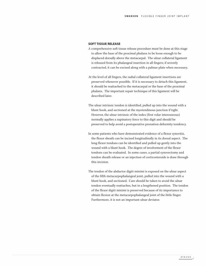
BONE RESECTION AND PREPARATION
The base of the proximal phalanx is resected including marginal
osteophytes which might interfere with the implant. All cartilage is
removed from the base of the proximal phalanx because it is believed
that progressive cartilage degeneration can eventually result in
recurrent synovitis.
The intramedullary canal of the metacarpal is prepared in a rectangular
fashion with a rasp, curet, broach, and air drill with a special bur. These
burs have a smooth leader point, which helps keep them in the canal
and prevents inadvertent perforation through the cortex. The
occasional construction in the intramedullary canal of the proximal
third of the metacarpal can be enlarged with the bur. The
intramedullary canal of the ring metacarpal is frequently quite small
and requires careful preparation. Care should be taken to avoid too
much reaming of the canals, especially in patients with thin bones.
A trial fit is made with the appropriate color coded sizers. The implant
stem should fit well down into the canal so that the transverse
midsection of the implant abuts against the bone end. The end of the
implant stem must not abut the end of the intramedullary canal and
the stem must be appropriately shortened. The largest implant possible
should be used. Implants of sizes 4 through 9 are generally used.
A rectangular hole is then made in the base of the proximal phalanx with
an osteotome, knife, broach, or air drill. The intramedullary canal is
reamed in the same fashion as the metacarpal to receive the distal stem
of the implant selected for the metacarpal.
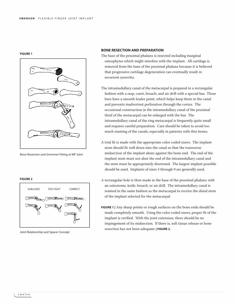
FIGURE 1 | Any sharp points or rough surfaces on the bone ends should be
made completely smooth. Using the color coded sizers, proper fit of the
implant is verified. With the joint extension, there should be no
impingement of its midsection. If there is, soft tissue release or bone
resection has not been adequate | FIGURE 2.
Bone Resection and Grommet Fitting at MP Joint
FIGURE 1
Joint Relationship and Spacer Concept
FIGURE 2
SUBLUXED TOO TIGHT CORRECT
S W A N S O N F L E X I B L E F I N G E R J O I N T I M P L A N T
t h i r t e e n
In the index and middle fingers, the intramedullary canal is reamed in a
rectangular shape, which is positioned high on the dorsal ulnar side of
the base of the proximal phalanx and low on its radial palmar side.
This proper rectangular and axial configuration of the canal stabilizes
the implant stems and helps maintain a slight supination of the digit to
prevent postoperative pronation deformity. Contrarily, in the little
finger, a position of slight pronation is desired and the intramedullary
rectangle is positioned high on the dorsoradial side and low on the
ulnar palmar side.
In patients selected to receive Swanson grommets, the implant sizer is
removed and the bone canals are prepared to allow a press-fit of the
appropriate sized grommet. The resected surfaces of the metacarpal
and proximal phalanx are shaped to obtain a precise fit of the grommet
sleeve and of its slightly curvilinear flanges against the resected bone
ends so that contact with overlying soft tissues is avoided. Both
surfaces are prepared and smoothened with a diamond bur or other
appropriate fine burs.
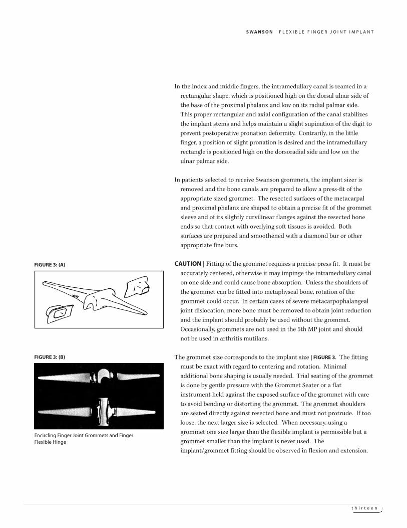
CAUTION | Fitting of the grommet requires a precise press fit. It must be
accurately centered, otherwise it may impinge the intramedullary canal
on one side and could cause bone absorption. Unless the shoulders of
the grommet can be fitted into metaphyseal bone, rotation of the
grommet could occur. In certain cases of severe metacarpophalangeal
joint dislocation, more bone must be removed to obtain joint reduction
and the implant should probably be used without the grommet.
Occasionally, grommets are not used in the 5th MP joint and should
not be used in arthritis mutilans.
The grommet size corresponds to the implant size | FIGURE 3. The fitting
must be exact with regard to centering and rotation. Minimal
additional bone shaping is usually needed. Trial seating of the grommet
is done by gentle pressure with the Grommet Seater or a flat
instrument held against the exposed surface of the grommet with care
to avoid bending or distorting the grommet. The grommet shoulders
are seated directly against resected bone and must not protrude. If too
loose, the next larger size is selected. When necessary, using a
grommet one size larger than the flexible implant is permissible but a
grommet smaller than the implant is never used. The
implant/grommet fitting should be observed in flexion and extension.
Encircling Finger Joint Grommets and FingerFlexible Hinge
FIGURE 3: (B)
FIGURE 3: (A)
S W A N S O N F L E X I B L E F I N G E R J O I N T I M P L A N T
f o u r t e e n
The implant must slide into the grommet and not be impinged by a too
narrow joint space or an uncorrected palmar subluxation. The
principal of the flexible hinge as a joint spacer must be respected
| FIGURE 2. The grommets and sizing unit are removed to carry out soft
tissue reconstruction.
SOFT TISSUE RECONSTRUCTION
Soft tissue reconstruction is a critical step of the operative procedure.
Proper balance of all the following structures must be attained:
collateral ligaments, palmar plate, capsule, intrinsic tendons, flexor, and
extensor mechanism.
Rheumatoid patients often present an inadequate first dorsal
interosseous muscle or have a tendency for pronation deformity of the
index finger and occasionally the middle finger, which can interfere
with the pinch and grasp mechanism.
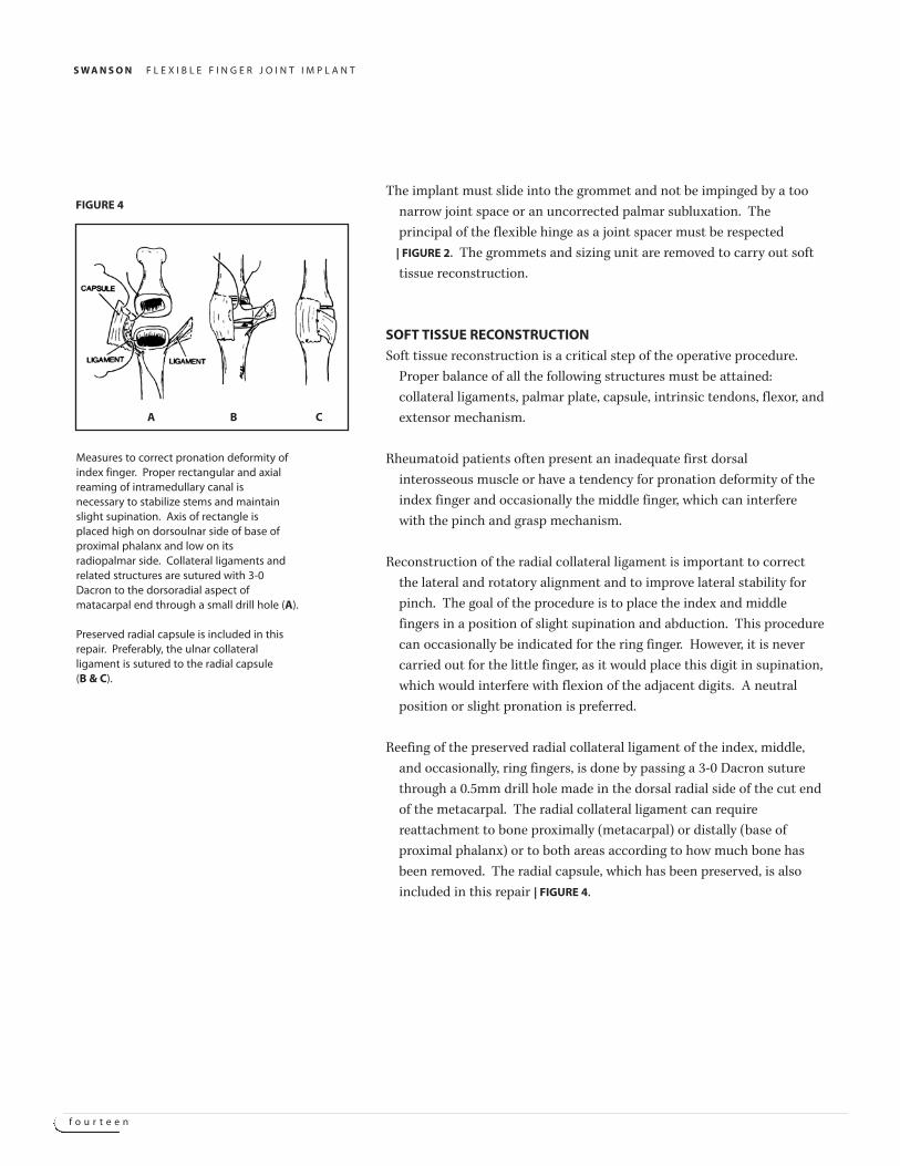
Reconstruction of the radial collateral ligament is important to correct
the lateral and rotatory alignment and to improve lateral stability for
pinch. The goal of the procedure is to place the index and middle
fingers in a position of slight supination and abduction. This procedure
can occasionally be indicated for the ring finger. However, it is never
carried out for the little finger, as it would place this digit in supination,
which would interfere with flexion of the adjacent digits. A neutral
position or slight pronation is preferred.
Reefing of the preserved radial collateral ligament of the index, middle,
and occasionally, ring fingers, is done by passing a 3-0 Dacron suture
through a 0.5mm drill hole made in the dorsal radial side of the cut end
of the metacarpal. The radial collateral ligament can require
reattachment to bone proximally (metacarpal) or distally (base of
proximal phalanx) or to both areas according to how much bone has
been removed. The radial capsule, which has been preserved, is also
included in this repair | FIGURE 4.
Measures to correct pronation deformity ofindex finger. Proper rectangular and axialreaming of intramedullary canal isnecessary to stabilize stems and maintainslight supination. Axis of rectangle isplaced high on dorsoulnar side of base ofproximal phalanx and low on itsradiopalmar side. Collateral ligaments andrelated structures are sutured with 3-0Dacron to the dorsoradial aspect ofmatacarpal end through a small drill hole (A).
Preserved radial capsule is included in thisrepair. Preferably, the ulnar collateralligament is sutured to the radial capsule(B & C).
FIGURE 4
A B C
S W A N S O N F L E X I B L E F I N G E R J O I N T I M P L A N T
f i f t e e n
An additional small drill hole can be made in the dorsal ulnar side of the cut end of the
metacarpal to secure the radial capsule with a 4-0 Dexon suture. However it is preferred
to suture the ulnar edge of the capsule to the ulnar collateral ligament to bring the
repaired capsule well ulnarward over the joint. The sutures are placed before the implant
is inserted and are tied as the finger is held in slight supination and abduction. Note
that the first dorsal interosseous muscle fibers become dorsally relocated with this
repair.
This procedure has seemed to be important in correction of pronation deformities and
provides some improved lateral stability for pinch. It seems to decrease flexion of the
index metacarpophalangeal joint by 10° to 20° by tightening the capsule, but this loss is
outweighed by increased stability and a better correction of the pronation deformity.
If the radial collateral ligament has been released from the base of the proximal phalanx it
should be reattached in a manner similar to that described above. If the radial collateral
ligament is inadequate, a portion of the palmar plate is used to reconstruct this
ligament. A distally-based flap made of the medial half of the palmar plate is prepared
and sutured in position through a drill hole made in the dorsoradial aspect of the cut
end of the metacarpal, similarly as described above.
Meticulous evaluation and correction of the balance of the capsuloligamentous and
musculotendinous structures will be rewarded by improved results.
IMPLANT INSERTION
The wound is thoroughly irrigated with triple antibiotic solution. The proximal grommet
is press-fit into the metacarpal and the distal grommet into the proximal phalanx
following the procedure described for trial insertion. Both grommets are firmly seated
against the resected bone by gentle pressure, taking care of avoid deforming the
grommet. The flexible implant is then inserted using blunt instruments and a "no-
touch" technique. First, the implant is inserted into the intramedullary canal of the
metacarpal, and then, by slight traction on the finger, the joint is distracted and the
implant is flexed so that the distal stem can easily be inserted into the proximal phalanx.
With the joint in extension, there should be no impingement of the implant. If there is,
soft tissue release or bone resection has been inadequate.
NOTE | Handling of the implant should be done with blunt instruments to avoid surface
trauma or contamination with foreign bodies. Reshaping of the implant should be
avoided because it can compromise or destroy the functional integrity and the
functionality of the implant.
S W A N S O N F L E X I B L E F I N G E R J O I N T I M P L A N T
s i x t e e n
EXTENSOR HOOD REEFING
The radial portion of the sagittal fibers of each extensor hood mechanism is reefed in an
overlapping fashion so that the extensor tendon is brought slightly to the radial side of
the center of the joint. Three to five 4-0 Dexon sutures with a buried-knot technique are
used. In the index and middle fingers, it is most important not to over-correct the
position of the extensor tendon too far radially to avoid deforming forces in favor of
pronation. In certain cases of severe or long-standing flexion deformity, the extensor
tendons may become stretched, and an extensor tendon lag may persist if not corrected.
In these cases, the extensor tendon should be reefed not only transversely as described,
but also longitudinally. Occasionally, the extensor tendon is tenodesed to the dorsal base
of the proximal phalanx through small drill holes. Perfect balance of the extensor
mechanism is essential. The juncturae tendinae which have been divided during the
release of the extensor tendons are meticulously re-approximated with 4-0 Dexon sutures
using inverted knots. This is done to further balance the extensor mechanism and
provide all possible extensor power.
CLOSURE AND DRESSING
The skin incision is closed with interrupted 5-0 nylon sutures; for small incision drains are
inserted into the wound subcutaneously. A non-adherent dressing, such as rayon, is
applied over the wound along with a Betadine or alternate gauze overlay. A voluminous
hand-conforming dressing is applied, avoiding pressure on the radial side of the index.
Gauze is placed between the fingers, but not down into the clefts which might cause
vascular constriction. A roll of Dacron batting is placed longitudinally across the dorsal
and palmar aspect of the forearm, wrist, hand, and fingers. Sheet wadding or Webril is
then applied. A narrow plaster or wooden splint is applied to the palmer aspect, and the
entire dressing is wrapped in a conforming bandage, such as Kling.
POSTOPERATIVE CARE AND BRACING
The ideal motion would provide adequate flexion of the ulnar digits, allowing the surface of
their pulps to touch the palm at the distal palmar crease for adequate grasp of smaller
objects. Full flexion of the index and middle fingers is less critical for grasping, as these
digits are mainly used for pinch activities. A degree of spreading of the finger, is
important. Full extension at these joints is also important to perform normal hand
activities and to maintain the balance of the distal joints. Chronic flexion deformity of
the metacarpophalangeal joints can further aggravate hyperextension tendencies at the
proximal interphalangeal joints. Pronation deformity of the index finger and,
occasionally, the middle finger can be a problem in the rheumatoid hand and can, to
some degree, be corrected in the postoperative program.
S W A N S O N F L E X I B L E F I N G E R J O I N T I M P L A N T
s e v e n t e e n
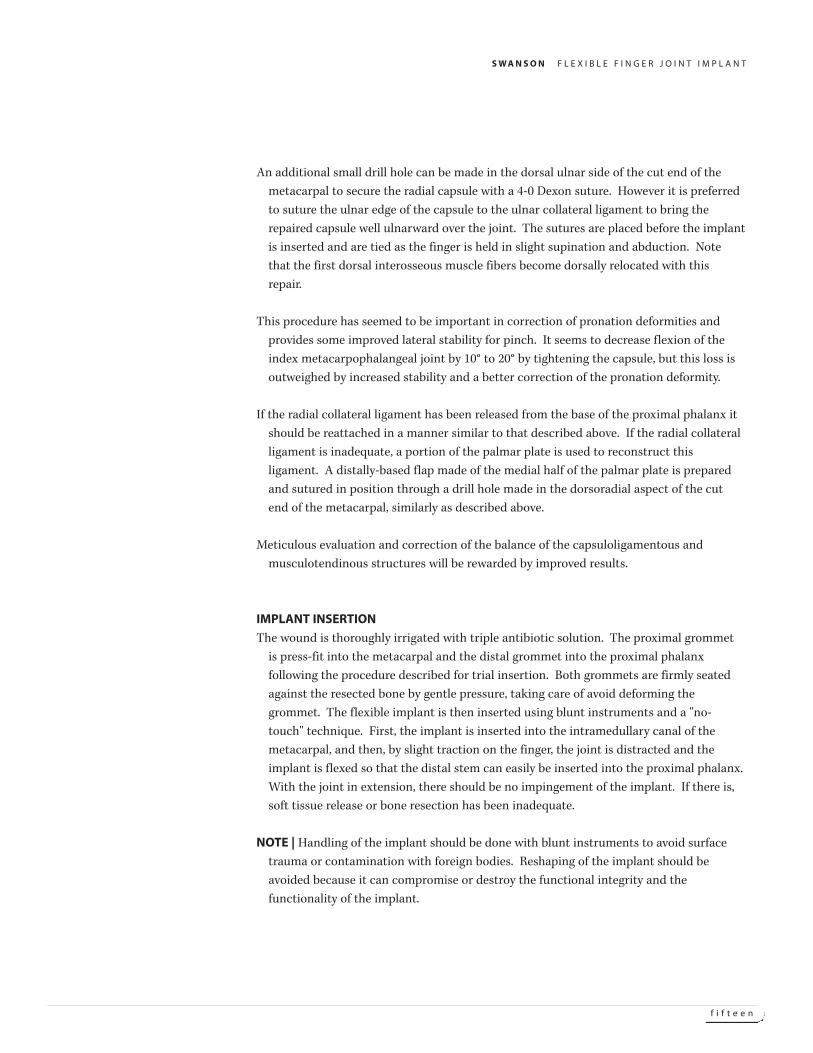
Immediate and continuous elevation of the hand and forearm during the
postoperative course is very important. The wound is usually checked
on the second day and drains removed. If swelling is minimal, use of
the dynamic brace can begin on the third to fifth postoperative day. A
light dressing is applied to the hand and forearm, and the dynamic
brace is fitted and adjusted, enabling the patient to start finger
movements in a protected arc. A ¼" felt pad is placed between the
forearm and the brace. If the splint is not available, guided early
motion may be obtained by applying a lightweight short arm case fitted
with outriggers and similar rubber band slings.
The rubber band slings are placed on the proximal phalanges to guide the
alignment of the digits into a slight radial direction to prevent
recurrent ulnar drift | FIGURE 5. The tension of the rubber bands should
be tight enough to support the digits and yet loose enough to allow 70°
of active flexion. This is especially true of the little finger which may
have weak flexion power. The brace may require adjustment once or
twice a day in the early postoperative course. Joint motion is measured
with a goniometer and recorded.
The thumb outrigger is applied in cases where the patient demonstrates
the tendency to bring the thumb over to the fingers on flexion. This
movement should be avoided because the pressure applied by the
thumb to the index finger would be in the ulnar direction, thus
aggravating the tendency toward ulnar drift deformity.
If there is a tendency toward medial rotation (pronation) of the
metacarpophalangeal joint of the index or middle fingers, additional
outrigger bars are applied to provide a supinatory force to the
joint | FIGURE 5.
The extension portion of the brace is worn continuously day and night for
the first three weeks, alternating with specific flexion exercises. The
exercises are started three days after surgery. However, if there is severe
flexor weakness of the little finger with adequate extension, the
extensor slings can be removed during the exercise periods.
Dynamic brace shown with additional lateralslings on the distal phalanx of the index andlong fingers to assist in correction of pronation deformity by forming a coupleproducing supinary torque force withoutinterfering with flexion and extensionmovements.
FIGURE 5
S W A N S O N F L E X I B L E F I N G E R J O I N T I M P L A N T
e i g h t e e n
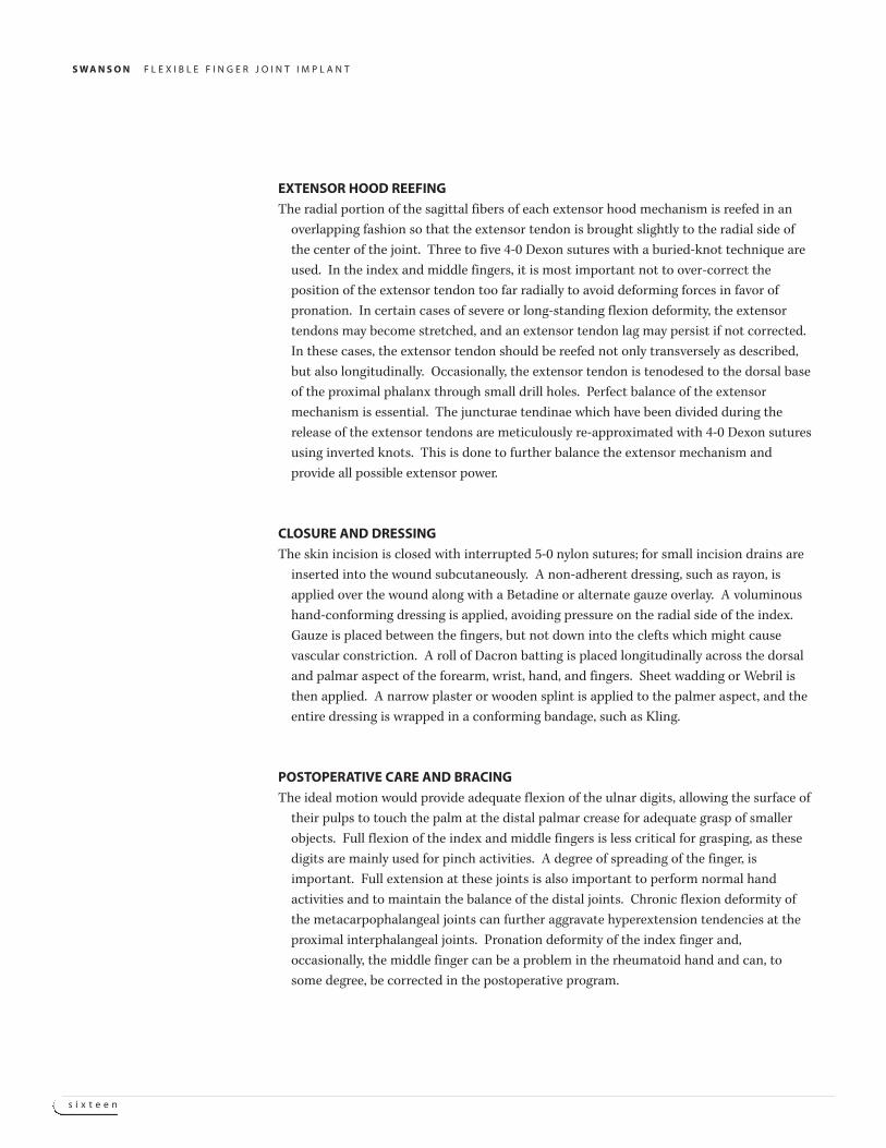
During the second and third weeks, the extension portion of the brace is
also worn continuously day and night. If there is severe flexor weakness
and good extension, the extensor slings can be removed one to two
hours a day to achieve greater active flexion of metacarpophalangeal
joints. Patients who have normal proximal interphalangeal joints
frequently will not gain the full-expected motion at the
metacarpophalangeal joint after arthroplasty because they tend to flex
the proximal interphalangeal joint during their exercise program and
thus relatively immobilize their metacarpophalangeal joints. To gain
active motion of the metacarpophalangeal joints in these patients,
small padded aluminum splints are taped on the dorsum of the
proximal interphalangeal joints during the exercise periods for the first
three or four weeks after surgery to encourage the patient to localize all
flexion forces at the metacarpophalangeal joint.
At three weeks any residual flexor weakness should be energetically
treated. In the presence of adequate extension, the preferred flexion
traction method consists of placing finger slings on the proximal
phalanges and attaching them volarly to the loop of a special Velcro
wrist strap or to that of the dynamic splint. When these flexion slings
are applied the figure eight elbow strap can be used to prevent distal
migration of the brace. If the distal and proximal interphalangeal joints
are stable, dressmaker hooks can be glued to the fingernails with a
cyanoacrylate adhesive. Individual rubber bands are then attached
from the loop of a special wrist strap to the nail hooks. Small band-aids
can be applied over the hooks for patient comfort between exercise
periods. These fraction methods give better control of alignment and
the desired amount of flexion pull for each individual finger. The
wearing schedule for these flexion devices varies with each patient but
usually should not exceed 20 minutes. If more flexion is required the
number of daily applications can be increased. In most cases 2-6 times
daily is sufficient.
Pre-Op x-ray reveals patient showing changesof rheumatoid arthritis involving metacarpophalangeal joints.
FIGURE 6: (A)
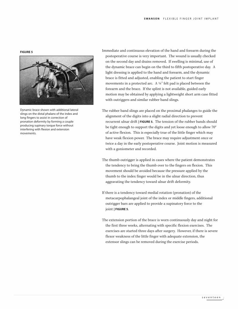
Post-Op radiogram shows one yearpostoperative reconstructive surgery withfusion of the MP of the thumb and siliconeimplant arthroplasty with encirclingtitanium gromments.
FIGURE 6: (B)
S W A N S O N F L E X I B L E F I N G E R J O I N T I M P L A N T
n i n e t e e n
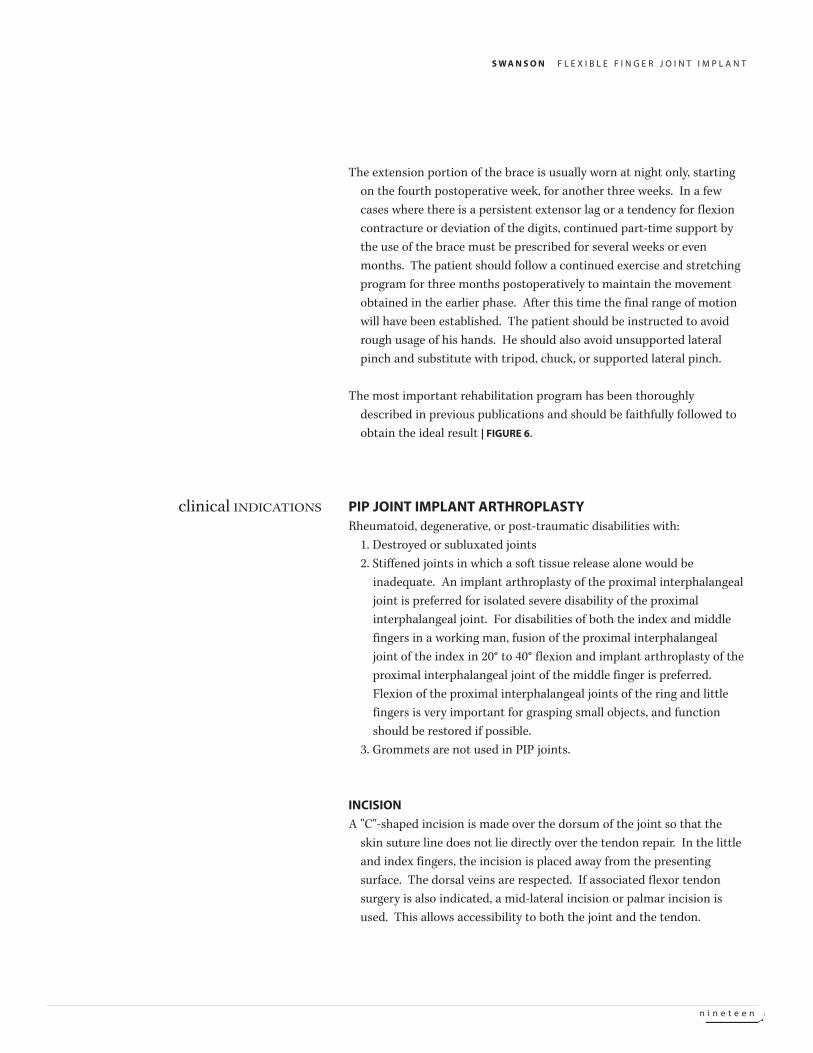
The extension portion of the brace is usually worn at night only, starting
on the fourth postoperative week, for another three weeks. In a few
cases where there is a persistent extensor lag or a tendency for flexion
contracture or deviation of the digits, continued part-time support by
the use of the brace must be prescribed for several weeks or even
months. The patient should follow a continued exercise and stretching
program for three months postoperatively to maintain the movement
obtained in the earlier phase. After this time the final range of motion
will have been established. The patient should be instructed to avoid
rough usage of his hands. He should also avoid unsupported lateral
pinch and substitute with tripod, chuck, or supported lateral pinch.
The most important rehabilitation program has been thoroughly
described in previous publications and should be faithfully followed to
obtain the ideal result | FIGURE 6.
PIP JOINT IMPLANT ARTHROPLASTYRheumatoid, degenerative, or post-traumatic disabilities with:
1. Destroyed or subluxated joints
2. Stiffened joints in which a soft tissue release alone would be
inadequate. An implant arthroplasty of the proximal interphalangeal
joint is preferred for isolated severe disability of the proximal
interphalangeal joint. For disabilities of both the index and middle
fingers in a working man, fusion of the proximal interphalangeal
joint of the index in 20° to 40° flexion and implant arthroplasty of the
proximal interphalangeal joint of the middle finger is preferred.
Flexion of the proximal interphalangeal joints of the ring and little
fingers is very important for grasping small objects, and function
should be restored if possible.
3. Grommets are not used in PIP joints.
INCISION
A "C"-shaped incision is made over the dorsum of the joint so that the
skin suture line does not lie directly over the tendon repair. In the little
and index fingers, the incision is placed away from the presenting
surface. The dorsal veins are respected. If associated flexor tendon
surgery is also indicated, a mid-lateral incision or palmar incision is
used. This allows accessibility to both the joint and the tendon.
clinical INDICATIONS
S W A N S O N F L E X I B L E F I N G E R J O I N T I M P L A N T
t w e n t y
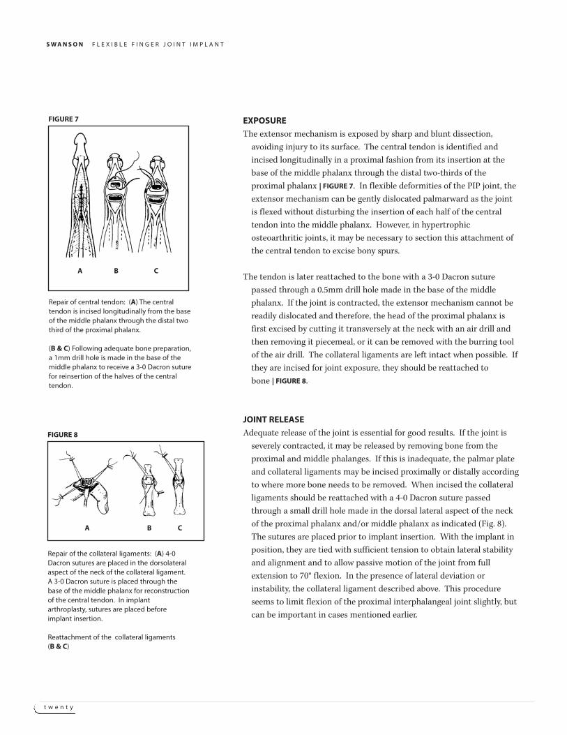
EXPOSURE
The extensor mechanism is exposed by sharp and blunt dissection,
avoiding injury to its surface. The central tendon is identified and
incised longitudinally in a proximal fashion from its insertion at the
base of the middle phalanx through the distal two-thirds of the
proximal phalanx | FIGURE 7. In flexible deformities of the PIP joint, the
extensor mechanism can be gently dislocated palmarward as the joint
is flexed without disturbing the insertion of each half of the central
tendon into the middle phalanx. However, in hypertrophic
osteoarthritic joints, it may be necessary to section this attachment of
the central tendon to excise bony spurs.
The tendon is later reattached to the bone with a 3-0 Dacron suture
passed through a 0.5mm drill hole made in the base of the middle
phalanx. If the joint is contracted, the extensor mechanism cannot be
readily dislocated and therefore, the head of the proximal phalanx is
first excised by cutting it transversely at the neck with an air drill and
then removing it piecemeal, or it can be removed with the burring tool
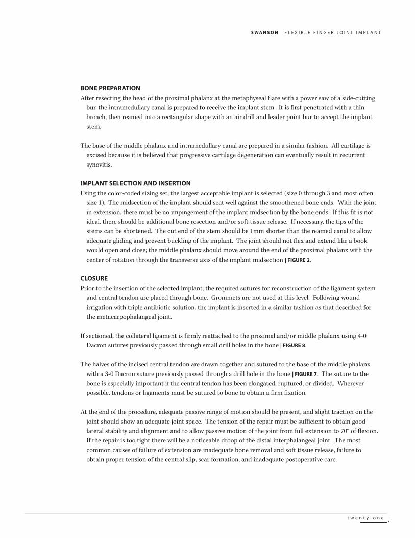
of the air drill. The collateral ligaments are left intact when possible. If
they are incised for joint exposure, they should be reattached to
bone | FIGURE 8.
JOINT RELEASE
Adequate release of the joint is essential for good results. If the joint is
severely contracted, it may be released by removing bone from the
proximal and middle phalanges. If this is inadequate, the palmar plate
and collateral ligaments may be incised proximally or distally according
to where more bone needs to be removed. When incised the collateral
ligaments should be reattached with a 4-0 Dacron suture passed
through a small drill hole made in the dorsal lateral aspect of the neck
of the proximal phalanx and/or middle phalanx as indicated (Fig. 8).
The sutures are placed prior to implant insertion. With the implant in
position, they are tied with sufficient tension to obtain lateral stability
and alignment and to allow passive motion of the joint from full
extension to 70° flexion. In the presence of lateral deviation or
instability, the collateral ligament described above. This procedure
seems to limit flexion of the proximal interphalangeal joint slightly, but
can be important in cases mentioned earlier.
Repair of central tendon: (A) The centraltendon is incised longitudinally from the baseof the middle phalanx through the distal twothird of the proximal phalanx.
(B & C) Following adequate bone preparation,a 1mm drill hole is made in the base of themiddle phalanx to receive a 3-0 Dacron suturefor reinsertion of the halves of the centraltendon.
FIGURE 7
Repair of the collateral ligaments: (A) 4-0Dacron sutures are placed in the dorsolateralaspect of the neck of the collateral ligament.A 3-0 Dacron suture is placed through thebase of the middle phalanx for reconstructionof the central tendon. In implantarthroplasty, sutures are placed beforeimplant insertion.
Reattachment of the collateral ligaments(B & C)
FIGURE 8
A B C
A B C
S W A N S O N F L E X I B L E F I N G E R J O I N T I M P L A N T
t w e n t y - o n e
BONE PREPARATION
After resecting the head of the proximal phalanx at the metaphyseal flare with a power saw of a side-cutting
bur, the intramedullary canal is prepared to receive the implant stem. It is first penetrated with a thin
broach, then reamed into a rectangular shape with an air drill and leader point bur to accept the implant
stem.
The base of the middle phalanx and intramedullary canal are prepared in a similar fashion. All cartilage is
excised because it is believed that progressive cartilage degeneration can eventually result in recurrent
synovitis.
IMPLANT SELECTION AND INSERTION
Using the color-coded sizing set, the largest acceptable implant is selected (size 0 through 3 and most often
size 1). The midsection of the implant should seat well against the smoothened bone ends. With the joint
in extension, there must be no impingement of the implant midsection by the bone ends. If this fit is not
ideal, there should be additional bone resection and/or soft tissue release. If necessary, the tips of the
stems can be shortened. The cut end of the stem should be 1mm shorter than the reamed canal to allow
adequate gliding and prevent buckling of the implant. The joint should not flex and extend like a book
would open and close; the middle phalanx should move around the end of the proximal phalanx with the
center of rotation through the transverse axis of the implant midsection | FIGURE 2.
CLOSURE
Prior to the insertion of the selected implant, the required sutures for reconstruction of the ligament system
and central tendon are placed through bone. Grommets are not used at this level. Following wound
irrigation with triple antibiotic solution, the implant is inserted in a similar fashion as that described for
the metacarpophalangeal joint.
If sectioned, the collateral ligament is firmly reattached to the proximal and/or middle phalanx using 4-0
Dacron sutures previously passed through small drill holes in the bone | FIGURE 8.
The halves of the incised central tendon are drawn together and sutured to the base of the middle phalanx
with a 3-0 Dacron suture previously passed through a drill hole in the bone | FIGURE 7. The suture to the
bone is especially important if the central tendon has been elongated, ruptured, or divided. Wherever
possible, tendons or ligaments must be sutured to bone to obtain a firm fixation.
At the end of the procedure, adequate passive range of motion should be present, and slight traction on the
joint should show an adequate joint space. The tension of the repair must be sufficient to obtain good
lateral stability and alignment and to allow passive motion of the joint from full extension to 70° of flexion.
If the repair is too tight there will be a noticeable droop of the distal interphalangeal joint. The most
common causes of failure of extension are inadequate bone removal and soft tissue release, failure to
obtain proper tension of the central slip, scar formation, and inadequate postoperative care.
S W A N S O N F L E X I B L E F I N G E R J O I N T I M P L A N T
t w e n t y - t w o
The skin is re-approximated with 5-0 Nylon and small Incision Drains are
inserted subcutaneously. The hand dressing is applied similarly as that
described for the metacarpophalangeal joint arthroplasty.
EXTENSOR MECHANISM IN COLLAPSE DEFORMITIES
Special consideration must be given to the extensor mechanism in
collapse deformities of the digits. Simply stated, in swan-neck
deformity the central tendon is relatively tight as compared to the
tension of the lateral tendons, and in the boutonniere deformity the
central tendon is relatively loose as compared to the tension of the
lateral tendons. Readjustment of the tension of these collapse
deformities. It should be noted that implant arthroplasty is seldom
indicated in swan-neck deformity.
BOUTONNIERE DEFORMITY
In a boutonniere deformity, the central tendon has usually been relatively
lengthened, and the lateral tendons displaced palmarward with the
connecting fibers stretched out. Implant arthroplasty is carried out as
indicated (Table II). The stretched out attachment of the central
tendon is sutured with a 4-0 Dacron suturing their connecting fibers or
overlapping any redundant fibers. A residual hyperextension deformity
of the distal joint can be corrected by lengthening or sectioning the
lateral tendons over the middle phalanx distal to the triangular
ligament.
SWAN-NECK DEFORMITY
Flexor tendon synovitis is treated before joint reconstruction. In swan-
neck deformity with combined involvement of the MP and PIP joints,
both joints are repaired at the same stage. Hyperextension of the PIP
joint is corrected through readjustment of the joint system. The tight
central tendon is lengthened, and the lateral bands are relocated
palmarward. At least 10° of flexion contracture should be obtained and
associated deformities of contiguous joints should be treated. In mild
flexible deformity in weak hands, dermadesis of the PIP joint is
indicated. In severe cases of swan-neck deformity, fusion of the joint is
preferred. Implant arthroplasty is rarely indicated | TABLE 1.
S W A N S O N F L E X I B L E F I N G E R J O I N T I M P L A N T
t w e n t y - t h r e e
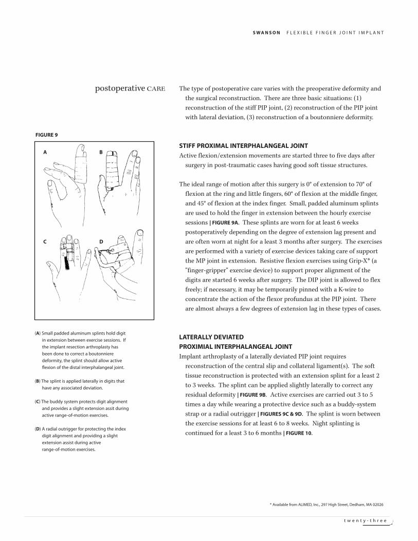
The type of postoperative care varies with the preoperative deformity and
the surgical reconstruction. There are three basic situations: (1)
reconstruction of the stiff PIP joint, (2) reconstruction of the PIP joint
with lateral deviation, (3) reconstruction of a boutonniere deformity.
STIFF PROXIMAL INTERPHALANGEAL JOINT
Active flexion/extension movements are started three to five days after
surgery in post-traumatic cases having good soft tissue structures.
The ideal range of motion after this surgery is 0° of extension to 70° of
flexion at the ring and little fingers, 60° of flexion at the middle finger,
and 45° of flexion at the index finger. Small, padded aluminum splints
are used to hold the finger in extension between the hourly exercise
sessions | FIGURE 9A. These splints are worn for at least 6 weeks
postoperatively depending on the degree of extension lag present and
are often worn at night for a least 3 months after surgery. The exercises
are performed with a variety of exercise devices taking care of support
the MP joint in extension. Resistive flexion exercises using Grip-X* (a
"finger-gripper" exercise device) to support proper alignment of the
digits are started 6 weeks after surgery. The DIP joint is allowed to flex
freely; if necessary, it may be temporarily pinned with a K-wire to
concentrate the action of the flexor profundus at the PIP joint. There
are almost always a few degrees of extension lag in these types of cases.
LATERALLY DEVIATED
PROXIMIAL INTERPHALANGEAL JOINT
Implant arthroplasty of a laterally deviated PIP joint requires
reconstruction of the central slip and collateral ligament(s). The soft
tissue reconstruction is protected with an extension splint for a least 2
to 3 weeks. The splint can be applied slightly laterally to correct any
residual deformity | FIGURE 9B. Active exercises are carried out 3 to 5
times a day while wearing a protective device such as a buddy-system
strap or a radial outrigger | FIGURES 9C & 9D. The splint is worn between
the exercise sessions for at least 6 to 8 weeks. Night splinting is
continued for a least 3 to 6 months | FIGURE 10.
postoperative CARE
(A) Small padded aluminum splints hold digit
in extension between exercise sessions. If
the implant resection arthroplasty has
been done to correct a boutonniere
deformity, the splint should allow active
flexion of the distal interphalangeal joint.
(B) The splint is applied laterally in digits that
have any associated deviation.
(C) The buddy system protects digit alignment
and provides a slight extension assit during
active range-of-motion exercises.
(D) A radial outrigger for protecting the index
digit alignment and providing a slight
extension assist during active
range-of-motion exercises.
FIGURE 9
* Available from ALIMED, Inc., 297 High Street, Dedham, MA 02026
A B
C D
S W A N S O N F L E X I B L E F I N G E R J O I N T I M P L A N T
t w e n t y - f o u r
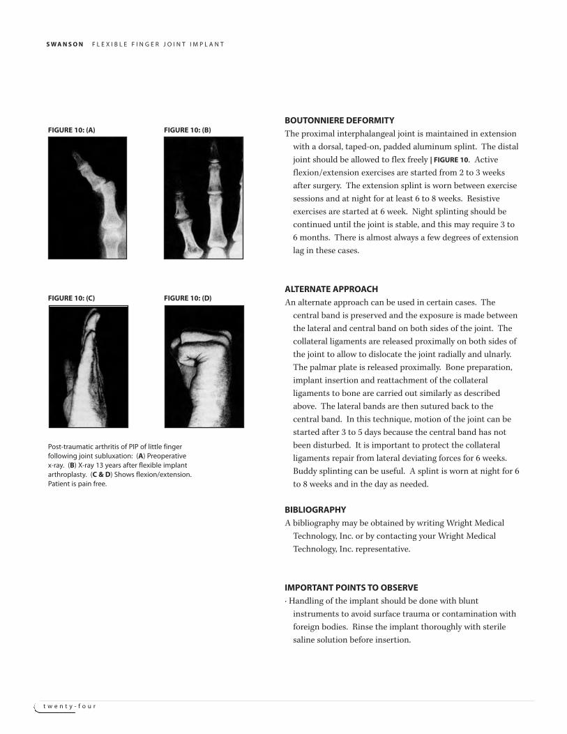
BOUTONNIERE DEFORMITY
The proximal interphalangeal joint is maintained in extension
with a dorsal, taped-on, padded aluminum splint. The distal
joint should be allowed to flex freely | FIGURE 10. Active
flexion/extension exercises are started from 2 to 3 weeks
after surgery. The extension splint is worn between exercise
sessions and at night for at least 6 to 8 weeks. Resistive
exercises are started at 6 week. Night splinting should be
continued until the joint is stable, and this may require 3 to
6 months. There is almost always a few degrees of extension
lag in these cases.
ALTERNATE APPROACH
An alternate approach can be used in certain cases. The
central band is preserved and the exposure is made between
the lateral and central band on both sides of the joint. The
collateral ligaments are released proximally on both sides of
the joint to allow to dislocate the joint radially and ulnarly.
The palmar plate is released proximally. Bone preparation,
implant insertion and reattachment of the collateral
ligaments to bone are carried out similarly as described
above. The lateral bands are then sutured back to the
central band. In this technique, motion of the joint can be
started after 3 to 5 days because the central band has not
been disturbed. It is important to protect the collateral
ligaments repair from lateral deviating forces for 6 weeks.
Buddy splinting can be useful. A splint is worn at night for 6
to 8 weeks and in the day as needed.
BIBLIOGRAPHY
A bibliography may be obtained by writing Wright Medical
Technology, Inc. or by contacting your Wright Medical
Technology, Inc. representative.
IMPORTANT POINTS TO OBSERVE
· Handling of the implant should be done with blunt
instruments to avoid surface trauma or contamination with
foreign bodies. Rinse the implant thoroughly with sterile
saline solution before insertion.
Post-traumatic arthritis of PIP of little fingerfollowing joint subluxation: (A) Preoperativex-ray. (B) X-ray 13 years after flexible implantarthroplasty. (C & D) Shows flexion/extension.Patient is pain free.
FIGURE 10: (A) FIGURE 10: (B)
FIGURE 10: (C) FIGURE 10: (D)
S W A N S O N F L E X I B L E F I N G E R J O I N T I M P L A N T
t w e n t y - f i v e
WARNING | Reshaping of the implant should be avoided because it can
compromise or destroy the structural integrity and the functionality of
the implant.
· Fitting of the grommet requires a precise press fit. It must be accurately
centered, otherwise it may impinge the cortex in the intramedullary
canal on one side and could cause bone absorption. Unless the
shoulders of the grommet can be fitted could occur. In certain cases of
severe metacarpophalangeal joint dislocation, more bone must be
removed to obtain joint reduction and the implant may have to be used
without the grommet.
· Grommets are not used at the PIP joint level or in the presence of bone
absorption or in arthritis mutilans.
HANDLING AND STERILIZATION
This product has been sterilized and should be considered sterile unless
the package has been opened or damaged. Remove from package, using
accepted sterile technique, only after the correct size has been
determined. Always handle the product with powder-free gloves, and
avoid contact with hard objects that may damage the product.
The sizing set is supplied nonsterile. The following sequential steps are
recommended to clean and sterlize the sizing set or to re-sterilize the
implant or grommets:
1. Scrub thoroughly with a clean, soft-bristled brush in a hot water-soap
solution to remove possible surface contaminants. Use a non-oily, mild
soap. Do not use synthetic detergents or oil-based soaps, as these soaps
may be absorbed and subsequently leach out to cause a tissue reaction.
2. Rinse thoroughly with distilled water.
3. If using a 270°F flash sterilization cycle, place the component on a
standard mesh sterilization tray.
4. If using a 250°F gravity or 270°F pulsing vacuum sterilization cycle,
double wrap the component in muslin or a similar type non-woven
medical grade wrapping material or place in a sealed sterilization
pouch.
5. Autoclave according to the following parameters:
MMeetthhoodd CCyyccllee TTeemmppeerraattuurree EExxppoossuurree
Steam Gravity 250°F/121°C 30 minutes
Steam Flash 270°F/132°C 10 minutes
Steam Pulsing-Vacuum 270°F/132°C 10 minutes
S W A N S O N F L E X I B L E F I N G E R J O I N T I M P L A N T
t w e n t y - s i x
After sterlization, remove the implant from its wrapping material/pouch or
the sterilization tray using accepted sterile technique. Ensure that the
implant is at room temperature prior to implantation.
These recommendations have been developed and tested using specific
equipment. The bioburden should not exceed 104 colony forming units
(CFU) per equipment, it must be demonstrated that these
recommendations produce sterility in your environment. If processing
conditions, wrapping materials or equipment changes occur, the
effectiveness of the sterilization process must be demonstrated.
Evidence suggests that repeated sterilization may affect the physical
characteristics of the implant. Accordingly, resterilization in excess of
three times is contraindicated.
This product is for single use only. An implant should never be sterilized
after contact with body tissues or fluids.
Do NOT sterilize by ethylene ozide as the residual sterilant may cause
adverse tissue reaction.
CAUTION | Federal (United States) law restricts this device to sale,
distribution and use by or on the order of a physician.
S W A N S O N F L E X I B L E F I N G E R J O I N T I M P L A N T
t w e n t y - s e v e n
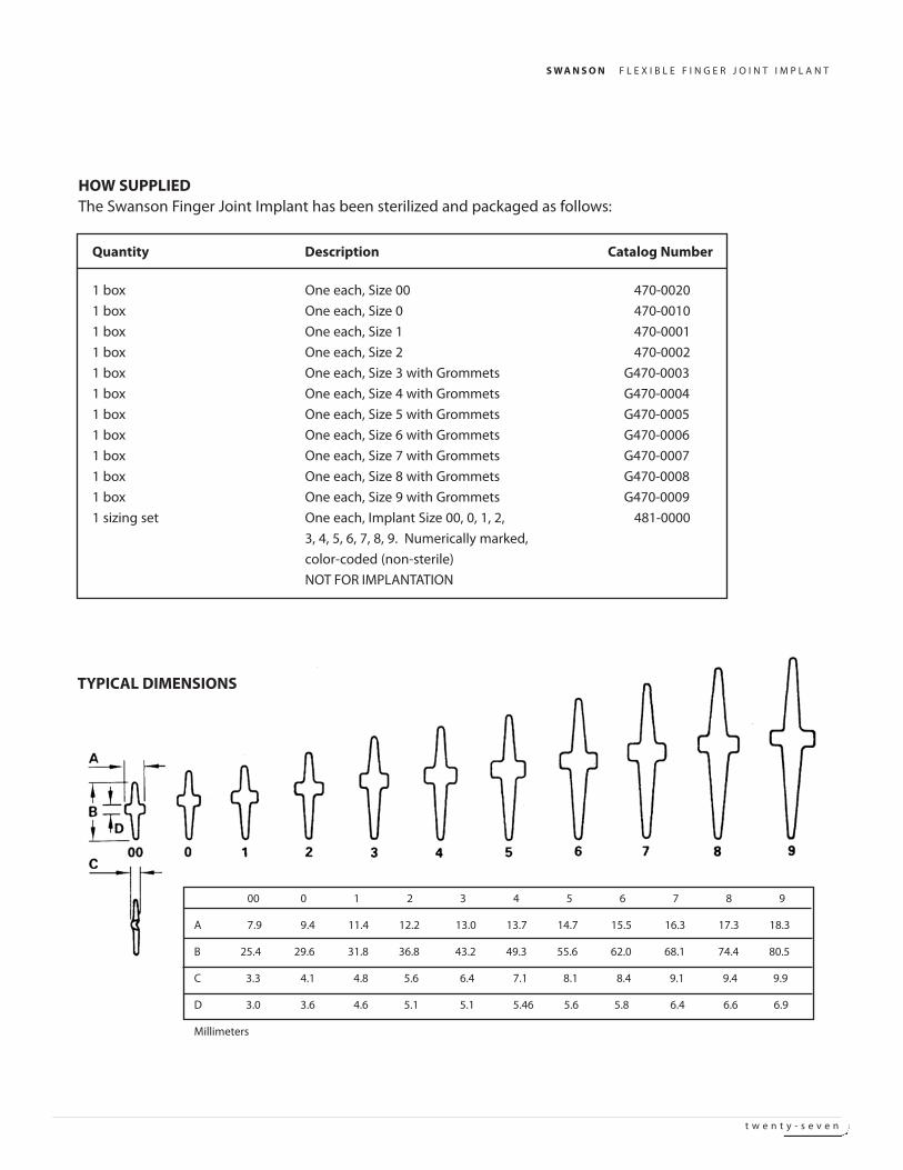
Quantity Description Catalog Number
1 box One each, Size 00 470-0020
1 box One each, Size 0 470-0010
1 box One each, Size 1 470-0001
1 box One each, Size 2 470-0002
1 box One each, Size 3 with Grommets G470-0003
1 box One each, Size 4 with Grommets G470-0004
1 box One each, Size 5 with Grommets G470-0005
1 box One each, Size 6 with Grommets G470-0006
1 box One each, Size 7 with Grommets G470-0007
1 box One each, Size 8 with Grommets G470-0008
1 box One each, Size 9 with Grommets G470-0009
1 sizing set One each, Implant Size 00, 0, 1, 2, 481-0000
3, 4, 5, 6, 7, 8, 9. Numerically marked,
color-coded (non-sterile)
NOT FOR IMPLANTATION
HOW SUPPLIEDThe Swanson Finger Joint Implant has been sterilized and packaged as follows:
TYPICAL DIMENSIONS
00 0 1 2 3 4 5 6 7 8 9
A 7.9 9.4 11.4 12.2 13.0 13.7 14.7 15.5 16.3 17.3 18.3
B 25.4 29.6 31.8 36.8 43.2 49.3 55.6 62.0 68.1 74.4 80.5
C 3.3 4.1 4.8 5.6 6.4 7.1 8.1 8.4 9.1 9.4 9.9
D 3.0 3.6 4.6 5.1 5.1 5.46 5.6 5.8 6.4 6.6 6.9
Millimeters
S W A N S O N F L E X I B L E F I N G E R J O I N T I M P L A N T
t w e n t y - e i g h t
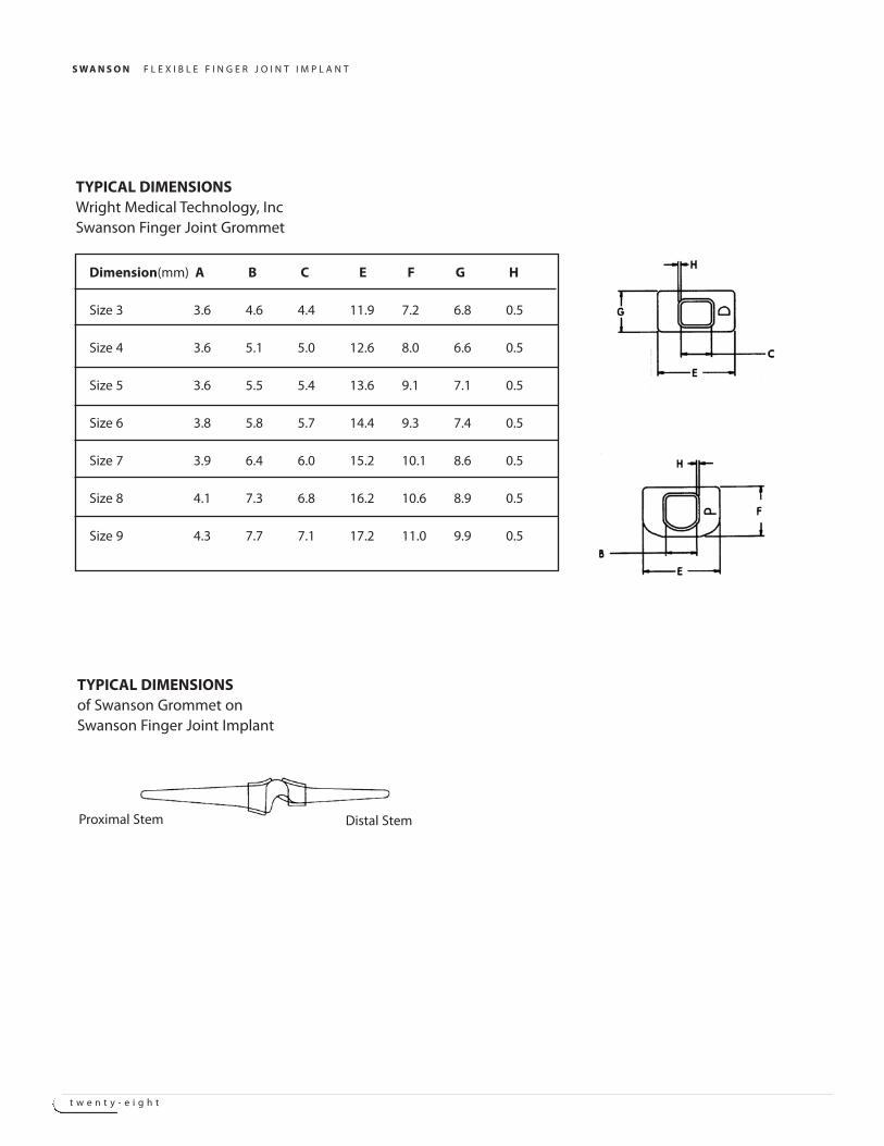
Dimension(mm) A B C E F G H
Size 3 3.6 4.6 4.4 11.9 7.2 6.8 0.5
Size 4 3.6 5.1 5.0 12.6 8.0 6.6 0.5
Size 5 3.6 5.5 5.4 13.6 9.1 7.1 0.5
Size 6 3.8 5.8 5.7 14.4 9.3 7.4 0.5
Size 7 3.9 6.4 6.0 15.2 10.1 8.6 0.5
Size 8 4.1 7.3 6.8 16.2 10.6 8.9 0.5
Size 9 4.3 7.7 7.1 17.2 11.0 9.9 0.5
TYPICAL DIMENSIONSWright Medical Technology, IncSwanson Finger Joint Grommet
TYPICAL DIMENSIONSof Swanson Grommet onSwanson Finger Joint Implant
Proximal Stem Distal Stem


Wright Medical Technology, Inc.5677 Airline RoadArlington, TN 38002901.867.9971 phone800.238.7188 [email protected]
Wright Cremascoli Ortho SAZone Industrielle la FarledeRue Pasteur BP 22283089 Toulon Cedex 09France011.33.49.408.7788 phone
SO STU003(Rev. 4-03)
TM Trademarks of Wright Medical Technology, Inc. Patents Pending.© 2003 Wright Medical Technology, Inc. All rights reserved.