Embed Size (px)
Citation preview

Sustained Submicromolar H2O2 Levels Induce Hepcidin viaSignal Transducer and Activator of Transcription 3 (STAT3)*□S
Received for publication, March 11, 2012, and in revised form, July 29, 2012 Published, JBC Papers in Press, August 29, 2012, DOI 10.1074/jbc.M112.358911
Gunda Millonig‡1, Ingo Ganzleben‡, Teresa Peccerella‡, Guillem Casanovas§¶, Lidia Brodziak-Jarosz�2,Katja Breitkopf-Heinlein**, Tobias P. Dick�, Helmut-Karl Seitz‡, Martina U. Muckenthaler§¶3, and Sebastian Mueller‡4
From the ‡Center for Alcohol Research, University of Heidelberg and Salem Medical Center, 69120 Heidelberg, the §Department ofPediatric Oncology, Hematology, Immunology, and Pulmonology and the ¶Molecular Medicine Partnership Unit, University ofHeidelberg, 69120 Heidelberg, the �Division of Redox Regulation, DKFZ-ZMBH Alliance, German Cancer Research Center,69120 Heidelberg, and **Molecular Hepatology-Alcohol Dependent Diseases II, Medical Clinic, Faculty of Medicine at Mannheim,University of Heidelberg, 68167 Mannheim, Germany
Background: Hepcidin, the systemic iron regulator, is induced during inflammation and leads to low circulating andincreased intracellular iron levels.Results: (Patho)physiologically relevant H2O2 levels up-regulate hepcidin via STAT3 in cultured liver cells.Conclusion: Intracellular and extracellular H2O2 acts similarly to IL-6 on hepcidin up-regulation and requires a functionalSTAT3-binding site.Significance: H2O2 is an important link between inflammation and iron metabolism.
The peptide hormone hepcidin regulates mammalian ironhomeostasis by blocking ferroportin-mediated iron export frommacrophages and the duodenum. During inflammation, hepci-din is strongly induced by interleukin 6, eventually leading tothe anemia of chronic disease.Herewe show that hepatoma cellsand primary hepatocytes strongly up-regulate hepcidin whenexposed to low concentrations of H2O2 (0.3–6 �M), concentra-tions that are comparable with levels of H2O2 released byinflammatory cells. In contrast, bolus treatment of H2O2 has noeffect at low concentrations and even suppresses hepcidin atconcentrations of>50�M.H2O2 treatment synergistically stim-ulates hepcidin promoter activity in combinationwith recombi-nant interleukin-6 or bone morphogenetic protein-6 and in amanner that requires a functional STAT3-responsive element.The H2O2-mediated hepcidin induction requires STAT3 phos-phorylation and is effectively blocked by siRNA-mediatedSTAT3 silencing, overexpression of SOCS3 (suppressor of cyto-kine signaling 3), and antioxidants such as N-acetylcysteine.Glycoprotein 130 (gp130) is required for H2O2 responsiveness,and Janus kinase 1 (JAK1) is required for adequate basal signal-ing, whereas Janus kinase 2 (JAK2) is dispensable upstream ofSTAT3. Importantly, hepcidin levels are also increased by intra-cellular H2O2 released from the respiratory chain in the pres-ence of rotenone or antimycin A. Our results suggest a novelmechanism of hepcidin regulation by nanomolar levels of sus-
tained H2O2. Thus, similar to cytokines, H2O2 provides animportant regulatory link between inflammation and ironmetabolism.
During infection and inflammation,mammalian ironmetab-olism undergoes typical changes. Plasma iron is rapidly with-drawn from the circulation and is safely stored intracellularly inthe form of ferritin. This redistribution limits iron availabilityfor erythropoiesis and eventually leads to the so-called anemiaof chronic disease (1). Hepcidin, a 25-amino acid peptide hasbeen identified as the central systemic iron-regulating hor-mone, and its discovery has provided new insights into themolecular mechanisms underlying hypoferremia and ironretention in the reticuloendothelial system during inflamma-tion (2–4). Under physiological conditions, replenished ironstores and inflammatory signals lead to hepcidin secretion byhepatocytes. Hepcidin then blocks duodenal uptake andmacrophage release of iron by binding to ferroportin, finallycausing its internalization and proteasomal degradation (5).The hepcidin-mediated blockage of ferroportin efficiently pre-vents iron export to the extracellular space and is amajormech-anistic pathway of the anemia of chronic disease (6).Among other cytokines, interleukin-6 (IL-6) is the most
potent inducer of hepcidin during inflammation (6, 7), aresponse controlled by STAT3 signaling (8). In addition, mul-tiple intra- and extracellular signals cross-talk to hepcidin, andthe hepcidin promoter contains binding sites for AP-1 (9),GATA-4 (10, 11), C/EBP�5 (8), and SMADs (mothers againstdecapentaplegic homologs; binding to the BMP-responsive ele-ments) (12) besides STAT3. Some of the promoter elements areessential for hepcidin basal expression, such as BMP-responsive
* This work was supported in part by the Dietmar Hopp-Stiftung and theManfred Lautenschläger-Stiftung.
□S This article contains supplemental Figs. S1–S3.1 Supported by an Olympia-Morata-Fellowship from the University of Heidel-
berg and a research grant from the YAEL Foundation. To whom corre-spondence may be addressed. Tel.: 49-6221-483-201; Fax: 49-6221-483-494; E-mail: [email protected].
2 Supported by the European Community’s Seventh Framework Programme(FP7/2007–2013) under Grant Agreement 215009.
3 Supported by the Dietmar Hopp Stiftung, eRARE-BMBF (HMA-IRON), andthe Network HepatoSys/Virtual Liver grant.
4 To whom correspondence may be addressed. Tel.: 49-6221-483-201; Fax:49-6221-483-494; E-mail: [email protected].
5 The abbreviations used are: C/EBP�, CCAAT/enhancer-binding protein �;BMP, bone morphogenetic protein; BMP-RE, BMP-responsive element;ROS, reactive oxygen species; H2O2ss, steady state H2O2; GOX, glucose oxi-dase; CAT, catalase; PG-1, Peroxy Green 1; NAC, N-acetylcysteine.
THE JOURNAL OF BIOLOGICAL CHEMISTRY VOL. 287, NO. 44, pp. 37472–37482, October 26, 2012© 2012 by The American Society for Biochemistry and Molecular Biology, Inc. Published in the U.S.A.
37472 JOURNAL OF BIOLOGICAL CHEMISTRY VOLUME 287 • NUMBER 44 • OCTOBER 26, 2012
by guest on March 16, 2019
http://ww
w.jbc.org/
Dow
nloaded from

elements (12) or C/EBP� (8), whereas others (e.g. the STAT3-binding site) are predominantly involved in hepcidin up-regula-tion upon inflammatory stimuli (8).Inflammatory conditions not only change the cytokine
milieu; they also increase the concentration of reactive oxygenspecies (ROS) locally and systemically. Upon activation, inflam-matory cells, such as neutrophils andmacrophages, undergo an“oxidative burst” that results in the release of large amounts ofreactive oxygen species to kill invading bacteria (13). Themem-brane-associated NADPH oxidase (NOX2) first generates super-oxide that is rapidly dismutated to the more stable H2O2 bysuperoxide dismutases (14). During inflammation, tissue H2O2concentrations are persistently elevated and can reachH2O2 con-centrations in the lowmicromolar range, mostly �10 �M (15).
During inflammation cells and tissues are exposed to persis-tently elevated concentrations of H2O2 demanding a tight reg-ulation of iron homeostasis to prevent tissue damage via Fentonand Fenton-like reactions. Previous studies showed direct reg-ulatory control of iron homeostasis by H2O2; the iron regula-tory protein 1 (IRP1), which usually stabilizes the mRNA ofTfR1 by binding to iron-responsive elements in the 3�-untrans-lated region (16), shows increased binding activity under per-sistently elevated H2O2 concentrations (17, 18). In addition,TfR1 is up-regulated independent of IRP-1 at the translationallevel under sustained levels of H2O2 (19). Both pathways finallylead to an iron shift into cells and removal of circulating freeiron.Little information is available how ROS regulate hepcidin
expression. Previous in vitro and in vivo studies on commonoxidative stress-associated liver diseases, such as alcoholic liverdisease (20) and chronic hepatitis C (21), demonstrated hepci-din down-regulation. However, a detailed mechanistic partici-pation of individual ROS on hepcidin down-regulation was notdemonstrated, and some reported observations were per-formed under artificially high ROS conditions. Miura et al. (21)exposed cultured cells to a single bolus of 100 �M H2O2, whichis commonly employed to dissect redox regulatory pathways.Such conditions, however, hardlymimic the continuous releaseof H2O2 from inflammatory cells, where H2O2 concentrationsdo not exceed 10 �M (15). Under normal culture conditions,H2O2 is rapidly degraded within 30 min (22), and artificiallyhigh H2O2 concentrations have to be used that may cause non-specific oxidative damage. We have previously established andoptimized an enzymatic system that allows the continuous gen-eration ofH2O2 at steady state levels over 24 h (19, 22–25). Thissystem allows mimicking the continuous H2O2 flux frominflammatory cells.Using this system, we show that in contrast to bolus applica-
tion of high doses ofH2O2, sustained levels ofH2O2 as low as 0.3�M rapidly and potently induce hepcidin independent of thecytokine network. Thus, H2O2 acts synergistically to inflamma-tory cytokines contributing to the anemia of chronic disease.
EXPERIMENTAL PROCEDURES
Cell Culture
Huh7 cells (from the Japanese Cancer Research ResourcesBank, Tokyo, Japan) weremaintained inDMEMwith 4.5 g/liter
glucose (PAA Laboratories, Pasching, Austria) and 10% fetalcalf serum (PAA Laboratories) under 5% CO2. Cells werescreened routinely formycoplasma contamination by PCR, andno infection could be detected. For experiments, cells wereseeded at a density of 6–7 � 104/well in 12-well plates with aworking volume of 1 ml/well. For immunoblotting experi-ments, we used 6-well plates with 1.8 � 105 cells/well with aworking volume of 2.5 ml/well, and for luciferase assays, cellswere kept in 96-well plates (5000 cells/well and a working vol-ume of 100 �l).
Cytokines and Reagents
IL-6 was purchased from Sigma-Aldrich and used diluted inculture medium at concentrations between 0.15 and 10 ng/ml.Bone morphogenetic protein 6 (BMP-6) was purchased fromR&D Systems Inc. (Minneapolis, MN) and used at concentra-tions between 10 and 50 ng/ml. N-Acetylcysteine was pur-chased from Sigma-Aldrich and used in a final concentration of2 mM. Rotenone and antimycin A were also purchased fromSigma-Aldrich and used at final concentrations of 10 and 3 �M,respectively.
Exposure of Cell Culture Cells to H2O2
H2O2 Bolus—Concentration of H2O2 (Merck) was deter-mined spectrophotometrically (�230 � 74 liters mol�1 cm�1).H2O2 was diluted into cell culture medium at concentrationsbetween 25 and 2000 �M.Steady State H2O2 (H2O2ss) Treatment Using Glucose Oxi-
dase and Catalase—Glucose oxidase (GOX) and catalase(CAT)were purchased fromSigma-Aldrich. Actual activities ofGOX and CAT were determined at very low H2O2 concentra-tions prior to the experiment using a sensitive chemilumines-cence technique (19, 22, 25, 26). During all experiments, kGOXwas kept at 4 � 10�8 M/s, whereas kCAT was adjusted to reachdefined H2O2ss concentrations between 0.15 and 6 �M.Because GOX metabolizes glucose and oxygen stochiometri-cally to H2O2 and �-gluconolactone, kGOX was selected to notdecrease glucose levels during 24 h by more than 3 mM, whichdoes not influence glucosemetabolism. In addition, no hypoxiawas observed under these conditions (27).
Measurement of H2O2 Concentrations in Cell Culture Medium
Huh7 cells were seeded at a density of 6–7� 104 cells/well in12-well plates. After 24 h, cell culture medium was changed tophenol red-free DMEM (4.5 g of glucose/liter) with 10% FCS.H2O2was added either as a bolus or generated byGOX/CAT asdescribed above. H2O2 determination was done as described byMueller et al. (15, 25). Briefly, a white 96-well plate (GreinerBio-One, Frickenhausen, Germany) was prefilled with 100�l/well of a 100 �M luminol, PBS solution (Sigma-Aldrich).Then 100 �l of cell culture supernatant from various timepoints of incubation with H2O2 and GOX/CAT, respectively,were added. Luminescence was then determined by adding 50�l of a 0.4 mM NaOCl solution by the injection device of theluminometer (Fluostar, BMG Labtech, Ortenberg, Germany).Luminescence was measured immediately after NaOCl injec-tion over 2 s.
Sustained Submicromolar Levels of H2O2 Induce Hepcidin
OCTOBER 26, 2012 • VOLUME 287 • NUMBER 44 JOURNAL OF BIOLOGICAL CHEMISTRY 37473
by guest on March 16, 2019
http://ww
w.jbc.org/
Dow
nloaded from

H2O2 Intracellular Imaging by Peroxy Green-1 (PG-1)
For imaging studies, Huh7 cells were seeded onto chamberslides (Nunc LabTek, Langenselbold, Germany) and grownovernight. The next day, cells were loaded with PG-1 (28) (akind gift of Christopher Chang (University of California, Berke-ley)) at a concentration of 5�M inHanks’ balanced salt solution,25 mM glucose for 20 min. Then the cells were exposed toH2O2ss ranging from 0.15 to 3 �M generated by the GOX/CATsystem inHanks’ balanced salt solution, 25mM glucose, 1% FCSfor 6 h. Confocal fluorescence imaging was performed with aZeiss LSM-710 laser-scanning microscope and a �40 (0.8numerical aperture) oil immersion objective lens. Excitation ofPG-1-loaded cells at 488 nm was carried out with an argonlaser, and emission was collected at 520 nm.
RNA Isolation, Reverse Transcription, and Real-timeQuantitative PCR
Cells were lysed with TriReagent (Molecular Research Cen-ter Inc. Cincinnati, OH), and RNAwas isolated with a standardchloroform/isopropyl alcohol extraction. RNA concentrationwas adjusted after photometric measurement, and 500 ng ofRNA were transcribed using Moloney murine leukemia virusreverse transcriptase, 50 pmol of random hexamer (both fromPromega, Mannheim, Germany), and 100 pmol of oligo(dT)primers (Carl Roth, Karlsruhe,Germany). RelativemRNA tran-script levels were quantified using the LightCycler FastStartDNAMaster Hybridization Probes kit on a LightCycler (RocheApplied Science) and applying the TaqMan methodology. Thehousekeeping genes �2-microglobulin and hypoxanthine-gua-nine-phosphoribosyltransferase were amplified in a parallelreaction for normalization. The TaqMan probes were posi-tioned on exon-exon boundaries of corresponding genes toexclude co-amplification of genomic DNA. Primers and probeswere designed using the Primer Express software (PerkinElmerLife Sciences) or the online Universal Probe Library AssayDesign Center (Roche Applied Science) and synthesized atEurofinsMWGOperon (Ebersbach, Germany). Sense and anti-sense primer (each at 0.5 �M) and 0.125 �M 5�-phosphorylatedprobe were labeled at their 5�-end with the reporter dye 6-car-boxyfluorescein and at the 3�-end with the quencher dye6-carboxytetramethylrhodamine.
Transfection Experiments
Huh7 cells were seeded in 96-well plates or 12-well platesdepending on the experiment. Cells were transfected usingLipofectamine 2000 (Invitrogen) according to the manufactur-er’s instructions. For promoter studies, hepcidin promoter con-structs containing firefly luciferase and a control plasmid con-taining Renilla luciferase were cotransfected. Details of theconstructs have been published previously (8, 12, 29). The cat-alase-containing plasmid was a kind gift of Thomas Kietzmann(University of Kaiserslautern, Germany) (30, 31). The SOCS3plasmid and the empty vector control were a kind gift from JuliaStrebovsky (University of Heidelberg, Germany).
Analysis of Hepcidin Promoter Activity
Huh7 cells were transfected as described above. 24 h aftertransfection, cells were treated with cytokines, steady state
H2O2, or a combination of both. After another 24 h, luciferaseactivity was assessed by the Dual Luciferase assay (Promega,Mannheim, Germany). Firefly luciferase expression was nor-malized to CMV promoter-controlled Renilla luciferase.Simultaneous overexpression of SOCS3 (suppressor of cyto-kine signaling 3) was performed by co-transfection of theSOCS3 expression vector or its corresponding empty controlvector.
RNA Silencing
For RNA silencing experiments, Huh7 cells were transfectedusing Lipofectamine 2000 and 10 nM siRNA. Validated siRNAfor STAT3, gp130, JAK1, and JAK2 were purchased by AppliedBiosystems Inc. (Foster City, CA). Equimolar concentrations ofuniversal negative siRNA (Sigma-Aldrich) were used as nega-tive control.
Immunoblotting
Cells were rinsed in ice-cold PBS and harvested in radioim-mune precipitation assay buffer, 1�Complete� protease inhib-itor with EDTA (Roche Applied Science) on ice. Protein con-centration was determined by a BCA assay (Pierce) andadjusted to 1 mg/ml in Laemmli buffer containing 0.2% 2-mer-captoethanol. Equal amounts of protein (20–40 �g/lane) wereseparated on 10% SDS-polyacrylamide gels and blotted onnitrocellulose membranes. Equal protein loading was con-firmed by protein staining with Ponceau-S solution. Mem-branes were blocked in 5% skim milk, PBS-Tween 0.05%(T-PBS) for 1 h at room temperature and then incubated inprimary antibody (see Table 1) diluted in skim milk/T-PBSovernight at 4 °C. 5% BSA, T-PBS was used for blocking of non-specific binding and for dilution of the anti-pSTAT3 antibody.After a wash in T-PBS for 20 min, the secondary antibody (seeTable 2) diluted in 5% skim milk was incubated at room tem-perature for 2 h followed by another washing step in T-PBS.The blot was developed using chemiluminescence (Rotilumin,Carl Roth, Karlsruhe, Germany) and exposed to autoradiogra-phy film for 1–3 min.
RESULTS
Sustained Non-toxic Levels of H2O2 Strongly Up-regulateHepcidin—In this study, we investigated whether hepatic cellsexposed to low non-toxic steady state levels of H2O2 as pro-duced by the glucose oxidase and catalase system (GOX/CAT)
TABLE 1Primer list
Primer Sequence
Human �2-microglobulinForward 5�-tga ctt tgt cac agc cca aga ta-3�Reverse 5�-aat cca aat gcg gca tct tc-3�Probe FAM-tga tgc tgc tta cat gtc tcg atc cca-TAMa
Human hepcidinForward 5�-cag gac aga gct gga gcc a -3�Reverse 5�-gca gca cat ccc aca ctt tg-3�Probe FAM-ctg ctg cgg ctg ctg tca tcg a-TAM
Human IL-6Forward 5�-gcc cag cta tga act cct tct-3�Reverse 5�-ctt ctc ctg ggg gta ctg g-3�Probe UPL 68 (Roche Applied Science)
a FAM, 6-carboxyfluorescein; TAM, 6-carboxytetramethylrhodamine.
Sustained Submicromolar Levels of H2O2 Induce Hepcidin
37474 JOURNAL OF BIOLOGICAL CHEMISTRY VOLUME 287 • NUMBER 44 • OCTOBER 26, 2012
by guest on March 16, 2019
http://ww
w.jbc.org/
Dow
nloaded from
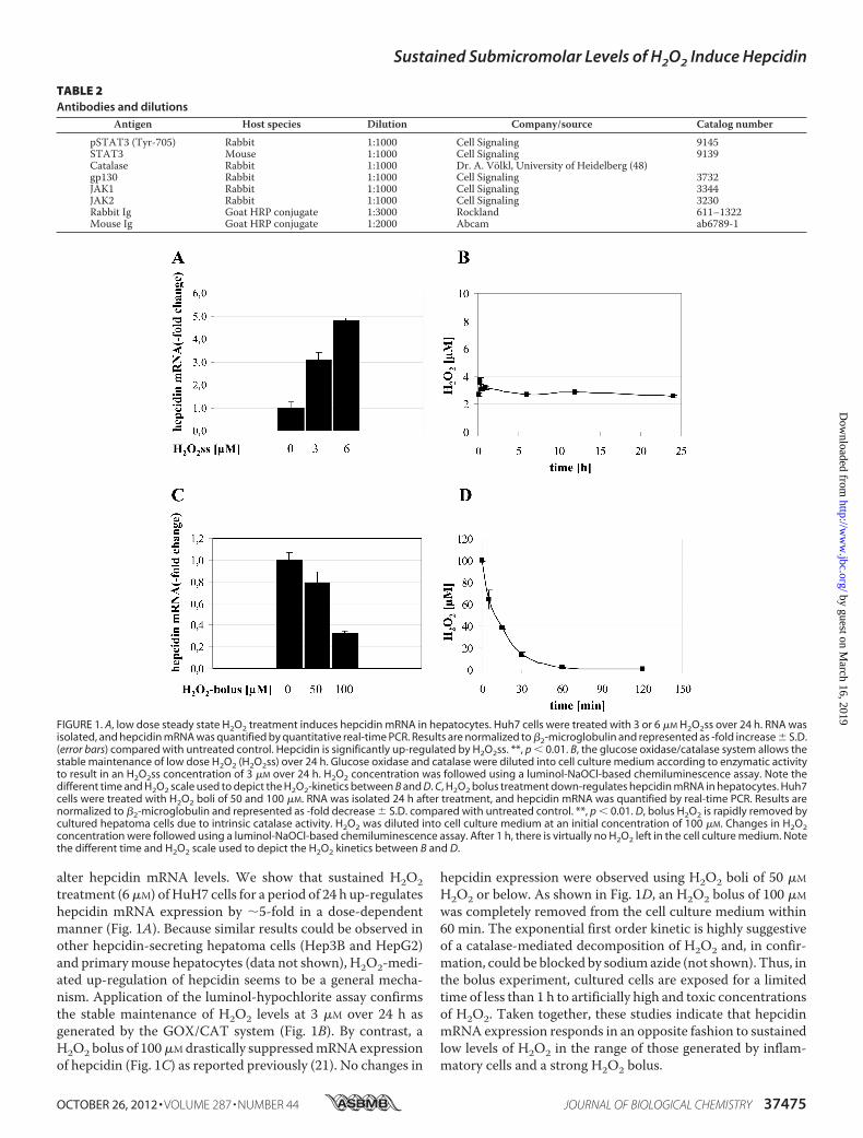
alter hepcidin mRNA levels. We show that sustained H2O2treatment (6�M) ofHuH7 cells for a period of 24 h up-regulateshepcidin mRNA expression by �5-fold in a dose-dependentmanner (Fig. 1A). Because similar results could be observed inother hepcidin-secreting hepatoma cells (Hep3B and HepG2)and primarymouse hepatocytes (data not shown), H2O2-medi-ated up-regulation of hepcidin seems to be a general mecha-nism. Application of the luminol-hypochlorite assay confirmsthe stable maintenance of H2O2 levels at 3 �M over 24 h asgenerated by the GOX/CAT system (Fig. 1B). By contrast, aH2O2 bolus of 100�Mdrastically suppressedmRNAexpressionof hepcidin (Fig. 1C) as reported previously (21). No changes in
hepcidin expression were observed using H2O2 boli of 50 �M
H2O2 or below. As shown in Fig. 1D, an H2O2 bolus of 100 �M
was completely removed from the cell culture medium within60 min. The exponential first order kinetic is highly suggestiveof a catalase-mediated decomposition of H2O2 and, in confir-mation, could be blocked by sodiumazide (not shown). Thus, inthe bolus experiment, cultured cells are exposed for a limitedtime of less than 1 h to artificially high and toxic concentrationsof H2O2. Taken together, these studies indicate that hepcidinmRNA expression responds in an opposite fashion to sustainedlow levels of H2O2 in the range of those generated by inflam-matory cells and a strong H2O2 bolus.
FIGURE 1. A, low dose steady state H2O2 treatment induces hepcidin mRNA in hepatocytes. Huh7 cells were treated with 3 or 6 �M H2O2ss over 24 h. RNA wasisolated, and hepcidin mRNA was quantified by quantitative real-time PCR. Results are normalized to �2-microglobulin and represented as -fold increase � S.D.(error bars) compared with untreated control. Hepcidin is significantly up-regulated by H2O2ss. **, p � 0.01. B, the glucose oxidase/catalase system allows thestable maintenance of low dose H2O2 (H2O2ss) over 24 h. Glucose oxidase and catalase were diluted into cell culture medium according to enzymatic activityto result in an H2O2ss concentration of 3 �M over 24 h. H2O2 concentration was followed using a luminol-NaOCl-based chemiluminescence assay. Note thedifferent time and H2O2 scale used to depict the H2O2-kinetics between B and D. C, H2O2 bolus treatment down-regulates hepcidin mRNA in hepatocytes. Huh7cells were treated with H2O2 boli of 50 and 100 �M. RNA was isolated 24 h after treatment, and hepcidin mRNA was quantified by real-time PCR. Results arenormalized to �2-microglobulin and represented as -fold decrease � S.D. compared with untreated control. **, p � 0.01. D, bolus H2O2 is rapidly removed bycultured hepatoma cells due to intrinsic catalase activity. H2O2 was diluted into cell culture medium at an initial concentration of 100 �M. Changes in H2O2concentration were followed using a luminol-NaOCl-based chemiluminescence assay. After 1 h, there is virtually no H2O2 left in the cell culture medium. Notethe different time and H2O2 scale used to depict the H2O2 kinetics between B and D.
TABLE 2Antibodies and dilutions
Antigen Host species Dilution Company/source Catalog number
pSTAT3 (Tyr-705) Rabbit 1:1000 Cell Signaling 9145STAT3 Mouse 1:1000 Cell Signaling 9139Catalase Rabbit 1:1000 Dr. A. Völkl, University of Heidelberg (48)gp130 Rabbit 1:1000 Cell Signaling 3732JAK1 Rabbit 1:1000 Cell Signaling 3344JAK2 Rabbit 1:1000 Cell Signaling 3230Rabbit Ig Goat HRP conjugate 1:3000 Rockland 611–1322Mouse Ig Goat HRP conjugate 1:2000 Abcam ab6789-1
Sustained Submicromolar Levels of H2O2 Induce Hepcidin
OCTOBER 26, 2012 • VOLUME 287 • NUMBER 44 JOURNAL OF BIOLOGICAL CHEMISTRY 37475
by guest on March 16, 2019
http://ww
w.jbc.org/
Dow
nloaded from
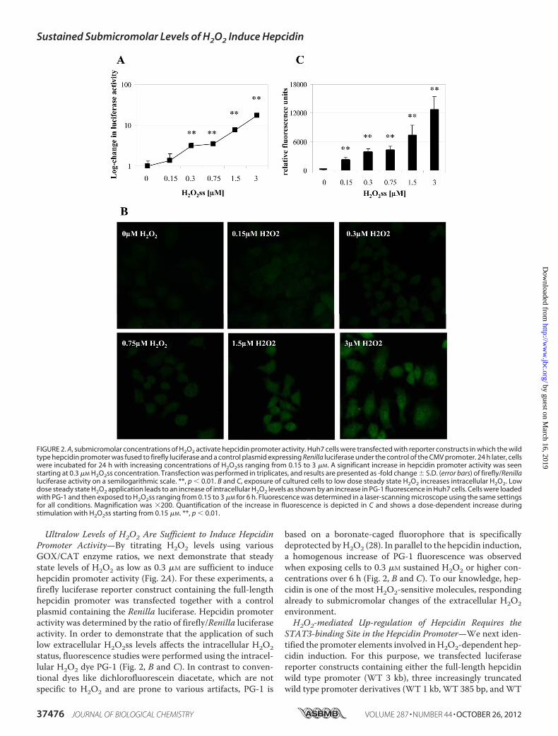
Ultralow Levels of H2O2 Are Sufficient to Induce HepcidinPromoter Activity—By titrating H2O2 levels using variousGOX/CAT enzyme ratios, we next demonstrate that steadystate levels of H2O2 as low as 0.3 �M are sufficient to inducehepcidin promoter activity (Fig. 2A). For these experiments, afirefly luciferase reporter construct containing the full-lengthhepcidin promoter was transfected together with a controlplasmid containing the Renilla luciferase. Hepcidin promoteractivity was determined by the ratio of firefly/Renilla luciferaseactivity. In order to demonstrate that the application of suchlow extracellular H2O2ss levels affects the intracellular H2O2status, fluorescence studies were performed using the intracel-lular H2O2 dye PG-1 (Fig. 2, B and C). In contrast to conven-tional dyes like dichlorofluorescein diacetate, which are notspecific to H2O2 and are prone to various artifacts, PG-1 is
based on a boronate-caged fluorophore that is specificallydeprotected byH2O2 (28). In parallel to the hepcidin induction,a homogenous increase of PG-1 fluorescence was observedwhen exposing cells to 0.3 �M sustained H2O2 or higher con-centrations over 6 h (Fig. 2, B and C). To our knowledge, hep-cidin is one of the most H2O2-sensitive molecules, respondingalready to submicromolar changes of the extracellular H2O2environment.H2O2-mediated Up-regulation of Hepcidin Requires the
STAT3-binding Site in the Hepcidin Promoter—We next iden-tified the promoter elements involved inH2O2-dependent hep-cidin induction. For this purpose, we transfected luciferasereporter constructs containing either the full-length hepcidinwild type promoter (WT 3 kb), three increasingly truncatedwild type promoter derivatives (WT 1 kb, WT 385 bp, andWT
FIGURE 2. A, submicromolar concentrations of H2O2 activate hepcidin promoter activity. Huh7 cells were transfected with reporter constructs in which the wildtype hepcidin promoter was fused to firefly luciferase and a control plasmid expressing Renilla luciferase under the control of the CMV promoter. 24 h later, cellswere incubated for 24 h with increasing concentrations of H2O2ss ranging from 0.15 to 3 �M. A significant increase in hepcidin promoter activity was seenstarting at 0.3 �M H2O2ss concentration. Transfection was performed in triplicates, and results are presented as -fold change � S.D. (error bars) of firefly/Renillaluciferase activity on a semilogarithmic scale. **, p � 0.01. B and C, exposure of cultured cells to low dose steady state H2O2 increases intracellular H2O2. Lowdose steady state H2O2 application leads to an increase of intracellular H2O2 levels as shown by an increase in PG-1 fluorescence in Huh7 cells. Cells were loadedwith PG-1 and then exposed to H2O2ss ranging from 0.15 to 3 �M for 6 h. Fluorescence was determined in a laser-scanning microscope using the same settingsfor all conditions. Magnification was �200. Quantification of the increase in fluorescence is depicted in C and shows a dose-dependent increase duringstimulation with H2O2ss starting from 0.15 �M. **, p � 0.01.
Sustained Submicromolar Levels of H2O2 Induce Hepcidin
37476 JOURNAL OF BIOLOGICAL CHEMISTRY VOLUME 287 • NUMBER 44 • OCTOBER 26, 2012
by guest on March 16, 2019
http://ww
w.jbc.org/
Dow
nloaded from

165 bp), or four full-length hepcidin promoters with selectivedeletions of specific binding sites, namely the STAT3-bindingsite (del-STAT3) and the BMP-responsive elements 1, 2, and 3(BMP-RE1, -2, and -3), respectively. As shown in Fig. 3A, nochanges of promoter activity in response to H2O2ss were
observed when truncating the hepcidin promoter stepwisefrom the 5�-end. However, a selective deletion of the STAT3-binding site almost completely prevented H2O2-mediated up-regulation of promoter activity, as did deletion of BMP-RE1.Selective deletions of BMP-RE2 and -3 still allowed for H2O2ss-
FIGURE 3. A, STAT3 is required for the H2O2-dependent increase of hepcidin promoter activity. Huh7 cells were transfected with hepcidin promoter constructscontaining the full-length wild type promoter (WT); promoter regions with decreasing length of the 5�-flanking region (WT 1kb, WT 385bp, and WT 165bp); orthe WT full-length promoter with specific deletions of transcription factor binding sites (i.e. the STAT3-binding site (delSTAT3)) and the BMP-responsiveelements 1–3 (del_bmp-RE1 to -3). Transfection and expression control was performed by a Renilla control plasmid. 24 h after transfection, cells were incubatedwith 3 �M H2O2ss for 24 h. Although increasing truncation of the WT promoter did not change responsiveness to H2O2ss, the deletion of the STAT3-binding sitecompletely abrogated H2O2ss-dependent promoter activation as did deletion of BMP-RE1. Because full STAT3 activation needs the presence of BMP-RE1,H2O2ss-induced hepcidin induction represents the typical profile of a STAT3-dependent activation. Transfections were performed in triplicates, and results arepresented as -fold change � S.D. (error bars) of firefly/Renilla luciferase activity compared with the untreated control of each construct. **, p � 0.01. B, H2O2bolus application induces STAT3 phosphorylation at supraphysiological doses. Huh7 cells were treated with H2O2 boli between 100 �M and 2 mM for 1 h. OnlyH2O2 bolus concentrations of 500 �M or above induced STAT3 phosphorylation after 1 h but led to cell death later on. Normalization was performed using aSTAT3 antibody. C, H2O2 bolus-induced down-regulation of hepcidin is STAT3-independent. Huh7 cells were transfected as described in A. 24 h after trans-fection, cells were incubated with H2O2 boli of 25, 50, or 100 �M for 24 h. Luciferase activity significantly decreased with increasing concentrations of bolus H2O2application in all promoter constructs. Transfections were performed in triplicates, and results are presented as -fold change � S.D. of firefly/Renilla luciferaseactivity compared with the untreated control of each construct.
Sustained Submicromolar Levels of H2O2 Induce Hepcidin
OCTOBER 26, 2012 • VOLUME 287 • NUMBER 44 JOURNAL OF BIOLOGICAL CHEMISTRY 37477
by guest on March 16, 2019
http://ww
w.jbc.org/
Dow
nloaded from

dependent promoter activation. This represents the typical pat-tern of STAT3-dependent hepcidin promoter activation, whichrequires both the STAT3-binding site and the BMP-RE1 (12,29). Because it is established that deletions of the STAT3-bind-ing site or the BMP-RE1 decrease basal expression hepcidinlevels (8, 29), the results are shown as -fold induction comparedwith the untreated control.Importantly, although H2O2 is able to induce STAT3 phos-
phorylation (Fig. 3B) as a bolus at toxic concentrations, pro-moter activity of the hepcidin promoter constructs was down-regulated. Thus, application of H2O2 boli between 25 and 100�M decreased hepcidin in a dose-dependent manner and inde-pendent of the STAT3-binding site (Fig. 3C). Taken together,only low and sustained and not high dose bolus H2O2 up-regu-lates hepcidin expression and requires the STAT3-responsiveelement, whereas down-regulation of hepcidin expression byhigh dose bolusH2O2 occurs independent of STAT3 binding tothe promoter.Low Levels of Sustained H2O2 Are Sufficient to Activate the
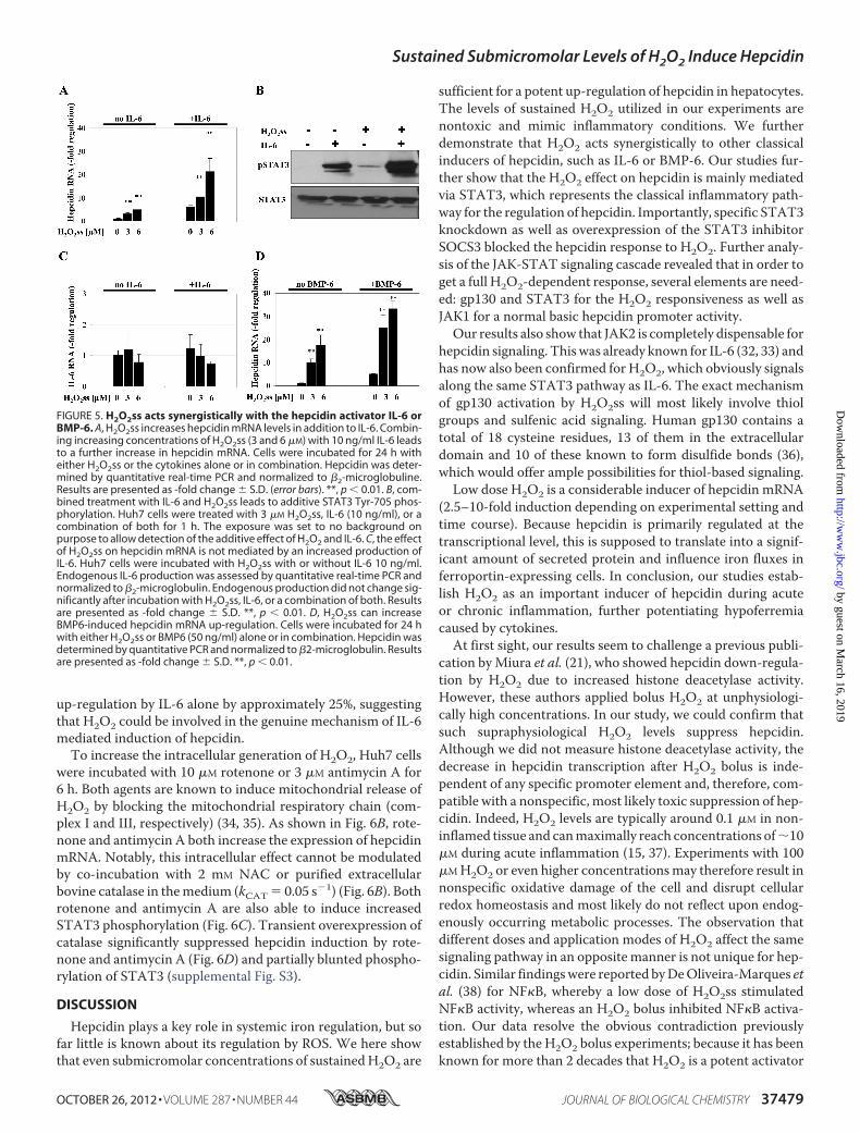
STAT3 Signaling Cascade—H2O2 boli higher than 500 �M areknown to induce STAT3 phosphorylation (Fig. 3B). We nowtested whether our low sustained H2O2 levels provided by theGOX/CAT system are likewise able to activate the STAT3 sig-naling cascade. Using a phospho-specific STAT3 antibody, wewere able to show that STAT3 is phosphorylated dose-depen-dently at the tyrosine residue at position 705 during a 6-h treat-ment of Huh7 cells with low and sustained levels of H2O2 (Fig.4A).We next tested whether the H2O2-mediated STAT3 phos-phorylation affects hepcidin regulation. Luciferase reporterconstructs with the wild type hepcidin promoter were trans-fected into HUH7 cells, and the STAT3 signaling cascade wasthen disrupted by siRNA-mediated silencing of each moleculeinvolved in the gp130-JAK-STAT3 signaling cascade. Theseexperiments identified three types of involvement in H2O2sssignaling (Fig. 4B and supplemental Fig. S1); gp130 and STAT3confer H2O2ss responsiveness, which is abrogated after silenc-ing of these twomolecules. STAT3 silencing even shows a com-plete suppression of H2O2ss-mediated hepcidin induction.JAK1 is responsible for basal hepcidin expression but notH2O2ss responsiveness. JAK2, finally, was shown to be com-pletely dispensable for H2O2ss as well as for IL-6 responsive-ness, as has been shown previously (32, 33). The importance ofSTAT3 for H2O2ss-mediated hepcidin expression was addi-tionally confirmed by overexpression of SOCS3 (suppressor ofcytokine signaling 3), the endogenous antagonist of STAT3 (seesupplemental Fig. S2). These experiments confirmed theimportant role of STAT3phosphorylation, gp130, and JAK1 forthe induction of hepcidin by H2O2.H2O2ss Induces Hepcidin Synergistically to IL-6 and BMP6—
Cytokines, such as IL-6, and bone morphogenetic proteins,such as BMP6, are powerful transcriptional activators of hepci-din promoter activity, with BMPs being required for basalexpression. As shown in Fig. 5A, H2O2ss-mediated up-regula-tion of hepcidin was drastically enhanced in the presence of 10ng/ml IL-6. Using immunoblotting for pSTAT3, we establishthat the combined application of low H2O2ss and IL-6 furtherincreases phosphorylation of STAT3 as compared with eitherof the stimuli alone (Fig. 5B). This effect is not dependent on
endogenous IL-6 production in hepatocytes after treatmentwithH2O2ss (Fig. 5C). In addition, H2O2ss further increases theBMP6-induced hepcidin response (Fig. 5D). We thus concludethat H2O2ss up-regulates hepcidin additively with IL-6 andBMP6 via increased STAT3 phosphorylation.Modulation of Hepcidin Expression by Intracellular H2O2—
Thus far, we could show that minuscule amounts of extracellu-lar H2O2ss suffice to induce hepcidin. We next analyzedwhether H2O2ss also affects hepcidin expression when modu-lated intracellularly (e.g. when produced from intracellularsources or when scavenged in the presence of an antioxidant,such as N-acetylcysteine (NAC)). 2 mM NAC completelyblocked up-regulation of hepcidin either by H2O2ss alone or incombination with IL-6 (Fig. 6A). This was not due to directinteraction of NAC with the GOX/CAT system because thiseffect was likewise observed when cells were pretreated withNAC for 24 h and, after washing, exposed to H2O2ss (data notshown). Interestingly, NAC alone also partly blocked hepcidin
FIGURE 4. A, STAT3 Tyr-705 is phosphorylated in response to low levels ofH2O2. Increasing H2O2ss concentrations cause increasing STAT3 phosphoryl-ation. Huh7 cells were treated with increasing concentrations of H2O2ss(range 0.15–3 �M) for 6 h. B and C, siRNA-mediated silencing of STAT3 andgp130 prevents H2O2ss-dependent activation of hepcidin. The H2O2-medi-ated effect on hepcidin induction is abolished completely by silencing ofSTAT3. Silencing of gp130 also diminished the H2O2 effect significantly. JAK1silencing decreases basal hepcidin promoter activity but does not influenceH2O2 responsiveness, whereas JAK2 is dispensable for H2O2-mediated as wellas IL-6-mediated hepcidin induction. Efficient silencing is shown by Westernblotting. Huh7 cells were transfected with a full-length wild type hepcidinpromoter fused to a firefly luciferase construct plus a Renilla control plasmid.Co-transfection with universal negative siRNA or siRNA directed specificallyagainst individual molecules of the JAK-STAT3 signaling cascade was per-formed at the same time. Cells were treated 24 h after transfection withH2O2ss (3 �M), IL-6 (10 ng/ml), or a combination of both. Hepcidin promoteractivity was assayed 12 h after treatment. Transfections were performed intriplicates, and results are presented as the ratio of firefly/Renilla luciferaseactivity � S.D. (error bars) Significant differences in H2O2-mediated promoteractivity compared with medium control are marked by asterisks (**, p � 0.01).
Sustained Submicromolar Levels of H2O2 Induce Hepcidin
37478 JOURNAL OF BIOLOGICAL CHEMISTRY VOLUME 287 • NUMBER 44 • OCTOBER 26, 2012
by guest on March 16, 2019
http://ww
w.jbc.org/
Dow
nloaded from
up-regulation by IL-6 alone by approximately 25%, suggestingthat H2O2 could be involved in the genuine mechanism of IL-6mediated induction of hepcidin.To increase the intracellular generation of H2O2, Huh7 cells
were incubated with 10 �M rotenone or 3 �M antimycin A for6 h. Both agents are known to induce mitochondrial release ofH2O2 by blocking the mitochondrial respiratory chain (com-plex I and III, respectively) (34, 35). As shown in Fig. 6B, rote-none and antimycin A both increase the expression of hepcidinmRNA. Notably, this intracellular effect cannot be modulatedby co-incubation with 2 mM NAC or purified extracellularbovine catalase in themedium (kCAT � 0.05 s�1) (Fig. 6B). Bothrotenone and antimycin A are also able to induce increasedSTAT3 phosphorylation (Fig. 6C). Transient overexpression ofcatalase significantly suppressed hepcidin induction by rote-none and antimycin A (Fig. 6D) and partially blunted phospho-rylation of STAT3 (supplemental Fig. S3).
DISCUSSION
Hepcidin plays a key role in systemic iron regulation, but sofar little is known about its regulation by ROS. We here showthat even submicromolar concentrations of sustainedH2O2 are
sufficient for a potent up-regulation of hepcidin in hepatocytes.The levels of sustained H2O2 utilized in our experiments arenontoxic and mimic inflammatory conditions. We furtherdemonstrate that H2O2 acts synergistically to other classicalinducers of hepcidin, such as IL-6 or BMP-6. Our studies fur-ther show that the H2O2 effect on hepcidin is mainly mediatedvia STAT3, which represents the classical inflammatory path-way for the regulation of hepcidin. Importantly, specific STAT3knockdown as well as overexpression of the STAT3 inhibitorSOCS3 blocked the hepcidin response to H2O2. Further analy-sis of the JAK-STAT signaling cascade revealed that in order toget a full H2O2-dependent response, several elements are need-ed: gp130 and STAT3 for the H2O2 responsiveness as well asJAK1 for a normal basic hepcidin promoter activity.Our results also show that JAK2 is completely dispensable for
hepcidin signaling. Thiswas already known for IL-6 (32, 33) andhas now also been confirmed for H2O2, which obviously signalsalong the same STAT3 pathway as IL-6. The exact mechanismof gp130 activation by H2O2ss will most likely involve thiolgroups and sulfenic acid signaling. Human gp130 contains atotal of 18 cysteine residues, 13 of them in the extracellulardomain and 10 of these known to form disulfide bonds (36),which would offer ample possibilities for thiol-based signaling.Low dose H2O2 is a considerable inducer of hepcidin mRNA
(2.5–10-fold induction depending on experimental setting andtime course). Because hepcidin is primarily regulated at thetranscriptional level, this is supposed to translate into a signif-icant amount of secreted protein and influence iron fluxes inferroportin-expressing cells. In conclusion, our studies estab-lish H2O2 as an important inducer of hepcidin during acuteor chronic inflammation, further potentiating hypoferremiacaused by cytokines.At first sight, our results seem to challenge a previous publi-
cation byMiura et al. (21), who showed hepcidin down-regula-tion by H2O2 due to increased histone deacetylase activity.However, these authors applied bolus H2O2 at unphysiologi-cally high concentrations. In our study, we could confirm thatsuch supraphysiological H2O2 levels suppress hepcidin.Although we did not measure histone deacetylase activity, thedecrease in hepcidin transcription after H2O2 bolus is inde-pendent of any specific promoter element and, therefore, com-patible with a nonspecific, most likely toxic suppression of hep-cidin. Indeed, H2O2 levels are typically around 0.1 �M in non-inflamed tissue and canmaximally reach concentrations of�10�M during acute inflammation (15, 37). Experiments with 100�MH2O2 or even higher concentrationsmay therefore result innonspecific oxidative damage of the cell and disrupt cellularredox homeostasis and most likely do not reflect upon endog-enously occurring metabolic processes. The observation thatdifferent doses and application modes of H2O2 affect the samesignaling pathway in an opposite manner is not unique for hep-cidin. Similar findingswere reported byDeOliveira-Marques etal. (38) for NF�B, whereby a low dose of H2O2ss stimulatedNF�B activity, whereas an H2O2 bolus inhibited NF�B activa-tion. Our data resolve the obvious contradiction previouslyestablished by the H2O2 bolus experiments; because it has beenknown for more than 2 decades that H2O2 is a potent activator
FIGURE 5. H2O2ss acts synergistically with the hepcidin activator IL-6 orBMP-6. A, H2O2ss increases hepcidin mRNA levels in addition to IL-6. Combin-ing increasing concentrations of H2O2ss (3 and 6 �M) with 10 ng/ml IL-6 leadsto a further increase in hepcidin mRNA. Cells were incubated for 24 h witheither H2O2ss or the cytokines alone or in combination. Hepcidin was deter-mined by quantitative real-time PCR and normalized to �2-microglobuline.Results are presented as -fold change � S.D. (error bars). **, p � 0.01. B, com-bined treatment with IL-6 and H2O2ss leads to additive STAT3 Tyr-705 phos-phorylation. Huh7 cells were treated with 3 �M H2O2ss, IL-6 (10 ng/ml), or acombination of both for 1 h. The exposure was set to no background onpurpose to allow detection of the additive effect of H2O2 and IL-6. C, the effectof H2O2ss on hepcidin mRNA is not mediated by an increased production ofIL-6. Huh7 cells were incubated with H2O2ss with or without IL-6 10 ng/ml.Endogenous IL-6 production was assessed by quantitative real-time PCR andnormalized to �2-microglobulin. Endogenous production did not change sig-nificantly after incubation with H2O2ss, IL-6, or a combination of both. Resultsare presented as -fold change � S.D. **, p � 0.01. D, H2O2ss can increaseBMP6-induced hepcidin mRNA up-regulation. Cells were incubated for 24 hwith either H2O2ss or BMP6 (50 ng/ml) alone or in combination. Hepcidin wasdetermined by quantitative PCR and normalized to �2-microglobulin. Resultsare presented as -fold change � S.D. **, p � 0.01.
Sustained Submicromolar Levels of H2O2 Induce Hepcidin
OCTOBER 26, 2012 • VOLUME 287 • NUMBER 44 JOURNAL OF BIOLOGICAL CHEMISTRY 37479
by guest on March 16, 2019
http://ww
w.jbc.org/
Dow
nloaded from

of the STAT3 pathway, the suppression of STAT-controlledhepcidin by H2O2 was difficult to comprehend.Although STAT3 seems to be responsible almost exclusively
for the H2O2-mediated up-regulation of hepcidin in our exper-iments, the hepcidin promoter contains other redox-sensitiveelements. For example, a C/EBP� binding site has been previ-ously suggested to down-regulate hepcidin in response to oxi-dative stress in mouse models for HCV and alcoholic liver dis-ease (39). However, elimination of the C/EBP� binding site atpositions �231 to �222 (40) in the truncated promoter con-structWT 165 kb neither prevented hepcidin down-regulationby H2O2 bolus application nor influenced hepcidin up-regula-tion by H2O2ss. Because in previous publications the type ofROS that activated C/EBP� has not been identified, the repres-sive effect might be exerted by a type of ROS other than H2O2.The hepcidin promoter also contains a putative AP-1 bindingsite, which is known to be sensitive to H2O2 (41). However,similar to C/EBP�, loss of the only AP-1 binding site in the
promoter constructs at positions �242 to �232 (8) had noeffect on the responsiveness to H2O2. Taken together, up-reg-ulation of hepcidin by H2O2 seems to be exclusively mediatedvia STAT3.The finding that hepcidin can be induced by very low levels of
H2O2 starting from 0.3 �M has wide reaching implications. Tothe best of our knowledge, hepcidin is one of the most H2O2-sensitive molecules described to date. Even the redox-sensitiveIRP1 required �10 times higher levels of H2O2 at �5 �M (42).Besides the amount of H2O2, the localization of the H2O2source is of critical importance for hepcidin regulation. It isespecially relevant for the question of whether H2O2 couldserve as an important if not mandatory intracellular signalingmolecule within the hepcidin pathway. By applying the intra-cellular H2O2 dye PG-1, we show that exposure of cells to sub-micromolarH2O2 concentrations affects the intracellularH2O2milieu. Furthermore, we demonstrate that N-acetylcysteineblocks up-regulation of hepcidin not only by H2O2 but also by
FIGURE 6. A, N-acetylcysteine efficiently blocks H2O2ss-induced hepcidin up-regulation. Huh7 cells were incubated with medium alone, 3 �M H2O2ss, or 3 �M
H2O2ss and 2 mM N-acetylcysteine (NAC) with or without IL-6 (10 ng/ml). Total RNA was isolated 6 h after treatment, and hepcidin mRNA was quantified byreal-time PCR. Results are normalized to �2-microglobulin and represented as -fold decrease � S.D. (error bars) compared with untreated control. Thehepcidin-inducing effect of H2O2ss was completely blocked by NAC. The additive effect of IL-6 and 3 �M H2O2ss was reduced to the effect of IL-6 alone whenNAC was added. *, p � 0.05; **, p � 0.01. B, intracellular H2O2 induces hepcidin mRNA. Huh7 cells treated with rotenone or antimycin A show a significantup-regulation of hepcidin mRNA. This increase in hepcidin cannot be reversed by adding either NAC or extracellular catalase to the cell culture medium. Resultsare normalized to �2-microglobulin and represented as -fold change � S.D. compared with untreated control. **, p � 0.01 compared with the medium control;#, no statistically significant change compared with rotenone or antimycin A treatment without NAC or extracellular catalase. C, treatment with rotenone orantimycin A increase STAT3 phosphorylation. Huh7 cells were treated for 6 h with either rotenone (10 �M) or antimycin A (3 �M). Cell lysates were then blottedfor pSTAT3 and STAT3. Both reagents led to increased STAT3 phosphorylation. D, overexpressed intracellular catalase blocks hepcidin up-regulation due tointracellular H2O2. Huh7 cells were transiently transfected with catalase. 24 h after transfection, cells were treated for 6 h with either rotenone (10 �M) orantimycin A (3 �M). Results are normalized to �2-microglobulin and represented as -fold change � S.D. compared with untransfected control. *, p � 0.05; **, p �0.01.
Sustained Submicromolar Levels of H2O2 Induce Hepcidin
37480 JOURNAL OF BIOLOGICAL CHEMISTRY VOLUME 287 • NUMBER 44 • OCTOBER 26, 2012
by guest on March 16, 2019
http://ww
w.jbc.org/
Dow
nloaded from

IL-6 alone. This suggests that H2O2 could be involved in IL-6signaling to hepcidin (e.g. by activation of an NADPH oxidase).The requirement of the NADPH oxidase in JAK/STAT-depen-dent signaling has been previously shown for angiotensin II(43). In addition, we demonstrate that also intracellularlyreleased H2O2 is able to induce hepcidin. H2O2 release by inhi-bition of the mitochondrial respiratory chain by rotenone(complex I) and antimycin A (complex III) (35) significantlyinduced hepcidin promoter activity, an effect that could beabrogated by overexpressed catalase.We further propose that apart from its role in inflammation,
H2O2 could also contribute to the regulation of hepcidin undernon-inflammatory conditions, including metabolic diseases.Indeed, a new iron overload entity termed dysmetabolic ironoverload syndrome has been introduced recently (44), which isoften associated with the metabolic syndrome and non-alco-holic fatty liver disease (45). In these patients, circulating hep-cidin levels are elevated for so far unknown reasons. They inter-rupt iron recycling and subsequently lead to iron accumulationin the reticuloendothelial system. Because insulin signaling(46), especially during conditions of insulin resistance (47), hasbeen associated with increased intracellular release of H2O2, itwould be very interesting to further investigate the role ofH2O2in dysmetabolic iron overload syndrome in future studies.Finally, although highly speculative, up-regulation of hepci-din by intracellular H2O2 could be an attractive mechanismof iron-mediated control of hepcidin. In this context, itshould be noted that it still remains unresolved why iron-mediated induction of hepcidin in vivo cannot be repro-duced under in vitro conditions.In summary, our results establish H2O2 as an important
upstream regulator and potent inducer of hepcidin that actsindependent of but synergistically with the cytokine network,thus contributing to the anemia of chronic disease. On a finalnote, our results also demonstrate the importance of choosingan appropriate H2O2model for studies of cellular signaling thatclosely mimics H2O2 conditions in vivo.
REFERENCES1. Cartwright, G. E., and Lauretsen,M.A. (1946) The anemia associatedwith
chronic infection. Science 103, 722. Ganz, T. (2002) The role of hepcidin in iron sequestration during infec-
tions and in the pathogenesis of anemia of chronic disease. Isr.Med. Assoc.J. 4, 1043–1045
3. Nemeth, E., Valore, E. V., Territo, M., Schiller, G., Lichtenstein, A., andGanz, T. (2003) Hepcidin, a putative mediator of anemia of inflammation,is a type II acute-phase protein. Blood 101, 2461–2463
4. Nicolas, G., Bennoun, M., Porteu, A., Mativet, S., Beaumont, C., Grand-champ, B., Sirito, M., Sawadogo, M., Kahn, A., and Vaulont, S. (2002)Severe iron deficiency anemia in transgenic mice expressing liver hepci-din. Proc. Natl. Acad. Sci. U.S.A. 99, 4596–4601
5. Nemeth, E., Tuttle,M. S., Powelson, J., Vaughn,M. B., Donovan, A.,Ward,D. M., Ganz, T., and Kaplan, J. (2004) Hepcidin regulates cellular ironefflux by binding to ferroportin and inducing its internalization. Science306, 2090–2093
6. Nemeth, E., Rivera, S., Gabayan, V., Keller, C., Taudorf, S., Pedersen, B. K.,and Ganz, T. (2004) IL-6 mediates hypoferremia of inflammation by in-ducing the synthesis of the iron regulatory hormone hepcidin. J. Clin.Invest. 113, 1271–1276
7. Roy, C. N., Custodio, A. O., deGraaf, J., Schneider, S., Akpan, I.,Montross,L. K., Sanchez, M., Gaudino, A., Hentze, M. W., Andrews, N. C., and
Muckenthaler, M. U. (2004) An Hfe-dependent pathway mediates hypo-sideremia in response to lipopolysaccharide-induced inflammation inmice. Nat. Genet. 36, 481–485
8. Verga Falzacappa, M. V., Vujic Spasic, M., Kessler, R., Stolte, J., Hentze,M.W., andMuckenthaler,M.U. (2007) STAT3mediates hepatic hepcidinexpression and its inflammatory stimulation. Blood 109, 353–358
9. Truksa, J., Lee, P., and Beutler, E. (2007) The role of STAT, AP-1, E-boxandTIEGmotifs in the regulation of hepcidin by IL-6 and BMP-9. Lessonsfrom human HAMP and murine Hamp1 and Hamp2 gene promoters.Blood Cells Mol. Dis. 39, 255–262
10. Island, M. L., Fatih, N., Leroyer, P., Brissot, P., and Loreal, O. GATA-4transcription factor regulates hepatic hepcidin expression. Biochem. J.437, 477–482
11. Bagu, E. T., and Santos, M. M. Friend of GATA suppresses the GATA-induced transcription of hepcidin in hepatocytes through a GATA-regu-latory element in the HAMP promoter. J. Mol. Endocrinol. 47, 299–313
12. Casanovas, G., Mleczko-Sanecka, K., Altamura, S., Hentze, M. W., andMuckenthaler, M. U. (2009) Bonemorphogenetic protein (BMP)-respon-sive elements located in the proximal and distal hepcidin promoter arecritical for its response to HJV/BMP/SMAD. J. Mol. Med. 87, 471–480
13. Klebanoff, S. J. (1988) Phagocytic cells. Products of oxygenmetabolism. inInflammation: Basic Principles and Clinical Correlates (Snyderman, R.,ed) pp. 391–444, Raven Press, New York
14. Geiszt, M., and Leto, T. L. (2004) The Nox family of NAD(P)H oxidases.Host defense and beyond. J. Biol. Chem. 279, 51715–51718
15. Mueller, S., and Arnhold, J. (1995) Fast and sensitive chemiluminescencedetermination of H2O2 concentration in stimulated human neutrophils.J. Biolumin. Chemilumin. 10, 229–237
16. Hentze, M. W., Muckenthaler, M. U., and Andrews, N. C. (2004) Balanc-ing acts. Molecular control of mammalian iron metabolism. Cell 117,285–297
17. Martins, E. A., Robalinho, R. L., andMeneghini, R. (1995) Oxidative stressinduces activation of a cytosolic protein responsible for control of ironuptake. Arch. Biochem. Biophys. 316, 128–134
18. Pantopoulos, K., and Hentze, M. W. (1995) Rapid responses to oxidativestress mediated by iron regulatory protein. EMBO J. 14, 2917–2924
19. Andriopoulos, B., Hegedusch, S., Mangin, J., Riedel, H. D., Hebling, U.,Wang, J., Pantopoulos, K., andMueller, S. (2007) Sustained hydrogen per-oxide induces iron uptake by transferrin receptor-1 independent of theiron regulatory protein/iron-responsive element network. J. Biol. Chem.282, 20301–20308
20. Harrison-Findik, D. D., Schafer, D., Klein, E., Timchenko, N. A., Kulaksiz,H., Clemens, D., Fein, E., Andriopoulos, B., Pantopoulos, K., and Gollan, J.(2006) Alcohol metabolism-mediated oxidative stress down-regulateshepcidin transcription and leads to increased duodenal iron transporterexpression. J. Biol. Chem. 281, 22974–22982
21. Miura, K., Taura, K., Kodama, Y., Schnabl, B., and Brenner, D. A. (2008)Hepatitis C virus-induced oxidative stress suppresses hepcidin expressionthrough increased histone deacetylase activity.Hepatology 48, 1420–1429
22. Mueller, S., Millonig, G., andWaite, G. N. (2009) The GOX/CAT system.A novel enzymatic method to independently control hydrogen peroxideand hypoxia in cell culture. Adv. Med. Sci. 54, 121–135
23. Mutze, S., Hebling, U., Stremmel, W., Wang, J., Arnhold, J., Pantopoulos,K., and Mueller, S. (2003) Myeloperoxidase-derived hypochlorous acidantagonizes the oxidative stress-mediated activation of iron regulatoryprotein 1. J. Biol. Chem. 278, 40542–40549
24. Mueller, S., Pantopoulos, K., Hubner, C. A., Stremmel, W., and Hentze,M. W. (2001) IRP1 activation by extracellular oxidative stress in the per-fused rat liver. J. Biol. Chem. 276, 23192–23196
25. Mueller, S. (2000) Sensitive and nonenzymatic measurement of hydrogenperoxide in biological systems. Free Radic. Biol. Med. 29, 410–415
26. Mueller, S., Riedel, H. D., and Stremmel, W. (1997) Determination ofcatalase activity at physiological hydrogen peroxide concentrations.Anal.Biochem. 245, 55–60
27. Millonig, G., Hegedusch, S., Becker, L., Seitz, H. K., Schuppan, D., andMueller, S. (2009) Hypoxia-inducible factor 1 � under rapid enzymatichypoxia. Cells sense decrements of oxygen but not hypoxia per se. FreeRadic. Biol. Med. 46, 182–191
Sustained Submicromolar Levels of H2O2 Induce Hepcidin
OCTOBER 26, 2012 • VOLUME 287 • NUMBER 44 JOURNAL OF BIOLOGICAL CHEMISTRY 37481
by guest on March 16, 2019
http://ww
w.jbc.org/
Dow
nloaded from

28. Miller, E. W., Tulyathan, O., Isacoff, E. Y., and Chang, C. J. (2007) Molec-ular imaging of hydrogen peroxide produced for cell signaling.Nat. Chem.Biol. 3, 263–267
29. Verga Falzacappa, M. V., Casanovas, G., Hentze, M.W., and Muckentha-ler, M. U. (2008) A bone morphogenetic protein (BMP)-responsive ele-ment in the hepcidin promoter controls HFE2-mediated hepatic hepcidinexpression and its response to IL-6 in cultured cells. J. Mol. Med. 86,531–540
30. Franke, K., Curth, K., Lenart, J., Knochenhauer, D., and Kietzmann, T.(2004) Enhanced plasminogen activator inhibitor-1 expression in trans-genic mice with hepatocyte-specific overexpression of superoxide dismu-tase or glutathione peroxidase. Antioxid. Redox Signal. 6, 721–728
31. BelAiba, R. S., Djordjevic, T., Bonello, S., Flugel, D., Hess, J., Kietzmann, T.,and Gorlach, A. (2004) Redox-sensitive regulation of the HIF pathwayunder non-hypoxic conditions in pulmonary artery smooth muscle cells.Biol. Chem. 385, 249–257
32. Murray, P. J. (2007) The JAK-STAT signaling pathway. Input and outputintegration. J. Immunol. 178, 2623–2629
33. Guschin, D., Rogers, N., Briscoe, J., Witthuhn, B., Watling, D., Horn, F.,Pellegrini, S., Yasukawa, K., Heinrich, P., and Stark, G. R. (1995) A majorrole for the protein tyrosine kinase JAK1 in the JAK/STAT signal trans-duction pathway in response to interleukin-6. EMBO J. 14, 1421–1429
34. Loschen, G., Flohe, L., and Chance, B. (1971) Respiratory chain linkedH2O2 production in pigeon heart mitochondria. FEBS Lett. 18, 261–264
35. Boveris, A., Oshino, R., Erecinska, M., and Chance, B. (1971) Reduction ofmitochondrial components by durohydroquinone. Biochim. Biophys.Acta 245, 1–16
36. Moritz, R. L., Hall, N. E., Connolly, L. M., and Simpson, R. J. (2001) Deter-mination of the disulfide structure and N-glycosylation sites of the extra-cellular domain of the human signal transducer gp130. J. Biol. Chem. 276,8244–8253
37. Test, S. T., and Weiss, S. J. (1984) Quantitative and temporal character-ization of the extracellular H2O2 pool generated by human neutrophils.J. Biol. Chem. 259, 399–405
38. deOliveira-Marques, V., Cyrne, L., Marinho, H. S., and Antunes, F. (2007)A quantitative study of NF-�B activation by H2O2. Relevance in inflam-mation and synergy with TNF-�. J. Immunol. 178, 3893–3902
39. Nishina, S., Hino, K., Korenaga,M., Vecchi, C., Pietrangelo, A.,Mizukami,Y., Furutani, T., Sakai, A., Okuda, M., Hidaka, I., Okita, K., and Sakaida, I.(2008) Hepatitis C virus-induced reactive oxygen species raise hepaticiron level in mice by reducing hepcidin transcription. Gastroenterology134, 226–238
40. Courselaud, B., Pigeon, C., Inoue, Y., Inoue, J., Gonzalez, F. J., Leroyer, P.,Gilot, D., Boudjema, K., Guguen-Guillouzo, C., Brissot, P., Loreal, O., andIlyin, G. (2002) C/EBP� regulates hepatic transcription of hepcidin, anantimicrobial peptide and regulator of iron metabolism. Cross-talk be-tween C/EBP pathway and iron metabolism. J. Biol. Chem. 277,41163–41170
41. Devary, Y., Gottlieb, R. A., Lau, L. F., and Karin, M. (1991) Rapid andpreferential activation of the c-jun gene during the mammalian UV re-sponse.Mol. Cell. Biol. 11, 2804–2811
42. Mueller, S., and Pantopoulos, K. (2002) Activation of iron regulatory pro-tein-1 (IRP1) by oxidative stress.Methods Enzymol. 348, 324–337
43. Schieffer, B., Luchtefeld, M., Braun, S., Hilfiker, A., Hilfiker-Kleiner, D.,and Drexler, H. (2000) Role of NAD(P)H oxidase in angiotensin II-in-duced JAK/STAT signaling and cytokine induction. Circ. Res. 87,1195–1201
44. Moirand, R., Mortaji, A. M., Loreal, O., Paillard, F., Brissot, P., and Deug-nier, Y. (1997) A new syndrome of liver iron overload with normal trans-ferrin saturation. Lancet 349, 95–97
45. Riva, A., Trombini, P., Mariani, R., Salvioni, A., Coletti, S., Bonfadini, S.,Paolini, V., Pozzi, M., Facchetti, R., Bovo, G., and Piperno, A. (2008) Re-valuation of clinical and histological criteria for diagnosis of dysmetaboliciron overload syndrome.World J. Gastroenterol. 14, 4745–4752
46. Livingston, J. N., Gurny, P. A., and Lockwood, D. H. (1977) Insulin-likeeffects of polyamines in fat cells. Mediation by H2O2 formation. J. Biol.Chem. 252, 560–562
47. Ikemura, M., Nishikawa, M., Hyoudou, K., Kobayashi, Y., Yamashita, F.,and Hashida, M. (2010) Improvement of insulin resistance by removal ofsystemic hydrogen peroxide by PEGylated catalase in obese mice. Mol.Pharm. 7, 2069–2076
48. Litwin, J. A., Volkl, A.,Muller-Hocker, J., Hashimoto, T., and Fahimi, H.D.(1987) Immunocytochemical localization of peroxisomal enzymes in hu-man liver biopsies. Am J Pathol 128, 141–150
Sustained Submicromolar Levels of H2O2 Induce Hepcidin
37482 JOURNAL OF BIOLOGICAL CHEMISTRY VOLUME 287 • NUMBER 44 • OCTOBER 26, 2012
by guest on March 16, 2019
http://ww
w.jbc.org/
Dow
nloaded from

U. Muckenthaler and Sebastian MuellerBrodziak-Jarosz, Katja Breitkopf-Heinlein, Tobias P. Dick, Helmut-Karl Seitz, Martina
Gunda Millonig, Ingo Ganzleben, Teresa Peccerella, Guillem Casanovas, Lidiaand Activator of Transcription 3 (STAT3)
Levels Induce Hepcidin via Signal Transducer2O2Sustained Submicromolar H
doi: 10.1074/jbc.M112.358911 originally published online August 29, 20122012, 287:37472-37482.J. Biol. Chem.
10.1074/jbc.M112.358911Access the most updated version of this article at doi:
Alerts:
When a correction for this article is posted•
When this article is cited•
to choose from all of JBC's e-mail alertsClick here
Supplemental material:
http://www.jbc.org/content/suppl/2012/09/14/M112.358911.DC1
http://www.jbc.org/content/287/44/37472.full.html#ref-list-1
This article cites 45 references, 18 of which can be accessed free at
by guest on March 16, 2019
http://ww
w.jbc.org/
Dow
nloaded from