Embed Size (px)
Citation preview

Heterodimerization with Different Jun Proteins Controlsc-Fos Intranuclear Dynamics and Distribution*□S
Received for publication, June 11, 2009, and in revised form, December 3, 2009 Published, JBC Papers in Press, January 6, 2010, DOI 10.1074/jbc.M109.032680
Cecile E. Malnou‡§¶1, Frederique Brockly‡§¶, Cyril Favard�, Gabriel Moquet-Torcy‡§¶, Marc Piechaczyk‡§¶2,3,and Isabelle Jariel-Encontre‡§¶2,4
From the ‡Institut de Genetique Moleculaire de Montpellier, UMR5535, CNRS, 1919 route de Mende, 34293 Montpellier Cedex 5,the §Universite Montpellier 2, Place Eugene Bataillon, 34095 Montpellier Cedex 5, the ¶Universite Montpellier 1,34967 Montpellier Cedex 2, and the �Institut Fresnel, CNRS UMR6133, Aix-Marseille Universite, Ecole Centrale Marseille,Campus de Saint Jerome, 13013 Marseille, France
The c-Fos proto-oncogenic transcription factor defines amultigene family controllingmanyprocesses both at the cell andthe whole organism level. To bind to its target AP-1/12-O-tet-radecanoylphorbol-13-acetate-responsive element or cAMP-responsive element DNA sequences in gene promoters andexert its transcriptional part, c-Fos must heterodimerize withother bZip proteins, its best studied partners being the Jun pro-teins (c-Jun, JunB, and JunD). c-Fos expression is regulated atmany transcriptional and post-transcriptional levels, yet little isknown on how its localization is dynamically regulated in thecell. Here we have investigated its intranuclear mobility usingfluorescence recovery after photobleaching, genetic, and bio-chemical approaches. Whereas monomeric c-Fos is highlymobile and distributed evenly with nucleolar exclusion in thenucleus, heterodimerization with c-Jun entails intranuclearredistribution and dramatic reduction in mobility of c-Foscaused by predominant association with the nuclear matrixindependently of anybinding toAP-1/12-O-tetradecanoylphor-bol-13-acetate-responsive element or cAMP-responsive ele-ment sequences. In contrast to c-Jun, dimerization with JunBdoes not detectably affect c-Fos mobility. However, dimeriza-tion with JunB affects intranuclear distribution with significantdifferences in the localization of c-Fos�c-Jun and c-Fos�JunBdimers. Moreover, c-Jun and JunB exert comparable effects onanother Fos family member, Fra-1. Thus, we report a novel reg-ulation, i.e. differentially regulated intranuclear mobility anddistribution of Fos proteins by their Jun partners, and suggestthe existence of intranuclear storage sites for latent c-Fos�c-Jun
AP-1 complexes. Thismay affect the numerous physiopatholog-ical functions these transcription factors control.
The AP-1 transcriptional complex comprises a large familyof dimeric transcription factors involved in the control ofnumerous physiological and pathological processes. Theseinclude among others cell proliferation, differentiation, apo-ptosis, responses to environmental cues, tumorigenesis, devel-opmental defects, and immune diseases (1–5). Its best studiedcomponents are the Fos (c-Fos, Fra-1, Fra-2, and FosB) and Jun(c-Jun, JunB, and JunD) family proteins, all of which necessitatedimerization via a leucine zipper (LZ)5 to acquire transcrip-tional competence. Fos proteins can only heterodimerize withother AP-1 components, whereas Jun proteins can alsohomodimerize, even thoughheterodimerizationwith any of theFos is favored (6, 7). Thanks to both the LZ and the adjacentbasic DNA-binding domains (DBD), Fos�Jun AP-1 dimers binddefinedDNAsequences known as 12-O-tetradecanoylphorbol-13-acetate-responsive elements (TREs) and less well to cAMP-responsive elements (CREs) that are found in many gene pro-moters, explaining the diversity of AP-1 effects. Importantly,AP-1 can act as a positive or a negative transcriptional regulatordepending on its composition, the target gene, the cell context,the extracellular environment, and which intracellular signal-ing cascades are activated (2, 6).c-Fos is the first discovered and best studied member of the
Fos family (8, 9). It is constitutively expressed in a limited num-ber of tissues but is rapidly and transiently induced in manyother cell types by a large variety of stimuli (9). In the latter case,c-Fos accumulation is controlled at the level of transcription,mRNA turnover, and protein stability (8, 10, 11). Moreover,c-Fos intracellular localization (see below) and transcriptionalactivity are tightly regulated (8). In particular, transcriptionalactivity can be enhanced via phosphorylation of various serinesand threonines that may also participate in protein stabiliza-tion. The kinases include the MAPK p38, ERK1/2, and ERK5
* This work was supported by grants from the ARC, Association pour laRecherche sur le Cancer, the Agence Nationale de Recherches (ANR-08-BLAN-007– 01), the French Ligue Nationale Contre le Cancer, and by theCNRS.
□S The on-line version of this article (available at http://www.jbc.org) containssupplemental Fig. S1.
1 Present address: UFR SVT, Universite Paul Sabatier, 31062 Toulouse andINSERM U563, Centre de Physiopathologie de Toulouse Purpan, 31024Toulouse, France.
2 Both authors contributed equally to this work.3 Supported in part by a Equipe Labelisee grant from the French Ligue Natio-
nale contre le Cancer. To whom correspondence may be addressed: IGMM,UMR5535, 1919 route de Mende, 34293 Montpellier Cedex 5, France. Tel.:33-4-67-61-36-68; Fax: 33-4-67-04-02-31; E-mail: [email protected].
4 To whom correspondence may be addressed: IGMM, UMR5535, 1919 routede Mende, 34293 Montpellier Cedex 5, France. Tel.: 33-4-67-61-36-68; Fax:33-4-67-04-02-31; E-mail: [email protected], [email protected].
5 The abbreviations used are: LZ, leucine zipper; FRAP, fluorescence recoveryafter photobleaching; CRE, cAMP-responsive element; TRE, 12-O-tetradecano-ylphorbol-13-acetate-responsive element; DBD, DNA-binding domain(s);MAPK, mitogen-activated protein kinase; ERK, extracellular signal-regu-lated kinase; STAT, signal transducers and activators of transcription; EGFP,enhanced green fluorescent protein; PBS, phosphate-buffered saline;MNase, microccocal nuclease.
THE JOURNAL OF BIOLOGICAL CHEMISTRY VOL. 285, NO. 9, pp. 6552–6562, February 26, 2010© 2010 by The American Society for Biochemistry and Molecular Biology, Inc. Printed in the U.S.A.
6552 JOURNAL OF BIOLOGICAL CHEMISTRY VOLUME 285 • NUMBER 9 • FEBRUARY 26, 2010
by guest on May 6, 2018
http://ww
w.jbc.org/
Dow
nloaded from

(12–25), where the role of ERK5 is disputed (26), as well as theERK1/2-activated kinases Rsk1/2 (12, 14, 16, 22) and I�B kinase(27). In contrast, c-Fos can be transcriptionally repressed bysumoylation at a specific lysine (28, 29). Interestingly, sumoy-lation of this lysine is antagonistic to a nearby transcription-activating phosphorylation (28).Usually, c-Fos accumulates predominantly, if not exclu-
sively, within the nucleus. However, it can also localizewithin the cytoplasm under certain conditions (17, 24, 25, 30,31). Cytoplasmic c-Fos can even associatewith the endoplasmicreticulum to activate phospholipid metabolism in a transcrip-tion activity-independent manner (32, 33). c-Fos intracellularlocalization is regulated by both intra- and extracellular signalswith demonstrated roles for cAMP-dependent protein kinaseA(34), the p38 MAPK (25), and the STAT3 transcription factorwhen ERK5 is inactivated (24). Consistent with this dual intra-cellular localization, we and others (24, 35, 36) have shown thatc-Fos can shuttle between the nucleus and the cytoplasm. Entryinto the nucleus is controlled by at least twonuclear localizationsignals: a conventional basic nuclear localization signal (35–37)that most likely utilizes the nuclear import receptor Imp�1 (35,36) and an unconventional nuclear localization signal located inthe N-terminal moiety of the protein that requires the nuclearimportin Transportin 1 (35, 36). Depending on the conditions,both Crm-1 exportin-dependent (24) and -independent (36)mechanisms involving different nuclear export signals areresponsible for c-Fos nuclear exit. Interestingly, this involvesprimarilymonomeric c-Fos and is inhibited upon heterodimer-ization with the Jun proteins (36). This indicated that dimeriza-tion is important, not only for the formation of active AP-1transcription complexes but also to keep them in the nucleuswhere they play their transcriptional part. c-Fos nuclear reten-tion, however, varies according to its Jun partner. It is muchstronger with c-Jun than with JunB or JunD. This correlateswith the strength of interaction of the various c-Fos�Jun dimers(38). Here, as a first step to understand why c-Fos nucleo-cyto-plasmic shuttling is differentially affected by the Juns, we usedfluorescence recovery after photobleaching (FRAP) togetherwith genetic and biochemical approaches to study the intranu-clear mobility of different Fos�Jun dimers.
MATERIALS AND METHODS
Plasmids, Cloning, andMutagenesis—Cloning andmutagen-esis were performed using standard PCR-basedmethods. All ofthe constructs were entirely sequenced. Plasmids and relatedinformation are available on request. pcDNA3-based expres-sion plasmids for wild type and mutant rat c-Fos and mouseJunB-FLAG and c-Jun-FLAG were previously described (16,36). EGFP chimeras were obtained by insertion of either ratc-Fos or human Fra1 coding sequences into the pEGFP-C1 vec-tor (Clontech).Cell Culture and Transfection—Conditions for culturing and
transfecting human HeLa cells by the calcium phosphate co-precipitation technique were previously described (36). 3 �g ofplasmid/106 cells was routinely used, and the transfection timewas limited to 16 h to avoid protein overexpression.Immunofluorescence and Confocal Microscopy—To set up the
conditions of FRAP experiments on cells expressing levels of
EGFP-c-FosorEGFPsimilar to thatof endogenousc-Fos,wecom-pared the expression level of transfected EGFP-c-Fos with that ofendogenous c-Fos expressed upon 1 h of serum stimulation. Tothisaim,HeLacellswereeither transfected for16hwith theEGFP-or EGFP-c-Fos-encoding vector or stimulatedwith 20% serum for1 h. The cells were fixed in 4% paraformaldehyde and permeabi-lized in the presence of 0.2% Triton X-100. They were then suc-cessively incubated with the H125 rabbit anti-c-Fos antibody(Santa Cruz Biotechnology), detecting equally c-Fos and EGFP-c-Fos and anAlexa 647-labeled anti-rabbit antibody (MolecularProbe). The nuclei were stained with Hoechst 33342, and cov-erslips were mounted in Permafluor. Cells expressing EGFP-c-Fos and emitting a fluorescence signal in the far red channelsimilar to that of cells expressing endogenous c-Fos wereselected to set up the signal intensity range in the green channel.This allowed us to select under the microscope cells expressingEGFP or EGFP-c-Fos at a level comparable with that of endog-enous c-Fos in the green channel. The far red fluorescence(Alexa 647) was monitored using a 560-nm long wavelengthpath filter, whereas EGFP fluorescence was monitored using a505–550-nm wavelength band pass filter. Twelve-bit imageacquisition was performed using a LSM510 meta microscope(Zeiss) equipped with a plan Apochromat 40� water immer-sion lens (1.2 numeric aperture) and with a confocal plane of5 �m.FRAP Experiments—FRAP analyses were performed at 37 °C
using a Zeiss LSM 510Meta microscope equipped with a heat-ing chamber and a planApochromat 40�water immersion lens(1.2 numeric aperture). To monitor EGFP fluorescence, thecells were excitedwith an argon laser at awavelength of 488 nm,and emission was collected using a 505–550-nm wavelengthband pass filter. The experiment was divided in three sequenc-es: (i) a prebleaching period, during which 15 images wereacquired to define the initial level of fluorescence; (ii) photo-bleaching, whichwas carried out on a 2-�mradius circular areaof the targeted nucleus using the 488-nm wavelength laser atmaximal power with 100 iterations of 256 �s/pixel; and (iii) apostbleaching period, during which fluorescence recovery wasmonitored every 150 ms for 30 s. Fluorescence recovery wasthen extracted on recorded images from the bleach area andcorrected for experimental fluctuations during acquisition.Correction was carried out by dividing the value of the fluores-cence in the bleach area by that of another region in the nucleusfar away from the bleached volume. Finally, fluorescence intensi-ties in the bleach area were normalized against the prebleach levelof fluorescence intensity. Data fitting was performed using theLevenberg-Marquadt algorithm to determine both themean half-time of fluorescence recovery (�t1⁄2�) and the mobile fraction ofthe protein (Fmob) using themodel in Equation 1,
I�t� � I0� A1exp��t
�1� � A2exp��t
�2�� � I30s�A1�1 � exp��t
�1��
� A2�1 � exp��t
�2��� (Eq. 1)
where I0 is the fluorescence intensity at t� 0 (immediately afterbleaching) and I∞ is the fluorescence intensity at 30 s (end of
Intranuclear Mobility of c-Fos
FEBRUARY 26, 2010 • VOLUME 285 • NUMBER 9 JOURNAL OF BIOLOGICAL CHEMISTRY 6553
by guest on May 6, 2018
http://ww
w.jbc.org/
Dow
nloaded from

recording) and with�t1⁄2� �A1�1 �A2�2 and with Equation 2.
Fmob30 �I30s � I0
1 � I0(Eq. 2)
The choice of this model was conditioned to a c2 statistical testshowing the best fit for all of the data as comparedwith amono-exponential or a two-dimensional diffusion under a Gaussianillumination model. This model was used without any a prioriknowledge of either the geometry of the bleaching or the pro-cess of fluorescence recovery. It was therefore used as a modelto comparemeanhalf-times of fluorescence recovery. Finally, I0was determined by bleaching under the same experimentalconditions as those described above for c-Fos-EGFP-express-ing cells after their fixation with 4% paraformaldehyde for 30min at room temperature. I0 was set to 0.4.Cell Fractionation in the Presence of Triton X-100—Cell frac-
tionation procedures are those described in reference (39).Approximately 107 cells were scrapped in PBS (150 mM NaCl,10 mM sodium phosphate, pH 7) on ice, harvested by low speedcentrifugation, resuspended in 200 �l of Buffer A (10 mM
Hepes, pH 7.9, 10 mM KCl, 1.5 mM MgCl2, 0.34 M sucrose, 10%glycerol, 1 mM dithiothreitol, 1 Complete Mini protease inhib-itor mixture tablet (Roche Applied Science)/10 ml of buffer)and let on ice for 8 min. The cells were lysed in the presence of0.15% Triton X-100 on ice for 4 min and subjected to centrifu-gation (3500 rpm for 5 min). The supernatant (S) containedboth the cytoplasmic and the soluble nuclear fractions. Thenuclei were then washed once in 0.15% Triton X-100-contain-ing buffer A and recentrifuged at 3500 rpm for 5 min. Afterresuspension in 200�l of Buffer B (3mM EDTA, 0.2mM EDTA,1 mM dithiothreitol, 1 Complete Mini protease inhibitor mix-ture tablet/10ml of buffer), the nuclei suspensionwas left on icefor 30 min for membrane disruption and then centrifuged at4000 rpm for 5min. The supernatant corresponded to the washfraction (W). The pellet (P) was subjected to a round of washingin buffer B and centrifugation and finally resuspended in Lae-mmli electrophoresis loading buffer. The various fractionswerethen submitted to immunoblotting analyses as previouslydescribed (36).Immunoprecipitation and Immunoblotting Experiments—
Immunoprecipitations were performed as described in Ref. 28.107 cells were lysed in 600 �l of radioimmune precipitationassay buffer (50 mM Tris-HCl, pH 8.0, 150 mM NaCl, 0.02%NaN3, 0.1% SDS, 1% Nonidet P-40, 0.5% sodium deoxycholate,1 Complete Mini protease inhibitor mixture tablet/10 ml ofbuffer). To immunoprecipitate FLAG-tagged proteins, 200 �lof lysates were incubated for 3 h in the presence of 30 �l ofanti-FLAG M2 affinity gel from Sigma. After centrifugation ofcell extract-containing suspensions, the supernatants were col-lected, whereas the pellets were subjected to five cycles of wash-ing in radioimmune precipitation assay buffer and centrifuga-tion. For immunoblotting analyses, total extracts, supernatants,and immunoprecipitated fractions were electrophoresedthrough 12% polyacrylamide gels containing SDS and electro-transferred onto polyvinylidene difluoride membranes. Immu-nodetections were carried out with appropriate dilutions of thevarious following primary antibodies: rabbit anti-c-Fos H125
(sc-7202), rabbit anti-c-Jun H79 (sc-1694), and goat anti-JunB(sc-46G) from Santa Cruz Biotechnology. We also used rabbitantisera directed to topoisomerase I (kind gift fromDr J. Soret).The anti-Phax mouse monoclonal and the anti-lamin rabbitpolyclonal antibodies were kindly provided by Drs. D. Lener,and H. Wodrich, respectively. Secondary horseradish peroxi-dase-coupled antibodies were either from Santa Cruz Biotech-nology (sc-2313 anti-rabbit and sc-2033 anti-goat horseradishperoxidase conjugates) or from Sigma (A-9044 anti-mousehorseradish peroxidase conjugate). Chemoluminescence wasdetected with the Chemoluminescence reagent Plus kit fromPerkinElmer Life Sciences using Biomax XAR Kodak films.Chromatin and Nuclear Matrix Fractionation: Biochemical
and Microscope Analyses—For analysis of endogenous c-Fos,HeLa cells were grown for 4 days without any change of culturemedium and stimulated for 1 h by the addition of 20% freshserum. For analysis of exogenous c-Fos, HeLa cells were trans-fected with a vector for EGFP-c-Fos alone or in combinationwith vectors for either c-Jun or JunB in a 1:2 ratio. Our fraction-ation procedure combined the methods described in Refs. 40and 41. For immunoblotting analyses, 4 � 106 cells/samplewere rinsed with PBS and then scraped from plates in ice-coldCSK buffer (10 mM Tris-HCl, pH 7.4, 300 mM sucrose, 100 mM
NaCl, 3 mMMgCl2, 1 mM EGTA). After centrifugation, the pel-lets were rinsed oncewith 1ml of CSKbuffer and recentrifuged,and cell lysis was allowed to proceed for 10 min at 0 °C (107cells/ml) in CSK buffer containing 0.5% Triton X-100, 1 �g/mlleupeptin, 1�g/ml aprotinin, 1�g/ml pepstatinA/ml, 25�g/ml4-(2-aminoethyl) benzenesulfonyl fluoride hydrochloride, and400 units/ml RNasin. Half of each sample was mixed with Lae-mmli sample buffer to constitute the total cell extract (T). Thenuclei of the other half were pelleted by centrifugation at 5000rpm for 2 min at 4 °C. The supernatants (S1), which containedsolubilized cytoplasmic and nuclear proteins, were collected.The nuclei were washed once with 200 �l of the above ice-coldlysis buffer and pelleted by centrifugation. Centrifugationsupernatants corresponded to the wash fractions (W). Thenuclei were then treated with microccocal nuclease (MNase;Roche Applied Science) to remove chromatin- and DNA-bound proteins. To this aim, they were resuspended (107nuclei/ml) in ice-cold CSK containing 0.5% Triton X-100, pro-tein, and RNase inhibitors as above, 10 mM CaCl2 and 200units/ml of MNase. After a 10-min incubation at 30 °C, theywere centrifuged, and supernatants (S2) were collected. Afterresuspension in CSK buffer containing 2 M NaCl and proteaseand RNase inhibitors as above, they were let for 5min at 0 °C toremove the remainingDNAandhistones. Pellets (P) containingnuclear matrix and associated proteins were collected by cen-trifugation. Supernatants (S3) contained nuclear proteins solu-ble in 2 MNaCl. Fluorescencemicroscope analyses of cells wereconducted in parallel. To this aim, the coverslips were placed inculture dishes before cell seeding and processed separately atthe time of biochemical cell/nucleus fractionation following thesame steps as those described above. After rinsing in the pres-ence of PBS, the cells were subsequently fixed in the presence of4% paraformaldehyde (i) before lysis, (ii) after Triton X-100 celllysis, (iii) after MNase treatment, and (iv) after 2 M NaCl treat-ment.When necessary (non-Triton X-100 lysed cells), an addi-
Intranuclear Mobility of c-Fos
6554 JOURNAL OF BIOLOGICAL CHEMISTRY VOLUME 285 • NUMBER 9 • FEBRUARY 26, 2010
by guest on May 6, 2018
http://ww
w.jbc.org/
Dow
nloaded from
tional permeabilization treatment with 0.2% Triton X-100 inPBS for 5 min at room temperature was added before micro-scopic analysis. EGFP-c-Fos was followed up using direct fluo-rescence, whereas endogenous c-Fos required indirect fluores-cence analysis using the sc-52 (Santa Cruz Biotechnology),anti-c-Fos antibody, and an Alexa 488-labeled anti-rabbitantibody (Molecular Probes). The nuclei were stained with
Hoechst 33342 at a concentrationof 0.2 �g/ml. The coverslips weremounted in Permafluor.Fluorescence Analysis of Fixed
Cells—Observations were per-formed using a Leica DMRAmicro-scope equipped with a 63� oil lensand with an enlargement of 1.6�,leading to a magnification of 100�.12-Bit image acquisition was per-formed using Metamorph software.All of the images were acquiredusing the same exposure time tocompare cells with equivalent c-Foslevels.
RESULTS
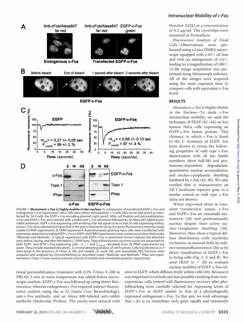
Monomeric c-Fos IsHighlyMobilein the Nucleus—To study c-Fosintranuclear mobility, we used thetechnique of FRAP (42–44) on livehuman HeLa cells expressing anEGFP-c-Fos fusion protein. Thischimera, in which c-Fos is fusedto the C terminus of EGFP, hasbeen shown to retain the follow-ing properties of wild type c-Fos:dimerization with all Jun familymembers, short half-life and pro-teasome-dependent degradation,quantitative nuclear accumulation,and nucleo-cytoplasmic shuttlinginhibited by c-Jun (16, 36). We alsoverified that it transactivates anAP-1-luciferase reporter gene to asimilar extend as wild type c-Fos(data not shown).When expressed alone in tran-
sient transfection assays, c-Fosand EGFP-c-Fos are essentially mo-nomeric (36) and predominantlynuclear despite their active nu-cleo-cytoplasmic shuttling (36).Moreover, they show a typical dif-fuse distribution with nucleolarexclusion, as assayed both by indi-rect immunofluorescence (36) or bydirect visualization of EGFP-c-Fosin living cells (Fig. 1, A and B). Weused FRAP (n � 20) to evaluatenuclear mobility of EGFP-c-Fos rel-
ative to EGFP, which diffuses freely within cells (45). Because itwas important to exclude any bias possibly resulting from over-expression, cells treated with fluorescence recovery after pho-tobleaching were carefully selected for expressing levels ofEGFP-c-Fos or EGFP similar to that of a physiologicallyexpressed endogenous c-Fos. To this aim, we took advantagethat c-fos is an immediate early gene rapidly and transiently
FIGURE 1. Monomeric c-Fos is highly mobile in the nucleus. A, comparison of transfected EGFP-c-Fos andendogenous c-Fos expressions. HeLa cells were either stimulated for 1 h with 20% serum (left panel) or trans-fected for 16 h with the EGFP-c-Fos-encoding plasmid (right panel). After cell fixation and permeabilization,c-Fos and EGFP-c-Fos were stained with a rabbit anti-c-Fos antiserum followed by an Alexa 647-labeled anti-rabbit antiserum. EGFP-c-Fos-expressing cells emitting a far red signal close to that of cells expressing endog-enous c-Fos were selected and observed in the green channel to set up the green fluorescence intensity rangeusable in FRAP experiments. B, FRAP experiment. Asynchronously growing HeLa cells were transfected withexpression plasmids encoding EGFP-c-Fos or EGFP, and FRAP experiments were carried out as described under“Materials and Methods.” A typical experiment with EGFP-c-Fos is presented. Arrows indicate the bleachedarea, before, during, and after the bleach. C, FRAP data. Typical fluorescence recovery curves are presented forboth EGFP- and EFGP-c-Fos-expressing cells. �t1⁄2� and Fmob30 calculated from 20 FRAP experiments aregiven. They include standard deviations. D, immunoblotting analysis of cell fractions. Cells transfected as in Awere lysed in the presence of Triton X-100, and soluble (S), wash (W), and nonsoluble (NS) fractions wereprepared and analyzed by immunoblotting as described under “Materials and Methods.” Phax and topoi-somerase I (Topo 1) were used as internal controls of soluble and nonsoluble proteins, respectively.
Intranuclear Mobility of c-Fos
FEBRUARY 26, 2010 • VOLUME 285 • NUMBER 9 JOURNAL OF BIOLOGICAL CHEMISTRY 6555
by guest on May 6, 2018
http://ww
w.jbc.org/
Dow
nloaded from

induced upon growth factor stimulation (8, 9) and comparedexogenous EGFP-c-Fos and EGFP abundance in transfectedHeLa cells with that of endogenous c-Fos in HeLa cells stimu-lated for 1 h by serum using standardized fluorescence acquisi-tion procedures, as detailed under “Materials and Methods”and shown Fig. 1A. Typical FRAP experiments are presented inFig. 1B. Mean half-time fluorescence recovery (�t1⁄2�), as wellas the mobile fractions 30 s after the bleach (Fmob30), were cal-culated for EGFP and EGFP-c-Fos using a two-exponential fit,as described under “Materials andMethods.” Fmob30 were 99 2 and 97 4% for EGFP and EGFP-c-Fos, respectively, indicat-ing that themolecules are highly mobile in this time frame (Fig.1C). The �t1⁄2� were 0.27 0.05 s for EGFP and 0.98 0.12 sfor EGFP-c-Fos. In fact, EFGP was so mobile that its fluores-cence recovered partially before the first post-bleach experi-mental measurement. This gave the impression of a less effi-cient photobleaching and led to an overestimation of the�t1⁄2�(compare EGFPminimal fluorescence value to that of EGFP-c-Fos in Fig. 1C). This indicated that EGFP-c-Fos, despite its highmobility, moves at least 4-fold more slowly than EGFP in thenucleus. If the mobility of the proteins is due to Brownian dif-fusion, this difference in mean half-time fluorescence recoverycannot be due to the increased mass of the chimeric protein ascompared with EGFP because the diffusion constant varies lit-tle with mass (Da1/M3, also see Ref. 45). Instead, the reducedmobility of EGFP-c-Fos would reflect weak binding to nuclearstructures.To complement these FRAP studies, we performed fraction-
ationexperiments on transfectedHeLacells, comparing thedistri-bution of EGFP-c-Fos with that of c-Fos in Triton X-100-solubleand -insoluble fractions.MonomericEGFP-c-Fos andmonomericc-Fos were principally found in the soluble fraction (S) togetherwith the nucleoplasmic protein Phax (46) taken as a control (Fig.1D; also see Fig. 2, D and E). Sometimes, a minor proportion ofmonomeric EGFP-c-Fos or c-Fos remained in the insoluble frac-tion (NS), which was monitored using chromatin-bound topoi-somerase I (47) (Fig. 1D; also see Fig. 2, D and E). Thus, the highmobility ofmonomeric c-Fos in thenucleus correlateswith its lackof strong interaction with intranuclear structures.c-Jun Reduces c-Fos Intranuclear Mobility—We next tested
whether dimerization with c-Jun altered c-Fos intranuclearmobility. Fos and Jun family members undergo dimerization/dedimerization cycles in vivo. Nevertheless, they interact for atleast several minutes (48). Thus, the mobility of Fos�Jun dimerscan be analyzed in short (30 s) FRAP experiments. Moreover,the formation of c-Fos�c-Jun heterodimers is strongly favoredover that of c-Jun�c-Jun homodimers (6, 49), meaning that aslight excess of c-Junwill ensure quantitative c-Fos recruitmentin heterodimers. Because c-Fos and c-Jun show half-lives of1h (48), this is easily accomplished by transfecting an excess ofc-Jun expression vector over that for c-Fos, provided that plas-mid backbones are identical.Strikingly, co-expression of c-Jun led to a dramatic intranu-
clear redistribution of EGFP-c-Fos, which became less homog-enous showing irregular areas of accumulation (Fig. 2A). WethenperformedFRAPexperiments onHeLa cells, where EGFP-c-Fos was expressed together with varying amounts of c-Jun.Interestingly, the mobile fraction of EGFP-c-Fos progressively
diminished as the amount of c-Jun increased. At 2- and 5-foldexcesses of c-Jun expression plasmid, the Fmob30 of EGFP-c-Foswas 40% that of monomeric c-Fos (Fig. 2B). Moreover, therecovery of fluorescence was still not complete 10 min afterbleaching (not shown). This effect was specific to the c-Fos�c-Jun interaction because co-transfection of c-Jun and EGFP didnot alter the mobility of EGFP in FRAP assays (Fig. 2C). Thesedata suggested that dimerization with c-Jun causes c-Fos toassociate with nuclear components, which was a notion furthersupported by cell fractionation experiments, because EGFP-c-Fos was principally recovered in the Triton X-100-insolublefraction (Fig. 2D). Importantly, a similar distribution was foundfor wild type c-Fos in the presence of c-Jun in parallel experi-ments (Fig. 2E), ruling out an artifact caused by the fusion toEGFP. Because a 2-fold excess of c-Jun expression plasmid overthat for c-Fos showed sufficient to obtainmaximal reduction inc-Fosmobility (Fig. 2B), this plasmid ratiowas used in the abovefractionation experiments aswell as in our further experiments.We noted that only 60% of c-Fos remained immobile during
the time course of the experiments, even though co-immuno-
FIGURE 2. Reduced intranuclear mobility of c-Fos in the presence of c-Jun.A, alteration of intranuclear distribution of EGFP-c-Fos in the presence ofc-Jun. HeLa cells were transfected with EGFP-c-Fos in the absence or in thepresence of a 2-fold excess of c-Jun expression plasmid and analyzed by con-focal microscopy on living cells. B, Fmob30 of EGFP-c-Fos in the presence ofvarying amounts of c-Jun. HeLa cells were co-transfected with c-Jun andEGFP-c-Fos expression plasmids in the indicated ratios. FRAP experimentswere performed as in Fig. 1A. Fmob30 values were calculated 30 s after the endof the bleach from 20 experiments for each condition and presented as his-tograms. The error bars indicate standard deviations. C, FRAP experiment.Asynchronously growing HeLa cells were transfected with expression plas-mids encoding EGFP with or without a 2-fold excess of c-Jun vector. Eachcurve corresponds to the averages of 20 FRAP experiments. D and E, cellfractionation experiments. Fractionation experiments were carried out as inFig. 1C using HeLa cells co-transfected with expression plasmids for (i) EGFP-c-Fos in the absence or in the presence of a 2-fold excess of c-Jun expressionplasmid (D) or (ii) c-Fos in the absence or in the presence of a 2-fold excess ofc-Jun expression plasmid (E). S, soluble; W, wash; NS, nonsoluble.
Intranuclear Mobility of c-Fos
6556 JOURNAL OF BIOLOGICAL CHEMISTRY VOLUME 285 • NUMBER 9 • FEBRUARY 26, 2010
by guest on May 6, 2018
http://ww
w.jbc.org/
Dow
nloaded from
precipitation experiments indicated quantitative association ofc-Fos with c-Jun. Thus, 40% of dimers appeared mobile for the30-s post-bleach. This likely reflected saturation of the nuclearbinding sites for c-Fos�c-Jun dimers in the window of time ana-lyzed possibly in association with slow exchange rate betweenbound and unbound dimers. Consistent with the idea of mobil-ity for a fraction of dimers, some c-Fos and EGFP-c-Fos (albeitless than 40%) were found in the soluble fraction in our cellfractionation experiments (Fig. 2, D and E).Reduced c-Fos Intranuclear Mobility upon Heterodimeriza-
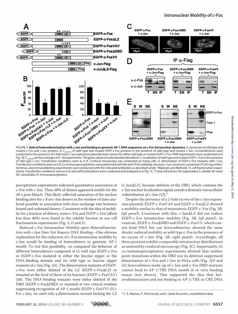
tion with c-Jun Does Not Require DNA Binding—One obviousexplanation for the reduction of c-Fos intranuclear mobility byc-Jun would be binding of heterodimers to genomic AP-1motifs. To test this possibility, we compared the behavior ofdifferent heterodimers composed of (i) wild type EGFP-c-Fosor EGFP-c-Fos mutated in either the leucine zipper or theDNA-binding domain and (ii) wild type or leucine zippermutants of c-Jun (Fig. 3A). The dimerizationmutants of EGFP-c-Fos were either deleted of the LZ (EGFP-c-Fos�LZ) ormutated at the level of three of its leucines (EGFP-c-FosVAV)(50). The DNA-binding mutants were either deleted of theDBD (EGFP-c-Fos�DBD) or mutated at two critical residuessuppressing recognition of AP-1 motifs (EGFP-c-FosVV) (51).For c-Jun, we used only a dimerization mutant lacking the LZ
(c-Jun�LZ), because deletion of the DBD, which contains thec-Junnuclear localization signal, entails a dramatic intracellularredistribution of c-Jun (52).6
Despite the presence of a 2-fold excess of the c-Jun expres-sion plasmid, EGFP-c-FosVAV and EGFP-c-Fos�LZ showeda mobility similar to that of monomeric EGFP-c-Fos (Fig. 3B,left panel). Consistent with this, c-Jun�LZ did not reduceEGFP-c-Fos intranuclear mobility (Fig. 3B, left panel). Incontrast, EGFP-c-Fos�DBD and EGFP-c-FosVV, which can-not bind DNA but can heterodimerize, showed the samedrastic reduced mobility as wild type c-Fos in the presence ofan excess of c-Jun (Fig. 3B, right panel). Accordingly, allthree proteins exhibit comparable intranuclear distributionsas assessed by confocal microscopy (Fig. 3C). Importantly, (i)co-immunoprecipitation experiments showed that neitherpoint mutations within the DBD nor its deletion suppresseddimerization of c-Fos and c-Jun in HeLa cells (Fig. 3D) and(ii) heterodimers made up of c-Jun and c-Fos DBD mutantscannot bind to AP-1/TRE DNA motifs in in vitro bindingassays (not shown). This supported the idea that het-erodimerization and not binding to AP-1/TRE or CRE DNA
6 C. E. Malnou, F. Piechaczyk, and I. Jariel-Encontre, unpublished data.
FIGURE 3. Role of heterodimerization with c-Jun and binding to genomic AP-1 DNA sequences on c-Fos intranuclear dynamics. A, structures of wild type andmutant c-Fos and c-Jun proteins. B, Fmob30 of wild type and mutant EGFP-c-Fos proteins in the presence of wild type and mutant c-Jun. Co-transfections wereconducted in the presence of 2-fold more c-Jun expression plasmids than vectors for either wild type or mutant EGFP-c-Fos. FRAP experiments were conducted as inFig. 1B. Fmob30 are the averages of 6–20 experiments. The given values include standard deviations. C, localization of wild type and mutant EGFP-c-Fos in the presenceof wild type c-Jun. Transfection conditions were as in B. Confocal microscopy was conducted on living cells. D, dimerization of EGFP-c-Fos mutants with c-Jun.Transfection conditions were as in B. Co-immunoprecipitations were performed with the anti-FLAG antibody, because c-Jun constructs contained a FLAG tag at the Cterminus, and immunoblotting experiments were conducted with the indicated antibodies as described under “Materials and Methods.” E, cell fractionation experi-ments. Transfection conditions were as in B, and cell fractionations were conducted and analyzed as in Fig. 1C. T, total cell extract; SN, supernatant; S, soluble; W, wash;NS, nonsoluble; IP, immunoprecipitation.
Intranuclear Mobility of c-Fos
FEBRUARY 26, 2010 • VOLUME 285 • NUMBER 9 JOURNAL OF BIOLOGICAL CHEMISTRY 6557
by guest on May 6, 2018
http://ww
w.jbc.org/
Dow
nloaded from

sequences is the primary event responsible for this c-Jun-mediated reduction of c-Fos mobility. Furthermore, cellfractionation experiments showed that EGFP-c-Fos�DBDand EGFP-c-FosVV are redistributed in the insolublenuclear fraction in the presence of c-Jun (Fig. 3E).JunB Does Not Affect c-Fos IntranuclearMobility—It seemed
important to test whether another dimerization partner couldhave the same effect on c-Fos intranuclearmobility. JunB is alsoa highly documented c-Fos partner (6, 15, 48, 53). Therefore, weanalyzed its effects on EGFP-c-Fos intracellular localizationand mobility as we did with c-Jun. Under conditions of quanti-tative association with JunB, as assayed by co-immunoprecipi-tation (Fig. 4A), EGFP-c-Fos was redistributed within thenucleus, as seen upon dimerization with c-Jun, albeit with a
reproducible significantly different distribution (Fig. 4B; com-pare with Fig. 2A). FRAP experiments showed both an initialrecovery of fluorescence (Fig. 4C) and an Fmob30 (Fig. 4D) sim-ilar for JunB�EGFP-c-Fos andmonomeric EGFP-c-Fos. Despitetheir high mobility, JunB�EGFP-c-Fos heterodimers associatedprincipally with the Triton X-100-insoluble nuclear fraction(Fig. 4E), unlike monomeric c-Fos (Fig. 1D). Thus, our dataindicate that c-Jun and JunB alter c-Fos intranuclear behaviorwith, however, different outcomes.Fra-1 Intranuclear Distribution andMobility Are Affected by
c-Jun and JunB in a Manner Similar to c-Fos—We then testedwhether c-Jun and JunB similarly influenced the behavior of theFos family protein Fra-1, because endogenous c-Jun and JunB
FIGURE 5. Alteration of Fra-1 intranuclear distribution and mobility byc-Jun and JunB. A, dimerization of EGFP-Fra-1 with c-Jun and JunB. Het-erodimerization assays were conducted as in Fig. 3D using HeLa cells trans-fected with plasmids for either c-Jun-FLAG � EGFP-Fra-1 or JunB-FLAG �EGFP-Fra-1 in a ratio of 2. B, intracellular localization of EGFP-Fra-1 in thepresence of c-Jun and JunB in a 2-fold excess. Intracellular localization wasassessed on living cells by confocal microscopy analysis of HeLa cells trans-fected as in A. C, FRAP experiments. FRAP experiments were conducted inHeLa cells transfected with expression plasmids for either EGFP-Fra1 or EGFP-Fra1 and a 2-fold excess of JunB- or c-Jun plasmid. The curves correspond tothe averages of 10 –20 FRAP experiments. D, Fmob30 of EGFP-Fra-1 in the pres-ence of c-Jun and JunB. Transfection conditions were as in A. Fmob30 werecalculated from more than 10 FRAP experiments. E, fractionation experi-ments. Fractionation and analyses of HeLa cells transfected as in A were con-ducted as in Fig. 1C. T, total cell extract; SN, supernatant; IP, immunoprecipi-tation; S, soluble; W, wash; NS, nonsoluble.
FIGURE 4. Effect of JunB on c-Fos intranuclear distribution and mobility.A, heterodimerization of JunB-FLAG with EGFP-c-Fos. The expression plasmidfor EGFP-c-Fos was transfected in HeLa cells in the presence of a 2-fold excessof JunB-FLAG construct. Co-immunoprecipitations were conducted with theanti-FLAG antibody as in Fig. 3D. The antibodies used in immunoblottinganalyses are indicated. B, intranuclear distribution of EGFP-c-Fos in the pres-ence of JunB. EGFP-c-Fos localization in the presence of JunB-FLAG wasassessed by confocal microscopy on living cells. The JunB versus EGFP-c-Fosexpression plasmid ratio was of 2. C, FRAP experiments. FRAP experimentswere conducted in HeLa cells transfected with expression plasmids for eitherEGFP-c-Fos or EGFP-c-Fos � JunB. In the latter case, the plasmid ratio was 2.The curves correspond to the averages of 20 FRAP experiments in each case.D, mobility of EGFP-c-Fos in the presence of different amounts of JunB. HeLacells were transfected in the presence of different ratios of JunB versus EGFP-c-Fos plasmids as indicated. Fmob30 were calculated from 15–20 FRAP exper-iments in each case. E, cell fractionation experiments. Cell fractionation exper-iments of cells transfected with plasmids encoding JunB and EGFP-c-Fos in aratio of 2 were conducted and analyzed as in Fig. 1D. T, total cell extract; SN,supernatant; IP, immunoprecipitation; S, soluble; W, wash; NS, nonsoluble.
Intranuclear Mobility of c-Fos
6558 JOURNAL OF BIOLOGICAL CHEMISTRY VOLUME 285 • NUMBER 9 • FEBRUARY 26, 2010
by guest on May 6, 2018
http://ww
w.jbc.org/
Dow
nloaded from
are known to interact with endogenous Fra-1 in a variety ofsituations (6, 48, 53). EGFP-Fra-1 quantitatively dimerizes withectopic c-Jun and JunB in HeLa cells (Fig. 5A). Interestingly,monomeric EGFP-Fra-1 was weakly cytoplasmic, whichwas nolonger seen in the presence of co-transfected c-Jun and JunB(Fig. 5B). Like for c-Fos, FRAP analyses indicated that Fra-1intranuclear mobility was dramatically reduced when co-ex-pressed with c-Jun but was unaffected by co-expression of JunB(Fig. 5C). Themobile fraction of Fra-1 decreased to40% in thepresence of c-Jun and remained unchanged in the presence ofJunB (Fig. 5D). EGFP-Fra-1 associated with the Triton X-100-insoluble nuclear fraction when co-expressed with either Jundimerization partner (Fig. 5E). Thus, c-Jun and JunB affectFra-1 intranuclear distribution and mobility in a manner simi-lar to c-Fos.Differential Association of c-Fos�c-Jun and c-Fos�JunB Het-
erodimers with the Nuclear Matrix—In a final step, we testedwhether c-Fos�c-Jun and c-Fos�JunB heterodimers could asso-ciate differentially with the chromatin and/or the nuclearmatrix.First, we conducted classical biochemical fractionation ex-
periments (see “Materials andMethods”) followed by immuno-blotting assays. In these experiments: (i) S1 corresponded toboth cytoplasmic and nuclear proteins solubilized upon celllysis in the presence of 0.5% Triton X-100, (ii) S2 correspondedto the chromatin proteins released upon subsequent extensiveDNA hydrolysis by MNase, and (iii) S3 corresponded to theremnant of chromatin proteins andDNAsolubilized after addi-tional high salt (2 M NaCl) treatment, and (P) corresponded tothe nuclear-matrix-containing fraction. Fractionation patternsof (i) the nucleosoluble Phax protein, (ii) the weakly chromatin-associated HCF-1 protein (54), (iii) tightly chromatin-associ-ated histoneH3, and (iv) the nuclear lamina-constituting laminA/C proteins confirmed the efficiency of the procedure (Fig. 6).In contrast to EGFP-c-Fos expressed alone, which localizedpredominantly in the S1 fraction, EGFP-c-Fos�c-Jun dimerswere essentially found associated with the nuclear matrix.However, a veryminor fraction of EGFP-c-Fos was found in theP fraction. This may be due to dimerization of the protein with
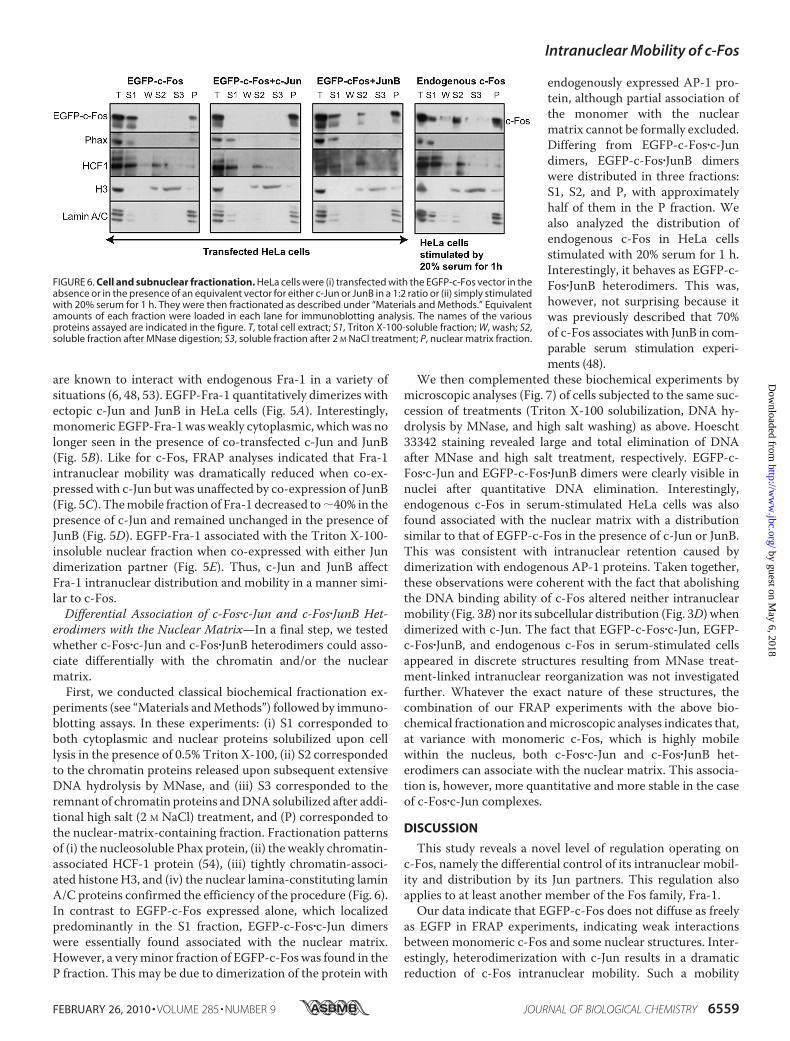
endogenously expressed AP-1 pro-tein, although partial association ofthe monomer with the nuclearmatrix cannot be formally excluded.Differing from EGFP-c-Fos�c-Jundimers, EGFP-c-Fos�JunB dimerswere distributed in three fractions:S1, S2, and P, with approximatelyhalf of them in the P fraction. Wealso analyzed the distribution ofendogenous c-Fos in HeLa cellsstimulated with 20% serum for 1 h.Interestingly, it behaves as EGFP-c-Fos�JunB heterodimers. This was,however, not surprising because itwas previously described that 70%of c-Fos associates with JunB in com-parable serum stimulation experi-ments (48).
We then complemented these biochemical experiments bymicroscopic analyses (Fig. 7) of cells subjected to the same suc-cession of treatments (Triton X-100 solubilization, DNA hy-drolysis by MNase, and high salt washing) as above. Hoescht33342 staining revealed large and total elimination of DNAafter MNase and high salt treatment, respectively. EGFP-c-Fos�c-Jun and EGFP-c-Fos�JunB dimers were clearly visible innuclei after quantitative DNA elimination. Interestingly,endogenous c-Fos in serum-stimulated HeLa cells was alsofound associated with the nuclear matrix with a distributionsimilar to that of EGFP-c-Fos in the presence of c-Jun or JunB.This was consistent with intranuclear retention caused bydimerization with endogenous AP-1 proteins. Taken together,these observations were coherent with the fact that abolishingthe DNA binding ability of c-Fos altered neither intranuclearmobility (Fig. 3B) nor its subcellular distribution (Fig. 3D) whendimerized with c-Jun. The fact that EGFP-c-Fos�c-Jun, EGFP-c-Fos�JunB, and endogenous c-Fos in serum-stimulated cellsappeared in discrete structures resulting from MNase treat-ment-linked intranuclear reorganization was not investigatedfurther. Whatever the exact nature of these structures, thecombination of our FRAP experiments with the above bio-chemical fractionation andmicroscopic analyses indicates that,at variance with monomeric c-Fos, which is highly mobilewithin the nucleus, both c-Fos�c-Jun and c-Fos�JunB het-erodimers can associate with the nuclear matrix. This associa-tion is, however, more quantitative and more stable in the caseof c-Fos�c-Jun complexes.
DISCUSSION
This study reveals a novel level of regulation operating onc-Fos, namely the differential control of its intranuclear mobil-ity and distribution by its Jun partners. This regulation alsoapplies to at least another member of the Fos family, Fra-1.Our data indicate that EGFP-c-Fos does not diffuse as freely
as EGFP in FRAP experiments, indicating weak interactionsbetween monomeric c-Fos and some nuclear structures. Inter-estingly, heterodimerization with c-Jun results in a dramaticreduction of c-Fos intranuclear mobility. Such a mobility
FIGURE 6. Cell and subnuclear fractionation. HeLa cells were (i) transfected with the EGFP-c-Fos vector in theabsence or in the presence of an equivalent vector for either c-Jun or JunB in a 1:2 ratio or (ii) simply stimulatedwith 20% serum for 1 h. They were then fractionated as described under “Materials and Methods.” Equivalentamounts of each fraction were loaded in each lane for immunoblotting analysis. The names of the variousproteins assayed are indicated in the figure. T, total cell extract; S1, Triton X-100-soluble fraction; W, wash; S2,soluble fraction after MNase digestion; S3, soluble fraction after 2 M NaCl treatment; P, nuclear matrix fraction.
Intranuclear Mobility of c-Fos
FEBRUARY 26, 2010 • VOLUME 285 • NUMBER 9 JOURNAL OF BIOLOGICAL CHEMISTRY 6559
by guest on May 6, 2018
http://ww
w.jbc.org/
Dow
nloaded from

change is not the result of dimerization per se but is selectivebecause JunB does not detectably alter c-Fos intranucleardynamics (as visualized by FRAP) under conditions wherec-Fos is quantitatively associated with JunB (as shown by co-immunoprecipitation). Although the FRAP data presented inFigs. 2–4 were obtained in human epithelial HeLa cells, similardifferential effects of c-Jun and JunB were observed in Balb/C3T3 mouse embryo fibroblasts (supplemental Fig. S1), indicat-ing that immobilization of c-Fos by c-Jun is neither cell- norspecies-specific.
It is most often considered that reduced intranuclear mobil-ity of transcription factors is largely determined by binding totarget DNA sequences because disabled DNA-binding domainmutants of proteins such as NF-�B p65 (55), Acep1 (56), inter-feron regulatory factor 8 (57), the androgen receptor (58, 59), orthe glucocorticoid receptor (60) are more mobile than theirwild type counterparts. However, immobilization of c-Fos byc-Jun is not primarilymediated by binding toAP-1/TREorCREtarget sequences (also see below) because EGFP-c-Fos andEGFP-c-Fos�DBD behave similarly in FRAP experiments,when quantitatively heterodimerized with c-Jun. Strengthen-ing the idea of a DNA-independent mechanism responsible forc-Fosmobility reduction, cell fractionation experiments (Figs. 6and 7) showed that c-Fos�c-Jun dimers predominantly associatewith the nuclear matrix, which is, by definition, devoid of chro-matin and DNA (61, 62).There may be an apparent paradox between the well known
strong transcriptional activity of c-Fos�c-Jun dimers, especiallyunder the transfection conditions used in thiswork, and the factthat a large majority of these complexes were found in thenuclear matrix fraction during the course of this work. Theobservation that 40% of c-Fos�c-Jun complexes are mobile (asassayed 30 s post-bleach), however, helps resolve it with a likelyscenario as follows. transcriptional activity at a given timewould be due to aminor fraction of c-Fos�c-Jundimers, whereasa large majority of these complexes would be latent and storedin intranuclear non-chromatin domains. Storage would, how-ever, be relatively limited in time, as indicated by FRAP exper-iments, at least for a fraction of them. Important challenges willnow be to elucidate whether these nuclear matrix storage sitesare located near transcription factories or not andwhy and howlatent c-Fos�c-Jun dimers are released from the nuclear matrixand addressed to the transcription sites. Along this line, it isof note that 50% of endogenous c-Fos is found associatedwith the nuclear matrix at the peak of expression in serum-stimulated cells, whereas the rest equally distributesbetween soluble and chromatin fractions (Fig. 6). Althoughthe respective roles of the latter two populations of c-Fos mol-ecules still require clarification, this observation is consistentwith the idea of storage sites for latent AP-1 complexes underphysiological conditions.Very interestingly, Andres and co-workers (63, 64) have
recently reported that c-Fos can associate with the nuclearmatrix component lamin A/C. This was proposed to constitutean AP-1 suppression mechanism. Although the authors did notaddress c-Fos mobility directly, their data are consistent with ourcell fractionation experiments showing interaction with thenuclear matrix. From in vitro pull-down experiments, theseauthorshypothesized thatc-Foswould interactwith laminA/Cviaits leucine zipper at the detriment of dimerization with its Junpartners (63). Although not questioning the principle of AP-1inactivation by lamin A/C, our data, however, argue against LZ-mediated sequestration of monomeric c-Fos, because we clearlyshow that (i)monomeric c-Fos is highlymobile and quantitativelyfound in the soluble protein fraction and (ii) only dimeric c-Fos isfound associated with the nuclear matrix.The behavioral differences between c-Fos�c-Jun and c-
Fos�JunB dimers are interesting to consider. First, in addition to
FIGURE 7. Microscopic analysis of c-Fos and EGFP-c-Fos intranuclearlocalization in fractionated cells. These experiments were carried out inparallel with those presented in Fig. 6 with HeLa cells seeded and grown oncoverslips. Endogenous c-Fos was detected using sequentially the sc52 rabbitanti-c-Fos antibody and an Alexa 488-labeled anti-rabbit secondary antibody.The nuclei were stained with Hoechst 33342. All of the pictures in the EGFPchannel were acquired using the same time exposure.
Intranuclear Mobility of c-Fos
6560 JOURNAL OF BIOLOGICAL CHEMISTRY VOLUME 285 • NUMBER 9 • FEBRUARY 26, 2010
by guest on May 6, 2018
http://ww
w.jbc.org/
Dow
nloaded from
partly different intranuclear distribution, as visualized by directfluorescence analysis of transfected cells (Figs. 2A and 4B),c-Fos�JunBdimers showedmore heterogeneous in our fraction-ation experiments than c-Fos�c-Jun ones. Whereas c-Fos�c-Jundimers were found quantitatively associated with the nuclearmatrix, c-Fos�JunBdimerswere detected not only in the nuclearmatrix fraction (50%) but also in the soluble and chromatinones (Figs. 6 and 7). If one takes into consideration that dimer-ization with JunB little affects c-Fos mobility, as assayed byFRAP (Fig. 4C), it ensues that association of c-Fos�JunB dimerswith the nuclear matrix must be muchmore dynamic than thatof c-Fos�c-Jun dimers. Thus, interaction with the nuclearmatrix may differentially control the abundances of the differ-ent AP-1 dimers available for gene regulation. It will next beinteresting to establish (i) whether this is due to differentialaffinity for the same nuclear matrix sites or to interaction withdifferent sites and (ii) whether the slowed down mobility ofc-Fos is contributed by both c-Fos and c-Jun or principally byc-Jun, which would, in the later case, serve as an anchor tonuclear structures. Unfortunately, we could not answer thisquestion in FRAP experiments, because EGFP-c-Jun chimerasbehave aberrantly because they do not show the same intracel-lular distribution as c-Jun.6c-Fos constitutes an heterogeneous collection of molecules
at any moment in any cell because of dimerization with multi-ple AP-1 proteins, interaction with multiple partners, diversepost-translational modifications, widespread distribution inthe nucleus, the multiple genes to which it can bind, etc. Animportant technical pointmust therefore be taken into accountwhen considering the FRAP experiments presented in thiswork. Those could only address the behavior of the bulk ofc-Fos and not those of the various (and possibly many) sub-populations of this protein, which may be different. In particu-lar, it is possible that the small fraction of transcriptionallyactive c-Fos�c-Jun dimersmay be immobilized on AP-1/TRE orCRE sequences for periods of time longer than those of associ-ation with the nuclear matrix for efficient stimulation of targetgene transcription. In support of such a possibility, severalother transcription factors were previously shown to displaylow mobility in their presumably active states. This was, forexample, the case of nuclear hormone receptors, such as andro-gen receptor (58, 65), glucocorticoid receptor (60), estrogenreceptor � and � (66, 67), liver X receptors � and � (68), perox-isome proliferator-activated receptor y (69), and progesteronereceptor (70) after ligand binding and/or association with tran-scriptional co-activators and/or chromatin remodeling com-plexes (60, 67, 68, 70). In the same vein, intranuclear diffusion ofCREB (71) and liver X receptor � (68) depends on the integrityof specific trans-activation domains, the intranuclear mobilityof interferon regulatory factor 8 is decreased upon associationwith PU.1 and interferon regulatory factor 1 (57), and that of�-catenin is reduced upon association with its transcriptionalpartner TCF4 (72).Finally, by demonstrating stronger association of c-Fos�c-Jun
dimers than c-Fos�JunB dimers to intranuclear structures, weprovide here a straightforward explanation of our previousobservation of more efficient inhibition of c-Fos nuclear exportby c-Jun than by JunB (36), even though an additional reduced
c-Jun-dependent recognition of c-Fos by the nuclear exportmachinery cannot be formally excluded. This resembles thesituation of �-catenin, where interaction with TCF4 leads toreduced nucleo-cytoplasmic shuttling primarily reflecting adecrease in intranuclearmobility and not that of nuclear exportefficiency per se (72). Further work will determine the role ofassociation with intranuclear components in the transcrip-tional activity of c-Fos�c-Jun heterodimers.
Acknowledgments—We thank Dr. R. Hipskind for critical reading ofthe manuscript. All of the imaging analyses were performed on theMontpellier RIO Imaging (Infrastructures Biologie Sante et Agrono-mie) imaging platform.
REFERENCES1. Eferl, R., and Wagner, E. F. (2003) Nat. Rev. Cancer 3, 859–8682. Hess, J., Angel, P., and Schorpp-Kistner, M. (2004) J. Cell Sci. 117,
5965–59733. Jochum, W., Passegue, E., and Wagner, E. F. (2001) Oncogene 20,
2401–24124. Shaulian, E., and Karin, M. (2002) Nat. Cell Biol. 4, E131–E1365. Zenz, R., Eferl, R., Scheinecker, C., Redlich, K., Smolen, J., Schonthaler,
H. B., Kenner, L., Tschachler, E., and Wagner, E. F. (2008) Arthritis Res.Ther. 10, 201–211
6. Chinenov, Y., and Kerppola, T. K. (2001) Oncogene 20, 2438–24527. Vinson, C., Acharya, A., and Taparowsky, E. J. (2006) Biochim. Biophys.
Acta 1759, 4–128. Jariel-Encontre, I., and Piechaczyk, M. (2008) Targeted Protein Database
[22540], 10.2970/tpdb. 2009.02189. Piechaczyk, M., and Blanchard, J. M. (1994)Crit. Rev. Oncol. Hematol. 17,
93–13110. Basbous, J., Jariel-Encontre, I., Gomard, T., Bossis, G., and Piechaczyk, M.
(2008) Biochimie 90, 296–30511. Gomard, T., Jariel-Encontre, I., Basbous, J., Bossis, G.,Mocquet-Torcy, G.,
and Piechaczyk, M. (2008) Biochem. Soc. Trans. 36, 858–86312. Bossis, G., Ferrara, P., Acquaviva, C., Jariel-Encontre, I., and Piechaczyk,
M. (2003)Mol. Cell Biol. 23, 7425–743613. Chalmers, C. J., Gilley, R., March, H. N., Balmanno, K., and Cook, S. J.
(2007) Cell Signal. 19, 695–70414. Chen, R. H., Abate, C., and Blenis, J. (1993) Proc. Natl. Acad. Sci. U.S.A. 90,
10952–1095615. Deng, T., and Karin, M. (1994) Nature 371, 171–17516. Ferrara, P., Andermarcher, E., Bossis, G., Acquaviva, C., Brockly, F., Jariel-
Encontre, I., and Piechaczyk, M. (2003) Oncogene 22, 1461–147417. Higashi, N., Kunimoto, H., Kaneko, S., Sasaki, T., Ishii,M., Kojima, H., and
Nakajima, K. (2004) Genes Cells 9, 233–24218. Mackeigan, J. P., Murphy, L. O., Dimitri, C. A., and Blenis, J. (2005) Mol.
Cell Biol. 25, 4676–468219. Monje, P., Hernandez-Losa, J., Lyons, R. J., Castellone,M.D., andGutkind,
J. S. (2005) J. Biol. Chem. 280, 35081–3508420. Monje, P., Marinissen, M. J., and Gutkind, J. S. (2003) Mol. Cell Biol. 23,
7030–704321. Murphy, L. O., MacKeigan, J. P., and Blenis, J. (2004) Mol. Cell Biol. 24,
144–15322. Murphy, L. O., Smith, S., Chen, R. H., Fingar, D. C., and Blenis, J. (2002)
Nat. Cell Biol. 4, 556–56423. Okazaki, K., and Sagata, N. (1995) EMBO J. 14, 5048–505924. Sasaki, T., Kojima, H., Kishimoto, R., Ikeda, A., Kunimoto, H., and Naka-
jima, K. (2006)Mol. Cell 24, 63–7525. Tanos, T., Marinissen,M. J., Leskow, F. C., Hochbaum, D.,Martinetto, H.,
Gutkind, J. S., and Coso, O. A. (2005) J. Biol. Chem. 280, 18842–1885226. Gilley, R., March, H. N., and Cook, S. J. (2009) Cell Signal. 21, 969–97727. Koga, K., Takaesu, G., Yoshida, R., Nakaya, M., Kobayashi, T., Kinjyo, I.,
and Yoshimura, A. (2009) Immunity 30, 372–38328. Bossis, G., Malnou, C. E., Farras, R., Andermarcher, E., Hipskind, R., Ro-
Intranuclear Mobility of c-Fos
FEBRUARY 26, 2010 • VOLUME 285 • NUMBER 9 JOURNAL OF BIOLOGICAL CHEMISTRY 6561
by guest on May 6, 2018
http://ww
w.jbc.org/
Dow
nloaded from

driguez,M., Schmidt, D.,Muller, S., Jariel-Encontre, I., and Piechaczyk,M.(2005)Mol. Cell Biol. 25, 6964–6979
29. Tempe, D., Piechaczyk, M., and Bossis, G. (2008) Biochem. Soc. Trans 36,874–878
30. Roux, P., Blanchard, J. M., Fernandez, A., Lamb, N., Jeanteur, P., and Piec-haczyk, M. (1990) Cell 63, 341–351
31. Vriz, S., Lemaitre, J. M., Leibovici, M., Thierry, N., andMechali, M. (1992)Mol. Cell Biol. 12, 3548–3555
32. Bussolino, D. F., Guido, M. E., Gil, G. A., Borioli, G. A., Renner, M. L.,Grabois, V. R., Conde, C. B., and Caputto, B. L. (2001) FASEB J. 15,556–558
33. Gil, G. A., Bussolino, D. F., Portal, M. M., Pecchio, A. A., Renner, M. L.,Borioli, G. A., Guido, M. E., and Caputto, B. L. (2004) Mol. Biol. Cell 15,1881–1894
34. Roux, P., Carillo, S., Blanchard, J. M., Jeanteur, P., and Piechaczyk, M.(1994) The c-fos and c-jun Families of Transcription Factors, pp. 87–95,CRC Press Inc.
35. Arnold, M., Nath, A., Wohlwend, D., and Kehlenbach, R. H. (2006) J. Biol.Chem. 281, 5492–5499
36. Malnou, C. E., Salem, T., Brockly, F., Wodrich, H., Piechaczyk, M., andJariel-Encontre, I. (2007) J. Biol. Chem. 282, 31046–31059
37. Tratner, I., and Verma, I. M. (1991) Oncogene 6, 2049–205338. Mason, J. M., Schmitz, M. A., Muller, K. M., and Arndt, K. M. (2006) Proc.
Natl. Acad. Sci. U.S.A. 103, 8989–899439. Wysocka, J., Reilly, P. T., and Herr, W. (2001) Mol. Cell Biol. 21,
3820–382940. Burch, P.M., Yuan, Z., Loonen, A., andHeintz, N. H. (2004)Mol. Cell Biol.
24, 4696–470941. Spector, D. L., Goldman, R. D., and Leinwand, L. A. (1998) Cells: A Labo-
ratory Manual, Vol. 1, pp. 44.2–44.6, Cold Spring Harbor Laboratory,Cold Spring Harbor, NY
42. Houtsmuller, A. B., and Vermeulen, W. (2001) Histochem. Cell Biol. 115,13–21
43. Reits, E. A., and Neefjes, J. J. (2001) Nat. Cell Biol. 3, E145–E14744. van Drogen, F., and Peter, M. (2004)Methods Mol. Biol. 284, 287–30645. Sprague, B. L., and McNally, J. G. (2005) Trends Cell Biol. 15, 84–9146. Ohno, M., Segref, A., Bachi, A., Wilm, M., and Mattaj, I. W. (2000) Cell
101, 187–19847. Christensen, M. O., Krokowski, R. M., Barthelmes, H. U., Hock, R., Boege,
F., and Mielke, C. (2004) J. Biol. Chem. 279, 21873–2188248. Kovary, K., and Bravo, R. (1991)Mol. Cell Biol. 11, 2451–245949. Vinson, C., Myakishev, M., Acharya, A., Mir, A. A., Moll, J. R., and Bonov-
ich, M. (2002)Mol. Cell Biol. 22, 6321–6335
50. Schuermann, M., Neuberg, M., Hunter, J. B., Jenuwein, T., Ryseck, R. P.,Bravo, R., and Muller, R. (1989) Cell 56, 507–516
51. Ransone, L. J., Visvader, J., Wamsley, P., and Verma, I. M. (1990) Proc.Natl. Acad. Sci. U.S.A. 87, 3806–3810
52. Waldmann, I., Walde, S., and Kehlenbach, R. H. (2007) J. Biol. Chem. 282,27685–27692
53. Kovary, K., and Bravo, R. (1992)Mol. Cell Biol. 12, 5015–502354. Julien, E., and Herr, W. (2004)Mol. Cell 14, 713–72555. Schaaf, M. J., Willetts, L., Hayes, B. P., Maschera, B., Stylianou, E., and
Farrow, S. N. (2006) J. Biol. Chem. 281, 22409–2242056. Karpova, T. S., Chen, T. Y., Sprague, B. L., andMcNally, J. G. (2004)EMBO
Rep. 5, 1064–107057. Laricchia-Robbio, L., Tamura, T., Karpova, T., Sprague, B. L., McNally,
J. G., and Ozato, K. (2005) Proc. Natl. Acad. Sci. U.S.A. 102, 14368–1437358. Farla, P., Hersmus, R., Geverts, B., Mari, P. O., Nigg, A. L., Dubbink, H. J.,
Trapman, J., and Houtsmuller, A. B. (2004) J. Struct. Biol. 147, 50–6159. Farla, P., Hersmus, R., Trapman, J., and Houtsmuller, A. B. (2005) J. Cell
Sci. 118, 4187–419860. Schaaf, M. J., and Cidlowski, J. A. (2003)Mol. Cell Biol. 23, 1922–193461. Albrethsen, J., Knol, J. C., and Jimenez, C. R. (2009) J. Proteomics 72,
71–8162. Lever, E., and Sheer, D. (2010) J. Pathol. 220, 114–12563. Gonzalez, J. M., Navarro-Puche, A., Casar, B., Crespo, P., and Andres, V.
(2008) J. Cell Biol. 183, 653–66664. Ivorra, C., Kubicek, M., Gonzalez, J. M., Sanz-Gonzalez, S. M., Alvarez-
Barrientos, A., O’Connor, J. E., Burke, B., andAndres, V. (2006)GenesDev.20, 307–320
65. Marcelli, M., Stenoien, D. L., Szafran, A. T., Simeoni, S., Agoulnik, I. U.,Weigel, N. L., Moran, T., Mikic, I., Price, J. H., and Mancini, M. A. (2006)J. Cell Biochem. 98, 770–788
66. Damdimopoulos, A. E., Spyrou, G., and Gustafsson, J. A. (2008) Endocri-nology 149, 339–345
67. Maruvada, P., Baumann, C. T., Hager, G. L., and Yen, P. M. (2003) J. Biol.Chem. 278, 12425–12432
68. Prufer, K., Hernandez, C., and Gilbreath, M. (2008) Exp. Cell Res. 314,2652–2660
69. Feige, J. N., Gelman, L., Tudor, C., Engelborghs, Y., Wahli, W., and Des-vergne, B. (2005) J. Biol. Chem. 280, 17880–17890
70. Rayasam, G. V., Elbi, C., Walker, D. A., Wolford, R., Fletcher, T. M., Ed-wards, D. P., and Hager, G. L. (2005)Mol. Cell Biol. 25, 2406–2418
71. Mayr, B. M., Guzman, E., and Montminy, M. (2005) J. Biol. Chem. 280,15103–15110
72. Krieghoff, E., Behrens, J., and Mayr, B. (2006) J. Cell Sci. 119, 1453–1463
Intranuclear Mobility of c-Fos
6562 JOURNAL OF BIOLOGICAL CHEMISTRY VOLUME 285 • NUMBER 9 • FEBRUARY 26, 2010
by guest on May 6, 2018
http://ww
w.jbc.org/
Dow
nloaded from

Piechaczyk and Isabelle Jariel-EncontreCécile E. Malnou, Frédérique Brockly, Cyril Favard, Gabriel Moquet-Torcy, Marc
Dynamics and DistributionHeterodimerization with Different Jun Proteins Controls c-Fos Intranuclear
doi: 10.1074/jbc.M109.032680 originally published online January 6, 20102010, 285:6552-6562.J. Biol. Chem.
10.1074/jbc.M109.032680Access the most updated version of this article at doi:
Alerts:
When a correction for this article is posted•
When this article is cited•
to choose from all of JBC's e-mail alertsClick here
Supplemental material:
http://www.jbc.org/content/suppl/2010/01/06/M109.032680.DC1
http://www.jbc.org/content/285/9/6552.full.html#ref-list-1
This article cites 71 references, 34 of which can be accessed free at
by guest on May 6, 2018
http://ww
w.jbc.org/
Dow
nloaded from