Embed Size (px)
Citation preview
f u n g a l b i o l o g y 1 1 7 ( 2 0 1 3 ) 1 1 2e1 2 3
journa l homepage : www.e lsev ier . com/ loca te / funb io
Survival of Phytophthora cinnamomi as oospores, stromata,and thick-walled chlamydospores in roots of symptomaticand asymptomatic annual and herbaceous perennial plantspecies
Michael CRONE*, Jen A. McCOMB, Philip A. O’BRIEN, Giles E. St J. HARDY
Centre for Phytophthora Science and Management (CPSM), School of Biological Sciences and Biotechnology, Murdoch University,
90 South Street, Murdoch, WA 6150, Australia
a r t i c l e i n f o
Article history:
Received 10 August 2012
Received in revised form
21 November 2012
Accepted 14 December 2012
Available online 11 January 2013
Corresponding Editor:
Hermann Voglmayr
Keywords:
Biotrophic growth
Endophyte
Facultative homothallic
Haustoria
Lifecycle
Phytophthora cinnamomi
Survival structures
* Corresponding author. Tel.: þ61 8 9360 6961E-mail addresses: [email protected]
(P. A. O’Brien), [email protected] (G1878-6146/$ e see front matter ª 2013 The Bhttp://dx.doi.org/10.1016/j.funbio.2012.12.004
a b s t r a c t
Studies were conducted to determine how Phytophthora cinnamomi survives during hot and
dry Mediterranean summers in areas with limited surviving susceptible hosts.
Two Western Australian herbaceous perennials Chamaescilla corymbosa and Stylidium diur-
oides and one Western Australian annual Trachymene pilosa were collected weekly from
a naturally infested site from the Eucalyptus marginata (jarrah) forest from winter to spring
and less frequently during summer 2011/2012. Selfed oospores, thick-walled chlamydo-
spores, and stromata of P. cinnamomi were observed in each species. Oospores and thick-
walled chlamydospores germinated in planta confirming their viability. This is the first re-
port of autogamy by P. cinnamomi in naturally infected plants. Stromata, reported for the
first time for P. cinnamomi, were densely aggregated inside host cells, and germinated in
planta with multiple germ tubes with hyphae capable of producing oospores and chla-
mydospores. Trachymene pilosa was completely asymptomatic, S. diuroides did not develop
root lesions but some plants wilted, whilst C. corymbosa remained asymptomatic above
ground but lesions developed on some tubers. The presence of haustoria suggests that
P. cinnamomi grows biotrophically in some hosts. Asymptomatic, biotrophic growth of
P. cinnamomi in some annual and herbaceous perennials and the production of a range of
survival structures have implications for pathogen persistence over summer and its
management.
ª 2013 The British Mycological Society. Published by Elsevier Ltd. All rights reserved.
Introduction ecosystems worldwide including 15 global biodiversity hot-
Phytophthora cinnamomi is a soil-borne root pathogen with
a broad host range and necrotrophic mode of infection
(Zentmyer 1980; Cahill et al. 2008) and results in the death of
many susceptible plant species and the degradation of
; fax: þ61 8 9360 6303.u.au (M. Crone), J.McCom. E. St J. Hardy).ritish Mycological Societ
spots (Dunstan et al. 2010). Phylogenetically and taxonomi-
cally, this pathogen belongs to the class Oomycetes in which
swimming zoospores are produced and released from sporan-
gia (Hardham 2005). This key event in the asexual lifecycle de-
pends on the availability of free water during warm periods.
[email protected] (J. A. McComb), [email protected]
y. Published by Elsevier Ltd. All rights reserved.
Survival of P. cinnamomi as oospores, stromata, and thick-walled chlamydospores in roots 113
As the pathogen is able to quickly produce zoospores it may
cause severe disease outbreaks even in Mediterranean areas
with only short conducive periods (Cahill et al. 2008). One ex-
ample is in the Western Australian Eucalyptus marginata (jar-
rah) forest, where the spread of the disease increased from
approximately 1.5 % of the forest area in 1940 (Dell et al.
2005) to 6 % in 1972 (Podger 1972), and more recently 14 %
(Davison & Shearer 1989).
Whilst the role of zoospores in the spread of P. cinnamomi is
well documented (Shearer & Tippett 1989), there is no satisfac-
tory information on the structures used for survival over
unfavourable seasons such as the long, hot, and dry summers
ofMediterranean ecosystems. It is known that P. cinnamomi re-
mains viable in woody tissues of dead susceptible species for
up to 34 m (Collins et al. 2012), however the type of inoculum
was not identified.
Phytophthora cinnamomi can also form asexual chlamydo-
spores and sexual oospores. It is commonly accepted that
chlamydospores play a role in survival as they can form thick
walls and are separated from the hyphae by a septum. How-
ever, definitive evidence is lacking (McCarren et al. 2005). Oo-
spores are commonly considered unimportant for survival
as the heterothallic P. cinnamomi requires two mating types
for oospore formation. The A1 mating type has a narrow dis-
tribution (Zentmyer 1980), has never been reported from the
jarrah forest (G. Hardy, pers. comm.), and even where both
mating types have been found, there is no evidence of mating
(Dobrowolski et al. 2003). It has been suggested that genetic
isolation ofmating types has occurred as a result of a degener-
ating compatibility system and further, that the A2 type dom-
inates due to the ability to form selfed oospores (Brasier 1975).
Selfing of P. cinnamomi A2 has been reported to occur in the
presence of biotic triggers such as the presence of certain Tri-
choderma species (Reeves & Jackson 1972; Brasier 1975) or suit-
able plant extracts (Zentmyer 1979). Furthermore, ageing
(Ashby 1929) or mechanical injury (Reeves & Jackson 1974)
are known abiotic stimuli. However, this autogamy has been
only reported under experimental conditions (Zentmyer
1980; Jayasekera et al. 2007). The observation of oospores in
naturally infected avocado roots obtained from plantations
(Mircetich & Zentmyer 1966) are indicative that these could
be also formed in natural environments, however no further
tests were conducted on the identity of these oospores and re-
lied on the fact that only P. cinnamomi was recovered from
these roots.
Discussions about alternative survival structures are rare.
Lignitubers, also known as papillae have been observed as
a response to P. cinnamomi infection and are known to form
as a general plant response to isolate cells from penetrating
hyphae even though these encasements alone do not com-
pletely prevent infection (Cahill & Weste 1983) but may pro-
tect the embedded hyphal fragments of P. cinnamomi from
dehydration and microbial attack (T. Jung, pers. comm.). Re-
cently, a survival structure analogous to stromata of true fungi
was detected in inoculation experiments of Phytophthora ramo-
rum for the first time in a Phytophthora species and these stro-
mata were shown to produce dense clusters of
chlamydospores and sporangia (Moralejo et al. 2006). Persis-
tence of P. cinnamomi in ‘field-resistant’ species was postu-
lated by Phillips & Weste (1984) based on P. cinnamomi
recoveries from three native monocotyledon species they
had inoculated. However, the 10 d experimental period did
not aim to assess formation of survival propagules and these
species were not tested for P. cinnamomi infection in the natu-
ral environment.
We have recently reported that P. cinnamomi infects at least
15 native annual and herbaceous perennial plant species in
the jarrah forest and that most of these were asymptomatic
and previously not considered to be hosts (Crone et al. 2012).
Inoculation experiments (Crone et al. 2013) further demon-
strated that P. cinnamomi colonises a range of annual and her-
baceous perennial plant species from the jarrah forestwithout
causing symptoms and that survival propagules are formed.
In the present study, we test whether survival structures are
formed in the natural environment within annual and herba-
ceous perennial species, allowing P. cinnamomi to persist on
highly impacted jarrah forest sites even though susceptible
woody plant species have almost been eliminated from such
sites.
We challenge the concept that P. cinnamomi is solely
a necrotroph, discuss the observation of haustoria and pro-
vide evidence for the formation of abundant, viable, selfed oo-
spores in nature as well as thick-walled chlamydospores.
Further, stromata are reported for the first time for this
species.
Material and methods
Collection of Phytophthora cinnamomi positive rootmaterial
One native annual, Trachymene pilosa (Araliaceae) and two her-
baceous perennial plant species, Chamaescilla corymbosa
(Asparagaceae) and Stylidium diuroides (Stylidiaceae)
(Western Australian Herbarium 1998) were collected weekly
during winter (28th Jun. 2011) to spring 2011 (1st Nov. 2011)
and then less frequently at the end of spring and summer
2011/2012 from naturally P. cinnamomi infested black gravel
sites within the Eucalyptus marginata (jarrah) forest ofWestern
Australia (32�50024.5000S 116�03050.6500E) in areas prone to tem-
porary waterlogging and some free-draining sites.
Briefly, whole root systems were removed from the soil,
cleanedandplated immediately in thefield onto a Phytophthora
selective NARPH medium (H€uberli et al. 2000) but without
1,2,3,4,5 Pentachloro-6-nitrobenzene (PCNB). The plates were
incubated in the dark at 21 �C (�1 �C) and examined daily for
the presence of hyphae typical of P. cinnamomi.
Microscopic examination
Examination of root materialAfter 2 or more days, as soon as outgrowth of hyphae typical
of Phytophthora cinnamomi was observed from plated roots,
these were removed from the agar and processed in one of
four ways as quickly as possible, to minimise the formation
of structures which were not present in the roots at the time
of harvest. The agar plates were observed for an additional
2e3 d to ensure these hyphae were forming P. cinnamomi col-
onies. For the examination, 65 Stylidium diuroides, 74
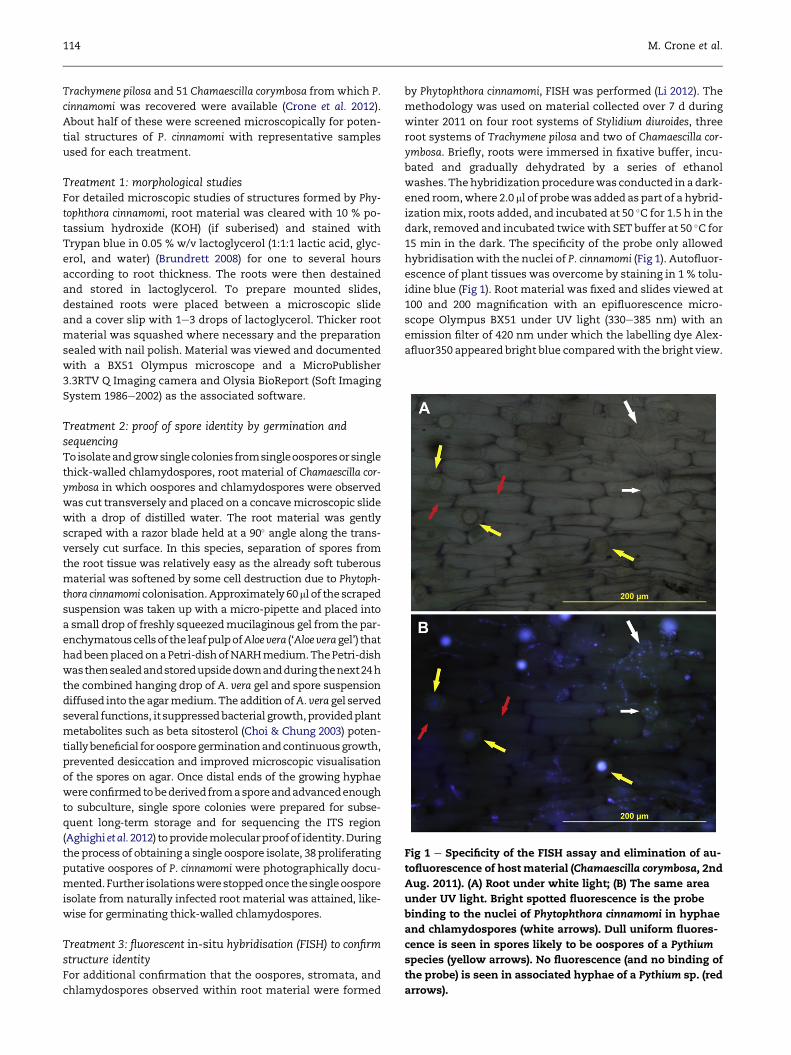
Fig 1 e Specificity of the FISH assay and elimination of au-
tofluorescence of host material (Chamaescilla corymbosa, 2nd
Aug. 2011). (A) Root under white light; (B) The same area
under UV light. Bright spotted fluorescence is the probe
binding to the nuclei of Phytophthora cinnamomi in hyphae
and chlamydospores (white arrows). Dull uniform fluores-
cence is seen in spores likely to be oospores of a Pythium
species (yellow arrows). No fluorescence (and no binding of
the probe) is seen in associated hyphae of a Pythium sp. (red
arrows).
114 M. Crone et al.
Trachymene pilosa and 51 Chamaescilla corymbosa from which P.
cinnamomi was recovered were available (Crone et al. 2012).
About half of these were screened microscopically for poten-
tial structures of P. cinnamomi with representative samples
used for each treatment.
Treatment 1: morphological studiesFor detailed microscopic studies of structures formed by Phy-
tophthora cinnamomi, root material was cleared with 10 % po-
tassium hydroxide (KOH) (if suberised) and stained with
Trypan blue in 0.05 % w/v lactoglycerol (1:1:1 lactic acid, glyc-
erol, and water) (Brundrett 2008) for one to several hours
according to root thickness. The roots were then destained
and stored in lactoglycerol. To prepare mounted slides,
destained roots were placed between a microscopic slide
and a cover slip with 1e3 drops of lactoglycerol. Thicker root
material was squashed where necessary and the preparation
sealed with nail polish. Material was viewed and documented
with a BX51 Olympus microscope and a MicroPublisher
3.3RTV Q Imaging camera and Olysia BioReport (Soft Imaging
System 1986e2002) as the associated software.
Treatment 2: proof of spore identity by germination andsequencingToisolateandgrowsinglecolonies fromsingleoosporesorsingle
thick-walled chlamydospores, root material of Chamaescilla cor-
ymbosa in which oospores and chlamydospores were observed
was cut transversely and placed on a concavemicroscopic slide
with a drop of distilled water. The root material was gently
scraped with a razor blade held at a 90� angle along the trans-
versely cut surface. In this species, separation of spores from
the root tissue was relatively easy as the already soft tuberous
material was softened by some cell destruction due to Phytoph-
thora cinnamomi colonisation.Approximately 60ml of the scraped
suspension was taken up with a micro-pipette and placed into
a small drop of freshly squeezedmucilaginous gel from the par-
enchymatouscellsof the leafpulpofAloevera (‘Aloeveragel’) that
hadbeenplacedonaPetri-dishofNARHmedium.ThePetri-dish
wasthensealedandstoredupsidedownandduringthenext24h
the combined hanging drop of A. vera gel and spore suspension
diffused into theagarmedium.The addition ofA. vera gel served
several functions, it suppressedbacterial growth,providedplant
metabolites such as beta sitosterol (Choi & Chung 2003) poten-
tiallybeneficial foroosporegerminationandcontinuousgrowth,
prevented desiccation and improved microscopic visualisation
of the spores on agar. Once distal ends of the growing hyphae
wereconfirmedtobederivedfromasporeandadvancedenough
to subculture, single spore colonies were prepared for subse-
quent long-term storage and for sequencing the ITS region
(Aghighi et al.2012) toprovidemolecularproofof identity.During
the process of obtaining a single oospore isolate, 38 proliferating
putative oospores of P. cinnamomi were photographically docu-
mented.Further isolationswerestoppedonce thesingleoospore
isolate from naturally infected root material was attained, like-
wise for germinating thick-walled chlamydospores.
Treatment 3: fluorescent in-situ hybridisation (FISH) to confirmstructure identityFor additional confirmation that the oospores, stromata, and
chlamydospores observed within root material were formed
by Phytophthora cinnamomi, FISH was performed (Li 2012). The
methodology was used on material collected over 7 d during
winter 2011 on four root systems of Stylidium diuroides, three
root systems of Trachymene pilosa and two of Chamaescilla cor-
ymbosa. Briefly, roots were immersed in fixative buffer, incu-
bated and gradually dehydrated by a series of ethanol
washes. Thehybridizationprocedurewas conducted in adark-
ened room,where 2.0 ml of probewas added as part of a hybrid-
izationmix, roots added, and incubated at 50 �C for 1.5 h in the
dark, removed and incubated twicewith SET buffer at 50 �C for
15 min in the dark. The specificity of the probe only allowed
hybridisationwith the nuclei of P. cinnamomi (Fig 1). Autofluor-
escence of plant tissues was overcome by staining in 1 % tolu-
idine blue (Fig 1). Root material was fixed and slides viewed at
100 and 200 magnification with an epifluorescence micro-
scope Olympus BX51 under UV light (330e385 nm) with an
emission filter of 420 nm under which the labelling dye Alex-
afluor350 appeared bright blue comparedwith the bright view.
Survival of P. cinnamomi as oospores, stromata, and thick-walled chlamydospores in roots 115
Images were taken using an Olympus DP70 digital camera (Ja-
pan) with DP Controller and DP Manager software.
Treatment 4: ultrastructural transmission electron microscopicanalysis of putative haustoriaA longitudinally sectioned asymptomatically infected root
piece of Trachymene pilosa collected on the 19th Jul. 2011 was
used for ultrastructural studies to support themicroscopic ob-
servations of putative haustoria from the three examined spe-
cies. The material was fixed in 3 % glutaraldehyde in 0.025 M
phosphate buffer (pH 7.0) followed by 2 % osmium tetroxide
in 0.025M phosphate buffer then dehydrated through increas-
ing concentrations of acetone (5e100 % acetone), infiltrated
with increasing concentrations of Spurr’s epoxy resin before
embedding in fresh 100 % Spurr’s resin. Areas of interest
were initially viewed by 1 mm thick light microscope sections
stainedwith 1 %methylene blue and 1 % Azur II in 1 % sodium
tetra borate (Richardson et al. 1960). Transmission electronmi-
croscope sections (80e90 nm thick) were contrasted with ura-
nyl acetate followed by lead citrate and viewed between 1600�to 52000� with a Philips CM 100 Bio transmission electron
microscope.
Determination of mating type
IsolatesOut of 219 Phytophthora cinnamomi isolations from root sys-
tems of annual and herbaceous perennial plant species, eight
representative isolates obtained from different host plants,
locations within the study sites and collection times during
2011 were sequenced and confirmed to be P. cinnamomi
(Crone et al. 2012). Host plants included Chamaescilla corym-
bosa, Trachymene pilosa, Stylidium diuroides and two additional
jarrah forest species Pterochaeta paniculata (Asteraceae, an-
nual) and Hypocalymma angustifolium (Myrtaceae, woody pe-
rennial). These eight isolates were used to determine the
mating type to ascertain whether the oospores were pro-
duced by selfing or due to mating with the opposite mating
type.
Mating tests were set up with two replicates including
a positive control where the tester strains P. cinnamomi A1
‘MUCC 794’ (GenBank Accession number: JX454790) and P. cin-
namomi A2 ‘MUCC 795’ (GenBank Accession number:
JX454791), obtained from the Murdoch University culture col-
lection (sequenced (Aghighi et al. 2012) and confirmed to be P.
cinnamomi), were used for mating the isolates. Both tester
strainswere used as negative controls by pairing A1mycelium
with A1 and A2 with A2.
Preparation of isolatesIsolates were aseptically taken out of long-term water storage
and plugs of each isolate as well as the A1 and A2 Phytophthora
cinnamomi tester strains were inserted approximately 0.5 cm
deep into a surface sterilised apple (cv. Granny Smith) to revi-
talise the isolates. After 10 d all isolates had caused large le-
sions, from which four pieces (approx. 0.5 cm2) from
underneath the apple skin were plated on NARH and incu-
bated. Outgrowing hyphae were subcultured for each isolate
after 2 d on V8 agar. Parafilmª sealed plates were stored in
the dark at 21 �C (�1 �C) for 7 d.
MatingAfter 7 d 0.5 cm2 plugs were taken from the edge of each col-
ony and placed approximately 0.5 cm away from a similar
plug containing either the Phytophthora cinnamomi A1 or A2
tester strain on V8 agar plates amendedwith 30mg l�1 beta si-
tosterol (Practical Grade, MP Biomedicals, Ohio, USA) to stim-
ulate oospore formation (Ribeiro 1978). The beta sitosterol was
dissolvedwith approximately 2ml of 100% ethanol in a heated
water bath before adding to the medium before autoclaving.
All plates were sealed with Parafilmª, placed within two zip
lock bags and wrapped in aluminium foil to exclude light.
The two replicates for each mating combination were kept
in two different locations with room temperatures conducive
for oospore development (between 20 �C and 26 �C). The plates
were inspected after 2 and 6 weeks.
Morphologically similar structures in Phytophthoracinnamomi-free plants
As part of a series of inoculation experiments on annual and
herbaceous perennial plant species, not-inoculated plants free
of P. cinnamomiwere available for comparisonwith the infected
plants. Microscopic examination allowed to observe whether
structures seen in P. cinnamomi positive roots were absent in
roots of these negative controls. Briefly, seeds of ten annual
and herbaceous perennial plant species were sown into pas-
teurised black gravel soil contained in free-draining square
pots (10cmheight�7cm)andwatereddaily tocontainer capac-
ity inanevaporatively cooledglasshouse (9e24 �C� 2 �C). Therewas an average of six plants per species as a negative control.
Thenot-inoculatedseedlings (onaverage39d-old)wereflooded
for 24handmock-inoculatedbyexposure toagarplugs freeofP.
cinnamomi, harvested at 56 d and plated on NARH agar to ob-
serve hyphal outgrowths. Roots were then fixed and stained
for microscopic examination (see Treatment 1).
Results
Microscopic observations of field isolations
OverviewFrom the end of Jun. to the beginning of Aug., Phytophthora cin-
namomiwas recovered from the naturally infected asymptom-
atic roots of Trachymene pilosa, Stylidium diuroides, and
Chamaescilla corymbosa. However, from Aug. onwards, whilst
P. cinnamomi infected individuals of T. pilosa remained asymp-
tomatic until natural senescence, root lesions were observed
for C. corymbosa although above ground tissues remained
symptomless, and S. diuroides wilted without visible root le-
sions (Crone et al. 2012). In all cases, the root tissues appeared
to be extensively colonised as evident by hyphal outgrowth
onto selective agar from several areas of the root systems,
and these areas corresponded with the pathogen detection
within the root material. A range of morphological structures
of P. cinnamomi was observed in all three species.
Thick-walled chlamydospores and lignitubersThick-walled chlamydospores were detected within root tis-
sues of Chamaescilla corymbosa, Stylidium diuroides, and
116 M. Crone et al.
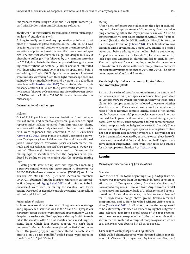
Trachymene pilosa and these germinated to produce viable col-
onies (Fig 2A). Whilst some chlamydospores were observed on
the surface of roots, there was evidence that they also oc-
curred within the root tissue. The clearest indicators that
chlamydospores were formed within host cells and not on
the root surface were that in some cases the chlamydospores
were elongated as a result of host cell constriction (Fig 2B),
while in other cases the host cell walls had distorted and ex-
panded due to the developing chlamydospore (Fig 2C).
A single thick-walled chlamydospore isolate from S. diur-
oides (collection: 26th Jul. 2011) was obtained by subculturing
the proximal end from one out of two proliferating germ
tubes. The ITS region was sequenced and Basic Local Se-
quence Alignment Tool for nucleotides (Blast n) confirmed
the identity of the isolate as Phytophthora cinnamomi (GenBank
Accession number: JX113308; Murdoch University storage
MUCC 793).
One germinated chlamydospore with hyphae typical of P.
cinnamomi was detected in dead root tissue of C. corymbosa in
which the chlamydospore had persisted over the summer
(Fig 2D). Lignitubers were observed rarely in S. diuroides and
T. pilosa, in some cases in close proximity to structures of P.
cinnamomi (Fig 2E).
StromataBesides hyphae proliferating through the host tissue, hyphal
aggregations variable in density and appearance were ob-
served. These formations are novel to Phytophthora cinnamomi
andwere termed ‘stromata’ (singular stroma) according to the
closest analogous structure in ascomycetes and basidiomy-
cetes. Typically stromata were confined to one cell, but it
was also observed that emerging hyphae penetrated new
root cells in close proximity to form more stromata (Fig 3B).
Within some stromata hyphae formed chlamydospores
Fig 2 e Chlamydospores and lignitubers of Phytophthora cinnam
root tissue of Chamaescilla corymbosa showing several germ tub
chlamydospore elongated due to the restriction of the plant cel
chlamydospore distorting host cell (S. diuroides, 13th Sep. 2011);
and germinated (arrows) the following spring (C. corymbosa, 4th
encapsulated by a lignituber (arrow) (Trachymene pilosa, 2nd Aug
root of Podotheca angustifolia. Scale bars: (A) 100 mm; (BeC), (EeF
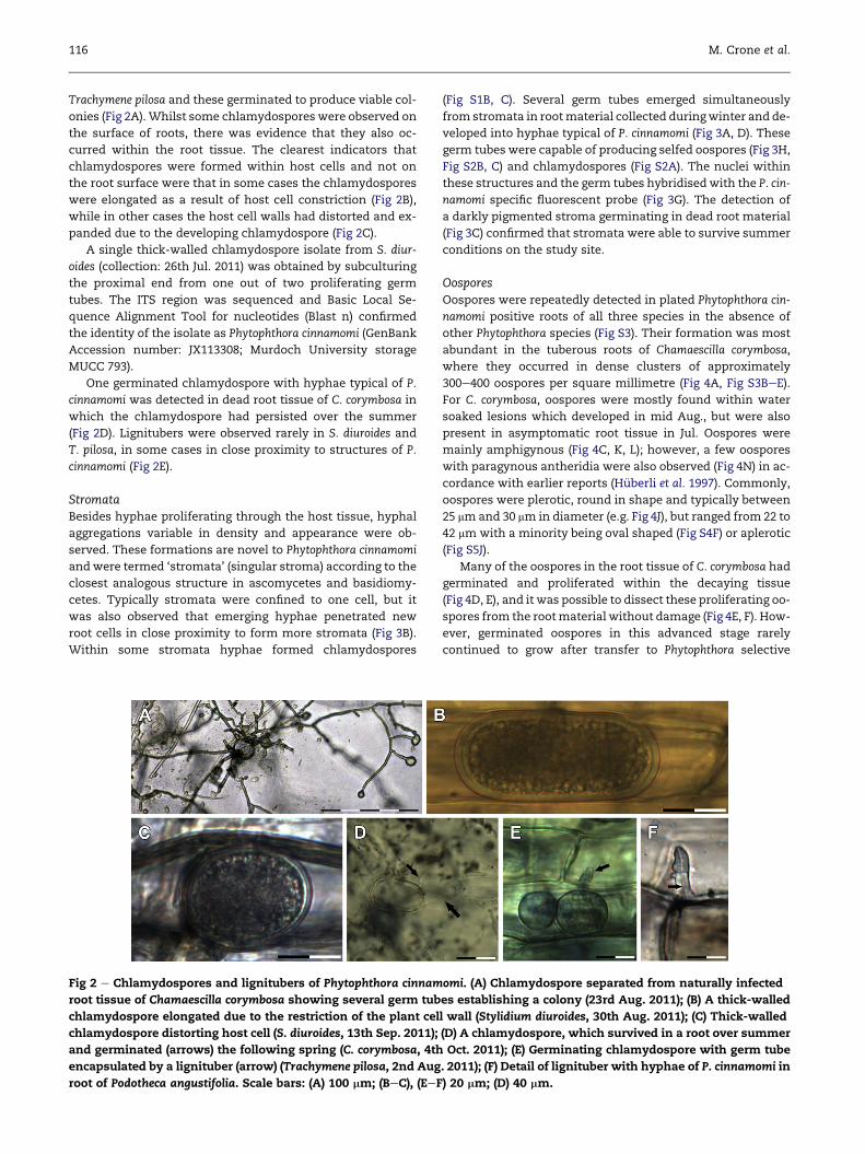
(Fig S1B, C). Several germ tubes emerged simultaneously
from stromata in rootmaterial collected duringwinter and de-
veloped into hyphae typical of P. cinnamomi (Fig 3A, D). These
germ tubes were capable of producing selfed oospores (Fig 3H,
Fig S2B, C) and chlamydospores (Fig S2A). The nuclei within
these structures and the germ tubes hybridisedwith the P. cin-
namomi specific fluorescent probe (Fig 3G). The detection of
a darkly pigmented stroma germinating in dead root material
(Fig 3C) confirmed that stromata were able to survive summer
conditions on the study site.
OosporesOospores were repeatedly detected in plated Phytophthora cin-
namomi positive roots of all three species in the absence of
other Phytophthora species (Fig S3). Their formation was most
abundant in the tuberous roots of Chamaescilla corymbosa,
where they occurred in dense clusters of approximately
300e400 oospores per square millimetre (Fig 4A, Fig S3BeE).
For C. corymbosa, oospores were mostly found within water
soaked lesions which developed in mid Aug., but were also
present in asymptomatic root tissue in Jul. Oospores were
mainly amphigynous (Fig 4C, K, L); however, a few oospores
with paragynous antheridia were also observed (Fig 4N) in ac-
cordance with earlier reports (H€uberli et al. 1997). Commonly,
oospores were plerotic, round in shape and typically between
25 mmand 30 mm in diameter (e.g. Fig 4J), but ranged from 22 to
42 mm with a minority being oval shaped (Fig S4F) or aplerotic
(Fig S5J).
Many of the oospores in the root tissue of C. corymbosa had
germinated and proliferated within the decaying tissue
(Fig 4D, E), and it was possible to dissect these proliferating oo-
spores from the rootmaterial without damage (Fig 4E, F). How-
ever, germinated oospores in this advanced stage rarely
continued to grow after transfer to Phytophthora selective
omi. (A) Chlamydospore separated from naturally infected
es establishing a colony (23rd Aug. 2011); (B) A thick-walled
l wall (Stylidium diuroides, 30th Aug. 2011); (C) Thick-walled
(D) A chlamydospore, which survived in a root over summer
Oct. 2011); (E) Germinating chlamydospore with germ tube
. 2011); (F) Detail of lignituber with hyphae of P. cinnamomi in
) 20 mm; (D) 40 mm.
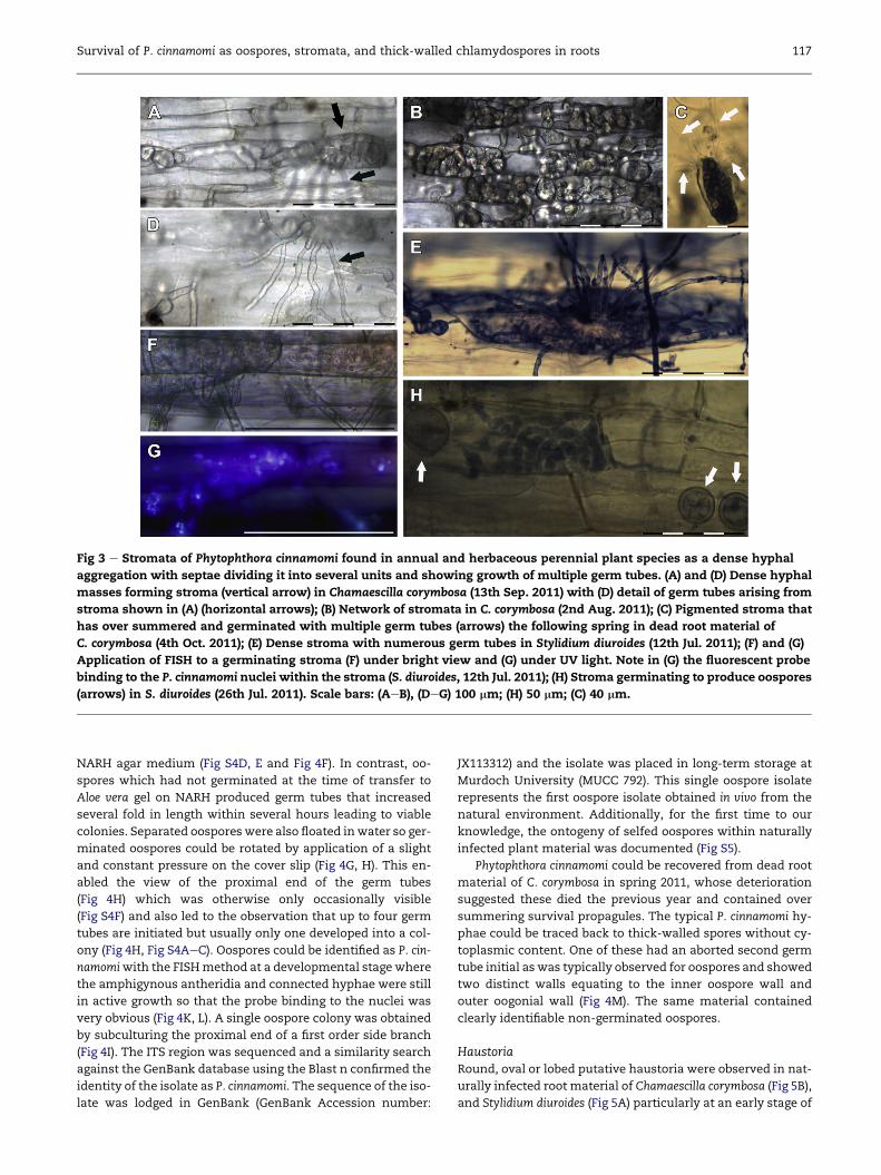
Fig 3 e Stromata of Phytophthora cinnamomi found in annual and herbaceous perennial plant species as a dense hyphal
aggregation with septae dividing it into several units and showing growth of multiple germ tubes. (A) and (D) Dense hyphal
masses forming stroma (vertical arrow) in Chamaescilla corymbosa (13th Sep. 2011) with (D) detail of germ tubes arising from
stroma shown in (A) (horizontal arrows); (B) Network of stromata in C. corymbosa (2nd Aug. 2011); (C) Pigmented stroma that
has over summered and germinated with multiple germ tubes (arrows) the following spring in dead root material of
C. corymbosa (4th Oct. 2011); (E) Dense stroma with numerous germ tubes in Stylidium diuroides (12th Jul. 2011); (F) and (G)
Application of FISH to a germinating stroma (F) under bright view and (G) under UV light. Note in (G) the fluorescent probe
binding to the P. cinnamomi nuclei within the stroma (S. diuroides, 12th Jul. 2011); (H) Stroma germinating to produce oospores
(arrows) in S. diuroides (26th Jul. 2011). Scale bars: (AeB), (DeG) 100 mm; (H) 50 mm; (C) 40 mm.
Survival of P. cinnamomi as oospores, stromata, and thick-walled chlamydospores in roots 117
NARH agar medium (Fig S4D, E and Fig 4F). In contrast, oo-
spores which had not germinated at the time of transfer to
Aloe vera gel on NARH produced germ tubes that increased
several fold in length within several hours leading to viable
colonies. Separated oospores were also floated inwater so ger-
minated oospores could be rotated by application of a slight
and constant pressure on the cover slip (Fig 4G, H). This en-
abled the view of the proximal end of the germ tubes
(Fig 4H) which was otherwise only occasionally visible
(Fig S4F) and also led to the observation that up to four germ
tubes are initiated but usually only one developed into a col-
ony (Fig 4H, Fig S4AeC). Oospores could be identified as P. cin-
namomiwith the FISHmethod at a developmental stage where
the amphigynous antheridia and connected hyphae were still
in active growth so that the probe binding to the nuclei was
very obvious (Fig 4K, L). A single oospore colony was obtained
by subculturing the proximal end of a first order side branch
(Fig 4I). The ITS region was sequenced and a similarity search
against the GenBank database using the Blast n confirmed the
identity of the isolate as P. cinnamomi. The sequence of the iso-
late was lodged in GenBank (GenBank Accession number:
JX113312) and the isolate was placed in long-term storage at
Murdoch University (MUCC 792). This single oospore isolate
represents the first oospore isolate obtained in vivo from the
natural environment. Additionally, for the first time to our
knowledge, the ontogeny of selfed oospores within naturally
infected plant material was documented (Fig S5).
Phytophthora cinnamomi could be recovered from dead root
material of C. corymbosa in spring 2011, whose deterioration
suggested these died the previous year and contained over
summering survival propagules. The typical P. cinnamomi hy-
phae could be traced back to thick-walled spores without cy-
toplasmic content. One of these had an aborted second germ
tube initial as was typically observed for oospores and showed
two distinct walls equating to the inner oospore wall and
outer oogonial wall (Fig 4M). The same material contained
clearly identifiable non-germinated oospores.
HaustoriaRound, oval or lobed putative haustoria were observed in nat-
urally infected root material of Chamaescilla corymbosa (Fig 5B),
and Stylidium diuroides (Fig 5A) particularly at an early stage of
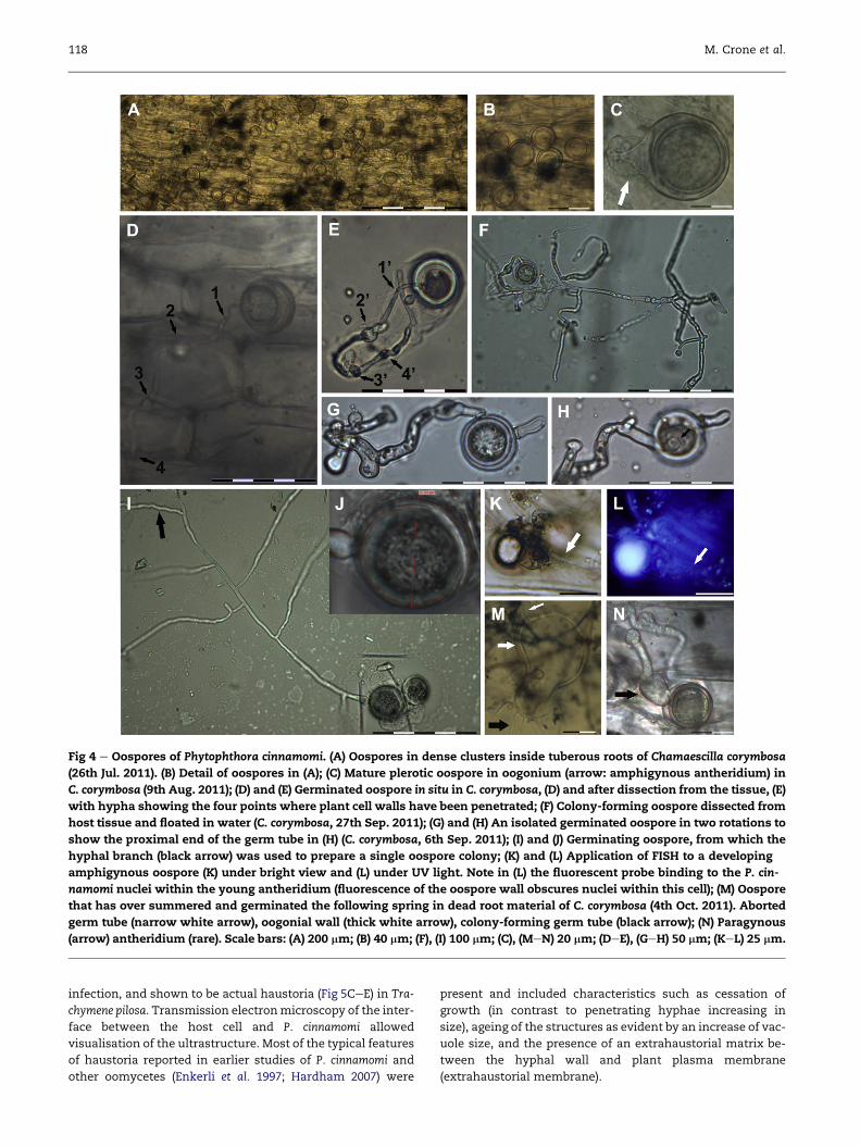
Fig 4 e Oospores of Phytophthora cinnamomi. (A) Oospores in dense clusters inside tuberous roots of Chamaescilla corymbosa
(26th Jul. 2011). (B) Detail of oospores in (A); (C) Mature plerotic oospore in oogonium (arrow: amphigynous antheridium) in
C. corymbosa (9th Aug. 2011); (D) and (E) Germinated oospore in situ in C. corymbosa, (D) and after dissection from the tissue, (E)
with hypha showing the four points where plant cell walls have been penetrated; (F) Colony-forming oospore dissected from
host tissue and floated in water (C. corymbosa, 27th Sep. 2011); (G) and (H) An isolated germinated oospore in two rotations to
show the proximal end of the germ tube in (H) (C. corymbosa, 6th Sep. 2011); (I) and (J) Germinating oospore, from which the
hyphal branch (black arrow) was used to prepare a single oospore colony; (K) and (L) Application of FISH to a developing
amphigynous oospore (K) under bright view and (L) under UV light. Note in (L) the fluorescent probe binding to the P. cin-
namomi nuclei within the young antheridium (fluorescence of the oospore wall obscures nuclei within this cell); (M) Oospore
that has over summered and germinated the following spring in dead root material of C. corymbosa (4th Oct. 2011). Aborted
germ tube (narrow white arrow), oogonial wall (thick white arrow), colony-forming germ tube (black arrow); (N) Paragynous
(arrow) antheridium (rare). Scale bars: (A) 200 mm; (B) 40 mm; (F), (I) 100 mm; (C), (MeN) 20 mm; (DeE), (GeH) 50 mm; (KeL) 25 mm.
118 M. Crone et al.
infection, and shown to be actual haustoria (Fig 5CeE) in Tra-
chymene pilosa. Transmission electronmicroscopy of the inter-
face between the host cell and P. cinnamomi allowed
visualisation of the ultrastructure. Most of the typical features
of haustoria reported in earlier studies of P. cinnamomi and
other oomycetes (Enkerli et al. 1997; Hardham 2007) were
present and included characteristics such as cessation of
growth (in contrast to penetrating hyphae increasing in
size), ageing of the structures as evident by an increase of vac-
uole size, and the presence of an extrahaustorial matrix be-
tween the hyphal wall and plant plasma membrane
(extrahaustorial membrane).
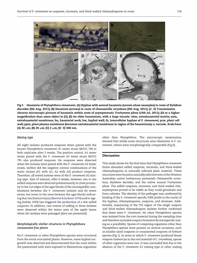
Fig 5 e Haustoria of Phytophthora cinnamomi. (A) Hyphae with several haustoria (arrows show examples) in roots of Stylidium
diuroides (9th Aug. 2011); (B) Haustoria (arrows) in roots of Chamaescilla corymbosa (9th Aug. 2011); (CeE) Transmission
electron microscopic pictures of haustoria within roots of asymptomatic Trachymene pilosa (19th Jul. 2011); (D) at a higher
magnification than same object in (C); (E) An older haustorium, with a large vacuole. ehm, extrahaustorial matrix; exm,
extrahaustorial membrane; hn, haustorial neck; hw, hyphal wall; ih, intercellular hyphae of P. cinnamomi; pcw, plant cell
wall; ppm, plant plasmamembrane (becomes extrahaustorial membrane in region of the haustorium); v, vacuole. Scale bars:
(A) 40 mm; (B) 20 mm; (C) 2 mm; (DeE) 500 nm.
Survival of P. cinnamomi as oospores, stromata, and thick-walled chlamydospores in roots 119
Mating type
All eight isolates produced oospores when paired with the
known Phytophthora cinnamomi A1 tester strain MUCC 794 in
both replicates after 2 weeks. The positive control, A1 tester
strain paired with the P. cinnamomi A2 tester strain MUCC
795 also produced oospores. No oospores were observed
when the isolates were paired with the P. cinnamomi A2 tester
strain, neither did the negative control combinations of the
tester strains (A1 with A1; A2 with A2) produce oospores.
Therefore, all tested isolates were of the P. cinnamomi A2 mat-
ing type. Also of interest, after 6 weeks, between one to ten
selfed oosporeswere detected predominantly in close proxim-
ity to the cut edges of the agar blocks of the incompatible com-
binations between the P. cinnamomi isolates and A2 tester
strain, but never in the zone between the agar blocks indicat-
ing thatmechanical injury (Reeves& Jackson 1974) and/or age-
ing (Ashby 1929) has triggered the production of a few selfed
oospores. In addition, rare events of selfing in three isolates
were observed within the lesion area of the apple tissue
when the isolates were passaged (data not presented).
Morphologically similar structures in Phytophthoracinnamomi-free plants
No P. cinnamomi or other Phytophthora species were recovered
from the mock-inoculated plants. However, some hyphal out-
growth was observed and demonstrated that the roots within
the pasteurised soils were exposed to filamentous organisms
other than Phytophthora. The microscopic examination
showed that whilst some structures were dissimilar to P. cin-
namomi, others were morphologically comparable (Fig 6).
Discussion
This study shows for the first time that Phytophthora cinnamomi
forms abundant selfed oospores, stromata, and thick-walled
chlamydospores in naturally infected plant material. These
structureswere found innaturally infected rootsof theWestern
Australian native herbaceous perennials Chamaescilla corym-
bosa, Stylidium diuroides, and the native annual Trachymene
pilosa. The selfed oospores, stromata, and thick-walled chla-
mydospores proved to be viable as they could germinate and
form colonies. The identity of the pathogen was confirmed by
binding of the P. cinnamomi specific FISH probe to the nuclei of
the hyphae, chlamydospores, oospores, and stromata. Addi-
tionally, sequencing of the ITS region of the single oospore
and thick-walled chlamydospore isolates further confirmed
that these were P. cinnamomi. No other Phytophthora species
was isolated from the root material during the sampling time
and therefore excludedoospore formationby intraspecificmat-
ing as a possibility. Spores of competing organisms other than
Phytophthora species were present on several occasions, such
as smaller sized oospores or ornamented oospores of Pythium
species (Fig 1). In areas in which P. cinnamomi produced dense
oospore clusters (as in the rootmaterial ofC. corymbosa), spores
of other organisms were rare. It was concluded that due to the
absence of the P. cinnamomi A1 mating type or other mating
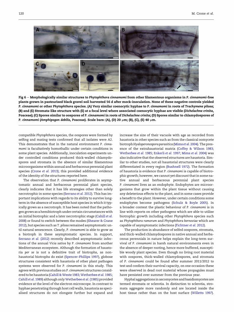
Fig 6 e Morphologically similar structures to Phytophthora cinnamomi from other filamentous organisms in P. cinnamomi-free
plants grown in pasteurised black gravel soil harvested 56 d after mock-inoculation. None of these negative controls yielded
P. cinnamomi or other Phytophthora species. (A) Very similar coenocytic hyphae to P. cinnamomi in roots of Trachymene pilosa;
(B) and (E) Stromata-like structure with (E) at a focal level where associated coenocytic hyphae are visible (Dichelachne crinita,
Poaceae); (C) Spores similar to oospores of P. cinnamomi in roots of Dichelachne crinita; (D) Spores similar to chlamydospores of
P. cinnamomi (Amphipogon debilis, Poaceae). Scale bars: (A), (D) 20 mm; (B), (C), (E) 40 mm.
120 M. Crone et al.
compatible Phytophthora species, the oospores were formed by
selfing and mating tests confirmed that all isolates were A2.
This demonstrates that in the natural environment P. cinna-
momi is facultatively homothallic under certain conditions in
some plant species. Additionally, inoculation experiments un-
der controlled conditions produced thick-walled chlamydo-
spores and stromata in the absence of similar filamentous
microorganismswithinannual andherbaceousperennial plant
species (Crone et al. 2013); this provided additional evidence
of the identity of the structures reported here.
The observation that P. cinnamomi proliferates in asymp-
tomatic annual and herbaceous perennial plant species,
clearly indicates that it has life strategies other than solely
necrotrophic in some species (Serrano et al. 2012). This has im-
portant implications with regards to its ability to survive long-
term in the absence of susceptible host species inwhich it typ-
ically grows as a necrotroph. It has been noted that the patho-
gengrowsasahemibiotrophundercertaincircumstanceswith
an initial biotrophic and a later necrotrophic stage (Cahill et al.
2008) or found to switch between the modes (Shearer & Crane
2012), but species such as T. pilosa remained asymptomatic un-
til natural senescence. Clearly, P. cinnamomi is able to grow as
a biotroph in these asymptomatic species. In support,
Serrano et al. (2012) recently described asymptomatic infec-
tions of the annual Vicia sativa by P. cinnamomi from another
Mediterranean ecosystem. Although the formation of hausto-
ria per se is not a definitive trait of biotrophs, as non-
haustorial biotrophs do exist (Spencer-Phillips 1997), globose
structures consistent with haustoria of other plant pathogen
systems were observed for P. cinnamomi in this study. This
agreeswithpreviousstudiesonP. cinnamomi structures consid-
ered tobehaustoria (Cahill&Weste1983;Wetherbee et al. 1985;
Cahill et al. 1989) althoughonlyWetherbee et al. (1985) provided
evidence at the level of the electronmicroscope. In contrast to
hyphaepenetrating throughhost cellwalls, haustoria as speci-
alised structures do not elongate further but expand and
increase the size of their vacuole with age as recorded from
haustoria in other species such as from the classical oomycete
biotrophHyaloperonosporaparasitica (Mims et al.2004).Thepres-
ence of the extrahaustorial matrix (Coffey & Wilson 1983;
Wetherbee et al. 1985; Enkerli et al. 1997; Mims et al. 2004) was
also indicative that the observed structures arehaustoria. Sim-
ilar to other studies, not all haustorial structures were clearly
differentiated in every region (Bushnell 1972). The formation
of haustoria is evidence that P. cinnamomi is capable of biotro-
phic growth; however,we cannot yet discount that in somena-
tive annual and herbaceous perennial plant species
P. cinnamomi lives as an endophyte. Endophytes are microor-
ganisms that grow within the plant tissue without causing
any deleterious effects to the plant, and in some cases provide
a benefit to the plant. However, under certain conditions some
endophytes become pathogens (Schulz & Boyle 2005). In
a broader context the plastic behaviour of P. cinnamomi is in
line with reports on other pathogens which are able to utilise
biotrophic growth including other Phytophthora species such
as Phytophthora ramorum and Phytophthora kernoviae which are
capable of asymptomatic infections (Fichtner et al. 2012).
The production in abundance of selfed oospores, stromata,
and thick-walled chlamydospores in native annual and herba-
ceous perennials in nature helps explain the long-term sur-
vival of P. cinnamomi in harsh natural environments even in
the absence of deeper rooting, hence more buffered, suscepti-
ble woody plant species. Even though no living root material
with oospores, thick-walled chlamydospores, and stromata
of P. cinnamomi could be found after summer 2011/2012 to
test and confirm their survival capacity, on one occasion these
were observed in dead root material whose propagules must
have persisted over summer from the previous year.
Hyphalaggregations inascomycetesandbasidiomycetesare
termed stromata or sclerotia. In distinction to sclerotia, stro-
mata aggregate more randomly and are located inside the
host tissue rather than on the host surface (Willetts 1997).
Survival of P. cinnamomi as oospores, stromata, and thick-walled chlamydospores in roots 121
Stromata have been further described as morphologically vari-
able formations in regard to size, compactness, and degree of
differentiation and a fungal species might only produce them
under certain conditions (Willetts 1997). Functionally, it is sug-
gested that due to the hyphal density of the stromata their ca-
pacity to store nutrients acquired from the host material is
significant, resulting in the high production of mycelium and
spores when conditions are favourable for germination
(Willetts 1997). Stromata also act as survival propagules
(Willetts 1997). Convergent evolution, resulting in analogous
structureshas beendemonstrated for unrelated taxa occupying
the samenicheand this is also the case for true fungi andoomy-
cetes (Tyler 2008) as they belong to two different kingdoms.
Consequently, it is appropriate toconsider that thesehyphalag-
gregates produced by P. cinnamomi are stromata.
Stromata formation by a Phytophthora specieswas observed
for the first time for P. ramorumunder experimental conditions
and described as small hyphal aggregates formed by repeated
branching, budding, swelling, and interweaving (Moralejo
et al. 2006). These usually darkened with time and were occa-
sionally found to produce sporangia or dense clusters of chla-
mydospores (Moralejo et al. 2006). In the case of P. cinnamomi,
the stromata produced clusters of oospores and chlamydo-
spores but as yet sporangia have not been observed.
One darkened stroma whose hyphae were typical of P. cin-
namomi was detected from dead host material from where P.
cinnamomi was recovered, which is very indicative of their
role as direct survival propagules (Fig 3K). Repeated observa-
tions are desirable in future to confirm this observation, but
detection in degraded host material is difficult as these thin
roots are subject to rapid breakdown.
The contribution of stromata to the long-term survival of P.
cinnamomi is definite based on the observation that stromata
occurred commonly, together with oospore clusters within
the same area (Fig S3D, E), and from the observation that
selfed oospores and chlamydospores (thin and thick-walled)
were formed by stromata.
Due to their rare occurrence, no conclusions could be
reached about the role of lignitubers in the survival of P. cinna-
momi in the jarrah forest. Their formation should be also
assessed in woody plant species, where they have been ob-
served in larger numbers (T. Jung, pers. comm.). These struc-
tures were first described as lignitubers by Fellows (1928)
and shown to originate from the plant and are a response to
any pathogen attack. As these encapsulate short hyphal
strands, they might preserve hyphae during summer. Germi-
nation would most likely occur from the proximal end with
the least plant cell deposits around the hyphae.
Future studies on isolation and germinationof oospores and
chlamydospores should consider the use of Aloe vera gel as it
appeared to have several advantages for P. cinnamomi andmay
have wider application for other oomycetes and true fungi.
Conclusion
This study has shown for the first time the importance of
selfed oospores, thick-walled chlamydospores, and stromata
produced by Phytophthora cinnamomi in asymptomatic annual
and herbaceous perennial species for the long-term survival
of P. cinnamomi. It has also increased our understanding of
a biotrophic and/or endophytic lifestyle of P. cinnamomi in
these plant species not previously recognised as hosts of this
pathogen. The findings suggest future research on whether
P. cinnamomi grows and survives in these groups of plants sim-
ilarly in horticulture and other impacted Mediterranean bi-
omes or other conducive ecosystems where it is present. The
frequency of the biotrophic/endophytic mode of growth in
plant species in its suggested areas of origin where ‘pathogen’
and host are in relative equilibrium, compared to regions in
which it has been recently introduced would also be of inter-
est. Further, the findings might apply to other hemibiotrophic
or necrotrophic oomycetes, which could potentially grow
asymptomatically as biotrophs in certain hosts. Finally, due
to the difficulty of studying microscopically the larger suber-
ised roots of woody perennial species, it is possible that selfed
oospores and thick-walled chlamydospores are also produced
in these species but have been overlooked. It is postulated that
the necrotrophic mode of growth cannot provide enough nu-
trients to produce the dense clusters of oospores and large
numbers of chlamydospores observed in the asymptomatic
plants. The biotrophic mode of growth with nutrient acquisi-
tion aided through haustoria, followed by formation of dense
masses of hyphae in stromatamay be a prerequisite to the for-
mation of the high numbers of oospores and chlamydospores.
From a disease management perspective, infected small root
systems of annual and herbaceous perennial plant species in
the surface soil provide a source of propagules easily spread
by wind and water movement of soil, and by animal foraging.
Acknowledgements
ThisworkwaspossiblewithanAPAI scholarship fromtheAus-
tralian Research Council (LP0776740) for the senior author, as
well as research funding fromAlcoa of Australia. Many thanks
to Dr V. Stokes and Dr I. Colquhoun with field assistance and
supervision, G. Thomson for assistance with the electron mi-
croscopy, and Dr T. Burgess and D. White for the molecular
work, Dr A. Li for assistance with the FISH work and
Dr T. Jung for discussions on P. cinnamomi survival propagules.
Appendix A. Supplementary data
Supplementary data related to this article can be found at
http://dx.doi.org/10.1016/j.funbio.2012.12.004.
r e f e r e n c e s
Aghighi S, Hardy GESt J, Scott JK, Burgess TI, 2012. Phytophthorabilorbang sp. nov., a new species associated with the decline ofRubus anglocandicans (European blackberry) in Western Aus-tralia. European Journal of Plant Pathology 133: 841e855.
Ashby SF, 1929. The production of sexual organs in pure culturesof Phytophthora cinnamomi Rands and Blepharospora cambivoraPetri. Transactions of the British Mycological Society 14: 260e263.
122 M. Crone et al.
Brasier CM, 1975. Stimulation of sex organ formation in Phytoph-thora by antagonistic species of Trichoderma II. Ecological im-plications. New Phytologist 74: 195e198.
Brundrett M, 2008. Mycorrhizal Associations: the web resource [WWWdocument]. http://mycorrhizas.info [accessed 16 June 2012].
Bushnell WR, 1972. Physiology of fungal haustoria. Annual Reviewof Phytopathology 10: 151e176.
Cahill DM, Rookes JE, Wilson BA, Gibson L, McDougall KL, 2008.Phytophthora cinnamomi and Australia’s biodiversity: impacts,predictions and progress towards control. Australian Journal ofBotany 56: 279e310.
Cahill D, Legge N, Grant B, Weste G, 1989. Cellular and histologicalchanges induced by Phytophthora cinnamomi in a group of plantspecies ranging fully susceptible to fully resistant. Phytopa-thology 79: 417e424.
Cahill D, Weste G, 1983. Formation of callose deposits as a re-sponse to infection with Phytophthora cinnamomi. Transactionsof the British Mycological Society 80: 23e29.
Choi S, Chung M-H, 2003. A review on the relationship betweenAloe vera components and their biologic effects. Seminars inIntegrative Medicine 1: 53e62.
Collins S, McComb JA, Howard K, Shearer BL, Colquhoun IJ,Hardy GESt J, 2012. The long-term survival of Phytophthoracinnamomi in mature Banksia grandis killed by the pathogen.Forest Pathology 42: 28e36.
Coffey MD,Wilson UE, 1983. An ultrastructural study of the late-blight fungus Phytophthora infestans and its interaction with thefoliage of two potato cultivars possessing different levels of gen-eral (field) resistance. Canadian Journal of Botany 61: 2669e2685.
Crone M, McComb JA, O’Brien PA, Hardy GEStJ, 2012. Annual andherbaceous perennial native Australian plant species areasymptomatic hosts of Phytophthora cinnamomi in the Eucalyp-tus marginata (jarrah) forest of Western Australia. Plant Pa-thology, http://dx.doi.org/10.1111/ppa.12016.
Crone M, McComb JA, O’Brien PA, Hardy GEStJ, 2013. Assessmentof Australian native annual/herbaceous perennial plantspecies as asymptomatic or symptomatic hosts of Phytoph-thora cinnamomi under controlled conditions. Forest Pathology,http://dx.doi.org/10.1111/efp.12027.
DavisonEM,ShearerBL, 1989.Phytophthora spp. in indigenous forestsin Australia. New Zealand Journal of Forestry Science 19: 277e289.
Dell B, Hardy GESt J, Vear K, 2005. History of Phytophthora cinna-momimanagement in Western Australia. In: Calver MC, Bigler-Cole H, Bolton G, Dargavel J, Gaynor A, Horwitz P, Mills J,Wardell-Johnston G (eds), A Forest Consciousness: proceedings6th national conference of the Australian forest history society inc.Millpress Science Publishers, Rotterdam, pp. 391e406.
Dobrowolski MP, Tommerup IC, Shearer BL, O’Brien PA, 2003.Three clonal lineages of Phytophthora cinnamomi in Australiarevealed by microsatellites. Phytopathology 93: 695e704.
DunstanWA, Rudman T, Shearer BL, Moore NA, Paap T, Calver MC,Dell B, Hardy GESt J, 2010. Containment and spot eradication ofa highly destructive, invasive plant pathogen (Phytophthora cin-namomi) in natural ecosystems. Biological Invasions 12: 913e925.
Enkerli K, Hahn MG, Mims CW, 1997. Ultrastructure of compatibleand incompatible interactions of soybean roots infected withthe plant pathogenic oomycete Phytophthora sojae. CanadianJournal of Botany 75: 1493e1508.
Fellows H, 1928. Some chemical and morphological phenomenaattending infection of the wheat plant by Ophiobolus graminis.Journal of Agricultural Research 37: 647e661.
Fichtner EJ, Rizzo DM, Kirk SA, Webber JF, 2012. Infectivity andsporulation potential of Phytophthora kernoviae to select NorthAmerican native plants. Plant Pathology 61: 224e233.
Hardham AR, 2005. Pathogen profile: Phytophthora cinnamomi.Molecular Plant Pathology 6: 589e604.
Hardham AR, 2007. Cell biology of planteoomycete interactions.Cellular Microbiology 9: 31e39.
H€uberli D, Tommerup IC, Hardy GESt J, 1997. The role of paragy-nous and amphigynous antheridia in sexual reproduction ofPhytophthora cinnamomi. Mycological Research 101: 1383e1388.
H€uberli D, Tommerup IC, Hardy GESt J, 2000. False-negative iso-lations or absence of lesions may cause mis-diagnosis of dis-eased plants infected with Phytophthora cinnamomi.Australasian Plant Pathology 29: 164e169.
Jayasekera AU,McComb JA, Shearer BL, Hardy GESt J, 2007. In plantaselfing and oospore production of Phytophthora cinnamomi in thepresence of Acacia pulchella.Mycological Research 111: 355e362.
Li AY, 2012. Role of the Feral Pig (Sus scrofa) in the Dissemination of Phy-tophthora cinnamomi in south-western Australia. PhD thesis, MurdochUniversity,Perth.http://researchrepository.murdoch.edu.au/9801/.
McCarren KL, McComb JA, Shearer BL, Hardy GESt J, 2005. The roleof chlamydospores of Phytophthora cinnamomi - a review. Aus-tralasian Plant Pathology 34: 333e338.
Mims CW, Richardson EA, Holt III BF, Dangl JL, 2004. Ultrastruc-ture of the host-pathogen interface in Arabidopsis thalianaleaves infected by the downy mildew Hyaloperonospora para-sitica. Canadian Journal of Botany 82: 1001e1008.
Mircetich SM, Zentmyer GA, 1966. Production of oospores andchlamydospores of Phytophthora cinnamomi in roots and soil.Phytopathology 56: 1076e1078.
Moralejo E, Puig M, Garc�ıa JA, Descals E, 2006. Stromata, sporan-giomata and chlamydosori of Phytophthora ramorum on inoc-ulated Mediterranean woody plants. Mycological Research 110:1323e1332.
Phillips D, Weste G, 1984. Field resistance in three native mono-cotyledon species that colonize indigenous sclerophyll forestafter invasion by Phytophthora cinnamomi. Australian Journal ofBotany 32: 339e352.
Podger FD, 1972. Phytophthora cinnamomi. A cause of lethal diseasein indigenous plant communities in Western Australia. Phy-topathology 62: 972e981.
Reeves RJ, Jackson RM, 1972. Induction of Phytophthora cinnamomioospores in soil by Trichoderma viride. Transactions of the BritishMycological Society 59: 156e159.
Reeves RJ, Jackson RM, 1974. Stimulation of sexual reproductionin Phytophthora cinnamomi by damage. Journal of General Micro-biology 84: 303e310.
RibeiroOK, 1978.ASource Book of the Genus Phytophthora. J. Cramer,Vaduz.
Richardson KC, Jarret L, Finke EH, 1960. Embedding in epoxy resinfor ultrathin sectioning in electron microscopy. Stain Technol-ogy 35: 313e323.
Schulz B, Boyle C, 2005. The endophytic continuum. MycologicalResearch 109: 661e686.
Serrano MS, Fern�andez-Rebollo P, De Vita P, S�anchez ME, 2012.Susceptibility of common herbaceous crops to Phytophthoracinnamomi and its influence on Quercus root rot in rangelands.European Journal of Plant Pathology 134: 409e414.
Shearer BL, Crane CE, 2012. Phytophthora cinnamomi visible ne-crotic lesionecolonisation relationships in native flora. Aus-tralasian Plant Pathology 41: 47e57.
Shearer BL, Tippett JT, 1989. Jarrah Dieback: the dynamics andmanagement of Phytophthora cinnamomi in the jarrah (Euca-lyptus marginata) forest of south-western Australia. Departmentof Conservation and Land Management, Perth.
Spencer-Phillips PTN, 1997. Function of fungal haustoria in epi-phytic and endophytic infections. Advances in Botanical Re-search 24: 309e333.
Tyler BM, 2008. Genomics of fungale and oomyceteesoybeaninteractions. In: Stacey G (ed.), Genetics and Genomics of Soybean.Springer, New York, pp. 243e267.
Western Australian Herbarium, 1998. FloraBase - the WesternAustralian flora. Department of Environment and Conserva-tion, Perth, Australia, [WWW document]. http://florabase.dec.wa.gov.au/ [accessed 28 May 2012].

Survival of P. cinnamomi as oospores, stromata, and thick-walled chlamydospores in roots 123
Wetherbee R, Hinch JM, Bonig I, Clarke AE, 1985. Response of Zeamays roots to infection with Phytophthora cinnamomi II. Thecortex and stele. Protoplasma 126: 188e197.
Willetts HJ, 1997. Morphology, development and evolution ofstromata/sclerotia and macroconidia of the Sclerotiniaceae.Mycological Research 101: 939e952.
Zentmyer GA, 1979. Stimulation of sexual reproduction in the A2mating type of Phytophthora cinnamomi by a substance in avo-cado roots. Phytopathology 69: 1029e1031.
Zentmyer GA, 1980. Phytophthora cinnamomi and the Diseases itCauses. Monograph 10. American Phytopathological SocietyPress, St Paul, MN.