Embed Size (px)
Citation preview
SURVIVAL AND GROWTH OF SALMONELLA POONA ON AND IN TISSUES OF
CANTALOUPES CO-INFECTED WITH PLANT PATHOGENIC MOLDS AND YEASTS
by
GLENNER MARIE RICHARDS
(Under the Direction of LARRY R. BEUCHAT)
ABSTRACT
Multistate outbreaks of salmonellosis associated with consumption of fresh cantaloupes from
salad bars suggest that contamination occurred early in the farm to fork chain, rather than
immediately before consumption. Factors that may influence the adherence, survival, and
growth of Salmonella enterica serotype Poona on and in cantaloupes were investigated. The
effects of temperature differentials between cantaloupes and S. Poona suspensions at 4°C and
30°C, on changes in fruit weight and populations of the pathogen recovered from rinds and stem
scar tissues of Eastern and Western type cantaloupes were assessed. The weight of immersed
cantaloupes increased by 0.13 – 0.43%, with Western cantaloupes showing greater increases.
Initial temperature of the inoculum and cantaloupe affected weight increase by Eastern
cantaloupes, but not Western type cantaloupes. The histology of cantaloupe rind and stem scar
tissues augments attachment and penetration by contaminating S. Poona, potentially reducing
effectiveness of sanitizer treatments. Proteolytic activity and changes in pH of cantaloupe rind
caused by growth of the phytopathogens Alternaria alternata, Cladosporium cladosporioides,
Epicoccum nigrum, Geotrichum candidum, and Penicillium expansum were studied. Survival
and growth characteristics of S. Poona on the surface rind and in wounded tissue as affected by
co-infection with molds and storage at 4°C and 20°C for up to 21 days were determined.
Populations of S. Poona on intact and wounded rind tissues at 4°C decreased by 1 – 2 logs, but
increased by 3 – 6 logs at 20°C. Co-infection with molds did not affect populations of S. Poona
recovered from cantaloupe rinds. The pathogen migrated from wounded tissues in the rind
through pulp tissues to distances as great as 3 – 4 cm below the surface, with or without co-
infection with phytopathogens. Migration and survival of S. Poona in cantaloupes were
enhanced by co-inoculation with C. cladosporioides and, to a lesser extent, P. expansum. Ten
yeasts were screened for antagonistic activity against S. Poona in cantaloupe juice and decay by
C. cladosporioides and G. candidum, in wounds on the surface of cantaloupe rind. Some of the
yeasts demonstrated their potential to restrict colonization of wounded tissues by
phytopathogenic molds, particularly at low storage temperatures. Test yeasts did not markedly
restrict growth of S. Poona in cantaloupe juice.
INDEX WORDS: Produce-related outbreaks, Salmonella, Cantaloupes, Metabiotic
associations, Phytopathogenic molds, Biological control
SURVIVAL AND GROWTH OF SALMONELLA POONA ON AND IN TISSUES OF
CANTALOUPES CO-INFECTED WITH PLANT PATHOGENIC MOLDS AND YEASTS
by
GLENNER MARIE RICHARDS
B.Sc., The University of the West Indies, Jamaica, 1988
M.Phil., The University of the West Indies, Jamaica, 1993
A Dissertation Submitted to the Graduate Faculty of The University of Georgia in Partial
Fulfillment of the Requirements for the Degree
DOCTOR OF PHILOSOPHY
ATHENS, GEORGIA
2003
© 2003
Glenner Marie Richards
All Rights Reserved
SURVIVAL AND GROWTH OF SALMONELLA POONA ON AND IN TISSUES OF
CANTALOUPES CO-INFECTED WITH PLANT PATHOGENIC MOLDS AND YEASTS
by
GLENNER MARIE RICHARDS
Approved:
Major Professor: Larry R. Beuchat
Committee: James W. Buck Joseph F. Frank Mark A. Harrison Ynes R. Ortega
Electronic Version Approved:
Maureen Grasso Dean of the Graduate School The University of Georgia December 2003
iv
DEDICATION
This dissertation and all the work involved is dedication to:
My God whose faithfulness is new every morning.
Mom and Daddy Robinson, you sowed the seed for this dream. Daddy, Osmond Ransford
Robinson (4/21/34 - 9/14/02), I am very sorry that I lost you before you could say, 'Cheers, to
success'. Mom, Madge Yvonne Robinson, thanks for keeping the light aglow in the midst of our
loss.
Mom and Dad Richards, thanks for teaching us to aim for the stars.
Bruce, Jonathan and Rachel, your love and support carried me all the way.
THANK YOU
v
ACKNOWLEDGEMENTS
I take this opportunity to express gratitude to:
Dr. Larry Beuchat, my major advisor and mentor, for the knowledge that he
graciously imparted and time invested to ensure my success.
My advisory committee: Dr. James Buck, Dr. Joseph Frank, Dr. Mark
Harrison and Dr. Ynes Ortega, for agreeing to serve as members of my committee.
ALL my teachers in Jamaica who patiently made their invaluable contribution
to my primary, secondary and tertiary education.
Andrea, Arlene, Audrey, Dave, Karen, Les, André, Lyndsey, and Alana, for
making our family a fun unit and a wonderful respite from work.
Prof. & Mrs. George Sidrak, Mr. & Mrs. Jervis Blanding, Mr. & Mrs. Peter
Bell, Mrs. Joan Hollingsworth, Ms. Rosemary Woodel, and Ms. Jacqueline Minus
for their friendship, encouragement, and support.
Beatrice Ayebah, Gloria Tetteh, Manan Sharma, Steve Kenney, Kaye Sy,
JeeHoon Ryu, for being supportive colleagues.
David Harrison, Kim Hortz, and Barbara Adler, for your assistance.
vi
TABLE OF CONTENTS
Page
ACKNOWLEDGEMENTS.............................................................................................................v
CHAPTER
1 INTRODUCTION AND LITERATURE REVIEW ...................................1
2 ATTACHMENT TO AND INFILTRATION OF CANTALOUPE RIND AND STEM SCAR TISSUES BY SALMONELLA POONA AS AFFECTED BY TEMPERATURE OF FRUIT AND INOCULUM........41
3 METABIOTIC ASSOCIATIONS OF MOLDS AND SALMONELLA
POONA ON INTACT AND WOUNDED CANTALOUPE RIND .........65 4 INFECTION OF CANTALOUPE RIND WITH CLADOSPORIUM
CLADOSPORIOIDES AND PENICILLIUM EXPANSUM, AND ASSOCIATED MIGRATION OF SALMONELLA POONA INTO EDIBLE TISSUES...................................................................................101
5 EXAMINATION OF YEASTS FOR ANTAGONISTIC ACTIVITY
AGAINST SALMONELLA POONA IN CANTALOUPE SUSPENSION AND IN WOUNDS IN RINDS CO-INFECTED WITH PHYTOPATHOGENIC MOLDS............................................................128
6 SUMMARY AND CONCLUSIONS ...........................................................185

1
CHAPTER 1
INTRODUCTION AND LITERATURE REVIEW
2
Introduction
The association of cancer and cardiovascular disease with consumption of saturated fat
has led to significant diet modifications by many Americans. The meat-and-potato diet that
accompanied the post war boom of the 1950s has been abandoned for a new American diet that
emphasizes fruits, vegetables and grains, while de-emphasizing meats and foods with a high fat
content. Public information campaigns such as Five-a-Day for Better Health by the National
Cancer Institute promote increased consumption of fresh fruits and vegetables (Hedberg et al.,
1994; Sewell and Farber, 2001). Consumers have been demanding and purchasing more fruits
and vegetables in recent years (Brackett, 1999).
Dietary changes have coincided with a dramatic increase in the variety of food items,
including fruits and vegetables, available to the consumer. The increased demand for fresh
produce has resulted in changes in the operation of food service establishments (Hedberg et al.,
1994). Restaurants have begun offering more raw produce in the form of salad bars and raw
vegetarian main courses. While progress has been made in promoting heart-healthy diets in the
past 40 years, these dietary changes have also altered the epidemiology of foodborne diseases in
the United States (Hedberg et al., 1994; Brackett, 1999).
Traditionally, foods implicated in outbreaks were undercooked meat, poultry or seafood,
or unpasteurized milk and dairy products. This has changed and foods previously thought to be
safe are now being recognized as being potentially hazardous (Tauxe, 1997). Fresh produce has
a short shelf life and often has been consumed or discarded before the outbreak is recognized.
Although still limited, an increasing proportion of reported outbreaks are being traced to fresh
produce. Because fresh produce is widely distributed, most produce-related outbreaks are
multistate events (Tauxe et al., 1997).
3
Meeting the increased worldwide demand for fresh produce has required the
implementation of intensive cultivation, harvesting and distribution measures. Local, national
and international competition between producers may also result in cost-reduction measures in
agricultural areas that have traditionally relied on low-paid workers. These measures could
potentially increase the likelihood of produce becoming contaminated in the field or during
packaging or distribution (Hedberg et al., 1994). Fresh produce is now being mass-produced and
distributed through large and complex networks. The size and complexity of these operations
can greatly magnify the public health significance of contamination with foodborne pathogens.
The presence of foodborne pathogens on fruits and vegetables results from exposure to human or
animal waste, or to irrigation water or other water that has been contaminated by these sources.
In the farm-to-table production, processing and distribution chain, there are various possible
sources of pathogens and points of contamination of fruits and vegetables with disease-causing
microorganisms. These sources include irrigation water, runoff water from livestock farms
adjacent to fields and orchards, manure, wash water, handling by workers, and contact with
contaminated surfaces, animal fertilizers applied in previous growing seasons, and feces of
rodents and ruminants (Guo et al., 2002).
Mechanisms of produce-borne diseases share several common features. Contamination
often occurs early in the production or handling process, rather than immediately before
consumption. Widespread consumer demand and global food markets have resulted in the use of
ingredients from many different locales and/or countries being combined in a single dish, making
the specific source of contamination difficult to trace and the concomitant outbreaks hard to
identify. All types of produce have the potential to harbor pathogens, but Shigella spp.,
Salmonella spp., enterotoxigenic and enterohemorrhagic Escherichia coli, Campylobacter spp.,
4
Listeria monocytogenes, Yersinia enterocolitica, Bacillus cereus, Clostridium botulinum, viruses,
and parasites such as Giardia lamblia, Cyclospora cayetanensis and Crytosporidium parvum are
of greatest public health concern (Beuchat, 2002). When compared with processed foods, fresh
produce typically has fewer barriers, such as reduced water activities or preservatives, to inhibit
microbial growth. A single and simple contravention can make the food unsafe, particularly with
respect to pathogens such as E. coli O157:H7 and Shigella that have low infectious doses.
Another notable influence on produce-related disease outbreaks is the introduction of new
packaging technologies. In an attempt to extend the shelf life of fresh-cut fruits and vegetables,
modified atmosphere packaging is used. Under these conditions, pathogens can potentially grow
at a faster rate than food spoilage microorganisms, whereas under normal atmospheric
conditions, pathogens may not be given an opportunity to multiply (Sewell and Farber, 2001).
This can be potentially perilous because produce may appear safe while it is sustaining survival
or growth of human pathogens.
Increased documentation of outbreaks associated with produce may be attributed to
changes in the produce industry, social demographics, food consumption patterns, awareness of
fresh fruits and vegetables as vehicles of infection (Beuchat and Ryu, 1997) and improved
epidemiology and identification of foodborne pathogens (Sewell and Farber, 2001).
Documentation of produce-related foodborne disease outbreaks has indicated that some produce
(e.g., lettuce, watermelon, sprouts, cantaloupes, and tomatoes), enteric pathogens (Salmonella, E.
coli O157:H7, and Shigella), and certain produce/pathogen combinations that are more
frequently reported (Beuchat, 1996). The survival or growth of human pathogens on the surface
of raw produce depends on the presence of free moisture, relative humidity and temperature
(Brackett, 1987). Human pathogens contaminating produce during cultivation or at harvest are
5
likely to survive, and may increase in numbers during storage before consumption (Nguyen-the
and Carlin, 2000).
Fruits and vegetables are perishable and maintain an active metabolism after harvesting
(Arul, 1994; Wills et al., 1998). Fungal infection, senescence and transpiration are the major
factors that contribute to the early termination of the storage life of fresh produce. In fleshy
fruits and vegetables, field infections may continue to develop after harvest. While in storage,
new infections may be caused by the same or other pathogens (Agrios, 1997). Factors that
accelerate senescence and favor microbial growth, such as physiological and mechanical injuries
as well as exposure to high ambient temperature and humidity, are the major promoters of
postharvest decay (Arul, 1994). Fleshy fruits and vegetables are generally kept at high relative
humidity to avoid shrinkage, therefore providing conditions favorable for growth of pathogenic
microorganisms, especially when wounds, cuts and bruises offer readily accessible nutrients.
However, penetration through natural openings and directly through the cuticle and epidermis
may also occur, especially when sound and infected produce are in contact. Once a fruit or
vegetable becomes infected, development and spread of the infection increase as the storage
temperature increases. At lower temperatures, pathogens and the diseases they cause develop
more slowly or cease to develop at all (Agrios, 1997).
Preharvest infections require that the pathogen circumvent inherent or induced resistance
of the host, or morphological barriers that inhibit the formation of an active disease lesion.
Alternatively, the pathogen may enter a latent state on or in the host. The quiescent pathogen
persists until the resistance of the host declines with advancing maturity or until a wound to the
host tissue or some mechanism that weakens the host defenses activates the pathogen (Eckert and
Ogawa, 1985). If physical injury is sustained, an active wound-healing process involving the
6
formation of corky cells to inhibit microbial invasion may ensue (Snowdon, 1990). Some of
these factors change with time and, if the microorganism remains viable, invasion and complete
colonization may eventually take place. Many molds are unable to penetrate intact skin of
produce, but readily invade any break in the skin. The damage is often microscopic, but is
sufficient for plant pathogens to gain access. In addition, tissue of produce at the point where it
is severed from the mother plant can act as a route of entry.
Several intrinsic and extrinsic factors affect the rate of plant tissue decay. Environmental
factors such as temperature, humidity and concentration of respiratory gases affect the rapidity
and severity of tissue decay. Warm temperature and high humidity favor the development of
postharvest decay and chilling injury generally predisposes tropical and subtropical produce to
postharvest decay. In contrast, low temperature, low oxygen, high carbon dioxide and low
humidity can restrict postharvest decay by either retarding the rate of ripening or senescence,
depressing growth of the pathogen or both. Ripened fruits are more susceptible than immature
fruits to microbial decay. Treatments that retard the rate of ripening will also slow the rate of
growth of decay microorganisms.
Interactions between foodborne human pathogens and background microflora have been
studied in meat and dairy products (Nguyen-the and Carlin, 1994; Francis et al., 1999), but
relationships on fruits and vegetables are ill defined (Beuchat, 2002). The antagonistic
properties of lactic acid bacteria (Francis et al., 1999) and pseudomonads toward pathogens on
ready-to-use vegetables have been reviewed (Nguyen-the and Carlin, 1994). Metabiotic
associations of molds and Clostridium botulinum on tomatoes (Draughon et al., 1988) and molds
and Listeria monocytogenes on fresh-cut apples (Conway et al., 2000; Chardonnet et al., 2002)
have been reported. Meager information has been published describing interactions between
7
Salmonella and E. coli O157:H7 and other microorganisms (Wells and Butterfield, 1997;
Riordan et al., 2000). E. coli O157:H7 is hypothetically more resistant than Salmonella to the
acidic fermentation end-products of lactic acid bacterial populations (Francis et al., 1999), but
the behavior of pathogens on or in produce characterized by pH change caused by metabiotic
activities of indigenous microflora has not been thoroughly investigated.
Interactions among microflora may have significant effects on the survival and growth
of pathogens. These interactions need to be elucidated to ensure that traditional and novel mild
preservation technologies can continue to be applied without compromising the safety of raw
fruits and vegetables.
Indigenous microflora on produce
The initial microflora of fresh fruits and vegetables originates in the field and from
harvesting and transportation equipment. Field sources include air, soil, insects, plant tissue
exudates (ICMSF, 1998) and animals. Fresh produce that has not been pasteurized or sterilized
has a microflora consisting of a specific association of microorganisms from the environment.
The origin, development and succession of this association are governed by ecological factors
that influence the physiological expression of microbial cells. Only microorganisms that possess
the necessary physiological attributes to respond to intrinsic and extrinsic environmental
pressures will survive (Goepfert, 1980). Eventually, given suitable environmental conditions, a
particular microbial community will develop. If environmental factors, inclusive of the unique
ecological factors associated with the specific produce permit, microflora will evolve to a
specific spoilage association (Goepfert, 1980; Deak and Beuchat, 1996). The numbers and types
of microorganisms present on freshly harvested fruit is dependent on environmental factors such
8
as the weather, the season, time of harvest within a season and type of fruit and its proximity to
the ground, as well cultural practices inclusive of irrigation and preharvest treatment with
chemicals such as fungicides.
As the flower bud develops into a flower and eventually matures into a fruit, surfaces are
colonized by a succession of microorganisms. The location of these microorganisms is mostly
limited to the surface of whole, sound fruits. The internal tissues are generally free from
contamination. The cuticular protective cover on many fruits and vegetables and structural
integrity due to cellulose and pectin, as well as the low pH by some produce, are important
intrinsic factors that influence the microbial ecology. Only a few microbial species are capable
of invading internal tissues of the fruit while it is attached to the plant.
Subsequent to harvest, fruits and vegetables are considered food products, although they
continue to be living plant tissue. Being compartmentalized, plant tissues limit the diffusion of
solutes and influence the survival and growth of microorganisms. Fruits and vegetables may
contain chemical compounds that are inhibitory to microorganisms and may induce defense
reactions similar to those found in growing plants against microbial invasion (Nguyen-the and
Carlin, 2000). Only the microorganisms that possess the necessary physiological attributes to
respond to intrinsic and extrinsic environmental pressures will survive (Goepfert, 1980).
Yeasts and molds are widely distributed in the environment and are present in the natural
microflora of most leaves, flowers, tree exudates, and fruit surfaces. The population and relative
proportion of species varies between commodities and is influenced by environmental,
harvesting and storage conditions (Deak and Beuchat, 1996). Freshly harvested fruits often have
a large yeast population. Mycological analysis has shown yeast population of 103 – 105 cfu/g of
sound, mature apples and 106 – 107 cfu/g of grapes (Splittstoesser, 1996; Lund and Snowdon,
9
2000). On freshly harvested soft fruits (strawberries, raspberries and blackberries), the number
of yeasts was 104 – 106 cfu/g; mold population was up to 104 cfu/g and bacterial population 105 –
106 cfu/g. With respect to shelf life and spoilage of most fruits, yeasts and molds play the most
significant role. Various types of injury caused by weather, insects, birds, rodents and farm
implements can be sustained before, during and after harvest (Snowdon, 1990). Breaking the
skin of fruits during harvesting or handling operations can significantly modify the environment
and allow the establishment of saprophytes (Mercier and Wilson, 1994).
Fruits that have been damaged are likely to have elevated yeast populations. Yeast cells
introduced to exposed tissues are able to utilize sugars and other nutrients to support growth
(Splittstoesser, 1996). Carbon dioxide and ethanol are the predominant metabolic by-products of
many yeasts, but glycerol, acetaldehyde, pyruvic acid and α-ketoglutaric acid are also formed.
Although yeasts may possess hydrolytic enzymes that degrade pectins, starch and certain
proteins, enzymatic activity is usually much less than that exhibited by other aciduric
microorganisms such as molds. Yeast populations on fruits have been extensively studied (Deak
and Beuchat, 1996). With a few exceptions, yeasts are not capable of attacking plant tissues.
Molds represent the most important group of microorganisms within the microflora that
cause decay of raw fruits. They are the main invaders causing fruits to rot, lose moisture and
becoming mummified (Lund and Snowdon, 2000). Genera of molds (Alternaria, Botrytis,
Botrysphaeria, Colletotrichum, Diplodia, Monilinia, Penicillium, Phomopsis, Rhizopus and
Sclerotinia) and bacteria (Erwinia and Pseudomonas) are the main plant pathogens. A few
molds, e.g., Colletotrichum, are able to penetrate the skin of healthy produce. Often the
relationship between the host (fruit or vegetable) and the pathogen is reasonably specific. For
example, Penicillium digitatum infects only citrus fruits and P. expansum primarily infects
10
apples and pears. Complete loss of the commodity occurs when one or a few pathogens invade
and breakdown the tissues. This initial attack is rapidly followed by a broad spectrum of weak
pathogens that magnify the damage caused by primary pathogens. Surface lesions caused by
plant pathogenic microorganisms, without the internal tissues being affected, may cause
deterioration of many commodities (Wills et al., 1998).
Some molds cause latent infections that do not produce visible symptoms until
postharvest. Defense mechanisms in the fruit are highly effective against nearly all fungi;
however, some molds are highly specialized pathogens, attacking only one or two kinds of fruit.
Others have a more general ability to invade fruit tissue (ICMSF, 1998). Only relatively few
genera and species are able to invade a particular fruit type and cause extensive losses. Some
species are plant pathogens that cause postharvest diseases of fruits while in transit or storage
and result in extensive economic loss (Agrios, 1997). Mold-infected raw fruit may become soft
after processing because pectinases were not inactivated by the thermal treatment. Some molds
are xerophilic and are therefore potential spoilage agents of foods of low water activity such as
dried fruits and fruit juice concentrate (Splittstoesser, 1996).
Control of Postharvest Diseases
Postharvest deterioration is a problem that persists from the time of harvest throughout
the fruit and vegetable distribution chain, affecting the cost and availability of produce to the
consumer and the ability of the producer to service distant markets (Arul, 1994). Control
measures aimed at inhibiting deterioration of the produce until it is consumed generally begin in
the field and continue through marketing. These include elimination of sources of infection,
preharvest and/or postharvest spraying with fungicides for the control or elimination of causal
11
microorganisms and careful handling during harvest to minimize mechanical damage and
subsequent microbial invasion.
The effectiveness of physical and chemical treatments used to control postharvest loss of
fruits and vegetables depends on the ability of the treatment or agent to reach the pathogen.
Physical treatments used to control postharvest deterioration include treatment with growth
regulators that delay tissue senescence, low and high temperatures, low-oxygen atmosphere,
modified relative humidity, ionizing radiation, good sanitation, and development of wound
barriers (Eckert and Ogawa, 1985; Wills et al., 1998). Handling, storage, and transport at low
temperature is the most important physical method to control postharvest deterioration because it
delays ripening, physiological deterioration, and postharvest decay of fruits and vegetables
(Smith, 1962). The remaining physical interventions are considered to be additional hurdles
(Wills et al., 1998).
Chemical control
Chemical treatments to reduce postharvest decay of fruits and vegetables may be applied
pre- or postharvest. The successful use of chemical treatment is dependent on the initial mold
spore population, growth rate, depth of the infection within the host tissue, temperature,
humidity, and depth to which the chemical can penetrate the host tissues. Efficacy of a chemical
is dependent on its cost effectiveness, solubility if used as a solution, ability to inhibit growth of
the pathogen and ability to kill the pathogen quickly. In addition, chemicals should not injure the
product, leave toxic or unattractive residues, or impair the sensory qualities (Smith, 1962; Wills
et al., 1998). Addition of chemicals to wash water was one of the earliest postharvest fungicide
application methods, and is still extensively used at present. Chemicals may also be impregnated
12
into wraps or box-liners, or applied as fumigants, solutions and suspensions, or in wax (Wills et
al., 1998). Most of the chemicals used are fungistatic in action, inhibiting or reducing the rate of
germination of spores or conidia and growth after germination, rather than causing death of the
mold. A few sanitizers such as chlorine and sulfur dioxide are true fungicides. Sodium
carbonate (Wills et al., 1998) and borax (sodium tetraborate) were the first chemicals to be
extensively tested and accepted for use to reduce postharvest decay (Smith, 1962; Wills et al.,
1998). Borax was predominantly used in the citrus industry to control Penicillium digitatum
(green mold) and P. italicum (blue mold). Cantaloupe molds were also effectively reduced
during transit and storage by washing for 0.5 to 2 min in warm water (37 – 42ºC) containing 2.5
– 5% borax (Smith, 1962).
Considered a more effective fungicide with broad-spectrum activity, sodium o-
phenylphenate (SOPP) superseded sodium carbonate and borax in the 1950s. SOPP is not
phytotoxic but is converted to a fungitoxic free phenol at appropriate sites. The o-phenylphenate
anion (OPP) diffuses selectively into injury sites and is converted to the undissociated form,
preventing infection at these sites during storage and marketing. SOPP can be applied as a dip or
foam, or incorporated into wax applied before or after washing. The SOPP-OPP system was
found to require pH control to prevent damage to the fruits. A residual tolerance level of 10 – 12
µg/g has been established.
Biphenyl, a compound with fungistatic activity, was subsequently introduced.
Impregnated into fruit wraps or into paper sheets placed in the fruit containers, biphenyl
sublimes into the atmosphere surrounding the fruits and prevents sporulation of Penicillium on
the surface of infected fruits and transfer of infection to neighboring fruits. It is also used as a
complement to SOPP on export fruits. The disadvantages associated with biphenyl include a
13
characteristic hydrocarbon odor, residues on the fruit surface sometimes exceeding that
permissible limit of 70 – 100 µg/g, and development of resistant strains by Penicillium and
Diplodia on fruits stored for up to 4 months. The use of biphenyl has been banned in some
countries (Wills et al., 1998).
The benzimidazole group of chemicals, introduced in the late 1960s, includes
thiabendazole (TBZ), benomyl, thiophanate methyl, and carbendazim. Benzimidazoles show
systemic and residual activity, are not phytotoxic and have low mammalian toxicity. They also
have a wide spectrum of antifungal activity at low concentrations, but are inactive against
Rhizopus, Alternaria, Geotrichum and soft rot bacteria (Eckert and Ogawa, 1985; Wills et al.,
1998). Major disadvantages of these fungicides are that they have limited solubility in water and
resistant strains of Penicillium have developed as a result of their specific action that interferes
with polymerization of the protein tubulin. The latter problem influenced the search for
alternative treatments (Wills et al., 1998).
Sec-butylamine (2-AB) has limited use as an alternative fungicide, due to its relatively
narrow spectrum of antifungal activity, and has been withdrawn from application. Guazantine
offers control of benzimidazole-resistant molds, but multiple resistances have been reported.
Triazoles, which include imazalil and prochloraz, are effective against a wide range of fungi,
particularly benzimidazole-resistant strains. Their mechanism involves inhibition of
demethylation during ergosterol biosynthesis. However, resistance has been reported (Eckert
and Ogawa, 1985; Wills et al., 1998). The use of postharvest chemical treatments helps to
extend the storage time of fruits and significantly affects the sucrose content (Caño et al., 1987),
but little is known about their effects on survival and growth of human pathogens in response to
modifications in the behavior of indigenous microflora.
14
Biological control
Application of synthetic fungicides has been the primary method of controlling
postharvest diseases. Interest in the development of alternatives has been generated in recent
times due to concerns about carcinogenic risks and public safety (National Research Council,
1987), presence of chemical residues in the food chain, development of fungicide-resistant
strains of postharvest pathogens, and deregistration of some of the more effective fungicides
(Wilson and Wisniewski, 1989; Wilson et al., 1997; El-Ghaouth et al., 1998).
Essential oils and natural extracts from plants and microorganisms have been evaluated
for safety and efficacy as fungicides (Wilson et al., 1997). Sugar analogs such as 2-deoxy-D-
glucose have been known to effectively inhibit the growth of several yeasts and some
filamentous fungi in the absence of metabolizable sugars (El-Ghaouth et al., 1997).
Native microflora on the surfaces of fresh produce have been assumed to be important in
maintenance of quality and safety, by producing antimicrobial compounds, activating plant
defense mechanisms, and suppressing human pathogens (Liao and Fett, 2001). The use of
antimicrobial agents as alternatives to synthetic fungicides has shown significant potential for the
control of postharvest disease of fruits and vegetables. Antagonistic yeasts and bacteria that
display the capability to protect several types of fruits against postharvest pathogens have been
isolated. Currently, two antagonistic microorganisms are commercially available as biocontrol
agents. Candida oleophila Montrochus, a yeast, and Pseudomonas syringae, a bacterium are
marketed under the trade names Aspire and Biosave-110, respectively (El-Neshway and Wilson,
1997; El-Ghaouth et al., 1998). Yeasts possess a number of attributes that make them useful
biological control agents in the postharvest environment. They effectively colonize plant
surfaces and produce extracellular compounds such as polysaccharides, which enhance their
15
survival while restricting colonization sites and flow of germination signals to pathogenic fungal
propagules which may be present (Wilson et al., 1996). The mechanism by which antagonistic
yeasts exert their biocontrol activity is unclear; however, nutrient competition (El-Ghaouth et al.,
1998), site exclusion, direct parasitism and induced resistance have been suggested (Wilson et
al., 1996). The biological activity of Candida saitoana and its interaction with plant pathogens
Botrytis cinerea and Penicillium expansum in apple wounds were investigated (El-Ghaouth et
al., 1998). C. saitoana proliferated in apple wounds, prevented the proliferation of the plant
pathogens, stimulated several host defense reactions, and controlled decay of apple fruits. It was
postulated that C. saitoana affects the ability of B. cinerea cells to degrade host tissue and
establish a nutritional relationship (El-Ghaouth et al., 1998). The natural microflora was found
to be inhibitory to B. cinerea at wound sites on Red Delicious apples while enhancing the
biocontrol activity of C. oleophila (Mercier and Wilson, 1994).
The polypeptide antibiotic nisin was first used to prevent spoilage of Swiss cheese by
Clostridium butyricum. Nisin has been used safely as a food preservative and has been approved
for use in the United States since 1988 (Jay, 2000). Enhanced biocontrol activity of C. oleophila
in the presence of nisin to control apple rot suggested that nisin stimulated the antagonist,
inhibited the pathogen by preventing germination of spores, restricted growth of the germ tubes,
and reduced the susceptibility of the wounds to infection (El-Neshway and Wilson, 1997).
Candida sake is commonly found in nature and is a component of the epiphytic
community on mature fruits. This yeast has not been associated with warm-blooded animals. C.
sake effectively controlled major postharvest diseases on apples when applied at 1.6 x 10⁶ cfu/ml
under commercial storage conditions (Viñas et al., 1998).
16
Biologically based control measures have not performed as consistently chemical
applications and tend to have a limited range of application (El-Neshway and Wilson, 1997).
Living organisms acting as ‘biofungicides’ have the disadvantage of not providing the direct and
complete control offered by most synthetic fungicides (Wilson et al., 1996). Biological control
of postharvest diseases is complex and involves a number of variables such as the rate and timing
of application of biocontrol agents, the possible use of additives to enhance biocontrol activity,
the physiological state of the host and environmental conditions used to store the produce
(Mercier and Wilson, 1994).
There are limited reports describing the effect of biocontrol agents on human pathogens.
Growth of L. monocytogenes on potato slices was significantly limited by the presence of
fluorescent soft-rotting pseudomonads (Liao and Sapers, 1999). A saprophytic P. syringae strain
(L-59-66) was effective in reducing the growth of human and decay pathogens on wounded
apples (Janisiewicz et al., 1999). Growth of Salmonella Chester on pepper discs was not
affected by the presence of native and uncharacterized flora. However, when co-inoculated with
potential antagonistic yeast onto pepper discs, the population of S. Chester was reduced by
approximately 1 log (Liao and Fett, 2001).
Salmonella and Salmonellosis
Salmonella spp. are facultatively anaerobic gram-negative rods within the taxonomic
family Enterobacteriaceae (D’Aoust, 2001). Most members of the genus are motile by
peritrichous flagella, but there are nonflagellated variants and nonmotile strains resulting from
dysfunctional flagella (D’Aoust, 1997). Salmonella nomenclature has progressed through a
succession of taxonomic schemes based on biochemical and serological characteristics and
17
principles of numerical taxonomy and DNA homology. The genus consists of more than 2,463
serovars that are classified within two species, Salmonella enterica and Salmonella bongori
(D’Aoust, 1997).
Salmonellae grow optimally at 35°C to 37°C, catabolize a variety of carbohydrates into
acids and gas, use citrate as a sole carbon source, produce hydrogen sulfide, and decarboxylate
lysine and ornithine to cadaverine and putrescine, respectively. They are oxidase-negative and
catalase-positive, with 50 to 53 mole % guanine plus cytosine (G+C) DNA content (D’Aoust,
2000). The biochemical identification of foodborne and clinical Salmonella isolates is generally
coupled to serological confirmation involving agglutination of bacterial surface antigens with
Salmonella-specific antibodies. These include O lipopolysaccharides (LPS) on the external
surface of the bacterial outer membrane, H antigens associated with peritrichous flagella and the
capsular (Vi) antigen, which occurs only in S. Typhi, S. Paratyphi C, and S. Dublin. All
serovars are considered to be potentially pathogenic (Jay, 2000; D’Aoust, 2001).
Although the primary habitat of Salmonella is the intestinal tract of birds, reptiles,
domestic and wild animals, humans, and insects, they may be found occasionally on other parts
of the body. As intestinal forms, they are excreted in feces, which may be transmitted by insects
and other living creatures to a large number of destinations, including water and food. When
humans and other animals consume contaminated water and foods, salmonellae are again shed
through fecal matter with a continuation of the cycle (Jay, 2000).
The genus Salmonella consists of resilient microorganisms that readily adapt to extreme
environmental conditions. They have been shown to actively grow within a wide temperature
range of 2°C to 47°C (Doyle and Cliver 1990; D’Aoust, 2000) and exhibit psychrotrophic
properties as reflected in the ability to grow in foods stored at 2 to 4°C (Matches and Liston,
18
1968; D’Aoust, 1997). The physiological adaptability of Salmonella is demonstrated by its
ability to proliferate at pH values ranging from 4.5 to 9.5, with an optimum pH for growth of 6.5
to 7.5. The minimum growth pH is dependent on the acid used to lower the pH (Chung and
Goepfert, 1970; Doyle and Cliver, 1990; Asplund and Nurmi, 1991; D’Aoust, 1997; Jay, 2000).
Water activity is an important factor determining the pH range for Salmonella growth. Most
salmonellae can grow in foods in a water activity range of 0.945 - 0.999. As water activity shifts
from the optimum for Salmonella growth, the permissive pH range narrows. Typical of other
gram-negative bacteria, they are able to grow on a large number of culture media and produce
visible colonies within 24 h at 37°C (Doyle and Cliver, 1990).
With respect to oxidation-reduction (OR) potential and nutrients, salmonellae are not
fastidious. Salmonella can grow under aerobic as well anaerobic conditions, but growth can be
inhibited by OR potential below –30mV (Doyle and Cliver, 1990). Generally, salmonellae are
unable to ferment lactose, sucrose or salicin, but a few serovars are capable of lactose
fermentation as an alternative. Glucose and certain other monosaccharides can be fermented to
produce gas. Salmonella normally utilizes amino acids as nitrogen sources, but nitrate, nitrite
and ammonia will serve as a sole source of nitrogen for S. Typhimurium (Jay, 2000).
Salmonellosis is the major bacterial foodborne disease in many countries (Asplund and
Nurmi, 1991; D’Aoust, 2001) and has been increasing steadily as a public health problem over
the last 50 years in the United States (Tauxe, 1991). Non-typhoidal salmonellae were estimated
to cause approximately 1.5 million cases of infection, resulting in 15,000 hospitalizations and
500 deaths in the United States in 1999 (Mead et al., 1999). Foods of animal origin have
historically been recognized as potential vehicles for Salmonella. According to the U.S.
Department of Agriculture, the bacterium is acquired from meat, poultry, or eggs in 50 to 75% of
19
salmonellosis cases. Poultry is known to be the primary vehicle of transmission (FSIS, 1995;
Leverentz et al., 2001). Salmonella as a leading cause of foodborne diseases, emanates from its
ubiquity in the environment, its prominence in various sectors of the agriculture industry and
from escalating movement of food and food ingredients in international trade (D’Aoust, 2001).
Major outbreaks of foodborne salmonellosis in the last two decades have involved a wide
variety of foods and serovars (D’Aoust, 2001). Salmonellae remain well entrenched in the meat
and poultry industries (D’Aoust, 2001); however, consumption of fresh fruits and vegetables,
including seed sprouts (O’Mahony et al., 1990; Van Beneden et al, 1996; Mahon et al., 1997),
unpasteurized apple cider (CDC, 1975), unpasteurized orange juice (Cook et al., 1998; CDC,
1999), raw tomatoes (Wood et al., 1991; Wessinger et al., 2000), watermelons (Gayler et al,
1955; Larson et al., 1979; Blostein, 1993), and cantaloupes (Ries et al., 1990; CDC, 1991) have
also been linked to outbreaks of salmonellosis in recent years. Sources of Salmonella on produce
include animal and human feces, and cross contamination from raw meat, poultry, or eggs
(IFT/FDA, 2001).
The number of cells comprising the infectious dose ranges from 10 to 105 (IFT/FDA,
2001). The typical incubation period of non-typhoid salmonellosis is 18 to 72 h, and symptoms
may include abdominal cramps, diarrhea with watery and possibly mucoid stools tinged with
blood, chills, fever of short duration (< 48 h), nausea and vomiting that appear 8 to 72 h
following exposure to the bacterium (D’Aoust, 2000). The clinical symptoms of uncomplicated
enterocolitis usually subside within five days followed by a convalescent carrier state of one to
several months during which the asymptomatic individual continues to excrete salmonellae.
Non-typhoid salmonellosis can progress from an enterocolitic to a septicemic condition as a
20
result of the migration of salmonellae from the intestinal tract into deeper tissues through
vascular and lymphatic conduits (D’Aoust, 2000).
Survival, growth, and removal of salmonellae on fruits and vegetables
The condition of produce tissue (healthy, intact tissue compared to damaged, diseased
tissue) impacts attachment, infiltration and efficacy of sanitation practices. Microbial cells are
known to lodge disproportionately in damaged and cut tissues compared to sound tissues
(Weissinger et al., 2000). E. coli in damaged apples, for example, poses a greater risk for the
contamination of cider than surface contamination of apples (Dingman, 2000). The epidermis of
fruits and vegetables is covered with a multi-layered hydrophobic cuticle, which provides the
primary barrier against invasion, insect and physical damage, and desiccation. Bacterial
attachment is facilitated by the presence of stomata, lenticels, broken trichomes, or cracks in the
cuticle. E. coli O157:H7 was observed to preferentially attach and penetrate the interior edge of
cut surface of lettuce leaves as opposed to intact leaf surface (Seo and Frank, 1998). The rind of
cantaloupes is complex, presenting a variety surfaces to which bacterium may bind. The
ruptured cell surface presents a meshwork of raised tissue (the net) (Ukuku and Fett, 2002).
Decontamination treatments are less effective in killing bacteria attached to or located within
protective sites that occur naturally or as a result of injury.
Salmonella has been isolated from many different types of raw fruits and vegetables
(Ercolani, 1976; Andrews et al., 1979; Roberts et al., 1982; Garcia-Villanova Ruiz et al., 1987).
Several studies have been undertaken to assess the survival and growth of Salmonella in tissues
of fruits and vegetables. Salmonella was associated more often with soft-rotted plant tissue
(Wells and Butterfield, 1997) and injured tissue (Liao and Sapers, 2000; Liao and Cooke, 2001)
than with their healthy and intact counterparts. Scanning electron microscopic observations
21
showed that retention of S. Chester on injured apple tissue, the stem, and calyx may be due in
part to the physical entrapment of bacteria in the holes below the surface of the tissue (Liao and
Sapers, 2000). When raw tomato fruits were immersed in a S. Montevideo suspension, more
cells were retained in stem scar and core tissues than on tomato skin. Unlike bacteria inoculated
onto the skin, S. Montevideo cells inoculated into wounds or onto slices multiplied over a 24-h
period (Wei et al., 1995).
The core tissue internalized higher numbers of S. Montevideo cells when tomatoes at
25°C were dipped into a suspension at 10°C compared to tomatoes dipped at 25 or 37°C. The
population of viable S. Montevideo cells remained constant throughout subsequent storage for 18
days at 10°C, but storage at 20°C resulted in significant population increases. When tomatoes
were stored at 5°C for 216 h, populations of S. Montevideo and aerobic mesophilic
microorganisms remained constant. However, when stored at 20 and 30°C, significant increases
in populations were observed (Zhuang et al., 1995). Salmonella is capable of rapid and prolific
growth on cantaloupe, watermelon, and honeydew melons incubated at 23°C (Escartin et al.,
1989; Golden et al., 1993). However, when incubated at 5°C for 24-h period, populations of
Salmonella on the melons remained unchanged (Golden et al., 1993).
At present, little is known about the mechanism by which bacteria become attached to
surfaces of produce. Physical forces such as London-van der Waals attraction, electrostatic
attraction between two surfaces and net gain entropy have been suggested to be involved.
Bacterial age and culture conditions have also been identified as factors affecting attachment
(Liao and Sapers, 2000).
The efficacy of washing treatments on detachment or inactivation of Salmonella on
cantaloupe surfaces is dependent on the state and location of the cells on the outer surface of the
22
cantaloupes. S. Stanley was found to be able to survive on cantaloupe surfaces for at least 6
days, on root crops for 10 to 53 days, and on leafy vegetables for 1 to 40 days (Ukuku and
Sapers, 2001). Firm attachment of bacteria to stem and calyx cavities, and injured tissue
represent a major obstacle in developing effective methods for eliminating Salmonella from the
surface of contaminated plant tissue.
Chlorine is widely used in the minimally processed fruit and vegetable industry.
However, high concentrations may provoke irritation of eyes, skin or lungs of workers, be
corrosive to metal components of machines, and perhaps leave residues on produce. Several
serotypes of salmonellae inoculated onto shredded lettuce and diced tomatoes were able to grow
and could not be eliminated with treatments of up to 200 ppm of chlorine (Weissinger et al.,
2000). The effectiveness of chlorine on minimally processed spinach inoculated with S. Hadar
was found to increase through a range of treatments up to 25 – 125 ppm for 2 – 8 min (Pirovani
et al., 2000). Dipping tomatoes inoculated with S. Montevideo in solutions containing 60 ppm
and 110 ppm of free chlorine achieved significant population reductions. However, increased
concentration up to 320 ppm chlorine did not significantly improve effectiveness in killing the
pathogen (Zhuang et al., 1995; Beuchat et al., 1998). Chlorine was also reported to be less
effective at killing cells that had been internalized to the core tissue than those that were on the
surface tissue.
Trisodium phosphate (TSP) is known to be effective in killing Salmonella on poultry and
red meats when applied to carcasses in the form of a chill or rinse water during processing. TSP
was also observed to be highly effective in killing S. Montevideo on the surface of mature-green
tomatoes, but less effective against internalized cells (Zhuang and Beuchat, 1996).
23
Salmonellosis associated with melons
Melon fruits are in contact with the ground during their development, enhancing their
potential to be contaminated by pathogens. A field survey conducted by the Food and Drug
Administration (FDA) during 1990 and 1991 revealed that 0.76 and 1.06%, respectively, of the
melon rinds harbored several serovars of Salmonella (Madden 1992; Golden et al., 1993).
Selected fresh produce, including cantaloupes imported into the United States was surveyed. Of
151 cantaloupes sampled, 7.3% were contaminated with Salmonella or Shigella (FDA, 2001a).
The results of a similar survey of domestic fresh produce initiated in May 2000 indicated the
presence of pathogens on 4.3% of 92 cantaloupes sampled (FDA, 2001b).
There have been numerous national and international incidents of salmonellosis
associated with the consumption of fresh watermelons and cantaloupes. The association of
salmonellosis with salad bars and fruit salads suggests introduction of Salmonella into the fruit
from the rind by the slicing action, or the physical contact of cut pieces with contaminated rind.
The netted texture of cantaloupe melon rind enables effective attachment of microorganisms and
results in difficulty in sanitization. As few as 150 bacteria per cm2 of the netted rind surface has
been found to result in contamination of the interior pulp as a result of cutting whole cantaloupes
(Suslow and Cantwell, 2001).
The potential of the contaminated tissue to support growth of Salmonella is exacerbated
by display or storage at ambient temperature for an extended time (CDC, 1991; Golden et al.,
1993). The high water activity and sugar content of tissue juices promote rapid proliferation of
salmonellae. Cut cantaloupe is considered a potentially hazardous food in the FDA Food Code
because it is capable of supporting the growth of pathogens due to low acidity (pH 5.2 to 6.7)
and high water activity (0.97 to 0.99) (IFT/FDA, 2001).
24
An outbreak of salmonellosis involving S. Bareilly on watermelons occurred in 1950 in
Minnesota, but was not reported in medical literature (Gaylor et al., 1955; Blostein, 1993). S.
Miami was isolated from the internal flesh of watermelons that were responsible for 17 reported
cases and one death in Massachusetts in 1954 (Gaylor et al., 1955). In 1979, an outbreak
involving 18 cases of salmonellosis caused by S. Oranienberg linked to watermelons occurred in
Illinois. Salmonella was not isolated from the melons but investigations revealed that damaged
fruits were cut, covered with plastic film and displayed without refrigeration until sold (CDC,
1979). Cut cantaloupe from salad bars contaminated with S. Chester was associated with a large
multistate outbreak estimated to involve more than 25,000 individuals and two deaths in 1989-
1990 (Ries et al., 1990).
There were 26 primary cases and 13 secondary cases of salmonellosis associated with
watermelon contaminated with S. Javiana in Michigan in 1991. It is suspected that the rind was
contaminated and was not washed prior to cutting and serving at a picnic and school party over a
two-day period (Blostein, 1993). In 1991, consumption of fruit salads containing sliced
cantaloupes was epidemiologically linked to multistate outbreaks in the U.S. and Canada,
involving more than 400 cases. S. Poona was isolated from cantaloupes originating in either
Texas or Mexico (CDC, 1991). S. Saphra was isolated from cantaloupes sourced from Mexico
and implicated in a 1997 outbreak involving 24 cases in California. Multiple purchase sites
suggest that contamination occurred during production or harvest, and that retail storage at
ambient temperature was a contributor (Mohle-Boetani et al., 1999).
In 1998, there were 22 cases of salmonellosis attributed to S. Oranienberg on cantaloupes
consumed at various venues in Ontario, Canada (Deeks et al., 1998). Although the pathogen was
not isolated from the fruit, it has been theorized that pathogen from the surface contaminated the
25
flesh during slicing and that the cut fruit was stored at room temperature for an extended period
of time prior to consumption (IFT/FDA, 2001). S. Poona was associated with cantaloupes that
were epidemiologically linked to multistate outbreaks in 2000 involving 43 cases (Ukuku and
Sapers 2001; IFT/FDA, 2001), in 2001 with 50 cases (Walker, 2001), and in 2002 with 58 cases
(CDC, 2002).
Cantaloupes
Cultivation and postharvest handling
Cantaloupes (Cucumis melo L. var. reticulatus Naud.), also commonly called
muskmelons, are members of the taxonomic family Cucurbitaceae, which includes squash,
pumpkins, cucumbers, watermelons, and gourds (Boyhan et al., 1999; Orzolek et al., 2001). The
species Cucumis melo is subdivided into seven botanical variants: cantaloupensis, reticulatus,
inodorous, flexuosus, conomon, chito and dudaim. Only the reticulatus and inodorous variants
are of commercial significance in the United States. The inodorous variety includes honeydew
melons (Boyhan et al., 1999).
Cantaloupes are annual plants that produce long running, non-climbing vines that are
prostrate on the soil. Healthy plants have a canopy of large, soft, hairy leaves that are generally
lobed and heart-shaped (Anonymous, 1999). Individual plants exhibit andromonoecious
flowering, first producing groups of male flowers in leaf axils, and then producing single perfect
flowers (Peet, 2001). Bee pollination is required to make fruit set possible, as pollen is too heavy
and sticky to make wind pollination successful (Hemphill, 2002). Cantaloupes are round to oval
fruits with sizes ranging from 5 to 8 inches in diameter and length, and weighing 3 to 7 pounds.
They are characterized by the roughened appearance due to the network of corky surface tissue
26
(Anonymous, 1999; Orzolek et al., 2001; Hemphill, 2002) and the salmon-orange colored flesh
with a musky aroma (Anonymous, 1999).
In the United States, cantaloupes are categorized on the basis of their fruit type.
“Western” or “shipping” types have uniformly netted rinds lacking sutures and a firm, salmon-
colored flesh. Traditionally, this type was grown in western states and shipped to distant states;
however, in recent times they have been widely adapted and grown throughout the country. The
“eastern” or “jumbo” cantaloupes are grown for local markets. They are characterized by fruits
that are larger and less uniform in size than the western cantaloupes, with less uniform or no
netting, deep sutures on the rind, and orange flesh (Boyhan et al., 1999; Hemphill, 2002).
Contingent on the cultivar and environment, fruits mature 35 to 55 days after full bloom
and are harvested by maturity and not by size (Peet, 2001). The principal harvest indices are
surface color and development of the abscission zone (Kasmire and Cantwell, 1992). The
external color depends on variety and may still have a greenish cast at full slip (Hartz et al.,
1996). Commercial maturity is ideally at the firm-ripe stage or “¾ to full slip” when a clear
abscission from the stem occurs with light pressure, leaving no stem tissue attached to the fruit
(Suslow et al., 2000; Agblor and Waterer, 2001; Orzolek et al., 2001; Peet, 2001). Typically full
slip is at 42 days after flowering, when cantaloupes have high sugar content and good flavor and
aroma, but have a short storage life. For distant markets, less mature cantaloupes are harvested
at “half-slip”, but when the stem attachment area is smooth, rounded and lightly depressed.
Harvesting at approximately 36 days after flowering has been suggested as a compromise for
acceptable flavor and storage potential (Agblor and Waterer, 2001).
California, Arizona, Texas, Georgia and Indiana were the leading cantaloupe producing
states in 1993-1997 (Rhodes, 2001). Most of the cantaloupes harvested in the U.S. are sold as
27
fresh produce in wholesale markets, cooperatives, local retailers, roadside stands, and pick-your
own operations (Orzolek et al., 2001). Federal Grade Standards are based predominantly on
external appearances and soluble solids. There are four U.S. fruit grades: Fancy and No.1 grades
have a minimum of 11% and 10% soluble solids, respectively, while Commercial and No. 2.
grades have a minimum of 9% soluble solids (Hurst, 1999; Suslow et al., 2000).
Cantaloupes are generally harvested manually and under cool early morning or late
evening conditions (Agblor and Waterer, 2001; Peet, 2001). Examination by computerized
tomography (CT) indicates that poorly handled fruits suffer mechanical injury that cause
increased metabolic activity as measured by pectin esterase activity, fruit firmness, acidity and
total soluble solids values (Halloran et al., 1999a). Dropping cantaloupes at a distance of more
than 8 inches onto hard surfaces causes bruising and cracking (Hurst, 1999).
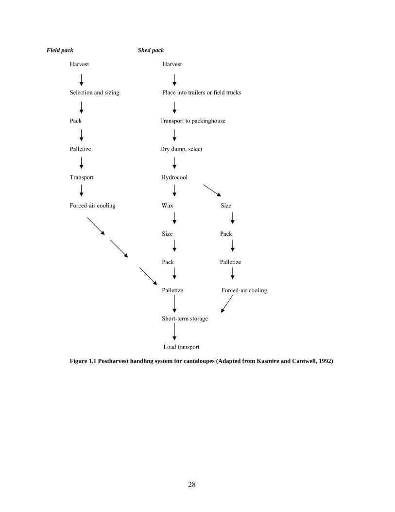
A flow diagram for postharvest handling systems for cantaloupes is shown in Figure 1.1.
Pre-cooling to a typical endpoint of 10°C or preferably to 4°C by cold water, cold air or ice
promptly after harvest is necessary to remove field heat, reduce respiration rate, and improve
shelf life. Cooling in a room without the use of forced air is usually avoided as it takes 24 to 36
h to cool cantaloupes to 10°C. Forced air-cooling is the most common practice (Suslow et al.,
2000), allowing cantaloupes to be cooled from 34°C to 16°C in 8 h (Agblor and Waterer, 2001).
Hydrocooling is the most efficient method to rapidly cool to 10°C. Cooled melons are generally
stored in a cold room and shipped in refrigerated trucks. Ice is blown in between rows of crates
or waxed cartons and over packed containers for shipment. Although cantaloupes are susceptible
to chilling injury, they are not injured by extended contact with ice (Hemphill, 2002). Typically 12
– 15 days of shelf life are attainable at storage temperatures of 2.2 – 5°C. Storage below this
optimum range usually results in chilling injury.
28
Field pack Shed pack
Harvest Harvest
Selection and sizing Place into trailers or field trucks
Pack Transport to packinghouse
Palletize Dry dump, select
Transport Hydrocool
Forced-air cooling Wax Size
Size Pack
Pack Palletize
Palletize Forced-air cooling
Short-term storage
Load transport
Figure 1.1 Postharvest handling system for cantaloupes (Adapted from Kasmire and Cantwell, 1992)
29
High relative humidity of 90 – 95% is essential to maximize postharvest quality and
prevent desiccation through scuffed and damaged surface netting (Hurst, 1999; Suslow et al.,
2000). Good quality is retained for approximately 14 – 21 days if cantaloupes are stored at 90 –
95% relative humidity and 8 – 12°C (Orzolek et al., 2001). Film wrapping is recommended to
improve the shelf life of fruits that are intended for long distance shipping, because desiccation is
a major cause of quality loss during shipping (Hemphill, 2002). Extended storage periods at
higher humidity or the formation of condensate encourages the growth of molds on stem-scar
tissue and rind surface (Suslow et al., 2000).
Consensus atmospheres of 3% O2 and 10% CO2 at 3°C with extended transit times of 14
– 21 days facilitates the benefit of delayed ripening, reduced respiration and associated sugar loss
and inhibition of surface mold and decay (Kader, 1992; Suslow et al., 2000). Controlled
atmosphere offers moderate benefits under most conditions; however, there is only limited use
on the commercial scale.
Postharvest diseases
Cantaloupes are susceptible to postharvest fungal rots, especially under warm, wet
conditions. Rots occur on the external surface of the fruit and gradually progress inwards in the
flesh. Fungal pathogens of major concern are Geotrichum, Alternaria, Penicillium,
Cladosporium, Rhizopus, and Fusarium (Suslow et al., 2000; Agblor and Waterer, 2001), and
Mucor to a lesser extent (Suslow et al., 2000). Alternaria and Cladosporium rots occur
frequently in cool storage. Dark brown or black lesions characterize Alternaria rot, and dark
green or black lesions characterize Cladosporium rots. Fusarium and Rhizopus rots are
problematic on fruit stored at room temperature. White and reddish hyphae on the netted surface
30
of fruits are symptoms of Fusarium rot. Softening and indentation of large areas of the flesh
with little mycelial growth characterize Rhizopus rots (Agblor and Waterer, 2001).
Dipping in hot water 52 – 55°C for 0.5 to 1.0 min (Suslow et al., 2000) or for 2 min
(Agblor and Waterer, 2001), or 59°C for 3 min (Hemphill, 2002) effectively reduces stem scar
and surface molds, and controls fungal and bacterial rots. Hemphill further reported that addition
of an undisclosed concentration of chlorine to hot water did not augment the benefits of hot
water treatment. Warm wet melons are subject to microbial invasion, and shelf life is reduced by
3 – 4 days. It is recommended that the fruits be dried quickly, wrapped in plastic film, sealed in
shipping boxes, and cooled to 4°C for up to 7 weeks of storage (Hemphill, 2002). Comparing
fungicide application with hot water treatment, cantaloupe fruits (Galia F1 cv.) treated with 3%
benomyl for 90 s and then packed into perforated polyethylene bags for cold storage at 2°C and
85 to 90% relative humidity scored satisfactorily with respect to weight loss, titratable acidity,
fruit firmness, pectin esterase, infection rate, and sensory quality. Hot water treated fruits had
higher fungal infection rates and earlier fruit softening (Halloran et al., 1999b).
Chilling injury, the most common physiological disorder of cantaloupes, typically occurs
after storage at less than 2°C for several days. Symptoms include pitting or sunken areas, failure
to ripen, off-flavors and increased surface decay (Suslow et al., 2000; Agblor and Waterer,
2001). Sensitivity to chilling injury decreases as melon maturity and ripeness increases (Suslow
et al., 2000).
Research Needs
The number of documented outbreaks of salmonellosis associated with consumption of
cantaloupes has increased in recent years. There is a dearth of information about the ecology of
fungal pathogens on cantaloupes in association with human pathogenic bacteria. The growth
31
habit, surface morphology, and postharvest conditions during processing, handling, and
distribution of cantaloupes provide opportunities for exposure of the fruits to plant pathogenic
molds and contamination of the rind and flesh by enteric pathogens such as Salmonella.
Research is needed to:
• Assess the effects of temperature differentials between cantaloupes and wash
water on changes in fruit weight and populations of Salmonella recovered from
rinds and stem scar tissues of Eastern and Western type cantaloupes.
• Examine the association and interaction between plant pathogenic molds and
Salmonella on rind and edible tissues of cantaloupe fruits.
• Determine the influence of postharvest processing, handling, and distribution
conditions on the infiltration, survival, and growth of Salmonella on sound and
injured tissues.
• Determine if the growth phytopathogenic molds in wounds on cantaloupe rinds
facilitates migration of S. Poona into sub-surface mesocarp tissues.
• Examine yeasts for their potential to impair survival and growth of S. Poona in
cell suspensions and in wounds on cantaloupe rind co-inoculated with
phytopathogenic molds.
Information gathered from research will be useful to predict the behavior of Salmonella
on damaged and sound tissues of cantaloupes and other produce, and to facilitate prudent
decisions pertaining to good agricultural practices and proper postharvest handling practices to
maximize microbiological safety of cantaloupes.
32
References
Agblor, S. and D. Waterer. June 2001. Muskmelons: Cantaloupe postharvest handling and storage. Agri-Food Innovation Fund (AFIF) Postharvest Specialist Program. University of Saskatchewan.
Agrios, G.N. 1997. Plant Pathology. Academic Press. San Diego, CA. Andrews, W.H., C.R. Wilson, P.L. Poelma, A. Romero, and P.B. Mislivec. 1979. Bacteriological
survey of sixty health foods. Appl. Environ. Microbiol. 37:559-566. Arul J. 1994. Emerging technologies for the control of postharvest diseases of fresh fruits and
vegetables, p. 1-13. In C.L. Wilson and M.E.Wisniewski (ed.), Biological control of postharvest disease. CRC Press. Boca Raton, FL.
Asplund, K. and E. Nurmi. 1991. The growth of salmonellae in tomatoes. Intl. J. Food Microbiol.
13:177-182. Anonymous. 1999. Cantaloupe. Purdue University, Center for New Crops and Plant Produce. 18
February 1999, [Internet, WWW], ADDRESS: http://www.hort.purdue.edu/newcrop/Crops/Cantaloupe.html. Accessed 25 March 2002.
Beuchat, L.R. 1996. Pathogenic microorganisms associated with fresh produce. J. Food Prot. 59:
204-216. Beuchat, L.R. and J-H. Ryu. 1997. Produce handling and processing practices. Emer. Infect. Dis.
3: 459-465. Beuchat, L.R., B.V. Nail, B.B. Adler, and M.R.S. Clavero.1998. Efficacy of spray application of
chlorinated water in killing pathogenic bacteria on raw apples, tomatoes, and lettuce. J. Food Prot. 61:1305-1311.
Beuchat, L.R. 2002. Ecological factors influencing survival and growth of human pathogens on
raw fruits and vegetables. Microbes Infect. 4: 413-423. Blostein, J. 1993. An outbreak of Salmonella javiana associated with consumption of
watermelon. J. Environ. Health 56:29-31. Boyhan, G.E., W.T. Kelley, and D.M. Granberry.1999. Culture- Cantaloupe and specialty
melons. The University of Georgia, College of Agricultural and Environmental Sciences Cooperative Extension Service. Bulletin 1179. October 1999, [Internet, WWW], ADDRESS: http://www.ces.uga.edu/pubcd/b1179.htm Accessed 25 March 2002.
Brackett, R.E. 1987. Microbiological consequences of minimally processed fruits and
vegetables. J. Food Qual.10:195-206.
33
Brackett, R.E. 1999. Incidence, contributing factors, and control of bacterial pathogens in produce. Postharvest Biol. & Tech. 15:305-311.
Caño, M.P., J.L. De la Plaza, and L. Munoz-Delgado. 1987. Effect of several postharvest
fungicide treatments on carbohydrate evolution of cold stored apples. Food Chem. 25:135-144.
Centers for Disease Control and Prevention (CDC). 1975. Salmonella typhimurium outbreak
traced to a commercial apple cider – New Jersey. Morb. Mortal. Wkly. Rep. 24:87-88. Centers for Disease Control and Prevention (CDC). 1991. Multistate outbreak of Salmonella
poona infections – United States and Canada, 1991. Morb. Mortal. Wkly. Rep. 40:549-552.
Centers for Disease Control and Prevention (CDC). 1999. Outbreaks of Salmonella serotype
Muenchen infections associated with unpasteurized orange juice – United States and Canada. Morb. Mortal. Wkly. Rep. 48:582-585.
Centers for Disease Control and Prevention (CDC). 2002. Multistate outbreaks of Salmonella
serotype Poona infections associated with eating cantaloupes from Mexico – United States and Canada, 2000 – 2002. Morbid. Mortal. Wkly. Rep. 51:1044-1047.
Chardonnet, C.O., E.E. Sams, W.S. Conway, F.A. Draughton, and J.R. Mount. 2002. Osmotic
dehydration of apple slices with CaCl2 and sucrose limits decay caused by Penicillium expansum, Colletotrichum acutatum and Botrytis cinerea and does not promote Listeria monocytogenes or total aerobic population growth. J. Food Prot. 65:172-177.
Chung, K.C. and J.M. Goepfert. 1970. Growth of Salmonella at low pH. J. Food Sci. 35:326-
328. Conway, W.S., B. Leverentz, R.A. Saftner, W.J. Janiewicz, C.E. Sams, and E. Leblanc. 2000.
Survival and growth of Listeria monocytogenes on fresh cut apples and its interaction with Glomerulla cingulata and Penicillium expansum. Plant Dis. 84:177-181.
Cook, K.A., T.E. Dobbs, W.G. Hlady, J.G. Wells, T.J. Barrett, N.D. Puhr, G.A. Lancette, D.W.
Bodager, B.L. Toth, C.A. Genese, A.K. Highsmith, K.E. Pilot, L. Finelli, and D.L. Swerdlow. 1998. Outbreak of Salmonella serotype Hartford infections associated with unpasteurized orange juice. J. Am. Med. Assoc. 280:1504-1509.
D’Aoust, J-Y. 1997. Salmonella species, p. 129-158. In M.P. Doyle, L.R. Beuchat,
and T.J. Montville (ed.), Food Microbiology: Fundamentals and Frontiers. ASM Press. Washington D.C.
D’Aoust, J-Y. 2000. Salmonella, p. 1233-1299. In B.M. Lund and G.W. Gould (ed.),
The microbiological safety and quality of food. Aspen Publishers. Gaithersburg, MD.
34
D’Aoust, J-Y. 2001. Foodborne salmonellosis: current international concerns. Food Safety Magazine 7(2):10-17, 51.
Deak, T. and L.R. Beuchat. 1996. Handbook of food spoilage yeasts. CRC Press. Boca Raton,
FL. Deeks, S., A. Ellis, B. Ciebin, R. Khakhria, M, Naus, and J. Hockin. 1998. Salmonella
Oranienberg, Ontario. Can. Comm. Dis. Rep. 24:177-179. Dingman, D.W. 2000. Growth of Escherichia coli O157:H7 in bruised apple (Malus domestica)
tissue as influenced by cultivar, date of harvest and source. Appl. Environ. Microbiol. 66:1077-1083.
Doyle, M.P. and D.O. Cliver. 1990. Salmonella, p.185-204. In D.O. Cliver (ed.),
Foodborne diseases. Academic Press. Inc. San Diego, CA. Draughon, F.A., S. Chen, and J.O. Mundt. 1988. Metabiotic association of Fusarium, Alternaria
and Rhizoctonia with Clostridium botulinum in fresh tomatoes. J. Food Sci. 53:120-123. Eckert, J.W. and J.M. Ogawa. 1985. The chemical control of post-harvest diseases: subtropical
and tropical fruits. Ann. Rev. Phytopathology 23: 421-454. El-Ghaouth, A., C.L., Wilson, and M. Wisniewski. 1997. Antifungal activity of 2-deoxy-D-
glucose on Botrytis cinerea, Penicillium expansum, and Rhizopus stolonifer: Ultrastructural and cytochemical aspects. Phytopathol. 87:772-779.
El-Ghaouth, A., C.L., Wilson, and M. Wisniewski. 1998. Ultrastructural and cytochemical
aspects of the biological control of Botrytis cinerea by Candida saitoana in apple fruit. Phytopathol. 88:282-291.
El-Neshway, S.M. and C.L. Wilson. 1997. Nisin enhancement of biocontrol of postharvest
diseases of apple with Candida oleophila. Post. Biol. and Tech.10:9-14. Ercolani, G.L. 1976. Bacteriological quality assessment of fresh marketed lettuce and fennel.
Appl. Environ. Microbiol. 55:832-836. Escartin, E.F., A. C. Ayala, and J.S. Lozano. 1989. Survival and growth of Salmonella and
Shigella on sliced fresh fruit. J. Food Prot. 52:471-472. Food and Drug Administration. 2001a. FDA Survey of imported fresh produce. FY 1999 Field
assignment. US Food and Drug Administration. Center for Food Safety and Applied Nutrition. Office of Plant and Dairy Foods and Beverages. January 30, 2001. [Internet, WWW], ADDRESS: http://www.cfsan.gov/~dms/prodsur6.html. Accessed 25 January 2002.
35
Food and Drug Administration. 2001b. FDA Survey of domestic fresh produce: interim results. US Food and Drug Administration. Center for Food Safety and Applied Nutrition. June 30, 2001. [Internet, WWW], ADDRESS: http://vm.cfsan.fda.gov/~dms/prodsur8.html Accessed 25 January 2002.
Food Safety and Inspection Service (FSIS). 1995. Pathogen reduction: hazard analysis and
critical control point (HACCP) systems; proposed rule. Fed. Regis. 60:6774-6889. Francis, G.A., C. Thomas, and D. O’Bierne. 1999. The microbiological safety of minimally
processed vegetables. Int. J. Food Sci. Tech. 34:1-22. Garcia-Villanova Ruiz, B., R. Galvez Varga, and R. Garcia-Villanova Ruiz. 1987.
Contamination of fresh vegetables during cultivation and marketing. Int. J. Food Microbiol. 4:285-291.
Gaylor, G.E., R.A. MacCready, J.P. Reardon, and B.F. Mc Kernan. 1955. An outbreak of
salmonellosis traced to watermelon. Public Health Rep. 70:311-313. Goepfert, J.M. 1980. Vegetables, fruit, nuts and their products, p. 606-642. In International
Commission on Microbiological Specifications for Foods. Microbial ecology of foods, Food commodities. vol. 2. Academic Press. NY, NY.
Golden, D.A., E.J. Rhodehamel, and D.A. Kautter. 1993. Growth of Salmonella spp. in
cantaloupe, watermelon, and honeydew melons. J. Food Prot. 56:194-196. Guo, X., J. Chen, R.E. Brackett, and L.R. Beuchat. 2002. Survival of Salmonella on tomatoes
stored at high relative humidity, in soil, and on tomatoes in contact with soil. J. Food Prot. 65:274-279.
Halloran, N., M.U. Kasim, and R. Çagiran. 1999a. Determination of mechanical injury and
effects of bruising on postharvest quality of cantaloupes. Acta Hort. (ISHS) 492:105-112. [Internet, WWW], ADDRESS: http://www.actahort.org/books/492/492_12.htm Accessed 26 March 2002
Halloran, N., M.U. Kasim, R. Çagiran, and A. Karakaya. 1999b. The effect of postharvest
treatments on storage duration of cantaloupes. Acta Hort. (ISHS) 492:207-212. [Internet, WWW], ADDRESS http://www.actahort.org/books/492/492_26.htm Accessed 25 March 2002
Hartz, T.K., K.S. Mayberry, and J. Valencia. 1996. Cantaloupe production in California.
Vegetable research and information center. Vegetable production series. University of California, Davis. Division of Agriculture and Natural Resources. Publication 7218.
Heard, G. 1999. Microbial safety of ready-to-eat salads and minimally processed vegetables and
fruits. Food Australia 51:414-420.
36
Hedberg, C.W., K.L. MacDonald, and M.T. Osterholm. 1994. Changing epidemiology of foodborne disease: a Minnesota perspective. Clin. Infect. Dis.18:671-682.
Hemphill, D. 2002. Melons. Commercial Vegetable Production Guides. Oregon State University.
[Internet, WWW], ADDRESS: http://www.orst.edu/Dept/NWREC/melon.html Accessed 25 March 2002.
Hurst, W.C. 1999. Good agricultural practices in the harvest, handling and packing of
cantaloupes. Cantaloupe and specialty melons. The University of Georgia, College of Agricultural and Environmental Sciences Cooperative Extension Service. Bulletin 1179. October 1999, [Internet, WWW], ADDRESS: http://www.ces.uga.edu/pubcd/b1179.htm Accessed 25 March 2002.
Institute of Food Technologists/Food and Drug Administration of the United States Department
of Health and Human Services (IFT/FDA). 2001. Analysis and evaluation of preventative control measures for the control and reduction/elimination of microbial hazards on fresh and fresh-cut produce. IFT/FDA Contract No. 223-98-2333. Task Order No. 3. Institute of Food Technologists. Chicago, IL.
International Commission on Microbiological Specifications for Foods (ICMSF). 1998.
Microorganisms in foods. p. 216-251. Microbial ecology of commodities, vol. 6, Blackie Academic & Professional, London, UK.
Janisiewicz, W.J., W.S. Conway, and B. Leverentz. 1999. Biological control of postharvest
decays of apple can prevent growth of Escherichia coli O157:H7 in apple wounds. J. Food Prot. 62:1372-1375.
Jay, J. 2000. Modern Food Microbiology. 6th ed. Aspen Publishers. Gaithersburg, MD. Kader, A. A.1992. Modified atmosphere during transport and storage, p. 85-95. In
A.A. Kader (ed.), Postharvest technology of horticultural crops. University of California, Division of Agriculture and Natural Resources. Publication 3311.
Kasmire, R.F. and M. Cantwell. 1992. Postharvest handling systems: Fruit vegetables, p. 261-
266. In A.A. Kader (ed.), Postharvest technology of horticultural crops. University of California, Division of Agriculture and Natural Resources. Publication 3311.
Larson, A., J. Wallis, C. Lewandowski, D. Jenson, S. Potsic, M.K. Nickels, M. Lesko, C.
Langkop, R.J. Martin, T. Endo, H.B. Ehrhard, and B.J. Francis. 1979. Salmonella Oranienberg gastroenteritis associated with consumption of precut watermelons- Illinois. Morb. Mortal. Wkly. Rep. 28:522-523.
Leverentz, B., W.S. Conway, Z. Alavidze, W.J. Janisiewicz, Y. Fuchs, M.J. Camp, E.
Chighladze, and A. Sulakvelidze. 2001. Examination of bacteriophage as a biocontrol method for Salmonella on fresh-cut fruit: A model study. J. Food Prot. 64:1116-1121.
37
Liao, C-H. and G.M. Sapers. 1999. Influence of soft rot bacteria on growth of Listeria monocytogenes in potato tuber slices. J. Food Prot. 62:343-348.
Liao, C-H. and G.M. Sapers. 2000. Attachment and growth of Salmonella Chester on apple fruit
and in vivo response of attached bacteria to sanitizer treatments. J. Food Prot. 63: 876-883.
Liao, C-H. and P.H. Cooke. 2001. Response to trisodium phosphate treatment of Salmonella
Chester to fresh-cut green pepper slices. Can. J. Microbiol. 47:25-32. Liao, C-H. and W.F. Fett. 2001. Analysis of native microflora and selection of strains
antagonistic to human pathogens on fresh produce. J. Food Prot. 64:1110-1115. Lund, B.M. and A.L. Snowdon. 2000. Fresh and processed fruits, p. 738-758. In B.L. Lund, T.C.
Baird –Parker, and G.W. Gould (ed.), The microbiological safety and quality of food, vol. 1. Aspen Publishers, Gaithersburg, MD.
Madden, J.M. 1992. Microbial pathogens in fresh produce – the regulatory perspective.
J. Food Prot. 55:821-823. Mahon, B.E., A. Ponka, W. Hall, K. Komatsu, S. Dietrich, A. Siitonen, G. Cage, M. Lambert-
Fair, P. Hayes, N. Bean, P. Griffin, and L. Slutsker. 1997. An international outbreak of Salmonella infections caused by alfalfa sprouts grown from contaminated seed. J. Infect. Dis.175:876-882.
Matches, J.R. and J. Liston. 1968. Low temperature growth of Salmonella. J. Food Sci. 33:641-
645. Mead, P.S., L. Slutsker, V. Dietz, L.F. McCaig, J.S. Bresee, C. Shapiro, P. M. Griffin, and R.V.
Tauxe. 1999. Food-related illness and death in the United States. Emerg. Infect. Dis. 5:607-625.
Mercier, J. and C.L. Wilson. 1994. Colonization of apple wounds by naturally occurring
microflora and introduced Candida oleophila and their effect on infection by Botrytis cinerea during storage. Biol. Contr. 4:138-144.
Mohle-Boetani, J.C., R. Reporter, S.B. Werner, S. Abbott, J. Farrar, S.H. Waterman, and D.J.
Vugia. 1999. An outbreak of Salmonella serogroup Saphra due to cantaloupes from Mexico. J. Infect. Dis. 180:1361-1364.
National Research Council, Board of Agriculture. 1987. Regulating pesticides in food- The
Delaney paradox, National Academy Press, Washington DC. Nguyen-the, C. and F. Carlin. 1994. The microbiology of minimally processed fresh fruits and
vegetables. Crit. Rev. Food. Sci. Nutri. 34:371-401.
38
Nguyen-the, C. and F. Carlin. 2000. Fresh and processed vegetables, p. 620-684. In B.L. Lund, T.C. Baird –Parker, and G.W. Gould (ed.), The microbiological safety and quality of Food, vol.1. Aspen Publishers, Gaithersburg, MD.
O’Mahony, P., J. Cowden, B. Smyth, D. Lynch, M. Hall, B. Rowe, E.L. Teare, R.E. Tettmar,
A.M. Rampling, M. Coles, R.J. Gilbert, E. Kingcott, and C.L.R. Bartlet. 1990. An outbreak of Salmonella Saint-paul infection associated with bean sprouts in the UK. Epidemiol. Infect. 104:229-235.
Orzolek, M.D., G.L. Greaser, and J.K. Harper. 2001. Cantaloupes. Penn State Cooperative
Extension. [Internet, WWW], ADDRESS: http://agalternatives.aers.psu.edu/crops/cantaloupe/ Accessed 25 March 2002.
Peet, M. 2001. Muskmelon and watermelon; Sustainable practices for vegetable production in
the south. North Carolina State University. 4 October 2001, [Internet, WWW], ADDRESS: http://www.cals.ncsu.edu/sustainable/peet/profiles/c12melon.html Accessed 12 April 2002.
Pirovani, M.E., D.R. Güemes, J.H. Di Pentima, and M.A. Tessi. 2000. Survival of Salmonella
Hadar after washing disinfection of minimally processed spinach. Lett. in Appl. Microbiol. 31:143-148.
Rhodes, D. 23 December 2001. US Cantaloupe and honeydew melon production statistics (1993-
1997). Purdue University. School of Agriculture. Department of Horticulture and Landscape Architecture.
Ries, A.A., S. Zaza, C. Langkop, R.V. Tauxe, and P.A. Blake. 1990. A multistate outbreak of
Salmonella Chester linked to imported cantaloupe. Abstract. 915, p. 238. In Programs and abstracts of 30th Interscience Conference on Antimicrobial Agents and Chemotherapy. American Society for Microbiology, Washington, D.C.
Riordan, D.C.R., G.M. Sapers, and B.A. Annous. 2000. The survival of Escherichia coli
O157:H7 in the presence of Penicillium expansum and Glomerulla cingulata in wounds on apple surfaces. J. Food Prot. 63:1637-1642.
Roberts, D., G.N. Watson, and R.J. Gilbert. 1982. Contamination of food plants and plant
products with bacteria of public health significance, p170-195. In M. Rhodes-Roberts and F.A. Skinner (ed.). Bacteria and plants. Soc. Appl. Bacteriol. Symp. Series No. 10. Academic Press, London.
Seo, K.H. and Frank, J.F. 1998. Attachment of Escherichia coli O157:H7 to lettuce leaf surface
and bacterial viability in response to chlorine treatment as demonstrated by using confocal scanning electron laser microscopy. J. Food Prot. 62:3-9.
39
Sewell, A.M. and J.M. Farber. 2001. Foodborne outbreaks in Canada linked to produce. J. Food Prot. 64:1863-1877.
Smith Jr., W.L. 1962. Chemical treatments to reduce postharvest spoilage of fruits and
vegetables. The Botanical Review. 28:411-445. Snowdon, A.L. 1990. A color atlas of post-harvest diseases and disorders of fruits and
vegetables. Volume 1: General introduction and fruits. Wolfe Scientific, Barcelona, Spain.
Splittstoesser, D.F. 1996. Microbiology of fruit products, p. 261-292. In L.P. Somogyi, H.S.
Ramaswamy, and Y.H. Hui (ed.), Processing fruits: Science and Technology. Biology, principles and applications. Technomic Publishing Co., Lancaster, PA.
Suslow, T.V., M. Cantwell, and J. Mitchell. 2000. Cantaloupes: Recommendations for
maintaining postharvest quality. University of California, Davis. Postharvest Technology Research and Information Center. 26 June 2000, [Internet, WWW], ADDRESS: http://postharvest.ucdavis.edu/Produce/Produce facts/cantalope.html Accessed 25 March 2002.
Suslow, T. and Cantwell, M. 2001. Recent findings on fresh-cut cantaloupe and honeydew
melon. Fresh cut. April 2001. Pp. 12, 20, 32-33. Tauxe, R.V. 1991. Salmonella: A postmodern pathogen. J. Food Prot. 54:563-568. Tauxe, R.V. 1997. Emerging foodborne diseases: An evolving public health challenge. Dairy
Food Environ. Sanit. 17:788-795. Tauxe, R., H. Kruse, C. Hedberg, M, Potter, J. Madden, and K. Wachsmuth. 1997. Microbial
hazards and emerging issues associated with produce; a preliminary report to the National Advisory Committee on Microbiologic Criteria for Foods. J. Food Prot. 60:1400-1408.
Ukuku, D.O. and W. F. Fett. 2002. Relationship of cell surface charge and hydrophobicity to
strength of attachment of bacteria to cantaloupe rind. J. Food Prot. 65:1093-1099. Ukuku, D.O. and G.M. Sapers. 2001. Effect of sanitizer treatments on Salmonella Stanley
attached to the surface of cantaloupe and cell transfer to fresh-cut tissues during cutting practices. J. Food Prot. 64:1286-1291.
Van Beneden, C.A., W.E. Keene, D.H. Werker, A.S. King, P.R. Cieslak, K. Hedberg, R.A.
Strang, A. Bell, and D. Fleming. 1996. A health food fights back: an international outbreak of Salmonella Newport infections due to alfalfa sprouts, K47. Abstr. 36th, p. 259. Intersci. Conf. Antimicrob. Agents Chemother. American Society for Microbiology, Washington, D.C.
40
Viñas, I., J. Usall, N. Teixidó, and V. Sanchis. 1998. Biological control of major postharvest pathogens on apple with Candida sake. Intl. J. Food Microbiol. 40: 9-16.
Walker, B. 2001. Salmonella outbreak prompts reminder to scrub cantaloupe before eating. State
of Idaho. Department of Health and Welfare. 23 May 2001, [Internet, WWW], ADDRESS: http://www2.state.id.us/dhw/news/2001/01may23-cantaloupe.htm. Accessed 25 March 2002.
Wei, C.I., T.S. Huang, J.M. Kim, W.F. Lin, M.L. Tamlin, and J.A. Bartz. 1995. Growth and
survival of Salmonella Montevideo on tomatoes and disinfection with chlorinated water. J. Food Prot. 58:829-836.
Weissinger, W.R., W. Chantarapanont, and L.R. Beuchat. 2000. Survival and growth of
Salmonella Baildon in shredded lettuce and diced tomatoes, and effectiveness of chlorinated water as a sanitizer. Intl. J. Food Microbiol. 62:123-131.
Wells, J.M. and J.E. Butterfield. 1997. Salmonella contamination associated with bacterial soft
rot of fresh fruits and vegetables in the marketplace. Plant Dis. 81:867-872. Wills, R.B.H., D. Graham, B. McGlasson, and D. Joyce. 1998. Postharvest: An Introduction to
the Physiology and Handling of Fruit, Vegetables and Ornamentals. CAB International. London, UK.
Wilson, C.L. and M.E. Wisniewski. 1989. Biological control of postharvest diseases of fruits and
vegetables: An emerging technology. Annu. Rev. Phytopathol. 27: 425-441. Wilson, C.L., M.E. Wisniewski, A. El-Ghaouth, S. Droby, and E. Chalutz. 1996.
Commercialization of antagonistic yeasts for their biological control of postharvest diseases of fruits and vegetables. SIM News, September/October 1996. 46: 237-242.
Wilson, C.L., J.M. Solar, A. El-Ghaouth, and M.E. Wisniewski. 1997. Rapid evaluation of plant
extracts and essential oils for antifungal activity against Botrytis cinerea. Plant Dis. 81: 204-210.
Wood, R.C., C. Hedberg, and K.White. 1991. A multistate outbreak of Salmonella javiana
infections associated with raw tomatoes. (Abstracts) In CDC Epidemic Intelligence Service, 40th Ann. Conf., Atlanta. U.S. Dept. of Health and Human Services, Public Health Science. p.69
Zhuang, R-Y., L.R. Beuchat, and F.J. Angulo. 1995. Fate of Salmonella Montevideo on and in
raw tomatoes as affected by temperature and treatment with chlorine. Appl. Environ. Microbiol. 61: 2127-2131.
Zhuang, R-Y. and L.R. Beuchat. 1996. Effectiveness of trisodium phosphate for killing
Salmonella Montevideo on tomatoes. Lett. Appl. Microbiol. 22: 97-100.
41
CHAPTER 2
ATTACHMENT TO AND INFILTRATION OF CANTALOUPE RIND AND
STEM SCAR TISSUES BY SALMONELLA POONA AS AFFECTED BY
TEMPERATURE OF FRUIT AND INOCULUM1
______________________
1 Richards, Glenner M. and Larry R. Beuchat. 2003. To be submitted to Journal of Food Protection.
42
ABSTRACT
A negative temperature differential between fruits and water in which they are immersed has
been shown to enhance infiltration of water and microorganisms into fruit tissues. The effect of
temperature differentials on infiltration of Salmonella into cantaloupe rind has not been
described. Using a full-factorial experimental design, we assessed the effects of temperature
differentials between cantaloupes and suspensions of Salmonella Poona, each at 4°C and 30°C,
on changes in fruit weight and populations of the pathogen recovered from rinds and stem scar
tissues of Eastern and Western type cantaloupes. When the initial temperature of inoculum was
4°C, mean percent increase in Eastern cantaloupe weight caused by immersion for 5 min was
significantly higher (P < 0.05) than when initial temperature of inoculum was 30°C, regardless of
cantaloupe temperature. However, the initial temperature of inoculum and cantaloupe did not
significantly affect weight gain by Western type cantaloupes. The mean weight gain by Western
type cantaloupes was significantly greater (P < 0.05) than that of Eastern cantaloupes for all
cantaloupe / inoculum temperature combinations. Populations of S. Poona recovered from stem
scar tissues of Eastern and Western type cantaloupes were not significantly different (P > 0.05),
regardless of cantaloupe / inoculum temperature combination. Attachment to or infiltration of
Eastern but not Western type cantaloupe rind by S. Poona is enhanced when the cantaloupe is at
4°C compared to 30°C, regardless of the temperature of immersion suspension. The number of
S. Poona cells recovered from rind tissue of Western cantaloupes at 30°C immersed in inoculum
at 30°C was significantly less (P < 0.05) than that recovered from rinds of cantaloupes at 4°C or
30°C that were immersed in inoculum at 4°C. Results demonstrate that S. Poona in immersion
water can adhere to and/or infiltrate rind and stem scar tissues of Eastern and Western type
cantaloupes. However, the number of cells adhering to or infiltrating tissues is not dictated by

43
temperature differentials between cantaloupes and immersion suspensions but rather by
structures unique to rind surface tissues.
44
INTRODUCTION
Salmonellae are considered to be a threat to public health worldwide and are a leading
cause of foodborne disease in the United States. While the pathogen has been isolated from
foods of animal and plant origin (Garcia-Villanova Ruiz et al., 1987), animal products and by-
products have been more frequently associated with transmission of salmonellosis (Bacon et al.,
2002). Most raw fruits and some raw vegetables have been historically considered to be among
the safest foods because their acidic pH prevents the growth of most human pathogens and
normal spoilage microflora usually have a competitive advantage (Brackett, 1987; Ukuku et al.,
2001). Produce safety has also been attributed in part to protection of subsurface tissues by
physical and chemical barriers provided by the skin, rind, or cuticle that prevent or retard
infiltration and propagation (Leverentz et al., 2001). Human pathogens are highly sensitive to
stresses such as periodic drying of plant surfaces, which has helped to limit their population size
on plants (Suslow, 2002).
Coupled with changes in population demography, there have been changes in food
consumption patterns. Many consumers in the United States are demanding more fresh and
minimally processed produce. To satisfy the escalating consumer demand, there have been
significant changes in methods used for growing, harvesting, processing, and distributing fresh
and fresh-cut produce. The increased number of foodborne illness outbreaks associated with
consumption of raw fruits and vegetables may be attributed, in part, to these changes (Beru and
Salsbury, 2002; FDA, 1998).
Washing with unchlorinated or chlorinated water is a common postharvest practice used
to clean and sanitize fruits and vegetables. However, water has the potential to be a source of
contamination with human pathogens and a vehicle for spreading localized contaminants in field,
45
processing, transportation, and preparation environments (Suslow, 2002). In one study,
contamination of cantaloupes and honeydew melons was traced to the primary wash tank or
hydrocooler (Gagliardi et al., 2003). Use of water containing enteric bacteria, helminthes,
protozoa, and viruses has been linked to contamination of produce (FDA, 1998). Salmonella has
been isolated from water after washing raw fruits and vegetables (Ercolani, 1976; Wells and
Butterfield, 1997). An outbreak of 250 cases of Salmonella Chester infection was linked to fresh
and cut cantaloupes produced in Latin America (Brackett et al., 1993). It was hypothesized that
contaminated water was used to wash the fruits. In 1990 and 1993, two multistate outbreaks of
salmonellosis involving at least 300 cases were linked to consumption of fresh tomatoes (CDC,
1993; Wood et al., 1991). Tomatoes involved in both outbreaks were traced to single packing
facilities where wash water appeared to be the likely source of contamination.
Surveys conducted by the United States Food and Drug Administration revealed that
rinds of 7.3% of imported cantaloupes and 4.3% of domestically grown cantaloupes were
contaminated with Salmonella or Shigella (FDA, 2001a; 2001b). Numerous national and
international outbreaks of salmonellosis have been epidemiologically linked to fresh cantaloupes
(CDC, 1991; CDC, 2002; Deeks et al., 1998; IFT/FDA, 2001; Mohle-Boetani et al., 1999).
Salmonella Poona was the predominant serotype responsible for these outbreaks. The
association of these outbreaks with consumption of cantaloupes from salad bars and in fruit
salads suggests that Salmonella is introduced into the fruit flesh from the rind during cutting or
physical contact of cut pieces with the contaminated rind. Ukuku and Sapers (2001) showed that
Salmonella Stanley on the surface of cantaloupes is transferred to internal tissues when
cantaloupes are cut in preparation for consumption. As few as 150 Salmonella /cm2 of netted
46
rind surface have been found to result in contamination of the interior pulp as a result of cutting
whole cantaloupes (Suslow and Cantwell, 2001).
Within a few hours of harvesting, commercially grown Eastern type cantaloupe are
commonly washed in large dump tanks containing chlorinated water at 45 - 50°C to remove soil
and provide some level of sanitization, thereby reducing postharvest microbial spoilage. The
temperature and probably the concentration of free chlorine in the water fluctuate during the
course of the day. Factors such as air and water temperature, organic matter, and turbulence
contribute to chlorine loss by chemical reaction and evaporation (Gagliardi et al., 2003). On
average, fruits are immersed in water for 5 min before being brushed and partially dried using
overhead fans at ambient temperature. Eastern cantaloupes are then cooled to 4 - 8°C with
forced air before transporting to wholesale or retail markets. Postharvest handling of
commercially grown Western (shipper type) cantaloupes is more diverse. Removal of field heat
is mainly accomplished by forced-air cooling; however, hydrocooling and top icing are methods
currently utilized in the industry to rapidly attain a temperature of 2 - 4°C. Some Western
cantaloupes are coated with food grade vegetable-, petroleum-, beeswax- and / or shellac-based
wax or resin immediately after the cooling process. To date, outbreaks of salmonellosis
associated with cantaloupes in the United States have been associated with the Western type, but
Eastern type cantaloupes may also serve as vehicles.
The extent of infiltration of water into fruits and vegetables is generally dependent on
factors such as length of exposure, magnitude of temperature differential, immersion depth,
agitation, viscosity of the external environment, and size and number of portals leading to
internal airspaces (Bartz and Showalter, 1981). A negative temperature differential, i.e., the
temperature of the fruit is higher than the temperature of the water in which it is immersed,
47
theoretically enhances infiltration of water and microorganisms it may contain. Infiltration of
water and plant pathogens into tomatoes has also been shown to be influenced by time- and
temperature-independent hydrostatic forces in addition to time-dependent temperature
differential phenomena (Bartz, 1982; Bartz and Showalter, 1981). The effect of temperature
differentials on infiltration of Salmonella into cantaloupe rind has not been described.
The objective of this study was to assess the effects of temperature differentials between
cantaloupes and suspensions of S. Poona on changes in fruit weight and populations of the
pathogen recovered from rinds and stem scar tissues of Eastern and Western type cantaloupes.
The performance of two direct plating media for recovering S. Poona from inoculated
cantaloupes was evaluated.
MATERIALS AND METHODS
Preparation of S. Poona and inoculum. Five strains of Salmonella Poona isolated from
patients in cantaloupe-associated outbreaks of salmonellosis were used. Strains 00A3207,
01A3923, and 02A3275 were obtained from the State of California, Department of Health
Services; strains 00A3279 and 01A242 were obtained from Dr. Trevor Suslow, University of
California, Davis. Each strain was progressively made resistant to nalidixic acid by a series of
nine transfers in tryptic soy broth (TSB; BBL/Difco, Sparks, MD) supplemented with increasing
concentrations of nalidixic acid up to 50 µg /ml (TSBN). Cultures were incubated at 37°C for 24
h before transferring to TSB containing a higher concentration of nalidixic acid. Nalidixic acid-
resistant cells were surface plated on tryptic soy agar (TSA; BBL/Difco) supplemented with 50
µg of nalidixic acid/ml (TSAN) and incubated at 37°C for 24 h. Stock cultures were stored on
TSAN at 4°C.
48
Stock cultures were streaked on TSAN and incubated at 37°C for 24 h before transferring
a single colony of each strain to 10 ml of TSBN and incubating at 37°C for 24 h. Two
consecutive transfers of 24-h cultures were made via loop inocula into 10 ml of TSBN. The third
transfer was made by depositing 2.0 ml of the 24-h TSBN culture in 198 ml of TSBN in a 500-
ml Erlenmeyer flask. After incubation at 37°C for 24 h, cells were harvested by centrifugation at
5,500 x g for 15 min at 4°C in a Model J2-MI centrifuge (Beckman; Palo Alto, CA). Cells were
washed three times in 200 ml of sterilized deionized water, then suspended in 200 ml of sterile
deionized water. The population (CFU/ml) in each suspension was determined by serially
diluting suspensions in sterile 0.1% (w/v) peptone water and surface plating samples (0.1 ml in
duplicate) on TSAN. Plates were incubated at 37°C for 24 h before colonies were counted.
Random presumptive positive colonies of Salmonella were confirmed using triple sugar iron,
lysine iron agar, and a Salmonella latex agglutination assay (Oxoid; Basingstoke, Hampshire,
UK).
Equal volumes (400 ml) of suspensions of each strain were combined to generate 4 liters
of a five-strain mixture of S. Poona containing approximately equal populations (9 log10
CFU/ml) of each strain. The mixture was divided into eight 500-ml portions, four of which were
held at 4°C and the other four at 30°C for 3 h to attain desired inoculation temperatures.
Immediately before being used to inoculate cantaloupes, each 500-ml suspension was combined
with 11.5 liters of sterile deionized water of the same temperature (4°C or 30°C) to give a five-
strain population of approximately 7 log10 CFU/ml.
Preparation and inoculation of cantaloupes. Full slip Eastern type cantaloupes
(Cucumis melo L. var. reticulatus Naud; cultivar Athena) weighing 910 – 2680 g were purchased
in August, 2002 from three vendors at the Georgia State Farmers’ Market, Forest Park, GA.
49
These cantaloupes were used in three replicate experiments. Western (shipper type) cantaloupes
(Cucumis melo L. var. reticulatus Naud; cultivar Ovassion) weighing 990 – 1500 g were
purchased in January, 2003 from a single produce broker at the Georgia State Farmers’ Market,
Forest Park, GA. Eastern and Western cantaloupes were stored at 4°C for 4 – 6 days before
using in experiments. For each study, sixteen Eastern and sixteen Western cantaloupes free of
visible wounds, cuts, and bruises were randomly divided into four groups of eight. One group of
each type was stored at 4°C and the other group was stored at 30°C for 24 h in forced air
incubators. Inoculum (12 liters at 4°C or 30°C) was poured into polyethylene bags (61 cm x 91
cm; Fisher Scientific; Pittsburgh, PA) and placed in a 34-liter plastic container (32 cm long x 25
cm wide x 45 cm high; Rubbermaid; Wooster, OH). Four cantaloupes at 4°C or 30°C were
randomly selected from groups of eight cantaloupes of each type, weighed, immersed in the
suspension, and constantly agitated with gloved hands for 5 min to facilitate uniform exposure of
the surface to the inoculum. A full-factorial experimental design using both types of cantaloupes
and inocula, each at 4°C and 30°C, was followed.
After immersion in inocula for 5 min, the cantaloupes were removed and placed on
elevated sterile mesh screens. The temperature of the suspensions was measured and
approximately 2 min later, when inoculum on the surface of the cantaloupes had stopped
dripping, cantaloupes were weighed. The surface of cantaloupes was dried by placing fruits in a
laminar flow biosafety hood (class II, type A/B3) for 1 h at 22 ± 1°C before analyzing for
populations of S. Poona.
Microbiological analysis. Samples of rind from two locations on each fruit were
analyzed for populations of S. Poona. A piece of rind tissue (4.3 cm x 4.3 cm x ca. 0.5 cm deep)
was excised using a sterile stainless steel knife. A sample of stem scar tissue (ca. 9 g) consisting
50
of the entire surface of the stem scar and a cone-shaped portion of subsurface tissue was removed
using a sterile stainless steel spatula. Each sample was placed in a sterile stomacher 80 bag (10.1
cm x 15.2 cm) (Seward Medical Ltd.; London, UK) and pummeled with a small hammer to
macerate the tissues. Forty milliliters of sterile lactose broth (LB; BBL/Difco) containing 50 µg
of nalidixic acid/ml (LBN) was added and the mixture was pummeled for 1 min at normal speed
in a Model 80 Stomacher (Seward Medical). The homogenate liquid was serially diluted in
sterile 0.1% peptone water and surface plated (0.25 ml in quadruplicate and 0.1 ml in duplicate)
on TSAN and bismuth sulfite agar (BSA; BBL/Difco) supplemented with 50 µg of nalidixic
acid/ml (BSAN). Plates were incubated at 37°C for 24 h before presumptive S. Poona colonies
were counted. Random presumptive-positive colonies were selected for confirmation using
triple sugar iron, lysine iron agar, and a Salmonella latex agglutination assay (Oxoid).
Statistical analysis. Three replicate experiments were conducted. Data were analyzed to
determine the effect of fruit and inoculum temperature differentials on the number of cells
adhering to or infiltrating the surface and subsurface areas of the rind and stem scar tissue, as
well as changes in weight of cantaloupes after immersion using general linear models on SAS
software (Statistical Analysis Systems Institute; Cary, NC). Significant differences (P < 0.05)
between mean values were determined using Duncan’s multiple range tests.
RESULTS AND DISCUSSION
Changes in temperature of inoculum and weight of cantaloupes. Listed in Table 2.1
are temperatures of cantaloupes and inocula before immersion of cantaloupes for 5 min and the
temperature of inocula after immersion of cantaloupes. The initial temperature of inoculum and
cantaloupe affected the mean percent increase in weight of Eastern type cantaloupes, but not the
Western type. When the initial temperature of the inoculum was 4°C, the mean percent increase
51
in Eastern cantaloupe weight caused by immersion for 5 min was significantly higher (P < 0.05)
than when the initial temperature of the inoculum was 30°C, regardless of the cantaloupe
temperature. The significantly higher weight gain (0.24%) of Eastern cantaloupes at 30°C
immersed in inoculum at 4°C (negative pressure differential), compared to weight gains of
cantaloupes subjected to cantaloupe / inoculum temperature combinations of 4°C / 30°C or 30°C
/ 30°C, was expected. Based on the prediction that a negative temperature differential should
result in uptake of more inoculum by cantaloupes compared to a positive temperature
differential, the lack of significant difference in weight change by Western cantaloupes subjected
to both differentials was not expected. Reasons for the lack of a significant difference between
weight gain of Eastern cantaloupes at 30°C immersed in inoculum at 4°C versus cantaloupes at
4°C immersed in inoculum at 4°C are not clear. The temperature of the rind of cantaloupes after
subjecting to the 4°C / 4°C treatment system would be lower than the temperature of the rind of
cantaloupes after subjecting to the other three temperature differential systems. Exposure of the
4°C / 4°C system cantaloupes to an atmosphere with high relative humidity at 22 + 1 °C after
removal from the inoculum may have resulted in condensation of atmospheric moisture on the
surface, thereby increasing the weight of the cantaloupes before they were weighed.
It is hypothesized that this aberrant weight increase may not have occurred during
immersion of cantaloupes, but rather during the 2-min period between immersion and weight
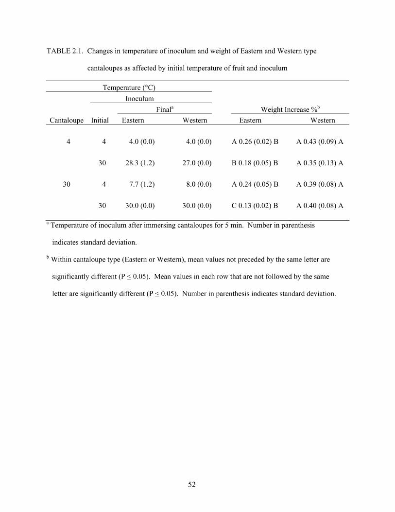
52
TABLE 2.1. Changes in temperature of inoculum and weight of Eastern and Western type
cantaloupes as affected by initial temperature of fruit and inoculum
Temperature (°C) Inoculum Finala Weight Increase %b
Cantaloupe Initial Eastern Western Eastern Western
4 4 4.0 (0.0) 4.0 (0.0) A 0.26 (0.02) B A 0.43 (0.09) A 30 28.3 (1.2) 27.0 (0.0) B 0.18 (0.05) B A 0.35 (0.13) A
30 4 7.7 (1.2) 8.0 (0.0) A 0.24 (0.05) B A 0.39 (0.08) A 30 30.0 (0.0) 30.0 (0.0) C 0.13 (0.02) B A 0.40 (0.08) A
a Temperature of inoculum after immersing cantaloupes for 5 min. Number in parenthesis
indicates standard deviation.
b Within cantaloupe type (Eastern or Western), mean values not preceded by the same letter are
significantly different (P < 0.05). Mean values in each row that are not followed by the same
letter are significantly different (P < 0.05). Number in parenthesis indicates standard deviation.
53
measurement. The stomata on leafy produce normally close after a small amount of water is lost,
but rapid cooling of some tissues can cause the stomata to remain open (Wills et al., 1981). A
rind surface meshwork of cantaloupe rind tissue, commonly referred to as the net, is an elaborate
system of lenticels and cork cells comprising the complementary tissue of lenticels and the
epiderm (Webster and Craig, 1976). Deviations from the norm in terms of uptake of water by
cantaloupes as affected by temperature differential may be attributable in part to the complex
lenticel structure of the rind net and stem scar tissues unique to cantaloupes. The surface of
cantaloupe rind is replete with lenticels and cork cells that do not possess a mechanism for
closing. A pervasive network of airspaces connecting the internal tissues of cantaloupe rind to
the external atmosphere supplies air for normal respiratory processes. Gaseous exchange is
facilitated by lenticular net development. When cantaloupes at a given temperature are
immersed in water at a lower temperature, the decreased internal gas pressure theoretically
causes an influx of water via portals such as lenticels and perhaps also the stem scar tissue. By
extension, this behavior could contribute to retention of water by cantaloupes immersed in
inoculum at 4°C.
The percentage weight gained by Western type cantaloupes as a result of immersion was
significantly greater (P < 0.05) than that for Eastern cantaloupes for all cantaloupe / inoculum
temperature combinations. The dissimilarity in weight gain response between Eastern and
Western type cantaloupes is attributable to differences in fruit morphology and density of rind
netting. Western type cantaloupes lack distinct sutures and the rinds are more densely and
evenly netted than are rinds of Eastern cantaloupes. Eastern cantaloupes have more prominent
crests and ridges, and rinds have a less uniform distribution of netting compared to rinds of
Western cantaloupes. Areas on the rind of Eastern type cantaloupes that lack netting would be
54
expected to imbibe less water, but be more sensitive to the effects of temperature differentials
owing to the lack of insulation assumed to be imparted by dense net tissue. Compared to Eastern
type cantaloupes, the uniformity of exposed surface area and intensity of netting of Western
cantaloupes could obscure the effects of temperature on water uptake. The dense netting of
Western type cantaloupes is a manifestation of greater disruption of the cuticle, enabling
retention of more water than netting of Eastern type cantaloupes.
Recovery of S. Poona from cantaloupes. Populations of S. Poona recovered from the
rind tissue and stem scar tissue of cantaloupes were, respectively, 0.10 – 1.05 log10 CFU/cm2 and
0.15 – 0.85 log10 CFU/g higher on TSAN than on BSAN, regardless of the fruit type or
temperature of fruit or inoculum (Table 2). In only one of sixteen cantaloupe type / temperature /
tissue type combinations (Western type, 4°C / 4°C, stem scar tissue) was the population
recovered on TSAN significantly (P < 0.05) higher than on BSAN. The lower number of S. Poona
recovered on BSAN is attributed to the inability of damaged or stressed cells to resuscitate when
exposed to selective conditions imposed by this medium. Populations recovered on TSAN and
BSAN from cantaloupes subjected to all other combinations of test parameters were not
significantly different (P > 0.05). Tryptic soy agar is a nutrient-rich medium used for isolating
and cultivating fastidious microorganisms whereas BSAN contains ingredients that are inhibitory
to the growth of gram-positive bacteria and perhaps also injured Salmonella. Nalidixic acid was
added to both media as a marker to eliminate or reduce the growth of gram negative
microorganisms naturally occurring on the cantaloupe surface. Gram-negative bacteria are
sensitive to nalidixic acid, having minimal inhibiting concentrations of < 10 µg/ml, unless
adapted to grow in media containing higher concentrations.
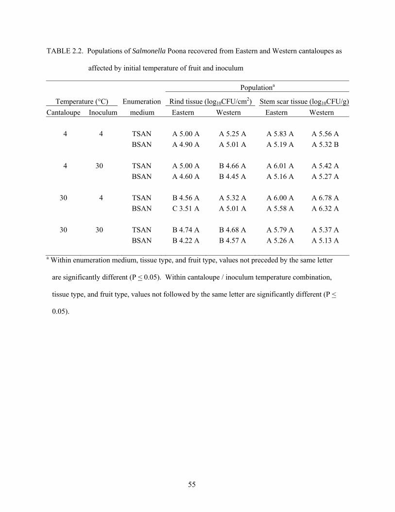
55
TABLE 2.2. Populations of Salmonella Poona recovered from Eastern and Western cantaloupes as
affected by initial temperature of fruit and inoculum
Populationa
Temperature (°C) Enumeration Rind tissue (log10CFU/cm2) Stem scar tissue (log10CFU/g)Cantaloupe Inoculum medium Eastern Western Eastern Western
4 4 TSAN A 5.00 A A 5.25 A A 5.83 A A 5.56 A
BSAN A 4.90 A A 5.01 A A 5.19 A A 5.32 B
4 30 TSAN A 5.00 A B 4.66 A A 6.01 A A 5.42 A BSAN A 4.60 A B 4.45 A A 5.16 A A 5.27 A
30 4 TSAN B 4.56 A A 5.32 A A 6.00 A A 6.78 A BSAN C 3.51 A A 5.01 A A 5.58 A A 6.32 A
30 30 TSAN B 4.74 A B 4.68 A A 5.79 A A 5.37 A BSAN B 4.22 A B 4.57 A A 5.26 A A 5.13 A
a Within enumeration medium, tissue type, and fruit type, values not preceded by the same letter
are significantly different (P < 0.05). Within cantaloupe / inoculum temperature combination,
tissue type, and fruit type, values not followed by the same letter are significantly different (P <
0.05).
56
Populations of S. Poona recovered from the rind of Eastern type cantaloupes after
immersion in suspensions at 4°C or 30°C were significantly higher (P < 0.05) when cantaloupes
were at 4°C compared to 30°C before immersion (Table 2.2). The population of S. Poona
recovered on BSAN from rind of Eastern cantaloupes at 30°C was significantly higher (P < 0.05)
when fruits were immersed in a warm (30°C) inoculum compared to populations recovered from
fruits immersed in chilled (4°C) inoculum, although the low number of cells may be attributable
in part to inability of injured cells to resuscitate and form colonies. Observations on populations
recovered on both media were contrary to predictions based on observations on infiltration of
microorganisms into tomatoes (Bartz, 1982; Bartz and Showalter, 1981) and apples (Buchanan et
al., 1999; Burnett et al., 2000; Kenney and Beuchat, 2002) as affected by temperature
differences between fruits and immersion suspensions. Results of these studies support the
contention that uptake of a larger volume of water by warm fruits exposed to cold water,
compared to cold fruits exposed to warm water, would allow more bacterial cells to adhere to or
infiltrate fruits during hydrocooling. Comparative recovery of S. Poona from the rind and stem
scar tissues of Eastern and Western cantaloupes exposed to positive and negative temperature
differentials did not conform to these predictions. We recovered significantly higher (P < 0.05)
numbers of S. Poona from the rind of chilled Eastern cantaloupes immersed in a warm
suspension of the pathogen (positive temperature differential) than from warm cantaloupes
immersed in a cool suspension (negative temperature differential). Our study indicates that
adherence to or infiltration of Eastern but not Western type cantaloupe rind by S. Poona is
enhanced when the cantaloupe is at 4°C compared to 30°C, regardless of the temperature of the
immersion suspension. The higher number of cells adhering to or infiltrating cantaloupe rind at
4°C, compared to 30°C, apparently masks any effect temperature differential might have on
57
extent of adherence or infiltration. The number of S. Poona cells recovered from rind tissue of
Western cantaloupes at 30°C immersed in inoculum at 30°C was significantly less (P < 0.05)
than the number of cells recovered from rinds of cantaloupes at 4°C or 30°C that were immersed
in inoculum at 4°C, indicating that immersion of warm fruits in warm inoculum diminishes
adherence of S. Poona. As with Eastern cantaloupes, the effect of temperature differential
between Western cantaloupes and inoculum did not conform to expectations based on
observations made by others on infiltration of spoilage and pathogenic bacteria into tomatoes and
apples.
The rind surface of Eastern and Western type cantaloupes is scabrous in texture due to
disruption of the cuticular layer and presence of the net. The presence of these complex surface
structures apparently negates predictions of infiltration based on the effects of temperature
differentials on infiltration of bacterial populations into apples and tomatoes, which have a
relatively even distribution of cuticle and smoother, much less porous surfaces. We hypothesize
that the atypical comparative recovery of S. Poona from the rind and stem scar tissues of Eastern
and Western type cantaloupes exposed to positive and negative temperature differentials is due
largely to their unique surface tissue composition, structure, and respiratory activity.
The observation that a significantly higher (P < 0.05) population of S. Poona was
recovered from Eastern cantaloupes at 4°C before immersion, compared to those at 30°C,
concurs with observations on the attachment of Escherichia coli O157:H7 to lettuce leaves as
affected by temperature (Takeuchi and Frank, 2000). They postulated that warm temperature
promotes active respiration of lettuce tissues, and that the resultant gas movement produces a
counterforce to resist penetration of E. coli O157:H7 cells. The attachment of E. coli O157:H7
to lettuce has also been attributed to changes in cell physiology and attachment mechanisms at
58
reduced temperature (Takeuchi et al., 2001). S. Poona may have exhibited a similar behavior on
cantaloupe rind. Cooling of superficial air on the porous surface of rind during the drying
process could have resulted in greater cell ingress of the tissue of warm Western cantaloupes that
were immersed in the 4°C suspension compared to the warm cantaloupes that were immersed in
30°C suspension. If a reduction in temperature during drying enhances attachment or penetration
of bacterial cells into damaged plant tissues, we postulate that a greater number of cells adhered
to cooled Western cantaloupe rind tissues.
The outer surface of cantaloupes presents a variety of structures and surfaces to which
bacteria may attach. As cantaloupe fruits mature, the hard epicarp undergoes progressive
modification that involves the development of a network of corklike tissue originating from
multi-layered hypodermal tissues (Webster and Craig, 1976). The surface becomes extensively
cracked and deep fissures disrupt the cuticle and some of the subtending epidermal and
hypodermal cells. The thickened and pitted cell walls have microscopic local thickenings and
warts. Additionally, there are depressions and grooves with jointed conical hairs up to 375 µm in
length on the net surface (Barber, 1909; Webster and Craig, 1976). It has been hypothesized that
the presence of cuticle on fruits and other plant parts prevents or retards penetration by
foodborne pathogens (Burnett and Beuchat, 2001; Kenney et al., 2001) and phytopathogenic
microorganisms (Beattie, 2002; Morris et al., 2002). This function is attributed to the chemical
nature of cutin, a highly polymerized hydrophobic material consisting of fatty acids. However,
extensive disruption of the cuticle on cantaloupes would compromise such effectiveness
(Webster and Craig, 1976). Damaged cuticle has been shown to enhance the ability of bacteria
to adhere to plant tissues (Kenney et al., 2001). Microorganisms have been observed to
concentrate in areas on fresh cut spinach where the cuticle and underlying tissues are damaged,
59
causing infection of the internal palisade parenchyma (Babic et al., 1996). When the
topographical structures on apple skin was examined with scanning electron microscopy,
attachment of S. Chester was primarily on surfaces of injured tissue and on stem and calyx parts,
but rarely on the unbroken skin (Liao and Sapers, 2000). Preferential attachment of S. Chester
was attributed to differences in topographical structures and physicochemical properties.
Populations of S. Poona recovered from the stem scar tissues of Eastern and Western type
cantaloupes were not significantly different (P > 0.05), regardless of cantaloupe / inoculum
temperature combination (Table 2.2). The stem scar tissue of tomatoes, on the other hand, is a
primary site of infiltration of bacteria (Bartz, 1982; Bartz and Showalter, 1981; Zhuang and
Beuchat, 1996). When raw tomato fruits were immersed in a suspension of Salmonella
Montevideo, more cells were retained in the stem scar and core tissues than on tomato skin (Wei
et al., 1995). Cantaloupe fruits mature on the vine with the peak of the respiratory climacteric
coinciding with abscission at approximately 42 days after anthesis. The entire middle lamella
surrounding cells at the stem / fruit tissue junction breaks down, and pectin dissolution and
changes in polysaccharides result in a simultaneous separation of contiguous parenchyma cells
within the abscission zone (Webster, 1975). Disjunct parenchyma cells soon collapse and
several small cavities, which are separated from each other by intact parenchyma and vascular
tissues, become evident. The anatomical and histochemical changes that occur in postharvest
cantaloupes could affect attachment or infiltration of bacterial cells. Irregularities such as
abrasions, crevices, and pits on the surface of cantaloupes enhance the ease of bacterial cell
attachment and interfere with their removal (Ukuku et al., 2001). These processes may have
occurred in Eastern and Western cantaloupes examined in our study, although the number of S.
60
Poona adhering to or infiltrating the stem scar surface and tissue was unaffected by differences in
temperatures of cantaloupes and inocula.
Attachment of E. coli O157:H7 to the surface of lettuce (Takeuchi and Frank, 2000) and
cantaloupes (Ukuku et al., 2001) is influenced by the inoculum size. Attachment of bacteria to
cantaloupe has also been attributed to surface charge and hydrophobicity of cells, as well as
associated flagella, fimbriae, outer membrane proteins, and extracellular polysaccharides (Ukuku
and Fett, 2002). The presence of waxes and suberin in cork cells within lenticels is thought to
render the rind net impervious to water (Webster and Craig, 1976). However, the porous netted
texture of cantaloupe rind promotes attachment and harborage of microorganisms, which results
in difficulty in sanitization (Gagliardi et al., 2003).
This study demonstrates that S. Poona in immersion water can adhere to cantaloupe rind
and stem scar tissues. The histology of the cantaloupe rind and stem scar tissues augments
attachment and penetration, and may thereby reduce the effectiveness of sanitizer treatments.
Salmonella and other foodborne pathogens attached to the external surfaces of cantaloupes are of
great concern because they can be transferred to the edible pulp during cutting and removal of
the rind. The abundance of nutrients and the near neutral pH of mature cantaloupe flesh would
be expected to enhance the survival and growth of Salmonella and other foodborne pathogens,
even at refrigeration temperature. It is important to develop and implement production,
processing, and preparation practices that include vigilant monitoring of the microbiological
quality of water that comes in contact with cantaloupes and prevent cross-contamination with
pathogens from other sources at all points in the preharvest and postharvest system.
61
REFERENCES
Babic, I., S. Roy, A.E. Watada, and W.P. Wergin. 1996. Changes in microbial population on fresh cut spinach. Int. J. Food Microbiol. 31:107-119.
Bacon, R.T., J.N. Sofos, K.E. Belk, D.R. Hyatt, and G.C. Hyatt. 2002. Prevalence and antibiotic
susceptibility of Salmonella isolated from beef animal hides and carcasses. J. Food Prot. 65:284-290.
Barber, K.G. 1909. Comparative histology of fruit and seeds of certain species of Cucurbitaceae. Bot. Gaz. 47:263-310.
Bartz, J.A. 1982. Infiltration of tomatoes immersed at different temperatures to different depths
in suspensions of Erwinia caratovora subsp. caratovora. Plant Dis. 66:302-305. Bartz, J.A. and R.K. Showalter. 1981. Infiltration of tomatoes by aqueous suspensions.
Phytopathology 71:515-518. Beattie, G.A. 2002. Leaf surface waxes and the process of leaf colonization by microorganisms,
p. 3-23. In S.E. Lindow, E.I. Hecht-Poinar, and V.J. Elliot (Ed.); Phyllosphere microbiology, APS Press, St. Paul, MN.
Beru, N. and P.A. Salsbury. 2002. FDA’s produce safety activities. Food Safety Magazine. p.14,
February/March 2002. Brackett, R.E. 1987. Microbiological consequences of minimally processed fruits and
vegetables. J. Food Qual.10:195-206. Brackett, R.E., D.M. Smallwood, S.M. Fletcher, and D.L. Horton. 1993. Food safety: Critical
points within the production and distribution system. In R.L. Shewfelt and S.E. Prussia (Ed.); Postharvest handling: A systems approach. Academic Press, San Diego, California.
Buchanan, R.L., S.G. Edelson, R.L. Miller, and G.M. Sapers. 1999. Contamination of intact
apples after immersion in an aqueous environment containing Escherichia coli O157:H7. J. Food Prot. 62:444-450.
Burnett, S.L. and L.R. Beuchat. 2001. Human pathogens associated with raw produce and unpasteurized juices, and difficulties in decontamination. J. Ind. Microbiol. Biotechnol. 27:104-110.
Burnett, S.L., J. Chen, and L.R. Beuchat. 2000. Attachment of Escherichia coli O157:H7 to the surface and internal structures of apples as demonstrated by confocal scanning laser microscopy. Appl. Environ. Microbiol. 66:4679-4687.
62
Centers for Disease Control and Prevention (CDC). 1991. Multistate outbreak of Salmonella poona infections – United States and Canada, 1991. Morbid. Mortal. Wkly. Rep. 40:549- 552.
Centers for Disease Control and Prevention (CDC). 1993. Multistate outbreak of Salmonella
serotype Montevideo infections. EPI-AID 93-79, 1993. Centers for Disease Control and Prevention (CDC). 2002. Multistate outbreaks of Salmonella
serotype Poona infections associated with eating cantaloupes from Mexico – United States and Canada, 2000 – 2002. Morbid. Mortal. Wkly. Rep. 51:1044-1047.
Deeks, S., A. Ellis, B. Ciebin, R. Khakhria, M. Naus, and J. Hockin. 1998. Salmonella Oranienberg, Ontario. Can. Comm. Dis. Rep. 24:177-179.
Ercolani, G.L. 1976. Bacteriological quality assessment of fresh marketed lettuce and fennel.
Appl. Environ. Microbiol. 55:832-836. Food and Drug Administration (FDA). 1998. Guidance for industry. Guide to minimize
microbial food safety hazards for fresh fruits and vegetables. U.S. Food and Drug Administration, Center for Food Safety and Applied Nutrition. October 26, 1998. [Internet, WWW], ADDRESS: http://www.cfsan.gov/~dms/prodguid.html. Accessed 4 February 2002.
Food and Drug Administration (FDA). 2001a. FDA Survey of imported fresh produce. FY 1999 Field assignment. US Food and Drug Administration. Center for Food Safety and Applied Nutrition. Office of Plant and Dairy Foods and Beverages. January 30, 2001. [Internet, WWW], ADDRESS: http://www.cfsan.gov/~dms/prodsur6.html. Accessed 25 January 2002.
Food and Drug Administration (FDA). 2001b. FDA Survey of domestic fresh produce: interim results. US Food and Drug Administration. Center for Food Safety and Applied Nutrition. June 30, 2001. [Internet, WWW], ADDRESS: http://vm.cfsan.fda.gov/~dms/prodsur8.html Accessed 25 January 2002.
Gagliardi, J.V., P.D. Miller, G. Lester, and D. Ingram. 2003. On-farm and postharvest processing sources of bacterial contamination to melon rinds. J. Food Prot. 66:82-87.
Garcia-Villanova Ruiz, B., R. Galvez Vargas, and R. Garcia-Villanova. 1987. Contamination
on fresh vegetables during cultivation and marketing. Intl. J. Food Microbiol. 4:285-291. Institute of Food Technologists/Food and Drug Administration of the United States Department
of Health and Human Services (IFT/FDA). 2001. Analysis and evaluation of preventative control measures for the control and reduction/elimination of microbial hazards on fresh and fresh-cut produce. IFT/FDA Contract No. 223-98-2333. Task Order No. 3. Institute of Food Technologists. Chicago, IL.
63
Kenney, S.J. and L.R. Beuchat. 2002. Survival of Escherichia coli O157:H7 and Salmonella Muenchen on apples as affected by application of commercial fruit waxes. Intl. J. Food Microbiol. 77:223-231.
Kenney, S.J., S.L. Burnett, and L.R. Beuchat. 2001. Location of Escherichia coli O157:H7 on
and in apples as affected by bruising, washing, and rubbing. J. Food. Prot. 64:1328-1333. Leverentz, B., W.S. Conway, Z. Alavidze, W.J. Janisiewicz, Y. Fuchs, M.J. Camp, E.
Chighladze, and A. Sulakvelidze. 2001. Examination of bacteriophage as a biocontrol method for Salmonella on fresh-cut fruit: A model study. J. Food Prot. 64:1116-1121.
Liao, C.-H. and G. M. Sapers. 2000. Attachment and growth of Salmonella Chester on apple
fruits and in vivo response of attached bacteria to sanitizer treatments. J. Food Prot. 63:876-883.
Mohle- Boetani, J.C. R. Reporter, S.B. Werner, S. Abbott, J. Farrar, S.H. Waterman, and D.J. Vugia. 1999. An outbreak of Salmonella serogroup Saphra due to cantaloupes from Mexico. J. Infect. Dis. 180:1361-1364.
Morris, C.F., M.H. Barnes, and R.J.C. McLean. 2002. Biofilms on leaf surfaces: Implications for the biology, ecology, and management of populations of epiphytic bacteria, p.139-155. In S.E. Lindow, E.I. Hecht-Poinar, and V.J. Elliot (Ed.); Phyllosphere microbiology. APS Press, St. Paul, MN.
Suslow, T.V. 2002. Production practices affecting the potential for persistent contamination of plants by microbial foodborne pathogen, p. 241-256. In S.E. Lindow, E.I. Hecht-Poinar, and V.J. Elliot (Ed.); Phyllosphere microbiology. APS Press, St. Paul, MN.
Suslow, T. and M. Cantwell. 2001. Recent findings on fresh-cut cantaloupe and honeydew melon. Fresh cut. April 2001. Pp. 12, 20, 32-33.
Takeuchi, K. and J.F. Frank. 2000. Penetration of Escherichia coli O157:H7 into lettuce tissues
as affected by inoculum size and temperature and the effect of chlorine treatment on cell viability. J. Food Prot. 63:434-440.
Takeuchi, K. A.N. Hassan, and J.F. Frank. 2001. Penetration of Escherichia coli O157:H7 into
lettuce as influenced by modified atmosphere and temperature. J. Food Prot. 64:1820-1823.
Ukuku, D.O., V. Pilizota, and G.M. Sapers. 2001. Influence of washing treatment on native microflora and Escherichia coli population of inoculated cantaloupes. J. Food Safety. 21:31-37.
Ukuku, D.O. and G.M. Sapers. 2001. Effect of sanitizer treatments on Salmonella Stanley
64
attached to the surface of cantaloupe and cell transfer to fresh-cut tissues during cutting practices. J. Food Prot. 64:1286-1291.
Ukuku, D.O. and W. F. Fett. 2002. Relationship of cell surface charge and hydrophobicity to strength of attachment of bacteria to cantaloupe rind. J. Food Prot. 65:1093-1099.
Webster, B.D. 1975. Anatomical histochemical modifications associated with abscission of
Cucumis fruits. J. Amer. Soc. Hort. Sci. 100:180-184. Webster, B.D. and M.E. Craig. 1976. Net morphogenesis and characteristics of the surface of
muskmelon fruit. J. Amer. Soc. Hort. Sci. 101:412-415. Wei, C.I., T.S., Huang, J.M. Kim, W.F. Lin, M.L. Tamlin, and J.A. Bartz. 1995. Growth and
survival of Salmonella Montevideo on tomatoes and disinfection with chlorinated water. J. Food Prot. 58:829-836.
Wells, J.M., and J.E. Butterfield. 1997. Salmonella contamination associated with bacterial soft rot of fresh fruits and vegetables in the marketplace. Plant Dis. 81:867- 872.
Wills, R.B.H., T.H Lee, D. Graham, W.B. Mc Glasson and E.G. Hall. 1981. Postharvest. An
introduction to the physiology and handling of fruit and vegetables. AVI publishing Company. New South Wales University Press, Australia.
Wood, R.C., C. Hedberg, and K. White. 1991. A multistate outbreak of Salmonella Javiana infections associated with raw tomatoes, p. 69. Abstr. Epidemic Intelligence Serv. 40th Annu. Conf. Centers for Disease Control and Prevention, Atlanta.
Zhuang, R.Y. and L.R. Beuchat. 1996. Effectiveness of trisodium phosphate for killing Salmonella Montevideo on tomatoes. Lett. Appl. Microbiol. 22:97-100.
65
CHAPTER 3
METABIOTIC ASSOCIATIONS OF MOLDS AND SALMONELLA POONA
ON INTACT AND WOUNDED CANTALOUPE RIND1
______________________
1 Richards, Glenner M. and Larry R. Beuchat. 2003. To be submitted to International Journal of Food Microbiology.
66
ABSTRACT
Salmonella Poona, a serotype rarely implicated in human infections, has recently caused
several cantaloupe-associated outbreaks of salmonellosis in the United States and Canada.
Metabiotic associations of molds and foodborne pathogens on produce have been reported. We
tested proteolytic activity and measured changes in the pH of cantaloupe rind caused by growth
of Alternaria alternata, Cladosporium cladosporioides, Epicoccum nigrum, Geotrichum
candidum, and Penicillium expansum, molds commonly involved in postharvest decay, and
determined survival and growth characteristics of S. Poona co-infected with each mold on the
surface rind and in wounded rind tissue as affected by temperature. C. cladosporioides, G.
candidum, and P. expansum, but not A. alternata and E. nigrum, showed proteolytic activity on
agar media containing gelatin and/or casein, with concurrent increases in pH. Intact and
mechanically wounded tissue of cantaloupe rinds were inoculated with a five-strain mixture of S.
Poona and/or test mold. Five inoculation schemes were used: mold only, S. Poona only, mold
and S. Poona simultaneously, mold then S. Poona 3 days later, and S. Poona then mold 3 days
later. The pH of cantaloupe rinds inoculated with molds and stored at 20°C for 14 days was
significantly higher (P < 0.05) than on day 0. However, only the pH of rinds inoculated with C.
cladosporioides or G. candidum was significantly higher (P < 0.05) on day 21 than on day 0,
when cantaloupes were stored at 4°C. An initial population of S. Poona increased from 3.3 log10
CFU/sample (ca. 7 cm2) of cantaloupe rind to populations as high as 9.5 log10 CFU/sample
during storage at 20°C for up 14 days, regardless of co-inoculation with molds. Populations of S.
Poona decreased or remained constant at 4°C for up to 21 days. Results demonstrate that S.
Poona can persist and grow on intact, wounded, and decaying cantaloupe rind at refrigerated and
ambient temperatures and is not affected by the presence of test molds.
67
INTRODUCTION
Fresh produce, historically regarded as being microbiologically safe, is now recognized
as a potential vehicle for foodborne pathogens. Numerous pathogenic microorganisms have
been isolated from raw fruits and vegetables, and cantaloupes are among the produce that have
been implicated in outbreaks of human infections (Beuchat, 1996; 2002). Cantaloupes provide
variety, flavor, aesthetic appeal, and essential nutrients to diets. Minimally processed fresh-cut
cantaloupe is widely accepted in the United States, largely because it offers convenience in
preparation and serving. The per capita consumption of cantaloupes has increased in the United
States in recent years. Domestic production and importation of cantaloupes have steadily
increased to adequately meet the growing demand. However, postharvest decay limits shelf life
in the fresh-fruit market (Sapers et al., 2001).
Several national and international outbreaks of salmonellosis have been
epidemiologically linked to consumption of fresh cantaloupes (CDC, 1991; CDC, 2002; Deeks et
al., 1998, IFT/FDA, 2001, Mohle-Boetani et al., 1999; Ries et al., 1990). In October 2002, the
United States Food and Drug Administration (FDA) issued an alert on importation of
cantaloupes originating in Mexico. Outbreaks of salmonellosis were traced to cantaloupes
originating in Mexico in 2000, 2001, and 2002 (FDA, 2002). Salmonella Poona, a relatively rare
serotype, has caused four of seven cantaloupe-associated salmonellosis outbreaks. Typically,
human infection with S. Poona is associated with exposure to reptiles. Three outbreaks of S.
Poona infections were epidemiologically linked to cantaloupes grown on Mexican farms in the
same region, suggesting the possibility of a unique natural reservoir of this serotype in the
production, harvesting, or packing environment. It has been hypothesized that reptiles such as
iguanas are attracted to feed on melons in the field or in packing sheds, thereby cross-
68
contaminating preharvest fruits or equipment and water used in washing and cooling processes
(CDC, 2002).
Cantaloupe fruits may be in direct contact with the ground during their development on
long, running, non-climbing vines that are prostrate on the soil. The growth habit of cantaloupes
enhances the potential for fruits to be contaminated by pathogens that may be present in the soil.
Postharvest handling may also bring cantaloupes in direct contact with various sources of
foodborne pathogens. Fruits are particularly subject to contamination when rind surface integrity
is compromised through disease, bruising, cutting, or peeling. Inadequate washing, storage, and
handling augment the potential for growth of pathogens and cross contamination (Sewell and
Faber, 2001). Field surveys conducted by the FDA during 1990 and 1991 revealed that 0.76%
and 1.06%, respectively, of cantaloupe rinds harbored several serovars of Salmonella (Madden,
1992; Golden et al., 1993). More recently, 7.3% of 151 imported cantaloupes were found to
harbor Salmonella or Shigella (FDA, 2001a). The results of a survey of domestic cantaloupes
indicated the presence of these pathogens on 4.3% of 92 fruits (FDA, 2001b). Studies by Ukuku
and Sapers (2001) indicate that outbreaks of salmonellosis may result from fresh-cut cantaloupes
consumed 3 days after contamination, because cells attached to the cantaloupe rind for 3 or more
days were difficult to kill using chlorine or hydrogen peroxide treatments.
The association of salmonellosis with cantaloupes on salad bars and fruit salads suggests
an introduction of Salmonella into the edible flesh of fruits during slicing, or direct contact of the
flesh with contaminated rind (Golden et al., 1993). As few as 150 Salmonella/cm2 of netted rind
have been shown to contaminate the pulp as a result of cutting whole cantaloupes (Suslow and
Cantwell, 2001). The potential for growth of Salmonella on tissue is exacerbated by displaying
or storing cut cantaloupes at ambient temperature for an extended time (CDC, 1991; Golden et
69
al., 1993). Cut cantaloupe is considered a potentially hazardous food in the FDA Food Code
because its low acidity (pH 5.2 to 6.7) and high water activity (0.97 to 0.99) enable the growth of
pathogens (IFT/FDA, 2001).
By virtue of their differences in physical structure, intact plant tissues are resistant to
infiltration and infection by most microorganisms. Plant diseases are the exception but occur
only when several conditions are fulfilled (Snowdon, 1990). Extreme temperatures alter the
physiology of raw fruits and vegetables, increasing their susceptibility to certain plant pathogens.
Disease incidence and severity is dependent on the capability and population of microorganisms
in the inoculum. Fungi are overwhelmingly responsible for postharvest diseases of fruits
(Sommer et al., 1992). Cantaloupes are susceptible to postharvest fungal rots, especially under
wet, warm conditions. Rots occur on the external surface of the fruit and gradually progress
inward to the flesh. Phytopathogenic fungi of major concern in cantaloupes include species of
Alternaria, Cladosporium, Fusarium, Geotrichum, Penicillium, Rhizopus, and Mucor (Ceponis
et al., 1986; Salunkhe and Desai, 1984; Suslow et al., 2000). These molds are also involved in
spoilage of foods that are not necessarily of plant origin.
Infection of cantaloupes by plant pathogenic fungi and contamination with foodborne
bacteria may occur before harvesting, at the time harvest, during handling, storage, transport, and
marketing, or after purchase by the consumer. Metabiosis refers to circumstances in which
growth of one microorganism creates favorable conditions for growth of another (Odlaug and
Pflug, 1979). Metabiotic associations of molds and Clostridium botulinum on tomatoes
(Draughon et al., 1988) and Escherichia coli O157:H7 and Listeria monocytogenes on fresh-cut
apples (Chardonnet et al., 2002; Conway et al., 2000; Riordan et al., 2000) have been reported.
The behavior of foodborne pathogens such as Salmonella on or in cantaloupes as affected by
70
metabiotic activities of plant pathogens has not been investigated. During transportation,
storage, and display at the retail level, physically sound and rotting fruits can come in direct
contact with each other. Cross contamination with Salmonella, followed by growth of the
pathogen, would increase the safety risk to the consumer. Thrifty consumers may be willing to
purchase fruits with decaying areas, pare away these areas, and consume the presumably safe
remainder.
The objective of this study was to examine the association between selected molds
pathogenic to cantaloupes and S. Poona on the surface of intact rind and in wounds in the rind.
Changes in pH caused by growth of molds were monitored, as were survival and growth of S.
Poona in co-infected tissue as affected by temperature.
MATERIALS AND METHODS
Culture conditions for S. Poona and preparation of inoculum. Five strains of S.
Poona isolated from patients in cantaloupe-associated outbreaks of salmonellosis were used.
Strains 00A3207, 01A3923, and 02A3275 were obtained from the State of California,
Department of Health Services, while strains 00A3279 and 01A242 were obtained from Dr.
Trevor Suslow, University of California, Davis. Each strain was progressively made resistant to
nalidixic acid by a series of nine transfers in tryptic soy broth (TSB; BBL/Difco, Sparks, MD)
supplemented with increasing concentrations of nalidixic acid up to 50 µg/ml (TSBN). Cultures
were incubated at 37°C for 24 h before transferring to TSB containing a higher concentration of
nalidixic acid. The nalidixic acid-resistant cells were surface plated on tryptic soy agar (TSA;
BBL/Difco) supplemented with 50 µg of nalidixic acid/ml (TSAN) and incubated at 37°C for 24
h. Stock cultures were stored on TSAN at 4°C.
71
Stock cultures were streaked on TSAN and incubated at 37°C for 24 h before transferring
cells from a single colony of each strain to 10 ml of TSBN and incubating at 37°C for 24 h.
Three consecutive 24-h transfers of suspensions were made via loop inocula into 10 ml of TSBN.
After incubation at 37°C for 24 h, the cells were harvested by centrifugation at 2,500 x g for 10
min in a Centra CL2 centrifuge (International Equipment Company, Needham Heights, MA).
Cells were washed two times in 10 ml of sterile deionized water, then resuspended in 10 ml of
sterile 0.1% (w/v) peptone water. The population (CFU/ml) in each suspension was determined
by serially diluting suspensions in sterile 0.1% peptone water and surface plating samples (0.1 ml
in duplicate) on TSAN. Plates were incubated at 37°C for 24 h before colonies were counted.
Random presumptive-positive colonies of Salmonella were confirmed using triple sugar iron,
lysine iron agar, and a Salmonella latex agglutination assay (Oxoid, Basingstoke, Hampshire,
UK).
Equal volumes (5 ml) of suspensions of each strain were combined to give 25 ml of a
five-strain mixture of S. Poona containing approximately equal populations (ca. 9 log10 CFU/ml)
of each strain. The suspension was serially diluted to give an inoculum that contained ca. 5 log10
CFU of each strain/ml.
Procedures for growing molds and preparation of inocula. Strains of A. alternata, C.
cladosporioides, E. nigrum, G. candidum, and P. expansum isolated from cantaloupes were
obtained from Dr. Trevor Suslow, University of California, Davis. Molds were maintained on
dichloran rose bengal chloramphenicol (DRBC) agar (BBL/Difco) and potato dextrose agar
(PDA) (BBL/Difco) slants at 4°C, and subcultured every 14 days by streaking on fresh DRBC
agar and PDA plates and incubating at 25°C for 5 days.
72
Cultures were streaked on DRBC agar and incubated at 25°C for 5 days. Suspensions of
conidia or arthroconidia (G. candidum) were prepared by pipetting 10 ml of sterile deionized
water containing 0.05% Tween 80 onto the surface of the 5-day-old cultures and gently
massaging for 1 min with a sterile glass rod. Using a sterile Pasteur pipette, the conidia
suspension was removed from the surface of the plate and deposited in a sterile test tube. The
number of conidia (CFU/ml) in each suspension was determined by serially diluting samples in
sterile deionized water and surface plating (0.1 ml in duplicate) on DRBC agar. Plates were
incubated at 25°C for 5 days before colonies were counted. The number of conidia in each
suspension was adjusted to ca. 4 – 5 log10 CFU/ml by diluting in sterile deionized water
containing 0.05% Tween 80.
Determination of proteolytic activity of molds. A. alternata, C. cladosporioides, E.
nigrum, G. candidum, and P. expansum were examined for extracellular proteolytic activity on
gelatin agar (GA, pH 5.9) and standard methods casienate (SMC, pH 6.3) agar. GA contains
(per liter of deionized water): 30 g of gelatin (BBL/Difco), 10 g of tryptone (BBL/Difco), 10 g
of sodium chloride (Fisher Scientific, Fair Lawn, NJ), and 15 g of agar (BBL/Difco). The
mixture was heated to dissolve ingredients, then autoclaved at 121°C for 15 min before cooling
to 50°C and pouring 18 ml into petri plates (100 x 15 mm). SMC agar contains (per liter of
deionized water): 23.4 g of standard methods agar (SMA; BBL/Difco), 10 g of sodium casienate
(ACROS; Morris Plains, NJ), 4.41 g hydrated trisodium citrate (ICN Biomedicals Inc., Aurora,
OH), and 4.38 g of hydrated calcium chloride (Fisher Scientific). Trisodium citrate was
dissolved in 1 liter of deionized water and divided into two 500-ml portions. SMA and calcium
chloride were added to one 500-ml portion of trisodium citrate and heated to dissolve. Sodium
casienate was added to the second 500-ml portion of sodium citrate and blended in a Model
73
56000 blender (Hamilton Beach / Procter Silex, Washington, NC) for 2 min. The two 500-ml
portions were combined in a 6-liter Erlenmeyer flask and sterilized at 121°C for 15 min. Twenty
milliliters of sterile 1 M calcium chloride solution was added to the cooled (50°C) agar and
thoroughly mixed before pouring (18 ml) into petri plates (100 x 15 mm).
Molds were inoculated into GA and SMC by stabbing two locations (approximately 2 cm
apart) on each plate with a probe containing conidia picked from 5-day-old colonies grown on
DRBC agar slants at 25°C. Plates were incubated upright at 20°C for 7 days or at 4°C for 21
days before examining for evidence of proteolytic activity. Proteolytic activity was manifested
by the development of a halo (zone of gelatin liquefaction or casienolysis) surrounding colonies
on GA and/or SMC agar. The halo on the transparent GA appeared as a cloudy area surrounding
the colonies, while on white, opaque SMC agar haloes appeared as translucent areas. Diameters
of colonies and haloes were measured with a dial caliper (Monostat, Zurich, Switzerland). The
surface pH of GA and SMC agar at the edge of each colony and 2 cm away from the colony was
measured using a surface pH probe.
Preparation and inoculation of cantaloupes. Full slip Eastern type cantaloupes
(Cucumis melo L. var. reticulatus Naud, cultivar Athena) were purchased in July – August, 2002
from a grocery in Griffin, GA and stored at 4°C for 2 days. Prior to using in experiments, the
cantaloupes were adjusted to 22 + 1°C over a 16 – 20 h period. Twenty-four unwashed
cantaloupes, free of visible wounds, cuts, and bruises, and supported by a 15-cm diameter plastic
potting saucer (Ivex, Tustin, CA), were randomly divided into eight groups of three fruits each
and placed in covered 27-liter polypropylene containers (59 cm long x 43 cm wide x 15 cm high;
Sterilite, Townsend, MA) in which twenty-eight holes (ca. 6 mm diameter) were made to
minimize the condensation of water on the surface of rinds and facilitate ventilation inside the
74
container. Each of the three fruits in the eight groups (treatments) represented a replicate. A
permanent ink marker was used to make six 3-cm diameter circles (two parallel rows of three, ca.
6 cm apart) on the rind of each cantaloupe, designating sites for inoculation. The end (8 mm
wide) of a sterile stainless steel spatula was used to create a wound 4 mm deep in the center of
three of the six circles. This procedure resulted in six inoculation sites (three intact and three
wounded) on each cantaloupe. Each pair of inoculation sites (one intact and one wounded)
represented sampling locations for each of three sampling days (days 7, 14, and 21) on each
replicate cantaloupe. The surface pH of the intact rind and the cut surface of wounded rind
within the circles was determined with a surface probe using a Basic pH meter (Denver
Instrument Company, Arvada, CO). Cantaloupes were inoculated with mold and/or S. Poona
suspensions using a Precision Microliter Pipette (Rainin, Woburn, MA). Inoculum was
deposited at 2 – 3 locations inside the 3-cm circles on the surface of intact rinds or directly in the
cut rind tissue. Five inoculation schemes were used:
(i) Mold only: Cantaloupes were inoculated with 20 µl of conidia suspension at each of
three intact and three wounded sites on six fruits on day 0. One group of three cantaloupes was
stored at 4°C and the other group of three cantaloupes was stored at 20°C.
(ii) S. Poona only: Cantaloupes were inoculated with 20 µl of S. Poona suspension at
each of three intact and three wounded sites on six fruits on day 0. One group of three
cantaloupes was stored at 4°C and the other group was stored at 20°C.
(iii) Mold and S. Poona: Cantaloupes were inoculated with 10 µl of conidia suspension
and 10 µl of S. Poona suspension at each of three intact and three wounded sites on six fruits on
day 0. One group of three cantaloupes was stored at 4°C and the other group was stored at 20°C.
75
(iv) Mold, then S. Poona 3 days later: Cantaloupes were inoculated with 20 µl of mold
conidia suspension on day 0, followed by 20 µl S. Poona suspension on day 3 at each of three
intact and three wounded sites on three fruits. All three cantaloupes were stored at 20°C.
(v) S. Poona, then mold 3 days later: Cantaloupes were inoculated with 20 µl of S. Poona
suspension on day 0, followed by 20 µl of mold conidia suspension on day 3 at three intact and
three wounded sites on three fruits. All three cantaloupes were stored at 20°C.
The inoculum on the surface of cantaloupes was dried by placing the fruits in a laminar
flow hood (class II, type A/B3) for 2 h at 22 + 1°C before placing in polyethylene containers and
incubating at 4°C or 20°C for up to 21 days.
Microbiological analyses. Intact and wounded rinds of cantaloupes were analyzed for
the presence of test molds and the presence and number of S. Poona 7, 14, and 21 days after
inoculation. For cantaloupes inoculated with mold and S. Poona on two different days (days 0
and 3), analysis was done 7, 14, and 21 days after the second inoculation day.
After the pH of the site inoculated with mold or mold and S. Poona was measured,
samples were removed from the surface with a sterile inoculation loop, streaked on duplicate
DRBC agar, and incubated at 25°C for 5 days. Isolates were examined to confirm the presence
of test molds.
Samples of intact and wounded rind tissues (ca. 3 cm diameter, 4 mm deep) inoculated
with S. Poona or S. Poona and mold were excised using a sterile spatula and placed in separate
stomacher 80 bags (Seward Medical Ltd., London, UK). Forty milliliters of sterile lactose broth
(LB; BBL/Difco) containing 50 µg of nalidixic acid/ml (LBN) was added. The sample was
hand rubbed and macerated, then pummeled for 1 min at normal speed in a Model 80 Stomacher
(Seward Medical). The liquid portion of the homogenate was serially diluted in sterile 0.1%
76
peptone water and surface plated (0.25 ml of undiluted liquid in quadruplicate and 0.1 ml of
diluted liquid in duplicate) on TSAN. Plates were incubated at 37°C for 24 h before
presumptive-positive S. Poona colonies were counted. Random presumptive-positive colonies
were selected for confirmation as described above. Homogenates containing LBN and rind
tissue were incubated at 37°C for 24 h. For samples that did not yield S. Poona colonies on
TSAN, the LBN/rind mixture was examined for the presence of S. Poona. A 1-ml sample of the
enriched mixture was inoculated into 10 ml of selenite cysteine (SC) broth (BBL/Difco),
incubated at 37°C for 24 h, and surface plated on bismuth sulfite agar (BSA; BBL/Difco)
supplemented with 50 µg of nalidixic acid/ml (BSAN). Plates were incubated at 37°C for 24 h
before random presumptive-positive colonies were selected for confirmation as described above.
Statistical analysis. Three replicate experiments were conducted and three samples were
analyzed at each sampling time. Mean values were analyzed to determine significant differences
in pH of intact and wounded cantaloupe rind inoculated with mold and/or S. Poona. Significant
differences in populations of S. Poona on intact and wounded cantaloupe rind as affected by co-
infection with mold and storage temperature were also determined. General linear models on
SAS software (Statistical Analysis Systems Institute, Cary, NC) were used. Significant
differences (P < 0.05) between mean values were determined using Duncan’s multiple range
tests.
RESULTS AND DISCUSSION
Proteolytic activity of molds. The five molds selected for this study were examined for
their ability to hydrolyze gelatin and casein in GA and SMC agar, respectively. As evidenced by
the formation of haloes around colonies, C. cladosporioides and G. candidum were proteolytic at
4 and 20°C (Table 3.1). C. cladosporioides showed proteolytic activity at 4°C on GA but not on
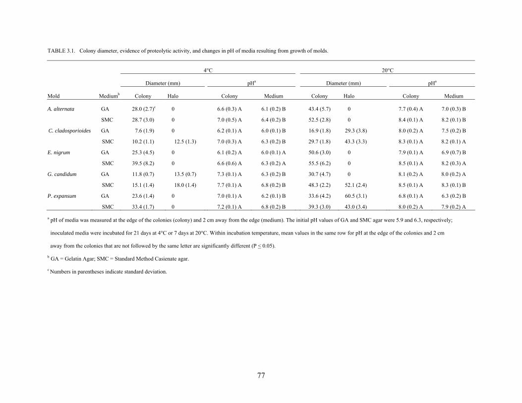
77
TABLE 3.1. Colony diameter, evidence of proteolytic activity, and changes in pH of media resulting from growth of molds.
4°C 20°C
Diameter (mm) pHa Diameter (mm) pHa
Mold Mediumb Colony Halo Colony Medium Colony Halo Colony Medium
A. alternata GA 28.0 (2.7)c 0 6.6 (0.3) A 6.1 (0.2) B 43.4 (5.7) 0 7.7 (0.4) A 7.0 (0.3) B
SMC 28.7 (3.0) 0 7.0 (0.5) A 6.4 (0.2) B 52.5 (2.8) 0 8.4 (0.1) A 8.2 (0.1) B
C. cladosporioides GA 7.6 (1.9) 0 6.2 (0.1) A 6.0 (0.1) B 16.9 (1.8) 29.3 (3.8) 8.0 (0.2) A 7.5 (0.2) B
SMC 10.2 (1.1) 12.5 (1.3) 7.0 (0.3) A 6.3 (0.2) B 29.7 (1.8) 43.3 (3.3) 8.3 (0.1) A 8.2 (0.1) A
E. nigrum GA 25.3 (4.5) 0 6.1 (0.2) A 6.0 (0.1) A 50.6 (3.0) 0 7.9 (0.1) A 6.9 (0.7) B
SMC 39.5 (8.2) 0 6.6 (0.6) A 6.3 (0.2) A 55.5 (6.2) 0 8.5 (0.1) A 8.2 (0.3) A
G. candidum GA 11.8 (0.7) 13.5 (0.7) 7.3 (0.1) A 6.3 (0.2) B 30.7 (4.7) 0 8.1 (0.2) A 8.0 (0.2) A
SMC 15.1 (1.4) 18.0 (1.4) 7.7 (0.1) A 6.8 (0.2) B 48.3 (2.2) 52.1 (2.4) 8.5 (0.1) A 8.3 (0.1) B
P. expansum GA 23.6 (1.4) 0 7.0 (0.1) A 6.2 (0.1) B 33.6 (4.2) 60.5 (3.1) 6.8 (0.1) A 6.3 (0.2) B
SMC 33.4 (1.7) 0 7.2 (0.1) A 6.8 (0.2) B 39.3 (3.0) 43.0 (3.4) 8.0 (0.2) A 7.9 (0.2) A a pH of media was measured at the edge of the colonies (colony) and 2 cm away from the edge (medium). The initial pH values of GA and SMC agar were 5.9 and 6.3, respectively;
inoculated media were incubated for 21 days at 4°C or 7 days at 20°C. Within incubation temperature, mean values in the same row for pH at the edge of the colonies and 2 cm
away from the colonies that are not followed by the same letter are significantly different (P < 0.05).
b GA = Gelatin Agar; SMC = Standard Method Casienate agar.
c Numbers in parentheses indicate standard deviation.
78
SMC agar. G. candidum showed proteolytic activity at 20°C on SMC agar but not on GA. P.
expansum was proteolytic on both media at 20°C, but neither at 4°C. A. alternata and E. nigrum
did not exhibit proteolytic activity, regardless of the medium or incubation temperature.
Growth of molds showing proteolytic activity caused a significant (P < 0.05) increase in
pH of media at the edge of colonies compared to the pH of media 2 cm away from colonies
(Table 3.1). Growth of G. candidum at 4°C caused the initial pH of GA and SMC agar at the
edges of colonies to increase from 5.9 and 6.3, respectively, to 7.3 and 7.7. A. alternata, C.
cladosporioides, and P. expansum also alkalinized GA and SMC agar at 4°C, but to a lesser
extent. At 20°C, the pH of GA at the edge of G. candidum colonies increased to 8.1; the pH of
the SMC agar at the edges of E. nigrum and G. candidum colonies increased to 8.5. When A.
alternata was incubated at 20°C for 7 days, the pH of media at the edge of the colonies was
significantly higher (P < 0.05) than the pH of 2 cm away from the edge. The pH at the edge of
C. cladosporioides and P. expansum colonies was significantly higher (P < 0.05) on GA, but not
on SMC agar, compared to the pH 2 cm away. Conversely, the pH at the edge of G. candidum
colonies was significantly higher (P < 0.05) than the pH 2 cm away on SMC agar, but not on
GA.
The lack of significant differences in pH at the two sites on GA or SMC agar inoculated
with C. cladosporioides, G. candidum, and P. expansum and incubated at 20°C is attributed, in
part, to extensive growth and colony development, causing the pH of media in the entire plates to
increase. Deamination of proteins by molds results in the production of ammonia. Evolution of
volatile alkalinizing compounds such as ammonia may have contributed to an increase in pH of
media at some distance from the colonies. The A. alternata and E. nigrum species used in this
study did not exhibit proteolytic activity, although growth at 20°C was accompanied by
79
alkalinization on GA and SMC agar. This indicates that biochemical activities other than gelatin
liquefaction or casienolytic activity result in by-products that cause an upward shift in pH. The
alkalinizing ability of molds is a species attribute (Mundt, 1978). Secretion of ammonia by
Colletotrichum species and A. alternata is thought to enhance mold pathogenicity because it
increases the pH to a more favorable level for growth (Eshel et al., 2002; Prusky et al., 2001).
Changes in pH of cantaloupe rind as affected by inoculation with mold and
temperature. The pH of the cantaloupe rind tissue inoculated with molds increased during
storage at 4 or 20°C for up to 21 days (Table 3.2). Changes were larger in cantaloupes incubated
at 20°C than at 4°C. At the time of inoculation (day 0), the pH values of intact rind surface and
wounded rind tissue were 5.55 – 6.47 and 5.25 – 6.49, respectively. After storage of cantaloupes
inoculated with molds for 21 days at 4°C, the pH of intact rind and wounded tissue was 5.84 –
6.81 and 5.38 – 7.42, respectively. The pH values of the surface of intact rind and wounded rind
tissue of cantaloupes inoculated with mold and stored for 14 days at 20°C were 6.05 – 8.26 and
6.29 – 8.12, respectively. The pH of the intact rinds inoculated with mold but not S. Poona and
incubated at 20°C did not change significantly or was significantly higher (P < 0.05) during 14
days of storage. The pH of intact rinds of cantaloupes inoculated only with C. cladosporioides
or G. candidum and stored at 4°C for 21 days was significantly higher (P < 0.05) than on day 0.
This is attributed to the alkalinizing ability of C. cladosporioides and G. candidum at 4°C, as
shown in Table 3.1. The pH of intact cantaloupe rinds inoculated with A. alternata, E. nigrum,
or P. expansum and stored at 4°C did not change significantly. This concurs with the lack of
proteolytic activity on GA and SMC agar incubated at 4°C.
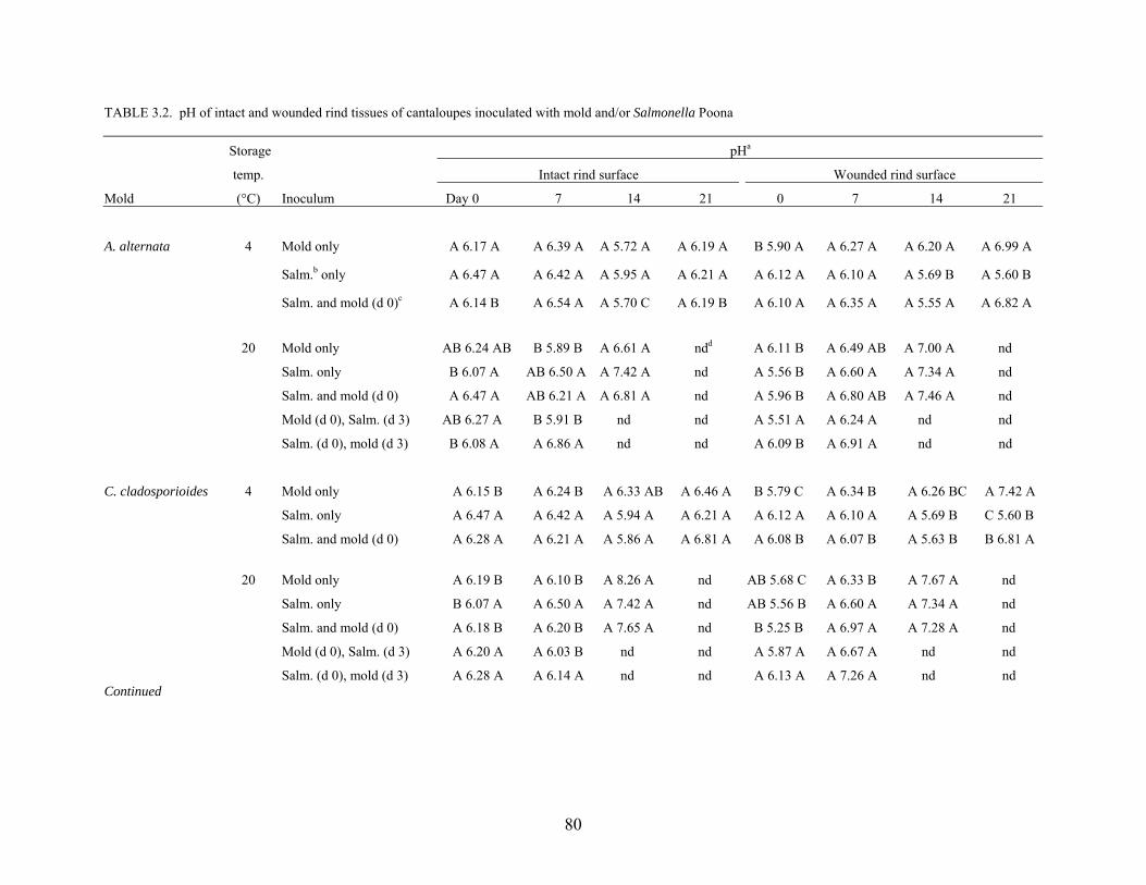
80
TABLE 3.2. pH of intact and wounded rind tissues of cantaloupes inoculated with mold and/or Salmonella Poona
Storage pHa
temp. Intact rind surface Wounded rind surface
Mold (°C) Inoculum Day 0 7 14 21 0 7 14 21
A. alternata 4 Mold only A 6.17 A A 6.39 A A 5.72 A A 6.19 A B 5.90 A A 6.27 A A 6.20 A A 6.99 A
Salm.b only A 6.47 A A 6.42 A A 5.95 A A 6.21 A A 6.12 A A 6.10 A A 5.69 B A 5.60 B
Salm. and mold (d 0)c A 6.14 B A 6.54 A A 5.70 C A 6.19 B A 6.10 A A 6.35 A A 5.55 A A 6.82 A
20 Mold only AB 6.24 AB B 5.89 B A 6.61 A ndd A 6.11 B A 6.49 AB A 7.00 A nd
Salm. only B 6.07 A AB 6.50 A A 7.42 A nd A 5.56 B A 6.60 A A 7.34 A nd
Salm. and mold (d 0) A 6.47 A AB 6.21 A A 6.81 A nd A 5.96 B A 6.80 AB A 7.46 A nd
Mold (d 0), Salm. (d 3) AB 6.27 A B 5.91 B nd nd A 5.51 A A 6.24 A nd nd
Salm. (d 0), mold (d 3) B 6.08 A A 6.86 A nd nd A 6.09 B A 6.91 A nd nd
C. cladosporioides 4 Mold only A 6.15 B A 6.24 B A 6.33 AB A 6.46 A B 5.79 C A 6.34 B A 6.26 BC A 7.42 A
Salm. only A 6.47 A A 6.42 A A 5.94 A A 6.21 A A 6.12 A A 6.10 A A 5.69 B C 5.60 B
Salm. and mold (d 0) A 6.28 A A 6.21 A A 5.86 A A 6.81 A A 6.08 B A 6.07 B A 5.63 B B 6.81 A
20 Mold only A 6.19 B A 6.10 B A 8.26 A nd AB 5.68 C A 6.33 B A 7.67 A nd
Salm. only B 6.07 A A 6.50 A A 7.42 A nd AB 5.56 B A 6.60 A A 7.34 A nd
Salm. and mold (d 0) A 6.18 B A 6.20 B A 7.65 A nd B 5.25 B A 6.97 A A 7.28 A nd
Mold (d 0), Salm. (d 3) A 6.20 A A 6.03 B nd nd A 5.87 A A 6.67 A nd nd
Salm. (d 0), mold (d 3) A 6.28 A A 6.14 A nd nd A 6.13 A A 7.26 A nd nd Continued
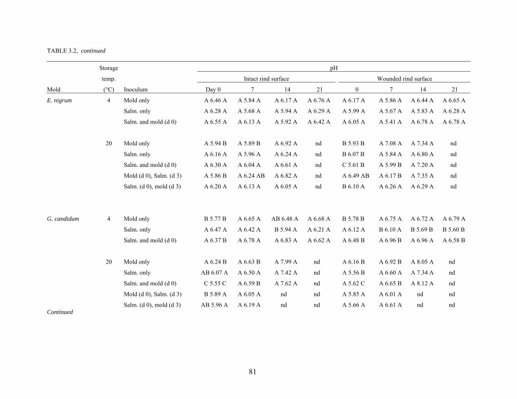
81
TABLE 3.2, continued
Storage pH
temp. Intact rind surface Wounded rind surface
Mold (°C) Inoculum Day 0 7 14 21 0 7 14 21
E. nigrum 4 Mold only A 6.46 A A 5.84 A A 6.17 A A 6.76 A A 6.17 A A 5.86 A A 6.44 A A 6.65 A
Salm. only A 6.28 A A 5.68 A A 5.94 A A 6.29 A A 5.99 A A 5.67 A A 5.83 A A 6.28 A
Salm. and mold (d 0) A 6.55 A A 6.13 A A 5.92 A A 6.42 A A 6.05 A A 5.41 A A 6.78 A A 6.78 A
20 Mold only A 5.94 B A 5.89 B A 6.92 A nd B 5.93 B A 7.08 A A 7.34 A nd
Salm. only A 6.16 A A 5.96 A A 6.24 A nd B 6.07 B A 5.84 A A 6.80 A nd
Salm. and mold (d 0) A 6.30 A A 6.04 A A 6.61 A nd C 5.61 B A 5.99 B A 7.20 A nd
Mold (d 0), Salm. (d 3) A 5.86 B A 6.24 AB A 6.82 A nd A 6.49 AB A 6.17 B A 7.35 A nd
Salm. (d 0), mold (d 3) A 6.20 A A 6.13 A A 6.05 A nd B 6.10 A A 6.26 A A 6.29 A nd
G. candidum 4 Mold only B 5.77 B A 6.65 A AB 6.48 A A 6.68 A B 5.78 B A 6.75 A A 6.72 A A 6.79 A
Salm. only A 6.47 A A 6.42 A B 5.94 A A 6.21 A A 6.12 A B 6.10 A B 5.69 B B 5.60 B
Salm. and mold (d 0) A 6.37 B A 6.78 A A 6.83 A A 6.62 A A 6.48 B A 6.96 B A 6.96 A A 6.58 B
20 Mold only A 6.24 B A 6.63 B A 7.99 A nd A 6.16 B A 6.92 B A 8.05 A nd
Salm. only AB 6.07 A A 6.50 A A 7.42 A nd A 5.56 B A 6.60 A A 7.34 A nd
Salm. and mold (d 0) C 5.55 C A 6.59 B A 7.62 A nd A 5.62 C A 6.65 B A 8.12 A nd
Mold (d 0), Salm. (d 3) B 5.89 A A 6.05 A nd nd A 5.85 A A 6.01 A nd nd
Salm. (d 0), mold (d 3) AB 5.96 A A 6.19 A nd nd A 5.66 A A 6.61 A nd nd Continued
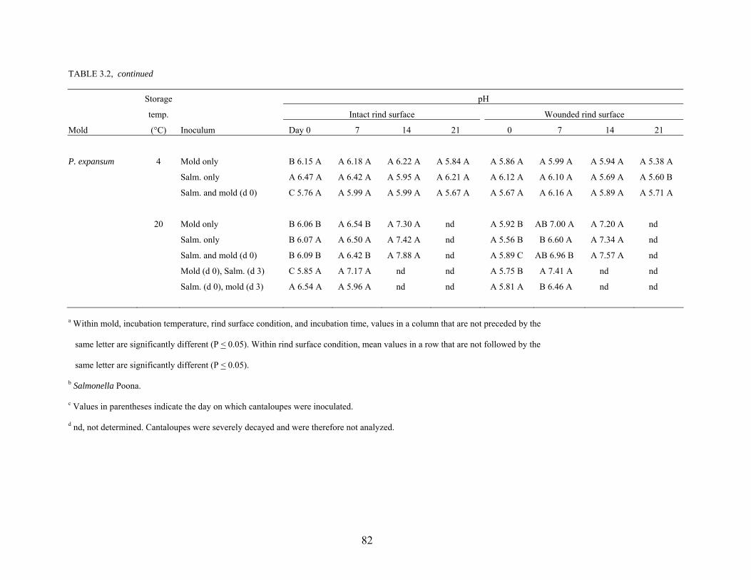
82
TABLE 3.2, continued
Storage pH
temp. Intact rind surface Wounded rind surface
Mold (°C) Inoculum Day 0 7 14 21 0 7 14 21
P. expansum 4 Mold only B 6.15 A A 6.18 A A 6.22 A A 5.84 A A 5.86 A A 5.99 A A 5.94 A A 5.38 A
Salm. only A 6.47 A A 6.42 A A 5.95 A A 6.21 A A 6.12 A A 6.10 A A 5.69 A A 5.60 B
Salm. and mold (d 0) C 5.76 A A 5.99 A A 5.99 A A 5.67 A A 5.67 A A 6.16 A A 5.89 A A 5.71 A
20 Mold only B 6.06 B A 6.54 B A 7.30 A nd A 5.92 B AB 7.00 A A 7.20 A nd
Salm. only B 6.07 A A 6.50 A A 7.42 A nd A 5.56 B B 6.60 A A 7.34 A nd
Salm. and mold (d 0) B 6.09 B A 6.42 B A 7.88 A nd A 5.89 C AB 6.96 B A 7.57 A nd
Mold (d 0), Salm. (d 3) C 5.85 A A 7.17 A nd nd A 5.75 B A 7.41 A nd nd
Salm. (d 0), mold (d 3) A 6.54 A A 5.96 A nd nd A 5.81 A B 6.46 A nd nd
a Within mold, incubation temperature, rind surface condition, and incubation time, values in a column that are not preceded by the
same letter are significantly different (P < 0.05). Within rind surface condition, mean values in a row that are not followed by the
same letter are significantly different (P < 0.05).
b Salmonella Poona.
c Values in parentheses indicate the day on which cantaloupes were inoculated.
d nd, not determined. Cantaloupes were severely decayed and were therefore not analyzed.
83
The pH of intact rinds of cantaloupes inoculated with S. Poona but not mold, then stored
at 4°C or 20°C, did not change significantly after 21 and 14 days, respectively (Table 3.2). In
some cases, simultaneously inoculating rind with S. Poona and a test mold resulted in increased
pH of intact rind during the storage period. Inoculation of intact cantaloupe rinds with S. Poona
and G. candidum simultaneously, followed by storage at 4°C or 20°C, resulted in a significant
increase (P < 0.05) in rind pH. The pH of intact rinds of cantaloupes inoculated with S. Poona
and C. cladosporioides or P. expansum on day 0 significantly increased (P < 0.05) during 14
days at 20°C, but not 21 days at 4°C. When the intact rinds were simultaneously inoculated with
S. Poona and A. alternata or E. nigrum, the rind pH did not change when the cantaloupes were
stored at 4°C or 20°C.
When intact rinds were inoculated with a mold 3 days before inoculating with S. Poona,
followed by storage at 20°C, changes in pH were variable for different molds (Table 3.2). The
pH of rind inoculated with A. alternata or C. cladosporioides decreased significantly (P < 0.05),
while inoculation of rind with E. nigrum resulted in a significant increase (P < 0.05); the pH of
intact rind inoculated with G. candidum or P. expansum did not change significantly. When
intact rinds were inoculated with S. Poona 3 days before inoculating with molds, the pH did not
change significantly within 7 or 14 days at 20°C.
Changes in pH of wounded cantaloupe rinds, as influenced by inoculation with S. Poona
and/or mold and storage temperature, were similar to those observed for intact rinds (Table 3.2).
The pH values of wounded cantaloupe rinds inoculated with C. cladosporioides or G. candidum
and then stored at 4°C or 20°C for 21 and 14 days, respectively, were significantly higher (P <
0.05) than initial (day 0) values. A. alternata, E. nigrum, and P. expansum did not cause a
84
significant change in the pH of wounded rind tissue of cantaloupes stored at 4°C, but
significantly increased the pH (P < 0.05) at 20°C.
The pH values of wounded rind tissue of cantaloupes inoculated with S. Poona only and
then incubated for 21 days at 4°C were unchanged or significantly reduced (P < 0.05). However,
the pH of wounded rind tissues of all cantaloupes inoculated with S. Poona only and stored at
20°C significantly increased (P < 0.05) after 14 days.
The effect of simultaneous inoculation of wounded tissues with S. Poona and a test mold
on the pH of the tissue was assessed. Wounded cantaloupe rind tissues inoculated
simultaneously with S. Poona and C. cladosporioides, but not other test molds, showed a
significant increase (P < 0.05) in the pH after storage at 4°C for 21 days. However, wounded
rind tissues of cantaloupes simultaneously inoculated with S. Poona and molds, followed by
storage at 20°C, showed significant increases (P < 0.05) in pH after 14 days. When wounded
rind tissues were inoculated with E. nigrum or P. expansum 3 days before inoculating with S.
Poona and storing at 20°C, the pH increased significantly (P < 0.05). Only the wounded
cantaloupe rinds inoculated with A. alternata 3 days after S. Poona was introduced into the
wound showed a significant increase (P < 0.05) in pH at 20°C.
Salmonellae are physiologically adaptable microorganisms, able to proliferate at pH 4.5 -
9.5, with 6.5 - 7.5 being the optimum pH for growth (D’Aoust, 1997). In this study, although
inoculated intact and wounded rinds of cantaloupes stored at 4°C did not show overt symptoms
of decay, the pH of inoculated tissues increased. The rise in pH would likely favor survival and
growth of S. Poona. Riordan et al. (2000), observed that enhanced growth of E. coli O157:H7 in
wounds on apples at 22 + 1°C co-inoculated with Glomerella cingulata was correlated with a
rise in pH at the infected site, even though decay was not evident. It was postulated that the
85
increase in pH of apple tissue was due to enzymatic activity of G. cingulata that led to the
production of alkaline end products or its ability to metabolize malic acid and lower acidity.
Aspergillus gracilis, Cladosporium, Fusarium, Penicillium, and Rhizoctonia have been reported
to raise the pH of tomato juice from levels that are normally inhibitory to levels that facilitate
growth and toxin production by Clostridium botulinum (Draughon et al., 1988; Huhtanen et al.,
1976; Odlaug and Pflug, 1979). The molds used in our study could have created
microenvironments within or adjacent to wounds on cantaloupe rinds that would favor survival
and growth S. Poona.
Recovery of molds from intact and wounded rinds. Populations of molds in inocula
deposited on the surface of intact cantaloupe rind or into wounds on the rind are shown in Table
3.3. Molds were recovered from intact and wounded rinds stored at 4°C for up to 21 days or
20°C for up to 14 days after inoculation. Molds were detected in a larger number of wounded
rinds than in intact rinds. This observation was most evident for rinds inoculated with E. nigrum
and P. expansum. E. nigrum, P. expansum, and G. candidum are not capable of readily
penetrating the cuticle and epidermis of the host but if they gain entry through the stem scar or a
wound or a natural opening, they can rapidly decay mature produce (Ceponis, 1966; Eckert,
1980; Snowdon, 1990; Wiant, 1937). Most of the test molds isolated from wounded cantaloupe
rind tissues were free of other molds, while isolates from the intact rinds consisted of test molds
together with other molds naturally present on the cantaloupes. This suggests that the test molds
competed more effectively with the natural mycoflora in wounded tissues than on intact tissues.
Recovery of A. alternata and C. cladosporioides from cantaloupes stored at 4°C was
consistently more frequent than recovery from cantaloupes at 20°C.
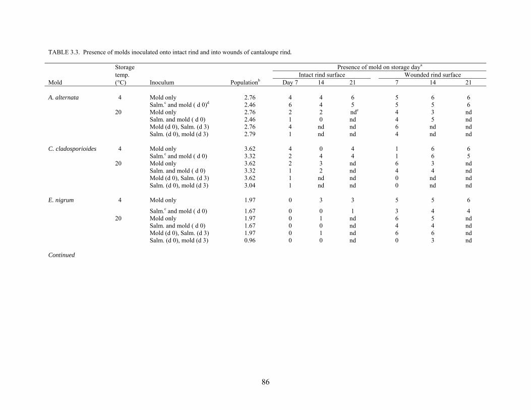
86
TABLE 3.3. Presence of molds inoculated onto intact rind and into wounds of cantaloupe rind. Storage Presence of mold on storage daya
temp. Intact rind surface Wounded rind surface Mold (°C) Inoculum Populationb Day 7 14 21 7 14 21 A. alternata 4 Mold only 2.76 4 4 6 5 6 6 Salm.c and mold ( d 0)d 2.46 6 4 5 5 5 6 20 Mold only 2.76 2 2 nde 4 3 nd Salm. and mold ( d 0) 2.46 1 0 nd 4 5 nd Mold (d 0), Salm. (d 3) 2.76 4 nd nd 6 nd nd Salm. (d 0), mold (d 3) 2.79 1 nd nd 4 nd nd C. cladosporioides 4 Mold only 3.62 4 0 4 1 6 6 Salm.c and mold ( d 0) 3.32 2 4 4 1 6 5 20 Mold only 3.62 2 3 nd 6 3 nd Salm. and mold ( d 0) 3.32 1 2 nd 4 4 nd Mold (d 0), Salm. (d 3) 3.62 1 nd nd 0 nd nd Salm. (d 0), mold (d 3) 3.04 1 nd nd 0 nd nd E. nigrum 4 Mold only 1.97 0 3 3 5 5 6
Salm.c and mold ( d 0) 1.67 0 0 1 3 4 4 20 Mold only 1.97 0 1 nd 6 5 nd Salm. and mold ( d 0) 1.67 0 0 nd 4 4 nd Mold (d 0), Salm. (d 3) 1.97 0 1 nd 6 6 nd Salm. (d 0), mold (d 3) 0.96 0 0 nd 0 3 nd Continued
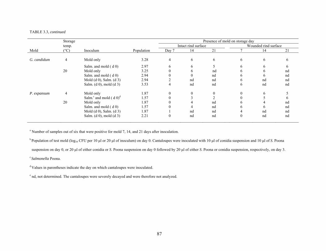
87
TABLE 3.3, continued Storage Presence of mold on storage day
temp. Intact rind surface Wounded rind surface Mold (°C) Inoculum Population Day 7 14 21 7 14 21 G. candidum 4 Mold only 3.28 4 6 6 6 6 6
Salm. and mold ( d 0) 2.97 6 6 5 6 6 6 20 Mold only 3.25 0 6 nd 6 6 nd Salm. and mold ( d 0) 2.94 0 0 nd 6 6 nd Mold (d 0), Salm. (d 3) 2.94 2 nd nd 6 nd nd Salm. (d 0), mold (d 3) 3.53 4 nd nd 6 nd nd P. expansum 4 Mold only 1.87 0 0 0 0 6 5 Salm.c and mold ( d 0)d 1.57 0 3 2 0 5 6 20 Mold only 1.87 0 4 nd 6 4 nd Salm. and mold ( d 0) 1.57 0 4 nd 6 6 nd Mold (d 0), Salm. (d 3) 1.87 1 nd nd 4 nd nd Salm. (d 0), mold (d 3) 2.21 0 nd nd 0 nd nd a Number of samples out of six that were positive for mold 7, 14, and 21 days after inoculation.
b Population of test mold (log10 CFU per 10 µl or 20 µl of inoculum) on day 0. Cantaloupes were inoculated with 10 µl of conidia suspension and 10 µl of S. Poona
suspension on day 0, or 20 µl of either conidia or S. Poona suspension on day 0 followed by 20 µl of either S. Poona or conidia suspension, respectively, on day 3.
c Salmonella Poona.
d Values in parentheses indicate the day on which cantaloupes were inoculated.
e nd, not determined. The cantaloupes were severely decayed and were therefore not analyzed.
88
Storage at low temperature retards decay by preserving resistance of the host to
parasitism and retarding growth of the pathogen directly (Eckert, 1980). Low temperature,
however, exacerbates decay of some fruits by Alternaria and Cladosporium (Morris and Wade,
1983; Snowdon, 1990).
Propagules of plant pathogenic molds are abundant on the surfaces of fruits and
vegetables as they approach maturity in the field. Removal of cantaloupes from the vine creates
stem scars through which molds, as well as foodborne pathogens, may enter the tissue and grow.
Damage to the surface of cantaloupe rind during harvesting is inevitable, thus creating additional
sites of entry for microorganisms. Sanitation is essential to reduce the population of
microorganisms on the surface of cantaloupes, especially if they are washed with potentially
contaminated water after harvest. The incidence of postharvest diseases that originate in wounds
may be substantially reduced by changing handling practices which damage the fruit surface and
by exposing crops to environmental conditions favoring the development of cellular barriers to
the penetration of pathogens (Eckert, 1980).
Preharvest cantaloupes may be infected by penetration of certain molds through the intact
cuticle or through wounds and natural openings on the surface of the fruit. Alternatively, many
postharvest diseases can be initiated through mechanical injuries sustained during harvest
(Eckert, 1980). Mature cantaloupes are susceptible to invasion by specific plant pathogens
because they are high in moisture and nutrient content and are no longer protected by some of
the intrinsic factors that confer resistance during development on the plant. Cantaloupes are
easily injured as they approach maturity and are therefore more vulnerable to pathogens that can
enter tissue through wounds. This situation is accelerated during transportation and storage
89
when cantaloupes are in close proximity, resulting in accelerated and uncontrolled ripening
caused by release of ethylene by decaying fruits.
Recovery of S. Poona from intact and wounded cantaloupe rinds. Populations of S.
Poona in 10 µl or 20 µl of inocula applied to intact rind of cantaloupes are shown in Table 3.4.
The population of S. Poona 7 days after inoculation was significantly lower (P < 0.05) than the
initial population, regardless of co-inoculation with mold or incubation temperature. Some cells
would have succumbed to environmental stresses during the drying period prior to storage and
throughout the 21-day storage period at 4°C. S. Poona was recovered from intact rinds of
cantaloupes stored at 4°C for up to 21 days however, and grew on cantaloupes stored at 20°C.
While the population of S. Poona recovered from cantaloupes stored at 20°C for 7 days was
significantly lower (P < 0.05) than the population on day 0, with one exception (inoculation with
S. Poona and E. nigrum on day 0), a significantly higher population (P < 0.05) was recovered
from cantaloupes stored for 14 days. Survival and growth of S. Poona at 4 and 20°C on intact
rinds were essentially unaffected by co-inoculation with mold, regardless of the order in which
inocula were applied. The near neutral pH, nutrient availability, and high aw on the surface of
the rind favored growth of S. Poona at 20°C, even in the absence phytopathogenic molds.
With the exception of wounded rind co-inoculated with G. candidum, the population of S.
Poona 21 days after inoculation and storage at 4°C was significantly lower (P < 0.05) than the
population initially applied, regardless of co-inoculation with mold (Table 3.5). Only wounded
tissue of rinds inoculated simultaneously with S. Poona and E. nigrum or P. expansum and stored
at 4°C required enrichment to detect viable S. Poona. Regardless of the inoculation scheme used,
the population of S. Poona recovered from wounds after storage for 7 and 14 days at 20°C was
significantly higher (P < 0.05) than the population initially applied.
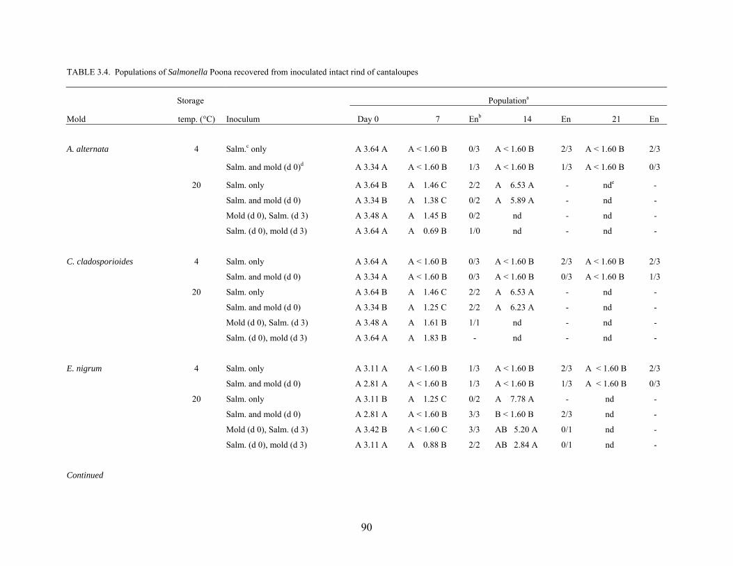
90
TABLE 3.4. Populations of Salmonella Poona recovered from inoculated intact rind of cantaloupes
Storage Populationa
Mold temp. (°C) Inoculum Day 0 7 Enb 14 En 21 En
A. alternata 4 Salm.c only A 3.64 A A < 1.60 B 0/3 A < 1.60 B 2/3 A < 1.60 B 2/3
Salm. and mold (d 0)d A 3.34 A A < 1.60 B 1/3 A < 1.60 B 1/3 A < 1.60 B 0/3
20 Salm. only A 3.64 B A 1.46 C 2/2 A 6.53 A - nde -
Salm. and mold (d 0) A 3.34 B A 1.38 C 0/2 A 5.89 A - nd -
Mold (d 0), Salm. (d 3) A 3.48 A A 1.45 B 0/2 nd - nd -
Salm. (d 0), mold (d 3) A 3.64 A A 0.69 B 1/0 nd - nd -
C. cladosporioides 4 Salm. only A 3.64 A A < 1.60 B 0/3 A < 1.60 B 2/3 A < 1.60 B 2/3
Salm. and mold (d 0) A 3.34 A A < 1.60 B 0/3 A < 1.60 B 0/3 A < 1.60 B 1/3
20 Salm. only A 3.64 B A 1.46 C 2/2 A 6.53 A - nd -
Salm. and mold (d 0) A 3.34 B A 1.25 C 2/2 A 6.23 A - nd -
Mold (d 0), Salm. (d 3) A 3.48 A A 1.61 B 1/1 nd - nd -
Salm. (d 0), mold (d 3) A 3.64 A A 1.83 B - nd - nd -
E. nigrum 4 Salm. only A 3.11 A A < 1.60 B 1/3 A < 1.60 B 2/3 A < 1.60 B 2/3
Salm. and mold (d 0) A 2.81 A A < 1.60 B 1/3 A < 1.60 B 1/3 A < 1.60 B 0/3
20 Salm. only A 3.11 B A 1.25 C 0/2 A 7.78 A - nd -
Salm. and mold (d 0) A 2.81 A A < 1.60 B 3/3 B < 1.60 B 2/3 nd -
Mold (d 0), Salm. (d 3) A 3.42 B A < 1.60 C 3/3 AB 5.20 A 0/1 nd -
Salm. (d 0), mold (d 3) A 3.11 A A 0.88 B 2/2 AB 2.84 A 0/1 nd -
Continued
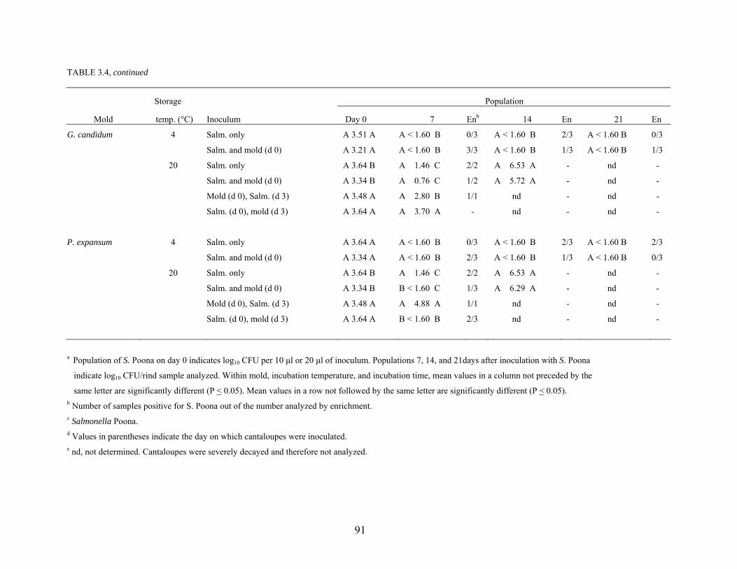
91
TABLE 3.4, continued
Storage Population
Mold temp. (°C) Inoculum Day 0 7 Enb 14 En 21 En
G. candidum 4 Salm. only A 3.51 A A < 1.60 B 0/3 A < 1.60 B 2/3 A < 1.60 B 0/3
Salm. and mold (d 0) A 3.21 A A < 1.60 B 3/3 A < 1.60 B 1/3 A < 1.60 B 1/3
20 Salm. only A 3.64 B A 1.46 C 2/2 A 6.53 A - nd -
Salm. and mold (d 0) A 3.34 B A 0.76 C 1/2 A 5.72 A - nd -
Mold (d 0), Salm. (d 3) A 3.48 A A 2.80 B 1/1 nd - nd -
Salm. (d 0), mold (d 3) A 3.64 A A 3.70 A - nd - nd -
P. expansum 4 Salm. only A 3.64 A A < 1.60 B 0/3 A < 1.60 B 2/3 A < 1.60 B 2/3
Salm. and mold (d 0) A 3.34 A A < 1.60 B 2/3 A < 1.60 B 1/3 A < 1.60 B 0/3
20 Salm. only A 3.64 B A 1.46 C 2/2 A 6.53 A - nd -
Salm. and mold (d 0) A 3.34 B B < 1.60 C 1/3 A 6.29 A - nd -
Mold (d 0), Salm. (d 3) A 3.48 A A 4.88 A 1/1 nd - nd -
Salm. (d 0), mold (d 3) A 3.64 A B < 1.60 B 2/3 nd - nd -
a Population of S. Poona on day 0 indicates log10 CFU per 10 µl or 20 µl of inoculum. Populations 7, 14, and 21days after inoculation with S. Poona
indicate log10 CFU/rind sample analyzed. Within mold, incubation temperature, and incubation time, mean values in a column not preceded by the
same letter are significantly different (P < 0.05). Mean values in a row not followed by the same letter are significantly different (P < 0.05). b Number of samples positive for S. Poona out of the number analyzed by enrichment. c Salmonella Poona. d Values in parentheses indicate the day on which cantaloupes were inoculated. e nd, not determined. Cantaloupes were severely decayed and therefore not analyzed.
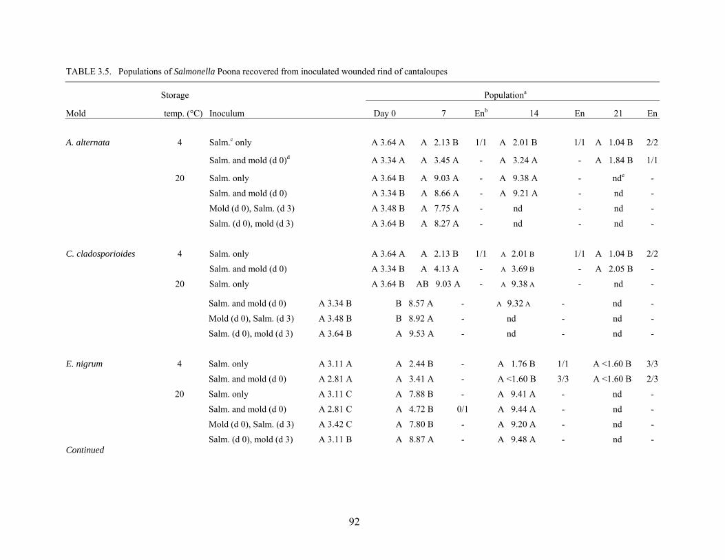
92
TABLE 3.5. Populations of Salmonella Poona recovered from inoculated wounded rind of cantaloupes
Storage Populationa
Mold temp. (°C) Inoculum Day 0 7 Enb 14 En 21 En
A. alternata 4 Salm.c only A 3.64 A A 2.13 B 1/1 A 2.01 B 1/1 A 1.04 B 2/2
Salm. and mold (d 0)d A 3.34 A A 3.45 A - A 3.24 A - A 1.84 B 1/1
20 Salm. only A 3.64 B A 9.03 A - A 9.38 A - nde -
Salm. and mold (d 0) A 3.34 B A 8.66 A - A 9.21 A - nd -
Mold (d 0), Salm. (d 3) A 3.48 B A 7.75 A - nd - nd -
Salm. (d 0), mold (d 3) A 3.64 B A 8.27 A - nd - nd -
C. cladosporioides 4 Salm. only A 3.64 A A 2.13 B 1/1 A 2.01 B 1/1 A 1.04 B 2/2
Salm. and mold (d 0) A 3.34 B A 4.13 A - A 3.69 B - A 2.05 B -
20 Salm. only A 3.64 B AB 9.03 A - A 9.38 A - nd -
Salm. and mold (d 0) A 3.34 B B 8.57 A - A 9.32 A - nd -
Mold (d 0), Salm. (d 3) A 3.48 B B 8.92 A - nd - nd -
Salm. (d 0), mold (d 3) A 3.64 B A 9.53 A - nd - nd -
E. nigrum 4 Salm. only A 3.11 A A 2.44 B - A 1.76 B 1/1 A <1.60 B 3/3
Salm. and mold (d 0) A 2.81 A A 3.41 A - A <1.60 B 3/3 A <1.60 B 2/3
20 Salm. only A 3.11 C A 7.88 B - A 9.41 A - nd -
Salm. and mold (d 0) A 2.81 C A 4.72 B 0/1 A 9.44 A - nd -
Mold (d 0), Salm. (d 3) A 3.42 C A 7.80 B - A 9.20 A - nd -
Salm. (d 0), mold (d 3) A 3.11 B A 8.87 A - A 9.48 A - nd - Continued
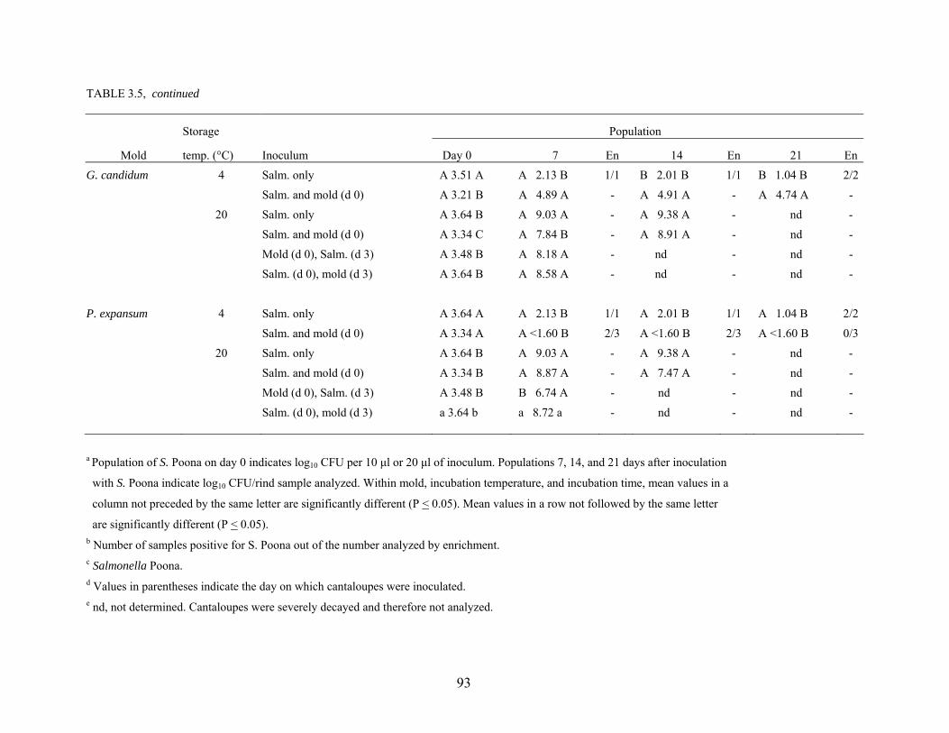
93
TABLE 3.5, continued
Storage Population
Mold temp. (°C) Inoculum Day 0 7 En 14 En 21 En
G. candidum 4 Salm. only A 3.51 A A 2.13 B 1/1 B 2.01 B 1/1 B 1.04 B 2/2
Salm. and mold (d 0) A 3.21 B A 4.89 A - A 4.91 A - A 4.74 A -
20 Salm. only A 3.64 B A 9.03 A - A 9.38 A - nd -
Salm. and mold (d 0) A 3.34 C A 7.84 B - A 8.91 A - nd -
Mold (d 0), Salm. (d 3) A 3.48 B A 8.18 A - nd - nd -
Salm. (d 0), mold (d 3) A 3.64 B A 8.58 A - nd - nd -
P. expansum 4 Salm. only A 3.64 A A 2.13 B 1/1 A 2.01 B 1/1 A 1.04 B 2/2
Salm. and mold (d 0) A 3.34 A A <1.60 B 2/3 A <1.60 B 2/3 A <1.60 B 0/3
20 Salm. only A 3.64 B A 9.03 A - A 9.38 A - nd -
Salm. and mold (d 0) A 3.34 B A 8.87 A - A 7.47 A - nd -
Mold (d 0), Salm. (d 3) A 3.48 B B 6.74 A - nd - nd -
Salm. (d 0), mold (d 3) a 3.64 b a 8.72 a - nd - nd -
a Population of S. Poona on day 0 indicates log10 CFU per 10 µl or 20 µl of inoculum. Populations 7, 14, and 21 days after inoculation
with S. Poona indicate log10 CFU/rind sample analyzed. Within mold, incubation temperature, and incubation time, mean values in a
column not preceded by the same letter are significantly different (P < 0.05). Mean values in a row not followed by the same letter
are significantly different (P < 0.05). b Number of samples positive for S. Poona out of the number analyzed by enrichment. c Salmonella Poona. d Values in parentheses indicate the day on which cantaloupes were inoculated. e nd, not determined. Cantaloupes were severely decayed and therefore not analyzed.
94
Larger populations of S. Poona were recovered from wounded rind than from intact rind
(Tables 3.5 and 3.4, respectively). The number of S. Poona recovered from wounded rind tissues
of cantaloupes stored at 4°C was 1 – 4 logs higher than that from intact tissues. The number of
S. Poona recovered from wounded tissue of cantaloupes stored at 20°C was 2 – 6 logs higher
than recovered from intact tissues. Nutrients were more available in wounds to facilitate
resuscitation and support growth of injured S. Poona cells. The growth of Salmonella Stanley on
the rind of cantaloupe cubes was also attributed to the availability of nutrients, especially when
storage temperature was favorable (Ukuku and Sapers, 2001). Foodborne pathogens have been
observed to consistently adhere preferentially to injured sites, cut edges, and blossom and stem
scars compared to intact surfaces of fresh produce (Annous et al., 2001; Lukasik et al., 2001;
Reina et al., 2002; Takeuchi and Frank, 2001). When soft rot and healthy tissues of potato,
carrot and pepper tissues were inoculated with Salmonella Typhimurium, the population of
viable cells multiplied 3- to 10-fold on soft rot tissues compared to healthy tissues (Wells and
Butterfield, 1997).
Storage temperature affected the survival and growth of S. Poona on intact and wounded
rinds of cantaloupes (Tables 3.4 and 3.5, respectively). Higher numbers of S. Poona were
recovered from rinds of cantaloupes stored at 20°C compared to cantaloupes stored at 4°C. Del
Rosario and Beuchat (1995) observed that the population of E. coli O157:H7 on inoculated
cantaloupe and watermelon cubes and rinds remained unchanged or decreased, respectively,
when stored at 5°C, but grew at 25°C. Similar observations were made when S. Stanley was
inoculated on fresh-cut cantaloupe cubes (Ukuku and Sapers, 2001). Salmonella has been
observed to grow on cut papaya, jimica, and watermelon held at 22 – 27°C (Escartin et al., 1989)
as well as cut cantaloupe and honeydew stored at 23°C (Golden et al., 1993). In the present
95
study, intact rinds of cantaloupes co-inoculated with S. Poona and test molds, then stored at 4°C,
required enrichment for detection of S. Poona. However, with the exception of wounded tissues
inoculated with S. Poona and E. nigrum or P. expansum on day 0, viable cells were recoverable
without enrichment of cantaloupes stored at 4°C (Table 3.5). Cold storage was reported to
adversely affect the survival of E. coli O157:H7 on injured apple tissues co-inoculated with G.
cingulata (Riordan et al., 2000).
The microbial ecology of fruits and vegetables can be influenced at every step from
production through the point of consumption. Traditionally, attention has been focused on
postharvest physiology and pathology of produce, recognizing that improper handling practices
can cause decay and extensive economic loss. Reports on the presence of human bacterial
pathogens on raw agricultural commodities have accentuated the need to consider fruits and
vegetables as vehicles of microorganisms capable of causing disease (Beuchat, 2002). Research
has shown that there are interactions between postharvest plant pathogens and human bacterial
pathogens. While P. expansum in decayed tissue of sliced apples impeded the survival of L.
monocytogenes by reducing the pH, G. cingulata was observed to stimulate growth at 10 and
20°C by increasing the pH of decaying tissue (Conway et al., 2000). A study of healthy and
soft-rotted fruits and vegetables in retail markets revealed that the incidence of Salmonella on
produce that had undergone bacterial soft rot was twice that of healthy samples (Wells and
Butterfield, 1997). Various practices employed in production, processing, and transportation of
fresh produce can influence microbial safety. Bacterial and fungal rots on fresh produce,
generally regarded as signs of poor handling, storage, and sanitation, could be warnings for
possible enteric bacterial contamination. Foodborne pathogens can persist on fresh produce
through the point of consumption. Treatments such as washing with chlorinated water cannot be
96
relied on to eliminate pathogens from produce (Brackett et al., 1993). It is unlikely that a
consumer would intentionally eat the decayed portion of a cantaloupe. However, removal of
bruised or moldy tissue, followed by cutting the remainder for consumption is not an uncommon
practice. The development of alkalinized tissue as a result of mold growth could be a plausible
explanation for retention of viability of Salmonella and associations of salmonellosis with raw
fruits and fruit products. Cellulases and pectinases produced by phytopathogens cause softening
and release of cell fluids from plant tissues. It is conceivable that release of cell sap and liquids
could allow dislocation and relocation of foodborne pathogens from wounded and decaying
tissues to otherwise wholesome tissue on the same fruit or adjacent fruits.
This study demonstrates that S. Poona can persist on intact, wounded, and decaying
cantaloupe rind at refrigerated and ambient temperatures. Molds commonly involved in
postharvest decay of cantaloupes increased the pH of intact and wounded rinds and enhanced the
survival of S. Poona. Storage at 4°C, compared to 20°C, reduced the rate of fruit senescence and
decay by phytopathogens, and enhanced the rate of death of S. Poona. Injury and high
temperature abuse enhanced the growth of S. Poona on cantaloupe rind. Recovery of larger
numbers of S. Poona from wounded tissues of cantaloupes, particularly fruits stored at 20°C,
emphasizes the importance of developing agronomic and postharvest handling practices that
prevent contamination, damage, and deterioration. The proteolytic activities of C.
cladosporioides and G. candidum, in particular, portend the need for further investigation of
potential metabiotic associations with S. Poona on cantaloupe rind and in cantaloupe cubes that
may promote survival and growth of foodborne pathogens in cantaloupes and other fruits.
97
REFERENCES Annous, B. A., G. Sapers, A. Mattrazzo, and D. Riordan. 2001. Efficacy of washing with a
commercial flatbed brush washer, using conventional and experimental washing agents, in reducing populations of Escherichia coli on artificially inoculated apples. J. Food Prot. 64:159-163.
Beuchat, L.R. 1996. Pathogenic microorganisms associated with fresh produce. J. Food Prot.
59:204-216. Beuchat, L.R. 2002. Ecological factors influencing survival and growth of human pathogens on
raw fruits and vegetables. Microbes Infect. 4:413-423. Brackett, R.E., D.M. Smallwood, S.M. Fletcher, and D.L. Horton. 1993. Food Safety: Critical
points within the production and distribution system. In R.L. Shewfelt and S. E. Prussia (Ed.), Postharvest Handling: a Systems Approach. Academic Press, San Diego, CA.
Centers for Disease Control and Prevention (CDC). 1991. Multistate outbreak of Salmonella
Poona infections – United States and Canada, 1991. Morbid. Mortal. Wkly. Rep. 40:549-552.
Centers for Disease Control and Prevention (CDC). 2002. Multistate outbreaks of Salmonella
Serotype Poona infections associated with eating cantaloupes from Mexico – United States and Canada, 2000 – 2002. Morbid. Mortal. Wkly. Rep. 51:1044-1047.
Ceponis, M.J. 1966. Occurrence of Geotrichum candidum in western melons on the New York
market. Plant Dis. Rep. 50:222-224. Ceponis, M.J., R.A. Cappellini, and G.W. Lightner. 1986. Disorders in muskmelon shipments to
the New York market, 1972 –1984. Plant Dis. 70:605-607. Chardonnet, C.O., E.E. Sams, W.S. Conway, F.A. Draughon, and J. R. Mount. 2002. Osmotic
dehydration of apple slices with CaCl2 and sucrose limits decay caused by Penicillium expansum, Colletotrichum acutatum and Botrytis cinerea and does not promote Listeria monocytogenes or total aerobic population growth. J. Food Prot. 65:172-177.
Conway, W.S., B. Leverentz, R.A. Saftner, W.J. Janisiewicz, C.E. Sams, and E. Leblanc. 2000.
Survival and growth of Listeria monocytogenes on fresh cut apples and its interaction with Glomerella cingulata and Penicillium expansum. Plant Dis. 84:177-181.
D’Aoust, J-Y. 1997. Salmonella species, p. 129-158. In M.P. Doyle, L.R. Beuchat,
and T.J. Montville (ed.), Food Microbiology: Fundamentals and Frontiers. ASM Press. Washington D.C.
Deeks, S., A. Ellis, B. Ciebin, R. Khakhria, M. Naus, and J. Hockin. 1998. Salmonella
Oranienberg, Ontario. Can. Comm. Dis. Rep. 24:177-179.
98
Del Rosario, B.A. and L.R. Beuchat. 1995. Survival and growth of Enterohemorrhagic Escherichia coli O157:H7 in cantaloupe and watermelon. J. Food Prot. 58:105-107.
Draughon, F.A., S. Chen, and J.O. Mundt. 1988. Metabiotic association of Fusarium, Alternaria,
and Rhizoctonia with Clostridium botulinum on fresh tomatoes. J. Food Sci. 53:120-123. Eckert, J.W. 1980. Postharvest diseases of fresh fruits and vegetables – etiology and control. In
N.F. Haard and D.K. Salunkhe (Ed.), Postharvest Biology and Handling of Fruits and Vegetables. AVI Publishing Company, Inc. Westport, CT.
Escartin, E.F., A.C. Ayala, and J.S. Lozano. 1989. Survival and growth of Salmonella and
Shigella on sliced fresh fruit. J. Food Prot. 52:471-472. Eshel, D., I Miyara, T. Ailing, A. Dinoor, and D. Prusky. 2002. pH regulates endoglucanase
expression in virulence of Alternaria alternata in persimmon fruit. Mol. Plant Microbe Interact. 15:774-779.
Food and Drug Administration. 2001a. FDA Survey of imported fresh produce. FY 1999 Field
Assignment. US Food and Drug Administration. Center for Food Safety and Applied Nutrition. Office of Plant and Dairy Foods and Beverages. January 30, 2001. [Internet, WWW], ADDRESS: http://www.cfsan.gov/~dms/prodsur6.html. Accessed 25 January 2002.
Food and Drug Administration. 2001b. FDA Survey of domestic fresh produce: interim results.
US Food and Drug Administration. Center for Food Safety and Applied Nutrition. June 30, 2001. [Internet, WWW], ADDRESS: http://vm.cfsan.gov/~dms/prodsur8.html. Accessed 25 January 2002.
Food and Drug Administration. 2002. FDA issue import alert on cantaloupes from Mexico. US
Food and Drug Administration. FDA Talk Paper. October 28, 2002. [Internet, WWW], ADDRESS: http://www.fda.gov/bbs/topics/ANSWERS/2002/ANS01167.html. Accessed 12 December 2002.
Golden, D.A., E.J. Rhodehamel, and D.A. Kautter. 1993. Growth of Salmonella spp. in
cantaloupe, watermelon, and honeydew melons. J. Food Prot. 56:194-196. Huhtanen, C.N., J. Naghski, C.S. Custer, and R.W. Russell. 1976. Growth and toxin production
by Clostridium botulinum in moldy tomato juice. Appl. Environ. Microbiol. 32:711-715. Institute of Food Technologists/Food and Drug Administration of the United States Department
of Health and Human Services (IFT/FDA). 2001. Analysis and evaluation of preventative control measures for the control and reduction/elimination of microbial hazards on fresh and fresh-cut produce. IFT/FDA Contract No. 223-98-2333. Task Order No. 3. Institute of Food Technologists. Chicago, IL.
99
Lukasik, J., M. Bradley, T. Scott, W-Y. Hsu, S. Farrah, and M. Tamplin. 2001. Elution, detection and quantification of Polio I, bacteriophages, Salmonella Montevideo, and Escherichia coli O157:H7 from seeded strawberries and tomatoes. J. Food Prot. 64:292-297.
Madden, J.M. 1992. Microbial pathogens in fresh produce – the regulatory perspective. J. Food
Prot. 55:821-823. Mohle-Boetani, J. C., R. Reporter, S.B. Werner, S. Abbott, J. Farrar, S.H. Waterman, and D.J.
Vugia. 1999. An outbreak of Salmonella serogroup Saphra due to cantaloupes from Mexico. J. Infect. Dis. 180:1361-1364.
Morris, S.C. and N.L. Wade. 1983. Control of postharvest disease in cantaloupes by treatment
with guazantine and benomyl. Plant Dis. 67:792-794. Mundt, J.O. 1978. Effect of mold growth on the pH of tomato juice. J. Food Prot. 41:267-268. Odlaug, T.E. and I.J. Pflug. 1979. Clostridium botulinum growth and toxin production in tomato
juice containing Aspergillus gracilis. Appl. Environ. Microbiol. 37:496-504. Prusky, D., J.L. McEvoy, B. Leverentz, and W.S. Conway. 2001. Local modulation of host pH
by Colletotrichum species as a medium to increase virulence. Mol. Plant Microbe Interact. 14:1105-1113.
Reina, L.D., H.P. Fleming, and F. Breidt, Jr. 2002. Bacterial contamination of cucumber fruit
through adhesion. J. Food Prot. 65:1881-1887. Ries, A.A., S. Zaza, C. Langkop, R.V. Tauxe, and P.A. Blake. 1990. A multistate outbreak of
Salmonella Chester linked to imported cantaloupe. Abstract. 915, p. 238. In Programs and abstracts of 30th Interscience Conference on Antimicrobial Agents and Chemotherapy. American Society for Microbiology, Washington, D.C.
Riordan, D.C.R., G.M. Sapers, and B.A. Annous. 2000. The survival of Escherichia coli
O157:H7 in the presence of Penicillium expansum and Glomerulla cingulata in wounds on apple surfaces. J. Food Prot. 63:1637-1642.
Salunkhe, D.K. and B.B. Desai. 1984. Postharvest biotechnology of vegetables. Volume 2. CRC
Press. Boca Raton, FL. Sapers, G.M., R.L. Miller, V. Pilizota, and A.M. Mattrazzo. 2001. Antimicrobial treatments for
minimally processed cantaloupe melon. J. Food Sci. 66:345-349. Sewell A.M. and J.M. Faber. 2001. Foodborne outbreaks in Canada linked to produce. J. Food
Prot. 64:1863-1877. Snowdon, A.L. 1990. A Colour Atlas of Post-Harvest Diseases and Disorders of Fruits and
100
Vegetables. Volume 1: General Introduction and Fruits. Wolf Scientific Ltd., Barcelona, Spain.
Sommer, N.F., R.J. Fortlage, and D.C. Edwards. 1992. Postharvest diseases of selected
commodities. In Adel A. Kader (Ed.), Postharvest Technology of Horticultural Crops. Publication 3311. University of California, Division of Agriculture and Natural Resources.
Suslow, T.V., M. Cantwell, and J. Mitchell. 2000. Cantaloupes: Recommendations for
maintaining postharvest quality. University of California, Davis. Postharvest Technology Research and Information Center. June 26, 2000. [Internet, WWW], ADDRESS: http://postharvest.ucdavis.edu/Produce/Producefacts/cantaloupe.html Accessed 25 March 2002.
Suslow, T. and M. Cantwell. 2001. Recent findings on fresh-cut cantaloupe and honeydew
melon. Fresh cut. April 2001. Pp.12, 20, 32-33. Takeuchi, K. and J. Frank. 2001. Expression of red-shifted green fluorescent protein by
Escherichia coli O157:H7 as a marker for the detection of cells on fresh product. J. Food Prot. 64:298-304.
Ukuku, D.O. and G.M. Sapers. 2001. Effect of sanitizer treatments on Salmonella Stanley
attached to the surface of cantaloupe and cell transfer to fresh-cut tissues during cutting practices. J. Food Prot. 64:1286-1291.
Wells J.M. and J.E. Butterfield. 1997. Salmonella contamination with bacterial soft rot of fresh
fruits and vegetables in the marketplace. Plant Dis. 81:867-872. Wiant, J.S. 1937. Investigations of the market diseases of cantaloupes and honeydew and honey
ball melons. United Sates Department of Agriculture. Technical Bulletin No. 573. September 1937.
101
CHAPTER 4
INFECTION OF CANTALOUPE RIND WITH CLADOSPORIUM CLADOSPORIOIDES AND
PENICILLIUM EXPANSUM, AND ASSOCIATED MIGRATION OF
SALMONELLA POONA INTO EDIBLE TISSUES1
______________________
1 Richards, Glenner M. and Larry R. Beuchat. 2003. To be submitted to International Journal of Food Microbiology.
102
ABSTRACT
Although cantaloupe fruits are in contact with the ground during their development,
enhancing their potential to be contaminated by pathogens, the internal tissues are expected to be
free from microorganisms. Fungal infections of the rind tissue gradually progress inward toward
the edible flesh. Salmonella Poona has been the predominant serotype responsible for national
and international outbreaks associated with consumption of fresh cantaloupes. This study was
undertaken to determine if the growth of two phytopathogens, Cladosporium cladosporioides
and Penicillium expansum, in wounds on cantaloupe rinds facilitates migration of S. Poona into
sub-surface mesocarp tissues. Wounded sites in cantaloupe rind were inoculated with S. Poona
only, S. Poona and mold simultaneously or mold followed by S. Poona 3 days later. A
cylindrical plug (ca. 3 cm diameter and 4 cm deep) was cut transversely into four segments (0 –1
cm, 1 – 2 cm, 2 – 3 cm, and 3 – 4 cm) representing distances from the rind surface. Regardless
of the type of inoculum and the time of storage subsequent to inoculation, the pH of the flesh
was significantly higher (P < 0.05) as the distance from the rind surface increased. Naturally-
occurring and test microorganisms on the rind surface introduced into internal tissues during
wounding as well as physiological changes in cantaloupe tissue contributed to these changes.
The test molds were recovered from the inoculated rind as well as the underlying tissues
throughout storage at 20°C for 10 days. S. Poona persisted and grew in the wounds in rinds on
inoculated cantaloupe incubated. Recovery of S. Poona from tissues 3 – 4 cm below the
inoculated wound supports the hypothesis that foodborne pathogens can migrate from the site of
inoculation into adjacent mesocarp tissues. Results demonstrate that migration and survival of S.
Poona in the internal tissues of cantaloupes were enhanced by co-inoculation with C.
cladosporioides and to a lesser extent, P. expansum.
103
INTRODUCTION
Salmonellosis is the major bacterial foodborne disease in many countries (Asplund and
Nurmi, 1991; D’Aoust, 2001) and has been increasing steadily as a public health problem over
the last 50 years in the United States (CDC, 2000). Non-typhoidal salmonellae were estimated to
cause approximately 1.5 million cases of infection, resulting in 15,000 hospitalizations and 500
deaths in the United States in 1999 (Mead et al., 1999). Salmonella, as a leading cause of
foodborne disease, is attributable in part to its presence in a wide range of environmental
sources, its prominence in various sectors of the agricultural and food industry, and from
escalating movement of food and food ingredients in international trade (D’Aoust, 2001).
Foods of animal origin have historically been recognized as potential vehicles for
Salmonella. However, consumption of fresh fruits and vegetables, including seed sprouts
(O’Mahony et al., 1990; Van Beneden et al., 1996; Mahon et al., 1997), unpasteurized apple
cider (CDC, 1975), unpasteurized orange juice (Cook et al., 1998; CDC, 1999), raw tomatoes
(Wood et al., 1991; Wessinger et al., 2000), watermelons (Gayler et al., 1955; Larson et al.,
1979; Blostein, 1993), and cantaloupes (CDC, 1991; CDC, 2002; Deeks et al., 1998; IFT/FDA,
2001; Mohle-Boetani et al., 1999; Ries et al., 1990) have also been linked to outbreaks of
salmonellosis in recent years. Sources of origin of Salmonella detected on produce include
animal and human wastes, irrigation water, runoff water from livestock farms adjacent to fields
and orchards, manure, wash water, handling by workers, and contact with contaminated surfaces,
animal fertilizers applied in previous growing seasons, and feces of rodents and ruminants (Guo
et al., 2002). Cross contamination from raw meat, poultry, or eggs has also been identified as a
potential mechanism for contamination of produce (IFT/FDA, 2001).
104
Cantaloupe fruits are in contact with the ground during their development, enhancing the
potential for contamination by pathogens. The netted texture of cantaloupe melon rind enables
effective attachment of microorganisms and results in difficulty in sanitization (Ukuku et al.,
2001). A field survey conducted by the Food and Drug Administration (FDA) during 1990 and
1991 revealed that 0.76 and 1.06%, respectively, of the melon rinds harbor several serovars of
Salmonella (Madden 1992; Golden et al., 1993). Selected fresh produce, including cantaloupes
imported into the United States, was surveyed. Of 151 cantaloupes sampled, 7.3% were
contaminated with Salmonella or Shigella (FDA, 2001a). The results of a similar survey of
domestic fresh produce initiated in May 2000 showed the presence of Salmonella and Shigella
on 4.3% of 92 cantaloupes sampled (FDA, 2001b). In a field study to assess the microbiological
quality of 433 samples of leafy green vegetables and cantaloupes, Salmonella was isolated from
3.3% of the cantaloupes examined (Kleman et al., 2003). Contamination of the rinds of
cantaloupes and honeydew melons has been traced to the primary wash tank or hydrocooler used
to clean fruits after harvest (Gagliardi et al., 2003). Salmonella Poona has been the predominant
serotype responsible for national and international outbreaks associated with consumption of
fresh cantaloupes.
The internal tissues of produce are generally free of microorganisms. The cuticular
protective cover on many fruits and vegetables and structural integrity afforded by cellulose and
pectin, as well as the low pH by some produce, are important intrinsic factors that influence the
microbial ecology. Being compartmentalized, plant tissues limit the diffusion of solutes that
influence the survival and growth of microorganisms. Fruits and vegetables may contain
chemical compounds that are inhibitory to microorganisms and may induce defense reactions
against microbial invasion (Nguyen-the and Carlin, 2000). Only the microorganisms that
105
possess the necessary physiological attributes to respond to intrinsic and extrinsic environmental
pressures will survive (Goepfert, 1980). The association of salmonellosis with salad bars and
fruit salads suggests introduction of Salmonella into the fruit from the rind by the slicing action,
or physical contact of cut pieces with contaminated rind (Suslow and Cantwell, 2001).
Cantaloupes are susceptible to postharvest fungal rots, especially under warm, wet
conditions. Infection of the external surface of the fruit gradually progresses inwards toward the
flesh. Fungal pathogens of major concern in cantaloupes are Geotrichum, Alternaria,
Penicillium, Cladosporium, Rhizopus, and Fusarium (Suslow et al., 2000; Agblor and Waterer,
2001) and, to a lesser extent, Mucor (Suslow et al., 2000). Complete loss of the commodity
occurs when one or a few fungal pathogens invade and begin to breakdown the tissues. This
initial attack is often rapidly followed by a broad spectrum of weak pathogens that magnify the
damage caused by primary pathogens.
Several studies have been undertaken to assess the survival and growth of Salmonella in
tissues of fruits and vegetables. Salmonella was associated more often with soft-rotted (Wells
and Butterfield, 1997) and injured plant tissues (Liao and Sapers, 2000; Liao and Cooke, 2001)
than with their healthy, intact counterparts. The potential of the contaminated tissue to support
growth of Salmonella is exacerbated by display or storage of cantaloupes at ambient temperature
for an extended time (CDC, 1991; Golden et al., 1993). The high water activity and sugar
content of tissue juices may promote rapid proliferation of salmonellae. Cut cantaloupe is
considered a potentially hazardous food in the FDA Food Code because it is capable of
supporting the growth of pathogens due to low acidity (pH 5.2 to 6.7) and high water activity
(0.97 to 0.99) (IFT/FDA, 2001). The study reported here was undertaken to determine if the
106
growth of two molds, Cladosporium cladosporioides and Penicillium expansum, in wounds on
cantaloupe rinds facilitates migration of S. Poona into sub-surface mesocarp tissues.
MATERIALS AND METHODS
Preparation of S. Poona and inoculum. Five strains of S. Poona isolated from patients
in cantaloupe-associated outbreaks of salmonellosis were used. Strains 00A3207, 01A3923, and
02A3275 were obtained from the State of California, Department of Health Services, while
strains 00A3279 and 01A242 were obtained from Dr. Trevor Suslow, University of California,
Davis. Each strain was progressively made resistant to nalidixic acid by a series of nine transfers
in tryptic soy broth (TSB; BBL/Difco, Sparks, MD) supplemented with increasing
concentrations of nalidixic acid up to 50 µg/ml (TSBN). Cultures were incubated at 37°C for 24
h before transferring to TSB containing a higher concentration of nalidixic acid. The nalidixic
acid-resistant cells were surface plated on tryptic soy agar (TSA; BBL/Difco) supplemented with
50 µg of nalidixic acid/ml (TSAN) and incubated at 37°C for 24 h. Stock cultures were stored
on TSAN at 4°C.
Stock cultures were streaked on TSAN and incubated at 37°C for 24 h before transferring
cells from a single colony of each strain to 10 ml of TSBN and incubating at 37°C for 24 h.
Three consecutive 24-h transfers of suspensions were made via loop inocula into 10 ml of TSBN.
After incubation at 37°C for 24 h, the cells were harvested by centrifugation at 2,500 x g for 10
min in a Centra CL2 centrifuge (International Equipment Company, Needham Heights, MA).
Cells were washed two times in 10 ml of sterile deionized water, then resuspended in 10 ml of
sterile 0.1% (w/v) peptone water. The population (CFU/ml) in each suspension was determined
by serially diluting suspensions in sterile 0.1% peptone water and surface plating samples (0.1 ml
in duplicate) on TSAN. Plates were incubated at 37°C for 24 h before colonies were counted.
107
Random presumptive-positive colonies of Salmonella were confirmed using triple sugar iron,
lysine iron agar, and a Salmonella latex agglutination assay (Oxoid, Basingstoke, Hampshire,
UK).
Equal volumes (10 ml) of suspensions of each strain were combined to give 50 ml of a
five-strain mixture of S. Poona containing approximately equal populations (9 log10 CFU/ml) of
each strain. The suspension was serially diluted to give an inoculum that contained ca. 5.7 log10
CFU/ml.
Procedures for growing molds and preparation of inocula. Strains of Cladosporium
cladosporioides and Penicillium expansum isolated from cantaloupes were obtained from Dr.
Trevor Suslow, University of California, Davis. Molds were maintained on dichloran rose
bengal chloramphenicol (DRBC) agar (BBL/Difco) and potato dextrose agar (PDA)
(BBL/Difco) slants at 4°C, and subcultured every 14 days by streaking on fresh DRBC agar and
PDA plates and incubating at 25°C for 5 days.
Each mold was streaked on DRBC agar and incubated at 25°C for 5 days. Suspensions
of conidia were prepared by pipetting 10 ml of sterile deionized water containing 0.05% Tween
80 onto the surface of the 5-day-old cultures and gently massaging for 1 min with a sterile glass
rod. Using a sterile Pasteur pipette, the conidial suspension was removed from the surface of the
plate and deposited in a sterile test tube. Based on observations from preliminary work (data not
shown), the number of conidia in each suspension was adjusted to ca. 4 – 5 log10 CFU/ml by
diluting in sterile deionized water containing 0.05% Tween 80. The number of conidia
(CFU/ml) in each suspension was determined by serially diluting samples in sterile deionized
water and surface plating (0.1 ml in duplicate) on DRBC agar. Plates were incubated at 25°C for
5 days before colonies were counted.
108
Preparation and inoculation of cantaloupes. Full slip Western type cantaloupes
(Cucumis melo L. var. reticulatus Naud) were purchased in September 2003 from a grocery in
Griffin, GA and stored at 4°C for 2 days. Prior to using in experiments, the cantaloupes were
adjusted to 22 + 1°C over a 16 to 20-h period. Twenty-four unwashed cantaloupes, free of
visible wounds, cuts, and bruises, and placed on a 15-cm diameter plastic potting saucer (Ivex,
Tustin, CA), were randomly divided into six groups of four fruits each and placed in covered 27-
liter polypropylene containers (59 cm long x 43 cm wide x 15 cm high; Sterilite, Townsend,
MA) in which twenty-eight holes (ca. 6 mm diameter) were made to minimize the condensation
of water on the surface of rinds and facilitate ventilation inside the container.
Each of the four fruits in the six groups (treatments) represented a sample for each day of
analysis (days 3, 5, 7, and 10). A permanent ink marker was used to make four 3-cm diameter
circles (ca. 4 cm apart) on the rind of each cantaloupe, designating sites for inoculation. The end
(8 mm wide) of a sterile stainless steel spatula was used to create a wound 4 mm deep in the
center of each circle. This procedure resulted in four inoculation sites on each cantaloupe, all of
which were analyzed at each sampling time.
The pH of the cut tissue of wounded rind was determined with a surface pH probe (Basic
pH meter, Denver Instrument Company, Arvada, CO). Two different lots of fruits were used.
One group of four cantaloupes was left untreated and the wounded tissues on the cantaloupes in
the other five groups were inoculated with S. Poona only or S. Poona and mold (C.
cladosporioides or P. expansum) suspensions using a Precision Microliter Pipette (Rainin,
Woburn, MA). Inoculum was deposited directly in the cut rind tissue. Three inoculation
schemes were used:
109
(i) S. Poona only: Wounded rind tissues on cantaloupes were inoculated with 10
µl of S. Poona suspension containing ca. 5.9 log10 CFU/ml on day 0.
(ii) S. Poona and mold: Wounded rind tissues on cantaloupes were inoculated with
10 µl of S. Poona suspension containing ca. 5.9 log10 CFU/ml and 10 µl of
mold suspension containing ca. 4 – 5 log10 CFU/ml on day 0.
(iii) Mold, then S. Poona 3 days later: Wounded rind tissues on cantaloupes were
inoculated with 10 µl of mold suspension containing ca. 4 – 5 log10 CFU/ml,
followed by 10 µl S. Poona suspension containing ca. 5.2 log10 CFU/ml, 3 days
later.
Inoculated cantaloupes were kept in a laminar flow hood (class II, type A/B3) for 2 h at
22 + 1°C, then incubated at 20°C for up to 10 days before analyzing for the presence of test
molds and presence/population of S. Poona.
Microbiological analyses. Wounded inoculated rinds of cantaloupes were analyzed for
the presence of C. cladosporioides or P. expansum and the presence and number of S. Poona 3,
5, 7, and 10 days after inoculation. For cantaloupes inoculated with mold on day 0 and S. Poona
on day 3, analysis was done 3, 5, 7, and 10 days after inoculating with S. Poona.
At each inoculation site, a plug of tissue (ca. 3 cm diameter and 4 cm deep) was excised
from the cantaloupe using a sterile stainless steel spatula. The cylindrical plug was placed on a
sterile petri dish and cut transversely into four equal portions (ca. 3 cm diameter and 1 cm
thick), representing segments four distances (0 – 1 cm, 1 – 2 cm, 2 – 3 cm, and 3 – 4 cm) from
the site at which the wounded rind was inoculated with S. Poona only or S. Poona and a test
mold. Each portion of the plug was placed into a separate sterile stomacher 80 bag (Seward
Medical Ltd., London, UK). The samples were hand rubbed and juice (ca. 0.2 ml) from the
110
macerated tissue was removed to determine the pH using a Basic pH meter (Denver Instrument
Company, Arvada, CO). A sterile inoculation loop was used to remove a sample from the
stomacher bag and streak on duplicate DRBC agar plates. DRBC agar plates were incubated at
25°C for 5 days before the examining for the presence of test molds. Twenty milliliters of sterile
lactose broth (BBL/Difco) supplemented with 50 µg of nalidixic acid/ml (LBN) were added to
the bags of macerated tissue samples and the mixture was pummeled for 1 min at normal speed
in a Model 80 Stomacher (Seward Medical). The liquid portion of the undiluted homogenate
was surface plated (0.25 ml in quadruplicate and 0.1 ml in duplicate) on TSAN; samples (0.1 in
duplicate) of homogenate diluted in 0.1% peptone were also surface plated on TSAN. Plates
were incubated at 37°C for 24 h before presumptive-positive S. Poona colonies were counted.
Random presumptive-positive colonies were selected for confirmation as described above.
Homogenates containing LBN and rind tissue were incubated at 37°C for 24 h. For samples that
did not yield S. Poona colonies on TSAN, the LBN/tissue mixture was examined for the presence
of S. Poona. A 1-ml sample of the pre-enriched mixture was inoculated into 10 ml of selenite
cysteine (SC) broth (BBL/Difco), incubated at 37°C for 24 h, and surface plated on bismuth
sulfite agar (BSA; BBL/Difco) supplemented with 50 µg of nalidixic acid/ml (BSAN). Plates
were incubated at 37°C for 24 h before random presumptive-positive colonies were selected for
confirmation as described above.
Statistical analysis. The experiment was done two times. Samples from four
cantaloupes were analyzed at each sampling time in both replicate trials. Mean values were
analyzed to determine if significant differences exist in pH of tissue and in populations of S.
Poona in each of the four segments of the plug at each storage time. Significant differences in
pH and in S. Poona population in a given segment of the plug as affected by storage time were
111
also determined. General linear models on SAS software (Statistical Analysis Systems Institute,
Cary, NC) were used. Significant differences (P < 0.05) between mean values were determined
using Duncan’s multiple range tests.
RESULTS AND DISCUSSION
pH of cantaloupe tissues as affected by inoculation with S. Poona or co-inoculation
with S. Poona and mold. The pH of cantaloupe tissues at the time of inoculation is contingent
on the distance of the tissue from the rind surface. Two different lots of fruits were used
resulting in different initial pH for cantaloupes for cantaloupes used in control and uninoculated
fruits. The pH of pulp tissues increased in each successive 1-cm segment from the site of
inoculation inwardly to the mesocarp tissue adjacent to the seed cavity (Table 4.1). This gradient
was unaffected by inoculation with S. Poona or co-inoculation with S. Poona and C.
cladosporioides or P. expansum. Regardless of the type of inoculum and the time of storage
subsequent to inoculation, the pH of the flesh further away from the rind surface (3 – 4 cm) was
significantly higher (P ≤ 0.05) than that of the tissue within 1 cm of the rind surface.
Ten days after wounding, the pH of the uninoculated (control) rind (0 – 1 cm) and
subsurface tissues (1 – 3 cm) was significantly higher (P < 0.05) than respective tissues on the
previous sampling days. However, the pH of tissues of control tissues on day 0 was not
significantly different (P > 0.05) in the four segments of the plug. The increase in pH is
attributed metabolic processes associated with tissues and to the growth of alkalinizing molds
that naturally occurred on the rind surface and were introduced into the fleshy tissue during the
wounding process. Prolific mold growth was observed in wounded rinds.
When wounds were inoculated with S. Poona only, the pH of the rind and the flesh
tissues (up to 3 cm deep) also increased significantly (P ≤ 0.05), peaking between days 5 and 7.
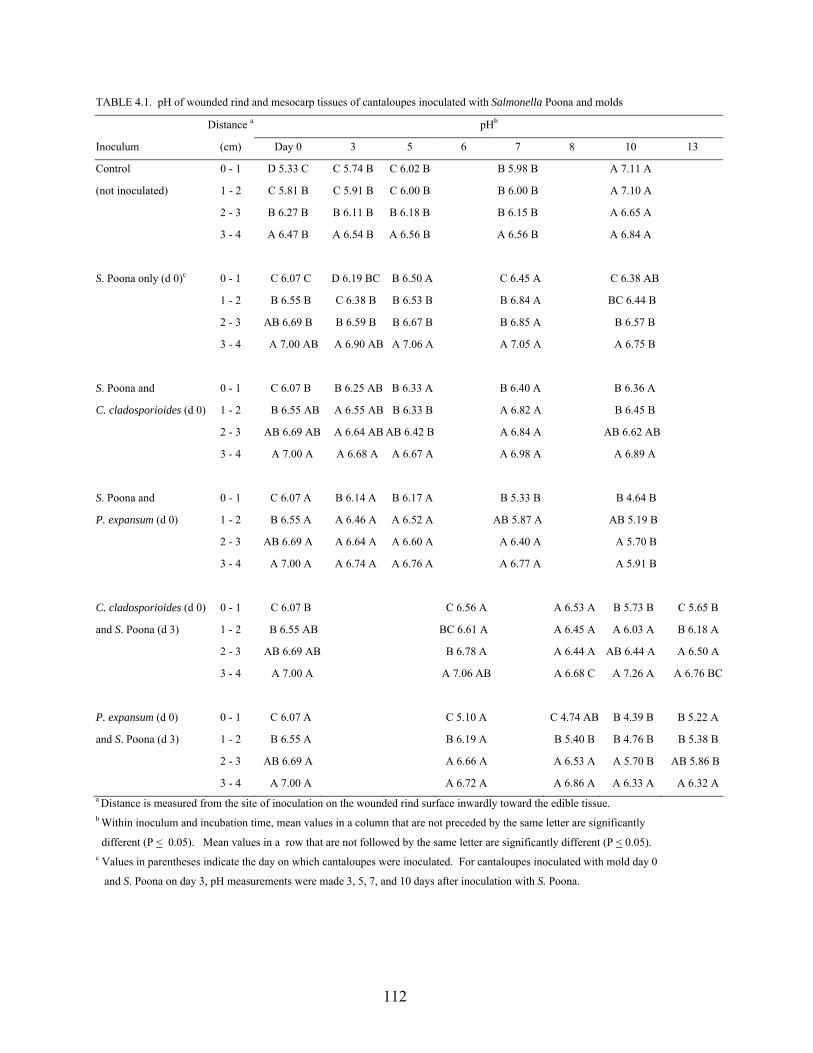
112
TABLE 4.1. pH of wounded rind and mesocarp tissues of cantaloupes inoculated with Salmonella Poona and molds
Distance a pHb
Inoculum (cm) Day 0 3 5 6 7 8 10 13
Control 0 - 1 D 5.33 C C 5.74 B C 6.02 B B 5.98 B A 7.11 A
(not inoculated) 1 - 2 C 5.81 B C 5.91 B C 6.00 B B 6.00 B A 7.10 A
2 - 3 B 6.27 B B 6.11 B B 6.18 B B 6.15 B A 6.65 A
3 - 4 A 6.47 B A 6.54 B A 6.56 B A 6.56 B A 6.84 A
S. Poona only (d 0)c 0 - 1 C 6.07 C D 6.19 BC B 6.50 A C 6.45 A C 6.38 AB
1 - 2 B 6.55 B C 6.38 B B 6.53 B B 6.84 A BC 6.44 B
2 - 3 AB 6.69 B B 6.59 B B 6.67 B B 6.85 A B 6.57 B
3 - 4 A 7.00 AB A 6.90 AB A 7.06 A A 7.05 A A 6.75 B
S. Poona and 0 - 1 C 6.07 B B 6.25 AB B 6.33 A B 6.40 A B 6.36 A
C. cladosporioides (d 0) 1 - 2 B 6.55 AB A 6.55 AB B 6.33 B A 6.82 A B 6.45 B
2 - 3 AB 6.69 AB A 6.64 AB AB 6.42 B A 6.84 A AB 6.62 AB
3 - 4 A 7.00 A A 6.68 A A 6.67 A A 6.98 A A 6.89 A
S. Poona and 0 - 1 C 6.07 A B 6.14 A B 6.17 A B 5.33 B B 4.64 B
P. expansum (d 0) 1 - 2 B 6.55 A A 6.46 A A 6.52 A AB 5.87 A AB 5.19 B
2 - 3 AB 6.69 A A 6.64 A A 6.60 A A 6.40 A A 5.70 B
3 - 4 A 7.00 A A 6.74 A A 6.76 A A 6.77 A A 5.91 B
C. cladosporioides (d 0) 0 - 1 C 6.07 B C 6.56 A A 6.53 A B 5.73 B C 5.65 B
and S. Poona (d 3) 1 - 2 B 6.55 AB BC 6.61 A A 6.45 A A 6.03 A B 6.18 A
2 - 3 AB 6.69 AB B 6.78 A A 6.44 A AB 6.44 A A 6.50 A
3 - 4 A 7.00 A A 7.06 AB A 6.68 C A 7.26 A A 6.76 BC
P. expansum (d 0) 0 - 1 C 6.07 A C 5.10 A C 4.74 AB B 4.39 B B 5.22 A
and S. Poona (d 3) 1 - 2 B 6.55 A B 6.19 A B 5.40 B B 4.76 B B 5.38 B
2 - 3 AB 6.69 A A 6.66 A A 6.53 A A 5.70 B AB 5.86 B
3 - 4 A 7.00 A A 6.72 A A 6.86 A A 6.33 A A 6.32 Aa Distance is measured from the site of inoculation on the wounded rind surface inwardly toward the edible tissue. b Within inoculum and incubation time, mean values in a column that are not preceded by the same letter are significantly
different (P < 0.05). Mean values in a row that are not followed by the same letter are significantly different (P < 0.05). c Values in parentheses indicate the day on which cantaloupes were inoculated. For cantaloupes inoculated with mold day 0
and S. Poona on day 3, pH measurements were made 3, 5, 7, and 10 days after inoculation with S. Poona.
113
Significant increases in pH of rind tissue (0 – 1 cm) occurred within 5 days after inoculation.
Simultaneous co-inoculation of wounds with S. Poona and C. cladosporioides caused a
significant increase in the pH of the cantaloupe rind tissue (0 – 1 cm) within 5 days. The pH of
flesh tissues (1 – 4 cm) was essentially unchanged during the 10-day storage period. The effect
of simultaneous co-inoculation of wounds in rind tissue with S. Poona and P. expansum on the
pH of the rind and flesh tissues was different from that of wounds caused by co-inoculation with
S. Poona and C. cladosporioides. Wounded tissues inoculated with S. Poona and P. expansum
showed a significant decrease in the pH of the rind tissue (0 – 1 cm) on days 7 and 10. The pH of
the mesocarp (1 – 4 cm) below the rind surface remained unchanged for 7 days but decreased
significantly between 7 and 10 days. Differences in changes in pH of tissues inoculated with the
two molds are attributed in part to differences in their alkalinizing capabilities. The production
of acids, such as lactic and pyruvic acids by S. Poona may also have contributed, to reductions in
pH.
The pH of rind tissue (1 – 2 cm) inoculated with C. cladosporioides 3 days before
inoculating with S. Poona decreased significantly between 5 and 7 days after inoculation with S.
Poona. The pH of the flesh 1 – 3 cm below the surface did not change significantly (P > 0.05);
however, at 3 – 4 cm, the pH fluctuated during the 10-day storage period. When the wounds
were inoculated with P. expansum 3 days prior to inoculation with S. Poona, the pH of rind tissue
(0 – 1 cm) decreased on day 7, then increased at day 10. The pH of mesocarp tissues (1 – 3 cm)
trended downward during storage for 10 days but the pH 3 – 4 cm below the surface of the rind
did not change significantly (P > 0.05). Fluctuations in pH of tissues in the four segments were
indicative of changes in microbial ecology during storage. The rind surface of cantaloupes is
inhabited by large numbers of yeast and mold species. Unlike molds, yeasts are generally
114
incapable of invading and infecting tissues and will act as secondary agents of spoilage
(Splittstoesser, 1987). In addition to differences in alkalinizing capabilities of C.
cladosporioides and P. expansum and growth of S. Poona in tissues, the decline in pH in the
latter stages of the storage period may also reflect metabolic activities of yeasts.
Overall changes in pH of tissues at or near the site of inoculation occurred before more
distant sites. This suggests that the inocula and naturally-occurring microorganisms were
responsible for biochemical changes in tissues that began at the site of inoculation and
progressed inwardly. Pseudomonads and lactic acid bacteria on fruits and vegetables capable of
producing pectin-degrading enzymes may cause maceration and release of juices from
cantaloupe tissues (Laminkanra et al., 2003). Wounding of plant tissues also induces elevated
ethylene production and consequently increases the rate of tissue respiration. Starch breakdown
is enhanced, stimulating the tricarboxylic acid cycle and the electron transport chain (Brecht,
1995). The escalated release of carbon dioxide and formation of carbonic acid causes the pH of
the wounded tissues to decrease. There would also be elevated levels of sugars available for
metabolizing by the microorganisms inoculated into the wounds and those that were naturally
resident on the rind surface. The metabolic activities of the microorganisms in the wounds
would elicit changes, such as increasing or decreasing the pH of the tissue environment,
depending on constituents utilized and by products accumulated. Change in pH of uninoculated
tissue reinforces the importance of preventing growth of molds that may influence the survival
and growth of Salmonella and other foodborne pathogens.
Recovery of molds from cantaloupe tissues. Populations of C. cladosporioides and P.
expansum deposited into wounds on the rind of cantaloupes are shown in Table 4.2. The molds
were recovered from inoculated rind tissue as well as the underlying mesocarp closest to the seed
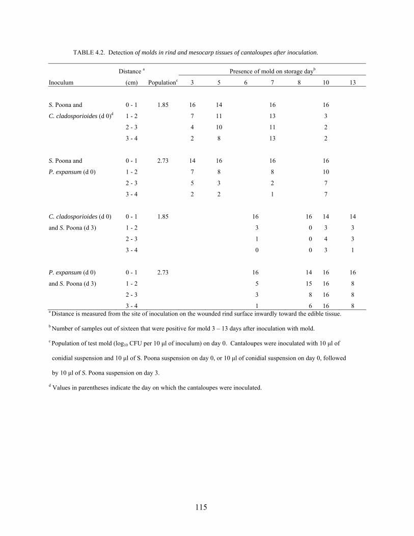
115
TABLE 4.2. Detection of molds in rind and mesocarp tissues of cantaloupes after inoculation.
Distance a Presence of mold on storage dayb
Inoculum (cm) Populationc 3 5 6 7 8 10 13
S. Poona and 0 - 1 1.85 16 14 16 16
C. cladosporioides (d 0)d 1 - 2 7 11 13 3
2 - 3 4 10 11 2
3 - 4 2 8 13 2
S. Poona and 0 - 1 2.73 14 16 16 16
P. expansum (d 0) 1 - 2 7 8 8 10
2 - 3 5 3 2 7
3 - 4 2 2 1 7
C. cladosporioides (d 0) 0 - 1 1.85 16 16 14 14
and S. Poona (d 3) 1 - 2 3 0 3 3
2 - 3 1 0 4 3
3 - 4 0 0 3 1
P. expansum (d 0) 0 - 1 2.73 16 14 16 16
and S. Poona (d 3) 1 - 2 5 15 16 8
2 - 3 3 8 16 8
3 - 4 1 6 16 8 a Distance is measured from the site of inoculation on the wounded rind surface inwardly toward the edible tissue.
b Number of samples out of sixteen that were positive for mold 3 – 13 days after inoculation with mold.
c Population of test mold (log10 CFU per 10 µl of inoculum) on day 0. Cantaloupes were inoculated with 10 µl of
conidial suspension and 10 µl of S. Poona suspension on day 0, or 10 µl of conidial suspension on day 0, followed
by 10 µl of S. Poona suspension on day 3.
d Values in parentheses indicate the day on which the cantaloupes were inoculated.
116
cavity (3 – 4 cm from the surface) after storage at 20°C for 10 days. Molds were detected in
consistently higher numbers of samples closer to the rind surface, regardless of inoculation
schedule or storage time after inoculation. As storage time increased, the detection of test molds
in tissues further from the inoculation site tended to increase.
When wounds on the rind surface were co-inoculated with S. Poona and C.
cladosporioides or P. expansum, the molds progressively penetrated the underlying mesocarp
tissue. Overall, detection of molds in tissues further away from the rind increased through days 7
– 10. Some decreases in number of positive samples were noted at extended storage times.
It has been reported that while lesions on fruits and vegetables caused by pathogenic
microorganisms may detract from the appearance, the internal tissues may remain unaffected
(Wills et al., 1998). However, results presented here show that C. cladosporioides and P.
expansum inoculated into wounds on the rinds of cantaloupes were capable of penetrating
through the mesocarp to depths as great as 4 cm. The effects of these molds on appearance of
mesocarp were different. C. cladosporioides did not affect the appearance of mesocarp tissue,
but tissues extending from the rind to the seed cavity were softened in texture. In some cases,
penetration of the mesocarp tissues by P. expansum hyphae was visible, causing discoloration,
reduction in the moisture content and hardening of the internal tissues.
Propagules of phytopathogens are abundant in the atmosphere and on the surface of fruits
and vegetables as they approach maturity in the field. Mature fruits and vegetables are highly
susceptible to invasion by many pathogenic fungi because they are high in moisture and nutrients
and are no longer protected by some of the intrinsic factors that confer resistance during their
development on the plant. Additionally, many tissues of fruits and vegetables become more
easily injured as they approach full maturity and therefore are more vulnerable to pathogens
117
entering through wounds. Abundance of C. cladosporioides and P. expansum conidia in the
tissue environment as well as and increasing susceptibility of mature cantaloupes during the 10-
day storage period modeled the infection process during storage of harvested fruits. During
storage of the cantaloupes, the fungal mycelia were advanced through the susceptible tissues.
Sanitation is essential to reduce pathogen populations available for infection, especially if
the crop is treated with water after harvest (Eckert, 1980). Harvesting of produce often results in
tissue injuries at the point of severance of stem and fruit, creating a point of entry for pathogens.
The incidence of postharvest diseases is enhanced by the presence of wounds could be
substantially reduced by modifying the handling practices that damage the fruit surface and by
exposing produce to environmental conditions that favor the development of cellular barriers to
the penetration of pathogens. The wounds created on the cantaloupe rinds disrupted the intrinsic
barriers and increased the availability of nutrients and moisture in the tissue environment. The
ability of C. cladosporioides and P. expansum to survive and advance to deeper tissues was
enhanced by the fact that the rind was disrupted.
Recovery of S. Poona from wounded rinds and mesocarp tissues. Populations of S.
Poona in 10 µl of inocula deposited into wounds in cantaloupe rinds are shown in Table 4.3. S.
Poona persisted and grew in the wounds on cantaloupes incubated at 20°C. Except for wounds
simultaneously or sequentially inoculated with P. expansum, the population of S. Poona
increased significantly higher (P < 0.05) within 3 days.
Recovery of S. Poona from tissues at depths up to 3 – 4 cm below the inoculated wound
supports the hypothesis that foodborne pathogens can migrate from the site of inoculation into
adjacent mesocarp tissues. In wounds in the rind tissue were inoculated with only S. Poona, the
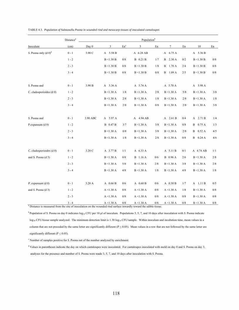
118
TABLE 4.3. Population of Salmonella Poona in wounded rind and mesocarp tissues of inoculated cantaloupes
Distancea Populationb
Inoculum (cm) Day 0 3 Enc 5 En 7 En 10 En
S. Poona only (d 0)d 0 - 1 3.90 C A 5.58 B A 6.28 AB A 6.75 A A 5.36 B
1 - 2 B <1.30 B 0/8 B 0.21 B 1/7 B 2.30 A 0/2 B <1.30 B 0/8
2 - 3 B <1.30 B 0/8 B <1.30 B 1/8 B 1.70 A 2/4 B <1.30 B 0/8
3 - 4 B <1.30 B 0/8 B <1.30 B 0/8 B 1.09 A 2/5 B <1.30 B 0/8
S. Poona and 0 - 1 3.90 B A 5.36 A A 5.74 A A 5.70 A A 5.98 A
C. cladosporioides (d 0) 1 - 2 B <1.30 A 1/8 B <1.30 A 2/8 B <1.30 A 5/8 B <1.30 A 3/8
2 - 3 B <1.30 A 2/8 B <1.30 A 1/8 B <1.30 A 2/8 B <1.30 A 1/8
3 - 4 B <1.30 A 2/8 B <1.30 A 0/8 B <1.30 A 2/8 B <1.30 A 3/8
S. Poona and 0 - 1 3.90 ABC A 5.97 A A 4.94 AB A 2.61 B 0/4 A 2.71 B 1/4
P.expansum (d 0) 1 - 2 B 0.47 B 3/7 B <1.30 A 3/8 B <1.30 A 0/8 B 0.75 A 1/3
2 - 3 B <1.30 A 0/8 B <1.30 A 3/8 B <1.30 A 2/8 B 0.52 A 4/5
3 - 4 B <1.30 A 1/8 B <1.30 A 2/8 B <1.30 A 0/8 B 0.24 A 4/6
C. cladosporioides (d 0) 0 - 1 3.20 C A 3.77 B 1/1 A 6.33 A A 5.11 B 0/1 A 4.74 AB 1/1
and S. Poona (d 3) 1 - 2 B <1.30 A 0/8 B 1.16 A 0/6 B 0.96 A 2/6 B <1.30 A 2/8
2 - 3 B <1.30 A 5/8 B <1.30 A 2/8 B <1.30 A 3/8 B <1.30 A 2/8
3 - 4 B <1.30 A 4/8 B <1.30 A 1/8 B <1.30 A 4/8 B <1.30 A 1/8
P. expansum (d 0) 0 - 1 3.20 A A 0.66 B 0/6 A 0.60 B 0/6 A 0.30 B 1/7 A 1.11 B 0/5
and S. Poona (d 3) 1 - 2 A <1.30 A 0/8 A <1.30 A 0/8 A <1.30 A 1/8 B <1.30 A 0/8
2 - 3 A <1.30 A 0/8 A <1.30 A 0/8 A <1.30 A 0/8 B <1.30 A 0/8
3 - 4 A <1.30 A 0/8 A <1.30 A 0/8 A <1.30 A 0/8 B <1.30 A 0/8 a Distance is measured from the site of inoculation on the wounded rind surface inwardly toward the edible tissue.
b Population of S. Poona on day 0 indicates log10 CFU per 10 µl of inoculum. Populations 3, 5, 7, and 10 days after inoculation with S. Poona indicate
log10 CFU/tissue sample analyzed. The minimum detection limit is 1.30 log10 CFU/sample. Within inoculum and incubation time, mean values in a
column that are not preceded by the same letter are significantly different (P < 0.05). Mean values in a row that are not followed by the same letter are
significantly different (P < 0.05).
c Number of samples positive for S. Poona out of the number analyzed by enrichment.
d Values in parentheses indicate the day on which cantaloupes were inoculated. For cantaloupes inoculated with mold on day 0 and S. Poona on day 3,
analyses for the presence and number of S. Poona were made 3, 5, 7, and 10 days after inoculation with S. Poona.
119
pathogen was recovered from the mesocarp tissues 2 – 3 cm below the rind surface 5 days after
inoculation. Within 7 days, S. Poona was recovered from tissues at a depth of 3 – 4 cm from the
rind surface. At 10 days, S. Poona was not detected in tissue 1 – 4 cm below the inoculation
site. The reason for the absence of the pathogen is not evident. In wounds simultaneously
inoculated with S. Poona and C. cladosporioides or P. expansum or sequentially inoculated with
C. cladosporioides and S. Poona, the pathogen was recovered from the tissues 3 – 4 cm below
the rind after 3 days of storage and persisted for up to 10 days. This suggests that C.
cladosporioides and P. expansum enhanced the migration of S. Poona away from inoculated
tissue and promoted its survival in subsurface tissues. Increased recovery of S. Poona from
deeper tissues could have been due to penetration by fungal hyphae or perhaps fluids as a result
of maceration of the tissues. Inoculation of wounds with P. expansum 3 days before inoculating
with S. Poona did not appear to facilitate the movement of S. Poona into sub-surface tissues.
C. cladosporioides appeared to promote survival and growth of S. Poona in cantaloupe
tissues more than P. expansum. This may be attributable in part to proteolytic activity by C.
cladosporioides, which would contribute to an increase the pH of the tissue, making it more
favorable for S. Poona. Hardening of the mesocarp tissues inoculated by P. expansum would
reduce the potential of S. Poona to move away from the site of inoculation. Although the pH of
wounds in rind tissue inoculated with P. expansum decreased to as low as 4.39 (sd. + 0.2), S.
Poona continued to survive in these tissues. The optimum pH for growth of Salmonella is 6.5 –
7.5, but physiological adaptability of the pathogen has been demonstrated by its ability to grow
at pH values ranging from 4.5 to 9.5 (D’Aoust, 1997; D’Aoust, 2000; Jay, 2000). Salmonella
Montevideo has been reported to grow in chopped tomatoes (pH 4.1) (Zhuang et al., 1995) and
Salmonella Baildon grew in diced tomatoes (pH 4.4) (Weissinger et al., 2000)
120
Microbial growth is among the factors that accelerate senescence of cantaloupes, causing
economic loss by producers and consumers. To reduce these losses, visually sound portions of
edible tissue at distances away from the site of infection are salvaged for consumption. This is a
potentially hazardous practice, as S. Poona and probably other salmonellae may migrate away
from the site of infection and contaminate apparently healthy tissues. The migration of S. Poona
in tissues of cantaloupes was more evident when co-inoculated with C. cladosporioides than with
P. expansum. This may be attributable to more extensive breakdown of tissues by C.
cladosporioides versus hardening of some tissues by P. expansum. S. Poona was not recovered
from tissues compacted as a result of growth of P. expansum.
Phytopathogens produce extracellular enzymes such as cellulases and pectinases to
decompose plant tissues that are not digestible by other organisms. Fruit decay pathogens
produce pectolytic enzymes that degrade cell wall pectin and macerate tissues (Bruton et al.,
1998). Release of fluids, softening of the tissues, and the creation of channels and cavities by
fungal mycelia would promote passive translocation of S. Poona from the site of inoculation in
the cantaloupe rind to tissues within the mesocarp. Salmonella is not fastidious with respect to
oxidation-reduction (OR) potential (Doyle and Cliver, 1990) and can therefore survive in
cantaloupe tissues with low OR potential. S. Montevideo has been reported to survive in internal
tissues of tomatoes stored at 20 and 30°C (Zhuang et al. 1995). Although the mode of entry of S.
Poona into internal cantaloupe tissues in the study presented here was different, viable S. Poona
was shown to be present for up to 10 days at a distance of 3 – 4 cm from the rind surface.
The effectiveness of treatments to detach or inactivate Salmonella on cantaloupe surfaces
is dependent on the location of the cells. Migration of S. Poona into sub-surface tissues would
reduce the efficacy of decontamination practices that are currently being used. Solutions
121
containing chlorine at concentrations of 60, 120, and 320 µg/ml, for example, were less effective
at killing S. Montevideo that had been internalized in the core tissue compared to those that were
on the surface of tomatoes (Zhuang et al., 1995). Aqueous chlorine at 100 µg/ml failed to kill S.
Montevideo inoculated into wounded sites on tomatoes (Wei et al., 1995). The efficacy of
chlorine in killing microorganisms on the surface of raw produce is notably reduced by contact
with organic matter (Beuchat et al., 1998). Therefore it is conceivable that application of
chlorine by dipping or spraying would be ineffective against Salmonella in sub-surface tissues.
Trisodium phosphate (TSP) was observed to be effective in killing S. Montevideo on the surface
of mature-green tomatoes, but less effective against internalized cells (Zhuang and Beuchat,
1996). Similarly, Salmonella Chester in injured tissue of pepper discs were not killed by
treatments of 3% to 12% TSP (Liao and Cooke, 2000). When apples were treated with hydrogen
peroxide for the purpose of killing or removing S. Chester, populations remaining in the stem
and calyx cavities were greater than on the skin (Liao and Sapers, 2000). Entrapment of bacteria
below the surface of the fruits and vegetables limits the accessibility of sanitizers to the bacterial
contaminants (Beuchat, 1999).
Salmonellosis associated with consumption of cantaloupes may be due to introduction of
Salmonella on the rind into the pulp tissue by physical contact or during cutting (Suslow and
Cantwell, 2001; Ukuku and Sapers, 2001). It is hypothesized that the stem scar tissues and
wounds on the rind would trap bacteria. In a previous study, S. Poona was shown to infiltrate the
intact rind and stem scar tissues during immersion in inocula. These cells could subsequently
migrate to deeper tissues. The study reported here shows that contamination of the flesh can also
result from migration of S. Poona from the wounded rind through the pulp tissues to distances as
far as 3 – 4 cm, with or without assistance from phytopathogens. Migration and survival of S.
122
Poona in the internal tissues of cantaloupes were enhanced by co-inoculation with C.
cladosporioides and to a lesser extent, P. expansum. Consumption of cantaloupes from which
diseased tissue has been removed is not advisable because S. Poona and perhaps other pathogens
may still be present in remaining tissues.
123
REFERENCES
Agblor, S. and D. Waterer. June 2001. Muskmelons: Cantaloupe postharvest handling and storage. Agri-Food Innovation Fund (AFIF) Postharvest Specialist Program. University of Saskatchewan.
Asplund, K. and E. Nurmi. 1991. The growth of salmonellae in tomatoes. Intl. J. Food Microbiol.
13:177-182. Beuchat, L.R. 1999. Surface decontamination of fruits and vegetables eaten raw: a review.
World Health Organization. WHO/FSF/FOS/98.2. Beuchat, L.R., B.V. Nail, B.B. Adler, and M.R.S. Clavero. 1998. Efficacy of spray application of
chlorinated water in killing pathogenic bacteria on raw apples, tomatoes, and lettuce. J. Food Prot. 61:1305-1311.
Blostein, J. 1993. An outbreak of Salmonella javiana associated with consumption of
watermelon. J. Environ. Health 56:29-31. Brecht, J.K. 1995. Physiology of lightly processed fruits and vegetables. Hort Sci. 30:18-22. Bruton, B.D. W.S. Conway, K.C. Gross, J.X. Zhang, C.L. Biles, and C.E. Sams. 1998.
Polygalacturonases of a latent and wound postharvest fungal pathogen of muskmelon fruit. Postharv. Biol. Technol. 13:205-214.
Centers for Disease Control and Prevention (CDC). 1975. Salmonella typhimurium outbreak
traced to a commercial apple cider – New Jersey. Morb. Mortal. Wkly. Rep. 24:87-88. Centers for Disease Control and Prevention (CDC). 1991. Multistate outbreak of Salmonella
poona infections – United States and Canada, 1991. Morb. Mortal. Wkly. Rep. 40:549-552.
Centers for Disease Control and Prevention (CDC). 1999. Outbreaks of Salmonella serotype
Muenchen infections associated with unpasteurized orange juice – United States and Canada. Morb. Mortal. Wkly. Rep. 48:582-585.
Centers for Disease Control and Prevention (CDC). 2000. Surveillance of foodborne disease
outbreaks – United States 1993 – 1997. Morb. Mortal. Wkly. Rep. Surveillance Summaries 49 (SS01):1-51.
Centers for Disease Control and Prevention (CDC). 2002. Multistate outbreaks of Salmonella
serotype Poona infections associated with eating cantaloupes from Mexico – United States and Canada, 2000 – 2002. Morbid. Mortal. Wkly. Rep. 51:1044-1047.
124
Cook, K.A., T.E. Dobbs, W.G. Hlady, J.G. Wells, T.J. Barrett, N.D. Puhr, G.A. Lancette, D.W. Bodager, B.L. Toth, C.A. Genese, A.K. Highsmith, K.E. Pilot, L. Finelli, and D.L. Swerdlow. 1998. Outbreak of Salmonella serotype Hartford infections associated with unpasteurized orange juice. J. Am. Med Assoc. 280:1504-1509.
D’Aoust, J-Y. 1997. Salmonella species, p. 129-158. In M.P. Doyle, L.R. Beuchat,
and T.J. Montville (ed.), Food microbiology: Fundamentals and frontiers. ASM Press. Washington D.C.
D’Aoust, J-Y. 2000. Salmonella, p. 1233-1299. In B.M. Lund and G.W. Gould (ed.),
The microbiological safety and quality of food. Aspen Publishers. Gaithersburg, MD. D’Aoust, J-Y. 2001. Foodborne salmonellosis: current international concerns. Food Safety
Magazine 7(2):10-17, 51. Deeks, S., A. Ellis, B. Ciebin, R. Khakhria, M, Naus, and J. Hockin. 1998. Salmonella
Oranienberg, Ontario. Can. Comm. Dis. Rep. 24:177-179. Doyle, M.P. and D.O. Cliver. 1990. Salmonella, p.185-204. In D.O. Cliver (ed.), Foodborne
diseases. Academic Press. Inc. Eckert, J.W. 1980. Postharvest diseases of fresh fruits and vegetables – Etiology and control,
p. 81-117. In N.F. Haard and D.K. Salunkhe (Ed.) Postharvest biology and handling of fruits and vegetables. The AVI Publishing Company, Inc., Westport, Connecticut.
Food and Drug Administration. 2001a. FDA Survey of imported fresh produce. FY 1999 Field
assignment. US Food and Drug Administration. Center for Food Safety and Applied Nutrition. Office of Plant and Dairy Foods and Beverages. January 30, 2001, [Internet, WWW], ADDRESS: http://www.cfsan.gov/~dms/prodsur6.html Accessed 25 January 2002.
Food and Drug Administration. 2001b. FDA Survey of domestic fresh produce: interim results.
US Food and Drug Administration. Center for Food Safety and Applied Nutrition. June 30, 2001, [Internet, WWW], ADDRESS: http://vm.cfsan.fda.gov/~dms/prodsur8.html Accessed 25 January 2002.
Gagliardi, J.V., P.D. Miller, G. Lester, and D. Ingram. 2003. On-farm and postharvest processing
sources of bacterial contamination to melon rinds. J. Food Prot. 66:82-87. Gayler, G.E., R.A. MacCready, J.P. Reardon, and B.F. Mc Kernan. 1955. An outbreak of
salmonellosis traced to watermelon. Public Health Rep. 70:311-313. Goepfert, J.M. 1980. Vegetables, fruit, nuts and their products. p. 606-642. In International
Commission on Microbiological Specifications for Foods. Microbial ecology of foods Volume 2. Food commodities.
125
Golden, D.A., E.J. Rhodehamel, and D.A. Kautter. 1993. Growth of Salmonella spp. in cantaloupe, watermelon, and honeydew melons. J. Food Prot. 56:194-196.
Guo, X., J. Chen, R.E. Brackett, and L.R. Beuchat. 2002. Survival of Salmonella on tomatoes
stored at high relative humidity, in soil, and on tomatoes in contact with soil. J. Food Prot. 65:274-279.
Institute of Food Technologists/Food and Drug Administration of the United States Department
of Health and Human Services (IFT/FDA). 2001. Analysis and evaluation of preventative control measures for the control and reduction/elimination of microbial hazards on fresh and fresh-cut produce. IFT/FDA Contract No. 223-98-2333. Task Order No. 3. Institute of Food Technologists. Chicago, IL.
Jay, J. 2000. Modern food microbiology. 6th ed. Aspen Publishers. Gaithersburg, MD. Kleman, L., L. Jaykus, D. Moll, C. Moe, C. Martinez, and J. Anciso. 2003. A field study of the
microbiological quality of fresh produce. Abstr. T33, p. 170. 90th Annual Meeting Intl. Assc. Food Prot. New Orleans, LA.
Laminkanra, O., B. Juavaez, M.A. Watson, and O.A. Richard. 2003. Effect of cutting and storage
on sensory traits of cantaloupe melon cultivars with extended postharvest shelf life. J. Sci. Food Agric. 83:702-708.
Larson, A., J. Wallis, C. Lewandowski, D. Jenson, S. Potsic, M.K. Nickels, M. Lesko, C.
Langkop, R.J. Martin, T. Endo, H.B. Ehrhard, and B.J. Francis. 1979. Salmonella Oranienberg gastroenteritis associated with consumption of precut watermelons- Illinois. Morb. Mortal. Wkly. Rep. 28:522-523.
Liao, C-H. and G.M. Sapers. 2000. Attachment and growth of Salmonella Chester on apple fruit
and in vivo response of attached bacteria to sanitizer treatments. J. Food Prot. 63: 876-883.
Liao, C-H. and P.H. Cooke. 2001. Response to trisodium phosphate treatment of Salmonella
Chester to fresh-cut green pepper slices. Can. J. Microbiol. 47:25-32. Madden, J.M. 1992. Microbial pathogens in fresh produce – the regulatory perspective. J. Food
Prot. 55:821-823. Mahon, B.E., A. Ponka, W. Hall, K. Komatsu, S. Dietrich, A. Siitonen, G. Cage, M. Lambert-
Fair, P. Hayes, N. Bean, P. Griffin, and L. Slutsker. 1997. An international outbreak of Salmonella infections caused by alfalfa sprouts grown from contaminated seed. J. Infect. Dis. 175:876-882.
Mead, P.S., L. Slutsker, V. Dietz, L.F. McCaig, J.S. Bresee, C. Shapiro, P. M. Griffin, and R.V.
Tauxe. 1999. Food-related illness and death in the United States. Emerg. Infect. Dis. 5:607-625.
126
Mohle-Boetani, J.C., R. Reporter, S.B. Werner, S. Abbott, J. Farrar, S.H. Waterman, and D.J. Vugia. 1999. An outbreak of Salmonella serogroup Saphra due to cantaloupes from Mexico. J. Infect. Dis. 180:1361-1364.
Nguyen-the, C. and F. Carlin. 2000. Fresh and processed vegetables, p. 620-684. In B.L. Lund,
T.C. Baird –Parker, and G.W. Gould (ed.), The microbiological safety and quality of food, vol. 1. Aspen Publishers, Gaithersburg, MD.
O’Mahony, P., J. Cowden, B. Smyth, D. Lynch, M. Hall, B. Rowe, E.L. Teare, R.E. Tettmar,
A.M. Rampling, M. Coles, R.J. Gilbert, E. Kingcott, and C.L.R. Bartlet. 1990. An outbreak of Salmonella Saint-Paul infection associated with bean sprouts in the UK. Epidemiol. Infect. 104:229-235.
Ries, A.A., S. Zaza, C. Langkop, R.V. Tauxe, and P.A. Blake. 1990. A multistate outbreak of
Salmonella Chester linked to imported cantaloupe, Abstr. 915. Abstr. 30th, p. 238. Intersci. Conf. Antimicrob. Agents Chemother. American Society for Microbiology, Washington, D.C.
Splittstoesser, D.F. 1987. Fruits and fruit products, p. 101-128. In L.R. Beuchat (Ed.). Food and
Beverage Mycology. AVI New York, NY. Suslow, T.V., M. Cantwell, and J. Mitchell. 2000. Cantaloupes: Recommendations for
maintaining postharvest quality. University of California, Davis. Postharvest Technology Research and Information Center. 26 June 2000, [Internet, WWW], ADDRESS: http://postharvest.ucdavis.edu/Produce/Produce facts/cantalope.html Accessed 25 March 2002.
Suslow, T. and Cantwell, M. 2001. Recent findings on fresh-cut cantaloupe and honeydew
melon. Fresh cut. April 2001. Pp. 12, 20, 32-33. Tauxe, R.V. 1991. Salmonella: A postmodern pathogen. J. Food Prot. 54:563-568. Ukuku, D.O., V. Pilizota, and G.M. Sapers. 2001. Influence of washing treatment on native
microflora and Escherichia coli population of inoculated cantaloupes. J. Food Safety 21:31-47.
Ukuku, D.O. and G.M. Sapers. 2001. Effect of sanitizer treatments on Salmonella Stanley
attached to the surface of cantaloupe and cell transfer to fresh-cut tissues during cutting practices. J. Food Prot. 64:1286-1291.
Van Beneden, C.A., W.E. Keene, D.H. Werker, A.S. King, P.R. Cieslak, K. Hedberg, R.A.
Strang, A. Bell, and D. Fleming. 1996. A health food fights back: an international outbreak of Salmonella Newport infections due to alfalfa sprouts, K47. Abstr. 36th, p. 259. Intersci. Conf. Antimicrob. Agents Chemother. American Society for Microbiology, Washington, D.C.
127
Wei, C.I., T.S. Huang, J.M. Kim, W.F. Lin, M.L. Tamlin, and J.A. Bartz. 1995. Growth and survival of Salmonella Montevideo on tomatoes and disinfection with chlorinated water. J. Food Prot. 58:829-836.
Weissinger, W.R., W. Chantarapanont, and L.R. Beuchat. 2000. Survival and growth of
Salmonella Baildon in shredded lettuce and diced tomatoes, and effectiveness of chlorinated water as a sanitizer. Intl. J. Food Microbiol. 62:123-131.
Wells, J.M. and J.E. Butterfield. 1997. Salmonella contamination associated with bacterial soft
rot of fresh fruits and vegetables in the marketplace. Plant Dis. 81:867-872. Wills, R.B.H., D. Graham, B. McGlasson, and D. Joyce. 1998. Postharvest: an introduction to
the physiology and handling of fruit, vegetables and ornamentals. CAB International. London, U.K.
Wood, R.C., C. Hedberg, and K.White. 1991. A multistate outbreak of Salmonella javiana
infections associated with raw tomatoes. (Abstracts) p.69. In CDC Epidemic Intelligence Service, 40th Ann. Conf., Atlanta. U.S. Dept. of Health and Human Services, Public Health Science.
Zhuang, R-Y., L.R. Beuchat, and F.J. Angulo. 1995. Fate of Salmonella Montevideo on and in
raw tomatoes as affected by temperature and treatment with chlorine. Appl. Env. Microbiol. 61: 2127-2131.
Zhuang, R-Y. and L.R. Beuchat. 1996. Effectiveness of trisodium phosphate for killing
Salmonella Montevideo on tomatoes. Lett. Appl. Microbiol. 22: 97-100
128
CHAPTER 5
EXAMINATION OF YEASTS FOR ANTAGONISTIC ACTIVITY AGAINST
SALMONELLA POONA IN CANTALOUPE SUSPENSION AND IN WOUNDS
IN RINDS CO-INFECTED WITH PHYTOPATHOGENIC MOLDS1
______________________
1 Richards, Glenner M. and Larry R. Beuchat. 2003. To be submitted to International Journal of Food Microbiology.
129
ABSTRACT
The application of yeasts as biocontrol agents to prevent fungal decay of fruits and
vegetables has been described. However, little is known about the potential antagonistic effects
of yeasts against human bacterial pathogens that may contaminate produce. Ten yeasts were
screened for antagonistic activity against survival and growth of Salmonella Poona at 20°C in
cantaloupe juice and decay by two phytopathogens, Cladosporium cladosporioides and
Geotrichum candidum, in wounds on the surface of cantaloupe rind stored at 4 and 20°C.
Cantaloupe juice was inoculated using five inoculation schemes: S. Poona only, high (3.93 –
5.21 log10 CFU/ml) or low populations (1.79 – 3.26 log10 CFU/ml) of yeasts only, and S. Poona
combined with high or low populations of yeasts. The populations of S. Poona and yeasts in
juice, as well as pH and °brix, were determined 6, 24, and 48 h after inoculation. High
populations of Debaryomyces hansenii, Pichia guilliermondii, and Pseudozyma sp. were
antagonistic to S. Poona in cantaloupe juice stored at 20°C after 48 h (P ≤ 0.05). With or without
yeasts, growth of S. Poona caused the pH of the cantaloupe juice to decrease (P ≤ 0.05). Wounds
in cantaloupe rinds were inoculated with yeast and mold or yeast, mold, and S. Poona and
incubated at 4°C for 14 days or 20°C for 7 days. The pH of the wounded rind tissue inoculated
with C. cladosporioides and yeasts increased significantly (P ≤ 0.05) at 20°C. Results indicate
that at 4°C, Candida oleophila, Candida sake, D. hansenii, P. guilliermondii, Pseudozyma sp.,
and Rhodotorula glutinis co-inoculated with G. candidum retarded the increase in pH and
prevented changes of the magnitude that occurred in tissue inoculated with G. candidum alone.
Wounds that were inoculated with P. guilliermondii together with C. cladosporioides or G.
candidum did not show visible mold growth. At 20°C, populations of S. Poona (6.40, 7.26, and
7.98 log10 CFU/sample) were lower in wounds co-inoculated with G. candidum and three of the
130
test yeasts (D. hansenii, P. guilliermondii, and Cr. albidus, respectively) compared to co-
inoculation with G. candidum (9.16 log10 CFU/sample) and the other yeasts (> 8.49 log10
CFU/sample). While C. oleophila and R. glutinis showed most promise for reducing the
population of S. Poona in the wounds in cantaloupes co-inoculated with G. candidum and stored
at 4°C, Cr. albidus, D. hansenii, and P. guilliermondii were most effective in controlling growth
at 20°C.
131
INTRODUCTION
Fresh and minimally processed produce are highly perishable. Postharvest deterioration
is a problem that persists from the point of harvest through the marketing chain, ultimately
resulting in increased cost and decreased availability of produce to consumers and delivery to
distant markets. Early termination of storage life of fresh produce is attributed to fungal
infection, senescence, and transpiration (Arul, 1994). Subsequent to harvest, physiological
senescence and microbial deterioration are accelerated. These processes are inextricably bound
because the ability of plant tissues to resist microbial invasion declines through senescence.
Worldwide postharvest losses of crops have been estimated to be in the range of 5 – 50% of the
harvested crop (Eckert and Ogawa, 1985; Salunkhe and Desai, 1984; Wilson and Wisniewski,
1989). Storage diseases, especially those caused by fungal pathogens infecting surface wounds,
are responsible for substantial postharvest losses of most fruits and vegetables (Droby et al.,
1996; Snowdon, 1990). Developments in the produce industry, such as mechanical harvesting
and bulk handling, can aggravate postharvest diseases because of increased wounding and the
creation of microenvironments that promote disease development (Arul, 1994).
Since the 1960s, synthetic fungicides have been the primary method of controlling
postharvest diseases (Eckert and Ogawa, 1985). In recent times, chemical control of plant pests
and diseases has come under scrutiny as there are indications that some pesticides and fungicides
pose potential oncogenic risks when applied to processed foods (National Research Council,
1987). Furthermore, some postharvest pathogens have developed resistance to fungicides that
have been registered for the control of postharvest diseases (El-Ghaouth, 1997; Wilson and
Wisniewski, 1989). Increasing awareness of the potential negative side-effects of fungicides on
the ecosystem, growing interest in pesticide-free agricultural products, and consumers’ demand
132
for food free of microbial toxins, pathogens, and chemical residues have resulted in an escalation
in research aimed at identifying alternative methods to improve the quality and shelf-life of fresh
produce.
Biological control refers to the process of curbing incidence and preventing spread of
plant diseases by applying a natural biological process and/or the product of a natural biological
process, either preharvest or postharvest (Wilson and Wisniewski, 1989; Wilson, 1997). Among
the possible approaches are the use of antagonistic microorganisms, inherent or induced
resistance, and natural plant- or animal-derived products (Wilson and Wisniewski, 1994). Ideal
antagonists need to meet several criteria to be successfully developed for commercial use on
harvested crops. These characteristics include: genetic stability, efficacy at low concentration,
not fastidious in nutrient requirements, the ability to survive adverse environmental conditions
such as low temperature and controlled atmosphere storage, effectiveness against a wide range of
pathogens on a variety of fruits and vegetables, amenable to production on an inexpensive
medium, easy to dispense, the microbe does not produce metabolites that are deleterious to
human health, resistance to pesticides, compatibility with commercial processing procedures,
and not pathogenic to plant tissues (Droby et al., 1996).
Biological control does not equal or surpass chemical control in the field, with respect to
efficiency or consistency (Wilson and Wisniewski, 1994). Inconsistent results of biological
control methods applied in the field have been attributed to the inherent variability in living
systems and vagaries of the environment. Prospects for biological control are better for
postharvest diseases than field infections because the postharvest environment is easier to
control, more conducive to the application of biological control agents, and more similar to
conditions in the laboratory than in the field (Wilson and Wisniewski, 1994). Effective
133
biological control of fungal pathogens by introduction of an antagonist has been reported on
apple, apricot, cherry, citrus, grape, nectarine, peach, pear, pepper, persimmon, plum, potato,
strawberry, and tomato (Janisiewicz and Korsten, 2002; Wilson and Wisniewski, 1989). A yeast,
Candida oleophila Montrocher, and a bacterium, Pseudomonas syringae, were registered by the
United States Environmental Protection Agency in 1995 for control of postharvest diseases of
pome and citrus fruits, and have been made commercially available under the trade names
AspireTM and Biosave 110TM, respectively. Yeasts are the main group of microorganisms being
investigated for use as artificially introduced antagonists because they can colonize the surface of
fruits and vegetables for long periods under reduced-moisture conditions, produce extracellular
polysaccharides that enhance their survival and restrict colonization sites and flow of
germination cues to fungal propagules, use available nutrients to rapidly proliferate, and are
minimally affected by pesticides (Droby et al., 1996). There are indications that the
mechanisms by which antagonistic yeasts exert their protectant effect involve competition for
nutrients, site exclusion, direct parasitism, and perhaps induced resistance (Droby et al., 1989;
Droby and Chalutz, 1994; El-Ghaouth, 1997; El-Ghaouth et al., 1998; Janisiewicz et al., 2000;
Walker et al., 2001; Wisniewski et al., 1991).
Although outbreaks of produce-associated foodborne illnesses continue to be fewer in
number compared to outbreaks linked to consumption of meat and meat products, microbial
contamination of fresh produce has been of increasing concern (Buck et al., 2003). The
percentage of outbreaks of foodborne illnesses that have been associated with consumption of
fresh and minimally processed fruits and vegetables or their products has been increasing. The
number of documented produce-related outbreaks doubled between 1973-1987 and 1988-1992.
Outbreaks with identified etiology were caused predominantly by bacteria, primarily Salmonella
134
(Buck et al., 2003). Results of experiments evaluating antimicrobial-producing lactic acid
bacteria such as Lactobacillus acidovorus, L. casei, L. plantarum, Lactococcus lactis, and
Pediococcus spp. to control Listeria monocytogenes, Salmonella Typhimurium, and
Staphylococcus aureus on meat, cheese, and vegetables are promising (Lewus et al., 1991;
Muriana, 1996; Stevens et al., 1991). Applications have positive and negative implications, as
compounds produced by the antagonists may initially kill foodborne pathogens, but pathogens
may eventually develop resistance to these compounds. The possibility of developing resistance
to a biocontrol agent whose mechanism of resistance is based on competitive exclusion is less
likely to occur (Janisiewicz et al., 1999a).
Metabiotic associations between phytopathogenic molds and foodborne bacteria on
apples and tomatoes have been described (Conway et al., 2000; Draughon et al., 1988; Huhtanen
et al., 1976; Mundt, 1978; Odlaug and Pflug, 1979; Wade and Beuchat, 2003). Some molds
produce proteolytic enzymes that release alkaline substances into the surrounding tissues and
cause the pH to increase. This may create a more favorable environment for enteric pathogens
such as Salmonella to survive and grow. In our previous studies, Salmonella Poona was shown
to persist on intact rind at refrigerated temperature and grow on intact, wounded, and decaying
cantaloupe rind at refrigerated and ambient temperatures. Injury and abusive storage
temperature enhance survival and growth of phytopathogenic molds and S. Poona on cantaloupe
rinds. Disruption of tissue and cell integrity in fresh cut and wounded fruits results in an
increased availability of nutrients and water to support contaminating microorganisms.
Modification of the pH and enhanced nutrient and moisture availability are factors that support S.
Poona on wounded cantaloupe rind tissues, especially at ambient temperature.
135
While the success of antagonistic yeasts in controlling fungal spoilage and decay of
produce has been demonstrated, much less is known about the antagonistic effect of yeasts
against bacteria. Bilinski et al. (1985) surveyed nearly 400 yeast strains belonging to 31 genera
for antibacterial activity. Kluyveromyces thermotolerans and Hanseniospora uvarum inhibited
the growth of Lactobacillus plantarum and Bacillus cereus, but not gram-negative bacteria.
Yeasts isolated from the phylloplane produced antibacterial compounds that inhibited the growth
of Pseudomonas fluorescens and S. aureus (McCormack et al., 1994). In light of concerted
efforts to develop biological control protocols to reduce the effects of postharvest fungal and
bacterial pathogens, studies to determine the response of human pathogens to potential
biocontrol microorganisms have been done. Saccharomyces boulardii was shown to be capable
of inhibiting multiplication of Salmonella and Shigella (Rodrigues et al., 1996). Decline in the
population of enteropathogenic Escherichia coli O157:H7 in apple cider was attributed to
interaction of the pathogen with naturally occurring yeasts (Janisiewicz et al., 1999a). P.
syringae L-59-66, the antagonist used to control postharvest decay of pome fruits, was reported
as preventing growth of E. coli O157:H7 on wounded apple tissue (Janisiewicz et al., 1999b).
The objective of this study was to examine yeasts for their potential to impair survival and
growth of S. Poona in cell suspensions and in wounds on cantaloupe rind co-inoculated with
phytopathogenic molds. Changes in the pH of cell suspensions and rind tissue and surface
diameter of inoculated wounds as affected by co-inoculation with yeasts, molds, and S. Poona
were determined.
MATERIALS AND METHODS
S. Poona inoculum. Five strains of S. Poona isolated from patients in cantaloupe-
associated outbreaks of salmonellosis were used. Strains 00A3207, 01A3923, and 02A3275
136
were obtained from the State of California, Department of Health Services, and strains 00A3279
and 01A242 were obtained from Dr. Trevor Suslow, University of California, Davis. Each strain
was progressively made resistant to nalidixic acid by a series of nine transfers in tryptic soy broth
(TSB; BBL/Difco) supplemented with increasing concentrations of nalidixic acid up to 50 µg/ml
(TSBN). Cultures were incubated at 37°C for 24 h before transferring to TSB containing a
higher concentration of nalidixic acid. The nalidixic acid-resistant cells were surface plated on
tryptic soy agar (TSA; BBL/Difco) supplemented with 50 µg of nalidixic acid/ml (TSAN) and
incubated at 37°C for 24 h. Stock cultures were stored on TSAN at 4°C.
Preparation of S. Poona inoculum for in vitro study. Stock cultures of S. Poona were
streaked on TSAN and incubated at 37°C for 24 h before transferring cells from a single colony
of each strain to 10 ml of TSBN and incubating at 37°C for 24 h. Three consecutive 24-h
transfers of suspensions were made via loop inocula into TBSN. After incubation at 37°C for 24
h, the cultures were serially diluted in sterile deionized water and finally cantaloupe juice (see
preparation below) to give inocula that contained 2.10 log10 CFU/ml. The population of S.
Poona was determined by surface plating serially diluted suspensions in cantaloupe juice (0.25
ml in quadruplicate and 0.1 ml in duplicate) on TSAN. Plates were incubated at 37°C for 24 h
before colonies were counted.
Preparation of S. Poona inoculum for wound inoculation. S. Poona cells were grown
as described above. Subsequent to the three consecutive 24-h transfers of cultures and
incubation in TSBN at 37°C for 24 h, cells were harvested by centrifugation at 2,500 x g for 10
min in a Centra CL2 centrifuge (International Equipment Company, Needham Heights, MA).
Cells were washed two times in 10 ml of sterile deionized water, then suspended in 10 ml of
sterile 0.1% (w/v) peptone water. The population (CFU/ml) in each suspension was determined
137
by serially diluting suspensions in sterile 0.1% peptone water and surface plating samples (0.1 ml
in duplicate) on TSAN. Plates were incubated at 37°C for 24 h before colonies were counted.
Random presumptive-positive colonies of Salmonella were confirmed using triple sugar iron,
lysine iron agar, and a Salmonella latex agglutination assay (Oxoid, Basingstoke, Hampshire,
U.K.).
Equal volumes (10 ml) of suspensions of each strain were combined to give 50 ml of a
five-strain mixture of S. Poona containing approximately equal populations (ca. 9 log10 CFU/ml)
of each strain. The suspension was serially diluted to give an inoculum that contained ca. 6.5
log10 CFU/ml.
Preparation of cantaloupe juice for in vitro study. Eastern type cantaloupes (Cucumis
melo L. var. reticulatus Naud.) were purchased in June – July, 2003 from a grocery in Griffin,
GA and stored at 4°C for 2 days before adjusting them to 22 ± 1°C over a 16 to 20-h period. The
rind was removed from four unwashed cantaloupes using a stainless steel knife. The seeds were
pared from the mesocarp and discarded. The mesocarp was diced and juice was extracted in a
commercial juice extractor (The Juiceman Jr., Trillium Health Products, J.M. Marketing Inc.
Seattle WA). The extracted juice, pulp, and fiber were filtered through a 5½-inch-diameter
strainer with 1-mm diameter holes (Inter-American Products, Cincinnati, OH). The filtrate was
filtered two additional times through four-ply graphics arts cheesecloth (Vertec, Graphic Arts
Products, Walpole, MA) to remove fibrous tissue and pulp. To allow sedimentation and removal
of large particles, 100-ml portions of the juice were centrifuged at 15,300 x g for 15 min at 4°C
in a Model J2-MI centrifuge (Beckman, Palo Alto, CA). The liquid portions were collected and
centrifuged twice. Using a vacuum, the juice was filtered through four sheets of 5.5-cm diameter
Whatman #2 Qualitative filter paper (Whatman Limited, England). To sterilize the cantaloupe
138
juice, the filtrate was successively vacuum filtered using microfilters with pore diameters of 0.65
µm (Millipore Filter Corporation, Bedford, MA), 0.45 µm (Millipore, Ireland) and 0.20 µm
(Nalgene Filtration Products, Nalge Nunc International, Rochester, NY). The pH and °brix of
the sterile cantaloupe juice were determined using a Basic pH meter (Denver Instrument
Company, Arvada, CO) and an ABBE-3L refractometer (Spectronic Instruments Inc., Rochester,
NY), respectively. Nine-milliliter aliquots of the cantaloupe juice were dispensed into sterile test
tubes (16 x 125 mm) and stored at 4°C for approximately 16 h. Immediately before inoculation,
the temperature of the sterile cantaloupe juice was adjusted to 22 ± 1°C.
Preparation of cantaloupes for wound inoculation. Western (shipper type)
cantaloupes were purchased in March – April, 2003 from a fresh produce broker at the Georgia
State Farmers’ Market, Forest Park, GA. The cantaloupes were stored at 4°C for 2 days, then
adjusted to 22 ± 1°C over a 16-to 20-h period before inoculating with test organisms. Forty-
eight unwashed cantaloupes, free of visible wounds, cuts, and bruises, and supported by a 9-cm
diameter plastic petri plate, were placed in 27-liter polypropylene containers (59 cm long x 43
cm wide x 15 cm high; Sterilite, Townsend, MA) in which twenty-eight holes (ca. 6 mm
diameter) were made to facilitate ventilation after a cover was applied. A permanent ink marker
was used to make six 3-cm diameter circles on the rind of each cantaloupe, designating sites for
inoculation. Circles did not encompass the stem end or blossom end. The end (8 mm wide) of a
sterile stainless steel spatula was used to create a wound 4 mm deep in the center of each circle.
This procedure resulted in six wounded sites for inoculation on each cantaloupe. Two
cantaloupes were used for each treatment and each cantaloupe represented a replicate. The rinds
of four other cantaloupes were not wounded. These cantaloupes were designated as intact, un-
139
inoculated controls. The pH of the cut surface of the wounded rind tissue and intact rind surface
was determined using a Basic pH meter (Denver Instrument Company, Arvada, CO).
Culture conditions for yeasts and preparation of inocula. Yeasts isolated from
various plants were obtained from Dr. James Buck, Department of Plant Pathology, University of
Georgia, Griffin, GA (Table 5.1). Some strains of these yeasts have been shown to inhibit the
growth of phytopathogens (Chand-Goyal and Spotts, 1997; Lima et al., 1998; Mercier and
Wilson, 1994; Viñas et al., 1998; Wilson et al., 1996; Wisniewski et al., 1991). The yeasts were
streaked on potato dextrose agar (PDA) (BBL/Difco, Sparks, MD) and incubated at 25°C for 48
h. Cells of a single colony of each yeast were transferred to a 250-ml Erlenmeyer flask
containing 50 ml of tryptone glucose yeast extract broth (TGY, pH 6.0) and incubated on a rotary
shaker at 25 + 1°C for 48 h. TGY broth contains (per liter of deionized water): 100 g of glucose,
5.0 g of tryptone, and 5.0 g of yeast extract.
To prepare yeast inocula for the in vitro cantaloupe juice study, cultures in TGY broth
were serially diluted in sterile deionized water and finally cantaloupe juice to give inocula that
contained 3.97 – 5.86 log10 CFU/ml for high population inocula and 1.87 – 3.26 log10 CFU/ml
for low population inocula. The populations of yeasts were determined by surface plating
serially diluted suspensions in cantaloupe juice (0.25 ml in quadruplicate and 0.1 ml in duplicate)
on dichloran rose bengal chloramphenicol (DRBC) agar (BBL/Difco). Plates were incubated at
25°C for 5 days before colonies were counted.
For the wound inoculation study, cells in 10 ml of 48-h cultures of yeasts were
centrifuged at 7,000 x g for 10 min at 4°C in a Model J1-MI centrifuge (Beckman, Palo Alto,
CA) and washed twice in sterile deionized water. Cells in pellets were resuspended in 10 ml
sterile deionized water and the population (CFU/ml) in each suspension was determined by
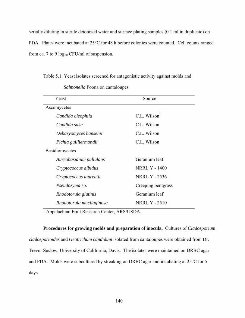
140
serially diluting in sterile deionized water and surface plating samples (0.1 ml in duplicate) on
PDA. Plates were incubated at 25°C for 48 h before colonies were counted. Cell counts ranged
from ca. 7 to 9 log10 CFU/ml of suspension.
Table 5.1. Yeast isolates screened for antagonistic activity against molds and
Salmonella Poona on cantaloupes Yeast Source
Ascomycetes
Candida oleophila C.L. Wilson1
Candida sake C.L. Wilson
Debaryomyces hansenii C.L. Wilson
Pichia guilliermondii C.L. Wilson
Basidiomycetes
Aureobasidium pullulans Geranium leaf
Cryptococcus albidus NRRL Y - 1400
Cryptococcus laurentii NRRL Y - 2536
Pseudozyma sp. Creeping bentgrass
Rhodotorula glutinis Geranium leaf
Rhodotorula mucilaginosa NRRL Y - 2510 1 Appalachian Fruit Research Center, ARS/USDA.
Procedures for growing molds and preparation of inocula. Cultures of Cladosporium
cladosporioides and Geotrichum candidum isolated from cantaloupes were obtained from Dr.
Trevor Suslow, University of California, Davis. The isolates were maintained on DRBC agar
and PDA. Molds were subcultured by streaking on DRBC agar and incubating at 25°C for 5
days.
141
Suspensions of conidia or arthroconidia were harvested by pipetting 10 ml of sterile
deionized water containing 0.05% Tween 80 onto the surface of the 5-day-old cultures and
gently rubbing for 1 min with a sterile glass rod. Using a sterile Pasteur pipette, conidial
suspensions were removed from the surface of plates and deposited in a sterile test tube. The
number of conidia of C. cladosporioides and arthroconidia and vegetative cells of G. candidum
(CFU/ml) in suspensions was determined by serially diluting samples in sterile deionized water
containing 0.05% Tween 80 and surface plating (0.1 ml in duplicate) on DRBC agar. Plates
were incubated at 25°C for 5 days before colonies were counted. The number of colony forming
units (CFUs) in each suspension was adjusted to ca. 4.64 log10 CFU/ml for C. cladosporioides
and 5.61 log10 CFU/ml for G. candidum by diluting in sterile deionized water containing 0.05%
Tween 80.
Effect of yeasts on S. Poona in cantaloupe juice (in vitro study). Test tubes containing
9 ml of cantaloupe juice were inoculated with 1 ml of a low-population inoculum of S. Poona
and/or 1 ml of high or low population of yeast inoculum, all made in cantaloupe juice.
Five inoculation schemes were used to inoculate cantaloupe juice:
(i) Low population of S. Poona only: Tubes containing 9 ml of cantaloupe juice were
inoculated with 1 ml of S. Poona suspension containing ca. 2.1 log10 CFU/ml.
(ii) High population of yeast only: Tubes containing 9 ml of cantaloupe juice were
inoculated with 1 ml of yeast suspension containing ca. 5.9 log10 CFU/ml.
(iii) Low population of yeast only: Tubes containing 9 ml of cantaloupe juice were
inoculated with 1 ml of yeast suspension containing ca. 3.5 log10 CFU/ml.
142
(iv) High population of yeast and low population of S. Poona: Tubes containing 9 ml of
cantaloupe juice were inoculated with 1 ml of yeast suspension containing ca. 5.9 log10 CFU/ml
and 1 ml of S. Poona suspension containing ca. 2.1 log10 CFU/ml.
(v) Low population of yeast and low population of S. Poona: Tubes containing 9 ml of
cantaloupe juice were inoculated with 1 ml of yeast suspension containing ca. 3.5 log10 CFU/ml
and 1 ml of S. Poona suspension containing ca. 2.1 log10 CFU/ml.
Immediately after inoculation, the pH and °brix of cantaloupe juice suspensions were
determined. A tube containing uninoculated cantaloupe juice was used as the control in all
treatments. Inoculated and uninoculated cantaloupe juice was incubated at 20°C.
Measurement of pH and °brix. The pH and °brix of the cantaloupe juice were
determined. A sterile 1-ml serological pipette was used to withdraw 0.2 ml of the suspension.
Three drops of suspension were dispensed on the sterile surface of a plastic petri dish for pH
determination, while two drops were placed on the measurement prism of the ABBE-3L
refractometer (Spectronic Instruments, Rochester, NY) for °brix determination.
Effect of yeasts on growth of molds in wounds. Cantaloupes were adjusted to 22 ± 1°C
before wounds were inoculated with yeast suspension. Using a Precision Microliter Pipette
(Rainin, Woburn, MA), 10 µl of yeast suspension was deposited into each wound in cantaloupe
rinds. The cantaloupes were kept at 22 ± 1°C for 2 h, followed by inoculation of wounds with 10
µl of mold suspension. Cantaloupes were kept at 22 ± 1°C for an additional 2 h before
incubating at 4°C for 21 days or 20°C for 7 days and analyzing for presence and populations of
test yeasts and molds.
143
The pH of inoculated tissues and using a dial caliper (Monostat, Zurich, Switzerland),
width of decayed area developing at wounded inoculation sites were measured initially and 7 and
14 days after inoculation and storage at 20°C and 4°C, respectively.
Test for antagonistic activity of yeasts against S. Poona in wounds. Wounds in
cantaloupe rinds were inoculated with yeast and mold or only mold as described above; 10 µl of
S. Poona suspension containing ca. 6.49 log10 CFU/ml was then deposited in each wound. The
cantaloupes were kept at 22 ± 1°C for 2 h before incubating at 4°C for 14 days or 20°C for 7
days.
Microbiological analyses of juice. Inoculated and uninoculated cantaloupe juice was
analyzed at 6, 24, and 48 h after inoculation for populations of S. Poona and test yeast. S. Poona
populations were determined by surface plating juice (0.25 ml in quadruplicate and 0.1 ml in
duplicate of undiluted juice and 0.1 ml in duplicate of suspensions serially diluted in 0.1%
peptone water) on TSAN. Plates were incubated at 37°C for 24 h before colonies were counted.
Random colonies were selected for confirmation as described above.
Yeast populations in inoculated juice were determined by surface plating undiluted juice
(0.25 ml in quadruplicate and 0.1 ml in duplicate) and juice serially diluted in 0.1% peptone
water (0.1 ml in duplicate) on DRBC agar. Plates were incubated at 25°C for 5 days before
colonies were counted.
Microbiological analyses of wound tissue. The pH of cut tissue in wounded,
uninoculated and inoculated sites on cantaloupes stored at 20°C for 7 days or 4°C for 14 days
was measured before removing samples for microbiological analysis. To test for the presence of
test yeast and mold inoculated into wounds, a sterile inoculation loop wetted in sterile deionized
water was used to remove samples from the surface of wound tissue. Samples were streaked on
144
duplicate DRBC agar plates and incubated at 25°C for 5 days. Isolates were examined to
confirm the presence of test yeasts and/or molds.
Samples of wounded rind tissues (ca. 3 cm diameter, 4 mm deep) inoculated with yeast,
mold, and S. Poona or mold and S. Poona were excised using a sterile spatula and placed in
separate stomacher 80 bags (Seward Medical Ltd., London, UK). Forty milliliters of sterile
lactose broth (LB; BBL/Difco) containing 50 µg of nalidixic acid/ml (LBN) was combined with
the tissue. The sample was hand rubbed and macerated, then pummeled for 1 min at normal
speed in a Model 80 Stomacher (Seward Medical Ltd.). The liquid portion of the homogenate
was serially diluted in sterile 0.1% peptone water and the undiluted homogenate (0.25 ml in
quadruplicate and 0.1 ml in duplicate) and diluted homogenate (0.1 ml in duplicate) were surface
plated on TSAN. Plates were incubated at 37°C for 24 h before presumptive-positive S. Poona
colonies were counted. Random colonies were selected for confirmation as described above.
Statistical analysis. Experiments for the in vitro study were replicated three times.
Mean values were analyzed to determine significant differences in populations of S. Poona in
cantaloupe juice inoculated with S. Poona only and co-inoculated with S. Poona and high or low
populations of yeasts. General linear models on SAS software (Statistical Analysis Systems
Institute, Cary, NC) were used. Significant differences (P < 0.05) between mean values were
determined using Duncan’s multiple range tests. Experiments on intact and wounded
cantaloupes were done two times.
RESULTS AND DISCUSSION
Growth of S. Poona and potentially antagonistic yeasts in cantaloupe juice. The initial
population of S. Poona inoculated into sterile cantaloupe juice was 1.10 log10 CFU/ml (Fig. 5.1).
With the exception of three test yeasts (D. hansenii, P. guilliermondii, and Pseudozyma sp.)
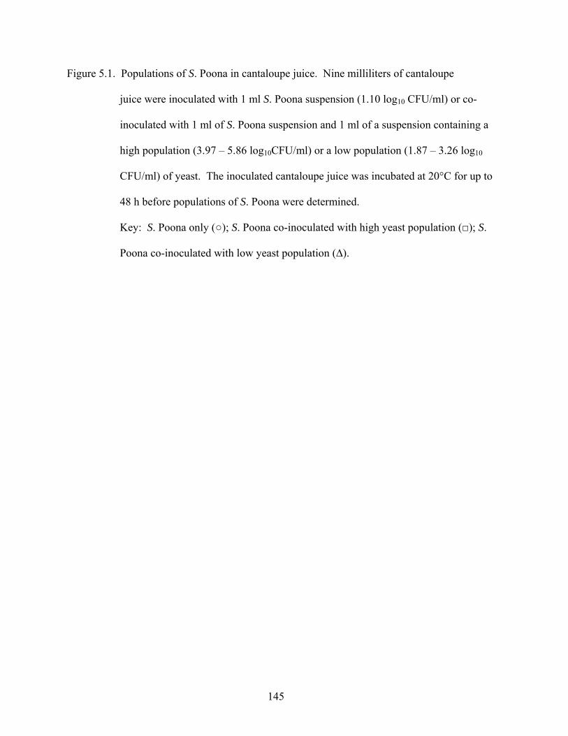
145
Figure 5.1. Populations of S. Poona in cantaloupe juice. Nine milliliters of cantaloupe
juice were inoculated with 1 ml S. Poona suspension (1.10 log10 CFU/ml) or co-
inoculated with 1 ml of S. Poona suspension and 1 ml of a suspension containing a
high population (3.97 – 5.86 log10CFU/ml) or a low population (1.87 – 3.26 log10
CFU/ml) of yeast. The inoculated cantaloupe juice was incubated at 20°C for up to
48 h before populations of S. Poona were determined.
Key: S. Poona only (○); S. Poona co-inoculated with high yeast population (□); S.
Poona co-inoculated with low yeast population (∆).
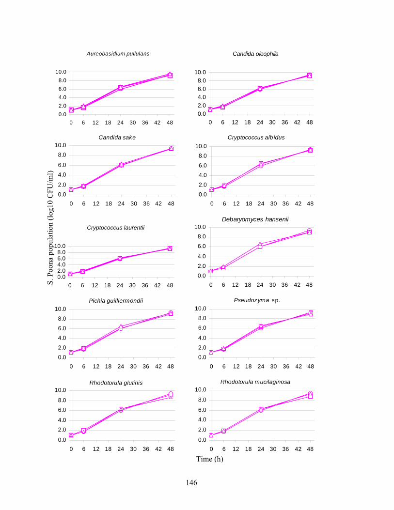
146
Aureobasidium pullulans
0.02.04.06.08.0
10.0
0 6 12 18 24 30 36 42 48
Candida oleophila
0.02.04.06.08.0
10.0
0 6 12 18 24 30 36 42 48
Candida sake
0.02.0
4.06.0
8.010.0
0 6 12 18 24 30 36 42 48
Cryptococcus alb idus
0.02.04.06.08.0
10.0
0 6 12 18 24 30 36 42 48
Cryptococcus laurentii
0.02.04.06.08.0
10.0
0 6 12 18 24 30 36 42 48
Debaryomyces hansenii
0.02.04.0
6.08.0
10.0
0 6 12 18 24 30 36 42 48
Pichia guilliermondii
0.02.04.06.08.0
10.0
0 6 12 18 24 30 36 42 48
Pseudozyma sp.
0.02.04.0
6.08.0
10.0
0 6 12 18 24 30 36 42 48
Rhodotorula glutinis
0.0
2.04.0
6.08.0
10.0
0 6 12 18 24 30 36 42 48
Rhodotorula mucilaginosa
0.0
2.0
4.06.0
8.0
10.0
0 6 12 18 24 30 36 42 48
Time (h)
S. P
oona
pop
ulat
ion
(log1
0 C
FU/m
l)
147
the presence and population of potentially antagonistic yeasts did not affect survival and growth
of S. Poona. Cantaloupe juice inoculated with only S. Poona contained populations of 1.70, 6.00,
and 9.41 log10 CFU/ml after incubation for 6, 24, and 48 h, respectively, at 20°C. Cantaloupe
juice inoculated with S. Poona and a high population of yeast (3.97 – 5.86 log10CFU/ml)
contained S. Poona populations of ca. 1.83, 6.22, and 9.17 log10 CFU/ml 6, 24, and 48 h,
respectively, after inoculation. S. Poona co-inoculated with a low population of yeast (1.87 –
3.26 log10 CFU/ml), were ca. 1.94, 6.40, and 9.21 log10 CFU/ml after 6, 24, and 48 h,
respectively.
Comparing S. Poona population in cantaloupe juice inoculated with S. Poona only with
that in juice co-inoculated with low populations of yeasts, there were no significant differences
in S. Poona populations (P > 0.05) at any sampling time. However, when co-inoculated with
high populations of D. hansenii, P. guilliermondii, or Pseudozyma sp., the S. Poona population
was significantly less (P < 0.05) at 48 h than in juice inoculated with S. Poona only. These
results indicate that while low populations of yeast did not affect the growth of S. Poona (Table
5.2), high populations of D. hansenii, P. guilliermondii, and Pseudozyma sp. are potentially
antagonistic to S. Poona in cantaloupe juice stored at 20°C statistically, but not practically
feasible (Table 5.3).
Yeasts inoculated into cantaloupe juice at high or low populations, or co-inoculated with
S. Poona were enumerated 6, 24, and 48 h after inoculation (Fig. 5.2). Yeast populations
changed by (-) 2.72 and 2.48 log10 CFU/ml and (-) 1.28 and 4.37 log10 CFU/ml, respectively, in
juice inoculated with high and low populations. Comparing the growth in juice inoculated with
the two populations, it is apparent that the low populations grew at a more rapid rate. This
observation is attributable to a comparatively greater availability, on a per cell basis,
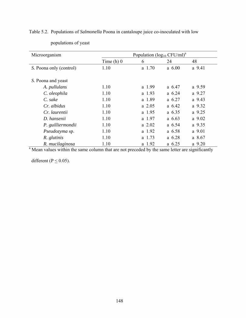
148
Table 5.2. Populations of Salmonella Poona in cantaloupe juice co-inoculated with low
populations of yeast
Microorganism Population (log10 CFU/ml)a
Time (h) 0 6 24 48 S. Poona only (control) 1.10 a 1.70 a 6.00 a 9.41 S. Poona and yeast A. pullulans 1.10 a 1.99 a 6.47 a 9.59 C. oleophila 1.10 a 1.93 a 6.24 a 9.27 C. sake 1.10 a 1.89 a 6.27 a 9.43 Cr. albidus 1.10 a 2.05 a 6.42 a 9.32 Cr. laurentii 1.10 a 1.95 a 6.35 a 9.25 D. hansenii 1.10 a 1.97 a 6.63 a 9.02 P. guilliermondii 1.10 a 2.02 a 6.54 a 9.35 Pseudozyma sp. 1.10 a 1.92 a 6.58 a 9.01 R. glutinis 1.10 a 1.73 a 6.28 a 8.67 R. mucilaginosa 1.10 a 1.92 a 6.25 a 9.20 a Mean values within the same column that are not preceded by the same letter are significantly
different (P < 0.05).
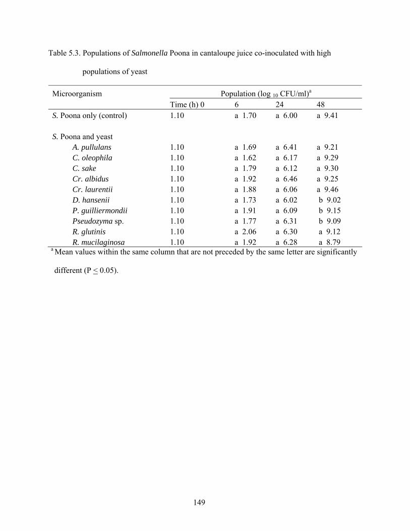
149
Table 5.3. Populations of Salmonella Poona in cantaloupe juice co-inoculated with high
populations of yeast
Microorganism Population (log 10 CFU/ml)a
Time (h) 0 6 24 48 S. Poona only (control) 1.10 a 1.70 a 6.00 a 9.41 S. Poona and yeast A. pullulans 1.10 a 1.69 a 6.41 a 9.21 C. oleophila 1.10 a 1.62 a 6.17 a 9.29 C. sake 1.10 a 1.79 a 6.12 a 9.30 Cr. albidus 1.10 a 1.92 a 6.46 a 9.25 Cr. laurentii 1.10 a 1.88 a 6.06 a 9.46 D. hansenii 1.10 a 1.73 a 6.02 b 9.02 P. guilliermondii 1.10 a 1.91 a 6.09 b 9.15 Pseudozyma sp. 1.10 a 1.77 a 6.31 b 9.09 R. glutinis 1.10 a 2.06 a 6.30 a 9.12 R. mucilaginosa 1.10 a 1.92 a 6.28 a 8.79 a Mean values within the same column that are not preceded by the same letter are significantly
different (P < 0.05).
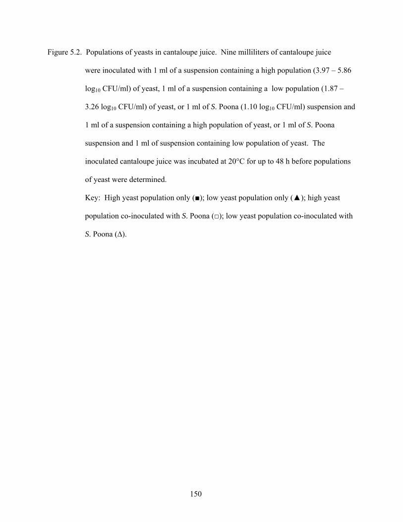
150
Figure 5.2. Populations of yeasts in cantaloupe juice. Nine milliliters of cantaloupe juice
were inoculated with 1 ml of a suspension containing a high population (3.97 – 5.86
log10 CFU/ml) of yeast, 1 ml of a suspension containing a low population (1.87 –
3.26 log10 CFU/ml) of yeast, or 1 ml of S. Poona (1.10 log10 CFU/ml) suspension and
1 ml of a suspension containing a high population of yeast, or 1 ml of S. Poona
suspension and 1 ml of suspension containing low population of yeast. The
inoculated cantaloupe juice was incubated at 20°C for up to 48 h before populations
of yeast were determined.
Key: High yeast population only (■); low yeast population only (▲); high yeast
population co-inoculated with S. Poona (□); low yeast population co-inoculated with
S. Poona (∆).
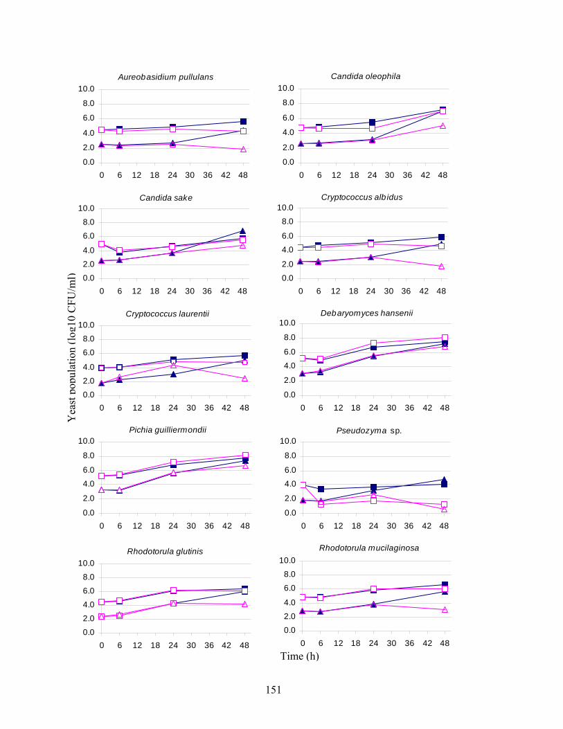
151
Aureobasidium pullulans
0.0
2.0
4.0
6.0
8.0
10.0
0 6 12 18 24 30 36 42 48
Candida oleophila
0.0
2.0
4.0
6.0
8.0
10.0
0 6 12 18 24 30 36 42 48
Candida sake
0.02.04.0
6.08.0
10.0
0 6 12 18 24 30 36 42 48
Cryptococcus alb idus
0.02.0
4.06.0
8.010.0
0 6 12 18 24 30 36 42 48
Cryptococcus laurentii
0.02.04.0
6.08.0
10.0
0 6 12 18 24 30 36 42 48
Debaryomyces hansenii
0.0
2.0
4.06.0
8.0
10.0
0 6 12 18 24 30 36 42 48
Pichia guilliermondii
0.0
2.0
4.06.0
8.0
10.0
0 6 12 18 24 30 36 42 48
Pseudozyma sp.
0.0
2.0
4.06.0
8.0
10.0
0 6 12 18 24 30 36 42 48
Rhodotorula glutinis
0.02.04.06.08.0
10.0
0 6 12 18 24 30 36 42 48
Rhodotorula mucilaginosa
0.02.04.0
6.08.0
10.0
0 6 12 18 24 30 36 42 48Time (h)
Yea
st p
opul
atio
n (lo
g10
CFU
/ml)
152
of nutrient sources in juice inoculated with a low population of yeasts. There is greater
competition for nutrients in juice inoculated high populations. Additionally, a higher initial yeast
population may result in a more rapid production and accumulation of metabolic by products
which may restrict the growth of cells.
While the populations of yeasts continued to increase between 24 and 48 h in juice
inoculated with high or low populations of yeast only, the populations of A. pullulans, Cry.
albidus, Cry. laurentii, Pseudozyma sp., R. glutinis, and R. mucilaginosa began to decrease after
24 h in juice co-inoculated with S. Poona (Fig. 5.2). At 6 and 24 h after inoculation, the
populations of yeast recovered from the juice were not significantly different from the
populations in juice co-inoculated with S. Poona (P > 0.05). With the exception of juice co-
inoculated with low populations of Cry. laurentii, D. hansenii, and P. guilliermondii, the
populations of yeast co-inoculated with S. Poona began to decrease at 48 h (P < 0.05).
With the exception of D. hansenii, P. guilliermondii, and Pseudozyma sp. at high initial
populations, overall, the presence and population of antagonistic yeasts did not appear to
influence the survival and growth of S. Poona in cantaloupe juice. Within 6, 24, and 48 h of
inoculation, the population of S. Poona increased by 0.72, 5.11, and 8.16 log10 CFU/ml,
respectively, in juice co-inoculated with yeasts. Results from our study indicate that, overall,
potentially antagonistic yeasts strains examined in this study do not compete effectively with S.
Poona in cantaloupe juice and therefore would not be effective in controlling the growth of S.
Poona on cantaloupes.
Changes in pH and total soluble solids content of cantaloupe juice as affected by
inoculation with S. Poona and yeasts. At the time of inoculation, the pH of cantaloupe juice
153
was 6.29 – 7.12, depending on the lot selected for examination (Fig. 5.3). The pH of
uninoculated juice increased from 6.78 to 7.04 over the 48-h storage period at 20°C. When
cantaloupe juice was inoculated with only S. Poona, there was a slight increase in pH during the
first 24 h, but a very rapid decrease to 5.44 between 24 and 48 h. Although the pH of cantaloupe
juice inoculated with C. oleophila, C. sake, D. hansenii, and P. guilliermondii trended downward
after 24 h, all the yeasts considered, the pH of the juice 48 h after inoculation with high or low
populations of yeasts was 6.28 – 7.45 and 6.43 – 7.51, respectively, indicating that the level of
inoculum did not have marked effect on the juice pH. However, when the cantaloupe juice was
co-inoculated with high or low populations of yeasts along with S. Poona, the pH decreased to
5.13 – 5.92 and 5.10 – 5.89, respectively, over the 48 h storage period. Growth of S. Poona
caused the pH of the environment to decrease.
Inoculation of cantaloupe juice with S. Poona, with or without yeasts, resulted in a sharp
decrease in juice pH. This may be attributable to metabolism of amino acids, resulting in
excretion of weak acids to maintain internal pH and consequently reduce the pH of the external
environment. Eighteen amino acids have been identified in cantaloupe fruits and the total amino
acid content decreased by 40% after storage at 20°C (Lamikanra et al., 2000). Some of these
amino acids were used to support bacterial growth, formation of other compounds, and catabolic
degradation. While utilization of acids by the yeasts would have caused the pH of the juice to
increase, excretion of acids by S. Poona would maintain the initial pH of the juice.
The initial total soluble solids content (TSSC, measured as °brix) in uninoculated
cantaloupe juice (4.6 – 8.7, mean = 7.6) decreased slightly to 7.4 (Fig. 5.4). When the juice was
inoculated with S. Poona, the TSSC decreased to ca. 7.1 over the 48-h storage period at 20°C.
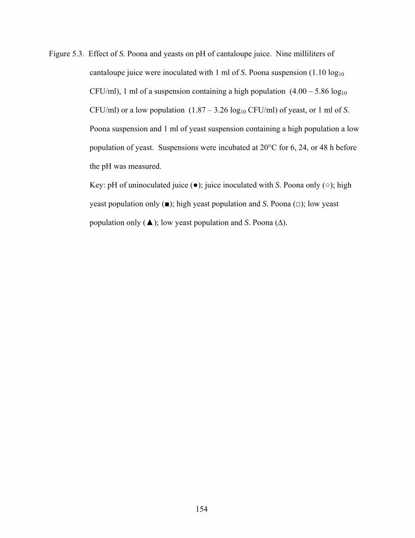
154
Figure 5.3. Effect of S. Poona and yeasts on pH of cantaloupe juice. Nine milliliters of
cantaloupe juice were inoculated with 1 ml of S. Poona suspension (1.10 log10
CFU/ml), 1 ml of a suspension containing a high population (4.00 – 5.86 log10
CFU/ml) or a low population (1.87 – 3.26 log10 CFU/ml) of yeast, or 1 ml of S.
Poona suspension and 1 ml of yeast suspension containing a high population a low
population of yeast. Suspensions were incubated at 20°C for 6, 24, or 48 h before
the pH was measured.
Key: pH of uninoculated juice (●); juice inoculated with S. Poona only (○); high
yeast population only (■); high yeast population and S. Poona (□); low yeast
population only (▲); low yeast population and S. Poona (∆).
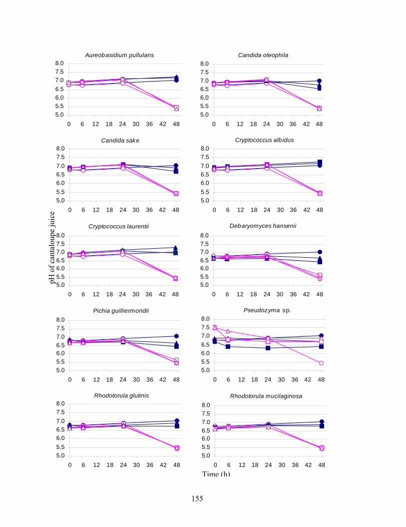
155
Aureobasidium pullulans
5.05.56.06.57.07.58.0
0 6 12 18 24 30 36 42 48
Candida oleophila
5.05.56.06.57.07.58.0
0 6 12 18 24 30 36 42 48
Candida sake
5.05.56.06.57.07.58.0
0 6 12 18 24 30 36 42 48
Cryptococcus alb idus
5.05.56.06.57.07.58.0
0 6 12 18 24 30 36 42 48
Cryptococcus laurentii
5.05.56.06.57.07.58.0
0 6 12 18 24 30 36 42 48
Debaryomyces hansenii
5.05.56.06.57.07.58.0
0 6 12 18 24 30 36 42 48
Pichia guilliermondii
5.05.56.06.57.07.58.0
0 6 12 18 24 30 36 42 48
Pseudozyma sp.
5.05.56.06.57.07.58.0
0 6 12 18 24 30 36 42 48
Rhodotorula glutinis
5.05.56.06.57.07.58.0
0 6 12 18 24 30 36 42 48
Rhodotorula mucilaginosa
5.05.56.06.57.07.58.0
0 6 12 18 24 30 36 42 48Time (h)
pH o
f can
talo
upe
juic
e
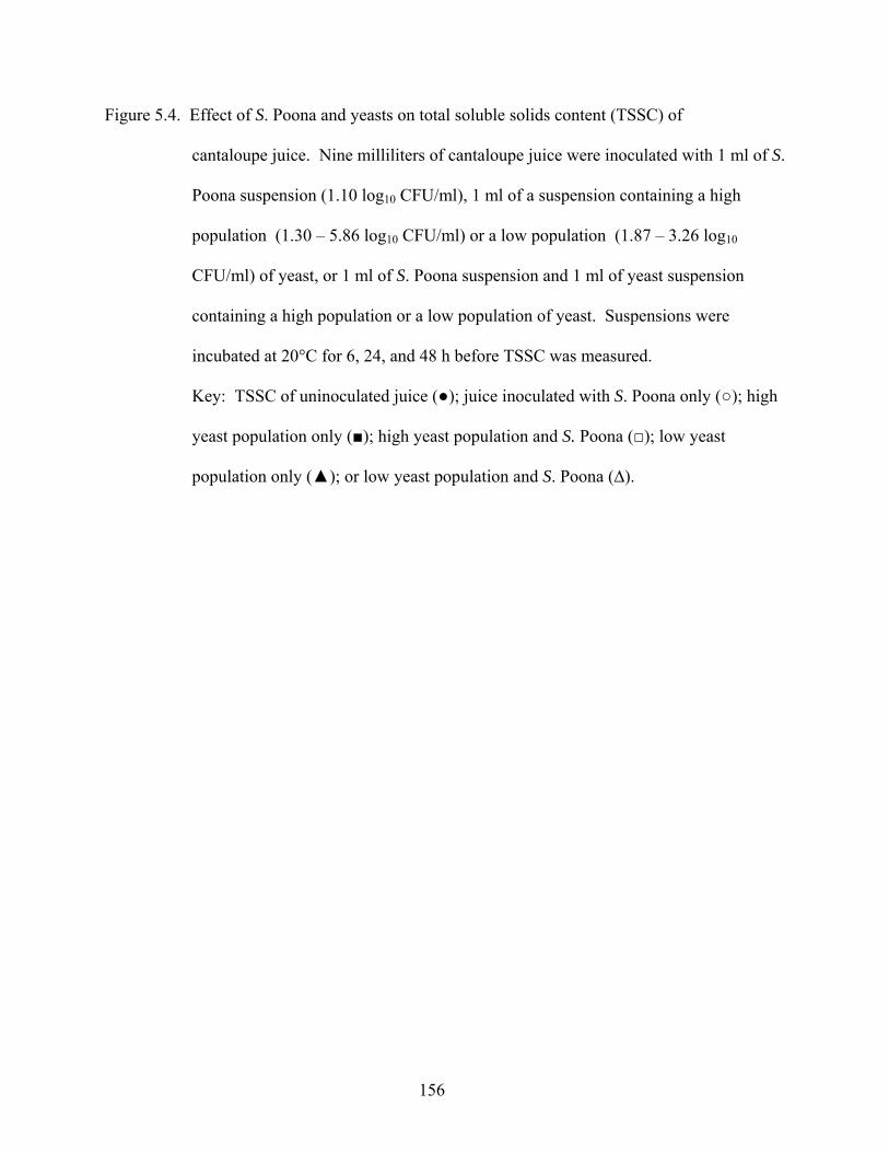
156
Figure 5.4. Effect of S. Poona and yeasts on total soluble solids content (TSSC) of
cantaloupe juice. Nine milliliters of cantaloupe juice were inoculated with 1 ml of S.
Poona suspension (1.10 log10 CFU/ml), 1 ml of a suspension containing a high
population (1.30 – 5.86 log10 CFU/ml) or a low population (1.87 – 3.26 log10
CFU/ml) of yeast, or 1 ml of S. Poona suspension and 1 ml of yeast suspension
containing a high population or a low population of yeast. Suspensions were
incubated at 20°C for 6, 24, and 48 h before TSSC was measured.
Key: TSSC of uninoculated juice (●); juice inoculated with S. Poona only (○); high
yeast population only (■); high yeast population and S. Poona (□); low yeast
population only (▲); or low yeast population and S. Poona (∆).
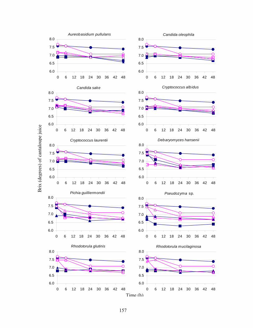
157
Aureobasidium pullulans
6.0
6.5
7.0
7.5
8.0
0 6 12 18 24 30 36 42 48
Candida oleophila
6.0
6.5
7.0
7.5
8.0
0 6 12 18 24 30 36 42 48
Candida sake
6.0
6.5
7.0
7.5
8.0
0 6 12 18 24 30 36 42 48
Cryptococcus alb idus
6.0
6.5
7.0
7.5
8.0
0 6 12 18 24 30 36 42 48
Cryptococcus laurentii
6.0
6.5
7.0
7.5
8.0
0 6 12 18 24 30 36 42 48
Debaryomyces hansenii
6.0
6.5
7.0
7.5
8.0
0 6 12 18 24 30 36 42 48
Pichia guilliermondii
6.0
6.5
7.0
7.5
8.0
0 6 12 18 24 30 36 42 48
Pseudozyma sp.
6.0
6.5
7.0
7.5
8.0
0 6 12 18 24 30 36 42 48
Rhodotorula glutinis
6.0
6.5
7.0
7.5
8.0
0 6 12 18 24 30 36 42 48
Rhodotorula mucilaginosa
6.0
6.5
7.0
7.5
8.0
0 6 12 18 24 30 36 42 48
Time (h)
Brix
(deg
rees
) of c
anta
loup
e ju
ice
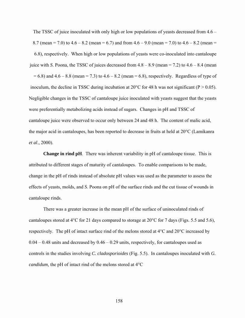
158
The TSSC of juice inoculated with only high or low populations of yeasts decreased from 4.6 –
8.7 (mean = 7.0) to 4.6 – 8.2 (mean = 6.7) and from 4.6 – 9.0 (mean = 7.0) to 4.6 – 8.2 (mean =
6.8), respectively. When high or low populations of yeasts were co-inoculated into cantaloupe
juice with S. Poona, the TSSC of juices decreased from 4.8 – 8.9 (mean = 7.2) to 4.6 – 8.4 (mean
= 6.8) and 4.6 – 8.8 (mean = 7.3) to 4.6 – 8.2 (mean = 6.8), respectively. Regardless of type of
inoculum, the decline in TSSC during incubation at 20°C for 48 h was not significant (P > 0.05).
Negligible changes in the TSSC of cantaloupe juice inoculated with yeasts suggest that the yeasts
were preferentially metabolizing acids instead of sugars. Changes in pH and TSSC of
cantaloupe juice were observed to occur only between 24 and 48 h. The content of malic acid,
the major acid in cantaloupes, has been reported to decrease in fruits at held at 20°C (Lamikanra
et al., 2000).
Change in rind pH. There was inherent variability in pH of cantaloupe tissue. This is
attributed to different stages of maturity of cantaloupes. To enable comparisons to be made,
change in the pH of rinds instead of absolute pH values was used as the parameter to assess the
effects of yeasts, molds, and S. Poona on pH of the surface rinds and the cut tissue of wounds in
cantaloupe rinds.
There was a greater increase in the mean pH of the surface of uninoculated rinds of
cantaloupes stored at 4°C for 21 days compared to storage at 20°C for 7 days (Figs. 5.5 and 5.6),
respectively. The pH of intact surface rind of the melons stored at 4°C and 20°C increased by
0.04 – 0.48 units and decreased by 0.46 – 0.29 units, respectively, for cantaloupes used as
controls in the studies involving C. cladosporioides (Fig. 5.5). In cantaloupes inoculated with G.
candidum, the pH of intact rind of the melons stored at 4°C
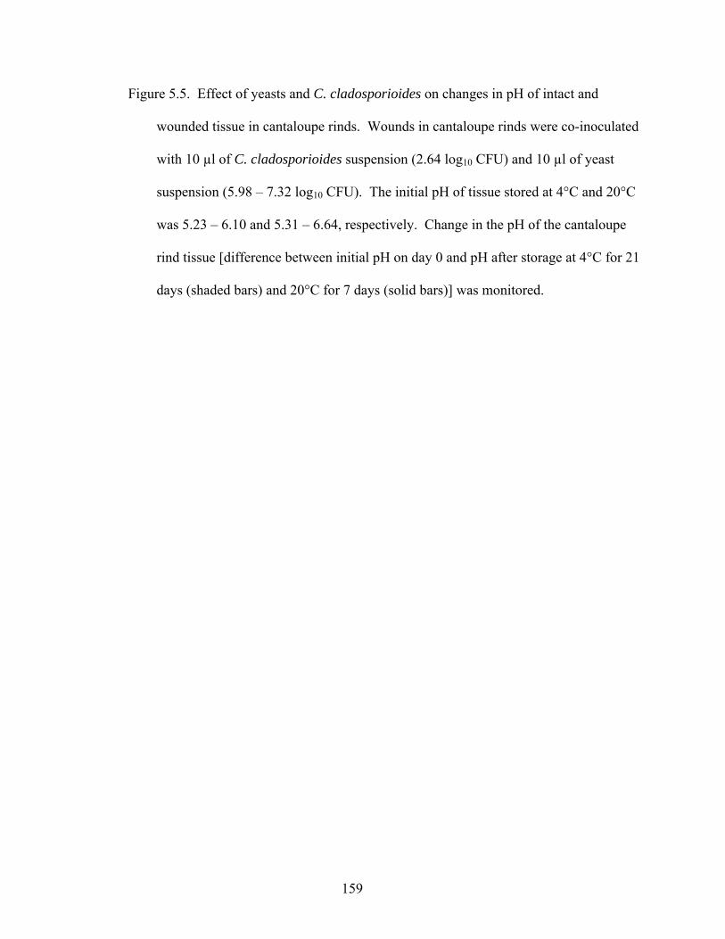
159
Figure 5.5. Effect of yeasts and C. cladosporioides on changes in pH of intact and
wounded tissue in cantaloupe rinds. Wounds in cantaloupe rinds were co-inoculated
with 10 µl of C. cladosporioides suspension (2.64 log10 CFU) and 10 µl of yeast
suspension (5.98 – 7.32 log10 CFU). The initial pH of tissue stored at 4°C and 20°C
was 5.23 – 6.10 and 5.31 – 6.64, respectively. Change in the pH of the cantaloupe
rind tissue [difference between initial pH on day 0 and pH after storage at 4°C for 21
days (shaded bars) and 20°C for 7 days (solid bars)] was monitored.
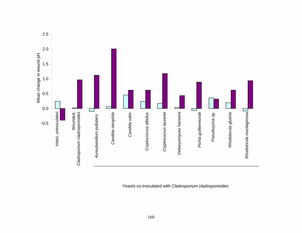
160
-0.5
0.0
0.5
1.0
1.5
2.0
2.5
Yeasts co-inoculated with Cladosporium cladosporioides
Mea
n ch
ange
in w
ound
pH
Inta
ct, u
nino
cula
ted
Wou
nded
, C
lado
spor
ium
cla
dosp
orio
ides
Aure
ubas
idiu
m p
ullu
lans
Can
dida
ole
ophi
la
Can
dida
sak
e
Cry
ptoc
occu
s al
bidu
s
Cry
ptoc
occu
s la
uren
ti i
Deb
aryo
myc
es h
anse
nii
Pich
ia g
uillie
rmon
dii
Pseu
dozy
ma
sp.
Rho
doto
rula
glu
tinis
Rho
doto
rula
muc
ilagi
nosa
____________________________________________________________________________
161
Figure 5.6. Effect of yeasts and G. candidum on changes in pH of intact and wounded
tissue in cantaloupe rinds. Wounds in cantaloupe rinds were co-inoculated with 10
µl of G. candidum suspension (3.51 log10 CFU) and 10 µl of yeast suspension (5.98
– 7.32 log10 CFU). The initial pH of tissue stored at 4°C and 20°C was 5.16 – 6.11
and 5.32 – 6.4, respectively. Change in the pH of the cantaloupe rind tissue
[difference between initial pH on day 0 and pH after storage at 4°C for 21 days
(shaded bars) and 20°C for 7 days (solid bars)] was monitored.
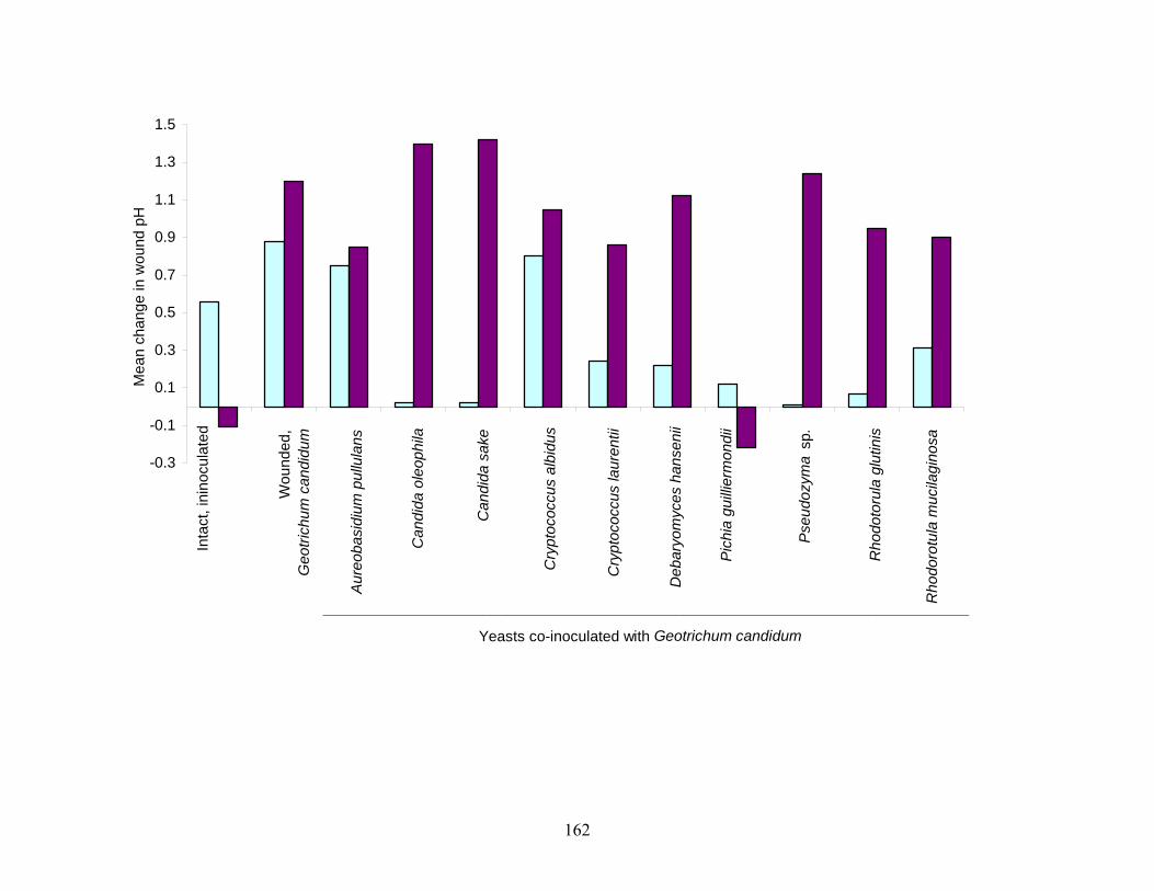
162
-0.3
-0.1
0.1
0.3
0.5
0.7
0.9
1.1
1.3
1.5
Yeasts co-inoculated with Geotrichum candidum
Mea
n ch
ange
in w
ound
pH
Inta
ct, i
nino
cula
ted
Wou
nded
,G
eotri
chum
can
didu
m
Aure
obas
idiu
m p
ullu
lans
Can
dida
ole
ophi
la
Can
dida
sak
e
Cry
ptoc
occu
s al
bidu
s
Cry
ptoc
occu
s la
uren
tii
Deb
aryo
myc
es h
anse
nii
Pich
ia g
uillie
rmon
dii
Pseu
dozy
ma
sp.
Rho
doto
rula
glu
tinis
Rho
doro
tula
muc
ilagi
nosa
_____________________________________________________________________________
163
increased by 0.12 – 1.23 units; melons stored at 20°C decreased by 0.23 or increased by 0.13
units (Fig. 5.6). The differences in pH between these two groups of control melons are attributed
to differences in the level of maturity of the fruits used in the two studies.
The pH of the uninoculated, intact rind surface of cantaloupes in which wounds were
inoculated with C. cladosporioides or with yeast and C. cladosporioides significantly increased
(P < 0.05) when cantaloupes were stored for 21 days at 4°C and decreased when at 20°C for 7
days (Fig. 5.5). In wounds on cantaloupe rinds inoculated with C. cladosporioides only or C.
cladosporioides and yeasts, the effect of storage temperature on change in pH showed the
opposite trend, i.e. the increase in pH was greater in cantaloupes stored at 20°C than at 4°C.
Inoculation of wounds with C. cladosporioides only, followed by storage at 4°C for 21
days, did not cause a significant change in pH (P > 0.05); at 20°C, however, the pH of wound
tissue increased significantly by 0.97 within 7 days (P < 0.05). When the wounds were co-
inoculated with C. cladosporioides and yeasts and stored at 20°C, the pH of wounded rind tissue
increased significantly (P < 0.05). The magnitude of increase in pH of wounded tissues was
lowest when C. cladosporioides was co-inoculated with C. oleophila. Increases in the pH
wounded rind of cantaloupes stored at 4°C after inoculation with C. cladosporioides and yeasts
were not significant (P > 0.05), except with C. sake and Pseudozyma sp. Wounds co-inoculated
with A. pullulans, P. guilliermondii, or R. mucilaginosa, and C. cladosporioides exhibited
decreased pH when the cantaloupes were stored at 4°C; however, these decreases were not
significant (P > 0.05).
Similarly, in wounded cantaloupe rinds inoculated with G. candidum only or G.
candidum and yeasts, with the exception of wounded tissue inoculated with G. candidum and P.
guilliermondii, the increase in pH was higher in cantaloupes stored at 20°C than at 4°C (Fig.
164
5.6). The pH of wounded rind tissue inoculated with only G. candidum increased significantly
(P < 0.05) by 0.88 and 1.20 units at 4°C and 20°C, respectively. In cantaloupes stored at 4°C,
and in some cases at 20°C, the pH of wounds inoculated with G. candidum and yeasts did not
increase as much as the pH of wounds inoculated with G. candidum only. Results indicate that
at 4°C, C. oleophila, C. sake, D. hansenii, P. guilliermondii, Pseudozyma sp., and R. glutinis
retarded the increase in pH and prevented changes of the magnitude that occurred in tissue
inoculated with G. candidum alone. Except in wounds co-inoculated with P. guilliermondii, the
pH of the wounded rind tissue co-inoculated with G. candidum and yeasts was significantly
higher (P < 0.05) after storage at 20°C for 7 days. P. guilliermondii limited the increase in pH of
wound tissue at 4°C but caused a decrease in the pH of wounded tissue of cantaloupes stored at
20°C.
Injury of rind and flesh of cantaloupes, either by cutting or infection with molds, causes
the integrity of cells to be disrupted and increases the availability of nutrients and water to
phytopathogens and other microorganisms that may occur naturally on the surface. Release of
products as a result of proteolysis and fermentation of tissue constituents, as well as utilization of
acids, may contribute to changes in the pH. Consequently, wounding eventually causes changes
the physiology, texture, and flavor of the fruit (Lamikanra et al., 2003). Oxalic, citric, malic, and
succinic acids, together with glucose, fructose, and sucrose are present in edible tissues of freshly
cut cantaloupe. In our previous study and that of Lamikanra et al. (2000), the pH of cut tissue of
cantaloupes stored at 20°C increased over a 5-day period, but the pH of cut tissue stored at 4°C
did not change substantially. The pH of wounded tissues increased more than that of the intact
surface tissue. This is attributed to an increased availability of nutrients on the cut surface,
which enhanced metabolic activities and consequently the production of by-products or
165
utilization of acids by yeasts and/or molds inoculated into the wounds. Some of the test yeasts,
as well as G. candidum and C. cladosporioides, are capable of producing extracellular
proteolytic enzymes that would catalyze biochemical reactions that produce alkaline substances
that increase the pH of the environment (Deak and Beuchat, 1996). Yeasts and bacteria are able
to take up nutrients more rapidly and in greater quantities than are the germ tubes of mycelial
fungi (Droby et al., 1989). Reduction in the amounts of exogenous nutrients restricts conidial
germination and hyphal development, which also gives yeasts a competitive advantage.
Fermentative yeasts could contribute to controlling increases in pH of tissues by producing acids.
C. oleophila, C. sake, and P. guilliermondii are fermentative, while the other test yeasts are non-
fermentative (Kurtzman and Fell, 1999). Among the test yeasts, P. guilliermondii was most
effective in limiting the increase in pH of wounds in the rinds of cantaloupes co-inoculated with
G. candidum.
Change in diameter of wounds. The diameter of wounds inoculated with C.
cladosporioides only or co-inoculated with C. cladosporioides and yeasts increased to 1.5 – 3.2
mm (mean = 2.3 mm) when the cantaloupes were stored at 4°C for 21 days and 3.0 – 5.0 mm
(mean = 3.4 mm) when they were stored at 20°C for 7 days (Fig. 5.7). When wounds in
cantaloupe rinds were inoculated with G. candidum only or co-inoculated with G. candidum and
yeasts, the approximate diameter of the infected surface area around the wounds increased from
1 mm (day 0) to 1.5 – 3.7 mm (mean = 2.5 mm) after 21 days at 4°C and 2.0 – 6.0 mm (mean =
3.7 mm) after 7 days at 20°C (Fig. 5.8).
The size of wounds inoculated with only C. cladosporioides or G. candidum increased to
2.5 and 3.0 mm, respectively, when cantaloupes were stored at 4°C and 20°C. Storage at 4°C
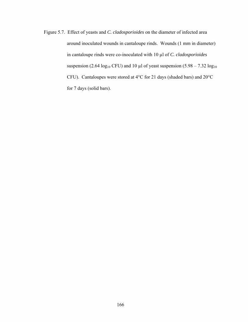
166
Figure 5.7. Effect of yeasts and C. cladosporioides on the diameter of infected area
around inoculated wounds in cantaloupe rinds. Wounds (1 mm in diameter)
in cantaloupe rinds were co-inoculated with 10 µl of C. cladosporioides
suspension (2.64 log10 CFU) and 10 µl of yeast suspension (5.98 – 7.32 log10
CFU). Cantaloupes were stored at 4°C for 21 days (shaded bars) and 20°C
for 7 days (solid bars).
167
0.0
1.0
2.0
3.0
4.0
5.0
6.0
Yeasts co-inoculated with Cladosporium cladosporioides
Mea
n di
amet
er o
f wou
nd (m
m)
Wou
nded
,C
lado
spor
ium
cla
dosp
orio
ides
Aure
obas
idiu
m p
ullu
lans
Can
dida
ole
ophi
la
Can
dida
sak
e
Deb
aryo
myc
es h
anse
nii
Pich
ia g
uillie
rmon
dii
Pseu
dozy
ma
sp.
Rho
doto
rula
glu
tinis
Cry
ptoc
occu
s al
bidu
s
Cry
ptoc
occu
s la
uren
tii
Rho
dotro
ula
muc
ilagi
nosa
_________________________________________________________________________
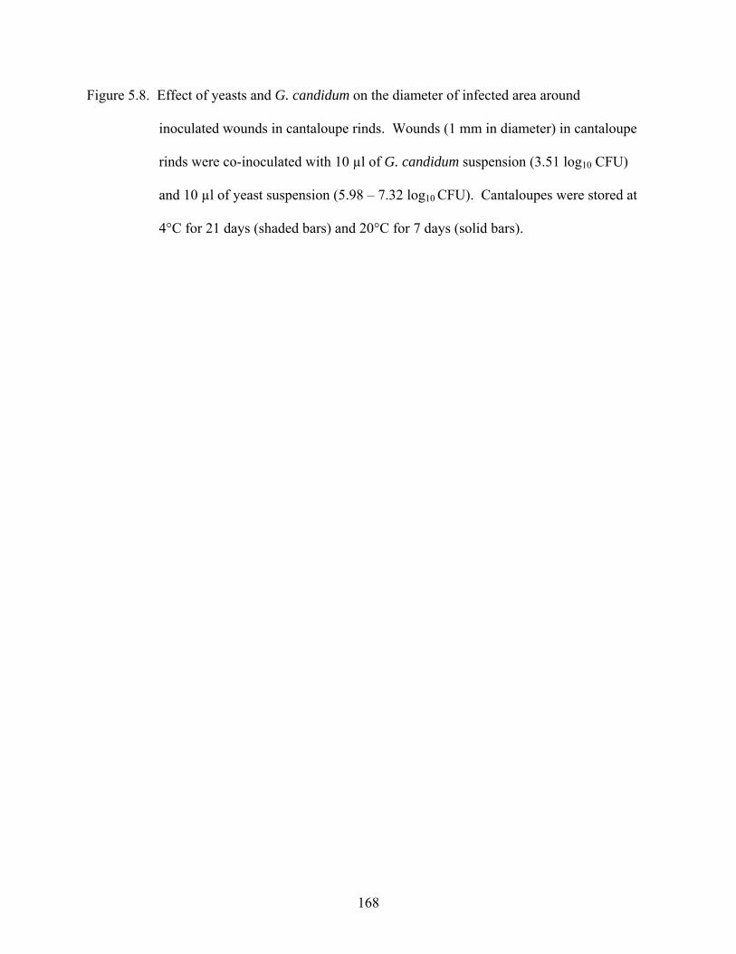
168
Figure 5.8. Effect of yeasts and G. candidum on the diameter of infected area around
inoculated wounds in cantaloupe rinds. Wounds (1 mm in diameter) in cantaloupe
rinds were co-inoculated with 10 µl of G. candidum suspension (3.51 log10 CFU)
and 10 µl of yeast suspension (5.98 – 7.32 log10 CFU). Cantaloupes were stored at
4°C for 21 days (shaded bars) and 20°C for 7 days (solid bars).
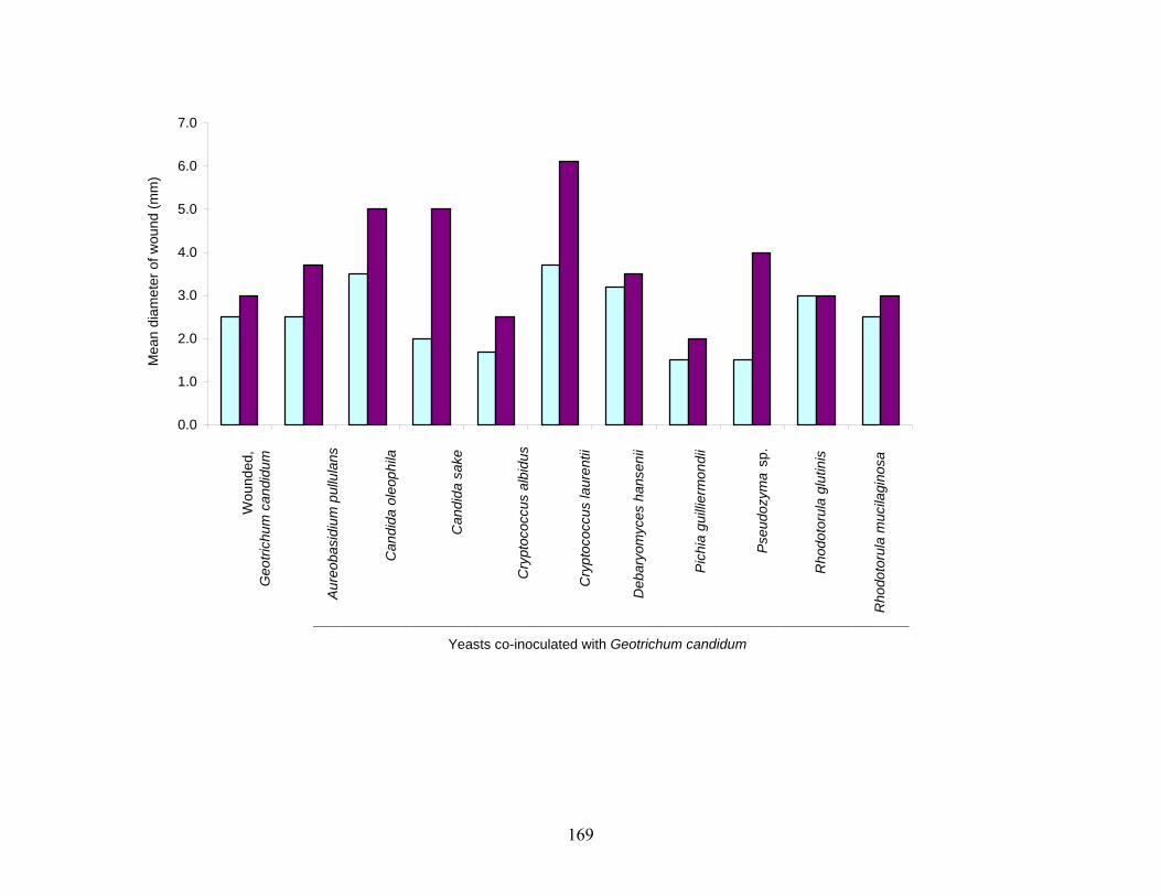
169
0.0
1.0
2.0
3.0
4.0
5.0
6.0
7.0
Yeasts co-inoculated with Geotrichum candidum
Mea
n di
amet
er o
f wou
nd (m
m)
Wou
nded
, G
eotri
chum
can
didu
m
Aure
obas
idiu
m p
ullu
lans
Can
dida
ole
ophi
la
Can
dida
sak
e
Cry
ptoc
occu
s al
bidu
s
Cry
ptoc
occu
s la
uren
tii
Deb
aryo
myc
es h
anse
nii
Pich
ia g
uilli
erm
ondi
i
Pseu
dozy
ma
sp.
Rho
doto
rula
glu
tinis
Rho
doto
rula
muc
ilagi
nosa
_____________________________________________________________________________
170
helped to minimize visible growth of the inoculated molds. Co-inoculation of wounds with
yeasts and C. cladosporioides or G. candidum did not inhibit decay at 20°C. However, D.
hansenii and R. mucilaginosa were the most effective among test yeasts in limiting visible
surface growth of C. cladosporioides (Fig. 5.7). Cr. albidus, P. guilliermondii, or Pseudozyma
sp. retarded the expansion of visible decay by G. candidum to a mean surface diameter of < 2
mm after 21 days at 4°C (Fig. 5.8).
Increased wound size can also be attributed, in part, to water loss, particularly during
extended storage. When wounds are created in the rinds, cells and tissues are ruptured, resulting
in the release of nutrients and water. Moisture loss by evaporation from the tissues causes
shrinkage of ruptured cells on and near the surface of wounds, thus expanding the wound size.
Plant tissues are in equilibrium with an atmosphere at a relative humidity of 99 to 99.5%. Any
reduction in water vapor pressure in an atmosphere below 99 – 99.5% would result in water loss.
In intact tissues, water in intercellular spaces is not directly exposed to the external atmosphere.
However, cutting or peeling a fruit or vegetable exposes interior tissues and drastically increases
the rate of water evaporation (Brecht, 1995). The higher relative humidity (70 – 80% RH) in
storage containers at 4°C compared to that in containers at 20°C (45%) in our study may have
contributed to some of the changes in wound size during storage.
Mycelia of G. candidum were not visually apparent in wounded rind of cantaloupes
stored at 4°C, regardless of co-inoculation with yeasts, but mycelial growth was obvious when
fruits were stored at 20°C. Introduction of C. cladosporioides and G. candidum into wounds
facilitated deterioration of the tissues as a result of production of cellulases and pectinases,
especially at abusive storage temperature (20°C), causing a release of fluids from tissues.
Rotting plant tissues are more likely to offer harborage to foodborne pathogens. In a survey of
171
more than 400 fruits and vegetables, the incidence of Salmonella in healthy tissues was reported
to be approximately 10%, whereas 20% of the samples with bacterial rot were confirmed
positive for the organism (Wells and Butterfield, 1997). Inoculation of soft rot and healthy
tissues of potato, carrot, and pepper with Salmonella Typhimurium resulted in a 3- to 10-fold
increase in population, compared to healthy tissues.
P. guilliermondii appeared to offer some control of visual mold deterioration of
cantaloupe tissues at 4 and 20°C. Wounds that were inoculated with P. guilliermondii, together
with C. cladosporioides or G. candidum, did not show obvious mold growth.
Populations and presence of yeasts and G. candidum in co-inoculated wounds.
Populations and presence of yeasts and G. candidum inoculated into wounds in cantaloupe rind
are listed in Table 5.4. Test yeasts were recovered from cantaloupes stored at 4°C for 7 and 14
days or 20°C for 7 days. Yeasts were more dominant than G. candidum in cantaloupes stored at
4°C. G. candidum as well as test yeasts were detected in a higher number of wounds on
cantaloupes stored at 20°C. Results indicate that the yeasts competed with G. candidum for
space and nutrients and their potential as antagonists against G. candidum was enhanced at low
temperature.
Change in pH and recovery of S. Poona from wounded cantaloupe rinds. In wounds
co-inoculated with G. candidum, yeasts, and S. Poona (Fig. 5.9), changes in pH were similar to
those occurring in wounds inoculated with G. candidum and yeasts (Fig. 5.6). Infection of
apples and tomatoes with molds has been reported to cause the pH of tissues to increase and
promote the growth of Clostridium botulinum (Draughon et al., 1988), Escherichia coli O157:H7
(Riordan et al., 2000), and Listeria monocytogenes (Conway et al., 2000). It was anticipated that
some of the test yeasts would produce ethanol or acids that would minimize increases in pH
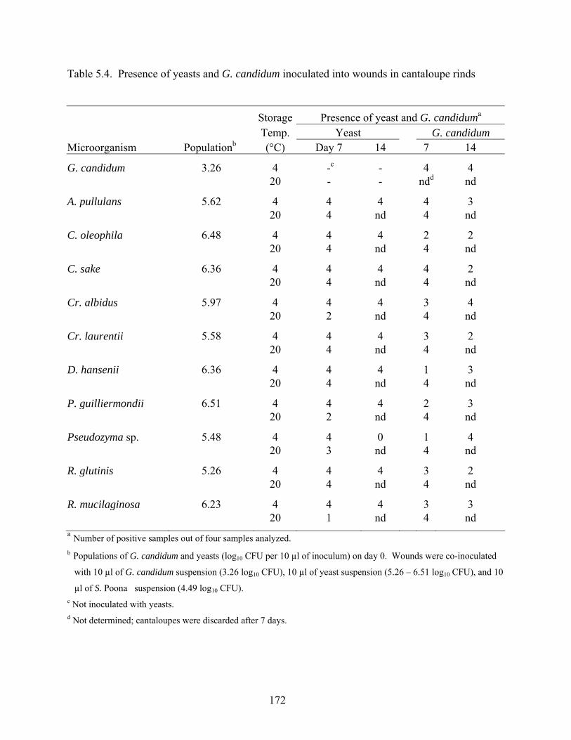
172
Table 5.4. Presence of yeasts and G. candidum inoculated into wounds in cantaloupe rinds
Storage Presence of yeast and G. candiduma
Temp. Yeast G. candidum Microorganism Populationb (°C) Day 7 14 7 14
G. candidum 3.26 4 -c - 4 4 20 - - ndd nd
A. pullulans 5.62 4 4 4 4 3 20 4 nd 4 nd
C. oleophila 6.48 4 4 4 2 2 20 4 nd 4 nd
C. sake 6.36 4 4 4 4 2 20 4 nd 4 nd
Cr. albidus 5.97 4 4 4 3 4 20 2 nd 4 nd
Cr. laurentii 5.58 4 4 4 3 2 20 4 nd 4 nd
D. hansenii 6.36 4 4 4 1 3 20 4 nd 4 nd
P. guilliermondii 6.51 4 4 4 2 3 20 2 nd 4 nd
Pseudozyma sp. 5.48 4 4 0 1 4 20 3 nd 4 nd
R. glutinis 5.26 4 4 4 3 2 20 4 nd 4 nd
R. mucilaginosa 6.23 4 4 4 3 3 20 1 nd 4 nda Number of positive samples out of four samples analyzed. b Populations of G. candidum and yeasts (log10 CFU per 10 µl of inoculum) on day 0. Wounds were co-inoculated
with 10 µl of G. candidum suspension (3.26 log10 CFU), 10 µl of yeast suspension (5.26 – 6.51 log10 CFU), and 10
µl of S. Poona suspension (4.49 log10 CFU). c Not inoculated with yeasts. d Not determined; cantaloupes were discarded after 7 days.
173
Figure 5.9. Effect of yeasts, G. candidum, and S. Poona on change in pH of tissue in
wounds in cantaloupe rinds. Wounds in cantaloupe rinds were co-inoculated with
10 µl of G. candidum suspension (3.26 log10 CFU), 10 µl of yeast suspension (5.26 -
6.51 log10 CFU), and 10 µl of S. Poona suspension (4.49 log10 CFU). The initial
pH values of tissue stored at 4°C and 20°C were 5.54 – 6.64 and 5.75 – 6.49,
respectively. Change in the pH of the cantaloupe wound issue [difference between
initial pH on day 0 and pH after storage at 4°C for 7days (shaded bars), 4°C for 14
days (solid bars) and 20°C for 7 days (open bars)] was monitored.
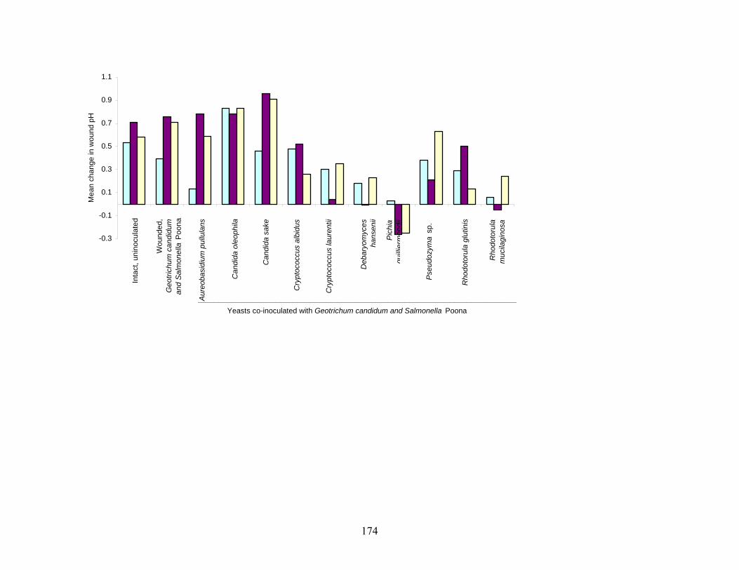
174
-0.3
-0.1
0.1
0.3
0.5
0.7
0.9
1.1
Yeasts co-inoculated with Geotrichum candidum and Salmonella Poona
Mea
n ch
ange
in w
ound
pH
Aure
obas
idiu
m p
ullu
lans
Inta
ct, u
nino
cula
ted
Wou
nded
, G
eotri
chum
can
didu
m
and
Salm
onel
la P
oona
Can
dida
ole
ophi
la
Can
dida
sak
e
Cry
ptoc
occu
s al
bidu
s
Cry
ptoc
occu
s la
uren
tii
Deb
aryo
myc
es
hans
enii
Pich
ia
guilli
erm
ondi
i
Pseu
dozy
ma
sp.
Rho
doto
rula
glu
tini s
Rho
doto
rula
m
ucila
gino
sa
_____________________________________________________________________________
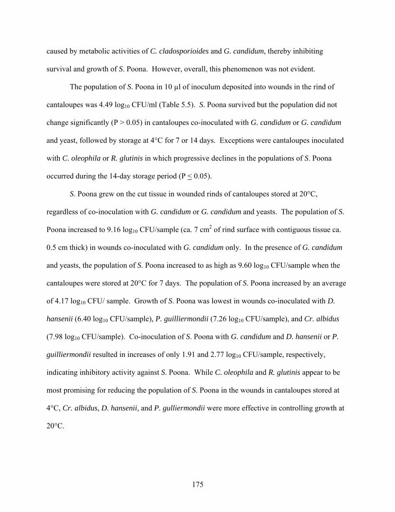
175
caused by metabolic activities of C. cladosporioides and G. candidum, thereby inhibiting
survival and growth of S. Poona. However, overall, this phenomenon was not evident.
The population of S. Poona in 10 µl of inoculum deposited into wounds in the rind of
cantaloupes was 4.49 log10 CFU/ml (Table 5.5). S. Poona survived but the population did not
change significantly (P > 0.05) in cantaloupes co-inoculated with G. candidum or G. candidum
and yeast, followed by storage at 4°C for 7 or 14 days. Exceptions were cantaloupes inoculated
with C. oleophila or R. glutinis in which progressive declines in the populations of S. Poona
occurred during the 14-day storage period (P < 0.05).
S. Poona grew on the cut tissue in wounded rinds of cantaloupes stored at 20°C,
regardless of co-inoculation with G. candidum or G. candidum and yeasts. The population of S.
Poona increased to 9.16 log10 CFU/sample (ca. 7 cm2 of rind surface with contiguous tissue ca.
0.5 cm thick) in wounds co-inoculated with G. candidum only. In the presence of G. candidum
and yeasts, the population of S. Poona increased to as high as 9.60 log10 CFU/sample when the
cantaloupes were stored at 20°C for 7 days. The population of S. Poona increased by an average
of 4.17 log10 CFU/ sample. Growth of S. Poona was lowest in wounds co-inoculated with D.
hansenii (6.40 log10 CFU/sample), P. guilliermondii (7.26 log10 CFU/sample), and Cr. albidus
(7.98 log10 CFU/sample). Co-inoculation of S. Poona with G. candidum and D. hansenii or P.
guilliermondii resulted in increases of only 1.91 and 2.77 log10 CFU/sample, respectively,
indicating inhibitory activity against S. Poona. While C. oleophila and R. glutinis appear to be
most promising for reducing the population of S. Poona in the wounds in cantaloupes stored at
4°C, Cr. albidus, D. hansenii, and P. gulliermondii were more effective in controlling growth at
20°C.
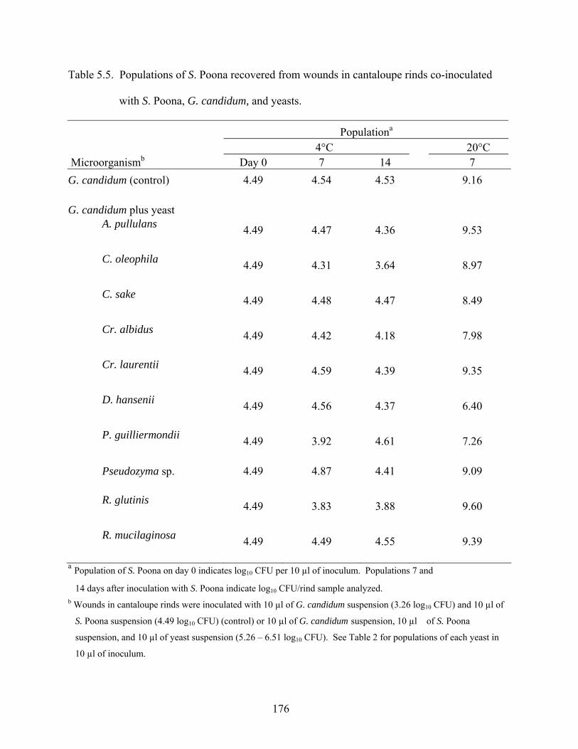
176
Table 5.5. Populations of S. Poona recovered from wounds in cantaloupe rinds co-inoculated
with S. Poona, G. candidum, and yeasts.
Populationa 4°C 20°C Microorganismb Day 0 7 14 7 G. candidum (control) 4.49 4.54 4.53 9.16 G. candidum plus yeast
A. pullulans 4.49 4.47 4.36 9.53
C. oleophila 4.49 4.31 3.64 8.97
C. sake 4.49 4.48 4.47 8.49
Cr. albidus 4.49 4.42 4.18 7.98
Cr. laurentii 4.49 4.59 4.39 9.35
D. hansenii 4.49 4.56 4.37 6.40
P. guilliermondii 4.49 3.92 4.61 7.26
Pseudozyma sp. 4.49 4.87 4.41 9.09
R. glutinis 4.49 3.83 3.88 9.60
R. mucilaginosa 4.49 4.49 4.55 9.39 a Population of S. Poona on day 0 indicates log10 CFU per 10 µl of inoculum. Populations 7 and
14 days after inoculation with S. Poona indicate log10 CFU/rind sample analyzed. b Wounds in cantaloupe rinds were inoculated with 10 µl of G. candidum suspension (3.26 log10 CFU) and 10 µl of
S. Poona suspension (4.49 log10 CFU) (control) or 10 µl of G. candidum suspension, 10 µl of S. Poona
suspension, and 10 µl of yeast suspension (5.26 – 6.51 log10 CFU). See Table 2 for populations of each yeast in
10 µl of inoculum.
177
The non-cellulosic neutral sugar composition of cell walls of mature cantaloupes is ca.
11.3 mg/100 mg of wall tissues (Gross and Sams, 1984). These sugars include rhaminose,
fructose, arabinose, xylose, mannose, galactose, and glucose. All test yeasts utilize glucose,
fructose, and mannose, but there are large differences among species with respect to utilization
of other sugars and amino acids (Deak and Beuchat, 1996). Generally, Salmonella are able to
ferment glucose and other monosaccharides and utilize a wide range of compounds as sources of
nitrogen. Yeasts can potentially be antagonistic by competing with bacteria for these nutrients in
fruit tissues. In studies conducted by Nunes et al. (2002), wounds in apples and pears were co-
inoculated with C. sake and the bacterium Pantoea agglomerans to assess their abilities to
enhance their biocontrol capabilities. C. sake dominated the wounds because of its higher
capacity to utilize nitrogen sources. The low nitrogen and vitamin contents in tissue fluids in
wound sites after colonization of C. sake caused a decline in the population of P. agglomerans.
Wounded apple tissue has been observed to be an excellent substrate for the growth of E.
coli, supporting exponential growth (Dingman, 2000; Janisiewicz et al., 1999b). Decreased
survival of E. coli O157:H7 in unpasteurized cider compared to sterile apple juice was attributed
to interaction with natural populations of yeasts (Janisiewicz et al. 1999b). The authors
recommended that explorative work be undertaken to identify yeasts that prevent potential
colonization of human pathogens.
Strains of the yeasts used in this study have been assessed by others to determine their
potential as antagonists and ability to prevent colonization of phytopathogens in wounds in fruits.
A. pullulans, Cr. laurentii, and R. glutinis were shown to produce antifungal and antibacterial
compounds that were inhibitory to P. fluorescens and S. aureus (McCormack et al., 1994).
These yeasts have also been shown to inhibit several fungal pathogens in wounds on apples,
178
pears, strawberry, kiwi, grapes, oranges, mandarins, and grapefruits (Lima et al., 1999).
Conidial germination and mycelial growth by Penicillium expansum were reduced by
rhodotorulic acid and siderophore produced by R. glutinis (Calvente et al., 1999). C. oleophila
has been used to develop commercial formulations for application to pome and citrus fruits
(Droby et al., 1998; Mercier and Wilson, 1994; Wilson et al., 1996) and C. sake is reported to be
an effective antagonist for the control of major postharvest pathogens on pome fruits at small and
semi-commercial scale trials in packinghouses (Abadias et al., 2001; Viñas et al., 1998).
Successful biocontrol activity of Pseudozyma flocculosa has been attributed to its ability to
produce antifungal fatty acids that affect the mycelial growth and conidial germination of fungi
(Avis and Bélanger, 2001).
Rapid colonization, competition for nutrients, and secretion of cell wall-degrading
enzymes have been suggested as the mechanisms by which D. hansenii and P. guilliermondii
(previously called D. hansenii) achieve biological control of phytopathogens (Droby et al., 1989;
Petersson and Schnürer, 1995). The ability of P. guilliermondii to attach firmly to the hyphae of
Botrytis cinerea and produce high levels of β- (1-3) glucanase activity were found to be
additional characteristics that improved its effectiveness as a biological control agent
(Wisniewski et al., 1991). P. guilliermondii has been reported to elicit the formation of
phytoalexins (scoparone and scopoletin) in citrus fruit wounds at concentrations that are
fungitoxic (Lima et al., 1999). These control methods would be acceptable for use in the
biological control of phytopathogens and foodborne pathogens because they do not involve
secretion of potentially harmful antibiotics.
Some of the yeasts tested by others (Droby et al., 1989; Droby et al., 1998; Lima et al.,
1999; Mercier and Wilson, 1994; McCormack et al., 1994; Petersson and Schnürer, 1995;
179
Wilson et al., 1996; Wisniewski et al., 1991) were demonstrated in this study to have potential to
restrict colonization and metabolic activities of plant pathogenic molds in wounds in cantaloupe
rinds, particularly at low storage temperatures. Results of our study indicate that C. oleophila
and R. glutinis at 4°C and Cr. albidus, D. hansenii, and P. gulliermondii at 20°C could also be
efficacious as antagonists to S. Poona in the wounds co-infected with phytopathogens. The
populations of S. Poona in cantaloupe juice co-inoculated with D. hansenii, P. guilliermondii,
and Pseudozyma sp. at high populations was significantly less than that of the control. However,
the overall results of the in vitro study suggest that the yeasts evaluated were unable to
vigorously compete and efficiently restrict the growth of S. Poona in cantaloupe juice. The
success of biological control depends on the ability of the antagonist to rapidly become
established in the environment, out-compete the pathogen for resources, and create a medium
that is unsuitable for the pathogen.
180
REFERENCES
Abadias, M., N. Teixidó, J. Usall, A. Benabarre, and I. Viñas. 2001. Viability, efficacy, and storage stability of freeze-dried biocontrol agent Candida sake using different protective and rehydration media. J. Food Prot. 64:856-861.
Arul, J. 1994. Emerging technologies for the control of postharvest diseases of fresh fruits
and vegetables. In C.L. Wilson and M.E. Wisniewski (Ed.), Biological Control of Postharvest Diseases. Theory and Practice. CRC Press, Boca Raton, FL.
Avis, T.J. and R.R. Bélanger. 2001. Specificity and mode of action of the antifungal fatty
acid cis-9-heptadecenoic acid produced by Pseudozyma flocculosa. Appl. Environ. Microbiol. 67:956-960.
Bilinski, C.A., G. Innamorato, and G.G. Stewart. 1985. Identification and characterization of
antimicrobial activity in two yeast genera. Appl. Environ. Microbiol. 50:1330-1332. Brecht, J.K. 1995. Physiology of lightly processed fruits and vegetables. HortSci. 30:18-22. Buck, J. W., R. R. Walcott, and L.R. Beuchat. 2003. Recent trends in microbiological safety
of fruits and vegetables. Plant Health Progress. http://www.apsnet.org. Accessed 29 January 2003.
Calvente, V., D. Benuzzi, and M.I.S. de Tosetti. 1999. Antagonistic action of siderophores
from Rhodotorula glutinis upon the postharvest pathogen Penicillium expansum. Int. Biodeter. Biodegrad. 43:167-172.
Chand-Goyal, T. and R.A. Spotts. 1997. Biological control of postharvest diseases of apples
and pear under semi-commercial and commercial conditions using three saprophytic yeasts. Biol. Control 10:199-206.
Conway, W.S., B. Leverentz, R.A. Saftner, W.J. Janisiewicz, C.E. Sams, and E. Leblanc.
2000. Survival and growth of Listeria monocytogenes on fresh cut apples and its interaction with Glomerella cingulata and Penicillium expansum. Plant Dis. 84:177-181.
Deak, T. and L.R. Beuchat. 1996. Handbook of Food Spoilage Yeasts. CRC Press, Boca
Raton, FL. Dingman, D. W. 2000. Growth of Escherichia coli O157:H7 in bruised apple (Malus
domestica) tissue as influenced by cultivar, date of harvest, and source. Appl. Environ. Microbiol. 66:1077-1083.
Draughon, F.A., S. Chen, and J.O. Mundt. 1988. Metabiotic association of Fusarium,
Alternaria, and Rhizoctonia with Clostridium botulinum on fresh tomatoes. J. Food Sci. 53:120-123.
181
Droby, S., E. Chalutz, C. L. Wilson, and M.E. Wisniewski. 1989. Characterization of the biocontrol activity of Debaryomyces hansenii in the control of Penicillium digitatum on grapefruit. Can. J. Microbiol. 35: 794-800.
Droby S. and E. Chalutz., 1994. Mode of action of biocontrol agents of postharvest diseases.
In C.L. Wilson and M.E. Wisniewski (Ed.), Biological Control of Postharvest Diseases. Theory and practice. CRC Press, Boca Raton, FL.
Droby, S., E. Chalutz, M.E. Wisniewski, and C.L. Wilson. 1996. Host response to
introduction of antagonistic yeasts used for control of postharvest decay. In C.E. Morris, P.C. Nicot, and C. Nguyen-the (Ed.), Aerial Plant Surface Microbiology. Plenum Press, New York, NY.
Droby, S., L. Cohen, A. Daus, B. Weiss, B. Horev, E. Chalutz, H. Katz, M. Keren-Tzur, and
A. Shachnai. 1998. Commercial testing of Aspire: A yeast preparation for the biological control of postharvest decay of citrus. Biol. Control 12:91-101.
Eckert, J.W. and J.M. Ogawa. 1985. The chemical control of postharvest diseases:
subtropical and tropical fruits. Ann. Rev. Phytopathol. 23:421-454.
El-Ghaouth, A. 1997. Biologically-based alternatives to synthetic fungicides for the control of postharvest diseases. J. Industrial Microbiol. Biotechnol. 19:160-162.
El-Ghaouth, C.L. Wilson, and M.E. Wisniewski. 1998. Ultrastructural and cytological
aspects of the biological control of Botrytis cinerea by Candida saitoana in apple fruit. Phytopathology 88: 282-291.
Gross, K.C. and C.E. Sams. 1984. Changes in cell wall neutral sugar composition during fruit
ripening: A species survey. Phytochemistry 23: 2457-2461. Huhtanen, C.N., J. Naghski, C.S. Custer, and R.W. Russell. 1976. Growth and toxin
production by Clostridium botulinum in moldy tomato juice. Appl. Environ. Microbiol. 32:711-715.
Janisiewicz, W.J., W.S. Conway, and B. Leverentz. 1999a. Biological control of postharvest
decays of apple can prevent growth of Escherichia coli O157:H7 in apple wounds. J. Food Prot. 62:1372-1375.
Janisiewicz, W.J. W.S. Conway, M.W. Brown, G.M. Sapers, P. Fratamico, and R.L.
Buchanan. 1999b. Fate of Escherichia coli O157:H7 on fresh-cut apple tissue and its potential for transmission by fruit flies. Appl. Environ. Microbiol. 65:1-5.
Janisiewicz, W.J., T.J. Tworkoski, and C. Sharer. 2000. Characterizing the mechanism of
biological control of postharvest diseases on fruits with a simple method to study competition for nutrients. Phytopathology 90:1196-1200.
182
Janisiewicz, W.J. and L. Korsten. 2002. Biological control of postharvest diseases of fruits. Annu. Rev. Phytopathol. 40:411-441.
Kurtzman, C.P. and J.W. Fell. 1999. The Yeasts, A Taxonomic Study. Elsevier, Amsterdam,
The Netherlands. Lamikanra, O., J.C. Chen, D. Banks, and P.A. Hunter. 2000. Biochemical and microbial
changes during the storage of minimally processed cantaloupe. J. Agric. Food Chem. 48:5955-5961.
Lamikanra, O., B. Juaraez, M.A. Watson, and O.A. Richard. 2003. Effect of cutting and
storage on sensory traits of cantaloupe melon cultivars with extended postharvest shelf life. J. Sci. Food Agric. 83:702-708.
Leverentz, B., W.S. Conway, Z. Alavidze, W.J. Janisiewicz, Y. Fuchs, M.J. Camp, E.
Chighladze, and A. Sulakvedidze. 2001. Examination of bacteriophage as a biocontrol method of Salmonella on fresh-cut Fruit: A model study. J. Food Prot. 64:1116-1121.
Lewus, C.B., A. Kaiser, and T.J. Montville. 1991. Inhibition of food-borne bacterial
pathogens by bacteriocins from lactic acid bacteria isolated from meat. Appl. Environ. Microbiol. 57:1683-1688.
Lima, G., S. Arru, F. De Curtis, and G. Arras. 1999. Influence of antagonist, host fruit and
pathogen on biological control of postharvest fungal diseases by yeasts. J. Indus. Microbiol. Biotechnol. 23:223-229.
McCormack, P.J., H.G. Wildman, and P. Jeffries. 1994. Production of antibacterial
compounds by phylloplane-inhabiting yeasts and yeastlike fungi. Appl. Environ. Microbiol. 60:927-931.
Mercier, J. and C.L. Wilson. 1994. Colonization of apple wounds by naturally occurring
microflora and introduced Candida oleophila and their effect on infection by Botrytis cinerea during storage. Biolog. Control 4:138-144.
Mundt, J.O. 1978. Effect of mold growth on the pH of tomato juice. J. Food Prot. 41:267-
268. Muriana, P.M. 1996. Bacteriocins for control of Listeria spp. in food. J. Food Prot. 56:944-
948. National Research Council, Board of Agriculture. 1987. Regulating pesticides in food – The
Delany Paradox, National Academy Press, Washington, DC. Nunes, C., J. Usall, N. Teixidó, R. Torres, and I. Viñas. 2002. Control of Penicillium
expansum and Botrytis cinerea on apples and pears with the combination of Candida sake and Pantoea agglomerans. J. Food Prot. 65:178-184.
183
Odlaug, T.E. and I.J. Pflug. 1979. Clostridium botulinum growth and toxin production in
tomato juice containing Aspergillus gracilis. Appl. Environ. Microbiol. 37:496-504.
Petersson, S. and J. Schnürer. 1995. Biocontrol of mold growth in high-moisture wheat stored under airtight conditions by Pichia anomala, Pichia guilliermondii, and Saccharomyces cerevisiae. Appl. Environ. Microbiol. 61:1027-1032.
Riordan, D.C.R., G.M. Sapers, and B.A. Annous. 2000. The survival and growth of
Escherichia coli O157:H7 in the presence of Penicillium expansum and Glomerella cingulata in wounds on apple surfaces. J. Food Prot. 63:1637-1642.
Rodrigues, A.C.P., R.M. Nardi, E.H. Bambirra, E.C. Vieira, and J.R. Nicoli. 1996. Effect of
Saccharomyces boulardii against experimental oral infection with Salmonella Typhimurium and Shigella flexneri in conventional and gnotobiotic mice. J. Appl. Bacteriol. 81:251-256.
Salunkhe, D.K. and B.B. Desai. 1984. Postharvest Biotechnology of Fruits. Vol. 1. CRC.
Boca Raton, FL. Snowdon, A. L. 1990. A Colour Atlas of Post-Harvest Diseases and Disorders of Fruits and
Vegetables. Volume 1: General Introduction and Fruits. CRC Press, Boca Raton, FL. Stevens, K.A., B.W. Sheldon, N.A. Klapes, and T.R. Klaenhammer. 1991. Nisin treatment
for inactivation of Salmonella species and other gram-negative bacteria. Appl. Environ. Microbiol. 57:3613-3615.
Viñas, I. J. Usall, N. Teixidó, and V. Sanchis. 1998. Biological control of major postharvest
pathogens on apple with Candida sake. Intl. J. Food Microbiol. 40:9-16. Wade, W.N. and L.R. Beuchat. 2003. Proteolytic fungi isolated from decayed and damaged
raw tomatoes and implications associated with changes in pericarp pH favorable for survival and growth of foodborne pathogens. J. Food Prot. 66:911-917.
Walker, R., C.M.J. Innes, and E.J. Allan. 2001. The potential biocontrol agent Pseudomonas
antimicrobica inhibits germination of conidia and outgrowth of Botrytis cinerea. Lett. Appl. Microbiol. 32:346-348.
Wells, J.M. and J.E. Butterfield. 1997. Salmonella contamination with bacterial soft rot of
fresh fruits and vegetables in the marketplace. Plant Dis. 81:867-872. Wilson, C.L. and M.E. Wisniewski. 1989. Biological control of postharvest diseases of fruits
and vegetables: An emerging technology. Annu. Rev. Phytopathol. 27:425-441. Wilson, C. L. and M.E. Wisniewski. 1994. Biological control of postharvest diseases. CRC
Press, Boca Raton, FL.
184
Wilson, C.L., M.E. Wisniewski, A. El Ghaouth, S. Droby, and E. Chalutz. 1996. Commercialization of antagonistic yeasts for the biological control of postharvest diseases of fruits and vegetables. J. Industrial Microbiol. 46:237-242.
Wilson, C.L., J.M. Solar, A. El-Ghaouth, and M.E. Wisniewski. 1997. Rapid evaluation of
plant extracts and essential oils for antifungal activity against Botrytis cinerea. Plant Dis. 81: 204-210.
Wisniewski, M., C. Biles, S. Droby, R. McLaughlin, C. Wilson, and E. Chalutz. 1991. Mode
of action of the postharvest biocontrol yeast, Pichia guilliermondii. I. Characterization of attachment to Botrytis cinerea. Phys. Mol. Plant Pathol. 39:245-258.

185
CHAPTER 6
SUMMARY AND CONCLUSIONS
186
Studies were undertaken to determine the survival and growth characteristics of
Salmonella Poona on and in cantaloupes as affected by conditions to which the fruits may be
exposed preceding cutting for preparation to consume. Specific objectives of the studies
documented in this dissertation were:
1. To determine if temperature differentials between cantaloupes and suspensions of S. Poona
influences infiltration of the pathogen into rind and stem scar tissues of Eastern and Western
type cantaloupes.
2. To determine survival and growth characteristics of S. Poona on the surface of intact rind and
in wounded rind tissue as affected by storage temperature and co-infection with
phytopathogenic molds.
3. To determine if S. Poona migrates from tissues in the cantaloupe rinds to edible internal
tissues.
4. To examine yeasts for their potential to impair survival and growth of S. Poona in cell
suspensions and in wounds in the cantaloupe rind co-inoculated with phytopathogenic molds.
This research demonstrated that S. Poona in wash water adheres to rind and stem scar tissues
of cantaloupes. The initial temperature of the inoculum and cantaloupe affected the mean
percent weight increase in weight by Eastern type cantaloupes, but not the Western type.
Adherence to or infiltration of Eastern but not Western type cantaloupe rind by S. Poona is
enhanced when the cantaloupe is at 4°C compared to 30°C. The histology of cantaloupe rind and
stem scar tissues augments attachment and penetration of S. Poona, and may therefore reduce the
effectiveness of sanitizer treatments.
S. Poona persists on intact, wounded, and decaying cantaloupe rind at refrigerated and
ambient temperatures. Phytopathogenic molds can increase the pH of cantaloupe tissue by
187
proteolytic activity and perhaps other alkalinizing activities. However, recovery of S. Poona
after incubation at 4°C and 30°C was not affected by co-infection with molds. Abusive storage
temperatures enhance survival and growth of S. Poona on wounded cantaloupe tissues. These
findings underscore the importance of proper postharvest handling and storage to prevent
contamination, physical damage, and deterioration.
S. Poona is capable of migrating from wounded tissues in the rind through the pulp tissues to
depths up to 4 cm in edible tissues, with or without co-inoculation with phytopathogens.
Migration and survival of S. Poona in cantaloupes were enhanced by co-inoculation with
Cladosporium cladosporioides and, to a lesser extent, Penicillium expansum.
Some of the yeasts examined in this study for biocontrol activity exhibited potential to
restrict colonization and metabolic activities of plant pathogenic molds in cantaloupe rinds,
particularly at low storage temperatures. Candida oleophila and Rhodotorula glutinis at 4°C and
Cryptococcus albidus, Debaryomyces hansenii, and Pichia guilliermondii at 20°C could also be
efficacious as antagonists to S. Poona in wounds co-infected with phytopathogenic molds. While
the populations of S. Poona in cantaloupe juice co-inoculated with D. hansenii, P. guilliermondii,
and Pseudozyma sp. at high populations were significantly less that of the control, the overall
results of the in vitro study suggest that the yeasts evaluated were unable to vigorously compete
and efficiently restrict the growth of S. Poona in cantaloupe juice.
Biological control activities by the yeasts investigated do not prevent survival and growth of
S. Poona in cantaloupe tissues. Adherence of S. Poona to rind and stem scar tissues, migration of
the pathogen from subsurface edible tissues, and recovery of larger populations from wounded
tissues, particularly from cantaloupes stored at abusive temperatures, underscore the importance
188
of proper postharvest handling and storage to prevent contamination, physical damage, and
deterioration.