Embed Size (px)
Citation preview
Surgical Short Cases
Jonny LenihanSurgical CT1 NWTD
Overview• Common pathologies• Examination technique • Presentation skills• Background Information• X-rays• Summary• Questions
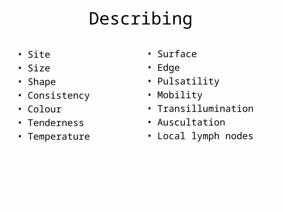
Describing
• Site• Size• Shape• Consistency• Colour• Tenderness• Temperature
• Surface• Edge • Pulsatility• Mobility• Transillumination• Auscultation• Local lymph nodes

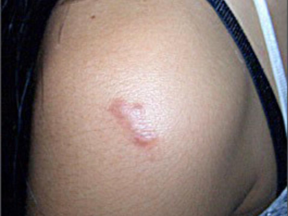
Hypertrophic and Keloid Scars• Types of wound prone to these:
– Infection; trauma; burns; tension
• Hypertrophic occur soon after insult; spontaneously regress
• Keloid scars appear months after and continue to grow
• Rx:– Mechanical pressure dressings with topical agents– Surgical excision– Intralesional steroid therapy
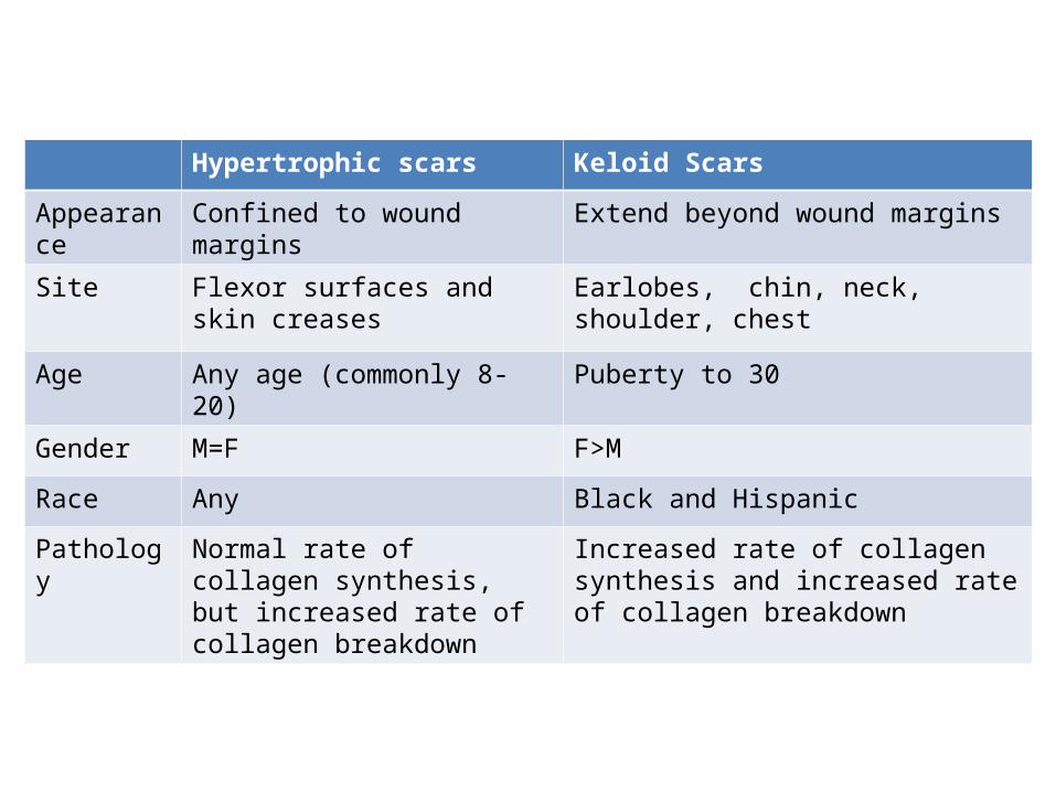
Hypertrophic scars Keloid Scars
Appearance Confined to wound margins Extend beyond wound margins
Site Flexor surfaces and skin creases Earlobes, chin, neck, shoulder, chest
Age Any age (commonly 8-20) Puberty to 30
Gender M=F F>M
Race Any Black and Hispanic
Pathology Normal rate of collagen synthesis, but increased rate of collagen breakdown
Increased rate of collagen synthesis and increased rate of collagen breakdown
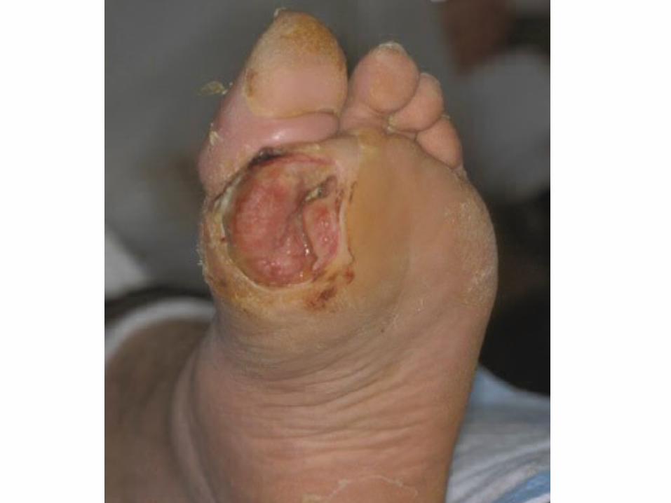
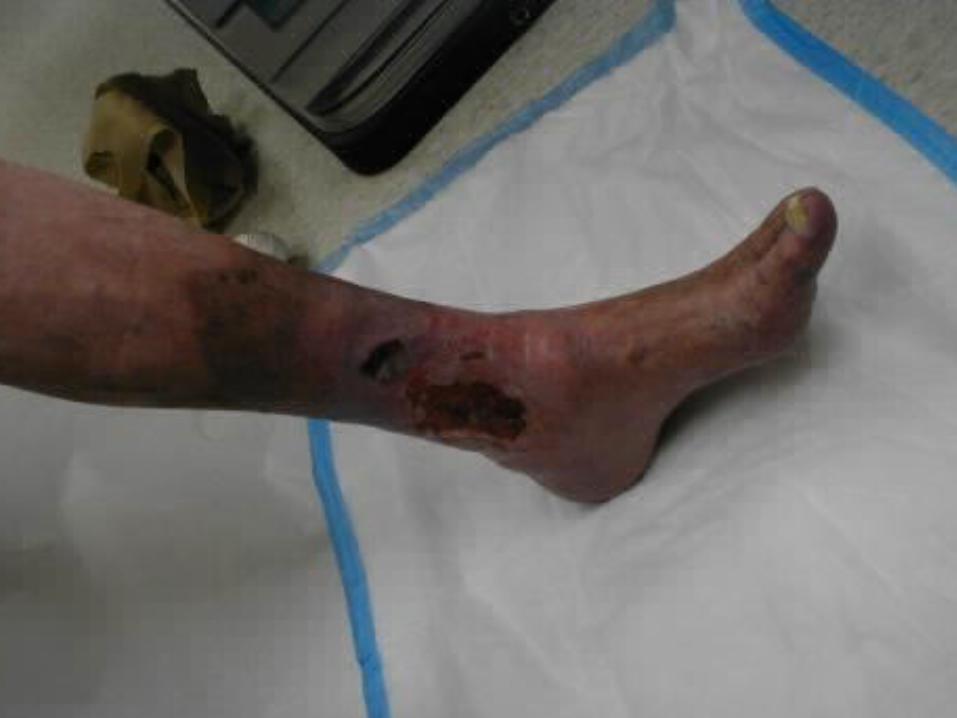
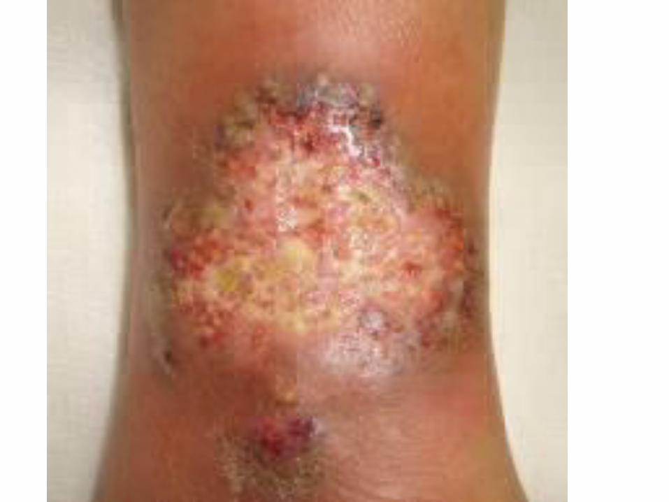
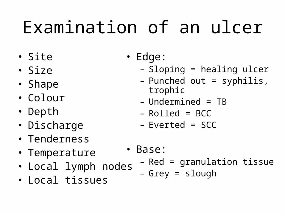
Examination of an ulcer• Site• Size• Shape• Colour• Depth• Discharge• Tenderness• Temperature• Local lymph nodes• Local tissues
• Edge:– Sloping = healing ulcer– Punched out = syphilis, trophic– Undermined = TB– Rolled = BCC– Everted = SCC
• Base:– Red = granulation tissue– Grey = slough
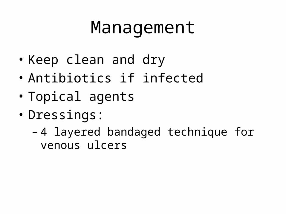
Management
• Keep clean and dry• Antibiotics if infected• Topical agents• Dressings:
– 4 layered bandaged technique for venous ulcers
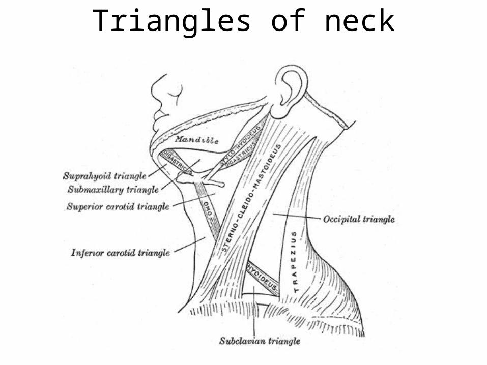
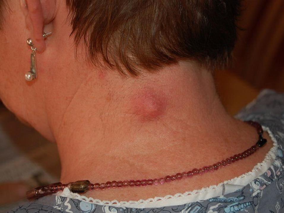
Triangles of neck
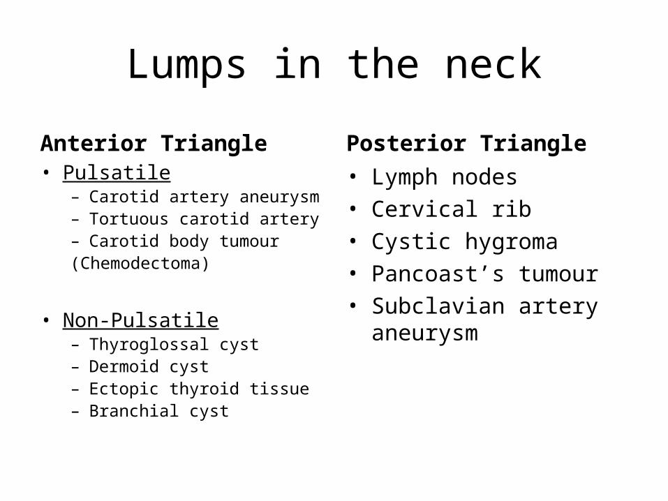
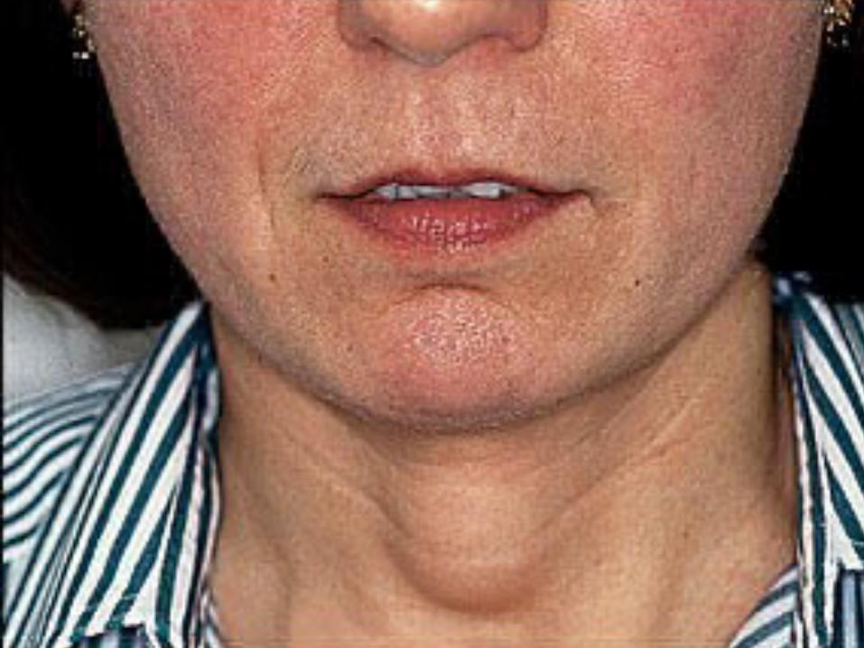
Lumps in the neck
Anterior Triangle• Pulsatile
– Carotid artery aneurysm– Tortuous carotid artery– Carotid body tumour(Chemodectoma)
• Non-Pulsatile– Thyroglossal cyst– Dermoid cyst– Ectopic thyroid tissue– Branchial cyst
Posterior Triangle• Lymph nodes• Cervical rib• Cystic hygroma• Pancoast’s tumour• Subclavian artery aneurysm
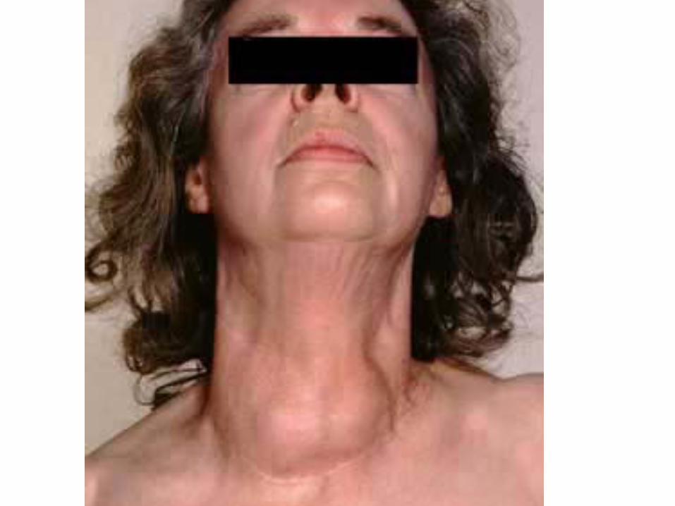
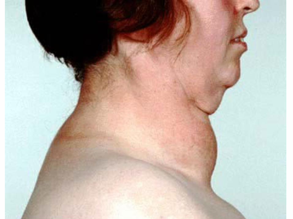
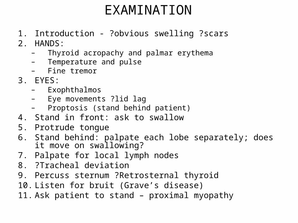
EXAMINATION
1. Introduction - ?obvious swelling ?scars2. HANDS:
– Thyroid acropachy and palmar erythema– Temperature and pulse– Fine tremor
3. EYES:– Exophthalmos– Eye movements ?lid lag– Proptosis (stand behind patient)
4. Stand in front: ask to swallow5. Protrude tongue6. Stand behind: palpate each lobe separately; does it move on
swallowing?7. Palpate for local lymph nodes8. ?Tracheal deviation9. Percuss sternum ?Retrosternal thyroid10. Listen for bruit (Grave’s disease)11. Ask patient to stand – proximal myopathy
Focused history• Symptoms of hyper/hypo – thyroidism:
– Weight, Appetite, Sweating, Tremor, Palpitations, Menstrual irregularities, Irritability, Diarrhoea
• Have they noticed a lump– Change in size over time?
• Change in voice? • Any pressure symptoms?
– Dyspnoea, Dysphagia
• Diet (deficient in Iodine)• Any history of radiation exposure?• Family history
INVESTIGATIONS• Biochemistry:
– Thyroid status: T3, T4 and TSH– FBC, U+Es, Ca2+, LFTs and ESR
• Radiology:– CXR– Ultrasound (solid, cystic masses)– CT scan
• Special:– Fine needle aspirate (not reliable for follicular
adenoma/carcinoma)– Tru-cut biopsy– Radioisotope scan (Tc99)– Laryngoscopy (?paralysis of vocal chords pre-operatively)
Management of Thyrotoxicosis• MEDICAL
– Pharmacological:– Carbimazole; Propylthiouracil; Propanolol
– Radioiodine (nb: teratogenic)– >50yrs old, recurrent episodes or post surgery
• SURGERY– Once medical therapy failed or pressure symptoms– Sub-total thyroidectomy (after antithyroid drugs)
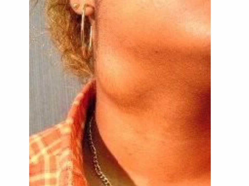

Dermoid cysts
1. Inclusion dermoids:– At site of embryological fusion: midline neck,
angle of orbit– Firm, not attached to skin – Rx = excise
2. Implantation dermoids:– Subcutaneous swellings after penetrating injury– Epidermal tissue introduced beneath skin
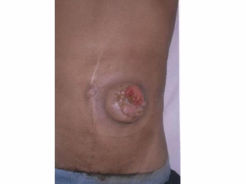
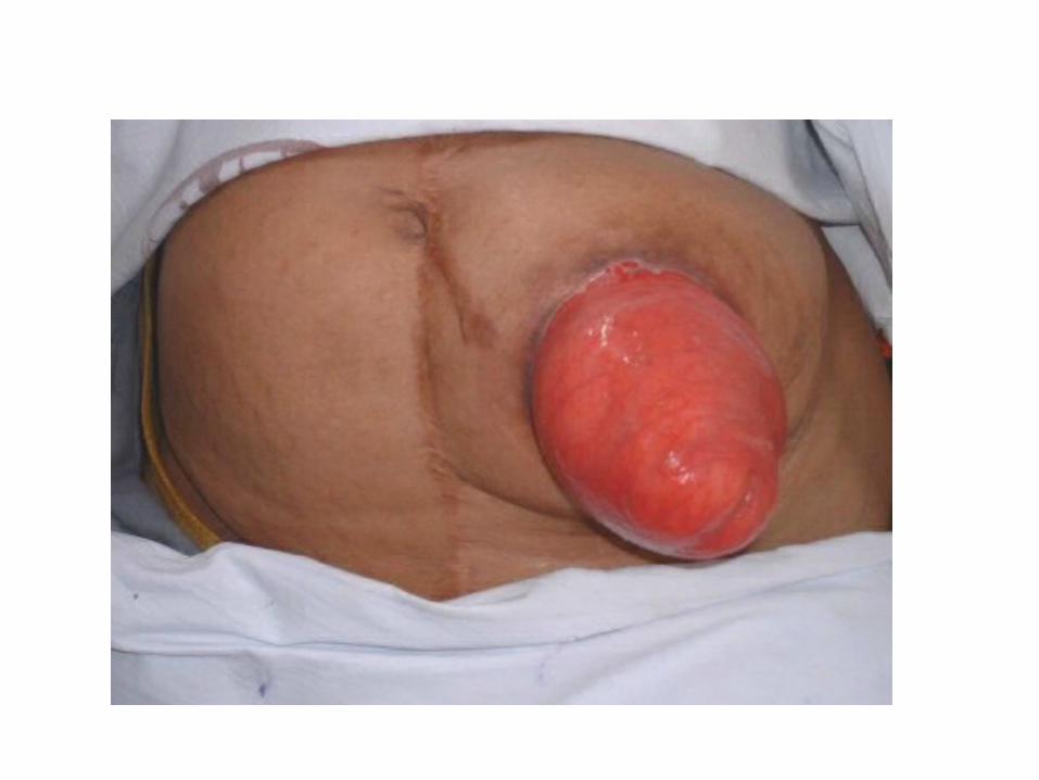
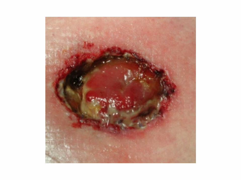
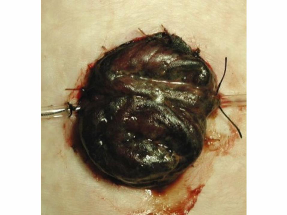
Complications
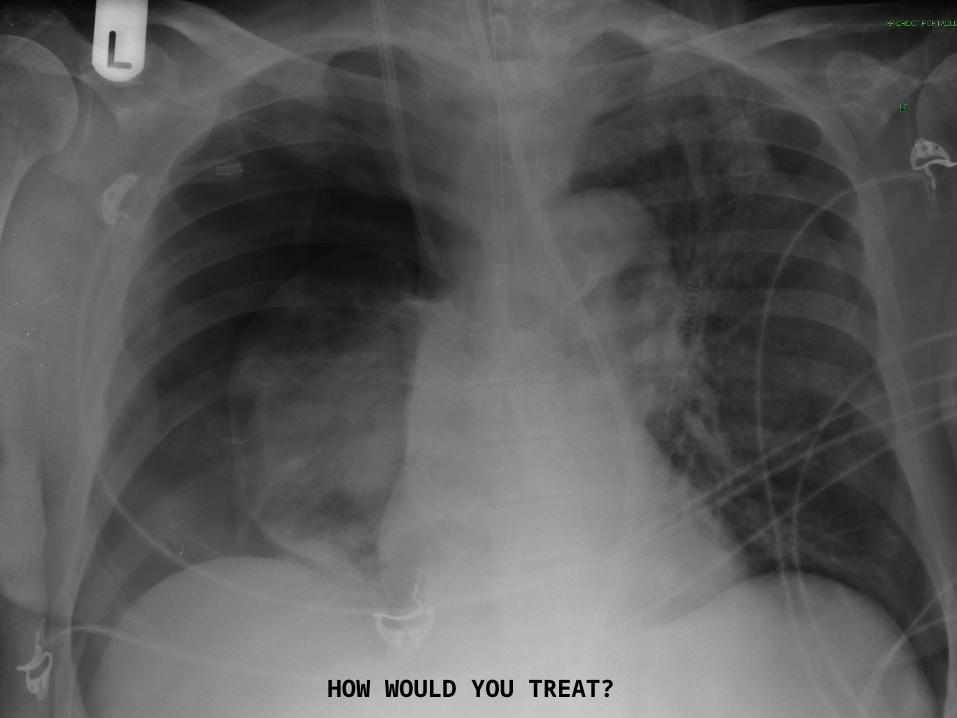
HOW WOULD YOU TREAT?
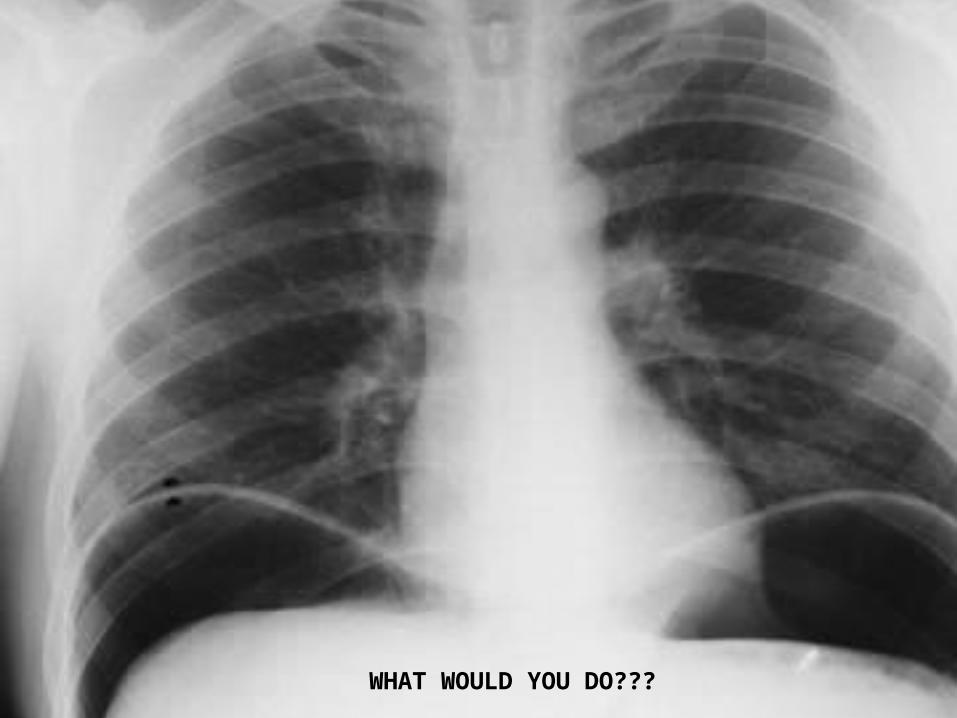
WHAT WOULD YOU DO???
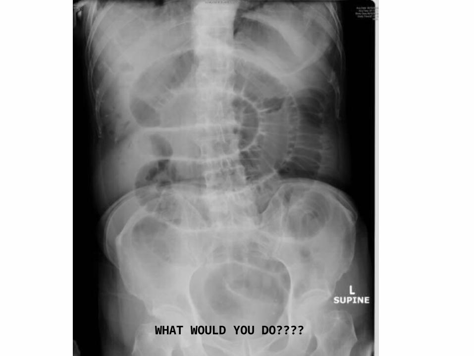
WHAT WOULD YOU DO????

WHAT WOULD YOU DO???
Summary
• Covered common presentations for Finals• Examination methods• Presenting your findings• Typical XRs in shorts• Google pathology• Questions?