Embed Size (px)
Citation preview
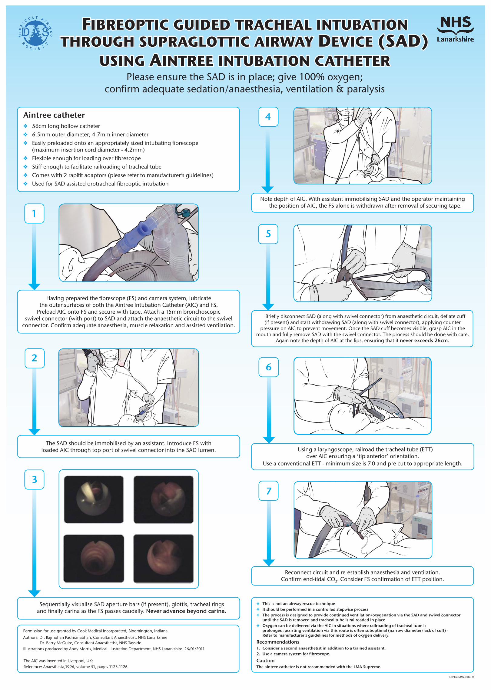
Fibreoptic guided tracheal intubation through supraglottic airway device (sad)
using aintree intubation catheterPlease ensure the SAD is in place; give 100% oxygen;
confirm adequate sedation/anaesthesia, ventilation & paralysis
Aintreecatheter v 56cm long hollow catheter v 6.5mm outer diameter; 4.7mm inner diameter v Easily preloaded onto an appropriately sized intubating fibrescope (maximum insertion cord diameter - 4.2mm) v Flexible enough for loading over fibrescope v Stiff enough to facilitate railroading of tracheal tube v Comes with 2 rapifit adaptors (please refer to manufacturer’s guidelines) v Used for SAD assisted orotracheal fibreoptic intubation
1
6
5
7
2
3
4
v Thisisnotanairwayrescuetechnique v Itshouldbeperformedinacontrolledstepwiseprocess v Theprocessisdesignedtoprovidecontinuedventilation/oxygenationviatheSADandswivelconnector untiltheSADisremovedandtrachealtubeisrailroadedinplace v OxygencanbedeliveredviatheAICinsituationswhererailroadingoftrachealtubeis prolonged;assistingventilationviathisrouteisoftensuboptimal(narrowdiameter/lackofcuff)- Refertomanufacturer’sguidelinesformethodsofoxygendelivery.
Recommendations 1. Considerasecondanaesthetistinadditiontoatrainedassistant. 2. Useacamerasystemforfibrescope.
Caution TheaintreecatheterisnotrecommendedwiththeLMASupreme.
Having prepared the fibrescope (FS) and camera system, lubricate the outer surfaces of both the Aintree Intubation Catheter (AIC) and FS.
Preload AIC onto FS and secure with tape. Attach a 15mm bronchoscopic swivel connector (with port) to SAD and attach the anaesthetic circuit to the swivel
connector. Confirm adequate anaesthesia, muscle relaxation and assisted ventilation.
The SAD should be immobilised by an assistant. Introduce FS with loaded AIC through top port of swivel connector into the SAD lumen.
Sequentially visualise SAD aperture bars (if present), glottis, tracheal rings and finally carina as the FS passes caudally. Neveradvancebeyondcarina.
Note depth of AIC. With assistant immobilising SAD and the operator maintaining the position of AIC, the FS alone is withdrawn after removal of securing tape.
Briefly disconnect SAD (along with swivel connector) from anaesthetic circuit, deflate cuff (if present) and start withdrawing SAD (along with swivel connector), applying counter
pressure on AIC to prevent movement. Once the SAD cuff becomes visible, grasp AIC in the mouth and fully remove SAD with the swivel connector. The process should be done with care.
Again note the depth of AIC at the lips, ensuring that it neverexceeds26cm.
Using a laryngoscope, railroad the tracheal tube (ETT) over AIC ensuring a ‘tip anterior’ orientation.
Use a conventional ETT - minimum size is 7.0 and pre cut to appropriate length.
Reconnect circuit and re-establish anaesthesia and ventilation. Confirm end-tidal CO2. Consider FS confirmation of ETT position.
Permission for use granted by Cook Medical Incorporated, Bloomington, Indiana.
Authors: Dr. Rajmohan Padmanabhan, Consultant Anaesthetist, NHS Lanarkshire Dr. Barry McGuire, Consultant Anaesthetist, NHS Tayside
Illustrations produced by Andy Morris, Medical Illustration Department, NHS Lanarkshire. 26/01/2011
CTP.PADMAN.71823.W
The AIC was invented in Liverpool, UK;Reference: Anaesthesia,1996, volume 51, pages 1123-1126.
![Intubation of Suspected/Confirmed COVID-19 Patients · V1 · Intubation Bin 2 Contents A. Supraglottic Airway Devices (SAD) (size 3.0, 4.0, 5.0) [e.g.: I-MA, Pro-Seal, I-Gel, etc.]](https://img.pdfslide.us/doc/110x75/606275599e6faf01743f477a/intubation-of-suspectedconfirmed-covid-19-patients-v1-intubation-bin-2-contents.jpg)