Embed Size (px)
Citation preview
S1
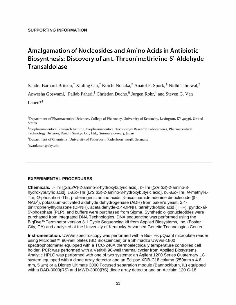
SUPPORTING INFORMATION
Sandra Barnard-Britson,† Xiuling Chi,
† Koichi Nonaka,
‡ Anatol P. Spork,
§ Nidhi Tibrewal,
†
Anwesha Goswami,† Pallab Pahari,
† Christian Ducho,
§ Jurgen Rohr,
† and Steven G. Van
Lanen*†
†Department of Pharmaceutical Sciences, College of Pharmacy, University of Kentucky, Lexington, KY 40536, United
States
‡Biopharmaceutical Research Group I, Biopharmaceutical Technology Research Laboratories, Pharmaceutical
Technology Division, Daiichi Sankyo Co., Ltd., Gunma 370-0503, Japan
§Department of Chemistry, University of Paderborn, Paderborn 33098, Germany
EXPERIMENTAL PROCEDURES
Chemicals. L-Thr [(2S,3R)-2-amino-3-hydroxybutyric acid], D-Thr [(2R,3S)-2-amino-3-hydroxybutyric acid], L-allo-Thr [(2S,3S)-2-amino-3-hydroxybutyric acid], DL-allo-Thr, N-methyl-L-
Thr, O-phospho-L-Thr, proteinogenic amino acids, -nicotinamide adenine dinucleotide (-NAD+), potassium-activated aldehyde dehydrogenase (ADH) from baker’s yeast, 2,4-dinitrophenylhydrazone (DPNH), acetaldehyde-2,4-DPNH, tetrahydrofolic acid (THF), pyridoxal-5'-phosphate (PLP), and buffers were purchased from Sigma. Synthetic oligonucleotides were purchased from Integrated DNA Technologies. DNA sequencing was performed using the BigDye™Terminator version 3.1 Cycle Sequencing kit from Applied Biosystems, Inc. (Foster City, CA) and analyzed at the University of Kentucky Advanced Genetic Technologies Center.
Instrumentation. UV/Vis spectroscopy was performed with a Bio-Tek µQuant microplate reader using Microtest™ 96-well plates (BD Biosciences) or a Shimadzu UV/Vis-1800 spectrophotometer equipped with a TCC-240A thermoelectrically temperature controlled cell holder. PCR was performed with a Veriti® 96-well thermal cycler from Applied Biosystems. Analytic HPLC was performed with one of two systems: an Agilent 1200 Series Quaternary LC system equipped with a diode array detector and an Eclipse XDB-C18 column (250mm x 4.6
mm, 5 m) or a Dionex Ultimate 3000 Focused separation module (Bannockburn, IL) equipped with a DAD-3000(RS) and MWD-3000(RS) diode array detector and an Acclaim 120 C-18
S2
column (4.6 mm x 100 mm, 3 m). Semipreparative HPLC was performed with a Waters 600 controller and pump (Milford, MA) equipped with a 996 diode array detector, 717plus
autosampler, and an Apollo C-18 column (250 mm x 10 mm, 5 m) purchased from Grace (Deerfield, IL). LC-electrospray ionization (ESI)-mass spectroscopy (MS) was performed using an Agilent 6120 Quadrupole MSD mass spectrometer (Agilent Technologies, Santa Clara, CA) equipped with an Agilent 1200 Series Quaternary LC system and an Eclipse XDB-C18 column
(150mm x 4.6 mm, 5 m, 80Å). High resolution (HR)-MS was performed at the Department of Chemistry Mass Spectrometry Service Laboratory, University of Minnesota, using a Bruker BioTOF II. NMR data were collected using a Varian Unity Inova 400, 500, or 600 MHz spectrometer (Varian, Inc., Palo Alto, CA).
Enzymes, strains, and growth media. Restriction enzymes and DNA modifying enzymes were purchased from Takara Bio Inc. or New England BioLabs Inc. (Ipswich, MA). Media, growth conditions, genomic DNA isolation, and recombinant DNA techniques for E. coli and actinomycetes were performed as described.1,2 Streptomyces albus was a gift from Prof. José A. Salas, Universidad de Oviedo.
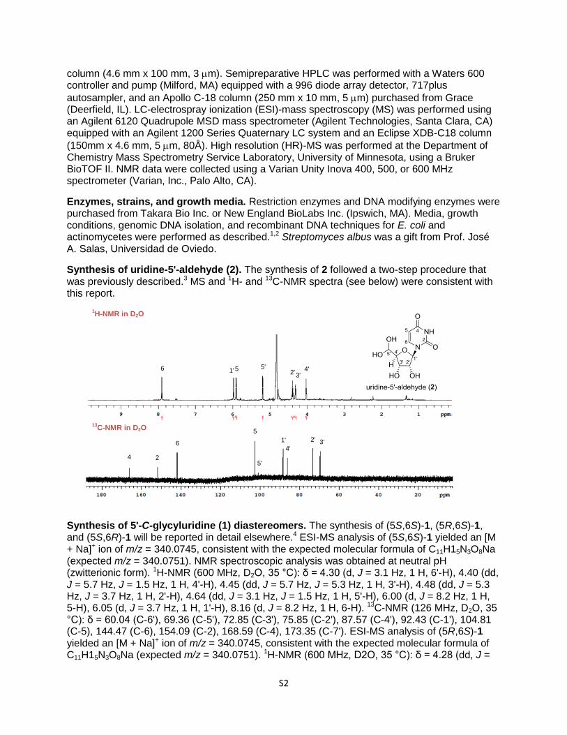
Synthesis of uridine-5'-aldehyde (2). The synthesis of 2 followed a two-step procedure that was previously described.3 MS and 1H- and 13C-NMR spectra (see below) were consistent with this report.
Synthesis of 5'-C-glycyluridine (1) diastereomers. The synthesis of (5S,6S)-1, (5R,6S)-1, and (5S,6R)-1 will be reported in detail elsewhere.4 ESI-MS analysis of (5S,6S)-1 yielded an [M + Na]+ ion of m/z = 340.0745, consistent with the expected molecular formula of C11H15N3O8Na (expected m/z = 340.0751). NMR spectroscopic analysis was obtained at neutral pH (zwitterionic form). 1H-NMR (600 MHz, D2O, 35 °C): δ = 4.30 (d, J = 3.1 Hz, 1 H, 6'-H), 4.40 (dd, J = 5.7 Hz, J = 1.5 Hz, 1 H, 4'-H), 4.45 (dd, J = 5.7 Hz, J = 5.3 Hz, 1 H, 3'-H), 4.48 (dd, J = 5.3 Hz, J = 3.7 Hz, 1 H, 2'-H), 4.64 (dd, J = 3.1 Hz, J = 1.5 Hz, 1 H, 5'-H), 6.00 (d, J = 8.2 Hz, 1 H, 5-H), 6.05 (d, J = 3.7 Hz, 1 H, 1'-H), 8.16 (d, J = 8.2 Hz, 1 H, 6-H). 13C-NMR (126 MHz, D2O, 35 °C): δ = 60.04 (C-6'), 69.36 (C-5'), 72.85 (C-3'), 75.85 (C-2'), 87.57 (C-4'), 92.43 (C-1'), 104.81 (C-5), 144.47 (C-6), 154.09 (C-2), 168.59 (C-4), 173.35 (C-7'). ESI-MS analysis of (5R,6S)-1 yielded an [M + Na]+ ion of m/z = 340.0745, consistent with the expected molecular formula of C11H15N3O8Na (expected m/z = 340.0751). 1H-NMR (600 MHz, D2O, 35 °C): δ = 4.28 (dd, J =
6 5 1'
1H-NMR in D2O
13C-NMR in D2O
5' 2' 3'
4'
4 2 5'
6
5
3' 2'
4'
1'
S3
7.9 Hz, J = 3.9 Hz, 1 H, 4'-H), 4.29 (d, J = 3.3 Hz, 1 H, 6'-H), 4.48 (dd, J = 7.9 Hz, J = 3.3 Hz, 1 H, 5'-H), 4.50 (dd, J = 5.6 Hz, J = 3.9 Hz, 1 H, 3'-H), 4.62 (dd, J = 5.6 Hz, J = 5.5 Hz, 1 H, 2'-H), 5.92 (d, J = 5.5 Hz, 1 H, 1'-H), 6.01 (d, J = 8.1 Hz, 1 H, 5-H), 7.81 (d, J = 8.1 Hz, 1 H, 6-H). 13C-NMR (126 MHz, D2O, 35 °C): δ = 58.96 (C-6'), 72.12 (C-5'), 73.23 (C-3'), 74.94 (C-2'), 85.51 (C-4'), 93.13 (C-1'), 104.94 (C-5), 145.28 (C-6), 154.09 (C-2), 168.65 (C-4), 172.17 (C-7'). ESI-MS analysis of (5S,6R)-1 yielded an [M + Na]+ ion of m/z = 340.0750, consistent with the expected molecular formula of C11H15N3O8Na (expected m/z = 340.0751). 1H-NMR (600 MHz, D2O, 35 °C): δ = 4.28 (dd, J = 5.4 Hz, J = 1.9 Hz, 1 H, 4'-H), 4.37 (d, J = 4.8 Hz, 1 H, 6'-H), 4.46 (dd, J = 5.4 Hz, J = 5.1 Hz, 1 H, 3'-H), 4.47 (dd, J = 5.1 Hz, J = 3.5 Hz, 1 H, 2'-H), 4.58 (dd, J = 4.8 Hz, J = 1.9 Hz, 1 H, 5'-H), 6.01 (d, J = 8.1 Hz, 1 H, 5-H), 6.03 (d, J = 3.5 Hz, 1 H, 1'-H), 8.14 (d, J = 8.1 Hz, 1 H, 6-H). 13C-NMR (126 MHz, D2O, 35 °C): δ = 60.59 (C-6'), 69.13 (C-5'), 72.61 (C-3'), 75.77 (C-2'), 85.99 (C-4'), 92.59 (C-1'), 104.89 (C-5), 144.55 (C-6), 154.14 (C-2), 168.68 (C-4), 172.62 (C-7').
Cloning of genes for heterologous expression. Genes were amplified by PCR using Expand
Long Template PCR System from Roche (Indianapolis, IN) with supplied Buffer 2, 200 M dNTPs, 5% DMSO, 10 ng DNA template, 5 U DNA polymerase, and 200 nM each of the following primer pairs: EcglyA (forward) 5'- GGTATTGAGGGTCGCATGTTAAAGCGTGAAATGAACATTG-3' / (reverse) 5'- AGAGGAGAGTTAGAGCCTTATGCGTAAACCGGGTAACG -3'; EcLTA (forward) 5'- GGTATTGAGGGTCGCATGTCCGTGCTGGGGCGG -3' / (reverse) 5'- AGAGGAGAGTTAGAGCCTTAACGCGCCAGGAATGCAC-3'; and lipK (forward) 5'- GGTATTGAGGGTCGCATGACGGTGGGGGCTGGTG-3' / (reverse) 5'- AGAGGAGAGTTAGAGCCTCACAGCCCGGCCTCCACTT-3'. DNA templates for PCR cloning
were E. coli DH5 genomic DNA for EcglyA and EcLTA and cosmid pN1 for lipK.5 The PCR program included an initial hold at 94 °C for 2 min, followed by 30 cycles of 94 °C for 10 s, 56 °C for 15 s, and 68 °C for 60 s per kb DNA. The gel-purified PCR product was inserted into pET-30 Xa/LIC using ligation-independent cloning as described in the provided protocol to yield pET30-EcglycA, pET30-EcglycA, and pET30-lipK. The genes were sequenced to confirm PCR fidelity.
Site-directed mutagenesis. A K235A point mutation of LipK was generated by PCR amplification using pET30-lipK as a template and the Expand Long Template PCR system (Roche Applied Science). Reactions were performed using the manufacturer's provided Buffer 2 with 5% DMSO, primers of 5'-CTTCGGCGGATCGACTCACGCGTCCTTCCCCGGGCCCC-3' and the reverse complement (the engineered Ala codon is underlined), and a PCR program consisting of an initial hold at 94 °C for 2 min followed by 20 cycles of 94 °C for 10 s, 56 °C for 20 s, and 68 °C for 7 min. The template DNA was digested with 10 units of DpnI (New England Biolabs) for 1 h at 37 °C and transformed into E. coli NovaBlue competent cells. The introduction of the correct point mutation and the sequence of the entire gene including 200 bp upstream and downstream were confirmed by DNA sequencing to yield pET30-lipK(K235A).
Heterologous production of proteins in E. coli. Plasmids pET30-EcglyA and pET30-EcLTA were introduced into E. coli BL21(DE3) cells, and the transformed strains were grown in LB
supplemented with 30 g/mL kanamycin. Following inoculation of 500 mL of LB with 30 g/mL kanamycin, the cultures were grown at 18 °C until the cell density reached an OD600 ~ 0.5 when expression was induced with 0.1 mM IPTG. Cells were harvested after an overnight incubation at 18 °C and lysed in 100 mM Tris-HCl (pH 8) and 300 mM KCl using a French Press with one pass at 16000 psi. Following centrifugation the protein was purified using affinity chromatography with Ni-NTA agarose from Qiagen (Valencia, CA), and the recombinant proteins were desalted into 50 mM phosphate (pH 7.5), 100 mM KCl, and 5 % glycerol using a PD-10 desalting column (GE Healthcare). The purified protein was concentrated using an
S4
Amicon Ultra 10000 MWCO centrifugal filter (Millipore) and stored as glycerol stocks (40%) at -20 °C. Protein purity was assessed as by 12% acrylamide SDS-PAGE; His6-tagged proteins were utilized without further modifications. Protein concentration was determined using Bradford protein assay or UV/Vis spectroscopy using the extension coefficients calculated using the
ProtParam tool available from ExPASY (LipK,280 = 28,880 M-1cm-1; EcGlyA, 280 = 46,300 M-
1cm-1; and EcLTA, 280 = 36,900 M-1cm-1.
Subcloning and expression in Streptomyces lividans TK-64 or Streptomyces albus. Plasmids pET30-lipK and pET30-lipK(K235A) were digested with NdeI-HindIII and the DNA fragment of the expected size was purified and ligated to the identical sites of pUWL201pw to yield pUWL201pw-lipK and pUWL201pw-lipK(K235A), respectively. The resulting plasmids were transformed into S. lividans TK-64 using PEG-mediated protoplast transformation and plated on R2YE. After 14 h at 28 °C, the plates were overlaid with 1 mL of soft nutrient agar supplemented
with 200 g of thiostrepton. After 3 additional days at 28 °C, single colonies were transferred to
fresh R2YE plates supplemented with 10 g/mL thiostrepton. After 4 days at 28 °C, positive transformants were confirmed by colony PCR using InstaGene Matrix from Bio-Rad (Hercules, CA) and LA-Taq polymerase with GC buffer II from Takara Bio Inc. (Shiga, Japan).
The recombinant strain was utilized to inoculate 50 mL R2YE containing 10 g/mL thiostrepton, grown for 2 days at 28 °C at 250 rpm, and 2 mL transferred to fresh 100 mL containing 10
g/mL thiostrepton. Following growth for 3 days at 28 °C at 250 rpm, the cells from 400 mL of culture were collected by centrifugation and washed with 60 mL of 100 mM potassium phosphate (pH 7.4), 300 mM KCl, and 1 mM PMSF and stored on ice for 10 min. Following centrifugation at 4,000 rpm for 15 min at 4 °C, the pellet was thoroughly resuspended in ice-cold 60 mL of 100 mM potassium phosphate (pH 7.4), 300 mM KCl, and 1 mM PMSF and ~25 mg of lysozyme was subsequently added to the suspension. After incubation on ice for 30 minutes, the cell suspension was mixed by pipeting and subjected to one pass through a French press at 18,000 psi. The cell debris was removed by centrifugation (17,500 rpm for 45 min), the cell-free extract filtered using a 0.45 mm PVDF syringe filter, and the clarified extract was loaded onto a gravity column containing 0.5 mL Ni-NTA resin that was pre-equilibrated with 100 mM potassium phosphate (pH 7.4) containing 300 mM KCl. The resin was washed with 10 volumes 100 mM potassium phosphate (pH 7.4) containing 300 mM KCl followed by 10 volumes of 100 mM potassium phosphate (pH 7.4) containing 300 mM KCl and 20 mM imidazole. The His6-tagged protein was eluted with 10 volumes of 100 mM potassium phosphate (pH 7.4) containing 300 mM KCl and 250 mM imidazole and concentrated to < 2.5 mL using an Amicon Ultra 10k MWCO. The sample was desalted into 35 mM potassium phosphate (pH 7.4) containing 100 mM KCl using a gravity flow PD-10 column following the manufacturer’s instructions. The purified protein was re-concentrated and stored as a 40% glycerol stock at -20 °C.
HPLC analysis of the reactions catalyzed by LipK. Routine reactions for LipK consisted of 50
mM HEPES or TES (pH 7.5), 2 mM 2, 5 mM co-substrate, 0.1 mM PLP, and 3.5 M LipK at 30 °C for the indicated time. Following removal of the protein by ultrafiltration or TCA precipitation (7%), the reaction components were analyzed using reverse-phase chromatography using the Agilent 1200 series HPLC equipped with an Eclipse XDB-C18 column. A series of linear gradients was developed from 0.1 % TFA in 0.5 % acetonitrile (A) to 0.1 % TFA in 90% acetonitrile (B) in the following manner (beginning time and ending time with linear increase to % B): 0-6 min, 0% B; 6-26 min, 100% B; 26-30 min, 100% B; 30-34 min, 0% B, and 34-39 min, 0% B. The flow rate was kept constant at 1 mL/min, and elution was monitored at 260 nm. Alternatively, the Dionex Ultimate 3000 equipped with an Acclaim 120 C-18 column was used. A series of linear gradients was developed in the following manner (beginning time and ending time with linear increase to % B): 0-7 min, 25% B; 7-25 min, 75% B; 25-40 min, 95% B; 40-45
S5
min, 0% B, and 45-50 min, 0% B. The flow rate was kept constant at 1 mL/min, and elution was monitored at 260 nm.
Isolation of the LipK product. A large-scale reaction (33 mL) consisted of 50 mM TES (pH 7.5), 100 mM KCl, 0.1 mM PLP, 1 mM DTT, 10 mM L-Thr, 2.5 mM 2, 3 mM NAD, 10 U ADH,
and 5 M LipK. Following incubation for 24 h at 30 °C, protein was removed by ultrafiltration and the product was partially purified by HPLC using a C-18 reverse-phase semi-preparative column. A series of linear gradients was developed from A to B in the following manner (beginning time and ending time with linear increase to % B): 0-4 min, 0% B; 4-24 min, 50% B; 24-26 min, 100% B; 26-32 min, 100% B; and 32-35 min, 0% B. The flow rate was kept constant at 3.5 mL/min, and elution was monitored at 260 nm. Fractions containing the desired product eluting near the column void volume were combined and lyophilized to yield a viscous, clear
solution. The amount of product was estimated using 262 nm = 10,100 M-1.1
Partially purified 1 was resuspended in 5 mL of 200 mM ammonium acetate (pH 8.8) and loaded onto a column containing 1 mL of immobilized boronic acid resin (Thermo-Fisher) that was equilibrated in 10 mL of 200 mM ammonium acetate (pH 8.8). Following a 3 mL wash with the same buffer, the bound 1 was eluted with 12 mL of 0.1 M formic acid. The purified sample was lyophilized prior to spectroscopic analysis.
Phosgene modification of GlyU. Phosgene modification of -hydroxy--amino acids was carried out as previously described.6 Approximately 20 mg of HPLC-purified 2 was dissolved in 1 M potassium hydroxide (6.5 mL), and the solution cooled to 5 °C prior to the addition of 6 mL of a 20% solution of phosgene in toluene. Following 5 min with rigorous stirring, the mixture was acidified with 10 drops of concentrated HCl, and the aqueous phase was removed and subjected to HPLC for purification using a C-18 reverse-phase semi-preparative column following the above conditions. The purified product was lyophilized and subjected to spectroscopic analysis (Figures S11-S14). 1H-NMR (D2O, 500 MHz): δ 7.56 (d, J = 8.5 Hz, 1H), 5.95 (d, J = 5.0 Hz, 1H), 5.90 (d, J = 8.0 Hz, 1H), 5.08 (dd, J = 5.0 Hz, J = 2.5 Hz, 1H), 4.55 (d, J = 5.5 Hz, 1H), 4.45 (t, J = 5.25 Hz, 1H), 4.41 (t, J = 5.0 Hz, 1H), 4.30 (dd, J = 5.25 Hz, J = 2.5 Hz, 1H). 13C-NMR (D2O, 100 MHz): δ 173.54, 165.85, 159.98, 151.51, 141.03, 102.60, 89.09, 83.33, 77.59, 72.71, 69.24, 62.38, 55.83.
For analytical analysis of synthetic 1 diastereomers, a 2 mM solution (50 L) of pure
diastereomer was prepared and chilled on ice for 5 min. After addition of 10 L of 5 M KOH, the
reaction was initiated by adding 50 L of 20% phosgene in toluene. After a 1 h incubation on ice with periodic mixing, the organic phase was removed and the aqueous phase analyzed by HPLC using the Agilent 1200 series equipped with an Eclipse XDB-C18 column and gradient
conditions described above for isolation of the LipK product.
Modification of aldehydes with DPNH. LipK reaction components were modified with DPNH as previously described.7 Following protein removal by centrifugation, the modified components were analyzed using reverse-phase chromatography using the Agilent 1200 series HPLC Agilent 1200 Series equipped with an Eclipse XDB-C18 column. A series of linear gradients was developed from A to B in the following manner (beginning time and ending time with linear increase to % B): 0-8 min, 0% B; 8-28 min, 100% B; 28-32 min, 100% B; and 32-35 min, 0% B. The flow rate was kept constant at 1 mL/min, and elution was monitored at 430 nm.
Kinetic analysis of LipK. Single-substrate kinetic analysis was carried out by monitoring the
reduction of -NAD+ using 340 = 6220 M-1cm-1. Reaction mixtures (90 L) consisting of 100 mM
TES (pH 7.5), 100 mM KCl, 1 mM DTT, 30 mM L-Thr, 0.1 mM PLP, 2 mM -NAD+, and variable
S6
2 (1.0 - 125 mM) were incubated at 30 oC for 1 min prior to initiation with a mixture (10 L)
consisting of 50 mM TES (pH 7.5), 100 mM KCl, 1 mM DTT, 2 mM -NAD+, 1 U ADH, and 250 nM LipK. Reactions were carried out for 10 min, and rates were calculated using linear regression analysis with data obtained under initial velocity conditions (< 10% product). Data were fitted to the Michaelis-Menten equation using GraphPad Prism 5.0.4 (GraphPad Software, Inc., La Jolla, CA). Each data point represents the average of a minimum of three independent measurements.
Bi-substrate kinetic analysis for LipK was performed using reactions as described above with
variable 2 (1.0 - 60 mM) and L-Thr (62 M – 15.6 mM). For mechanistic determination (ping pong versus sequential mechanism) the inverse data (1/[S] versus 1/v) were analyzed by simple linear regression analysis (Figure 5F). If data are fitted to an equation (eq 1)8 describing a random sequential mechanism as revealed for serine hydroxymethyltransferases, the global fit
yields a kinetic constant of Km = 31 M with respect to L-Thr and = 0.88 (is the factor by which the binding of one substrate alters the dissociation constant for the remaining substrate;
an value of unity indicates substrate binding has no effect on the binding of the respective substrate).
eq 1 1
𝑣=
∝ 𝐾𝐵𝑉𝑚𝑎𝑥
1 +𝐾𝐴[𝐴]
1
[𝐵]+
1
𝑉𝑚𝑎𝑥 1 +
∝ 𝐾𝐴[𝐴]
S7
SUPPORTING FIGURES
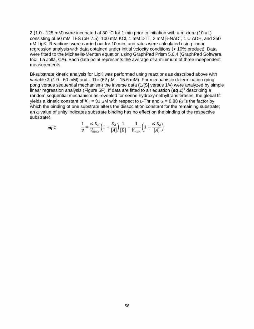
Figure S1. Enzymatic transformations of -hydroxy--amino acids (A) The canonical reaction catalyzed by SHMT results in the reversible conversion of Gly and N5,N10-methylene-tetrahydrofolate to L-Ser and tetrahydrofolate (THF) during amino acid metabolism. (B) The aldol-type reaction catalyzed by SHMT that is independent of THF. (C) The canonical reaction catalyzed by LTA resulting in the interconversion of Gly and L-Thr during amino acid metabolism.
C
B
A
S8
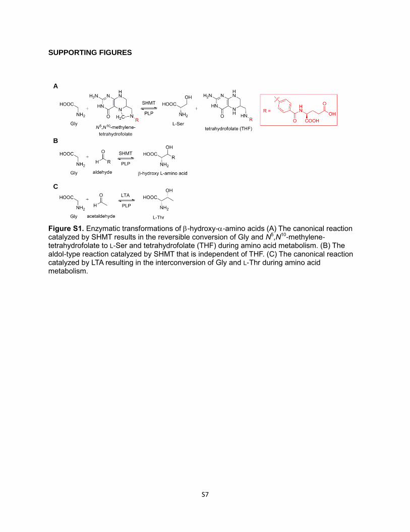
Figure S2. Sequence alignment of authentic and putative SHMTs. Sequences include the
putative SHMT LipK from the A-90289 gene cluster (BAJ05887); authentic SHMTs with solved
structures from Escherichia coli (EcGlyA, 1EQB_B) and Bacillus stearothermophilus (BsGlyA,
2VMV_A); and probable SHMTs identified from whole-genome sequencing from Streptomyces
coelicolor (ScGlyA, NP_628992) and Saccharopolyspora erythraea (SeGlyA, YP_001107812).
▼, conserved Lys required for formation of the internal aldimine with pyridoxal-5'-phosphate.
1 8010 20 30 40 50 60 70(1)
--------MTVGAGGKTSADADPLMLVRAIADADRRAAHALNLVPSENRISPLASLPLASDFYNRYFFNTDGDPLFWEFRLipK (1)
-----------MLKREMNIADYDAELWQAMEQEKVRQEEHIELIASENYTSPRVMQAQGSQLTNKYAEGYPGK----RYYEcGlyA (1)
---------------MKYLPQQDPQVFAAIEQERKRQHAKIELIASENFVSRAVMEAQGSVLTNKYAEGYPGR----RYYBsGlyA (1)
MPAEISPESTAYRAALDVIRAVEPRVADAIGQEVADQREMLKLIASENYASPATLLAMGNWFSDKYAEGTIGR----RFYScGlyA (1)
--------MSTPGQFNSELSAVDPEVAAAVGAELNRQQTTLEMIASENFAPQAVLQAQGSVLTNKYAEGYPGK----RYYSeGlyA (1)
81 16090 100 110 120 130 140 150(81)
GGEDIAHIEALGAA-ALRRMASARYCNVRPISGMSAMILTVAALS-P----------------P--------------GSLipK (73)
GGCEYVDIVEQLAIDRAKELFGADYANVQPHSGSQANFAVYTALLEP-------------------------------GDEcGlyA (66)
GGCEYVDIVEELARERAKQLFGAEHANVQPHSGAQANMAVYFTVLEH-------------------------------GDBsGlyA (62)
AGCRNVDTVESLAAEHARELFGARHAYVQPHSGIDANLVAFWAVLGARVEVPFLEKTGARQVNDLTDADWAELRQAFGNQScGlyA (77)
GGCEHVDVVEQLAIDRVKELFGASFANVQPHSGAQANAAAMFALLKP-------------------------------GDSeGlyA (69)
161 240170 180 190 200 210 220 230(161)
TVVSVDQNSGGHYATPALLGRLGRRSRLLNCKDG----EVDESELAEVLAPGDVALVYVDVQNCVRVPDFRRMSDVIREVLipK(121)
TVLGMNLAHGGHLTHGSPVNFSGKLYNIVPYGID-ATGHIDYADLEKQAKEHKPKMIIGGFSAYSGVVDWAKMREIADSIEcGlyA(115)
TVLGMNLSHGGHLTHGSPVNFSGVQYNFVAYGVDPETHVIDYDDVREKARLHRPKLIVAAASAYPRIIDFAKFREIADEVBsGlyA(111)
RMLGMSLDAGGHLTHGFRPNISGKMFDQRSYGTDPATGLIDYEALRASAREFKPLIIVAGYSAYPRLVNFRIMREIADEVScGlyA(157)
TIMGLDLAHGGHLTHGMRINFSGKLYNVVPYHVGDDDHRVDMDEVARLAREHRPRLIIAGWSAYPRQLDFARFREIADEVSeGlyA(118)
241 320250 260 270 280 290 300 310(241)
SPGTRLYVDASHYLGLVLGGLLAN--PLDCGADAFGGSTHKSFPGPHKGVIFTNA--EDVDESLRSAQFDLVSSHHFAETLipK(197)
G--AYLFVDMAHVAGLVAAGVYP---NPVPHAHVVTTTTHKTLAGPRGGLILAKGGSEELYKKLNSAVFPGGQGGPLMHVEcGlyA(194)
G--AYLMVDMAHIAGLVAAGLHP---NPVPYAHFVTTTTHKTLRGPRGGMILCQ---EQFAKQIDKAIFPGIQGGPLMHVBsGlyA(191)
G--ATLMVDMAHFAGLVAGKVLTGDFDPVPHAQIVTTTTHKSLRGPRGGMVLCD----DSLKDQVDRGCPMVLGGPLPHVScGlyA(237)
G--AYLMVDMAHFAGLVAAGLHP---NPVPHAHVVTTTTHKTLGGPRGGVILSAD--EEFTKKFNSAVFPGQQGGPLEHVSeGlyA(198)
321 400330 340 350 360 370 380 390(321)
LALSLAAL--EVEDRMGDYARATNDNARRLAGALADAG-FRVYGDSATGYTDTHQVWVELDGVA--AAYALSNRLAEGGILipK(273)
IAGKAVALKEAMEPEFKTYQQQVAKNAKAMVEVFLER-----GYKVVSGGTDNHLFLVDLVDKN-LTGKEADAALGRANIEcGlyA(269)
IAAKAVAFGEALQDDFKAYAKRVVDNAKRLASALQNE-----GFTLVSGGTDNHLLLVDLRPQQ-LTGKTAEKVLDEVGIBsGlyA(263)
MAAKAVALAEARRPAFQDYAQRIVDNARALAEGLTKR-----GATLVTGGTDNHLNLIDVASSYGLTGRQAEAALLDSGIScGlyA(311)
IAGKAVLFKLAAGEEFRDRQRRTLEGAKILAERLLADDAARAGVRLVSGGTDVHLVLVDLREAE-LDGKQAEDRLHEIGISeGlyA(271)
401 480410 420 430 440 450 460 470(401)
RVNLQSSMPGMSG----VHLRLGSNEVTFEGAGPQAIEELAGALVTARERALGP----------------RTVHEIRGRFLipK(348)
TVNKNSVPNDPKSPFVTSGIRVGTPAITRRGFKEAEAKELAGWMCDVLDSIN----------------DEAVIERIKGKVEcGlyA(343)
TVNKNTIPYDPESPGVTSGIRIGTAAVTTRGFGLEEMDEIAAIIGLVLKNVG----------------SEQALEEARQRVBsGlyA(337)
VTNRNAIPADPNGAWYTSGIRIGTPALTTRGLGTAEMDEVAGLIDRVLTATEPGTTKSGAPSKASHVLDAKVADEISHRAScGlyA(386)
TVNRNAVPNDPRPPMVTSGLRIGTPALATRGFGKTEFTEVADIIAEALKPDF----------------DEATSAKLRSRVSeGlyA(350)
481 497(481)
GAPFYTDPEKLKVEAGLLipK(408)
LDICARYPVYA------EcGlyA(407)
AALTD------------BsGlyA(401)
TDLVAGFPLYPEIDLG-ScGlyA(466)
EALAAGFPLYPNL----SeGlyA(414)
▼
S9
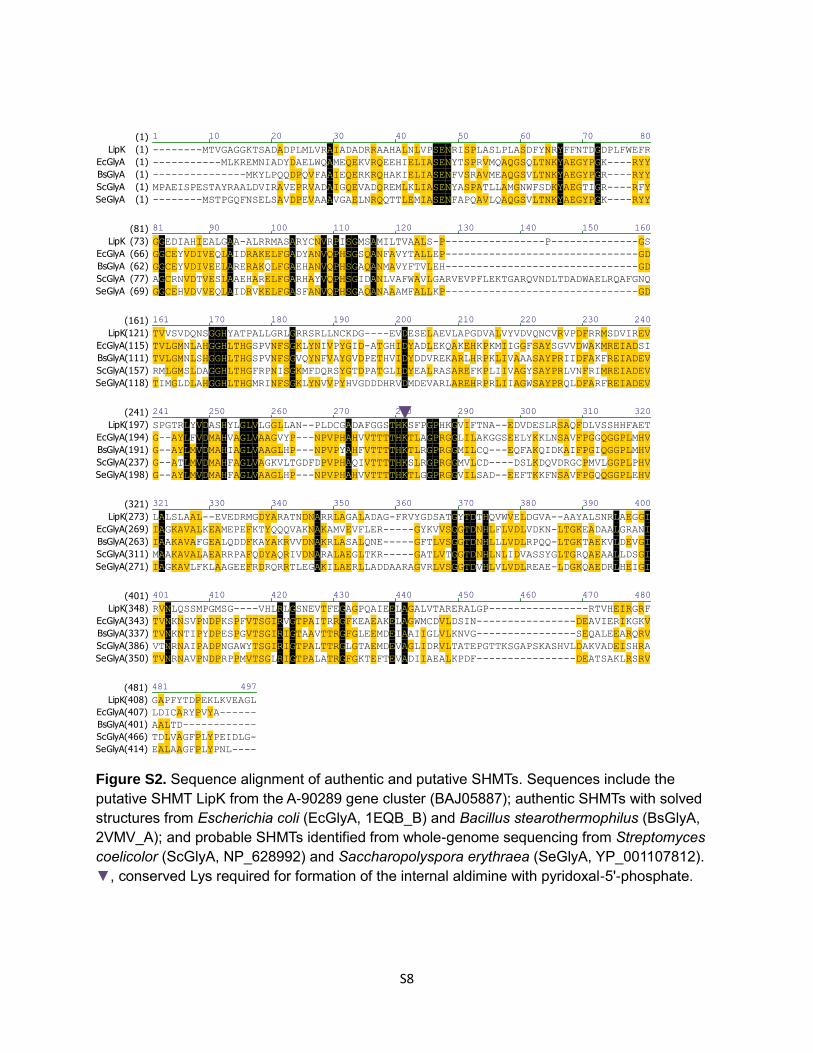
Figure S3. Production of recombinant enzymes. (A) SDS-PAGE analysis of purified His6-LipK from Streptomyces lividans TK64 (lane 2, expected MW of 50.1 kD). (B) SDS-PAGE analysis of partially purified His6-LipK(K235A) (lane 2, expected MW of 50.1 kD). (C) SDS-PAGE analysis of partially purified His6-LipK from Streptomyces albus (lane 2, expected MW of 50.1 kD). (D) SDS-PAGE analysis of purified His6-EcGlyA (lane 2, expected MW of 50.3 kD). (E) SDS-PAGE analysis of purified His6-EcLTA (lane 2, expected MW of 41.6 kD). For all gels, lane 1 consists of molecular weight standards from BioRad. The engineered N-terminal His6-tag contributes 5 kD to the predicted native molecular weight for each protein.
A B D C E
S10
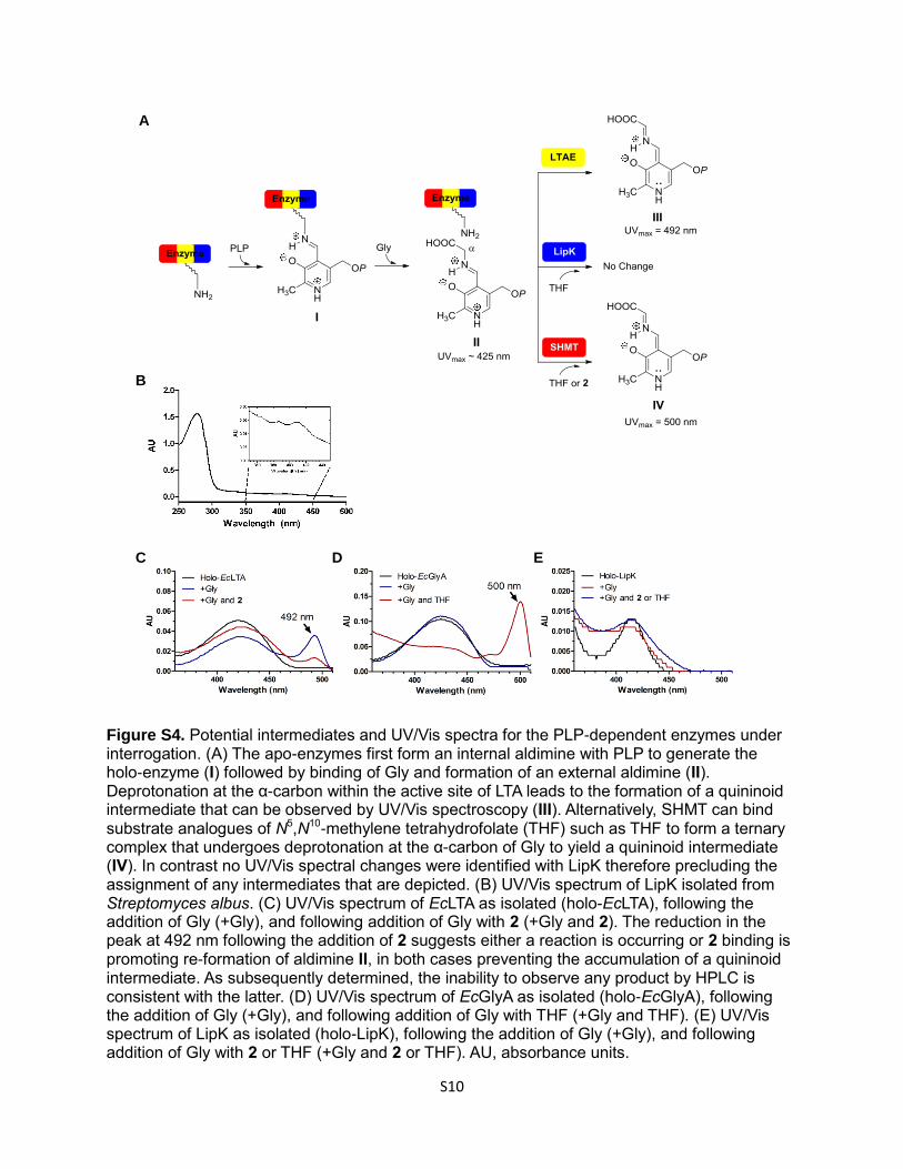
Figure S4. Potential intermediates and UV/Vis spectra for the PLP-dependent enzymes under interrogation. (A) The apo-enzymes first form an internal aldimine with PLP to generate the holo-enzyme (I) followed by binding of Gly and formation of an external aldimine (II). Deprotonation at the α-carbon within the active site of LTA leads to the formation of a quininoid intermediate that can be observed by UV/Vis spectroscopy (III). Alternatively, SHMT can bind substrate analogues of N5,N10-methylene tetrahydrofolate (THF) such as THF to form a ternary complex that undergoes deprotonation at the α-carbon of Gly to yield a quininoid intermediate (IV). In contrast no UV/Vis spectral changes were identified with LipK therefore precluding the assignment of any intermediates that are depicted. (B) UV/Vis spectrum of LipK isolated from Streptomyces albus. (C) UV/Vis spectrum of EcLTA as isolated (holo-EcLTA), following the addition of Gly (+Gly), and following addition of Gly with 2 (+Gly and 2). The reduction in the peak at 492 nm following the addition of 2 suggests either a reaction is occurring or 2 binding is promoting re-formation of aldimine II, in both cases preventing the accumulation of a quininoid intermediate. As subsequently determined, the inability to observe any product by HPLC is consistent with the latter. (D) UV/Vis spectrum of EcGlyA as isolated (holo-EcGlyA), following the addition of Gly (+Gly), and following addition of Gly with THF (+Gly and THF). (E) UV/Vis spectrum of LipK as isolated (holo-LipK), following the addition of Gly (+Gly), and following addition of Gly with 2 or THF (+Gly and 2 or THF). AU, absorbance units.
A
C E D
B
S11
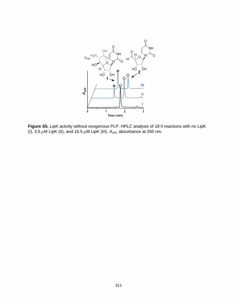
Figure S5. LipK activity without exogenous PLP. HPLC analysis of 18 h reactions with no LipK
(I), 3.5 M LipK (II), and 16.5 M LipK (III). A260, absorbance at 260 nm.
S12
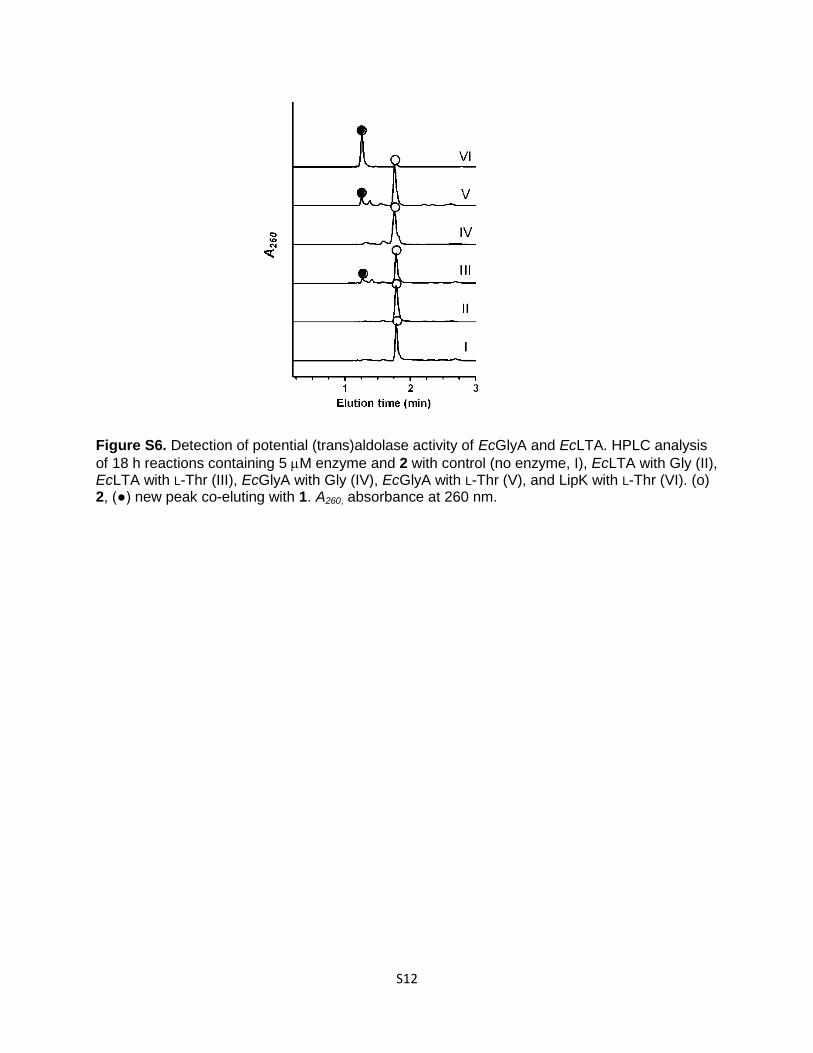
Figure S6. Detection of potential (trans)aldolase activity of EcGlyA and EcLTA. HPLC analysis
of 18 h reactions containing 5 M enzyme and 2 with control (no enzyme, I), EcLTA with Gly (II), EcLTA with L-Thr (III), EcGlyA with Gly (IV), EcGlyA with L-Thr (V), and LipK with L-Thr (VI). (o) 2, (●) new peak co-eluting with 1. A260, absorbance at 260 nm.
S13
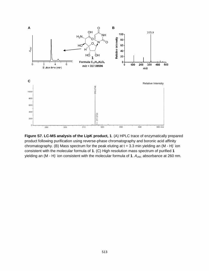
Figure S7. LC-MS analysis of the LipK product, 1. (A) HPLC trace of enzymatically prepared
product following purification using reverse-phase chromatography and boronic acid affinity
chromatography. (B) Mass spectrum for the peak eluting at t = 3.3 min yielding an (M - H)- ion
consistent with the molecular formula of 1. (C) High resolution mass spectrum of purified 1
yielding an (M - H)- ion consistent with the molecular formula of 1. A260, absorbance at 260 nm.
A B
Formula C11H15N3O8
m/z = 317.08596
C Relative Intensity
1
S14
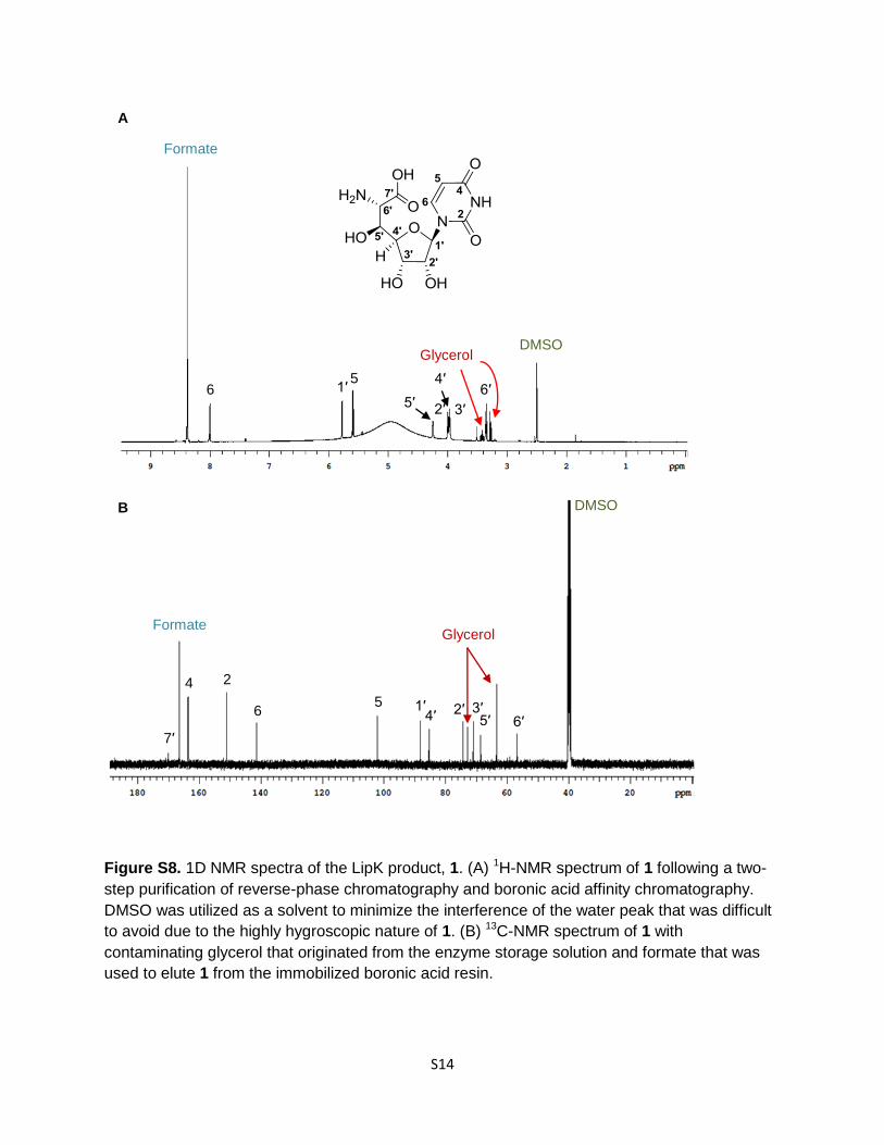
Figure S8. 1D NMR spectra of the LipK product, 1. (A) 1H-NMR spectrum of 1 following a two-
step purification of reverse-phase chromatography and boronic acid affinity chromatography.
DMSO was utilized as a solvent to minimize the interference of the water peak that was difficult
to avoid due to the highly hygroscopic nature of 1. (B) 13C-NMR spectrum of 1 with
contaminating glycerol that originated from the enzyme storage solution and formate that was
used to elute 1 from the immobilized boronic acid resin.
Glycerol
Glycerol
Formate
DMSO
7′
4 2
6 5 1′
4′ 2′ 5′
3′ 6′
DMSO
5 4′ 6′
3′ 5′
6 1′
2′
A
B
Formate
S15
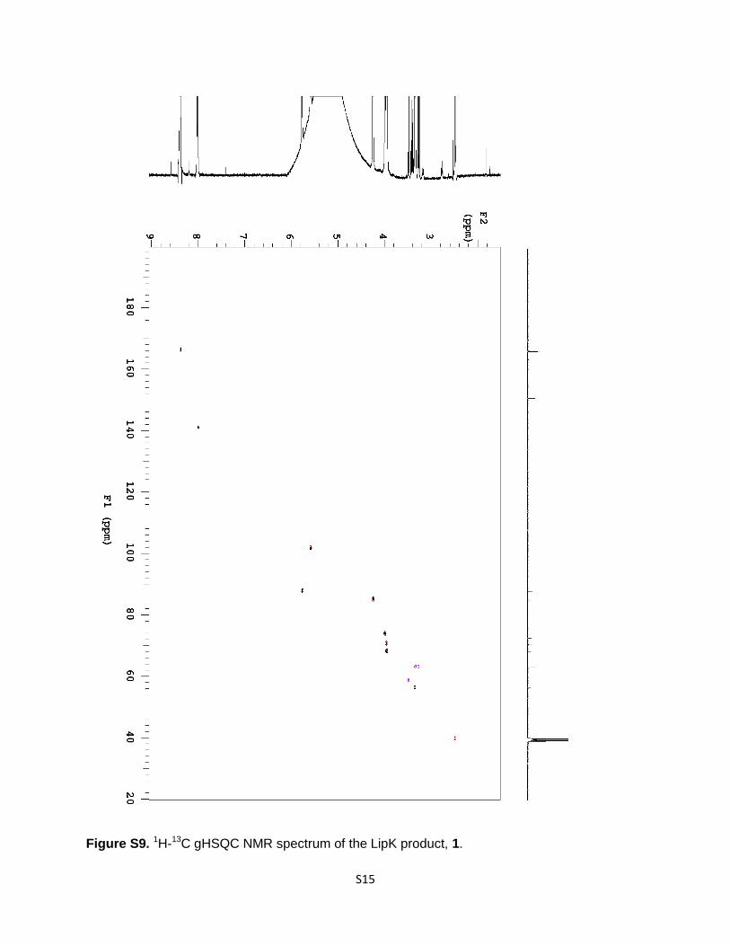
Figure S9. 1H-13C gHSQC NMR spectrum of the LipK product, 1.
S16
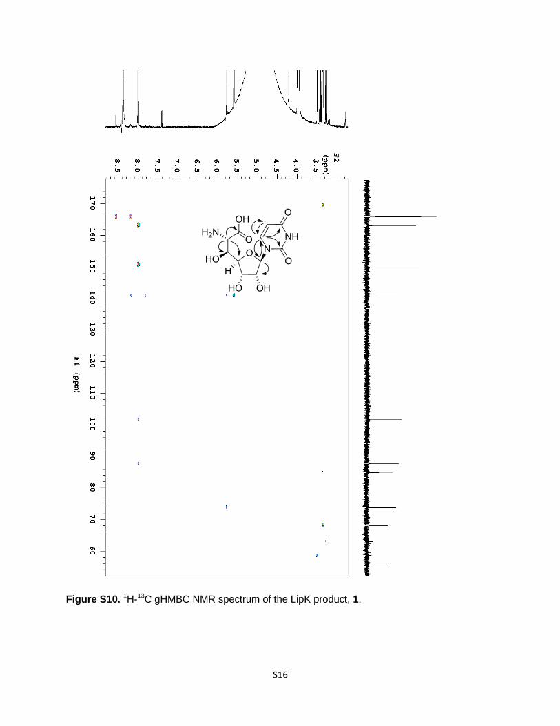
Figure S10. 1H-13C gHMBC NMR spectrum of the LipK product, 1.
S17
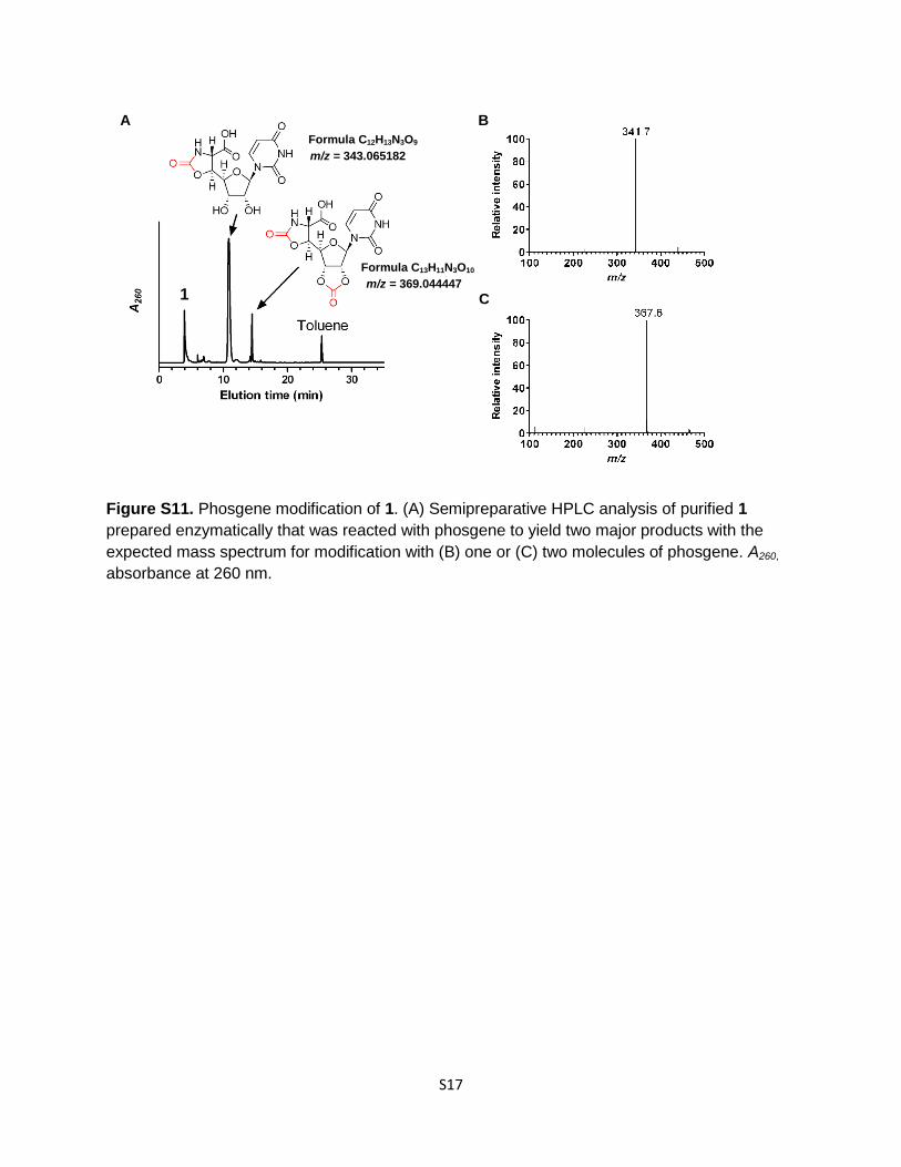
Figure S11. Phosgene modification of 1. (A) Semipreparative HPLC analysis of purified 1
prepared enzymatically that was reacted with phosgene to yield two major products with the
expected mass spectrum for modification with (B) one or (C) two molecules of phosgene. A260,
absorbance at 260 nm.
A B
Formula C12H13N3O9
m/z = 343.065182
Formula C13H11N3O10
m/z = 369.044447
C 1
S18
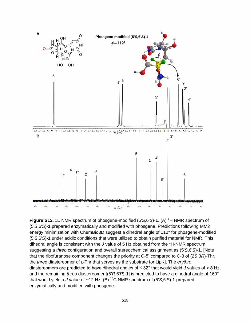
Figure S12. 1D NMR spectrum of phosgene-modified (5'S,6'S)-1. (A) 1H NMR spectrum of
(5'S,6'S)-1 prepared enzymatically and modified with phosgene. Predictions following MM2
energy minimization with ChemBio3D suggest a dihedral angle of 112° for phosgene-modified
(5'S,6'S)-1 under acidic conditions that were utilized to obtain purified material for NMR. This
dihedral angle is consistent with the J value of 5 Hz obtained from the 1H-NMR spectrum,
suggesting a threo configuration and overall stereochemical assignment as (5'S,6'S)-1. [Note
that the ribofuranose component changes the priority at C-5’ compared to C-3 of (2S,3R)-Thr,
the threo diastereomer of L-Thr that serves as the substrate for LipK]. The erythro
diastereomers are predicted to have dihedral angles of ≤ 32° that would yield J values of > 8 Hz,
and the remaining threo diastereomer [(5'R,6'R)-1] is predicted to have a dihedral angle of 160°
that would yield a J value of ~12 Hz. (B) 13C NMR spectrum of (5'S,6'S)-1 prepared
enzymatically and modified with phosgene.
B
A
°
4'
Phosgene-modified 5'S,6'S)-1
5 1'
6' 6
5'
3'
2'
7'
4 1''
2 6
5
1' 4'
5'
2'
3'
6'
S19
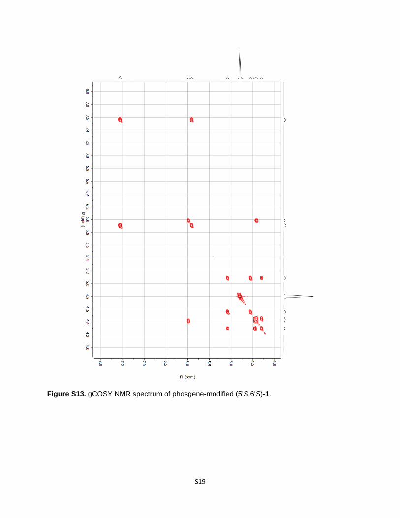
Figure S13. gCOSY NMR spectrum of phosgene-modified (5'S,6'S)-1.
S20
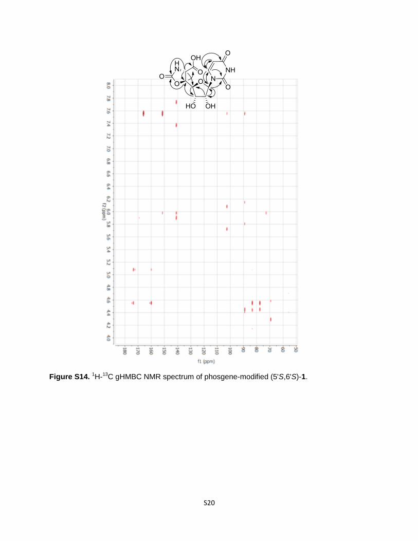
Figure S14. 1H-13C gHMBC NMR spectrum of phosgene-modified (5'S,6'S)-1.
S21
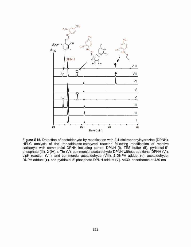
Figure S15. Detection of acetaldehyde by modification with 2,4-dinitrophenylhydrazine (DPNH). HPLC analysis of the transaldolase-catalyzed reaction following modification of reactive carbonyls with commercial DPNH including control DPNH (I), TES buffer (II), pyridoxal-5'-phosphate (III), 2 (IV), L-Thr (V), commercial acetaldehyde-DPNH without additional DPNH (VI), LipK reaction (VII), and commercial acetaldehyde (VIII). 2-DNPH adduct (○), acetaldehyde-
DNPH adduct (●), and pyridoxal-5'-phosphate-DPNH adduct (). A430, absorbance at 430 nm.
S22
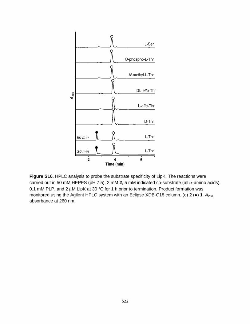
Figure S16. HPLC analysis to probe the substrate specificity of LipK. The reactions were
carried out in 50 mM HEPES (pH 7.5), 2 mM 2, 5 mM indicated co-substrate (all -amino acids),
0.1 mM PLP, and 2 M LipK at 30 °C for 1 h prior to termination. Product formation was
monitored using the Agilent HPLC system with an Eclipse XDB-C18 column. (o) 2 (●) 1. A260,
absorbance at 260 nm.
S23
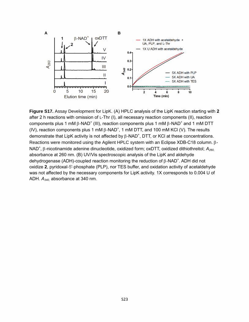
Figure S17. Assay Development for LipK. (A) HPLC analysis of the LipK reaction starting with 2
after 2 h reactions with omission of L-Thr (I), all necessary reaction components (II), reaction
components plus 1 mM -NAD+ (III), reaction components plus 1 mM -NAD+ and 1 mM DTT
(IV), reaction components plus 1 mM -NAD+, 1 mM DTT, and 100 mM KCl (V). The results
demonstrate that LipK activity is not affected by -NAD+, DTT, or KCl at these concentrations.
Reactions were monitored using the Agilent HPLC system with an Eclipse XDB-C18 column. -
NAD+, -nicotinamide adenine dinucleotide, oxidized form; oxDTT, oxidized dithiothreitol; A260,
absorbance at 260 nm. (B) UV/Vis spectroscopic analysis of the LipK and aldehyde
dehydrogenase (ADH)-coupled reaction monitoring the reduction of -NAD+. ADH did not
oxidize 2, pyridoxal-5'-phosphate (PLP), nor TES buffer, and oxidation activity of acetaldehyde
was not affected by the necessary components for LipK activity. 1X corresponds to 0.004 U of
ADH. A340, absorbance at 340 nm.
B A
S24
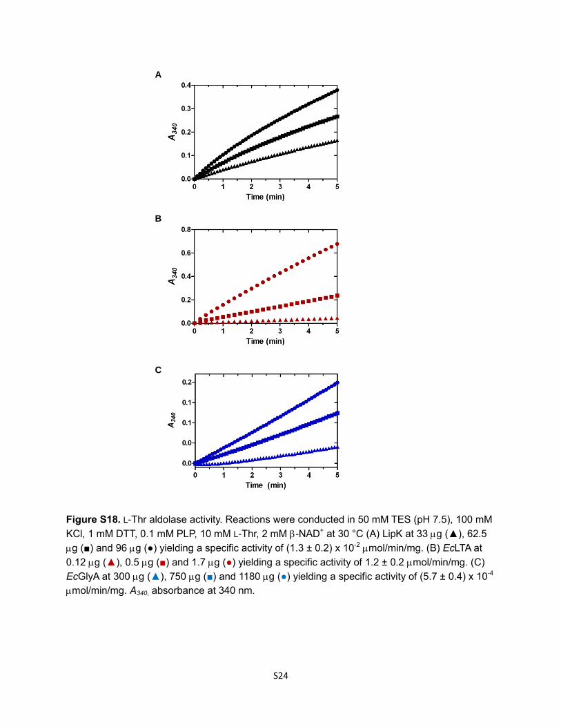
Figure S18. L-Thr aldolase activity. Reactions were conducted in 50 mM TES (pH 7.5), 100 mM
KCl, 1 mM DTT, 0.1 mM PLP, 10 mM L-Thr, 2 mM -NAD+ at 30 °C (A) LipK at 33 g (▲), 62.5
g (■) and 96 g (●) yielding a specific activity of (1.3 ± 0.2) x 10-2 mol/min/mg. (B) EcLTA at
0.12 g (▲), 0.5 g (■) and 1.7 g (●) yielding a specific activity of 1.2 ± 0.2 mol/min/mg. (C)
EcGlyA at 300 g (▲), 750 g (■) and 1180 g (●) yielding a specific activity of (5.7 ± 0.4) x 10-4
mol/min/mg. A340, absorbance at 340 nm.
B
A
C
S25
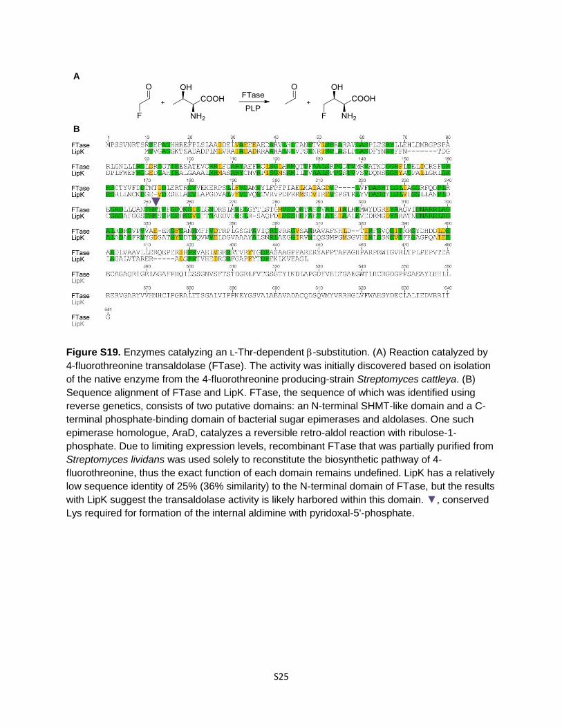
Figure S19. Enzymes catalyzing an L-Thr-dependent -substitution. (A) Reaction catalyzed by
4-fluorothreonine transaldolase (FTase). The activity was initially discovered based on isolation
of the native enzyme from the 4-fluorothreonine producing-strain Streptomyces cattleya. (B)
Sequence alignment of FTase and LipK. FTase, the sequence of which was identified using
reverse genetics, consists of two putative domains: an N-terminal SHMT-like domain and a C-
terminal phosphate-binding domain of bacterial sugar epimerases and aldolases. One such
epimerase homologue, AraD, catalyzes a reversible retro-aldol reaction with ribulose-1-
phosphate. Due to limiting expression levels, recombinant FTase that was partially purified from
Streptomyces lividans was used solely to reconstitute the biosynthetic pathway of 4-
fluorothreonine, thus the exact function of each domain remains undefined. LipK has a relatively
low sequence identity of 25% (36% similarity) to the N-terminal domain of FTase, but the results
with LipK suggest the transaldolase activity is likely harbored within this domain. ▼, conserved
Lys required for formation of the internal aldimine with pyridoxal-5'-phosphate.
A
▼
B
S26
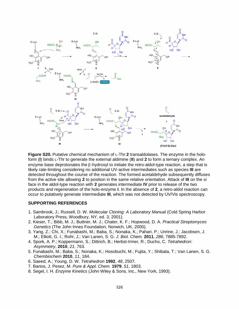
Figure S20. Putative chemical mechanism of L-Thr:2 transaldolases. The enzyme in the holo-form (I) binds L-Thr to generate the external aldimine (II) and 2 to form a ternary complex. An
enzyme base deprotonates the -hydroxyl to initiate the retro-aldol-type reaction, a step that is likely rate-limiting considering no additional UV-active intermediates such as species III are detected throughout the course of the reaction. The formed acetaldehyde subsequently diffuses from the active site allowing 2 to position in the same relative orientation. Attack of III on the si face in the aldol-type reaction with 2 generates intermediate IV prior to release of the two products and regeneration of the holo-enzyme I. In the absence of 2, a retro-aldol reaction can occur to putatively generate intermediate III, which was not detected by UV/Vis spectroscopy.
SUPPORTING REFERENCES
1. Sambrook, J.; Russell, D. W. Molecular Cloning: A Laboratory Manual (Cold Spring Harbor Laboratory Press, Woodbury, NY, ed. 3, 2001).
2. Kieser, T.; Bibb, M. J.; Buttner, M. J.; Chater, K. F.; Hopwood, D. A. Practical Streptomyces Genetics (The John Innes Foundation, Norwich, UK, 2000).
3. Yang, Z.; Chi, X.; Funabashi, M.; Baba, S.; Nonaka, K.; Pahari, P.; Unrine, J.; Jacobsen, J. M.; Elliott, G. I.; Rohr, J.; Van Lanen, S. G. J. Biol. Chem. 2011, 286, 7885-7892.
4. Spork, A. P.; Koppermann, S.; Dittrich, B.; Herbst-Irmer, R.; Ducho, C. Tetrahedron: Asymmetry, 2010, 21, 763.
5. Funabashi, M.; Baba, S.; Nonaka, K.; Hosobuchi, M.; Fujita, Y.; Shibata, T.; Van Lanen, S. G. Chembiochem 2010, 11, 184.
6. Saeed, A.; Young, D. W. Tetrahedron 1992, 48, 2507. 7. Bartos, J. Pesez, M. Pure & Appl. Chem. 1979, 51, 1803. 8. Segel, I. H. Enzyme Kinetics (John Wiley & Sons, Inc., New York, 1993).
S27
Complete Citation of Reference 11 in the Main Text (11) Yang, Z.; Chi, X.; Funabashi, M.; Baba, S.; Nonaka, K.; Pahari, P.; Unrine, J.; Jacobsen, J.
M.; Elliott, G. I.; Rohr, J.; Van Lanen, S. G. J. Biol. Chem. 2011, 286, 7885-7892.


![Lander techniques for deep-ocean biological researchdiscovery.ucl.ac.uk/1470451/1/Bagley et al _Lander techniques_art00003.pdf · e]jkaZd]k( LIPk( Yf\ gl`]j oaj] _] Yjk( l`[q [Yf](https://img.pdfslide.us/doc/110x75/5d2a15df88c993c66c8b933c/lander-techniques-for-deep-ocean-biological-et-al-lander-techniquesart00003pdf.jpg)
![PRUDNIKOVA, Regina Lithuania Documentation Project Lithuanian … · Lipkė in [19]40—and that Lipkė – Liurija was his last name, but people called him Lipk ė—had a daughter,](https://img.pdfslide.us/doc/110x75/5d2a15df88c993c66c8b9317/prudnikova-regina-lithuania-documentation-project-lithuanian-lipke-in-1940and.jpg)


![Work report 王军亮 20150530. I. The modification of pCT5e MH-G3Long MH-G3Short SOE PCR: MH-G3Long[NotI-BamHI], MH-G3Short[NotI-NdeI] Double digest: Conclusion:](https://img.pdfslide.us/doc/110x75/5697c02b1a28abf838cd8602/work-report-20150530-i-the-modification-of-pct5e-mh-g3long-mh-g3short.jpg)

![with PExonucleaseIIIwasusedto kilobase (kb) HindIII-Sal I fragment ofpir25.1; see ref. 21] generate progressively deleted sequencing templates (18). fromD.melanogasteras ahybridization](https://img.pdfslide.us/doc/110x75/60776df7b4ecf364957519c7/with-p-exonucleaseiiiwasusedto-kilobase-kb-hindiii-sal-i-fragment-ofpir251-see.jpg)