Embed Size (px)
Citation preview

RESEARCH ARTICLE
Storage Temperature Alters the Expression ofDifferentiation-Related Genes in CulturedOral KeratinocytesTor Paaske Utheim1,2,3,4, Rakibul Islam1,2, Ida G. Fostad2, Jon R. Eidet1,5, Amer Sehic2, OleK. Olstad1, Darlene A. Dartt6, Edward B. Messelt2, May Griffith7, Lara Pasovic1,8*
1 Department of Medical Biochemistry, Oslo University Hospital, Oslo, Norway, 2 Department of OralBiology, Faculty of Dentistry, University of Oslo, Oslo, Norway, 3 Department of Ophthalmology, VestreViken HF Trust, Drammen, Norway, 4 Faculty of Health Sciences, National Centre for Optics, Vision and EyeCare, Buskerud and Vestfold University College, Kongsberg, Norway, 5 Department of Ophthalmology, OsloUniversity Hospital, Oslo, Norway, 6 Schepens Eye Research Institute, Massachusetts Eye and Ear,Harvard Medical School, Boston, Massachusetts, United States of America, 7 Department of Clinical andExperimental Medicine, Linköping University, Linköping, Sweden, 8 Faculty of Medicine, University of Oslo,Oslo, Norway
Abstract
Purpose
Storage of cultured human oral keratinocytes (HOK) allows for transportation of cultured
transplants to eye clinics worldwide. In a previous study, one-week storage of cultured HOK
was found to be superior with regard to viability and morphology at 12°C compared to 4°C
and 37°C. To understand more of how storage temperature affects cell phenotype, gene
expression of HOK before and after storage at 4°C, 12°C, and 37°C was assessed.
Materials and Methods
Cultured HOK were stored in HEPES- and sodium bicarbonate-buffered Minimum Essential
Medium at 4°C, 12°C, and 37°C for one week. Total RNAwas isolated and the gene expres-
sion profile was determined using DNAmicroarrays and analyzed with Partek Genomics
Suite software and Ingenuity Pathway Analysis. Differentially expressed genes (fold change >
1.5 and P < 0.05) were identified by one-way ANOVA. Key genes were validated using qPCR.
Results
Gene expression of cultures stored at 4°C and 12°C clustered close to the unstored control
cultures. Cultures stored at 37°C displayed substantial change in gene expression com-
pared to the other groups. In comparison with 12°C, 2,981 genes were differentially
expressed at 37°C. In contrast, only 67 genes were differentially expressed between the
unstored control and the cells stored at 12°C. The 12°C and 37°C culture groups differed
most significantly with regard to the expression of differentiation markers. The Hedgehog
signaling pathway was significantly downregulated at 37°C compared to 12°C.
PLOS ONE | DOI:10.1371/journal.pone.0152526 March 29, 2016 1 / 24
OPEN ACCESS
Citation: Utheim TP, Islam R, Fostad IG, Eidet JR,Sehic A, Olstad OK, et al. (2016) StorageTemperature Alters the Expression of Differentiation-Related Genes in Cultured Oral Keratinocytes. PLoSONE 11(3): e0152526. doi:10.1371/journal.pone.0152526
Editor: Andrzej T Slominski, University of Alabama atBirmingham, UNITED STATES
Received: December 19, 2015
Accepted: March 15, 2016
Published: March 29, 2016
Copyright: © 2016 Utheim et al. This is an openaccess article distributed under the terms of theCreative Commons Attribution License, which permitsunrestricted use, distribution, and reproduction in anymedium, provided the original author and source arecredited.
Data Availability Statement: All relevant data arewithin the paper. Supporting Information files are notprovided.
Funding: Funding was received from Department ofOral Biology, Faculty of Dentistry, University of Osloand Department of Medical Biochemistry, OsloUniversity Hospital, Oslo, Norway.
Competing Interests: The authors have declaredthat no competing interests exist.

Conclusion
HOK cultures stored at 37°C showed considerably larger changes in gene expression com-
pared to unstored cells than cultured HOK stored at 4°C and 12°C. The changes observed
at 37°C consisted of differentiation of the cells towards a squamous epithelium-specific phe-
notype. Storing cultured ocular surface transplants at 37°C is therefore not recommended.
This is particularly interesting as 37°C is the standard incubation temperature used for cell
culture.
IntroductionThe stem cells of the cornea are located in the periphery, in a region known as the limbus. Lim-bal stem cells can be destroyed by a multitude of diseases, including certain autoimmune dis-eases and genetic conditions [1]. These cells can also be damaged by external factors, such aschemical or thermal burns, ultraviolet radiation, and infections (e.g. trachoma). Contingentupon the extent of damage to limbal stem cells, various clinical presentations of limbal stemcell deficiency (LSCD) may develop. In the most serious cases, patients may become blind andexperience substantial pain.
In 1997, LSCD was for the first time successfully treated by transplantation of ex vivo cul-tured limbal stem cells [2]. In unilateral LSCD, autologous limbal stem cells can be harvestedfrom the contralateral healthy cornea, but this is generally not feasible in bilateral LSCD, whichis by far the most common form. If allogeneic limbal stem cells are applied, immunosuppres-sion, which can have severe adverse effects [3], is required at least for a certain period of time[1]. This has urged researchers to the search for alternative autologous cell sources.
In 2004, oral keratinocytes were shown to be effective for treating LSCD in humans [4, 5].Since then, there have been 20 clinical reports confirming their potential to treat LSCD [6].Except for conjunctival cells, oral keratinocytes are the only non-limbal cell type that has beenused clinically. Accumulating evidence of the rationale for transplanting cultured oral keratino-cytes in LSCD substantiates the need to make this regenerative medicine technology availableworldwide. Currently, the treatment is restricted to a few centers of expertise [6]. Increasinglystricter regulations for cell therapy will likely lead to the centralization of culture units [7]. Cen-tralization requires effective transportation strategies [8], which calls for a practical method forstorage of cultured cells outside the incubator (Fig 1).
Storage in a small sealed container for some days offers a number of advantages. Theseinclude: 1) sufficient time for phenotypic assessment of the cultured transplants prior to sur-gery, which has become increasingly important with recent knowledge about the critical role ofthe phenotype of transplants for good clinical outcome [9]; 2) microbiological assessment afteraspiration of a storage medium sample from the septum of the hermetically sealed storage con-tainer; 3) increased flexibility for the surgeon in scheduling surgeries, which may be convenientif unforeseen factors related to the patient or cultured cells should occur [10, 11], and impor-tantly; 4) transportation of transplants to reach eye clinics worldwide.
In a previous study, one-week storage of cultured human oral keratinocytes (HOK) at 12°Cwas superior with regard to viability and morphology compared to storage at 4°C and 37°C[12]. In the present study, we have used genome-wide analysis of gene expression to: 1) investi-gate whether differences in temperature following one-week storage result in phenotypicchanges, and 2) explore potential mechanisms behind these differences. In the previous study,phenotype was analysed by immunocytochemistry and found to be preserved at both 4°C and
Storage Temperature Affects Differentiation of Oral Keratinocytes
PLOS ONE | DOI:10.1371/journal.pone.0152526 March 29, 2016 2 / 24

Fig 1. Possible steps in the treatment of limbal stem cell deficiency. An oral mucosa biopsy is removedfrom the mouth (A) and sent to a laboratory (B, C, D). Oral keratinocytes are then cultured in an incubator forsix days before the generated cell sheet is transferred to a sealed storage container where it can bepreserved for up to one week. This allows the cultured tissue to be returned to the patient (E) fortransplantation onto the diseased eye (F). Courtesy of Amer Sehic, Department of Oral Biology, University ofOslo.
doi:10.1371/journal.pone.0152526.g001
Storage Temperature Affects Differentiation of Oral Keratinocytes
PLOS ONE | DOI:10.1371/journal.pone.0152526 March 29, 2016 3 / 24

12°C, but not at 37°C [12]. Based on this finding, we hypothesize that cells stored at 37°C showsubstantial differences in gene expression compared to cells stored at 12°C.
Materials and MethodsFirst passage normal HOK, oral keratinocyte medium (OKM), oral keratinocyte growth sup-plement (OKGS), and penicillin/streptomycin solution (P/S) were purchased from ScienCellResearch Laboratories (San Diego, Carlsbad, CA, USA). Nunclon Δ-Cell culture flasks andpipettes were purchased from VWR International (West Chester, PA, USA). The MinimumEssential Medium (MEM) was obtained from Invitrogen (Carlsbad, CA, USA). Phosphate-buffered saline (PBS), 4-(2-hydroxyethyl)-1-piperazineethanesulfonic acid (HEPES), andsodium bicarbonate were all purchased from Sigma-Aldrich (St. Louis, MO). RNeasy PlusMini Kit and the QIAzol Lysis Reagent were provided by Qiagen (Hilden, Germany).
Culture and Storage of Human Oral KeratinocytesHuman oral keratinocytes were grown to confluence in T25 cell culture flasks in completeOKM (made by adding 5 mL OKGS and 5 mL P/S to 500 mL OKM), in a 37°C humidifiedincubator with 5% CO2 supply. The HOK were cultured in the dark, and the culture mediumwas changed every other day. All cells were cultured for six days. Control cells were immedi-ately processed for analysis, while the rest were randomized to storage at either 4°C, 12°C or37°C. These were stored for one week before being analyzed.
On day six of culture, when confluent cultures were obtained, the OKM was removed andthe cultures were rinsed with PBS before adding the storage medium. The storage medium con-sisted of 70 mL MEM, 25 mMHEPES, 600 mg/L sodium bicarbonate, and 50 μg/mL gentamy-cin (hereafter named MEM). The screw caps of the T-25 flasks were tightened to reduce airexchange and evaporation. The cultures were randomized for storage at three temperatures(4°C, 12°C, and 37°C) for one week. Cells cultured for six days, but not subjected to storage,served as controls in all experiments. Custom-built storage cabinets with a very small standarddeviation (±0.4°C) for the set temperatures were used for regulating temperature during stor-age [13]. The temperature inside each storage container was monitored throughout all experi-ments. The storage cabinets were kept in a cold room maintaining an ambient temperature of4°C. Each cabinet was equipped with a light bulb functioning as a heater, which increased thetemperature inside the box from the ambient room temperature (4°C) to the desired storagetemperature. The light bulbs were continuously regulated by a highly sensitive thermometer,and the storage containers were equipped with a small fan that ensured a homogeneous tem-perature inside the box by circulating the air. The light bulbs were separated from the cells bydark walls, which ensured that the cells were not directly exposed to light, and minimized indi-rect light exposure. However, we cannot exclude the possibility that cells stored at higher tem-peratures (12°C and 37°C) were to some extent exposed to the light.
Isolation of RNACultured HOK stored for one week at 4°C, 12°C, and 37°C, and control cultures that had notbeen subjected to storage, were rinsed with PBS and directly lysed with QIAzol Lysis Reagent.According to the manufacturer’s protocol, the fractions of total RNA were isolated using miR-Neasy Mini Kit (Qiagen). The concentrations of purified RNA were assayed using a NanodropND-1000 Spectrophotometer (Thermo Fisher Scientific, Wilmington, DE, USA). This yieldedRNA fractions exhibiting absorbance ratios—A260/280 and A260/230 –of at least 1.8 and 2.0,respectively. The quality of RNA in solutions was assessed using the Agilent-Bioanalyzer 2100
Storage Temperature Affects Differentiation of Oral Keratinocytes
PLOS ONE | DOI:10.1371/journal.pone.0152526 March 29, 2016 4 / 24

System and RNA 6000 Nano Assay (Agilent Technologies, Santa Clara, CA, USA). All solu-tions used had RNA integrity number (RIN) values of> 8.5.
Microarray AnalysisThe Affymetrix GeneChip Human Gene 1.0 ST Microarrays (Affymetrix, Santa Clara, CA,USA) used in this study included approximately 28,000 gene transcripts. Microarray analysiswas carried out in triplicate using cultured HOK stored for one week at 4°C, 12°C, and 37°C.Unstored control cultures served as control. Preparation of complementary DNA (cDNA) wascarried out using GeneChip HT One-Cycle cDNA Synthesis Kit (Affymetrix). Each of threemicroarrays was hybridized with cDNA prepared from 150 ng of total RNA from each result-ing solution. Biotinylated and fragmented single stranded cDNAs were hybridized to the Gene-Chips. The arrays were washed and stained using FS-450 fluidics station (Affymetrix).
Signal intensities were detected by Hewlett Packard Gene Array Scanner 3000 7G (HewlettPackard, Palo Alto, CA, USA). The scanned images were processed using the AGCC (Affyme-trix GeneChip Command Console) software and the CEL files were imported into PartekGenomics Suite software (Partek, Inc. MO, USA). The Robust Multichip Analysis (RMA) algo-rithm was applied to generate signal values and normalization. Gene transcripts with maximalsignal values of less than 32 across all arrays were removed to filter for low and non-expressedgenes, reducing the number of gene transcripts to 17,684. For expression comparisons of differ-ent groups, profiles were compared using a one-way ANOVA model. The results wereexpressed as fold changes (FC) with corresponding P-values.
Bioinformatic AnalysisBioinformatic analysis using Ingenuity Pathways Analysis (IPA) (Ingenuity Inc, IL) was carriedout to find molecular and cellular functions and canonical pathways that were significantlyassociated with differentially expressed genes. Briefly, the data set containing gene identifiersand corresponding fold changes and P-values was uploaded onto the web-delivered applicationand each gene identifier was mapped to its corresponding gene object in the Ingenuity Path-ways Knowledge Base (IPKB). Functional analysis identified the biological functions and/ordiseases that were significantly associated with the data sets. Fisher’s exact test was performedto calculate a P-value determining the probability that each biological function and/or diseaseassigned to the data set was due to chance alone. The data sets were mined for significant path-ways with the IPA library of canonical pathways, using IPA generated networks as graphicalrepresentations of the molecular relationships between genes and gene products.
Validation of Microarray Results by Quantitative Real-Time PCRThe differential gene expression data were validated for selected transcripts using TaqMan1
Gene Expression Assays and the Applied Biosystems1ViiA™ 7 Real-Time PCR system(Applied Biosystems, Life Technologies, Carlsbad, CA, USA). The genes encoding heat shock22kDa protein 8 (HSPB8), tumor protein p63 (TP63), and keratin 10 (KRT10) were selectedfor validation. Briefly, 200 ng of total RNA was reverse transcribed using qScript™ cDNA SuperMix (Quanta Biosciences Gaithersburg, MD) following the manufacturer’s instructions. Aftercompletion of cDNA synthesis, 1/10th of the first strand reaction was used for PCR amplifica-tion. A total amount of 9 μl of diluted cDNA (diluted in H2O), 1 μl of selected primer/ probesTaqMan1 Gene Expression Assays (Life Technologies), and 10 μl TaqMan1 Universal MasterMix (Life Technologies) were used, as per the manufacturer’s instructions. Transuducin-likeenhancer of split 1 (TLE1) was used as an endogenous control due to the low coefficient of vari-ation (CV) (0.444) in the Affymetrix study. Each gene was run in duplicates. TaqMan1 Gene
Storage Temperature Affects Differentiation of Oral Keratinocytes
PLOS ONE | DOI:10.1371/journal.pone.0152526 March 29, 2016 5 / 24

Expression Assays (Life Technologies) used assays detecting HSPB8(HSPB8-Hs00205056_m1), TP63 (TP63-Hs00978343_m1), KRT10 (KRT10-Hs01043114_g1),and TLE1 (Hs00270768_m1).
P-values were calculated using Student's t-test in Microsoft Excel (Redmond, WA, USA)using delta Ct-values. Normalized target gene expression levels were calculated using the for-mula: 2^(–ΔΔCt).
Results
Global Perspective of Microarray ResultsGene expression of cultures stored at 4°C and 12°C were clustered close to those of fresh cul-tures that had not been subjected to storage (control group) (Fig 2). Cultures stored at 37°Cdisplayed substantial change in gene expression compared to the other groups (Fig 3). In com-parison with 12°C, 2,981 genes were differentially expressed at 37°C (Table 1). In contrast, only67 and 117 genes were differentially expressed when comparing the 12°C group to the controland the 4°C group, respectively. While only 67 genes were differentially regulated at 12°C com-pared to control cells, almost twice as many (124) were differentially regulated at 4°C comparedto the control. Given the relatively small differences between the control, 4°C and 12°C butlarge differences between 12°C and 37°C, we have chosen to direct our focus primarily on thedifferential gene expression between 12°C and 37°C.
The Most Differentially Regulated GenesRepetin (RPTN) was the most differentially regulated gene, with a 136.9-fold upregulation at37°C compared to 12°C. Repetin is a constituent of the epidermal differentiation complex andfunctions in the cornified cell envelope formation [14]. Desmoglein (DSG1) was the second
Fig 2. Principal component analyses demonstrated clustering of the gene expression of unstored cultures (violet) and cultures stored for oneweek at 4°C (red) and 12°C (blue). In contrast, gene expression of cultures stored at 37°C (green) showed a distant clustering compared to the otherexperimental groups.
doi:10.1371/journal.pone.0152526.g002
Storage Temperature Affects Differentiation of Oral Keratinocytes
PLOS ONE | DOI:10.1371/journal.pone.0152526 March 29, 2016 6 / 24

most upregulated gene at 37°C compared to 12°C, with a 94.6-fold upregulation at this temper-ature. It is a constituent of the desmosome, providing cell-cell adhesion [15]. Keratinocyte dif-ferentiation-associated protein (KDAP) was upregulated 54.7-fold at 37°C compared to 12°C.It is a regulatory protein of keratinocyte differentiation and influences the stratification of epi-thelia [16, 17]. Keratin 10 (KRT10), involved in the differentiation of human oral keratinocytes[18], was upregulated 45.6-fold at 37°C compared to 12°C. Lipase K (LIPK), a gene which ishighly specific for the last step of keratinocyte differentiation [19], was upregulated 43.9-fold at37°C compared to 12°C. Cornulin (CRNN), another marker of late epidermal differentiation[20], was upregulated 43.2-fold at 37°C compared to 12°C (Table 2). Hence, the most differen-tially regulated proteins at 37°C compared to 12°C are directly associated with differentiationof epithelia.
Melatonin receptor 1A (MTNR1A) was significantly upregulated in the 12°C storage groupcompared to all other groups: 5.4-fold compared to control cultures, 4.8-fold compared to 4°Cand 5.9-fold compared to 37°C (Table 3). It was the single most upregulated gene when com-paring the 12°C group to the control and the 4°C group. Expression of MTNR1A in the skin ismodified by several factors, including UVB exposure [21]. A significant association between
Table 1. Number of differentially expressed genes (P < 0.05; FC > 1.5).
Comparison Number of genes changed >1.5-fold Number of genes downregulated (% of total) Number of genes upregulated (% of total)
Control vs. 12°C 67 25 (37.3%) 42 (62.7%)
4°C vs. 12°C 117 59 (50.4%) 58 (49.6%)
37°C vs. 12°C 2981 1486 (49.8%) 1495 (50.2%)
4°C vs. Control 124 76 (61.3%) 48 (38.7%)
doi:10.1371/journal.pone.0152526.t001
Fig 3. Hierarchical cluster analysis visualizing differences in gene expression between the cultures stored at 37°C and the remaining experimentalgroups (unstored cultures and cultures stored for one week at 4°C and 12°C).
doi:10.1371/journal.pone.0152526.g003
Storage Temperature Affects Differentiation of Oral Keratinocytes
PLOS ONE | DOI:10.1371/journal.pone.0152526 March 29, 2016 7 / 24

Table 2. Top ten upregulated genes during storage.
Gene Symbol Gene Description Affymetrix ID P-value Fold Change
Control vs. 12°C
mir-31 microRNA 31 8160439 2.31E-03 7.57
LCE3D late cornified envelope 3D 7920185 8.65E-03 6.91
mir-503 microRNA 503 8175261 1.64E-03 6.84
MIR205HG MIR205 host gene (non-protein coding) 7909422 2.71E-02 4.22
IFNE interferon, epsilon 8160435 2.13E-03 3.85
mir-21 microRNA 21 8008885 1.61E-02 3.53
TAS2R4 taste receptor, type 2, member 4 8136645 2.67E-02 3.27
VPS29 vacuolar protein sorting 29 homolog (S. cerevisiae) 7966343 3.73E-03 3.17
TRIM52 tripartite motif containing 52 8110666 1.61E-03 3.14
mir-24 microRNA 24–1 8034694 4.50E-02 2.92
4°C vs. 12°C
mir-31 microRNA 31 8160439 2.00E-03 7.95
HIST1H4B histone cluster 1, H4b 8124385 8.03E-04 5.20
SNORA74A small nucleolar RNA, H/ACA box 74A 8108420 1.68E-03 5.03
RPSA ribosomal protein SA 8078918 2.05E-02 4.99
mir-21 microRNA 21 8008885 4.87E-03 4.95
TAS2R4 taste receptor, type 2, member 4 8136645 6.94E-03 4.83
HIST2H4B histone cluster 2, H4b 8124521 7.31E-03 4.39
C9orf3 chromosome 9 open reading frame 3 8156571 3.64E-02 3.79
HIST1H4C histone cluster 1, H4c 8117368 2.81E-02 3.69
HIST1H4A histone cluster 1, H4a 8117334 1.47E-02 3.60
37°C vs. 12°C
RPTN repetin 7920146 1.84E-07 136.92
DSG1 desmoglein 1 8020724 7.86E-07 94.61
KDAP keratinocyte differentiation-associated protein 8036072 2.13E-05 54.65
KRT10 keratin 10 8015104 9.32E-05 45.63
LIPK lipase, family member K 7928994 2.46E-05 43.89
CRNN cornulin 7920178 8.85E-04 43.19
TMPRSS11B transmembrane protease, serine 11B 8100701 3.54E-03 38.52
SPINK7 serine peptidase inhibitor, Kazal type 7 (putative) 8109049 5.78E-03 36.93
MUC15 mucin 15, cell surface associated 7947156 8.52E-08 36.71
MAP2 microtubule-associated protein 2 8047926 1.47E-07 34.06
4°C vs. control
MT-TE mitochondrially encoded tRNA glutamic acid 8165707 3.67E-02 8.17
SNORA52 small nucleolar RNA. H/ACA box 52 7937483 3.84E-02 5.37
SNORA74A small nucleolar RNA. H/ACA box 74A 8108420 4.29E-03 5.13
SNORD14E small nucleolar RNA. C/D box 14E 7952335 8.97E-03 4.98
RNU4-2 RNA. U4 small nuclear 2 7967028 4.32E-02 4.92
RNA5SP242 RNA. 5S ribosomal pseudogene 242 8135943 3.77E-02 4.52
SCARNA9L small Cajal body-specific RNA 9-like 8171758 2.34E-03 3.45
HIST1H4J histone cluster 1. H4j 8117598 4.17E-02 3.45
RNA5SP191 RNA. 5S ribosomal pseudogene 191 8107857 4.44E-02 3.40
EIF4A2 eukaryotic translation initiation factor 4A2 8084708 3.40E-02 3.36
doi:10.1371/journal.pone.0152526.t002
Storage Temperature Affects Differentiation of Oral Keratinocytes
PLOS ONE | DOI:10.1371/journal.pone.0152526 March 29, 2016 8 / 24

Table 3. Top ten downregulated genes during storage.
Gene Symbol Gene Description Affymetrix ID P-value Fold Change
Control vs. 12°C
MTNR1A melatonin receptor 1A 8104074 4.71E-04 -5.41
GADD45B growth arrest and DNA-damage-inducible, beta 8024485 3.80E-04 -3.61
RNU11 RNA, U11 small nuclear 7899502 1.60E-02 -2.55
RPL13A ribosomal protein L13a 8030364 8.82E-03 -2.45
CSRNP1 cysteine-serine-rich nuclear protein 1 8086330 1.67E-02 -2.41
C9orf131 chromosome 9 open reading frame 131 8160912 4.45E-02 -2.38
NXF1 nuclear RNA export factor 1 7948839 1.11E-03 -2.30
VTRNA1-3 vault RNA 1–3 8108631 1.14E-02 -2.30
MAB21L3 mab-21-like 3 (C. elegans) 7904244 8.77E-03 -2.21
IFRD1 interferon-related developmental regulator 1 8135514 1.71E-02 -2.19
4°C vs. 12°C
MTNR1A melatonin receptor 1A 8104074 7.56E-04 -4.80
GADD45B growth arrest and DNA-damage-inducible, beta 8024485 1.52E-03 -2.81
mir-181 microRNA 181a-1 7923173 1.95E-02 -2.57
CSRNP1 cysteine-serine-rich nuclear protein 1 8086330 2.36E-02 -2.25
FBXO32 F-box protein 32 8152703 2.50E-02 -2.25
NXF1 nuclear RNA export factor 1 7948839 1.87E-03 -2.15
TGM2 transglutaminase 2 8066214 3.06E-01 -2.07
MAB21L3 mab-21-like 3 (C. elegans) 7904244 1.59E-02 -2.02
CCDC80 coiled-coil domain containing 80 8089544 4.72E-02 -1.96
IFI35 interferon-induced protein 35 8007446 4.02E-03 -1.85
37°C vs. 12°C
TFPI2 tissue factor pathway inhibitor 2 8141016 3.60E-08 -13.24
FKBP5 FK506 binding protein 5 8125919 1.62E-06 -12.53
ANPEP alanyl (membrane) aminopeptidase 7991335 5.25E-07 -12.44
CDC20 cell division cycle 20 7900699 7.36E-03 -12.19
RNU5D-1 RNA, U5D small nuclear 1 7915592 3.90E-04 -11.70
DTL denticleless E3 ubiquitin protein ligase homolog (Drosophila) 7909568 2.21E-02 -11.52
ANGPTL4 angiopoietin-like 4 8025402 2.70E-03 -11.15
PLK1 polo-like kinase 1 7994109 1.56E-02 -10.23
TPX2 TPX2, microtubule-associated 8061579 2.39E-03 -10.23
PLAT plasminogen activator, tissue 8150509 1.53E-03 -9.77
4°C vs. control
LCE3D late cornified envelope 3D 7920185 8.89E-03 -5.94
LCE3E late cornified envelope 3E 7920182 2.47E-02 -3.37
MIR222 microRNA 222 8172268 1.52E-02 -3.03
DEFB103A defensin beta 103A 8149172 3.12E-02 -3.00
MIR503 microRNA 503 8175261 4.12E-02 -2.72
KPRP keratinocyte proline rich protein 7905515 5.41E-03 -2.67
RNA5SP82 RNA. 5S ribosomal pseudogene 82 7925701 4.75E-02 -2.44
MIR181B1 microRNA 181b-1 7923173 2.38E-02 -2.40
VPS29 VPS29 retromer complex component 7966343 2.06E-02 -2.34
SLITRK6 SLIT and NTRK like family member 6 7972239 8.89E-03 -2.23
doi:10.1371/journal.pone.0152526.t003
Storage Temperature Affects Differentiation of Oral Keratinocytes
PLOS ONE | DOI:10.1371/journal.pone.0152526 March 29, 2016 9 / 24

MTNR1A polymorphisms and oral carcinogenesis has been demonstrated [22], and MTNR1Ahas been designated a putative tumor suppressor [23].
Cysteine-serine-rich nuclear protein 1 (CSRNP1) was upregulated 2.4-fold after storage at12°C compared to control cultures (Table 3). This result is in line with recent findings fromour research group, indicating that CSRNP1 is the second most upregulated gene (12.7-foldincrease) in retinal pigment epithelial cells, when stored at 16°C compared to unstored cells(unpublished data).
Late cornified envelope 3D (LCE3D) was downregulated 5.9-fold at 4°C and 6.9-fold at12°C compared to the control. Similarly, late cornified envelope 3E (LCE3E) and keratinocyteproline rich protein (KPRP) were downregulated 3.4-fold and 2.7-fold at 4°C compared to thecontrol, respectively. Genes that code for late cornified envelope proteins are enriched andclustered within the epidermal differentiation complex [24, 25]. The relatively low differencesin gene expression at 4°C and 12°C compared to the control stand in sharp contrast to themuch greater changes observed when comparing 12°C cultures to 37°C.
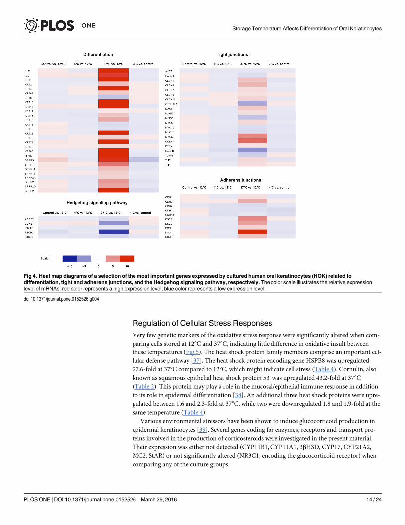
Expression of Differentiation MarkersIn addition to RPTN, KDAP, KRT10, LIPK, and CRNN, as presented in the previous section,the following genes associated with differentiation were upregulated: First, the oral mucosaldifferentiation marker keratin 4 (KRT4) [26] was upregulated 10.2-fold at 37°C storage com-pared to 12°C (Table 4 and Fig 4), indicative of a lower degree of differentiation in 12°C cul-tures. Second, keratin 6B, a specific marker of oral mucosal cells [26], was upregulated 2.5-foldat 37°C compared to 12°C cultures. Third, expression of keratin 19 (KRT19), a marker ofundifferentiated cells [27], was 1.5-fold higher in cells stored at 12°C compared to those storedat 37°C (Table 4). Taken together, a total of 17 keratins were differentially regulated at 37°Ccompared to 12°C; 14 of these were upregulated.
Other structural markers of keratinocyte differentiation had also changed during storage at37°C. Filaggrin (FLG), which aggregates keratin intermediate filaments in mammalian epider-mis [28], was upregulated 18.9-fold at 37°C compared to 12°C. Involucrin (IVL), a marker ofdifferentiated keratinocytes [17, 18, 27], was upregulated 8.8-fold in cells stored at 37°C com-pared to those stored at 12°C, indicating increased differentiation of cells stored at 37°C. Thelipase M (LIPM) gene, closely related to lipase K and exclusively expressed in the epidermis[19], was upregulated 3.7-fold at 37°C compared to 12°C (Table 4).
The cornified cell envelope is an insoluble protein layer that provides barrier function tostratified squamous epithelial cells [29]. Small proline-rich proteins (SPRRs) are constituentsof this structure, and their expression is restricted to terminally differentiating squamous cells[18, 30]. Eight SPRR genes were upregulated between 2- and 11-fold at 37°C compared to12°C, further indicating a more differentiated phenotype of cells stored at this temperature(Table 4). Apart from a 1.7-fold downregulation of TP63, a marker of undifferentiated cells, at37°C compared to 12°C, few stem cell markers seemed to be affected by storage temperature.Neither OCT-4, FGF2, nor Nanog were differentially expressed at 12°C compared to 37°C, sug-gesting no significant impact of storage temperature on these stem cell related genes.
Regulation of Cell-Cell ContactIdentified claudins (CLDN) 1, 4, 7, 9, and 16 were upregulated between 1.5 and 3.6-fold at37°C compared to 12°C. Genes encoding tight junction proteins 1 (TJP1) and 3 (TJP3) wereboth upregulated 1.8 and 1.6-fold at 37°C compared to 12°C, respectively (Table 4 and Fig 4).These changes indicate an increased synthesis of tight junctions in cells stored at 37°C.
Storage Temperature Affects Differentiation of Oral Keratinocytes
PLOS ONE | DOI:10.1371/journal.pone.0152526 March 29, 2016 10 / 24

Table 4. Differential regulation of genes in HOK cultures stored at 37°C compared to HOK cultures stored at 12°C.
Gene Symbol Gene Description Affymetrix ID P-value Fold Change
Differentiation
FLG filaggrin 7920165 1.71E-03 18.90
IVL involucrin 7905533 5.74E-04 8.76
KRT1 keratin 1 7963491 3.51E-03 13.49
KRT2 keratin 2 7963479 1.85E-02 2.88
KRT4 keratin 4 7963534 4.66E-03 10.21
KRT6B keratin 6B 7963406 1.76E-04 2.53
KRT8 keratin 8 7963567 4.64E-05 -2.74
KRT10 keratin 10 8015104 9.32E-05 45.63
KRT13 keratin 13 8015323 1.57E-04 26.79
KRT14 keratin 14 8015366 1.90E-01 1.10
KRT15 keratin 15 8015337 3.63E-03 2.64
KRT16 keratin 16 8015376 2.49E-04 4.11
KRT18 keratin 18 7969574 3.07E-02 -1.67
KRT19 keratin 19 8015349 3.96E-02 -1.50
KRT23 keratin 23 (histone deacetylase inducible) 8015133 6.84E-03 16.39
KRT75 keratin 75 7963396 1.32E-03 2.13
KRT78 keratin 78 7963555 5.24E-04 17.62
KRT79 keratin 79 7963545 3.75E-02 1.76
KRT80 keratin 80 7963333 2.32E-04 10.63
LIPM lipase, familiy member M 7929003 2,28E-02 3.72
RPTN repetin 7920146 1.84E-07 136.92
SPRR3 small proline-rich protein 3 7905548 2.11E-03 9.39
SPRR4 small proline-rich protein 4 7905536 9.45E-03 6.33
SPRR1A small proline-rich protein 1A 7905544 6.29E-04 3.01
SPRR1B small proline-rich protein 1B 7905553 1.12E-03 2.26
SPRR2A small proline-rich protein 2A 7920205 1.77E-03 2.05
SPRR2B small proline-rich protein 2B 7920210 5.78E-04 4.77
SPRR2D small proline-rich protein 2D 7920196 1.42E-03 4.22
SPRR2E small proline-rich protein 2E 7920214 9.27E-05 11.18
TP63 tumor protein p63 8084766 1,49E-03 -1.667
Tight junctions
ACTB actin, beta 8137979 7.57E-02 -1.16
CALM1 (includes others) calmodulin 1 (phosphorylase kinase, delta) 8029831 1.73E-03 -1.54
CLDN1 claudin 1 8092726 8.59E-03 2.86
CLDN4 claudin 4 8133360 7.88E-05 3.65
CLDN7 claudin 7 8012126 2.05E-02 1.71
CLDN9 claudin 9 7992782 3.54E-04 1.53
CLDN16 claudin 16 8084788 4.67E-03 2.11
CTNNAL1 catenin (cadherin-associated protein), alpha-like 1 8163063 1.14E-03 -4.02
MAGI1 membrane associated guanylate kinase, WW and PDZ domaincontaining 1
8088602 3.27E-03 2.46
MAGI3 membrane associated guanylate kinase, WW and PDZ domaincontaining 3
7904106 5.09E-03 1.57
MPDZ multiple PDZ domain protein 8160088 6.35E-05 -2.84
MYO6 myosin VI 8120783 2.30E-04 1.78
MYO10 myosin X 8111153 1.05E-02 -1.60
(Continued)
Storage Temperature Affects Differentiation of Oral Keratinocytes
PLOS ONE | DOI:10.1371/journal.pone.0152526 March 29, 2016 11 / 24
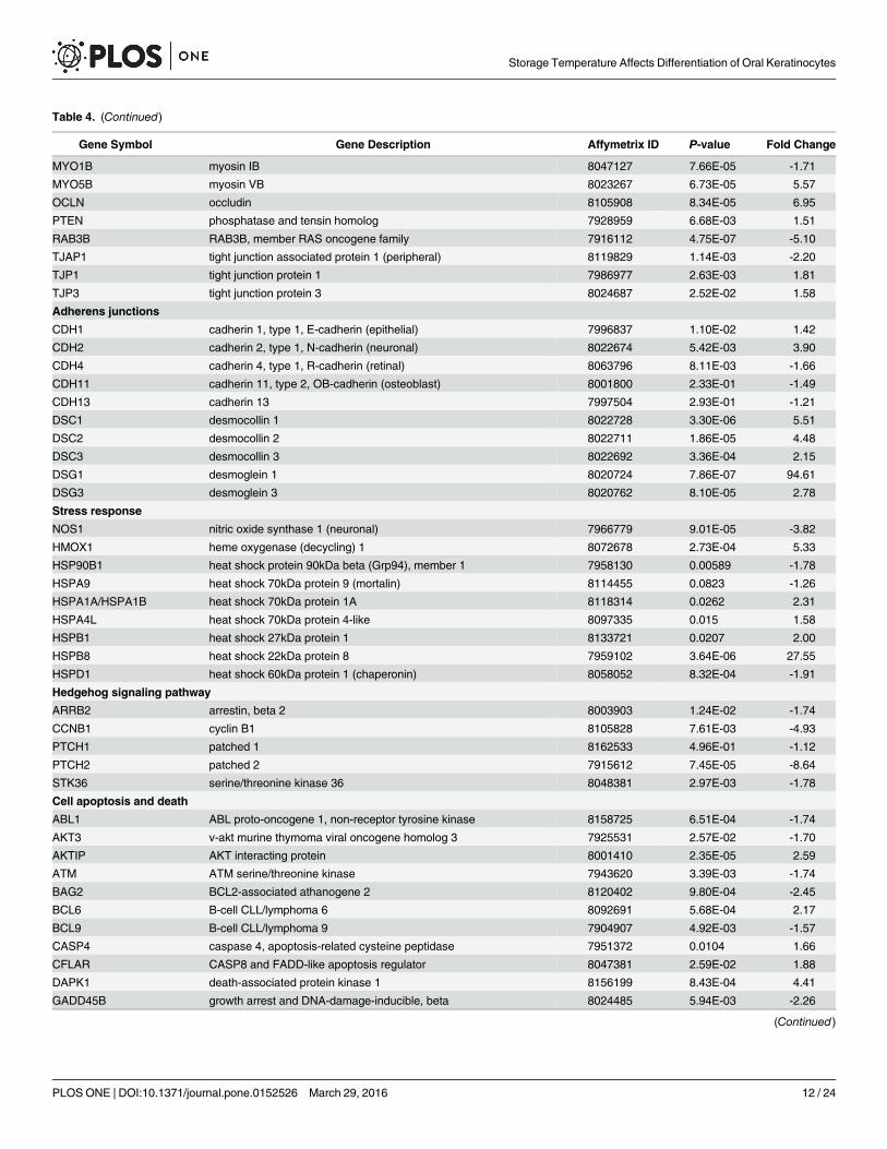
Table 4. (Continued)
Gene Symbol Gene Description Affymetrix ID P-value Fold Change
MYO1B myosin IB 8047127 7.66E-05 -1.71
MYO5B myosin VB 8023267 6.73E-05 5.57
OCLN occludin 8105908 8.34E-05 6.95
PTEN phosphatase and tensin homolog 7928959 6.68E-03 1.51
RAB3B RAB3B, member RAS oncogene family 7916112 4.75E-07 -5.10
TJAP1 tight junction associated protein 1 (peripheral) 8119829 1.14E-03 -2.20
TJP1 tight junction protein 1 7986977 2.63E-03 1.81
TJP3 tight junction protein 3 8024687 2.52E-02 1.58
Adherens junctions
CDH1 cadherin 1, type 1, E-cadherin (epithelial) 7996837 1.10E-02 1.42
CDH2 cadherin 2, type 1, N-cadherin (neuronal) 8022674 5.42E-03 3.90
CDH4 cadherin 4, type 1, R-cadherin (retinal) 8063796 8.11E-03 -1.66
CDH11 cadherin 11, type 2, OB-cadherin (osteoblast) 8001800 2.33E-01 -1.49
CDH13 cadherin 13 7997504 2.93E-01 -1.21
DSC1 desmocollin 1 8022728 3.30E-06 5.51
DSC2 desmocollin 2 8022711 1.86E-05 4.48
DSC3 desmocollin 3 8022692 3.36E-04 2.15
DSG1 desmoglein 1 8020724 7.86E-07 94.61
DSG3 desmoglein 3 8020762 8.10E-05 2.78
Stress response
NOS1 nitric oxide synthase 1 (neuronal) 7966779 9.01E-05 -3.82
HMOX1 heme oxygenase (decycling) 1 8072678 2.73E-04 5.33
HSP90B1 heat shock protein 90kDa beta (Grp94), member 1 7958130 0.00589 -1.78
HSPA9 heat shock 70kDa protein 9 (mortalin) 8114455 0.0823 -1.26
HSPA1A/HSPA1B heat shock 70kDa protein 1A 8118314 0.0262 2.31
HSPA4L heat shock 70kDa protein 4-like 8097335 0.015 1.58
HSPB1 heat shock 27kDa protein 1 8133721 0.0207 2.00
HSPB8 heat shock 22kDa protein 8 7959102 3.64E-06 27.55
HSPD1 heat shock 60kDa protein 1 (chaperonin) 8058052 8.32E-04 -1.91
Hedgehog signaling pathway
ARRB2 arrestin, beta 2 8003903 1.24E-02 -1.74
CCNB1 cyclin B1 8105828 7.61E-03 -4.93
PTCH1 patched 1 8162533 4.96E-01 -1.12
PTCH2 patched 2 7915612 7.45E-05 -8.64
STK36 serine/threonine kinase 36 8048381 2.97E-03 -1.78
Cell apoptosis and death
ABL1 ABL proto-oncogene 1, non-receptor tyrosine kinase 8158725 6.51E-04 -1.74
AKT3 v-akt murine thymoma viral oncogene homolog 3 7925531 2.57E-02 -1.70
AKTIP AKT interacting protein 8001410 2.35E-05 2.59
ATM ATM serine/threonine kinase 7943620 3.39E-03 -1.74
BAG2 BCL2-associated athanogene 2 8120402 9.80E-04 -2.45
BCL6 B-cell CLL/lymphoma 6 8092691 5.68E-04 2.17
BCL9 B-cell CLL/lymphoma 9 7904907 4.92E-03 -1.57
CASP4 caspase 4, apoptosis-related cysteine peptidase 7951372 0.0104 1.66
CFLAR CASP8 and FADD-like apoptosis regulator 8047381 2.59E-02 1.88
DAPK1 death-associated protein kinase 1 8156199 8.43E-04 4.41
GADD45B growth arrest and DNA-damage-inducible, beta 8024485 5.94E-03 -2.26
(Continued)
Storage Temperature Affects Differentiation of Oral Keratinocytes
PLOS ONE | DOI:10.1371/journal.pone.0152526 March 29, 2016 12 / 24

Regulatory changes of the constituents of the desmosomal adherens junction were noted(Table 4 and Fig 4). Desmosomes are intercellular junctions that link the intermediate fila-ments of the cytoskeleton of neighboring epithelial cells and consist of desmocollins, desmogle-ins, and cadherins [15]. Desmocollins (DSC) 1, 2, and 3 were upregulated 5.5, 4.5, and 2.2-foldat 37°C compared to 12°C, respectively. Loss of function of these genes is associated with skinbarrier defects [31] and metastasis of cancer cells [32]. Desmocollin 3 has been described as atumor suppressor of several types of cancer [33–36]. Desmogleins 1 and 3 were upregulated94.6-fold and 2.8-fold, respectively. Of the many identified cadherins in our material, only cad-herins 2 and 4 were differentially regulated, with a 3.9-fold upregulation and a 1.7-fold downre-gulation at 37°C, respectively (Table 4). Our findings point in the direction of increasedadherence between cells stored at 37°C compared to other temperatures.
Table 4. (Continued)
Gene Symbol Gene Description Affymetrix ID P-value Fold Change
IKBKE inhibitor of kappa light polypeptide gene enhancer in B-cells, kinaseepsilon
7909188 3.05E-03 1.72
IL1A interleukin 1, alpha 8054712 1.33E-02 -1.67
IL1B interleukin 1, beta 8054722 1.67E-02 -2.50
LIPH lipase, member H 8092541 3.72E-05 13.68
MAP3K1 mitogen-activated protein kinase kinase kinase 1, E3 ubiquitinprotein ligase
8105436 3.36E-05 2.03
MYC v-myc avian myelocytomatosis viral oncogene homolog 8148317 1.33E-03 -1.66
MYD88 myeloid differentiation primary response 88 8078729 9.49E-04 1.89
PARP1 poly (ADP-ribose) polymerase 1 7924733 8.67E-03 -2.13
PPP3CB protein phosphatase 3, catalytic subunit, beta isozyme 7934393 1.03E-02 -1.67
RIPK3 receptor-interacting serine-threonine kinase 3 7978312 1.35E-02 1.60
TNFRSF10A tumor necrosis factor receptor superfamily, member 10a 8149762 4.81E-04 -1.53
TNFRSF10B tumor necrosis factor receptor superfamily, member 10b 8149733 7.64E-03 -1.69
TNFRSF10D tumor necrosis factor receptor superfamily, member 10d, decoy withtruncated death domain
8149749 1.59E-02 -1.67
TNFRSF9 tumor necrosis factor receptor superfamily, member 9 7912145 7.42E-05 2.79
TNFSF10 tumor necrosis factor (ligand) superfamily, member 10 8092169 4.86E-04 4.66
TOP2A topoisomerase (DNA) II alpha 170kDa 8014974 3.96E-02 -6.14
TP53BP2 tumor protein p53 binding protein 2 7924526 1.61E-02 1.58
Squamous metaplasia
FLG filaggrin 7920165 1.71E-03 18.90
IVL involucrin 7905533 5.74E-04 8.76
MAPK1 mitogen-activated protein kinase 1 8074791 3.16E-01 1.23
MAPK3 mitogen-activated protein kinase 3 8000811 3.86E-01 1.20
MAPK7 mitogen-activated protein kinase 7 8005576 3.34E-02 1.40
MAPK8 mitogen-activated protein kinase 8 7927389 1.65E-01 1.15
MAPK9 mitogen-activated protein kinase 9 8116402 2.52E-01 -1.19
MAPK12 mitogen-activated protein kinase 12 8076962 5.05E-05 -2.39
TGM2 transglutaminase 2 8066214 5.79E-01 -1.47
TGM3 transglutaminase 3 8060432 3.02E-02 5.20
TGM5 transglutaminase 5 7988050 1.91E-02 5.95
doi:10.1371/journal.pone.0152526.t004
Storage Temperature Affects Differentiation of Oral Keratinocytes
PLOS ONE | DOI:10.1371/journal.pone.0152526 March 29, 2016 13 / 24
Regulation of Cellular Stress ResponsesVery few genetic markers of the oxidative stress response were significantly altered when com-paring cells stored at 12°C and 37°C, indicating little difference in oxidative insult betweenthese temperatures (Fig 5). The heat shock protein family members comprise an important cel-lular defense pathway [37]. The heat shock protein encoding gene HSPB8 was upregulated27.6-fold at 37°C compared to 12°C, which might indicate cell stress (Table 4). Cornulin, alsoknown as squamous epithelial heat shock protein 53, was upregulated 43.2-fold at 37°C(Table 2). This protein may play a role in the mucosal/epithelial immune response in additionto its role in epidermal differentiation [38]. An additional three heat shock proteins were upre-gulated between 1.6 and 2.3-fold at 37°C, while two were downregulated 1.8 and 1.9-fold at thesame temperature (Table 4).
Various environmental stressors have been shown to induce glucocorticoid production inepidermal keratinocytes [39]. Several genes coding for enzymes, receptors and transport pro-teins involved in the production of corticosteroids were investigated in the present material.Their expression was either not detected (CYP11B1, CYP11A1, 3βHSD, CYP17, CYP21A2,MC2, StAR) or not significantly altered (NR3C1, encoding the glucocorticoid receptor) whencomparing any of the culture groups.
Fig 4. Heat map diagrams of a selection of the most important genes expressed by cultured human oral keratinocytes (HOK) related todifferentiation, tight and adherens junctions, and the Hedgehog signaling pathway, respectively. The color scale illustrates the relative expressionlevel of mRNAs: red color represents a high expression level; blue color represents a low expression level.
doi:10.1371/journal.pone.0152526.g004
Storage Temperature Affects Differentiation of Oral Keratinocytes
PLOS ONE | DOI:10.1371/journal.pone.0152526 March 29, 2016 14 / 24
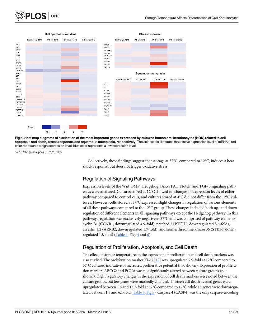
Collectively, these findings suggest that storage at 37°C, compared to 12°C, induces a heatshock response, but does not trigger oxidative stress.
Regulation of Signaling PathwaysExpression levels of the Wnt, BMP, Hedgehog, JAK/STAT, Notch, and TGF-β signaling path-ways were analyzed. Cultures stored at 12°C showed no changes in expression levels of eitherpathway compared to control cells, and cultures stored at 4°C did not differ from the 12°C cul-tures. However, cells stored at 37°C expressed slight changes in regulation of various elementsof all these pathways compared to the 12°C group. These changes included both up- and down-regulation of different elements in all signaling pathways except the Hedgehog pathway. In thispathway, regulation was exclusively negative at 37°C and was comprised of pathway elementscyclin B1 (CCNB1, downregulated 4.9-fold), patched 2 (PTCH2, downregulated 8.6-fold),arrestin, β2 (ARRB2, downregulated 1.7-fold), and serine/threonine kinase 36 (STK36, down-regulated 1.8-fold) (Table 4, Figs 4 and 6).
Regulation of Proliferation, Apoptosis, and Cell DeathThe effect of storage temperature on the expression of proliferation and cell death markers wasalso studied. The proliferation marker Ki-67 [18] was upregulated 7.9-fold at 12°C compared to37°C cultures, indicative of increased proliferative potential (not shown). Expression of prolifera-tion markers ABCG2 and PCNA was not significantly altered between culture groups (notshown). Slight regulatory changes in the expression of cell death markers were noted between theculture groups, but few genes were markedly changed. Thirteen cell death-related genes wereupregulated between 1.6 and 13.7-fold at 37°C compared to 12°C, while 15 genes were downregu-lated between 1.5 and 6.1-fold (Table 4, Fig 5). Caspase 4 (CASP4) was the only caspase-encoding
Fig 5. Heat map diagrams of a selection of the most important genes expressed by cultured human oral keratinocytes (HOK) related to cellapoptosis and death, stress response, and squamousmetaplasia, respectively. The color scale illustrates the relative expression level of mRNAs: redcolor represents a high expression level; blue color represents a low expression level.
doi:10.1371/journal.pone.0152526.g005
Storage Temperature Affects Differentiation of Oral Keratinocytes
PLOS ONE | DOI:10.1371/journal.pone.0152526 March 29, 2016 15 / 24

gene to be differentially expressed, with a 1.7-fold upregulation at 37°C compared to 12°C. LipaseH (LIPH), which is selectively upregulated in lung cancer and associated with increased survivalin lung cancer patients [40], was upregulated 13.7-fold at 37°C compared to 12°C.
Of note, of the 114 genes important for cellular function that were significantly regulated at37°C compared to 12°C (Table 4), only one was differentially regulated when comparing 4°Cstorage to 12°C storage (Growth arrest and DNA-damage-inducible beta (GADD45B); downre-gulated 2.81-fold), control cultures to 12°C storage (GADD45B; downregulated 3.6-fold), and4°C storage to the control (member RAS oncogene family (RAB3B); downregulated 1.53-fold).GADD45 is induced by environmental stress or DNA damage [41], while RAB3B localizes totight junctions where it has been suggested to contribute to the polarization of epithelia [42, 43].Hence, 4°C and 12°C storage do not induce notable changes in the regulation of the importantgenes analyzed herein, and the two storage groups seem to offer equivalent results.
Quantitative Real-Time PCR Validation of Microarray DataHSPB8, TP63 and KRT10 were selected for validation by qPCR (Table 5). The expression ofHSPB8 was substantially upregulated at 37°C compared to 12°C; a 123.6-fold upregulation byPCR compared to a 27.6-fold upregulation by microarray. The expression of TP63 was signifi-cantly downregulated 3.6-fold at 37°C compared to 12°C, which is in line with the microarrayresults demonstrating a 1.7-fold downregulation at 37°C. Keratin 10 expression was signifi-cantly upregulated 6.7-fold at 37°C compared to 12°C. Upregulation of this gene was higher inthe microarray analysis (45.6-fold). Expression levels of HSPB8, TP63, and KRT10 at theremaining temperatures were consistent between qPCR and microarray results, showing nosignificant differential regulation between temperatures (Table 5 and Fig 7).
DiscussionThe current study investigated the effects of storage temperature on gene expression in cul-tured HOK using microarray analysis. The temperatures selected included 4°C, the standard
Fig 6. Differential regulation of the Hedgehog signaling pathway at 37°C compared to 12°C. Pathwayelements marked in red are significantly downregulated at 37°C. There is no upregulation of pathwayelements.
doi:10.1371/journal.pone.0152526.g006
Storage Temperature Affects Differentiation of Oral Keratinocytes
PLOS ONE | DOI:10.1371/journal.pone.0152526 March 29, 2016 16 / 24

temperature of a refrigerator; 12°C, a temperature which previously gave the most optimalresults with regard to both morphology and viability of stored HOK; and 37°C, the temperatureof a standard cell culture incubator.
Five of the six most differentially regulated proteins at 37°C compared to 12°C are directlyassociated with epithelial differentiation. An epidermal differentiation profile of these HOKcells is regarded as a disadvantage when used for treating LSCD [9]. The RPTN gene is veryactive during the final steps of epidermal keratinocyte differentiation, since the repetin proteinis associated with the keratin intermediate filaments that are present in mature epidermal cells[14]. Keratinocyte differentiation-associated protein localizes to the stratum corneum of nor-mal skin, but is expressed in suprabasal keratinocytes in psoriatic lesions [17]. Expression ofthe KDAP gene is markedly upregulated during keratinocyte differentiation in vitro [17]. Filag-grin contributes to the hydration and pH homeostasis of the stratum corneum [28], and muta-tions of the filaggrin gene are associated with ichtyosis vulgaris [44] and eczema [45]. Cornulinis a squamous cell-specific polypeptide [37] which is downregulated in eczema [46] and is acomponent of the epithelial innate immune response [38]. This protein is a constituent of theheat shock response of esophageal squamous epithelial tissue, where it is known as the squa-mous epithelial heat shock protein 53 (SEP53) [37]. Cornulin’s expression increases markedlyas a consequence of heat shock, which might indicate activation of this cellular defense path-way in cell cultures stored at 37°C compared to those stored at 12°C. However, the activationof cornulin as a component of the differentiation process, and not primarily as a heat shockmodulating protein, is an alternative interpretation.
The oral mucosal marker keratin 6b [26] was upregulated 2.5-fold in cultures stored at 37°Ccompared to 12°C. Its relative downregulation in 12°C cultures might indicate a dedifferentia-tion of cells at these temperatures. The upregulation of keratin 6b has also been demonstratedin conjunctival epithelium of patients with Sjögren’s syndrome [47]. Keratin 6b is also used asa marker of activated keratinocytes, and its expression can be induced both by proliferative sig-nals and the proinflammatory cytokine TNF-α [48]. TNF was upregulated 1.4-fold at 37°Ccompared to 12°C (not shown), and several TNF-related proteins are differentially regulated at37°C (Table 4). Given the stable expression of proliferative markers at 37°C compared to 12°C,it is more likely that the keratin 6b upregulation might be a consequence of inflammatoryresponses rather than proliferative signals.
Table 5. Validation of microarray results by qPCR.
Gene Affymetrix PCR
Fold Change P-value Fold Change P-value
HSPB8
Control vs 12°C 1.45 0.25 2.45 0.13
4°C vs 12°C 1.38 0.31 -1.09 0.47
37°C vs 12°C 27.55 3.64E-06 123.63 1.00E-03
TP63
Control vs 12°C 1.00 1.00 -1.05 0.30
4°C vs 12°C -1.07 0.57 -1.22 0.10
37°C vs 12°C -1.67 1.49E-03 -3.6 5.00E-04
KRT10
Control vs 12°C -1.09 0.87 -1.12 0.06
4°C vs 12°C -1.01 0.99 -1.38 0.08
37°C vs 12°C 45.63 9.32E-05 6.67 0.04
doi:10.1371/journal.pone.0152526.t005
Storage Temperature Affects Differentiation of Oral Keratinocytes
PLOS ONE | DOI:10.1371/journal.pone.0152526 March 29, 2016 17 / 24

CSRNP1 was upregulated 2.4-fold after storage at 12°C compared to control cultures.CSRNP1 has been described as a tumor suppressor gene, its expression level decreased in sev-eral types of cancers [49]. Overexpression has been reported to halt cell cycle progression atmitosis [50]. Its function is essential for normal development of the brain [51], and it is aknown negative regulator of the Wnt pathway [52].
Both adhesion and tight junction-related genes were significantly upregulated at 37°C com-pared to 12°C and control cultures. The upregulation of DSC1 in cultures stored at 37°C is inline with our findings in retinal pigment epithelial cells [53]. These findings indicate a func-tional change towards a more tightly woven, squamous-like epithelium after storage at 37°C.
The expression of stress-related genes was also evaluated. Except for a few heat shock pro-teins, few genetic markers of the oxidative stress response were significantly altered when com-paring cells stored at 12°C and 37°C. Recent studies have described that epidermalkeratinocytes subjected to various environmental stressors can respond by upregulating theirglucocorticoid production [39, 54]. Expression of genes encoding enzymes, receptors andtransport proteins necessary for the production and effect of corticosteroids were either notdetected or were unaltered in our material.
Cultures stored at 37°C show both up- and downregulation of several important moleculesof the Wnt, BMP, JAK/STAT, Notch, and TGF-β signaling pathways, rendering the effects ofthese regulational changes inconclusive. However, the Hedgehog pathway was exclusivelydownregulated in the 37°C cell group compared to the 12°C group. The Hedgehog signaling
Fig 7. Validation of microarray expression results by qPCR. SelectedmRNAs (HSPB8, TP63 and KRT10) were differentially expressed in culturedRPE cells stored at 12°C compared to cultures that were stored at 37°C. Black bars indicate microarray expression values; grey bars represent PCRverification values. *P < 0.05.
doi:10.1371/journal.pone.0152526.g007
Storage Temperature Affects Differentiation of Oral Keratinocytes
PLOS ONE | DOI:10.1371/journal.pone.0152526 March 29, 2016 18 / 24

pathway is instrumental for vertebrate embryogenesis and has been demonstrated to regulatecell fate, proliferation, and survival in multiple cell types, especially those of neuroectodermalorigin such as cells of the retina and optic nerve [55]. The pathway also regulates adult stemcells in several self-renewing organs including the subventricular zone of the brain [56, 57].The deactivating effect of 37°C on this pathway may therefore perturb cellular function. Thesonic hedgehog protein, Shh, binds Patched, a transmembrane receptor of the target cell [58].Patched functions as a tumor suppressor in the hedgehog signaling pathway [58] and muta-tions of the gene have been detected in basal cell carcinomas and medulloblastomas, amongother cancers [55, 59]. The downregulation of Patched in the 37°C culture group compared to12°C might destabilize cellular quiescence, an undesirable event in cell preservation.
The patched protein interacts with cyclin B1 and participates in determining its cellularlocalization [60]. Cyclin B1 is a regulatory protein involved in the promotion of mitosis, transi-tioning the cell from the G2 to M phase. The effect of Patched on Cyclin 1 is inhibition of cellu-lar proliferation [60]. Arrestin β2, which was downregulated at 37°C, is a ubiquitouslydistributed protein with a critical role in the regulation of several important signaling path-ways, including Hedgehog [61]. The serine/threonine kinase 36 (STK36), also downregulatedat 37°C, plays a key role in the Hedgehog signaling pathway. Hence, several crucial constituentsof the pathway are downregulated at 37°C, contributing to a reduced activity of the pathway asa whole. The finding of perturbed signaling pathway elements of several major pathways in cul-tures stored at 37°C is in concordance with findings of similar changes in retinal pigment epi-thelial cells stored at 37°C (unpublished data).
The analysis of cell proliferation and death markers after storage at different temperaturesindicates a disturbance of the regulation of several cell death-related genes at 37°C. These changesmay be partly responsible for the reduced viability found in cultures stored at this temperature.
Expression of the melatonin receptor MTNR1A was significantly upregulated at 12°C com-pared to the other groups. Melatonin exerts numerous effects on a multitude of organ systems,including the skin. Its effects are mediated through both receptor dependent and independentmechanisms [62]. Importantly, the skin cannot be regarded a passive target for the effects ofmelatonin, but a vibrant site of its synthesis and metabolism [63, 64]. There is accumulatingevidence that the stringently regulated effects of melatonin in the skin are organized throughan interlaced local neuroendocrine system exploiting both auto- and paracrine mechanisms[63, 64]. Melatonin and its metabolites exert broad antioxidant effects, and melatonin is able toactivate cytoprotective molecules and enzymes, including glutathione [63, 65, 66]. It may alsoprotect DNA from oxidative damage, thereby providing anti-apoptotic and anti-carcinogeniceffects [63, 64, 67]. Specifically, both the initiation and promotion of skin carcinogenesis can bedecreased by melatonin [68]. There is also evidence that its oncostatic effects are dependent onthe MTNR1A receptor [69–71], and that the tumor suppressive effect of the MTNR1A gene issilenced in oral squamous cell carcinoma [23]. A significant association between MTNR1Apolymorphisms and oral carcinogenesis has been demonstrated [22], in which environmentalfactors (betel quid chewing and cigarette smoking) are required to increase the susceptibility tooral cancer in individuals with MTNR1A gene polymorphisms.
It has also been demonstrated that melatonin and its metabolites protect keratinocytes fromUV radiation [64, 72]. Following UVB exposure, MTNR1A expression has been shown to beupregulated in normal neonatal epidermal melanocytes and downregulated in melanoma lines[73]. In the current study, HOK were cultured in the dark except when being handled, but cul-tures stored at 12°C and 37°C were probably exposed to small amounts of light during heatingof the storage containers. This light exposure might have contributed in inducing MTNR1Aexpression in the 12°C group, but it does not explain why a similar upregulation in the 37°Cstorage group could not be detected.
Storage Temperature Affects Differentiation of Oral Keratinocytes
PLOS ONE | DOI:10.1371/journal.pone.0152526 March 29, 2016 19 / 24
While there are clear indications to the upregulation of differentiation-related genes at 37°Ccompared to 12°C, the gene regulation changes in the 4°C and 12°C groups compared to thecontrol are not as clear. However, the downregulation of some constituents of the epidermaldifferentiation complex at 4°C might indicate some degree of de-differentiation in these cul-tures, contrary to the effect of 37°C storage. For cells stored at 12°C, the evidence toward a lessdifferentiated phenotype was not as strong.
In conclusion, HOK cultures stored at 37°C demonstrated considerably larger changes inboth the amount of genes affected as well as their differential regulation levels compared tounstored cells than cultured HOK stored at 4°C and 12°C. Temporary storage of cell culturesin sealed containers at 37°C, rather than at 12°C, appears to promote differentiation similarlyto conventional cell culture, which employs a humidified incubator at 37°C with CO2 supply.Storage at 37°C may reduce the stemness, and thereby the therapeutic potential, of culturedcells intended for the treatment of LSCD [9]. Storage at 12°C also maintains the regulation ofgenes closer to control levels than storage at 4°C. However, storage at 4°C might steer cellstoward a less differentiated phenotype. Nevertheless, we conclude that storage at 4°C and 12°Care more suitable than storage at 37°C for preserving cultured HOK for transplantation. Ourfindings are in line with a recent study [12], demonstrating superior viability of HOK whenstored at 12°C compared to 4°C and 37°C. Thus, collectively, 12°C seems to be the most idealstorage temperature among those investigated.
Author ContributionsConceived and designed the experiments: TPU IF JRE DAD EMMG LP. Performed the experi-ments: TPU RI OKO. Analyzed the data: TPU AS OKO LP. Contributed reagents/materials/analysis tools: TPU RI OKO EM. Wrote the paper: TPU IF JRE AS OKO DAD EMMG LP.
References1. Utheim TP. Limbal epithelial cell therapy: past, present, and future. Methods in molecular biology (Clif-
ton, NJ). 2013; 1014:3–43. Epub 2013/05/22. doi: 10.1007/978-1-62703-432-6_1 PMID: 23690002.
2. Pellegrini G, Traverso CE, Franzi AT, Zingirian M, Cancedda R, De Luca M. Long-term restoration ofdamaged corneal surfaces with autologous cultivated corneal epithelium. Lancet (London, England).1997; 349(9057):990–3. Epub 1997/04/05. doi: 10.1016/s0140-6736(96)11188-0 PMID: 9100626.
3. Niethammer D, Kummerle-Deschner J, Dannecker GE. Side-effects of long-term immunosuppressionversus morbidity in autologous stem cell rescue: striking the balance. Rheumatology (Oxford). 1999; 38(8):747–50. Epub 1999/09/29. PMID: 10501425.
4. Nakamura T, Inatomi T, Sotozono C, Amemiya T, Kanamura N, Kinoshita S. Transplantation of culti-vated autologous oral mucosal epithelial cells in patients with severe ocular surface disorders. The Brit-ish journal of ophthalmology. 2004; 88(10):1280–4. Epub 2004/09/21. doi: 10.1136/bjo.2003.038497PMID: 15377551; PubMed Central PMCID: PMCPMC1772364.
5. Nishida K, Yamato M, Hayashida Y, Watanabe K, Yamamoto K, Adachi E, et al. Corneal reconstructionwith tissue-engineered cell sheets composed of autologous oral mucosal epithelium. The New Englandjournal of medicine. 2004; 351(12):1187–96. Epub 2004/09/17. doi: 10.1056/NEJMoa040455 PMID:15371576.
6. Utheim TP. Concise review: transplantation of cultured oral mucosal epithelial cells for treating limbalstem cell deficiency-current status and future perspectives. Stem Cells. 2015; 33(6):1685–95. doi: 10.1002/stem.1999 PMID: 25786664.
7. Daniels JT, Secker GA, Shortt AJ, Tuft SJ, Seetharaman S. Stem cell therapy delivery: treading the reg-ulatory tightrope. Regen Med. 2006; 1(5):715–19. PMID: 17465740
8. Ahmad S, Osei-Bempong C, Dana R, Jurkunas U. The culture and transplantation of human limbalstem cells. Journal of Cellular Physiology. 2010; 225(1):15–9. doi: 10.1002/jcp.22251 PMID: 20506173
9. Rama P, Matuska S, Paganoni G, Spinelli A, De Luca M, Pellegrini G. Limbal stem-cell therapy andlong-term corneal regeneration. The New England journal of medicine. 2010; 363(2):147–55. Epub2010/06/25. doi: 10.1056/NEJMoa0905955 PMID: 20573916.
Storage Temperature Affects Differentiation of Oral Keratinocytes
PLOS ONE | DOI:10.1371/journal.pone.0152526 March 29, 2016 20 / 24

10. O'Callaghan AR, Daniels JT. Limbal epithelial stem cell therapy: controversies and challenges. StemCells. 2011; 29(12):1923–32. doi: 10.1002/stem.756 PMID: 21997829
11. Utheim TP, Raeder S, Olstad OK, Utheim OA, de la Paz MF, Cheng R, et al. Comparison of the histol-ogy, gene expression profile, and phenotype of cultured human limbal epithelial cells from different lim-bal regions. InvestOphthalmolVisSci. 2009; 50(11):5165–72. iovs.08-2884 [pii]; doi: 10.1167/iovs.08-2884
12. Islam R, Jackson C, Eidet JR, Messelt EB, Corraya RM, Lyberg T, et al. Effect of Storage Temperatureon Structure and Function of Cultured Human Oral Keratinocytes. PLOS ONE. 2015; 10(6):e0128306.doi: 10.1371/journal.pone.0128306 PMID: 26052937; PubMed Central PMCID: PMCPMC4459984.
13. Pasovic L, Utheim TP, Maria R, Lyberg T, Messelt EB, Aabel P, et al. Optimization of Storage Tempera-ture for Cultured ARPE-19 Cells. Journal of Ophthalmology. 2013; 2013:11. doi: 10.1155/2013/216359
14. Huber M, Siegenthaler G, Mirancea N, Marenholz I, Nizetic D, Breitkreutz D, et al. Isolation and Charac-terization of Human Repetin, a Member of the Fused Gene Family of the Epidermal DifferentiationComplex. J Invest Dermatol. 2005; 124:998–1007. PMID: 15854042
15. Garrod DR, Merritt AJ, Nie Z. Desmosomal cadherins. Current Opinion in Cell Biology. 2002; 14:537–45. PMID: 12231347
16. Oomizu S, Sahuc F, Asahina K, Inamatsu M, Matsuzaki T, Sasaki M, et al. Kdap, a novel gene associ-ated with the stratification of the epithelium. Gene. 2000; 256:19–27. PMID: 11054531
17. Tsuchida S, Bonkobara M, McMillan JR, Akiyama M, Yudate T, Aragane Y, et al. Characterization ofKdap, a Protein Secreted by Keratinocytes. J Invest Dermatol. 2004; 122:1225–34. PMID: 15140226
18. Gibbs D, Ponec M. Intrinsic regulation of differentiation markers in human epidermis, hard palate andbuccal mucosa. Arch Oral Biochem. 2000; 45:149–58.
19. Toulza E, Mattiuzzo NR, Galliano MF, Jonca N, Dossat C, Jacob D, et al. Large-scale identification ofhuman genes implicated in epidermal barrier function. Genome biology. 2007; 8(6):R107. doi: 10.1186/gb-2007-8-6-r107 PMID: 17562024; PubMed Central PMCID: PMC2394760.
20. Contzler R, Favre B, Huber M, Hohl D. Cornulin, a NewMember of the ‘‘Fused Gene” Family, IsExpressed During Epidermal Differentiation. J Invest Dermatol. 2005; 124:990–97. PMID: 15854041
21. Slominski A, Tobin DJ, Zmijewski MA, Wortsman J, Paus R. Melatonin in the skin: synthesis, metabo-lism and functions. Trends Endocrinol Metab. 2008; 19(1):17–24. doi: 10.1016/j.tem.2007.10.007PMID: 18155917.
22. Lin FY, Lin CW, Yang SF, LeeWJ, Lin YW, Lee LM, et al. Interactions between environmental factorsand melatonin receptor type 1A polymorphism in relation to oral cancer susceptibility and clinicopatho-logic development. PLOS ONE. 2015; 10(3):e0121677. doi: 10.1371/journal.pone.0121677 PMID:25806809; PubMed Central PMCID: PMCPMC4373723.
23. Nakamura E, Kozaki K, Tsuda H, Suzuki E, Pimkhaokham A, Yamamoto G, et al. Frequent silencing ofa putative tumor suppressor gene melatonin receptor 1 A (MTNR1A) in oral squamous-cell carcinoma.Cancer Sci. 2008; 99(7):1390–400. doi: 10.1111/j.1349-7006.2008.00838.x PMID: 18452558.
24. Backendorf C, Hohl D. A common origin for cornified envelope proteins? Nature genetics. 1992; 2:91.PMID: 1303269
25. Jackson B, Tilli CM, Hardman MJ, Avilion AA, MacLeod MC, Ashcroft GS, et al. Late cornified envelopefamily in differentiating epithelia—response to calcium and ultraviolet irradiation. J Invest Dermatol.2005; 124(5):1062–70. doi: 10.1111/j.0022-202X.2005.23699.x PMID: 15854049.
26. Sugiyama H, Yamato M, Nishida K, Okano T. Evidence of the survival of ectopically transplanted oralmucosal epithelial stem cells after repeated wounding of cornea. Molecular therapy: the journal of theAmerican Society of Gene Therapy. 2014; 22(8):1544–55. doi: 10.1038/mt.2014.69 PMID: 24769908.
27. Calenic B, Ishkitiev N, Yaegaki K, Imai T, Costache M, Tovaru M, et al. Characterization of oral kerati-nocyte stem cells and prospects of its differentiation to oral epithelial equivalents. Rom J MorpholEmbryol. 2010; 51(4):641–45. PMID: 21103620
28. Thyssen JP, Kezic S. Causes of epidermal filaggrin reduction and their role in the pathogenesis ofatopic dermatitis. The Journal of allergy and clinical immunology. 2014; 134(4):792–9. doi: 10.1016/j.jaci.2014.06.014 PMID: 25065719.
29. Steinert PM, Candi E, Kartasova T, Marekov L. Small proline-rich proteins are cross-bridging proteinsin the cornified cell envelopes of stratified squamous epithelia. Journal of structural biology. 1998; 122(1–2):76–85. Epub 1998/09/02. doi: 10.1006/jsbi.1998.3957 PMID: 9724607.
30. Hohl D, de Viragh PA, Amiguet-Barras F, Gibbs S, Backendorf C, Huber M. The small proline-rich pro-teins constitute a multigene family of differentially regulated cornified cell envelope precursor proteins.J Invest Dermatol. 1995; 104(6):902–9. PMID: 7769256
31. Chidgey M, Brakebusch C, Gustafsson E, Cruchley A, Hail C, Kirk S, et al. Mice lacking desmocollin 1show epidermal fragility accompanied by barrier defects and abnormal differentiation. J Cell Biol. 2001;
Storage Temperature Affects Differentiation of Oral Keratinocytes
PLOS ONE | DOI:10.1371/journal.pone.0152526 March 29, 2016 21 / 24

155(5):821–32. Epub 2001/11/21. doi: 10.1083/jcb.200105009 PMID: 11714727; PubMed CentralPMCID: PMC2150874.
32. Shinohara M, Hiraki A, Ikebe T, Nakamura S, Kurahara S-I, Shirasuna K, et al. Immunohistochemicalstudy of desmosomes in oral squamous cell carcinoma: correlation with cytokeratin and E-cadherinstaining, and with tumour behaviour. J Pathol. 1998; 184:369–81. PMID: 9664902
33. Cui T, Chen Y, Yang L, Knosel T, Huber O, Pacyna-Gengelbach M, et al. The p53 target gene desmo-collin 3 acts as a novel tumor suppressor through inhibiting EGFR/ERK pathway in human lung cancer.Carcinogenesis. 2012; 33(12):2326–33. Epub 2012/09/04. doi: 10.1093/carcin/bgs273 PMID:22941060.
34. Oshiro MM, Kim CJ, Wozniak RJ, Junk DJ, Munoz-Rodriguez JL, Burr JA, et al. Epigenetic silencing ofDSC3 is a common event in human breast cancer. Breast Cancer Res. 2005; 7(5):R669–80. Epub2005/09/20. doi: 10.1186/bcr1273 PMID: 16168112; PubMed Central PMCID: PMC1242132.
35. Wang L, Liu T, Wang Y, Cao L, Nishioka M, Aguirre RL, et al. Altered expression of desmocollin 3, des-moglein 3, and beta-catenin in oral squamous cell carcinoma: correlation with lymph node metastasisand cell proliferation. Virchows Arch. 2007; 451(5):959–66. Epub 2007/09/12. doi: 10.1007/s00428-007-0485-5 PMID: 17846785.
36. Cui T, Chen Y, Yang L, Knosel T, Zoller K, Huber O, et al. DSC3 expression is regulated by p53, andmethylation of DSC3 DNA is a prognostic marker in human colorectal cancer. Br J Cancer. 2011; 104(6):1013–9. Epub 2011/03/03. doi: 10.1038/bjc.2011.28 PMID: 21364582; PubMed Central PMCID:PMC3065270.
37. Yagui-Beltran A, Craig AL, Lawrie L, Thompson D, Posipilova S, Johnston D, et al. The human oeso-phageal squamous epithelium exhibits a novel type of heat shock protein response. Eur J Biochem.2001; 268:5343–55. PMID: 11606197
38. Little TJ, Nelson L, Hupp T. Adaptive evolution of a stress response protein. PLOS ONE. 2007; 2(10):e1003. doi: 10.1371/journal.pone.0001003 PMID: 17925851; PubMed Central PMCID: PMC1994589.
39. Zhu G, Janjetovic Z, Slominski A. On the role of environmental humidity on cortisol production by epi-dermal keratinocytes. Exp Dermatol. 2014; 23(1):15–7. doi: 10.1111/exd.12275 PMID: 24372648;PubMed Central PMCID: PMCPMC3923494.
40. Seki Y, Yoshida Y, Ishimine H, Shinozaki-Ushiku A, Ito Y, Sumitomo K, et al. Lipase member H is anovel secreted protein selectively upregulated in human lung adenocarcinomas and bronchioloalveolarcarcinomas. Biochem Biophys Res Commun. 2014; 443(4):1141–7. doi: 10.1016/j.bbrc.2013.12.106PMID: 24380866.
41. TakekawaM, Saito H. A Family of Stress-Inducible GADD45-like Proteins Mediate Activation of theStress-Responsive MTK1/MEKK4MAPKKK. Cell. 1998; 95(4):521–30. PMID: 9827804
42. Weber E, Berta G, Tousson A, John PS, Green MW, Gopalokrishnan U, et al. Expression and PolarizedTargeting of a Rab3 Isoform in Epithelial Cells. J Cell Biol. 1994; 125(3):583–94. PMID: 8175882
43. Yamamoto Y, Nishimura N, Morimoto S, Kitamura H, Manabe S, Kanayama H-O, et al. Distinct roles ofRab3B and Rab13 in the polarized transport of apical, basolateral, and tight junctional membrane pro-teins to the plasmamembrane. Biochemical and Biophysical Research Communications. 2003; 308(2):270–5. doi: 10.1016/s0006-291x(03)01358-5 PMID: 12901864
44. Smith FJ, Irvine AD, Terron-Kwiatkowski A, Sandilands A, Campbell LE, Zhao Y, et al. Loss-of-functionmutations in the gene encoding filaggrin cause ichthyosis vulgaris. Nature genetics. 2006; 38(3):337–42. doi: 10.1038/ng1743 PMID: 16444271.
45. Palmer CN, Irvine AD, Terron-Kwiatkowski A, Zhao Y, Liao H, Lee SP, et al. Common loss-of-functionvariants of the epidermal barrier protein filaggrin are a major predisposing factor for atopic dermatitis.Nature genetics. 2006; 38(4):441–6. doi: 10.1038/ng1767 PMID: 16550169.
46. Lieden A, Ekelund E, Kuo IC, Kockum I, Huang CH, Mallbris L, et al. Cornulin, a marker of late epider-mal differentiation, is down-regulated in eczema. Allergy. 2009; 64(2):304–11. doi: 10.1111/j.1398-9995.2008.01856.x PMID: 19133922.
47. Kawasaki S, Kawamoto S, Yokoi N, Connon C, Minesaki Y, Kinoshita S, et al. Up-regulated geneexpression in the conjunctival epithelium of patients with Sjögren's syndrome. Exp Eye Res. 2003; 77(1):17–26. doi: 10.1016/s0014-4835(03)00087-3 PMID: 12823984
48. Komine M, Rao LS, Kaneko T, Tomic-Canic M, Tamaki K, Freedberg IM, et al. Inflammatory versus pro-liferative processes in epidermis. Tumor necrosis factor alpha induces K6b keratin synthesis through atranscriptional complex containing NFkappa B and C/EBPbeta. J Biol Chem. 2000; 275(41):32077–88.doi: 10.1074/jbc.M001253200 PMID: 10887174.
49. Ishiguro H, Tsunoda T, Tanaka T, Fujii Y, Nakamura Y, Furukawa Y. Identification of AXUD1, a novelhuman gene induced by AXIN1 and its reduced expression in human carcinomas of the lung, liver,colon and kidney. Oncogene. 2001; 20:5062–66. PMID: 11526492
Storage Temperature Affects Differentiation of Oral Keratinocytes
PLOS ONE | DOI:10.1371/journal.pone.0152526 March 29, 2016 22 / 24

50. Glavic A, Molnar C, Cotoras D, de Celis JF. Drosophila Axud1 is involved in the control of proliferationand displays pro-apoptotic activity. Mech Dev. 2009; 126(3–4):184–97. Epub 2008/12/17. doi: 10.1016/j.mod.2008.11.005 PMID: 19084594.
51. Feijoo CG, Sarrazin AF, Allende ML, Glavic A. Cystein-serine-rich nuclear protein 1, Axud1/Csrnp1, isessential for cephalic neural progenitor proliferation and survival in zebrafish. Dev Dyn. 2009; 238(8):2034–43. Epub 2009/06/23. doi: 10.1002/dvdy.22006 PMID: 19544579.
52. Wang K, Ling T, Wu H, Zhang J. Screening of candidate tumor-suppressor genes in 3p21.3 and investi-gation of the methylation of gene promoters in oral squamous cell carcinoma. Oncol Rep. 2013; 29(3):1175–82. Epub 2013/01/08. doi: 10.3892/or.2012.2213 PMID: 23292452.
53. Pasovic L, Eidet JR, Brusletto BS, Lyberg T, Utheim TP. Effect of Storage Temperature on Key Func-tions of Cultured Retinal Pigment Epithelial Cells. Journal of ophthalmology. 2015; 2015:263756. Epub2015/10/09. doi: 10.1155/2015/263756 PMID: 26448872; PubMed Central PMCID: PMCPmc4584032.
54. Takei K, Denda S, Kumamoto J, Denda M. Low environmental humidity induces synthesis and releaseof cortisol in an epidermal organotypic culture system. Exp Dermatol. 2013; 22(10):662–4. doi: 10.1111/exd.12224 PMID: 24079737.
55. Varjosalo M, Taipale J. Hedgehog: functions and mechanisms. Genes Dev. 2008; 22(18):2454–72.doi: 10.1101/gad.1693608 PMID: 18794343.
56. Ihrie RA, Shah JK, Harwell CC, Levine JH, Guinto CD, Lezameta M, et al. Persistent sonic hedgehogsignaling in adult brain determines neural stem cell positional identity. Neuron. 2011; 71(2):250–62.doi: 10.1016/j.neuron.2011.05.018 PMID: 21791285; PubMed Central PMCID: PMC3346180.
57. Palma V, Lim DA, Dahmane N, Sanchez P, Brionne TC, Herzberg CD, et al. Sonic hedgehog controlsstem cell behavior in the postnatal and adult brain. Development. 2005; 132(2):335–44. doi: 10.1242/dev.01567 PMID: 15604099; PubMed Central PMCID: PMC1431583.
58. Stone DM, Hynes M, Armanini M, Swanson TA, Gu Q, Johnson RL, et al. The tumour-suppressor genepatched encodes a candidate receptor for Sonic hedgehog. Nature. 1996; 384:129–33. PMID:8906787
59. Villavicencio EH, Walterhouse DO, Iannaccone PM. The Sonic Hedgehog–Patched–Gli Pathway inHuman Development and Disease. Am J HumGenet. 2000; 67:1047–54. PMID: 11001584
60. Barnes E, Kong M, Ollendorff V, Donoghue DJ. Patched1 interacts with cyclin B1 to regulate cell cycleprogression. Embo J. 2001; 20(9):2214–23. PMID: 11331587
61. Kovacs JJ, Hara MR, Davenport CL, Kim J, Lefkowitz RJ. Arrestin development: emerging roles forbeta-arrestins in developmental signaling pathways. Developmental cell. 2009; 17(4):443–58. doi: 10.1016/j.devcel.2009.09.011 PMID: 19853559; PubMed Central PMCID: PMC3221601.
62. Slominski RM, Reiter RJ, Schlabritz-Loutsevitch N, Ostrom RS, Slominski AT. Melatonin membranereceptors in peripheral tissues: distribution and functions. Mol Cell Endocrinol. 2012; 351(2):152–66.doi: 10.1016/j.mce.2012.01.004 PMID: 22245784; PubMed Central PMCID: PMCPMC3288509.
63. Slominski A, Wortsman J, Tobin DJ. The cutaneous serotoninergic/melatoninergic system: securing aplace under the sun. FASEB J. 2005; 19(2):176–94. doi: 10.1096/fj.04-2079rev PMID: 15677341.
64. Slominski AT, Kleszczynski K, Semak I, Janjetovic Z, Zmijewski MA, Kim TK, et al. Local melatoniner-gic system as the protector of skin integrity. Int J Mol Sci. 2014; 15(10):17705–32. doi: 10.3390/ijms151017705 PMID: 25272227; PubMed Central PMCID: PMCPMC4227185.
65. Rodriguez C, Mayo JC, Sainz RM, Antolín I, Herrera F, Martín V, et al. Regulation of antioxidantenzymes: a significant role for melatonin. J Pineal Res. 2004; 36(1):1–9. PMID: 14675124
66. Martín M, Macías M, Escames G, León J, Acuña-Castroviejo D. Melatonin but not vitamins C and Emaintains glutathione homeostasis in t-butyl hydroperoxide-induced mitochondrial oxidative stress.FASEB J. 2000; 14(12):1677–9. PMID: 10973915
67. Karbownik M, Lewinski A, Reiter RJ. Anticarcinogenic actions of melatonin which involve antioxidativeprocesses: comparison with other antioxidants. Int J BiochemCell Biol. 2001; 33(8):735–53. PMID:11404179
68. Kumar CA, Das UN. Effect of melatonin on two stage skin carcinogenesis in Swiss mice. Med SciMonit. 2000; 6(3):471–75. PMID: 11208355
69. Jones MP, Melan MA, Witt-Enderby PA. Melatonin decreases cell proliferation and transformation in amelatonin receptor-dependent manner. Cancer Lett. 2000; 151(2):133–43. PMID: 10738107
70. Ram PT, Dai J, Yuan L, Dong C, Kiefer TL, Lai L, et al. Involvement of the mt1 melatonin receptor inhuman breast cancer. Cancer Lett. 2002; 178(2):141–50.
71. Hill SM, Belancio VP, Dauchy RT, Xiang S, Brimer S, Mao L, et al. Melatonin: an inhibitor of breast can-cer. Endocr Relat Cancer. 2015; 22(3):R183–204. doi: 10.1530/ERC-15-0030 PMID: 25876649;PubMed Central PMCID: PMCPMC4457700.
Storage Temperature Affects Differentiation of Oral Keratinocytes
PLOS ONE | DOI:10.1371/journal.pone.0152526 March 29, 2016 23 / 24

72. Janjetovic Z, Nahmias ZP, Hanna S, Jarrett SG, Kim TK, Reiter RJ, et al. Melatonin and its metabolitesameliorate ultraviolet B-induced damage in human epidermal keratinocytes. J Pineal Res. 2014; 57(1):90–102. doi: 10.1111/jpi.12146 PMID: 24867336; PubMed Central PMCID: PMCPMC4106994.
73. Slominski A, Fischer TW, Zmijewski MA, Wortsman J, Semak I, Zbytek B, et al. On the Role of Melato-nin in Skin Physiology and Pathology. Endocrine. 2005; 27(2):137–47. PMID: 16217127
Storage Temperature Affects Differentiation of Oral Keratinocytes
PLOS ONE | DOI:10.1371/journal.pone.0152526 March 29, 2016 24 / 24


![Low-Frequency Error Extraction and Compensation for ... · installation Euler angle of CCD01 and APS01 relative to ASTRO 10 is [130.45 173.81 48.11 ] and [31.29 81.54 43.89 ], respectively](https://img.pdfslide.us/doc/110x75/5ec702f3eab8206a99713154/low-frequency-error-extraction-and-compensation-for-installation-euler-angle.jpg)


![UPDATED Autani Master Spec SECTION [26 09 43.19] 12-11-18 rev](https://img.pdfslide.us/doc/110x75/6184bad9dff2484ef53c9b4c/updated-autani-master-spec-section-26-09-4319-12-11-18-rev.jpg)
