Embed Size (px)
Citation preview
www.sciencemag.org/content/353/6306/1409/suppl/DC1
Supplementary Materials for
Screening in crystalline liquids protects energetic carriers in hybrid perovskites
Haiming Zhu, Kiyoshi Miyata, Yongping Fu, Jue Wang, Prakriti P. Joshi, Daniel Niesner,
Kristopher W. Williams, Song Jin, X.-Y. Zhu*
*Corresponding author. Email: [email protected]
Published 23 September 2016, Science 353, 1409 (2016) DOI: 10.1126/science.aaf9570
This PDF file includes:
Materials and Methods Supplementary Text Figs. S1 to S12 Table S1 References

2
Materials and Methods Synthesis and structural characterizations of APbBr3 (A=MA, FA or Cs) macro-crystals
Millimeter-scale single crystal samples of MAPbBr3, FAPbBr3, and CsPbBr3 were prepared under ambient conditions via a vapor diffusion method in which the vapor of an antisolvent slowly diffuses into a perovskite precursor solution (22). Specifically, the MAPbBr3 crystals were grown by the slow diffusion of dichloromethane (DCM) into a solution of PbBr2 and MABr (1:2 molar ratio, 0.5 M and 1.0 M) in N,N-dimethylformamide (DMF). The FAPbBr3 crystals were grown by diffusion of isopropanol into a solution of PbBr2 and NH=CHNH2�CH3COOH (1:1 molar ratio, 0.03 M) in aqueous HBr (48%). The CsPbBr3 crystals were grown by diffusion of nitromethane into a solution of PbBr2 and CsBr (1:1 molar ratio, 0.04 M) in DMF. The single crystalline nature of the perovskite samples were confirmed by single crystal X-ray diffraction (XRD). Optical images and reconstructed precession images are shown in Figure S1. Data were collected on an Agilent SuperNova single crystal X-ray diffractometer at room temperature. The lattice constants are given in Table S1. These are consistent with literature values and reflect the cubic phase for MAPbBr3 and FAPbBr3 and the orthorhombic phase for CsPbBr3 (50, 51). Synthesis and structural characterizations of single crystal APbBr3 (A=MA, FA or Cs) micro-plates.
The synthesis of these perovskite nanostructures followed our previously reported methods (23–25). Specifically, the MAPbBr3 or FAPbBr3 nanostructures were synthesized by immersing a piece of PbAc2 coated glass slide (~ 2-3 cm2) in 1 mL of 7 mg/mL MABr or FABr solution in isopropanol (IPA) at room temperature (22 °C) for ~48 h, with the PbAc2 coated side facing down. The PbAc2 thin film was prepared by drop casting 100 mg/mL PbAc2·3H2O aqueous solution on a glass slide and dried at 60 °C. We should note that we kept spreading out the lead acetate aqueous solution to obtain a uniform film on the substrate during the drying process, since the solution tended to shrink on the substrate. The CsPbBr3 nanostructures were synthesized using a similar method with some modifications to the lead precursor and solvent. Specifically, the CsPbBr3 nanostructures were synthesized by reacting a piece of lead precursor coated substrate (~ 2-3 cm2) with a 15 mg/mL CsBr solution in anhydrous methanol at room temperature for ~15 h, with the lead precursor coated side facing down. The lead precursor coated substrate was prepared by sequentially dropcasting 200 mg/mL PbBr2 solution in dimethylformamide and then 100 mg/mL PbAc2·3H2O solution in methanol on a glass slide. The mass loading of PbBr2 and PbAc2·3H2O was around 0.7 mg/cm2 and 1 mg/cm2, respectively. The coated substrate was then annealed on a hot plate at 100 °C for ~ 30 min before it was dipped into 1 mL CsBr solution in a reaction vial. After a certain reaction time, the substrate was taken out, and subsequently rinsed with the corresponding solvent and dried by a N2 stream.
The optical images of all nano- and microstructures were obtained on an Olympus BX51M optical microscope. The scanning electron microscopy (SEM) images were collected on a LEO SUPRA 55 VP field-emission scanning electron microscope operated at 3 kV. The PXRD data were collected on as-grown samples on glass substrates using a Bruker D8 Advance Powder X-ray Diffractometer with Cu Kα radiation. The
3
transmission electron microscope (TEM) characterizations were performed using a Tecnai TF-12 microscope operating at an accelerating voltage of 120 kV.
Samples and handling.
The freshly grown single crystal samples were packaged in a dry N2 environment and stored in a nitrogen glovebox (mBraun LabMaster 130). The single crystal samples were transferred to vacuum cryostats in the glovebox. Two vacuum cryostats were used in spectroscopic measurements: a Cryo Industries Model XEM Variable Temperature Continuous Flow CFM Microscopy Cryostat for all TR-PL measurements and a Janis Research ST-100 Standard Optical Cryostat for the TR-OKE measurements. All spectroscopic measurements were carried out under vacuum (≤10-6 torr) with the cryostat pumped by a turbo molecular pumping stage. In TR-PL measurements, the sample stage could be cooled by liquid nitrogen and heated resistively. All TR-OKE measurements were carried out with the sample at room temperature. For the micro-plate samples, we measured two batches from independent synthesis for MAPbBr3 and FAPbBr3 micro-plates, and one batch for CsPbBr3. We confirmed the results were reproducible for all the perovskites. For micro-plate crystals, we presented results from at least three samples for each crystal in the manuscript and supporting information (Fig. S5-S7). For the mm-sized single crystals, we measured multiple batches from independent synthesis. While the mm-sized single crystal shows less prominent PL above bandgap caused by vertical carrier diffusion and re-absorption due its thickness, we confirmed that mm-sized crystals show hot fluorescence for MAPbBr3 and FAPbBr3, and no hot fluorescence for CsPbBr3. We show representative spectra from each single crystals (Fig. S9).
Note that the samples used in SEM and X-ray diffraction are than those used in the TRPL and TR-OKE measurements, but there were from the same batch of growth in each case. Time-resolved photoluminescence measurements.
We recorded time resolved PL spectra using a home-built inverted microscope setup (Olympus, IX73 inverted microscope). The excitation light of different wavelengths was generated from a regenerative amplifier (Coherent RegA amplifier seeded by Coherent Mira oscillator, 100 fs) and Optical parameter amplifier (Coherent OPA 9450). The excitation light, after passing through a lens and a 40X, NA = 0.6 objective, was expanded onto the sample surface (with a beam diameter ~ 40 um). The excitation light had a photon energy of 3.08 eV with an energy density of 1.7 µJ.cm-2, corresponding to a carrier density of ~ 7×1016 cm-3, assuming an average thickness of 500 nm. The emission light was collected by the same objective and sent through a monochromator (Newport Cornerstone™ 130) and focused on a SPAD detector (IDQ, id100-50). We collected TR-PL decay kinetic traces for each wavelength (450 – 570 nm, 3 nm interval) using a time-correlated single photon counting module (B&H, SPC130) and re-constructed time resolved PL spectra after correcting the system collection efficiency at each wavelength with a calibrated quartz tungsten halogen lamp. We determined the instrument response function (IRF) of the whole system by collecting scattered pump light and the full width at half maximum (FWHM) of IRF was determined to be 90 ps; this gave an effective time resolution of ~20 ps.
4
Time-resolved optical Kerr effect measurements. We carried out femtosecond TR-OKE measurements using a home-built, two-color
pump-probe system described below. The outputs from two non-collinear optical parametric amplifiers (NOPAs) served as pump and probe laser pulses. The NOPAs were pumped by the second and third harmonic of a Yb-doped fiber laser (Clark-MXR, Inc. model Impulse) operated at 760 kHz. The center wavelengths of the visible pump and NIR probe pulses were in the range of 610-620 nm and 750-770 nm, respectively. Pulse energies were 4 nJ and 0.3 nJ for the pump and probe, respectively. Spot sizes were about 600 µμm and 120 µμm for the pump and probe, respectively. The pump and probe were focused on the sample with polarizations set to 45° with respect to each other, whereas a delay line was used to control the time delay between the pulses. The typical temporal response measured by the cross-correlation between the pump and probe pulses from a 1 mm thick glass plate was 70 ± 5 fs (FWHM). The pump beam intensity was modulated by a mechanical chopper at 5 kHz. The pump beam was blocked after the sample, whereas the probe beam was routed through a second polarizer, which was crossed-polarization against the first polarizer in front of the sample. To achieve heterodyne detection, a local oscillator was introduced by rotating the second polarizer by either +2 or -2° away from the homodyne orientation which gives the maximum extinction of the probe beam. The Kerr signal was detected by a large area amplified PIN photodiode (THORLABS, 100A) with 700 nm long-pass filter to eliminate any scattered light from the pump beam, and recorded by a lock-in amplifier (Stanford Research Systems, SR830) synchronized with the modulation of the pump beam intensity.
Supplementary Text Carrier temperature determination from PL by global fitting
In order to properly determine the carrier temperature from the PL spectra, we performed a spectral deconvolution of the data based on the model described below. The dependence of the photoluminescence cross-section on energy above the band gap is controlled by the joint density of occupied states, 𝑛(𝐸), for the electron or hole under the approximation of symmetric conduction and valence bands. This can be modeled by:
𝑛 𝐸 = 𝑔 𝐸 ∗ 𝑓(𝐸) (1) where
𝑔 𝐸 = !! !!!
𝑚∗! ! 𝐸 − 𝐸! (2)
is the joint density of states for symmetric and parabolic bands of effective mass 𝑚∗ and with a band gap energy 𝐸!, and
𝑓 𝐸 = !
!!!!!∆!!
∗
!"
(3)
is the Fermi-Dirac function which is the probability of state occupation for an electron or a hole at temperature 𝑇. ∆𝐸!∗ = 𝐸!"∗ − 𝐸!"∗ is the difference between the quasi-Fermi levels of the electron in the conduction band and the hole in the valence band.

5
In the case of photo-excited HOIP where there is a high density of hot carriers, we model the joint density of occupied states by a linear combination of two thermalized populations, for both electrons in the valence band and holes in the conduction band:
𝑛 𝐸 = 𝑎!𝑛! 𝐸 + 𝑎!𝑛! 𝐸 (4)
where the subscript 1 and 2 are assigned to bandedge and hot carriers, respectively. The intensity of photoluminescence, a two-body recombination process between electron and hole, is then
𝐼 𝐸 =!! !!!!
!!!!!∆!!
∗
!!!
+!!!! !!!!
!!!!!∆!!
∗
!!!
!
(5)
where T1,2 are thermal temperatures of bandedge and hot carriers. All the coefficients (population, effective mass, cross section) are absorbed in c1 and c2, where c1 is proportional to the total population and c2 is the (apparent) population ratio between hot and bandedge carriers, assuming shared effective mass and cross section between bandedge and hot carriers.
We globally fit the PL spectra from 0.00 to 10 ns to extract the dynamics of 𝑇!. During the fitting process, 𝑇! was fixed at 293 K (180 K) for the room temperature (180 K) experiments and all spectra at different delay times shared the same values of 𝑐!, 𝐸!, and ∆𝐸!∗. 𝑐! and 𝑇! were allowed to vary in time during the fitting procedure.

6
Fig. S1. Optical images of single crystalline MAPbBr3 (A), FAPbBr3 (B), and CsPbBr3 (C). Scale bars are 1 mm. Reconstructed precession images (D, E 0kl; F hk0 sections) for MAPbBr3 (D), FAPbBr3 (E), and CsPbBr3 (F).
7
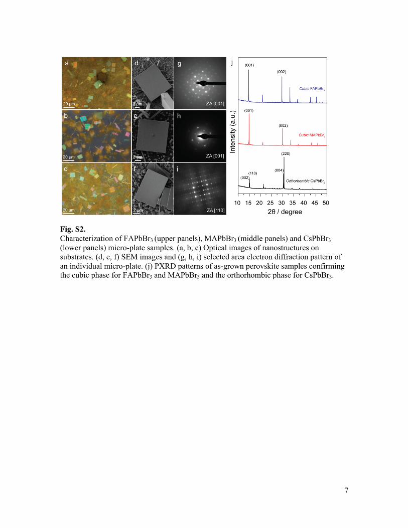
Fig. S2. Characterization of FAPbBr3 (upper panels), MAPbBr3 (middle panels) and CsPbBr3 (lower panels) micro-plate samples. (a, b, c) Optical images of nanostructures on substrates. (d, e, f) SEM images and (g, h, i) selected area electron diffraction pattern of an individual micro-plate. (j) PXRD patterns of as-grown perovskite samples confirming the cubic phase for FAPbBr3 and MAPbBr3 and the orthorhombic phase for CsPbBr3.
8
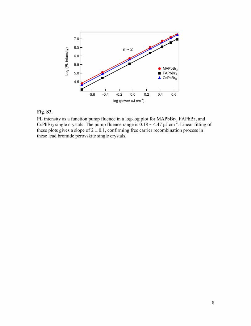
Fig. S3. PL intensity as a function pump fluence in a log-log plot for MAPbBr3, FAPbBr3 and CsPbBr3 single crystals. The pump fluence range is 0.18 ~ 4.47 µJ cm-2. Linear fitting of these plots gives a slope of 2 ± 0.1, confirming free carrier recombination process in these lead bromide perovskite single crystals.
7.0
6.5
6.0
5.5
5.0
4.5
Log
(PL
inte
nsity
)
-0.6 -0.4 -0.2 0.0 0.2 0.4 0.6
log (power uJ cm-2)
MAPbBr3 FAPbBr3 CsPbBr3
n ~ 2
9
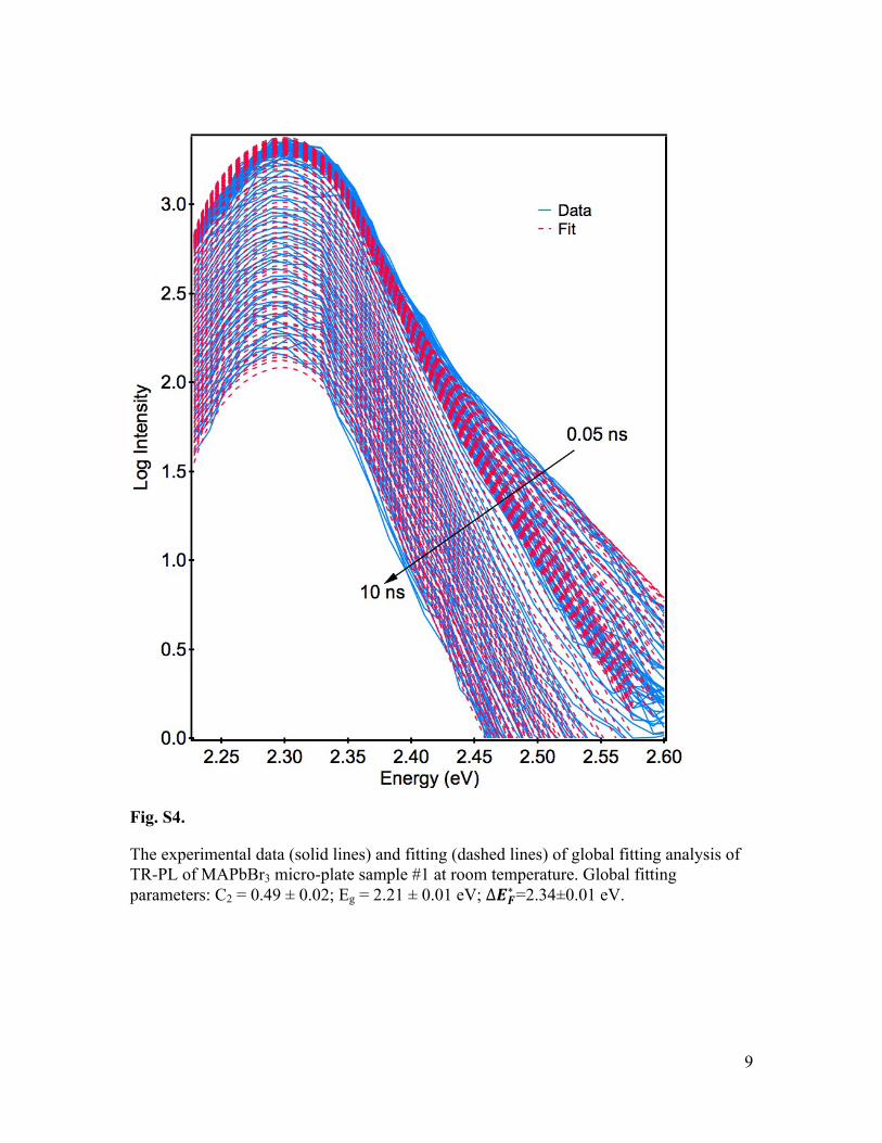
Fig. S4.
The experimental data (solid lines) and fitting (dashed lines) of global fitting analysis of TR-PL of MAPbBr3 micro-plate sample #1 at room temperature. Global fitting parameters: C2 = 0.49 ± 0.02; Eg = 2.21 ± 0.01 eV; ∆𝑬𝑭∗=2.34±0.01 eV.
10
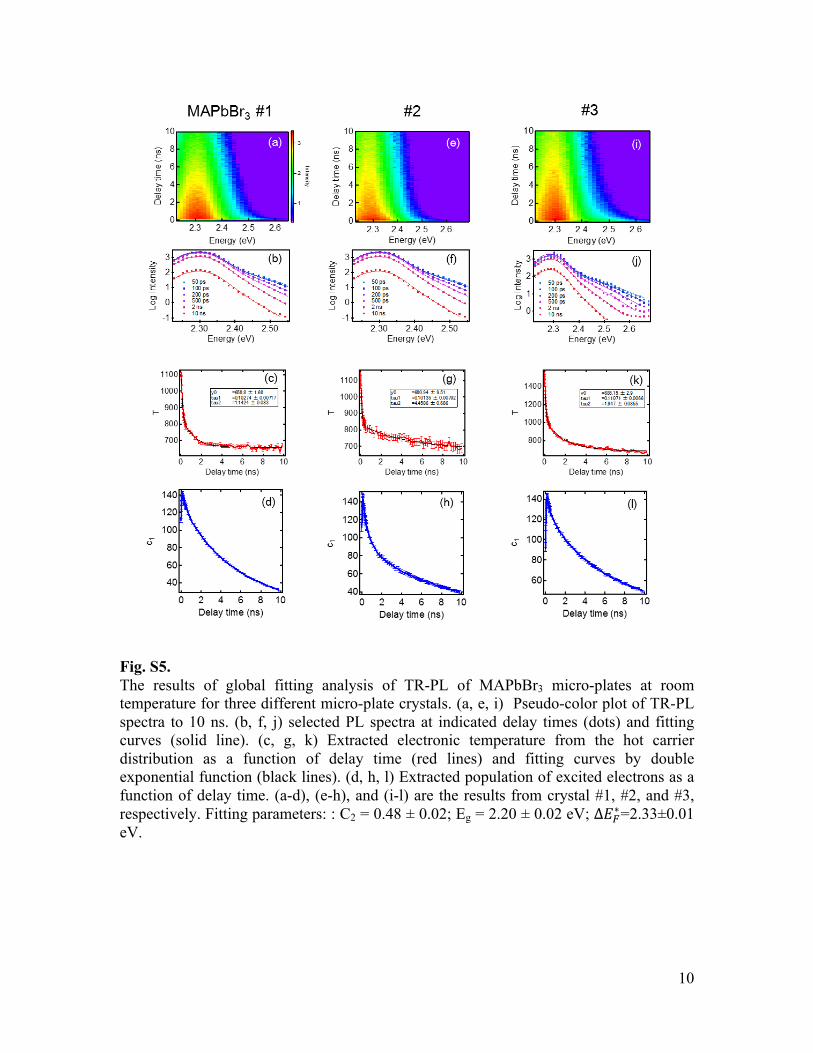
Fig. S5. The results of global fitting analysis of TR-PL of MAPbBr3 micro-plates at room temperature for three different micro-plate crystals. (a, e, i) Pseudo-color plot of TR-PL spectra to 10 ns. (b, f, j) selected PL spectra at indicated delay times (dots) and fitting curves (solid line). (c, g, k) Extracted electronic temperature from the hot carrier distribution as a function of delay time (red lines) and fitting curves by double exponential function (black lines). (d, h, l) Extracted population of excited electrons as a function of delay time. (a-d), (e-h), and (i-l) are the results from crystal #1, #2, and #3, respectively. Fitting parameters: : C2 = 0.48 ± 0.02; Eg = 2.20 ± 0.02 eV; ∆𝐸!∗=2.33±0.01 eV.

11
Fig. S6. Pseudo-color plot of TR-PL spectra of additional FAPbBr3 micro-plates at room temperature.
12
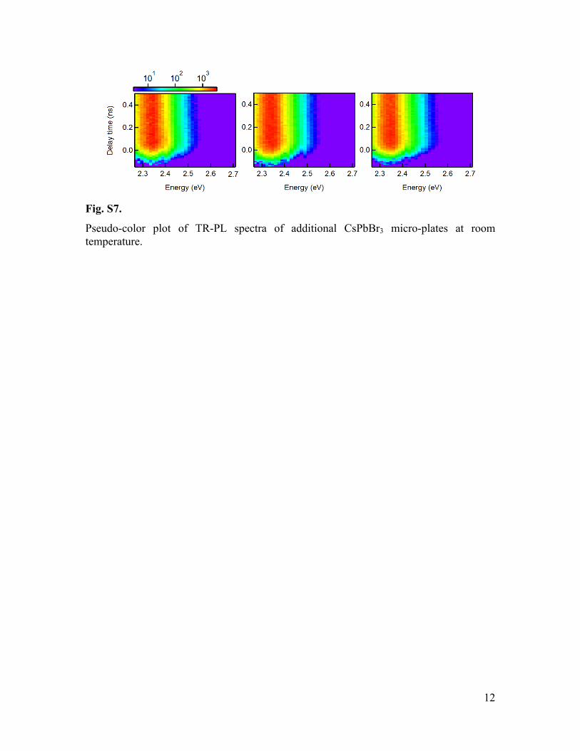
Fig. S7.
Pseudo-color plot of TR-PL spectra of additional CsPbBr3 micro-plates at room temperature.

13
Fig. S8. The results of global fitting analysis of TR-PL of FAPbBr3 micro-plates at room temperature. (a) Pseudo-color plot of TR-PL spectra to 10 ns. (b) selected PL spectra at indicated delay times (dots) and fitting curves (solid line). (c) Extracted electronic temperature from the hot carrier distribution as a function of delay time (red lines) and fitting curves by double exponential function (black lines). (d) Total PL decay kinetics. 𝑐!, 𝐸!, and ∆𝐸!∗ are, 0.30±0.01, 2.15±0.01 eV, and 2.31±0.01 eV, respectively.
14
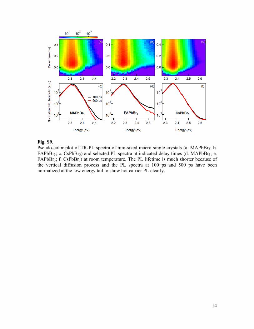
Fig. S9. Pseudo-color plot of TR-PL spectra of mm-sized macro single crystals (a. MAPbBr3; b. FAPbBr3; c. CsPbBr3) and selected PL spectra at indicated delay times (d. MAPbBr3; e. FAPbBr3; f. CsPbBr3) at room temperature. The PL lifetime is much shorter because of the vertical diffusion process and the PL spectra at 100 ps and 500 ps have been normalized at the low energy tail to show hot carrier PL clearly.

15
Fig. S10. The results of global fitting analysis of TR-PL of MAPbBr3 micro-plates at 180 K. (a) Pseudo-color plot of TR-PL spectra to 10 ns. (b) selected PL spectra at indicated delay times (dots) and fitting curves (solid line). (c) Extracted electronic temperature from the hot carrier distribution as a function of delay time (red lines) and fitting curves by double exponential function (black lines). (d) Extracted population of excited electrons as a function of delay time. 𝑐!, 𝐸!, and ∆𝐸!∗ are, 0.31±0.01, 2.22±0.01 eV, and 2.32±0.01 eV, respectively.

16
Fig. S11. PL spectra at early (100 ps) and later (500 ps) delay times for FAPbBr3; single crystals at different excitation fluences (a. 0.85 µJ cm-2; b. 4.40 µJ cm-2; c. 8.93 µJ cm-2)
17
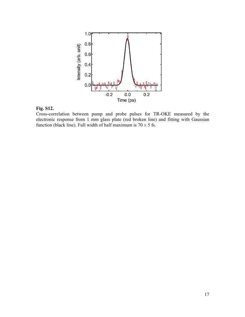
Fig. S12. Cross-correlation between pump and probe pulses for TR-OKE measured by the electronic response from 1 mm glass plate (red broken line) and fitting with Gaussian function (black line). Full width of half maximum is 70 ± 5 fs.

18
Table S1. Table S1. Lattice constants for cubic MAPbBr3 (space group Pm3m), cubic FAPbBr3
(space group Pm3m), and orthorhombic CsPbBr3 (space group Pbnm).
a b c
MAPbBr3 5.924 Å
FAPbBr3 5.987 Å
CsPbBr3 8.223 Å 8.243 Å 11.761 Å

19
References and Notes 1. M. M. Lee, J. Teuscher, T. Miyasaka, T. N. Murakami, H. J. Snaith, Efficient hybrid solar
cells based on meso-superstructured organometal halide perovskites. Science 338, 643–647 (2012). Medline doi:10.1126/science.1228604
2. S. D. Stranks, G. E. Eperon, G. Grancini, C. Menelaou, M. J. Alcocer, T. Leijtens, L. M. Herz, A. Petrozza, H. J. Snaith, Electron-hole diffusion lengths exceeding 1 micrometer in an organometal trihalide perovskite absorber. Science 342, 341–344 (2013). Medline doi:10.1126/science.1243982
3. G. Xing, N. Mathews, S. Sun, S. S. Lim, Y. M. Lam, M. Grätzel, S. Mhaisalkar, T. C. Sum, Long-range balanced electron- and hole-transport lengths in organic-inorganic CH3NH3PbI3. Science 342, 344–347 (2013). Medline doi:10.1126/science.1243167
4. T. M. Brenner, D. Egger, L. Kronik, G. Hodes, D. Cahen, Hybrid organic—inorganic perovskites: Low-cost semiconductors with intriguing charge-transport properties. Nat. Rev. Mater. 1, 15007 (2016). doi:10.1038/natrevmats.2015.7
5. K. Miyano, N. Tripathi, M. Yanagida, Y. Shirai, Lead Halide Perovskite Photovoltaic as a Model p-i-n Diode. Acc. Chem. Res. 49, 303–310 (2016). Medline doi:10.1021/acs.accounts.5b00436
6. T. J. Savenije, C. S. Ponseca Jr., L. Kunneman, M. Abdellah, K. Zheng, Y. Tian, Q. Zhu, S. E. Canton, I. G. Scheblykin, T. Pullerits, A. Yartsev, V. Sundström, Thermally activated exciton dissociation and recombination control the carrier dynamics in organometal halide perovskite. J. Phys. Chem. Lett. 5, 2189–2194 (2014). Medline doi:10.1021/jz500858a
7. H. T. Yi, X. Wu, X. Zhu, V. Podzorov, Intrinsic Charge Transport across Phase Transitions in Hybrid Organo-Inorganic Perovskites. Adv. Mater. 28, 6509–6514 (2016). 10.1002/adma.201600011 Medline doi:10.1002/adma.201600011
8. M. Karakus, S. A. Jensen, F. D’Angelo, D. Turchinovich, M. Bonn, E. Cánovas, Phonon-electron scattering limits free charge mobility in methylammonium lead iodide perovskites. J. Phys. Chem. Lett. 6, 4991–4996 (2015). Medline doi:10.1021/acs.jpclett.5b02485
9. J. Bardeen, W. Shockley, Deformation potentials and mobilities in non-polar crystals. Phys. Rev. 80, 72–80 (1950). doi:10.1103/PhysRev.80.72
10. Q. Dong, Y. Fang, Y. Shao, P. Mulligan, J. Qiu, L. Cao, J. Huang, Electron-hole diffusion lengths > 175 μm in solution-grown CH3NH3PbI3 single crystals. Science 347, 967–970 (2015). doi:10.1126/science.aaa5760
11. H. Oga, A. Saeki, Y. Ogomi, S. Hayase, S. Seki, Improved understanding of the electronic and energetic landscapes of perovskite solar cells: High local charge carrier mobility, reduced recombination, and extremely shallow traps. J. Am. Chem. Soc. 136, 13818–13825 (2014). Medline doi:10.1021/ja506936f
12. C. Wehrenfennig, M. Liu, H. J. Snaith, M. B. Johnston, L. M. Herz, Charge-carrier dynamics in vapour-deposited films of the organolead halide perovskite CH3NH3PbI3−xClx. Energy Environ. Sci. 7, 2269 (2014). doi:10.1039/c4ee01358a

20
13. A. M. Leguy, J. M. Frost, A. P. McMahon, V. G. Sakai, W. Kochelmann, C. Law, X. Li, F. Foglia, A. Walsh, B. C. O’Regan, J. Nelson, J. T. Cabral, P. R. Barnes, The dynamics of methylammonium ions in hybrid organic-inorganic perovskite solar cells. Nat. Commun. 6, 7124 (2015). Medline doi:10.1038/ncomms8124
14. I. P. Swainson, C. Stock, S. F. Parker, L. Van Eijck, M. Russina, J. W. Taylor, From soft harmonic phonons to fast relaxational dynamics in CH3NH3PbBr3. Phys. Rev. B 92, 100303 (2015). doi:10.1103/PhysRevB.92.100303
15. N. Onoda-Yamamuro, T. Matsuo, H. Suga, Dielectric study of CH3NH3PbX3 (X = Cl, Br, I). J. Phys. Chem. Solids 53, 935–939 (1992). doi:10.1016/0022-3697(92)90121-S
16. R. E. Wasylishen, O. Knop, J. B. Macdonald, Cation rotation in methylammonium lead halides. Solid State Commun. 56, 581–582 (1985). doi:10.1016/0038-1098(85)90959-7
17. D. Emin, Polarons (Cambridge Univ. Press, Cambridge, 2013).
18. X.-Y. Zhu, V. Podzorov, Charge carriers in hybrid organic-inorganic lead halide perovskites might be protected as large polarons. J. Phys. Chem. Lett. 6, 4758–4761 (2015). Medline doi:10.1021/acs.jpclett.5b02462
19. D. McMorrow, W. T. Lotshaw, G. Kenney-Wallace, Femtosecond optical Kerr studies on the origin of the nonlinear responses in simple liquids. IEEE J. Quantum Electron. 24, 443–454 (1988). doi:10.1109/3.144
20. J. Timmermans, Un nouvel état mésomorphe. Les cristaux organiques plastiques. J. Chim. Phys. 35, 331–344 (1938).
21. J. Even, M. Carignano, C. Katan, Molecular disorder and translation/rotation coupling in the plastic crystal phase of hybrid perovskites. Nanoscale 8, 6222–6236 (2016). Medline doi:10.1039/C5NR06386H
22. D. Shi, V. Adinolfi, R. Comin, M. Yuan, E. Alarousu, A. Buin, Y. Chen, S. Hoogland, A. Rothenberger, K. Katsiev, Y. Losovyj, X. Zhang, P. A. Dowben, O. F. Mohammed, E. H. Sargent, O. M. Bakr, Low trap-state density and long carrier diffusion in organolead trihalide perovskite single crystals. Science 347, 519–522 (2015). Medline doi:10.1126/science.aaa2725
23. H. Zhu, Y. Fu, F. Meng, X. Wu, Z. Gong, Q. Ding, M. V. Gustafsson, M. T. Trinh, S. Jin, X. Y. Zhu, Lead halide perovskite nanowire lasers with low lasing thresholds and high quality factors. Nat. Mater. 14, 636–642 (2015). Medline doi:10.1038/nmat4271
24. Y. Fu, H. Zhu, A. W. Schrader, D. Liang, Q. Ding, P. Joshi, L. Hwang, X. Y. Zhu, S. Jin, Nanowire lasers of formamidinium lead halide perovskites and their stabilized alloys with improved stability. Nano Lett. 16, 1000–1008 (2016). Medline doi:10.1021/acs.nanolett.5b04053
25. Y. Fu, H. Zhu, C. C. Stoumpos, Q. Ding, J. Wang, M. G. Kanatzidis, X. Zhu, S. Jin, Broad wavelength tunable robust lasing from single-crystal nanowires of cesium lead halide perovskites (CsPbX3, X = Cl, Br, I). ACS Nano 10, 7963–7972 (2016). 10.1021/acsnano.6b03916 doi:10.1021/acsnano.6b03916
26. Supplementary materials are available on Science Online.
21
27. Y. Yamada, T. Yamada, Q. Phuong, N. Maruyama, H. Nishimura, A. Wakamiya, Y. Murata, Y. Kanemitsu, Dynamic optical properties of CH₃NH₃PbI₃ single crystals as revealed by one- and two-photon excited photoluminescence measurements. J. Am. Chem. Soc. 137, 10456–10459 (2015). Medline doi:10.1021/jacs.5b04503
28. L. M. Pazos-Outón, M. Szumilo, R. Lamboll, J. M. Richter, M. Crespo-Quesada, M. Abdi-Jalebi, H. J. Beeson, M. Vrućinić, M. Alsari, H. J. Snaith, B. Ehrler, R. H. Friend, F. Deschler, Photon recycling in lead iodide perovskite solar cells. Science 351, 1430–1433 (2016). Medline doi:10.1126/science.aaf1168
29. M. Saba, M. Cadelano, D. Marongiu, F. Chen, V. Sarritzu, N. Sestu, C. Figus, M. Aresti, R. Piras, A. Geddo Lehmann, C. Cannas, A. Musinu, F. Quochi, A. Mura, G. Bongiovanni, Correlated electron-hole plasma in organometal perovskites. Nat. Commun. 5, 5049 (2014). Medline doi:10.1038/ncomms6049
30. C. Wehrenfennig, M. Liu, H. J. Snaith, M. B. Johnston, L. M. Herz, Homogeneous emission line broadening in the organo lead halide perovskite CH3NH3PbI3-xClx. J. Phys. Chem. Lett. 5, 1300–1306 (2014). Medline doi:10.1021/jz500434p
31. K. Chen, A. J. Barker, F. L. C. Morgan, J. E. Halpert, J. M. Hodgkiss, Effect of Carrier Thermalization Dynamics on Light Emission and Amplification in Organometal Halide Perovskites. J. Phys. Chem. Lett. 6, 153–158 (2015). Medline doi:10.1021/jz502528c
32. J. M. Frost, A. Walsh, What is moving in hybrid halide perovskite solar cells? Acc. Chem. Res. 49, 528–535 (2016). Medline doi:10.1021/acs.accounts.5b00431
33. R. Righini, Ultrafast optical kerr effect in liquids and solids. Science 262, 1386–1390 (1993). Medline doi:10.1126/science.262.5138.1386
34. P. Foggi, R. Righini, R. Torre, L. Angeloni, S. Califano, The dynamics of succinonitrile in the plastic phase by subpicosecond time‐resolved optical Kerr effect. J. Chem. Phys. 96, 110–115 (1992). doi:10.1063/1.462500
35. M. T. Weller, O. J. Weber, J. M. Frost, A. Walsh, Cubic Perovskite Structure of Black Formamidinium lead iodide, α-[HC(NH2)2]PbI3, at 298 K. J. Phys. Chem. Lett. 6, 3209–3212 (2015). doi:10.1021/acs.jpclett.5b01432
36. A. A. Bakulin, O. Selig, H. J. Bakker, Y. L. Rezus, C. Müller, T. Glaser, R. Lovrincic, Z. Sun, Z. Chen, A. Walsh, J. M. Frost, T. L. Jansen, Real-time observation of organic cation reorientation in methylammonium lead iodide perovskites. J. Phys. Chem. Lett. 6, 3663–3669 (2015). Medline doi:10.1021/acs.jpclett.5b01555
37. N. Balkan, Hot Electrons in Semiconductors (Oxford Univ. Press, 1998).
38. A. J. Nozik, Spectroscopy and hot electron relaxation dynamics in semiconductor quantum wells and quantum dots. Annu. Rev. Phys. Chem. 52, 193–231 (2001). Medline doi:10.1146/annurev.physchem.52.1.193
39. U. Bockelmann, G. Bastard, Phonon scattering and energy relaxation in two-, one-, and zero-dimensional electron gases. Phys. Rev. B Condens. Matter 42, 8947–8951 (1990). Medline doi:10.1103/PhysRevB.42.8947
22
40. Y. Yang, D. P. Ostrowski, R. M. France, K. Zhu, J. van de Lagemaat, J. M. Luther, M. C. Beard, Observation of a hot-phonon bottleneck in lead-iodide perovskites. Nat. Photonics 10, 53–59 (2016). doi:10.1038/nphoton.2015.213
41. M. B. Price, J. Butkus, T. C. Jellicoe, A. Sadhanala, A. Briane, J. E. Halpert, K. Broch, J. M. Hodgkiss, R. H. Friend, F. Deschler, Hot-carrier cooling and photoinduced refractive index changes in organic-inorganic lead halide perovskites. Nat. Commun. 6, 8420 (2015). Medline doi:10.1038/ncomms9420
42. L. Landau, On the motion of electrons in a crystal lattice. Phys. Z. Sowjetunion 3, 664 (1933).
43. G. R. Berdiyorov, A. Kachmar, F. El-mellouhi, M. A. Carignano, M. El-Amine Madjet, Role of cations on the electronic transport and optical properties of lead-iodide perovskites. J. Phys. Chem. C 120, 16259–16270 (2016). doi:10.1021/acs.jpcc.6b01818
44. Y. J. Chang, E. W. Castner, Fast responses from “slowly relaxing” liquids: A comparative study of the femtosecond dynamics of triacetin, ethylene glycol, and water. J. Chem. Phys. 99, 7289–7299 (1993). doi:10.1063/1.465710
45. Y. Zhou, L. You, S. Wang, Z. Ku, H. Fan, D. Schmidt, A. Rusydi, L. Chang, L. Wang, P. Ren, L. Chen, G. Yuan, L. Chen, J. Wang, Giant photostriction in organic-inorganic lead halide perovskites. Nat. Commun. 7, 11193 (2016). Medline doi:10.1038/ncomms11193
46. A. Pecchia, D. Gentilini, D. Rossi, M. Auf der Maur, A. Di Carlo, Role of ferroelectric nanodomains in the transport properties of perovskite solar cells. Nano Lett. 16, 988–992 (2016). Medline doi:10.1021/acs.nanolett.5b03957
47. C. Quarti, E. Mosconi, F. De Angelis, Structural and electronic properties of organo-halide hybrid perovskites from ab initio molecular dynamics. Phys. Chem. Chem. Phys. 17, 9394–9409 (2015). Medline doi:10.1039/C5CP00599J
48. S. A. March et al., https://arxiv.org/abs/1602.05186 (2016).
49. R. T. Ross, A. J. Nozik, Efficiency of hot-carrier solar energy converters. J. Appl. Phys. 53, 3813–3818 (1982). doi:10.1063/1.331124
50. F. C. Hanusch, E. Wiesenmayer, E. Mankel, A. Binek, P. Angloher, C. Fraunhofer, N. Giesbrecht, J. M. Feckl, W. Jaegermann, D. Johrendt, T. Bein, P. Docampo, Efficient planar heterojunction perovskite solar cells based on formamidinium lead bromide. J. Phys. Chem. Lett. 5, 2791–2795 (2014). Medline doi:10.1021/jz501237m
51. M. Rodová, J. Brožek, K. Knížek, K. Nitsch, Phase transitions in ternary caesium lead bromide. J. Therm. Anal. Calorim. 71, 667–673 (2003). doi:10.1023/A:1022836800820


![Multiphase Reacting Flows[1]](https://img.pdfslide.us/doc/110x75/577c78321a28abe0548f08d8/multiphase-reacting-flows1.jpg)