Embed Size (px)
Citation preview
Chemistry & Biology, Volume 19
Supplemental Information
Catalysis Uncoupling in a Glutamine
Amidotransferase Bienzyme
by Unblocking the Glutaminase Active Site Felix List, M. Cristina Vega, Adelia Razeto, Michaela C. Häger, Reinhard Sterner, and Matthias Wilmanns
Table S1: X-ray data collection and refinement statistics, related to Figure 2. Inventory of Supplemental Information
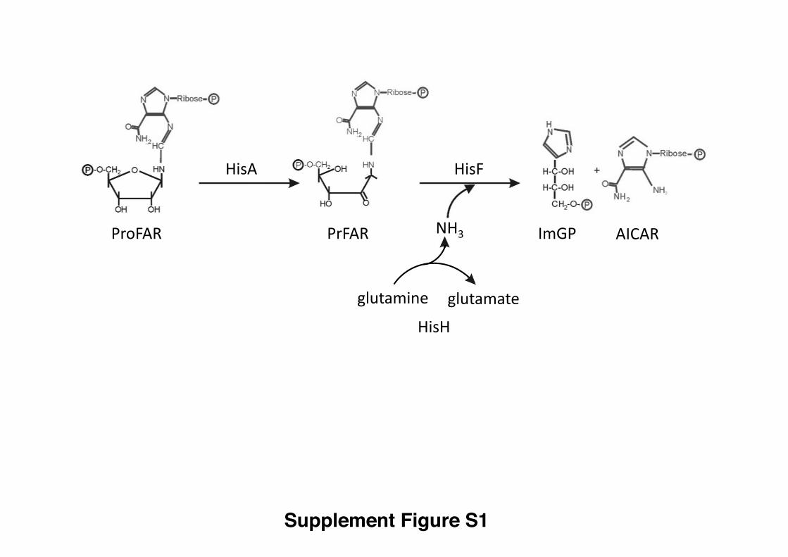
Figure S1. Schematic view of the reactions catalyzed by HisA and ImGP showing
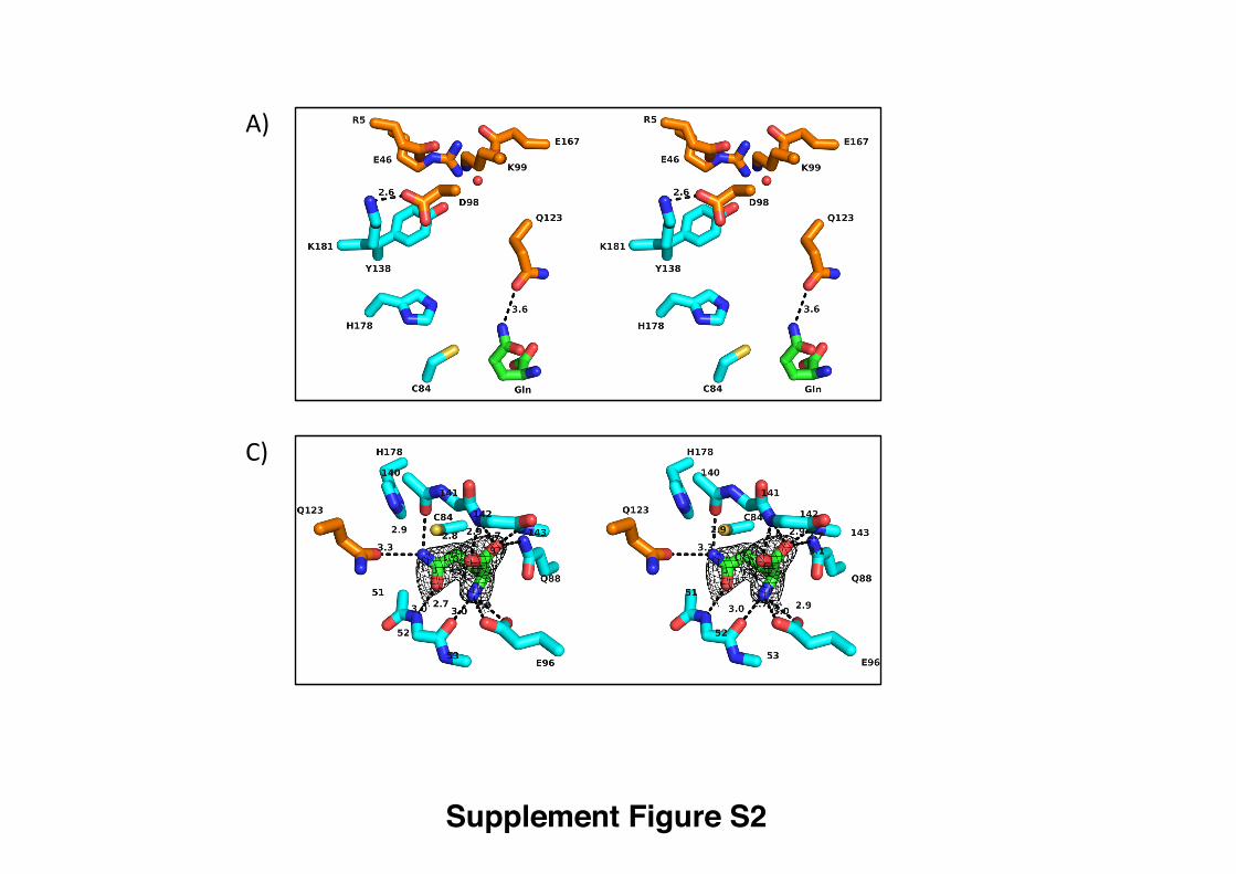
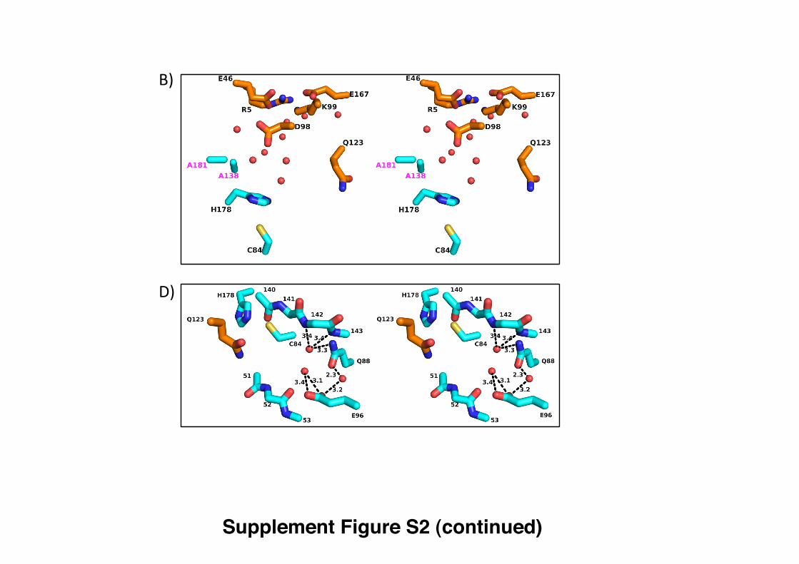
the structures of PrFAR, ProFAR, AICAR and ImGP, related to Figure 1. Figure S2. Stereo version of Figure 3, with hydrogen bond distances labeled,
related to Figure 3. Figure S3: Superimposed glutaminase active-site structures, related to Figure 3. Figure S4. Assays used for steady-state enzyme kinetic analysis, related to Figure
4.
S1
Supplemental Data
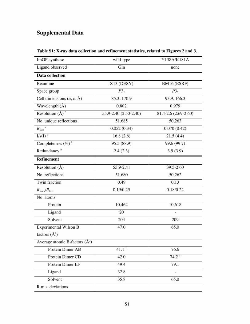
Table S1: X-ray data collection and refinement statistics, related to Figures 2 and 3.
ImGP synthase wild-type Y138A/K181A Ligand observed Gln none
Data collection
Beamline X13 (DESY) BM16 (ESRF) Space group P32 P32
Cell dimensions (a, c, Å) 85.3, 170.9 93.9, 166.3 Wavelength (Å) 0.802 0.979 Resolution (Å) * 55.9-2.40 (2.50-2.40) 81.4-2.6 (2.69-2.60) No. unique reflections 51,685 50,263 Rsym
a 0.052 (0.34) 0.070 (0.42)
I/s(I) a 16.8 (2.6) 21.5 (4.4) Completeness (%) b 95.5 (88.9) 99.6 (99.7) Redundancy b 2.4 (2.3) 3.9 (3.9)
Refinement
Resolution (Å) 55.9-2.41 39.5-2.60 No. reflections 51,680 50,262 Twin fraction 0.49 0.13 Rwork/Rfree 0.19/0.25 0.18/0.22 No. atoms Protein 10,462 10,618 Ligand 20 - Solvent 204 209 Experimental Wilson B factors (Å2)
47.0 65.0
Average atomic B-factors (Å2) Protein Dimer AB 41.1 † 76.6 Protein Dimer CD 42.0 74.2 † Protein Dimer EF 49.4 79.1 Ligand 32.8 - Solvent 35.8 65.0 R.m.s. deviations
S2
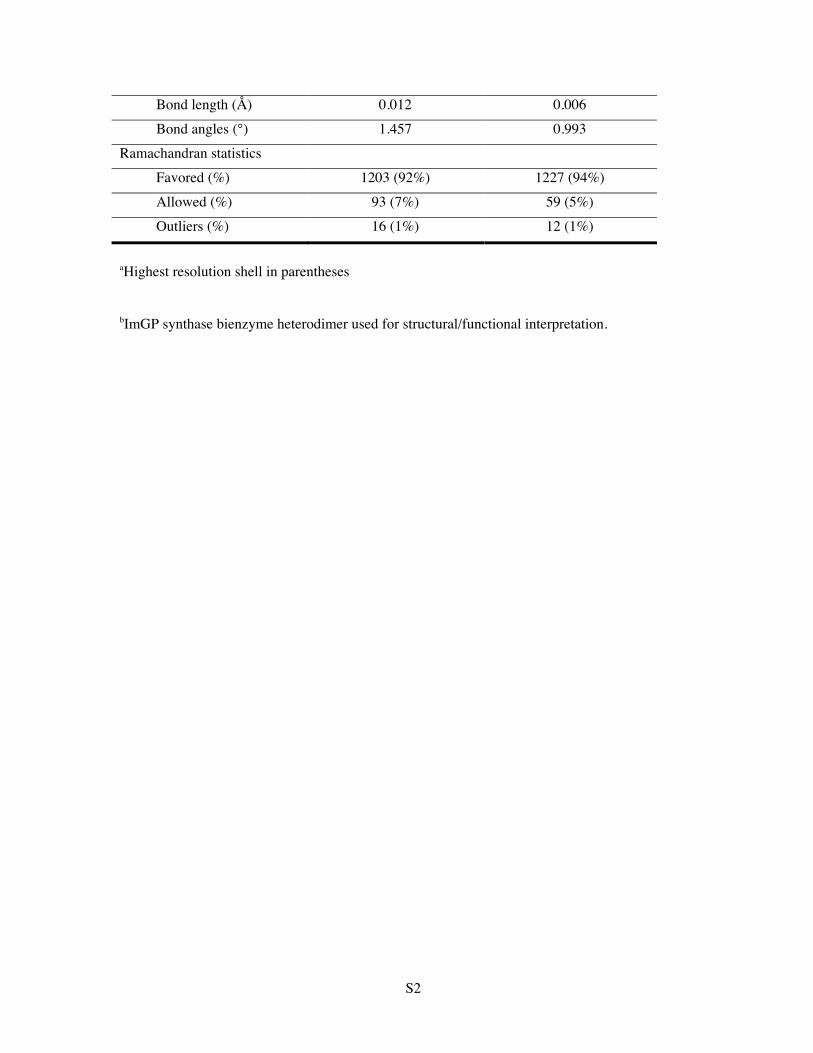
Bond length (Å) 0.012 0.006 Bond angles (°) 1.457 0.993 Ramachandran statistics Favored (%) 1203 (92%) 1227 (94%) Allowed (%) 93 (7%) 59 (5%) Outliers (%) 16 (1%) 12 (1%)
aHighest resolution shell in parentheses
bImGP synthase bienzyme heterodimer used for structural/functional interpretation.
S3
Supplementary Figures
Figure S1. Schematic view of the reactions catalyzed by HisA and ImGP showing
the structures of PrFAR, ProFAR, AICAR and ImGP, related to Figure 1.
Figure S2. Stereo version of Figure 3, with hydrogen bond distances labeled, related
to Figure 3.
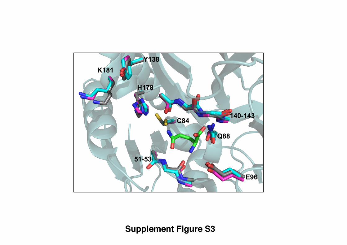
Figure S3: Superimposed glutaminase active-site structures, related to Figure 3.
Apo-ImGP synthase (Douangamath et al., 2002) (carbon atoms, magenta); ImGP
synthase-glutamine complex (carbon atoms cyan, this contribution); separate glutaminase
subunit of ImGP synthase (Douangamath et al., 2002) (carbon atoms, grey). Atom-
specific colors are used for all remaining atoms shown. Bound glutamine is also shown
(carbon atoms in green).
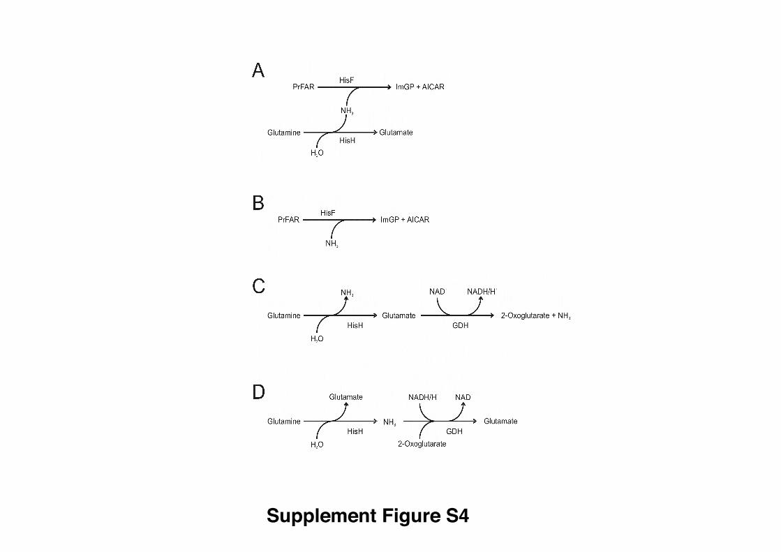
Figure S4. Assays used for steady-state enzyme kinetic analysis, related to Figure 4.
(A) Glutamine-dependent cyclase activity; (B) Ammonia-dependent cyclase activity; (C)
Glutaminase activity as recorded by glutamate production; (D) Glutaminase activity as
recorded by ammonia production. The relations of the assays to the overall reaction
scheme of ImGP synthase reaction are also indicated in Figure 1.
S4
Supplemental Experimental Procedures
Reaction conditions for steady state kinetics
Glutamine-dependent cyclase assay: 50 mM Tris-acetate buffer, pH 8.0, 0.2-2.0 µM
HisH, 0.1-1.0 µM HisF, 5-50 mM L-glutamine, 0-40 µM ProFAR.
Ammonia-dependent cyclase assay: 50 mM tris-acetate buffer, pH 8.5, 0.2 µM HisH,
0.1 µM HisF, 100 mM NH4CH3CO2 corresponding to 17.4 mM NH3, 0-40 µM
ProFAR.
Continuous glutaminase assay to monitor glutamate production: 50 mM tricine-
hydroxide buffer, pH 8.0, 2-8 µM HisF, 1-4 µM HisH, 40 µM ProFAR, 0-5 mM L-
glutamine, 3.8-6.7 mg/ml GDH, 11-20 mM NAD+; continuous glutaminase assay to
monitor ammonia production, 50 mM tricine-hydroxide buffer, pH 8.0, 2 µM HisF,
1 µM HisH, 40 µM ProFAR, 10 mM Gln, 2.1 mg/ml GDH, 10 mM α-ketoglutarate,
300 µM NADH/H+. GDH from beef liver was purchased from Roche Diagnostics GmbH
(Cat.no. 10197734001). According to the manufacturer’s certificate the ammonia
concentration was less than 1 ug / mg lyophilisate and the specific activity about 10U /
mg lyophilisate. The GDH concentration was adjusted in such a way that further addition
of GDH did not accelerate the observed turnover.
Discontinuous glutaminase assay: 0.15-15 µM HisF, 0.25-10 µM HisH, 10 mM L-
glutamine.
S5
Glutamate quantification: 100 µl of the filtrate were incubated with 10 mM NAD+ and
0.75 mg/ml GDH for at least one hour at 37 °C in a final volume of 750 µl.
Glutamate competition assay: ImGP synthase variants were incubated at 25°C with
1.25 µl [14C]-glutamine (specific activity 262 mCi/mmol) and 5 mM nonradioactive
glutamine in a total volume of 50 µl of 50 mM tricine-hydroxide buffer, pH 8.0. Samples
were prepared in the presence of non-radioactive glutamate at three different
concentrations (0, 5 and 20 mM), with/without 40 µM ProFAR. Reaction conditions,
ProFAR present: incubation time 5 min for 1µM HisH (all variants except wt) and 2 µM
HisF, or 30 µM wt HisH and 60 µM HisF; reaction conditions, ProFAR absent:
incubation time 20 min for 0.6 µM [Y138A(HisH),K181A(HisH)] and 1.2 µM HisF, 40
min for 15 µM Y138A(HisH) or K181A(HisH) and 30 µM HisF, 4.5 h for wt HisH and
90 µM HisF. A reaction mixture without enzyme was used as a control for spontaneous
glutamine hydrolysis. Following incubation, each of the reaction solutions was spun
through a 10 kDa filter to remove ImGP synthase. A total of 0.5 µl of each filtrate was
then spotted onto cellulose thin-layer chromatography (TLC) sheets (MN CEL 300-10).
The TLC sheets were developed using a solvent system of n-butanol : acetic acid : water
(12:3:5, by volume), dried and analyzed by using a phosphor imaging plate (Perkin
Elmer). Digital light units were read out with a Cyclone Storage Phosphor System
(Packard), using OptiQuant Software. Units, which were generated by [14C]-glutamate,
were normalized with overall units ([14C]-glutamate and [14C]-glutamine), within the
corresponding lanes after background subtraction. Normalized [14C]-glutamate units
formed in the presence of 5 or 20 mM non-radioactive glutamate are given relative to the
S6
corresponding units of [14C]-glutamate formed in the absence of non-radioactive
glutamate (Table S1).
X-ray structure determination
X-ray diffraction datasets were collected at the synchrotron radiation beamlines X13 and
BW7B at EMBL/DESY, Hamburg, Germany, and beamline BM16 at the ESRF,
Grenoble, at a temperature of 100 K. Intensities were integrated and scaled with
MOSFLM (Leslie, 2006) and SCALA (Leslie, 2006), or with HKL2000 (Otwinowski,
1997). Further statistics are summarized in Table 1. Submission of the structure factor
amplitudes to the Twin server (Yeates and Fam, 1999) revealed that all datasets were
pseudo-merohedrally twinned with twin fractions of 0.10-0.49. The three heterodimers of
wt and [Y138A(HisH), K181A(HisH)] mutant ImGP synthase were positioned and
orientated by molecular replacement with AMORE (Navaza, 2001) or with PHASER
(McCoy et al., 2007), using the previous apo structure of ImGP synthase (Douangamath
et al., 2002) as a search model.
After initial refinement of these coordinates in REFMAC5, phenix.refine (Adams et al.,
2004) was used for twin refinement, while retaining non-crystallographic symmetry
(NCS) restraints to ensure convergence of refinement at 2.4–2.8 Å. This substantially
improved the electron density of the third ImGP synthase hetero-dimer, and the R factor
statistics remained within acceptable values. Model building was carried out with Turbo-
Frodo (Roussel et al., 1990) and COOT (Emsley and Cowtan, 2004), and model
correctness was assessed with PROCHECK (Laskowski et al., 1993). All cyclase and
glutaminase subunits in the asymmetric unit are superimposable with an r.m.s.d. (Ca
S7
atoms) of 0.36 ± 0.02 Å and 0.41 ± 0.07 Å, respectively. When the three hetero-dimers
are compared, both as rigid and as flexible complexes, the r.m.s.d. is 0.40 Å (chains AB
versus CD) and 0.70 Å (chains AB or CD versus EF). It is worth noting that the two
glutamine complexes (chains AB and CD) are structurally more similar and better
ordered crystallographically than the third, glutamine-free hetero-dimer (chains EF). The
best defined hetero-dimeric glutaminase/cyclase complex from each crystal form, defined
by lowest overall temperature factors and assigned with chains A (cyclase) and B
(glutaminase), was used for further interpretation. We have noticed that the final average
residual atomic mobility factors in the refined Y138A(HisH), K181A(HisH)] mutant
ImGP synthase variant were substantially higher than those of the refined structure of the
wt bienzyme (Table S1). We confirmed this difference to be real, as the experimental
BWilson factors were estimated to be 47 Å2 for the wt bioenzyme and 65 Å2 for the
Y138A(HisH), K181A(HisH)] mutant.
S8
Supplemental References
Adams, P.D., Gopal, K., Grosse-Kunstleve, R.W., Hung, L.W., Ioerger, T.R., McCoy,
A.J., Moriarty, N.W., Pai, R.K., Read, R.J., Romo, T.D., et al. (2004). Recent
developments in the PHENIX software for automated crystallographic structure
determination. J Synchrotron Radiat 11, 53-55.
Amaro, R.E., Myers, R.S., Davisson, V.J., and Luthey-Schulten, Z.A. (2005). Structural
elements in IGP synthase exclude water to optimize ammonia transfer. Biophys J 89,
475-487.
Chaudhuri, B.N., Lange, S.C., Myers, R.S., Davisson, V.J., and Smith, J.L. (2003).
Toward understanding the mechanism of the complex cyclization reaction catalyzed
by imidazole glycerolphosphate synthase: crystal structures of a ternary complex and
the free enzyme. Biochemistry 42, 7003-7012.
Douangamath, A., Walker, M., Beismann-Driemeyer, S., Vega-Fernandez, M.C., Sterner,
R., and Wilmanns, M. (2002). Structural evidence for ammonia tunneling across the
(beta alpha)(8) barrel of the imidazole glycerol phosphate synthase bienzyme
complex. Structure 10, 185-193.
Emsley, P., and Cowtan, K. (2004). Coot: model-building tools for molecular graphics.
Acta Crystallogr D Biol Crystallogr 60, 2126-2132.
S9
Laskowski, R.A., MacArthur, M.W., and Thornton, J.M. (1993). PROCHECK: a
program to check the stereochemical quality of protein structures. J. Appl. Cryst. 26,
283-291.
Leslie, A.G. (2006). The integration of macromolecular diffraction data. Acta Crystallogr
D Biol Crystallogr 62, 48-57.
McCoy, A.J., Grosse-Kunstleve, R.W., Adams, P.D., Winn, M.D., Storoni, L.C., and
Read, R.J. (2007). Phaser crystallographic software. J Appl Crystallogr 40, 658-674.
Myers, R.S., Amaro, R.E., Luthey-Schulten, Z.A., and Davisson, V.J. (2005). Reaction
coupling through interdomain contacts in imidazole glycerol phosphate synthase.
Biochemistry 44, 11974-11985.
Navaza, J. (2001). Implementation of molecular replacement in AMoRe. Acta Crystallogr
D Biol Crystallogr 57, 1367-1372.
Otwinowski, Z., Minor, V. (1997). Processing of X-ray diffraction data collected in
oscillation mode. Meth. Enzym. 276, 307-326.
Roussel, A., Fontecilla-Camps, J.C., and Cambillau, C. (1990). CRYStallize: a
crystallographic symmetry display and handling subpackage in TOM/FRODO. J Mol
Graph 8, 86-88, 91.
Yeates, T.O., and Fam, B.C. (1999). Protein crystals and their evil twins. Structure 7,
R25-29.
Supplement Figure S1
ImGP AICARPrFARProFAR
glutamine glutamate
HisA HisF
HisH
NH3
Supplement Figure S2
A)
C)
Supplement Figure S2 (continued)
B)
D)
Y138 K181
H178
C84 140-143
E96
51-53
Q88
Supplement Figure S3
Supplement Figure S4