Embed Size (px)
Citation preview
PMA P180036: FDA Summary of Safety and Effectiveness Data Page 1
SUMMARY OF SAFETY AND EFFECTIVENESS DATA (SSED) I. GENERAL INFORMATION
Device Generic Name: Implantable Pulse Generator
Device Trade Name: OPTIMIZER Smart System
Device Procode: QFV
Applicant’s Name and Address: Impulse Dynamics (USA), Inc. 30 Ramland Rd, Suite 204 Orangeburg, NY 10962
Date(s) of Panel Recommendation: December 4, 2018
Premarket Approval Application (PMA) Number: P180036
Date of FDA Notice of Approval: March 21, 2019
Breakthrough Device: Granted breakthrough device status (formerly known as the Expedited Access Pathway, or EAP) on July 31, 2015, because the device treats a life-threatening disease (heart failure) and addresses an unmet medical need.
II. INDICATIONS FOR USE
The OPTIMIZER Smart System, which delivers Cardiac Contractility Modulation therapy, is indicated to improve 6-minute hall walk distance, quality of life, and functional status of NYHA Class III heart failure patients who remain symptomatic despite guideline directed medical therapy, who are in normal sinus rhythm, are not indicated for Cardiac Resynchro-nization Therapy, and have a left ventricular ejection fraction ranging from 25% to 45%.
III. CONTRAINDICATIONS
Use of the OPTIMIZER Smart system is contraindicated in: 1. Patients with permanent or long-standing persistent atrial fibrillation or flutter; 2. Patients with a mechanical tricuspid valve; and/or 3. Patients in whom vascular access for implantation of the leads cannot be obtained.
IV. WARNINGS AND PRECAUTIONS
The warnings and precautions can be found in the OPTIMIZER Smart labeling.
PMA P180036: FDA Summary of Safety and Effectiveness Data Page 2
V. DEVICE DESCRIPTION
The OPTIMIZER Smart system is comprised of the following components: OPTIMIZER Smart Implantable Pulse Generator (IPG) Commercially Available IS-1 Active Fixation Bipoar Pacing Leads OPTIMIZER Mini Charger OMNI II Programmer with OMNI SMART Software OPTIMIZER System Lead Extension Cable (Optional)
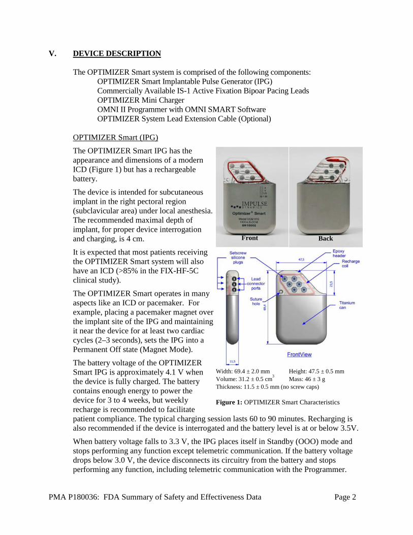
OPTIMIZER Smart (IPG) The OPTIMIZER Smart IPG has the appearance and dimensions of a modern ICD (Figure 1) but has a rechargeable battery. The device is intended for subcutaneous implant in the right pectoral region (subclavicular area) under local anesthesia. The recommended maximal depth of implant, for proper device interrogation and charging, is 4 cm. It is expected that most patients receiving the OPTIMIZER Smart system will also have an ICD (>85% in the FIX-HF-5C clinical study). The OPTIMIZER Smart operates in many aspects like an ICD or pacemaker. For example, placing a pacemaker magnet over the implant site of the IPG and maintaining it near the device for at least two cardiac cycles (2–3 seconds), sets the IPG into a Permanent Off state (Magnet Mode). The battery voltage of the OPTIMIZER Smart IPG is approximately 4.1 V when the device is fully charged. The battery contains enough energy to power the device for 3 to 4 weeks, but weekly recharge is recommended to facilitate patient compliance. The typical charging session lasts 60 to 90 minutes. Recharging is also recommended if the device is interrogated and the battery level is at or below 3.5V. When battery voltage falls to 3.3 V, the IPG places itself in Standby (OOO) mode and stops performing any function except telemetric communication. If the battery voltage drops below 3.0 V, the device disconnects its circuitry from the battery and stops performing any function, including telemetric communication with the Programmer.
Width: 69.4 ± 2.0 mm Height: 47.5 ± 0.5 mm Volume: 31.2 ± 0.5 cm3 Mass: 46 ± 3 g Thickness: 11.5 ± 0.5 mm (no screw caps) Figure 1: OPTIMIZER Smart Characteristics
Front Back
PMA P180036: FDA Summary of Safety and Effectiveness Data Page 3
However, it can be reactivated by the OPTIMIZER Mini Charger. The rechargeable battery in the OPTIMIZER IPG can be fully discharged without out causing any damage. Additional details pertaining to the operation of the IPG may be found in the Instructions for Use manuals.
Leads
Any commercially available IS-1, active fixation, bipolar pacing lead can be used with the OPTIMIZER Smart IPG. However, the two ventricular leads must meet the following requirements:
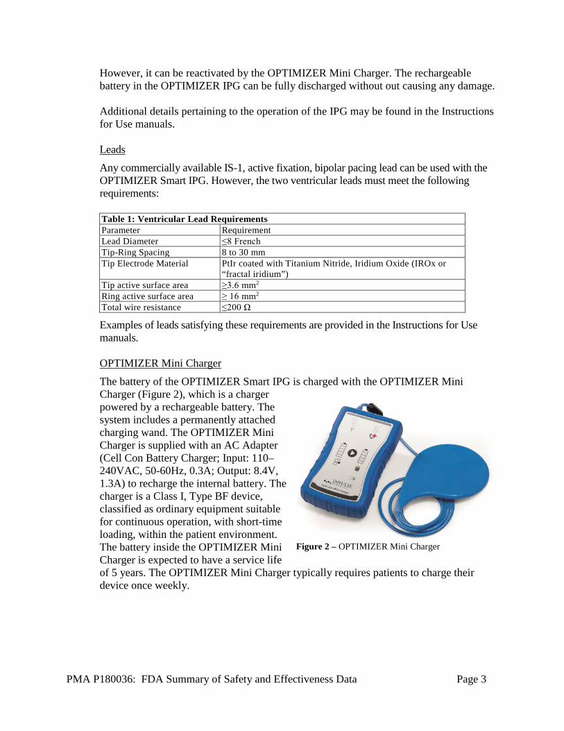
Examples of leads satisfying these requirements are provided in the Instructions for Use manuals. OPTIMIZER Mini Charger The battery of the OPTIMIZER Smart IPG is charged with the OPTIMIZER Mini Charger (Figure 2), which is a charger powered by a rechargeable battery. The system includes a permanently attached charging wand. The OPTIMIZER Mini Charger is supplied with an AC Adapter (Cell Con Battery Charger; Input: 110–240VAC, 50-60Hz, 0.3A; Output: 8.4V, 1.3A) to recharge the internal battery. The charger is a Class I, Type BF device, classified as ordinary equipment suitable for continuous operation, with short-time loading, within the patient environment. The battery inside the OPTIMIZER Mini Charger is expected to have a service life of 5 years. The OPTIMIZER Mini Charger typically requires patients to charge their device once weekly.
Table 1: Ventricular Lead Requirements Parameter Requirement Lead Diameter ≤8 French Tip-Ring Spacing 8 to 30 mm Tip Electrode Material PtIr coated with Titanium Nitride, Iridium Oxide (IROx or
“fractal iridium”) Tip active surface area ≥3.6 mm2 Ring active surface area ≥ 16 mm2 Total wire resistance ≤200 Ω
Figure 2 – OPTIMIZER Mini Charger
PMA P180036: FDA Summary of Safety and Effectiveness Data Page 4
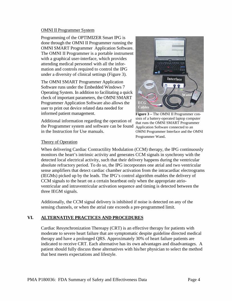
OMNI II Programmer System Programming of the OPTIMIZER Smart IPG is done through the OMNI II Programmer running the OMNI SMART Programmer Application Software. The OMNI II Programmer is a portable instrument with a graphical user-interface, which provides attending medical personnel with all the infor-mation and controls required to control the IPG under a diversity of clinical settings (Figure 3). The OMNI SMART Programmer Application Software runs under the Embedded Windows 7 Operating System. In addition to facilitating a quick check of important parameters, the OMNI SMART Programmer Application Software also allows the user to print out device related data needed for informed patient management. Additional information regarding the operation of the Programmer system and software can be found in the Instruction for Use manuals.
Theory of Operation When delivering Cardiac Contractility Modulation (CCM) therapy, the IPG continuously monitors the heart’s intrinsic activity and generates CCM signals in synchrony with the detected local electrical activity, such that their delivery happens during the ventricular absolute refractory period. To do so, the IPG incorporates one atrial and two ventricular sense amplifiers that detect cardiac chamber activation from the intracardiac electrograms (IEGMs) picked up by the leads. The IPG’s control algorithm enables the delivery of CCM signals to the heart on a certain heartbeat only when the appropriate atrio-ventricular and intraventricular activation sequence and timing is detected between the three IEGM signals. Additionally, the CCM signal delivery is inhibited if noise is detected on any of the sensing channels, or when the atrial rate exceeds a pre-programmed limit.
VI. ALTERNATIVE PRACTICES AND PROCEDURES
Cardiac Resynchronization Thereapy (CRT) is an effective therapy for patients with moderate to severe heart failure that are symptomatic despite guideline directed medical therapy and have a prolonged QRS. Approximately 30% of heart failure patients are indicated to receive CRT. Each alternative has its own advantages and disadvantages. A patient should fully discuss these alternatives with his/her physician to select the method that best meets expectations and lifestyle.
Figure 3 – The OMNI II Programmer con-sists of a battery-operated laptop computer that runs the OMNI SMART Programmer Application Software connected to an OMNI Programmer Interface and the OMNI Programmer Wand.
Wand
ECG Cables
PMA P180036: FDA Summary of Safety and Effectiveness Data Page 5
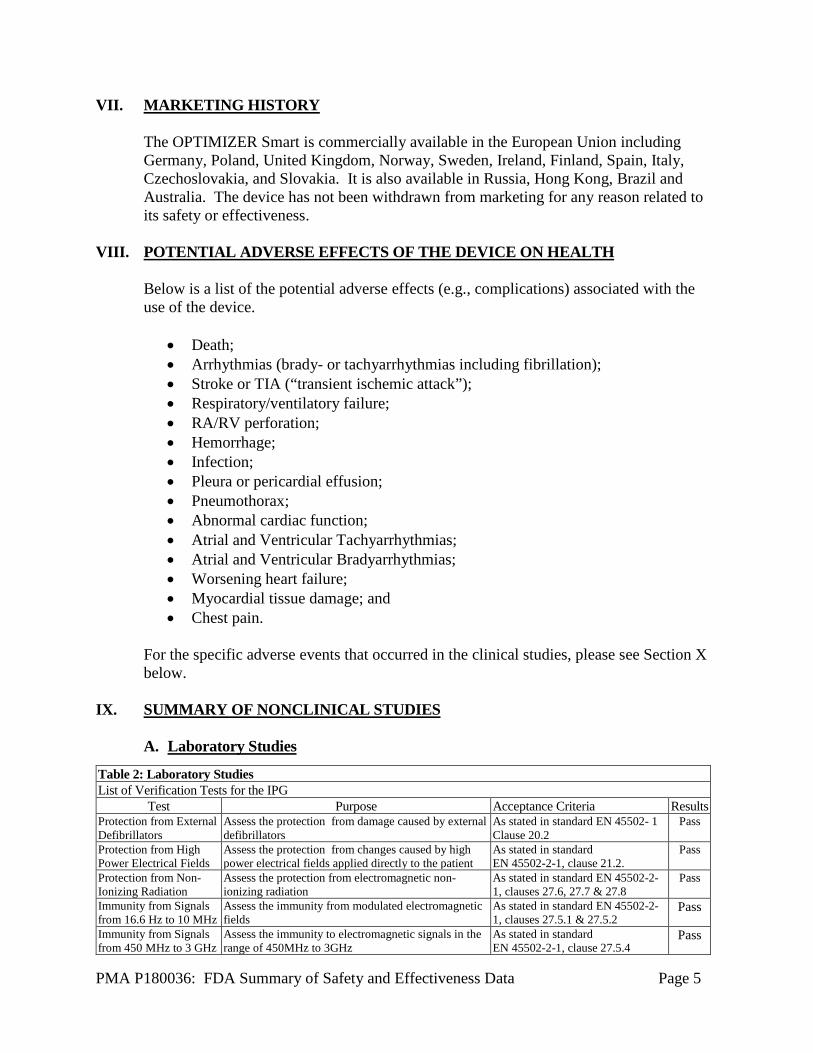
VII. MARKETING HISTORY
The OPTIMIZER Smart is commercially available in the European Union including Germany, Poland, United Kingdom, Norway, Sweden, Ireland, Finland, Spain, Italy, Czechoslovakia, and Slovakia. It is also available in Russia, Hong Kong, Brazil and Australia. The device has not been withdrawn from marketing for any reason related to its safety or effectiveness.
VIII. POTENTIAL ADVERSE EFFECTS OF THE DEVICE ON HEALTH
Below is a list of the potential adverse effects (e.g., complications) associated with the use of the device.
• Death; • Arrhythmias (brady- or tachyarrhythmias including fibrillation); • Stroke or TIA (“transient ischemic attack”); • Respiratory/ventilatory failure; • RA/RV perforation; • Hemorrhage; • Infection; • Pleura or pericardial effusion; • Pneumothorax; • Abnormal cardiac function; • Atrial and Ventricular Tachyarrhythmias; • Atrial and Ventricular Bradyarrhythmias; • Worsening heart failure; • Myocardial tissue damage; and • Chest pain.
For the specific adverse events that occurred in the clinical studies, please see Section X below.
IX. SUMMARY OF NONCLINICAL STUDIES
A. Laboratory Studies
Table 2: Laboratory Studies List of Verification Tests for the IPG
Test Purpose Acceptance Criteria Results Protection from External Defibrillators
Assess the protection from damage caused by external defibrillators
As stated in standard EN 45502- 1 Clause 20.2
Pass
Protection from High Power Electrical Fields
Assess the protection from changes caused by high power electrical fields applied directly to the patient
As stated in standard EN 45502-2-1, clause 21.2.
Pass
Protection from Non-Ionizing Radiation
Assess the protection from electromagnetic non- ionizing radiation
As stated in standard EN 45502-2-1, clauses 27.6, 27.7 & 27.8
Pass
Immunity from Signals from 16.6 Hz to 10 MHz
Assess the immunity from modulated electromagnetic fields
As stated in standard EN 45502-2-1, clauses 27.5.1 & 27.5.2
Pass
Immunity from Signals from 450 MHz to 3 GHz
Assess the immunity to electromagnetic signals in the range of 450MHz to 3GHz
As stated in standard EN 45502-2-1, clause 27.5.4
Pass
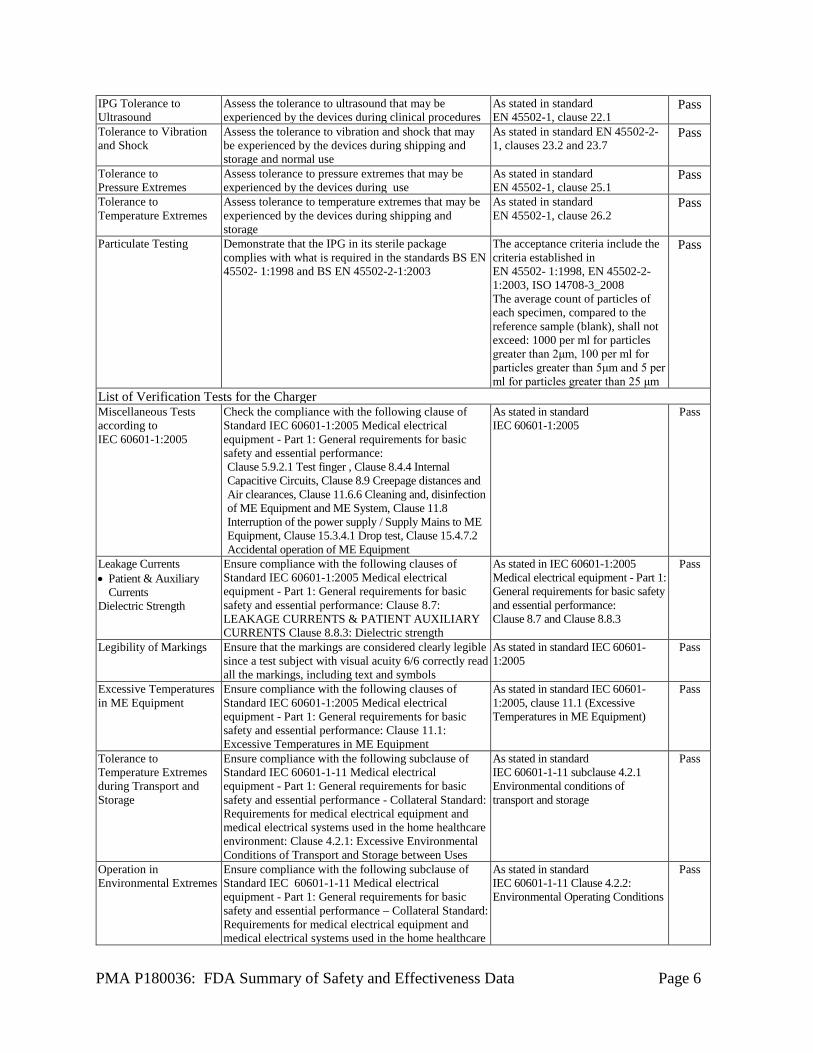
PMA P180036: FDA Summary of Safety and Effectiveness Data Page 6
IPG Tolerance to Ultrasound
Assess the tolerance to ultrasound that may be experienced by the devices during clinical procedures
As stated in standard EN 45502-1, clause 22.1
Pass
Tolerance to Vibration and Shock
Assess the tolerance to vibration and shock that may be experienced by the devices during shipping and storage and normal use
As stated in standard EN 45502-2-1, clauses 23.2 and 23.7
Pass
Tolerance to Pressure Extremes
Assess tolerance to pressure extremes that may be experienced by the devices during use
As stated in standard EN 45502-1, clause 25.1
Pass
Tolerance to Temperature Extremes
Assess tolerance to temperature extremes that may be experienced by the devices during shipping and storage
As stated in standard EN 45502-1, clause 26.2
Pass
Particulate Testing Demonstrate that the IPG in its sterile package complies with what is required in the standards BS EN 45502- 1:1998 and BS EN 45502-2-1:2003
The acceptance criteria include the criteria established in EN 45502- 1:1998, EN 45502-2-1:2003, ISO 14708-3_2008 The average count of particles of each specimen, compared to the reference sample (blank), shall not exceed: 1000 per ml for particles greater than 2μm, 100 per ml for particles greater than 5μm and 5 per ml for particles greater than 25 μm
Pass
List of Verification Tests for the Charger Miscellaneous Tests according to IEC 60601-1:2005
Check the compliance with the following clause of Standard IEC 60601-1:2005 Medical electrical equipment - Part 1: General requirements for basic safety and essential performance: Clause 5.9.2.1 Test finger , Clause 8.4.4 Internal Capacitive Circuits, Clause 8.9 Creepage distances and Air clearances, Clause 11.6.6 Cleaning and, disinfection of ME Equipment and ME System, Clause 11.8 Interruption of the power supply / Supply Mains to ME Equipment, Clause 15.3.4.1 Drop test, Clause 15.4.7.2 Accidental operation of ME Equipment
As stated in standard IEC 60601-1:2005
Pass
Leakage Currents • Patient & Auxiliary
Currents Dielectric Strength
Ensure compliance with the following clauses of Standard IEC 60601-1:2005 Medical electrical equipment - Part 1: General requirements for basic safety and essential performance: Clause 8.7: LEAKAGE CURRENTS & PATIENT AUXILIARY CURRENTS Clause 8.8.3: Dielectric strength
As stated in IEC 60601-1:2005 Medical electrical equipment - Part 1: General requirements for basic safety and essential performance: Clause 8.7 and Clause 8.8.3
Pass
Legibility of Markings Ensure that the markings are considered clearly legible since a test subject with visual acuity 6/6 correctly read all the markings, including text and symbols
As stated in standard IEC 60601-1:2005
Pass
Excessive Temperatures in ME Equipment
Ensure compliance with the following clauses of Standard IEC 60601-1:2005 Medical electrical equipment - Part 1: General requirements for basic safety and essential performance: Clause 11.1: Excessive Temperatures in ME Equipment
As stated in standard IEC 60601-1:2005, clause 11.1 (Excessive Temperatures in ME Equipment)
Pass
Tolerance to Temperature Extremes during Transport and Storage
Ensure compliance with the following subclause of Standard IEC 60601-1-11 Medical electrical equipment - Part 1: General requirements for basic safety and essential performance - Collateral Standard: Requirements for medical electrical equipment and medical electrical systems used in the home healthcare environment: Clause 4.2.1: Excessive Environmental Conditions of Transport and Storage between Uses
As stated in standard IEC 60601-1-11 subclause 4.2.1 Environmental conditions of transport and storage
Pass
Operation in Environmental Extremes
Ensure compliance with the following subclause of Standard IEC 60601-1-11 Medical electrical equipment - Part 1: General requirements for basic safety and essential performance – Collateral Standard: Requirements for medical electrical equipment and medical electrical systems used in the home healthcare
As stated in standard IEC 60601-1-11 Clause 4.2.2: Environmental Operating Conditions
Pass
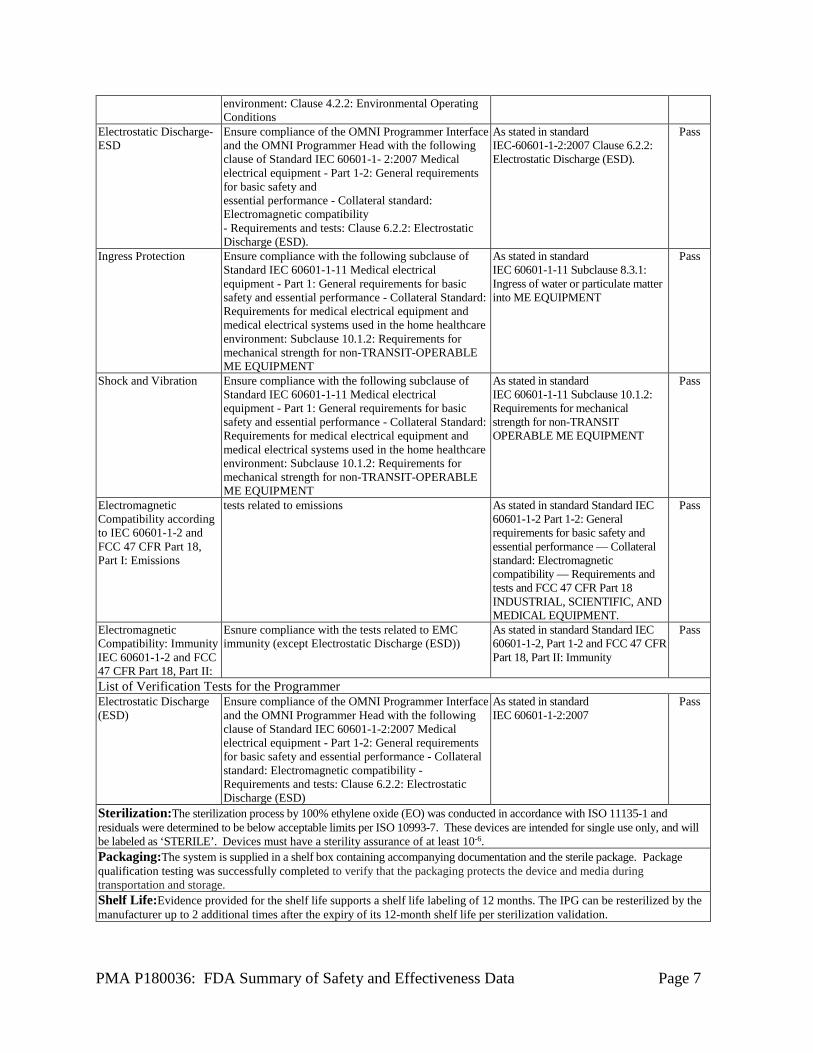
PMA P180036: FDA Summary of Safety and Effectiveness Data Page 7
environment: Clause 4.2.2: Environmental Operating Conditions
Electrostatic Discharge-ESD
Ensure compliance of the OMNI Programmer Interface and the OMNI Programmer Head with the following clause of Standard IEC 60601-1- 2:2007 Medical electrical equipment - Part 1-2: General requirements for basic safety and essential performance - Collateral standard: Electromagnetic compatibility - Requirements and tests: Clause 6.2.2: Electrostatic Discharge (ESD).
As stated in standard IEC-60601-1-2:2007 Clause 6.2.2: Electrostatic Discharge (ESD).
Pass
Ingress Protection Ensure compliance with the following subclause of Standard IEC 60601-1-11 Medical electrical equipment - Part 1: General requirements for basic safety and essential performance - Collateral Standard: Requirements for medical electrical equipment and medical electrical systems used in the home healthcare environment: Subclause 10.1.2: Requirements for mechanical strength for non-TRANSIT-OPERABLE ME EQUIPMENT
As stated in standard IEC 60601-1-11 Subclause 8.3.1: Ingress of water or particulate matter into ME EQUIPMENT
Pass
Shock and Vibration Ensure compliance with the following subclause of Standard IEC 60601-1-11 Medical electrical equipment - Part 1: General requirements for basic safety and essential performance - Collateral Standard: Requirements for medical electrical equipment and medical electrical systems used in the home healthcare environment: Subclause 10.1.2: Requirements for mechanical strength for non-TRANSIT-OPERABLE ME EQUIPMENT
As stated in standard IEC 60601-1-11 Subclause 10.1.2: Requirements for mechanical strength for non-TRANSIT OPERABLE ME EQUIPMENT
Pass
Electromagnetic Compatibility according to IEC 60601-1-2 and FCC 47 CFR Part 18, Part I: Emissions
tests related to emissions As stated in standard Standard IEC 60601-1-2 Part 1-2: General requirements for basic safety and essential performance — Collateral standard: Electromagnetic compatibility — Requirements and tests and FCC 47 CFR Part 18 INDUSTRIAL, SCIENTIFIC, AND MEDICAL EQUIPMENT.
Pass
Electromagnetic Compatibility: Immunity IEC 60601-1-2 and FCC 47 CFR Part 18, Part II:
Esnure compliance with the tests related to EMC immunity (except Electrostatic Discharge (ESD))
As stated in standard Standard IEC 60601-1-2, Part 1-2 and FCC 47 CFR Part 18, Part II: Immunity
Pass
List of Verification Tests for the Programmer Electrostatic Discharge (ESD)
Ensure compliance of the OMNI Programmer Interface and the OMNI Programmer Head with the following clause of Standard IEC 60601-1-2:2007 Medical electrical equipment - Part 1-2: General requirements for basic safety and essential performance - Collateral standard: Electromagnetic compatibility - Requirements and tests: Clause 6.2.2: Electrostatic Discharge (ESD)
As stated in standard IEC 60601-1-2:2007
Pass
Sterilization:The sterilization process by 100% ethylene oxide (EO) was conducted in accordance with ISO 11135-1 and residuals were determined to be below acceptable limits per ISO 10993-7. These devices are intended for single use only, and will be labeled as ‘STERILE’. Devices must have a sterility assurance of at least 10-6. Packaging:The system is supplied in a shelf box containing accompanying documentation and the sterile package. Package qualification testing was successfully completed to verify that the packaging protects the device and media during transportation and storage. Shelf Life:Evidence provided for the shelf life supports a shelf life labeling of 12 months. The IPG can be resterilized by the manufacturer up to 2 additional times after the expiry of its 12-month shelf life per sterilization validation.
PMA P180036: FDA Summary of Safety and Effectiveness Data Page 8
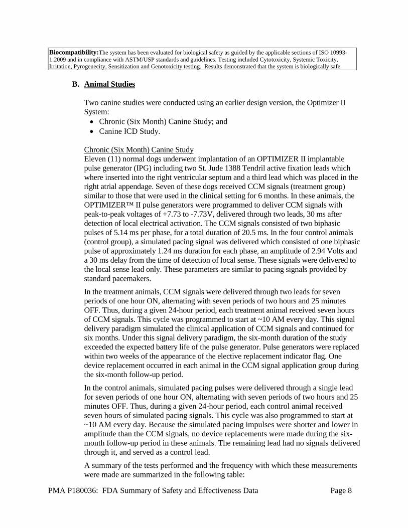
B. Animal Studies
Two canine studies were conducted using an earlier design version, the Optimizer II System: • Chronic (Six Month) Canine Study; and • Canine ICD Study.
Chronic (Six Month) Canine Study Eleven (11) normal dogs underwent implantation of an OPTIMIZER II implantable pulse generator (IPG) including two St. Jude 1388 Tendril active fixation leads which where inserted into the right ventricular septum and a third lead which was placed in the right atrial appendage. Seven of these dogs received CCM signals (treatment group) similar to those that were used in the clinical setting for 6 months. In these animals, the OPTIMIZER™ II pulse generators were programmed to deliver CCM signals with peak-to-peak voltages of +7.73 to -7.73V, delivered through two leads, 30 ms after detection of local electrical activation. The CCM signals consisted of two biphasic pulses of 5.14 ms per phase, for a total duration of 20.5 ms. In the four control animals (control group), a simulated pacing signal was delivered which consisted of one biphasic pulse of approximately 1.24 ms duration for each phase, an amplitude of 2.94 Volts and a 30 ms delay from the time of detection of local sense. These signals were delivered to the local sense lead only. These parameters are similar to pacing signals provided by standard pacemakers. In the treatment animals, CCM signals were delivered through two leads for seven periods of one hour ON, alternating with seven periods of two hours and 25 minutes OFF. Thus, during a given 24-hour period, each treatment animal received seven hours of CCM signals. This cycle was programmed to start at ~10 AM every day. This signal delivery paradigm simulated the clinical application of CCM signals and continued for six months. Under this signal delivery paradigm, the six-month duration of the study exceeded the expected battery life of the pulse generator. Pulse generators were replaced within two weeks of the appearance of the elective replacement indicator flag. One device replacement occurred in each animal in the CCM signal application group during the six-month follow-up period. In the control animals, simulated pacing pulses were delivered through a single lead for seven periods of one hour ON, alternating with seven periods of two hours and 25 minutes OFF. Thus, during a given 24-hour period, each control animal received seven hours of simulated pacing signals. This cycle was also programmed to start at ~10 AM every day. Because the simulated pacing impulses were shorter and lower in amplitude than the CCM signals, no device replacements were made during the six-month follow-up period in these animals. The remaining lead had no signals delivered through it, and served as a control lead. A summary of the tests performed and the frequency with which these measurements were made are summarized in the following table:
Biocompatibility:The system has been evaluated for biological safety as guided by the applicable sections of ISO 10993-1:2009 and in compliance with ASTM/USP standards and guidelines. Testing included Cytotoxicity, Systemic Toxicity, Irritation, Pyrogenecity, Sensitization and Genotoxicity testing. Results demonstrated that the system is biologically safe.
PMA P180036: FDA Summary of Safety and Effectiveness Data Page 9
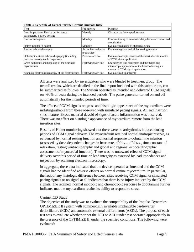
Table 3: Schedule of Events for the Chronic Animal Study Test Frequency Purpose Lead impedance, Device performance parameters, Battery voltage
Weekly Characterize device performance
Electrocardiograms Monthly Confirm timing of automatic daily device activation and deactivation
Holter monitor (4 hours) Monthly Evaluate frequency of abnormal beats. Resting echocardiography At implant and prior
to sacrifice Evaluate regional and global resting function
Dobutamine stress echocardiography (including invasive hemodynamic responses)
Prior to sacrifice Evaluate inotropic reserve of the heart after six months of CCM signal application.
Gross pathology and histology of the heart and myocardium
Following sacrifice Characterize lead placement and the macro and microscopic appearance of the heart following six months of CCM signal application
Scanning electron microscopy of the electrode tips Following sacrifice Evaluate lead tip integrity All tests were analyzed by investigators who were blinded to treatment group. The overall results, which are detailed in the final report included with this submission, can be summarized as follows. The System operated as intended and delivered CCM signals on >90% of beats during the intended periods. The pulse generator turned on and off automatically for the intended periods of time. The effects of CCM signals on gross and histologic appearance of the myocardium were indistinguishable from those observed with simulated pacing signals. At lead insertion sites, mature fibrous material devoid of signs of acute inflammation was observed. There was no effect on histologic appearance of myocardium remote from the lead insertion sites. Results of Holter monitoring showed that there were no arrhythmias induced during periods of CCM signal delivery. The myocardium retained normal inotropic reserve, as evidenced by normal resting function and normal response to dobutamine infusion (assessed by dose-dependent changes in heart rate, dP/dtmax, dP/dtmin, time constant of relaxation, resting ventriculography and global and regional echocardiographic assessment of myocardial function). There was no untoward effect of CCM signal delivery over this period of time on lead integrity as assessed by lead impedances and inspection by scanning electron microscopy. In aggregate, these data indicated that the device operated as intended and the CCM signals had no identified adverse effects on normal canine myocardium. In particular, the lack of any histologic difference between sites receiving CCM signal or simulated pacing signals or no signal at all indicates that there is no injury induced by the CCM signals. The retained, normal inotropic and chronotropic response to dobutamine further indicates mat the myocardium retains its ability to respond to stress. Canine ICD Study The objective of the study was to evaluate the compatibility of the Impulse Dynamics OPTIMIZER II system with commercially available implantable cardioverter defibrillators (ICDs) and automatic external defibrillators (AEDs). The purpose of each test was to evaluate whether or not the ICD or AED under test operated appropriately in the presence of the OPTIMIZE II under the specified conditions. The following were evaluated:
PMA P180036: FDA Summary of Safety and Effectiveness Data Page 10
1. Appropriate VF detection and delivery of defibrillation pulses despite the presence of the OPTIMIZER II CCM signals.
2. Appropriate sensing and pacing during CCM delivery with no double counting of events leading to a false arrhythmia detection by the ICD or AED.
3. Inhibition of CCM signal delivery by the OPTIMIZER II during arrhythmias despite the presence of the ICD.
4. Instances of reset to backup mode of the OPTIMIZE II by internal or external shocks.
Four dogs were implanted with ICDs. A comprehensive set of results were obtained from three of the dogs. One dog died during the protocol after non-resuscitatable ventricular fibrillation induction. All of the study end points were achieved with the conclusion that the OPTIMIZER II and the defibrillators do not adversely interfere with each other. The study also demonstrated the ability of the OPTIMIZER II to deliver CCM signals during VVI NSR, DDD NSR, DDD AP/VS, DDD AS/VP and DDD AP/VP as well as inhibition of CCM delivery during VVI RV pacing. No additional in-vivo animal studies were conducted for release of the OPTIMIZER Smart system because at the time the device was released to the clinic for study, over 1, 000 patients had already been treated with some version of the OPTIMIZER Family of devices and there was little that could be learned from further in vitro and in vivo studies. It should be noted that the device evolved from the OPTIMIZER II submitted with the original IDE application G030099 to the OPTIMIZER Smart submitted with PMA P180036, however, the CCM has been the same for all clinical studies conducted in the US. Parameters such as pulse amplitude, pulse width, dosage (total CCM therapy per day) have been kept the same for US studies to allow a comparison between these studies.
X. SUMMARY OF PRIMARY CLINICAL STUDIES
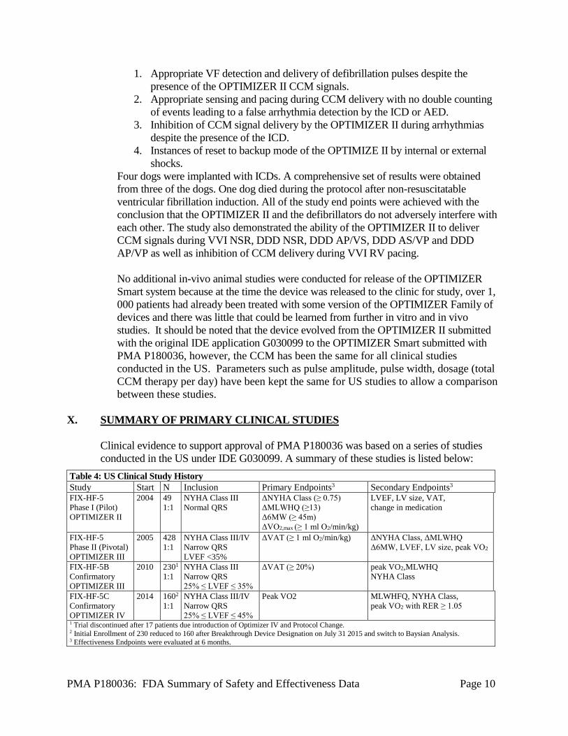
Clinical evidence to support approval of PMA P180036 was based on a series of studies conducted in the US under IDE G030099. A summary of these studies is listed below:
Table 4: US Clinical Study History Study Start N Inclusion Primary Endpoints3 Secondary Endpoints3 FIX-HF-5 Phase I (Pilot) OPTIMIZER II
2004 49 1:1
NYHA Class III Normal QRS
ΔNYHA Class (≥ 0.75) ΔMLWHQ (≥13) Δ6MW (≥ 45m) ΔVO2,max (≥ 1 ml O2/min/kg)
LVEF, LV size, VAT, change in medication
FIX-HF-5 Phase II (Pivotal) OPTIMIZER III
2005 428 1:1
NYHA Class III/IV Narrow QRS LVEF <35%
ΔVAT (≥ 1 ml O2/min/kg)
ΔNYHA Class, ΔMLWHQ Δ6MW, LVEF, LV size, peak VO2
FIX-HF-5B Confirmatory OPTIMIZER III
2010 2301
1:1 NYHA Class III Narrow QRS 25% ≤ LVEF ≤ 35%
ΔVAT (≥ 20%)
peak VO2,MLWHQ NYHA Class
FIX-HF-5C Confirmatory OPTIMIZER IV
2014 1602
1:1 NYHA Class III/IV Narrow QRS 25% ≤ LVEF ≤ 45%
Peak VO2 MLWHFQ, NYHA Class, peak VO2 with RER ≥ 1.05
1 Trial discontinued after 17 patients due introduction of Optimizer IV and Protocol Change. 2 Initial Enrollment of 230 reduced to 160 after Breakthrough Device Designation on July 31 2015 and switch to Baysian Analysis. 3 Effectiveness Endpoints were evaluated at 6 months.
PMA P180036: FDA Summary of Safety and Effectiveness Data Page 11
In the treatment arm of these studies, all patients were implanted with a 3-lead (one atrial, two ventricular) Optimizer System. CCM signals were delivered through the two ventricular leads for a total of 5 hours in a given 24 hour period; the therapy delivery duration was non-programmable. The delivery period started each day at 00:00 hours and was delivered in 1 hour increments. After each one hour period, the device withheld therapy for 3.8 hours. This cycle continued until 23:59 hours at which time the cycle starts again unless interrupted by certain preprogrammed safety features, such as High PVC count, aterial arrhythmia, etc. The pulse amplitude was set to 7.5 V unless chest wall, phrenic nerve or pocket stimulations were observed. Two biphasic pulses, each phase consiting of a 5.14 ms segment, were utilized (totaling 20.56 ms). These pulses were followed by a 40 ms charge balacing phase. Default device settings are provided in detail in the Instructions for Use. The FIX-HF-5 Phase II (Pivotal) trial failed its primary endpoint, but a subgroup analysis demonstrated an improvement in the subgroup with LVEF >25% and NYHA class III. Subsequently, this led to the FIX-HF-5C study which was conducted to further evaluate the benefit of CCM in patients with LVEF ranging from 25% to 45%. A Bayesian statistical approach was employed to leverage the data available from the FIX-HF5 trial, particularly the peak VO2 results. Additional assessments in the FIX-HF-5C study included quality of life as assessed by the Minnesota Living with Heart Failure Questionnaire (MLWHFQ), functional class (NYHA) and 6-minute walk. A summary of the clinical study is presented below. A. Study Design
Patients were treated between 2014 and 2017. The database for this PMA reflected data collected through August 2017 and included 160 patients. There were 60 investigational sites. FIX-HF-5C was a prospective, randomized, third-party blinded (CPX core lab), multicenter study. For the primary effectiveness endpoint, longitudinal data from the prospective study was analyzed together with 30% fixed borrowing of data from the 229 subjects with EF > 25% from the FIX- HF-5 Phase II study using a Bayesian modeling approach. Subjects (n=160) were randomly assigned to one of two groups (treatment or control) with an allocation ratio of 1:1. Block randomization by site and etiology of heart failure (ischemic versus non-ischemic cardiomyopathy) was used to ensure balanced enrollment between the two groups. An Events Adjudication Committee (EAC) was established to review records of adverse events, hospitalizations and deaths. This committee was composed of 3 independent cardiologists experienced in the adjudication process. The committee provided definitions for protocol-specified hospitalizations which included a hospital admission that resulted in a calendar date change or was related to an adverse event that caused a prolongation of the index hospitalization for device implantation. The committee also adjudicated the cardiac and heart failure relatedness of deaths and hospitalizations.
PMA P180036: FDA Summary of Safety and Effectiveness Data Page 12
An independent Data and Safety Monitoring Board (DSMB) reviewed aggregate safety data and monitored for the emergence of any significant safety concerns. The DSMB was composed of 5 members with clinical trial experience in heart failure, electrophysiology and statistics not otherwise participating in the study. The DSMB was unblinded to study group assignment.
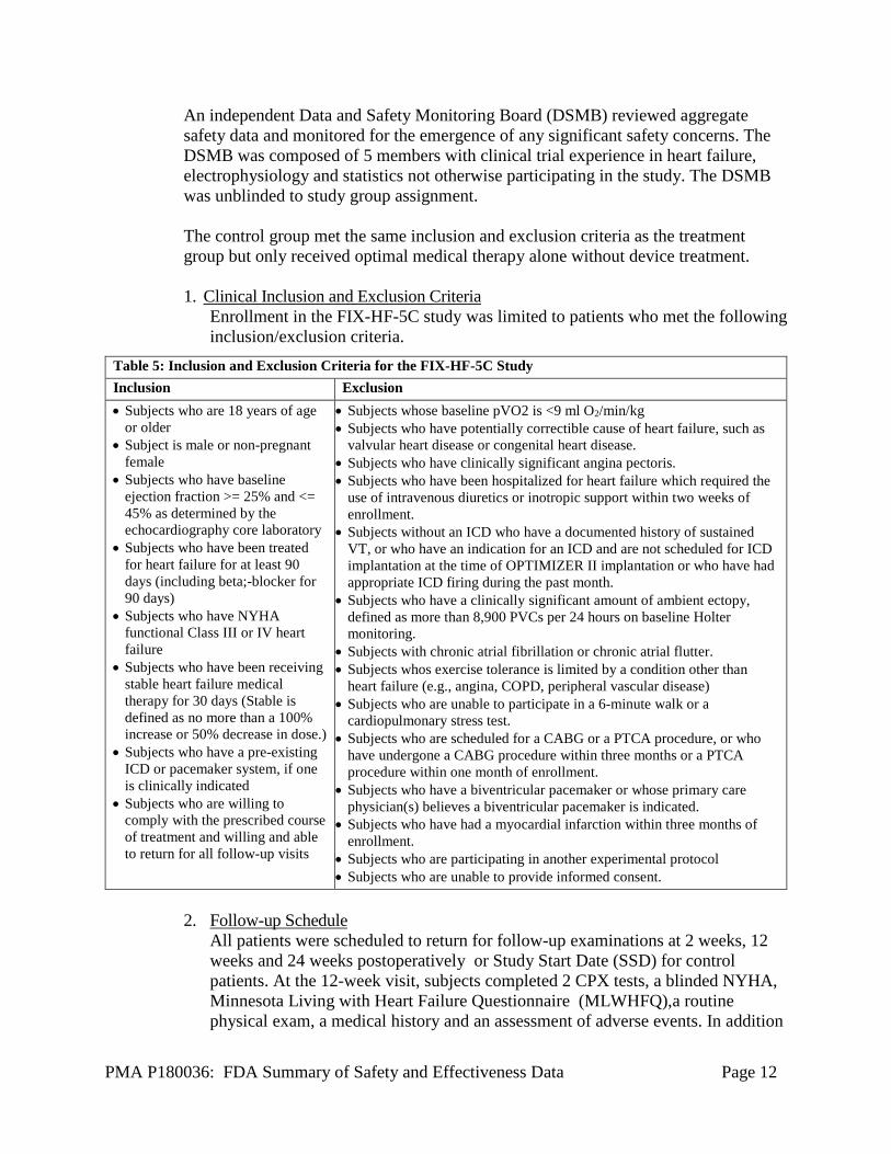
The control group met the same inclusion and exclusion criteria as the treatment group but only received optimal medical therapy alone without device treatment. 1. Clinical Inclusion and Exclusion Criteria
Enrollment in the FIX-HF-5C study was limited to patients who met the following inclusion/exclusion criteria.
2. Follow-up Schedule
All patients were scheduled to return for follow-up examinations at 2 weeks, 12 weeks and 24 weeks postoperatively or Study Start Date (SSD) for control patients. At the 12-week visit, subjects completed 2 CPX tests, a blinded NYHA, Minnesota Living with Heart Failure Questionnaire (MLWHFQ),a routine physical exam, a medical history and an assessment of adverse events. In addition
Table 5: Inclusion and Exclusion Criteria for the FIX-HF-5C Study Inclusion Exclusion • Subjects who are 18 years of age
or older • Subject is male or non-pregnant
female • Subjects who have baseline
ejection fraction >= 25% and <= 45% as determined by the echocardiography core laboratory
• Subjects who have been treated for heart failure for at least 90 days (including beta;-blocker for 90 days)
• Subjects who have NYHA functional Class III or IV heart failure
• Subjects who have been receiving stable heart failure medical therapy for 30 days (Stable is defined as no more than a 100% increase or 50% decrease in dose.)
• Subjects who have a pre-existing ICD or pacemaker system, if one is clinically indicated
• Subjects who are willing to comply with the prescribed course of treatment and willing and able to return for all follow-up visits
• Subjects whose baseline pVO2 is <9 ml O2/min/kg • Subjects who have potentially correctible cause of heart failure, such as
valvular heart disease or congenital heart disease. • Subjects who have clinically significant angina pectoris. • Subjects who have been hospitalized for heart failure which required the
use of intravenous diuretics or inotropic support within two weeks of enrollment.
• Subjects without an ICD who have a documented history of sustained VT, or who have an indication for an ICD and are not scheduled for ICD implantation at the time of OPTIMIZER II implantation or who have had appropriate ICD firing during the past month.
• Subjects who have a clinically significant amount of ambient ectopy, defined as more than 8,900 PVCs per 24 hours on baseline Holter monitoring.
• Subjects with chronic atrial fibrillation or chronic atrial flutter. • Subjects whos exercise tolerance is limited by a condition other than
heart failure (e.g., angina, COPD, peripheral vascular disease) • Subjects who are unable to participate in a 6-minute walk or a
cardiopulmonary stress test. • Subjects who are scheduled for a CABG or a PTCA procedure, or who
have undergone a CABG procedure within three months or a PTCA procedure within one month of enrollment.
• Subjects who have a biventricular pacemaker or whose primary care physician(s) believes a biventricular pacemaker is indicated.
• Subjects who have had a myocardial infarction within three months of enrollment.
• Subjects who are participating in another experimental protocol • Subjects who are unable to provide informed consent.
PMA P180036: FDA Summary of Safety and Effectiveness Data Page 13
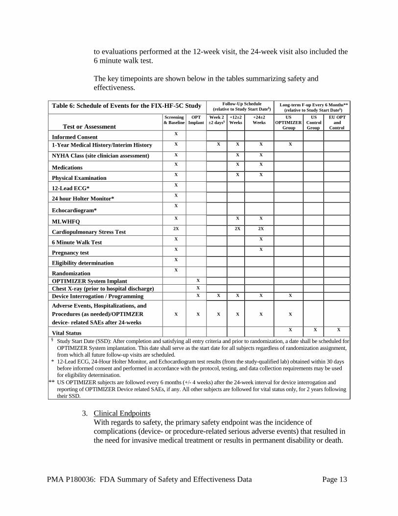
to evaluations performed at the 12-week visit, the 24-week visit also included the 6 minute walk test.
The key timepoints are shown below in the tables summarizing safety and effectiveness.
Table 6: Schedule of Events for the FIX-HF-5C Study Follow-Up Schedule
(relative to Study Start Date§) Long-term F-up Every 6 Months**
(relative to Study Start Date§)
Test or Assessment
Screening & Baseline
OPT Implant
Week 2 ±2 days§
+12±2 Weeks
+24±2 Weeks
US OPTIMIZER
Group
US Control Group
EU OPT and
Control
Informed Consent X
1-Year Medical History/Interim History X X X X X
NYHA Class (site clinician assessment) X X X
Medications X X X
Physical Examination X X X
12-Lead ECG* X
24 hour Holter Monitor* X
Echocardiogram* X
MLWHFQ X X X
Cardiopulmonary Stress Test 2X 2X 2X
6 Minute Walk Test X X
Pregnancy test X X
Eligibility determination X
Randomization X
OPTIMIZER System Implant X
Chest X-ray (prior to hospital discharge) X
Device Interrogation / Programming X X X X X
Adverse Events, Hospitalizations, and Procedures (as needed)/OPTIMZER device- related SAEs after 24-weeks
X
X
X
X
X
X
Vital Status X X X
§ Study Start Date (SSD): After completion and satisfying all entry criteria and prior to randomization, a date shall be scheduled for OPTIMIZER System implantation. This date shall serve as the start date for all subjects regardless of randomization assignment, from which all future follow-up visits are scheduled.
* 12-Lead ECG, 24-Hour Holter Monitor, and Echocardiogram test results (from the study-qualified lab) obtained within 30 days before informed consent and performed in accordance with the protocol, testing, and data collection requirements may be used for eligibility determination.
** US OPTIMIZER subjects are followed every 6 months (+/- 4 weeks) after the 24-week interval for device interrogation and reporting of OPTIMIZER Device related SAEs, if any. All other subjects are followed for vital status only, for 2 years following their SSD.
3. Clinical Endpoints
With regards to safety, the primary safety endpoint was the incidence of complications (device- or procedure-related serious adverse events) that resulted in the need for invasive medical treatment or results in permanent disability or death.
PMA P180036: FDA Summary of Safety and Effectiveness Data Page 14
The success criterion for the safety endpoint was set such that the therapy would be considered safe if 70% or higher of the implanted population was free of such a complication. There were five secondary safety endpoints: overall survival through 24 weeks, cardiac death survival through 24 weeks, freedom from all-cause mortality or all- cause hospitalization through 24 weeks, freedom from cardiac death and worsening heart failure hospitalization through 24 weeks and adjudicated serious adverse events by treatment group through 24 weeks. The survival analyses and freedom from event analysis were based on Kaplan-Meier analysis and the adverse events are tabulated by seriousness and treatment group with testing by Fisher's exact test. With regards to effectiveness, the primary effectiveness endpoint for the study was serial change in peak VO2 measured at baseline, 12 weeks and 24 weeks of follow up. Secondary effectiveness endpoints included quality of life assessed with the MLWHFQ, change in NYHA Class and peak VO2 among subjects with respiratory exchange ratio (RER)> 1.05.
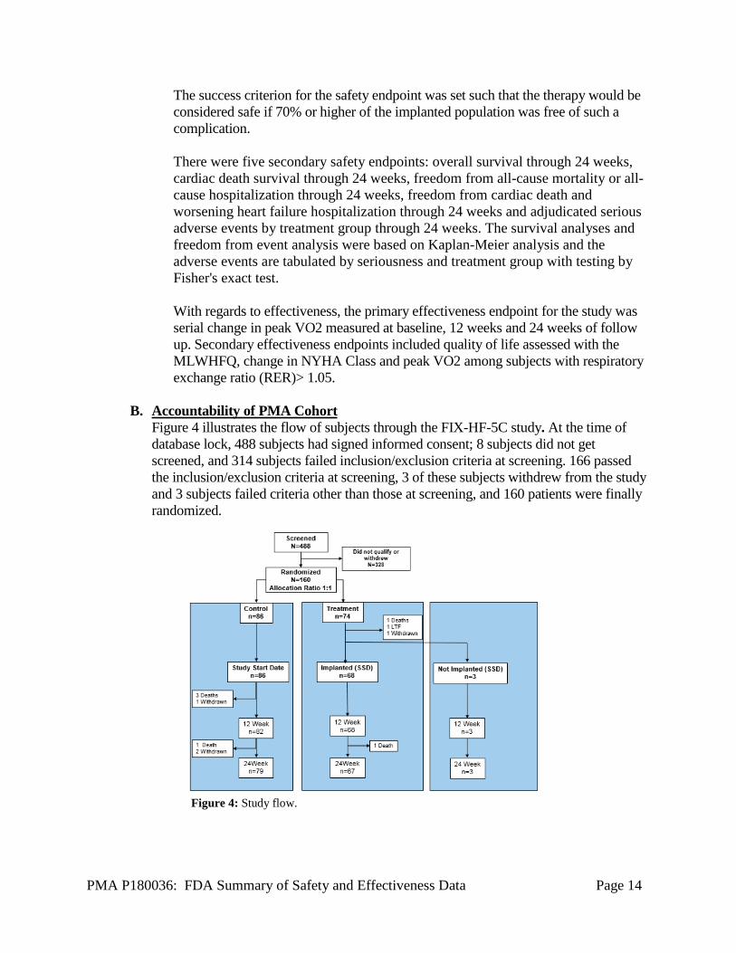
B. Accountability of PMA Cohort
Figure 4 illustrates the flow of subjects through the FIX-HF-5C study. At the time of database lock, 488 subjects had signed informed consent; 8 subjects did not get screened, and 314 subjects failed inclusion/exclusion criteria at screening. 166 passed the inclusion/exclusion criteria at screening, 3 of these subjects withdrew from the study and 3 subjects failed criteria other than those at screening, and 160 patients were finally randomized.
Figure 4: Study flow.
PMA P180036: FDA Summary of Safety and Effectiveness Data Page 15
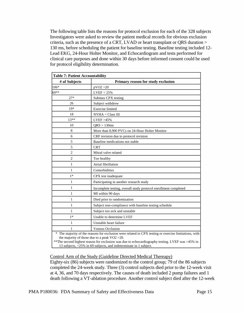
The following table lists the reasons for protocol exclusion for each of the 328 subjects Investigators were asked to review the patient medical records for obvious exclusion criteria, such as the presence of a CRT, LVAD or heart transplant or QRS duration > 130 ms, before scheduling the patient for baseline testing. Baseline testing included 12-Lead EKG, 24-Hour Holter Monitor, and Echocardiogram and tests performed for clinical care purposes and done within 30 days before informed consent could be used for protocol eligibility determination.
Table 7: Patient Accountability # of Subjects Primary reason for study exclusion
106* pVO2 >20 69** LVEF < 25%
27* Submax CPX testing 26 Subject withdrew
19* Exercise limited 18 NYHA < Class III
13** LVEF >45% 10 QRS > 130ms 8 More than 8,900 PVCs on 24-Hour Holter Monitor 6 CRF revision due to protocol revision 5 Baseline medications not stable 5 CRT 2 Mitral valve related 2 Too healthy 1 Atrial fibrillation 1 Comorbidities 1* CPX test inadequate 1 Participating in another research study 1 Incomplete testing, overall study protocol enrollment completed 1 MI within 90 days 1 Died prior to randomization 1 Subject non-compliance with baseline testing schedule 1 Subject too sick and unstable 1* Unable to determine LVEF 1 Unstable heart failure 1 Venous Occlusion
* The majority of the reasons for exclusion were related to CPX testing or exercise limitations, with the majority of those due to a peak VO2 >20.
**The second highest reason for exclusion was due to echocardiography testing. LVEF was >45% in 13 subjects, <25% in 69 subjects, and indeterminate in 1 subject.
Control Arm of the Study (Guideline Directed Medical Thereapy) Eighty-six (86) subjects were randomized to the control group; 79 of the 86 subjects completed the 24-week study. Three (3) control subjects died prior to the 12-week visit at 4, 36, and 70 days respectively. The causes of death included 2 pump failures and 1 death following a VT-ablation procedure. Another control subject died after the 12-week
PMA P180036: FDA Summary of Safety and Effectiveness Data Page 16
visit and prior to the 24-week visit at 117 days, due to a pulmonary complication following a non-cardiac procedure. One control subject withdrew prior to the 12-week visit at 77 days and 2 subjects withdrew after the 12-week visit and prior to the 24-week visit at 86 and 115 days respectively. Treatment Arm of the Study (Guideline Directed Medical Thereapy + Device) Seventy-four (74) subjects were randomized to the CCM™ Treatment group; 68 of these 74 subjects underwent device implantation. Six (6) subjects did not receive an implant. One subject died 2 days prior to the scheduled implant date, 1 subject was lost to follow-up prior to the scheduled implant date, 1 subject was deemed ineligible (interim assessment classified this patient as NYHA Class II) and was withdrawn, 1 subject was discovered to have an additional abandoned ICD lead and the implant was canceled (follow-up testing through 24-weeks performed) and 2 subjects elected not to undergo the implant procedure but follow-up testing through 24-weeks was performed. Thus, 3 of the six patients randomized to CCM™ treatment who did not undergo device implantation completed the 24-week study follow up visits. In addition to the subject that died just prior to the implant date, 1 subject died 164 days after the OPTIMIZER implantation due to sepsis following surgery for an incarcerated hernia.
C. Study Population Demographics and Baseline Parameters
The demographics of the study population are typical for a heart failure study performed in the US. The following three tables present comparisions between the treatment and control groups for continous and categorical variables as well as baseline medication.
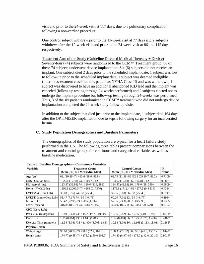
Table 8: Baseline Demographics - Continuous Variables Variable Treatment Group
Mean (SD) N / Med (Min, Max) Control Group
Mean (SD) N / Med (Min, Max) P-value
Age (yrs) 63. (10.89) 74 / 63.6 (38.0, 86.8) 62.79 (11.38) 86 /62.4 (09 30.7, 89.2) 0.71091 QRS Duration (ms) 102.50 (12.58) 74 / 100 (76, 128) 103.62 (12.10) 86 / 104 (80, 129) 0.58671 PR Interval (ms) 183.37 (36.86) 74 / 180.0 (114, 288) 184.57 (43.93) 86 / 178.0 (28, 320) 0.98091 Holter (PVCs/24hr) 1599.5 (2009.0) 74 / 668 (0, 7370) 1176.8 (1712.4) 86 / 277.5 (0, 8514) 0.43341 LVEF (%) (Core Lab) 33.08 (5.55) 74 / 32 (25, 45) 32.55 (5.18) 86 / 32 (25, 45) 0.57471 LVEDD (mm) (Core Lab) 58.47 (7.17) 74 / 59 (40, 75) 60.20 (7.01) 82 / 59 (44, 77) 0.19841 MLWHFQ 56.42 (22.95) 74 / 60.5 (1, 96) 57.35 (23.36) 86 / 60 (5, 99) 0.73651 6MW (meters) 316.85 (88.37) 74 / 308 (75, 462) 324.07 (89.71) 86 / 315 (120, 579) 0.87241 CPX (Core Lab) Peak VO2 (ml/kg/min) 15.49 (2.61) 733 / 15.70 (9.75, 19.70) 15.36 (2.82) 86 / 15.85 (9.10, 19.90) 0.80111 Peak RER 1.15 (0.064) 733 / 1.140 (1.015, 1315) 1.14 (0.074) 86 / 1.125 (0.975, 1.480) 0.44691 Exercise Time (minutes) 11.38 (3.08) 733 / 11.800 (3.208, 18.5) 10.58 (3.09) 86 / 11.163 (3.133, 18.03) 0.12861 Physical Exam Weight (kg) 99.60 (20.72) 74 /98.0 (52.7, 167.8) 100.33 (23.32) 86 / 96.8 (49.0, 155.1) 0.84421 Height (cm) 174.77 (9.58) 74 / 175.0 (150.0 208.0) 174.40 (8.97) 86 / 175.0 (142.0, 201.0) 0.90191
PMA P180036: FDA Summary of Safety and Effectiveness Data Page 17
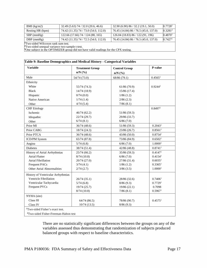
BMI (kg/m2) 32.49 (5.63) 74 / 32.0 (20.6, 46.6) 32.90 (6.90) 86 / 32.2 (19.1, 50.0) 0.77281 Resting HR (bpm) 74.42 (11.35) 74 / 73.0 (54.0, 112.0) 76.45 (14.84) 86 / 76.5 (45.0, 137.0) 0.32812 SBP (mmHg) 122.66 (17.66) 74 / 124 (88, 165) 126.04 (18.83) 86 / 122.(91, 196) 0.48701 DBP (mmHg) 74.42 (11.35) 74 / 72.5 (54.0, 112.0) 76.45 (14.84) 86 / 76.5 (45.0, 137.0) 0.74271
1Two-sided Wilcoxon rank sum test. 2Two-sided unequal variance two-sample t-test. 3One subject in the OPTIMIZER group did not have valid readings for the CPX testing. Table 9: Baseline Demographics and Medical History - Categorical Variables
Variable Treatment Group n/N (%)
Control Group n/N (%)
P-value
Male 54/74 (73.0) 68/86 (79.1) 0.45651 Ethnicity White Black Hispanic Native American Other
55/74 (74.3) 14/74 (18.9) 0/74 (0.0) 1/74 (1.4) 4/74 (5.4)
61/86 (70.9) 15/86 (17.4) 1/86 (1.2) 2/86 (2.3) 7/86 (8.1)
0.92442
CHF Etiology Ischemic Idiopathic Other
46/74 (62.2) 22/74 (29.7) 6/74 (8.1)
51/86 (59.3) 29/86 (33.7) 6/86 (7.0)
0.84972
Prior MI 36/74 (48.6) 51/86 (59.3) 0.20431 Prior CABG 18/74 (24.3) 23/86 (26.7) 0.85611 Prior PTCA 36/74 (48.6) 43/86 (50.0) 0.87541 ICD/PM System 65/74 (87.8) 73/86 (84.9) 0.65021 Angina 5/74 (6.8) 6/86 (7.0) 1.00001 Diabetes 38/74 (51.4) 42/86 (48.8) 0.87411 History of Atrial Arrhythmias Atrial Flutter Atrial Fibrillation Frequent PACs Other Atrial Abnormalities
25/74 (66.2) 8/74 (10.8) 20/74 (27.0) 3/74 (4.1) 2/74 (2.7)
35/86 (59.3) 6/86 (7.0) 27/86 (31.4) 1/86 (1.2) 3/86 (3.5)
0.41471 0.41541 0.60351 0.33651 1.00001
History of Ventricular Arrhythmias Ventricle Fibrillation Ventricular Tachycardia Frequent PVCs
26/74 (35.1) 5/74 (6.8) 19/74 (25.7) 8/74 (10.8)
28/86 (32.6) 8/86 (9.3) 19/86 (22.1) 7/86 (8.1)
0.74061 0.77291 0.7098 0.59671
NYHA (site) Class III Class IV
64/74 (86.5) 10/74 (13.5)
78/86 (90.7) 8/86 (9.3)
0.45751
1Two-sided Fisher’s exact test. 2Two-sided Fisher-Freeman-Halton test
There are no statistically significant differences between the groups on any of the variables assessed thus demonstrating that randomization of subjects produced balanced groups with respect to baseline characteristics.
PMA P180036: FDA Summary of Safety and Effectiveness Data Page 18
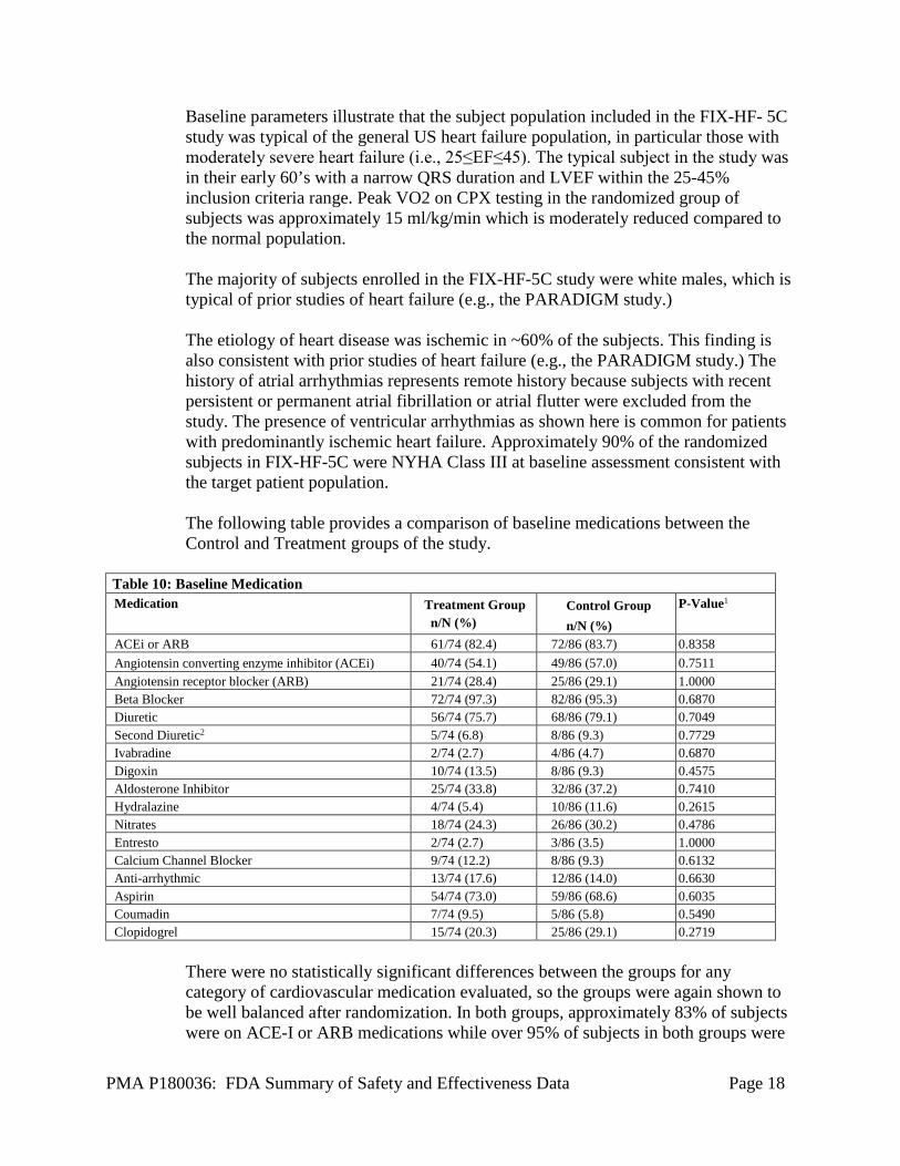
Baseline parameters illustrate that the subject population included in the FIX-HF- 5C study was typical of the general US heart failure population, in particular those with moderately severe heart failure (i.e., 25≤EF≤45). The typical subject in the study was in their early 60’s with a narrow QRS duration and LVEF within the 25-45% inclusion criteria range. Peak VO2 on CPX testing in the randomized group of subjects was approximately 15 ml/kg/min which is moderately reduced compared to the normal population.
The majority of subjects enrolled in the FIX-HF-5C study were white males, which is typical of prior studies of heart failure (e.g., the PARADIGM study.)
The etiology of heart disease was ischemic in ~60% of the subjects. This finding is also consistent with prior studies of heart failure (e.g., the PARADIGM study.) The history of atrial arrhythmias represents remote history because subjects with recent persistent or permanent atrial fibrillation or atrial flutter were excluded from the study. The presence of ventricular arrhythmias as shown here is common for patients with predominantly ischemic heart failure. Approximately 90% of the randomized subjects in FIX-HF-5C were NYHA Class III at baseline assessment consistent with the target patient population.
The following table provides a comparison of baseline medications between the Control and Treatment groups of the study.
Table 10: Baseline Medication Medication Treatment Group
n/N (%) Control Group n/N (%)
P-Value1
ACEi or ARB 61/74 (82.4) 72/86 (83.7) 0.8358 Angiotensin converting enzyme inhibitor (ACEi) 40/74 (54.1) 49/86 (57.0) 0.7511 Angiotensin receptor blocker (ARB) 21/74 (28.4) 25/86 (29.1) 1.0000 Beta Blocker 72/74 (97.3) 82/86 (95.3) 0.6870 Diuretic 56/74 (75.7) 68/86 (79.1) 0.7049 Second Diuretic2 5/74 (6.8) 8/86 (9.3) 0.7729 Ivabradine 2/74 (2.7) 4/86 (4.7) 0.6870 Digoxin 10/74 (13.5) 8/86 (9.3) 0.4575 Aldosterone Inhibitor 25/74 (33.8) 32/86 (37.2) 0.7410 Hydralazine 4/74 (5.4) 10/86 (11.6) 0.2615 Nitrates 18/74 (24.3) 26/86 (30.2) 0.4786 Entresto 2/74 (2.7) 3/86 (3.5) 1.0000 Calcium Channel Blocker 9/74 (12.2) 8/86 (9.3) 0.6132 Anti-arrhythmic 13/74 (17.6) 12/86 (14.0) 0.6630 Aspirin 54/74 (73.0) 59/86 (68.6) 0.6035 Coumadin 7/74 (9.5) 5/86 (5.8) 0.5490 Clopidogrel 15/74 (20.3) 25/86 (29.1) 0.2719
There were no statistically significant differences between the groups for any category of cardiovascular medication evaluated, so the groups were again shown to be well balanced after randomization. In both groups, approximately 83% of subjects were on ACE-I or ARB medications while over 95% of subjects in both groups were
PMA P180036: FDA Summary of Safety and Effectiveness Data Page 19
receiving beta blocker medication at baseline. Seventy-five percent of subjects were taking diuretics and approximately 35% of subjects were on an aldosterone inhibitor. The rate of diuretic therapy in FIX-HF-5C is similar to the rate reported in the PARADIGM study which was ~80%.
D. Safety and Effectiveness Results
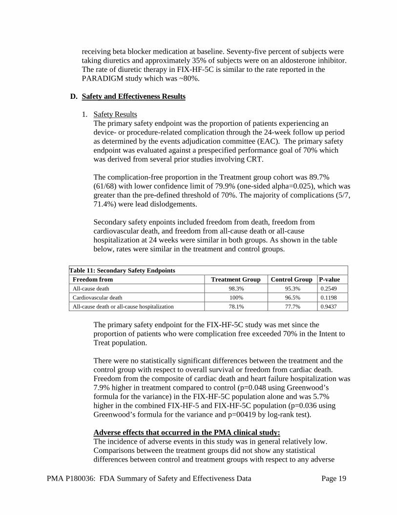
1. Safety Results The primary safety endpoint was the proportion of patients experiencing an device- or procedure-related complication through the 24-week follow up period as determined by the events adjudication committee (EAC). The primary safety endpoint was evaluated against a prespecified performance goal of 70% which was derived from several prior studies involving CRT. The complication-free proportion in the Treatment group cohort was 89.7% (61/68) with lower confidence limit of 79.9% (one-sided alpha=0.025), which was greater than the pre-defined threshold of 70%. The majority of complications (5/7, 71.4%) were lead dislodgements. Secondary safety enpoints included freedom from death, freedom from cardiovascular death, and freedom from all-cause death or all-cause hospitalization at 24 weeks were similar in both groups. As shown in the table below, rates were similar in the treatment and control groups.
Table 11: Secondary Safety Endpoints Freedom from Treatment Group Control Group P-value All-cause death 98.3% 95.3% 0.2549 Cardiovascular death 100% 96.5% 0.1198 All-cause death or all-cause hospitalization 78.1% 77.7% 0.9437
The primary safety endpoint for the FIX-HF-5C study was met since the proportion of patients who were complication free exceeded 70% in the Intent to Treat population. There were no statistically significant differences between the treatment and the control group with respect to overall survival or freedom from cardiac death. Freedom from the composite of cardiac death and heart failure hospitalization was 7.9% higher in treatment compared to control (p=0.048 using Greenwood’s formula for the variance) in the FIX-HF-5C population alone and was 5.7% higher in the combined FIX-HF-5 and FIX-HF-5C population (p=0.036 using Greenwood’s formula for the variance and p=00419 by log-rank test). Adverse effects that occurred in the PMA clinical study: The incidence of adverse events in this study was in general relatively low. Comparisons between the treatment groups did not show any statistical differences between control and treatment groups with respect to any adverse
PMA P180036: FDA Summary of Safety and Effectiveness Data Page 20
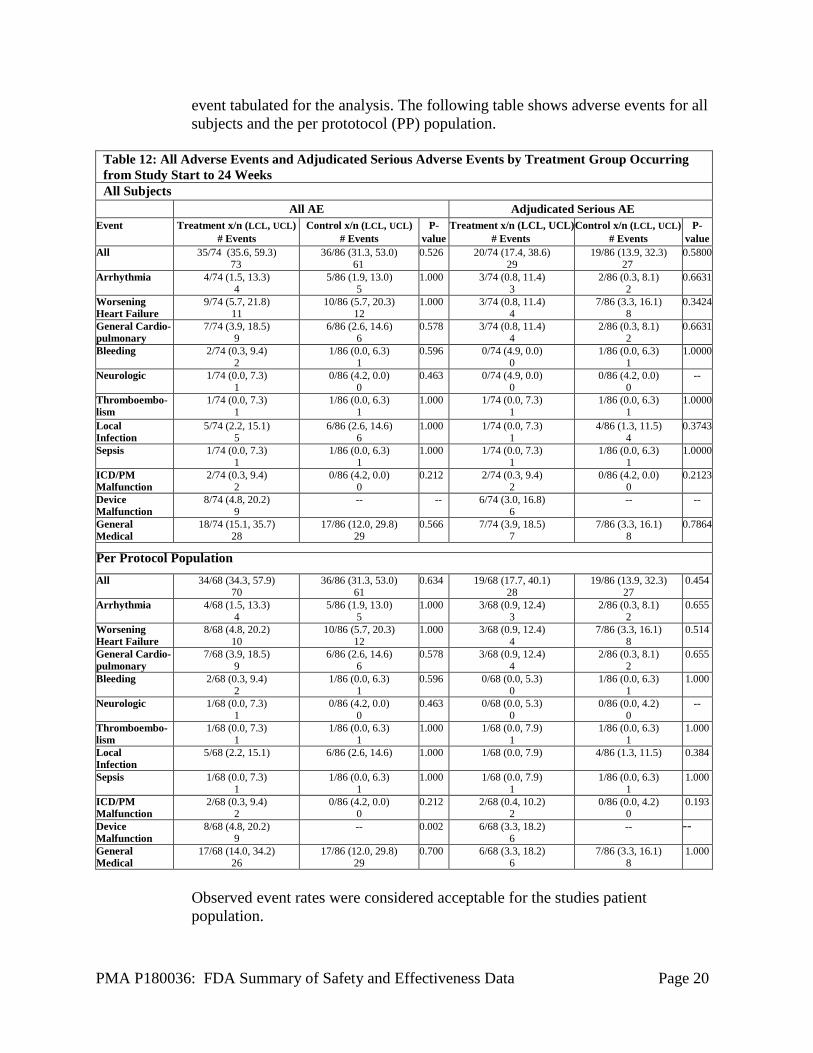
event tabulated for the analysis. The following table shows adverse events for all subjects and the per prototocol (PP) population.
Table 12: All Adverse Events and Adjudicated Serious Adverse Events by Treatment Group Occurring from Study Start to 24 Weeks All Subjects All AE Adjudicated Serious AE
Event Treatment x/n (LCL, UCL) # Events
Control x/n (LCL, UCL) # Events
P-value
Treatment x/n (LCL, UCL) # Events
Control x/n (LCL, UCL) # Events
P-value
All 35/74 (35.6, 59.3) 73
36/86 (31.3, 53.0) 61
0.526 20/74 (17.4, 38.6) 29
19/86 (13.9, 32.3) 27
0.5800
Arrhythmia 4/74 (1.5, 13.3) 4
5/86 (1.9, 13.0) 5
1.000 3/74 (0.8, 11.4) 3
2/86 (0.3, 8.1) 2
0.6631
Worsening Heart Failure
9/74 (5.7, 21.8) 11
10/86 (5.7, 20.3) 12
1.000 3/74 (0.8, 11.4) 4
7/86 (3.3, 16.1) 8
0.3424
General Cardio-pulmonary
7/74 (3.9, 18.5) 9
6/86 (2.6, 14.6) 6
0.578 3/74 (0.8, 11.4) 4
2/86 (0.3, 8.1) 2
0.6631
Bleeding 2/74 (0.3, 9.4) 2
1/86 (0.0, 6.3) 1
0.596 0/74 (4.9, 0.0) 0
1/86 (0.0, 6.3) 1
1.0000
Neurologic 1/74 (0.0, 7.3) 1
0/86 (4.2, 0.0) 0
0.463 0/74 (4.9, 0.0) 0
0/86 (4.2, 0.0) 0
--
Thromboembo-lism
1/74 (0.0, 7.3) 1
1/86 (0.0, 6.3) 1
1.000 1/74 (0.0, 7.3) 1
1/86 (0.0, 6.3) 1
1.0000
Local Infection
5/74 (2.2, 15.1) 5
6/86 (2.6, 14.6) 6
1.000 1/74 (0.0, 7.3) 1
4/86 (1.3, 11.5) 4
0.3743
Sepsis 1/74 (0.0, 7.3) 1
1/86 (0.0, 6.3) 1
1.000 1/74 (0.0, 7.3) 1
1/86 (0.0, 6.3) 1
1.0000
ICD/PM Malfunction
2/74 (0.3, 9.4) 2
0/86 (4.2, 0.0) 0
0.212 2/74 (0.3, 9.4) 2
0/86 (4.2, 0.0) 0
0.2123
Device Malfunction
8/74 (4.8, 20.2) 9
-- -- 6/74 (3.0, 16.8) 6
-- --
General Medical
18/74 (15.1, 35.7) 28
17/86 (12.0, 29.8) 29
0.566 7/74 (3.9, 18.5) 7
7/86 (3.3, 16.1) 8
0.7864
Per Protocol Population
All 34/68 (34.3, 57.9) 70
36/86 (31.3, 53.0) 61
0.634 19/68 (17.7, 40.1) 28
19/86 (13.9, 32.3) 27
0.454
Arrhythmia 4/68 (1.5, 13.3) 4
5/86 (1.9, 13.0) 5
1.000 3/68 (0.9, 12.4) 3
2/86 (0.3, 8.1) 2
0.655
Worsening Heart Failure
8/68 (4.8, 20.2) 10
10/86 (5.7, 20.3) 12
1.000 3/68 (0.9, 12.4) 4
7/86 (3.3, 16.1) 8
0.514
General Cardio-pulmonary
7/68 (3.9, 18.5) 9
6/86 (2.6, 14.6) 6
0.578 3/68 (0.9, 12.4) 4
2/86 (0.3, 8.1) 2
0.655
Bleeding 2/68 (0.3, 9.4) 2
1/86 (0.0, 6.3) 1
0.596 0/68 (0.0, 5.3) 0
1/86 (0.0, 6.3) 1
1.000
Neurologic 1/68 (0.0, 7.3) 1
0/86 (4.2, 0.0) 0
0.463 0/68 (0.0, 5.3) 0
0/86 (0.0, 4.2) 0
--
Thromboembo-lism
1/68 (0.0, 7.3) 1
1/86 (0.0, 6.3) 1
1.000 1/68 (0.0, 7.9) 1
1/86 (0.0, 6.3) 1
1.000
Local Infection
5/68 (2.2, 15.1) 6/86 (2.6, 14.6) 1.000 1/68 (0.0, 7.9) 4/86 (1.3, 11.5) 0.384
Sepsis 1/68 (0.0, 7.3) 1
1/86 (0.0, 6.3) 1
1.000 1/68 (0.0, 7.9) 1
1/86 (0.0, 6.3) 1
1.000
ICD/PM Malfunction
2/68 (0.3, 9.4) 2
0/86 (4.2, 0.0) 0
0.212 2/68 (0.4, 10.2) 2
0/86 (0.0, 4.2) 0
0.193
Device Malfunction
8/68 (4.8, 20.2) 9
-- 0.002 6/68 (3.3, 18.2) 6
-- --
General Medical
17/68 (14.0, 34.2) 26
17/86 (12.0, 29.8) 29
0.700 6/68 (3.3, 18.2) 6
7/86 (3.3, 16.1) 8
1.000
Observed event rates were considered acceptable for the studies patient population.
PMA P180036: FDA Summary of Safety and Effectiveness Data Page 21
2. Effectiveness Results The primary endpoint of the FIX-HF-5C study was the change in peak V02 at 24 weeks from baseline. Δ3 was defined as the mean difference in peak V02 between treatment and control groups at the third (24 week) visit, adjusting for baseline and 12-week peak V02 values. The following hypothesis was tested:
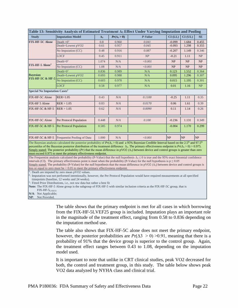
A Bayesian model was fitted to obtain the posterior distribution of Δ3. If the posterior probability that Δ3 is positive is greater than 0.975, that is 𝑃𝑃r(Δ3>0) > 0.975, the null hypothesis was rejected and the device was considered superior to control with respect to the primary endpoint. The baysian model incorporated 160 patients from the FIX-HF-5C study as well as a prior distribution of the treatment effect (Δ3) that was obtained from the FIX-HF-5LVEF25 229 patient subgroup, that is, the subgroup of the FIX-HF-5 study for which LVEF was greater than 25%. A posterior distribution of Δ3 for the FIX-HF-5LVEF25 group was determined and then combined with the FIX-HF-5C data but down-weighting the posterior by 70%. Using simulations, it was found that borrowing at 30% would result in an adequate power around 80% for a feasible sample size of 160 patients in the case of a 50% treatment effect based on the posterior prior effect of 1.08 ml/kg/min (~0.5 O2 ml/kg/min) or higher. Up to 10% of missing data, assumed to be “missing at random” (i.e., not informative about the missing treatment outcome), were considered in the simulations. A longitudinal mixed effects model was proposed to analyze peak VO2 without any imputation for missing data. With the within-subject correlation modeled by a random subject intercept, the random effect model would provide an unbiased estimate for treatment effect if the data are missing at random. However, there were a couple of situations in which missing data on peakVO2 could be informative about the treatment: missing due to death or missing due to heart failure hospitalization. In study FIX-HF-5C, there were 6 patients that had missing peak VO2 values at 24 weeks due to death (2 in the treatment group, and 4 in the control group) and there were no patients with missing pVO2 values at 24 weeks due to heart failure hospitalization. For the primary analysis of peak VO2, missing peak VO2 values due to death were imputed with zeros. Additional analyses with missing peak VO2 values imputed as the lowest observed value and with no imputation were performed as sensitivity analyses. The following table summarizes the results of a sensitivity analysis looking at various imputation and pooling conditions.
PMA P180036: FDA Summary of Safety and Effectiveness Data Page 22
The table shows that the primary endpoint is met for all cases in which borrowing from the FIX-HF-5LVEF25 group is included. Imputation plays an important role in the magnitude of the treatment effect, ranging from 0.58 to 0.836 depending on the imputation method use. The table also shows that FIX-HF-5C alone does not meet the primary endpoint, however, the posterior probabilities are 𝑃𝑃r(Δ3 > 0) >0.91, meaning that there is a probability of 91% that the device group is superior to the control group. Again, the treatment effect ranges between 0.43 to 1.08, depending on the imputation model used. It is important to note that unlike in CRT clinical studies, peak VO2 decreased for both, the control and treatment group, in this study. The table below shows peak VO2 data analyzed by NYHA class and clinical trial.
Table 13: Sensitivity Analysis of Estimated Treatment Δ3 Effect Under Varying Imputation and Pooling Study Imputation Model Δ3 Pr(Δ3 > 0) P-Value CI (LL) CI (UL) SE
FIX-HF-5C Alone Death =01 0.8 0.960 0.041 -0.099 1.684 0.455 Death=Lowest pVO2 0.61 0.957 0.045 -0.093 1.298 0.355 No Imputation (CC) 0.48 0.916 0.087 -0.207 1.149 0.346
LOCF 0.45 0.911 NP -0.21 1.11 NP
FIX-HF-5 Alone3
Death=01 1.074 N/A <0.001 NP NP NP
No Imputation (CC) 1.08 N/A <0.001 NP NP NP Bayesian FIX-HF-5C & HF-5
Death=01 0.836 0.989 N/A 0.123 1.552 0.364 Death=Lowest pVO2 0.693 0.988 N/A 0.095 1.296 0.307 No Imputation (CC) 0.603 0.978 N/A 0.015 1.195 0.301
LOCF 0.58 0.977 N/A 0.01 1.16 NP
Special No Imputation Cases2
FIX-HF-5C Alone
RER>1.05 0.43 N/A 0.1100 -0.25 1.11 0.35
FIX-HF 5 Alone RER > 1.05
0.83 N/A 0.0170 0.06 1.61 0.39 FIX-HF-5C & HF-5
RER > 1.05
0.62 N/A 0.0090 0.11
1.14
0.26
FIX-HF-5C Alone
Per Protocol Population 0.448 N/A 0.100 -0.236 1.131 0.349
FIX-HF-5C & HF-5
Per Protocol Population 0.585 0.974 -0.004 1.170 0.299
FIX-HF-5C & HF-5 Frequentist Pooling of Data 1.000 N/A <0.001 NP NP NP The Bayesian analysis calculated the posterior probability of Pr(Δ3 > 0) and a 95% Bayesian Credible Interval based on the 2.5th and 97.5th percentiles of the Bayesian posterior distribution of the treatment difference Δ3. The primary effectiveness endpoint is Pr(Δ3 > 0) > 0.975. Simply stated: The posterior probability (Pr) that the mean difference in pVO2 (Δ3) between device and control groups is greater than zero must exceed 0.975 to meet the primary effectiveness endpoint. The Frequentist analysis calculated the probability (P-Value) that the null hypothesis Δ3 ≤ 0 is true and the 95% exact binomial confidence intervals (CI). The primary effectiveness point is meet when the probability (P-Value) for the null hypothesis is p ≤ 0.05 Simply stated: The probability (P-Value) for the null hypothesis that the mean difference in pVO2 (Δ3) between device and control groups is less or equal to zero must be < 0.05 to meet the primary effectiveness endpoint. 1: Death are imputed by zero mean pVO2 values. 2: Imputation was not performed intentionally, however, the Per Protocol Population would have required measurements at all specified timepoints (baseline, 12 weeks and 24 weeks). 3: Fitted Prior Distributions, i.e., not raw data but rather a best fit Note: The FIX-HF-5 Alone group is the subgroup of FIX-HF-5 with similar inclusion criteria as the FIX-HF-5C group, that is FIX-HF-5LVEF25 N/A: Not Applicable; NP: Not Provided.
PMA P180036: FDA Summary of Safety and Effectiveness Data Page 23
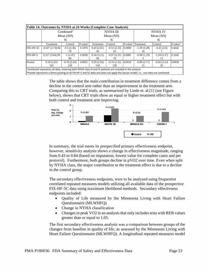
Table 14: Outcomes by NYHA at 24 Weeks (Complete Case Analysis) Combined1
Mean (SD) N
NYHA III Mean (SD)
N
NYHA IV Mean (SD)
N Treatment Control P-value1 Treatment Control P-value1 Treatment Control P-value1 FIX-HF-5C -0.027 (2.74) 66 -0.5 (2.36)
70 0.1870 0.20 (2.61)
56 -0.51 (2.33)
63 0.0690 -1.29 (3.28)
10 -0.41 (2.8)
7 0.4642
FIX-HF-5 0.317 (3.04) 99 -1 (2.47) 89
0.0038 0.34 (3.11) 94
-0.97 (2.31) 76
0.0081 -0.08 (1.19) 5
-1.18 (3.37) 13
0.3243
Pooled 0.18 (2.92) 165
-0.78 (2.43) 159
0.0025 0.29 (2.92) 150
-0.76 (2.32) 139
0.0018 -0.89 (2.77) 15
-0.92 (3.13) 20
0.8939
1Combined represents all data, meaning both NYHA class III and IV patients are included in the analysis 2Pooled represents a direct pooling of all FIX-HF-5 and 5C data and does not apply the basian model; i.e., raw data are combined.
The table shows that the main contribution in treatment difference comes from a decline in the control arm rather than an improvement in the treatment arm. Comparing this to CRT trials, as summarized by Linde et. al.[1] (see Figure below), shows that CRT trials show an equal or higher treatment effect but with both control and treatment arm improving.
In summary, the trial meets its prespecified primary effectiveness endpoint, however, sensitivity analysis shows a change in effectiveness magnitude, ranging from 0.43 to 0.84 (based on imputation, lowest value for complete cases and per protocol). Furthermore, both groups decline in pVO2 over time. Even when split by NYHA class, the major contribution to the treatment effect is due to a decline in the control group.
The secondary effectiveness endpoints, were to be analyzed using frequentist correlated repeated measures models utilizing all available data of the prospective FIX-HF-5C data using maximum likelihood methods. Secondary effectivness endpoints included:
• Quality of Life measured by the Minnesota Living with Heart Failure Questionnaire (MLWHFQ)
• Change in NYHA classification • Changes in peak VO2 in an analysis that only includes tests with RER values
greater than or equal to 1.05. The first secondary effectiveness analysis was a comparison between groups of the changes from baseline in quality of life, as assessed by the Minnesota Living with Heart Failure Questionnaire (MLWHFQ). A longitudinal repeated measures model
PMA P180036: FDA Summary of Safety and Effectiveness Data Page 24
was applied to the quality of life scores which assessed at baseline, 12 weeks and 24 weeks.
where Yit is the response of patient i at time t, t=1,2,3 for baseline, 12 weeks, and 24 weeks, respectively; 𝜃𝜃𝑡𝑡 is the mean control group response at time t; 𝛽𝛽𝑡𝑡 is the difference between treatment group and control at time t; 𝑍𝑍𝑖𝑖 is an indicator that equals 1 if patient i is in the test group and 0 if the control group; 𝑏𝑏𝑖𝑖~𝑁𝑁 (0, 𝜏𝜏𝑏𝑏) is a subject-specific random intercept with variance 𝜏𝜏𝑏𝑏; and 𝜖𝜖𝑖𝑖~𝑁𝑁 (0, 𝜏𝜏) is the residual error with variance 𝜏𝜏. The model accounts for baseline MLWHFQ, and a constraint 𝛽𝛽1=0 implies no group differences at baseline, which essentially allows the model to compare groups with respect to changes from baseline as opposed to raw differences at 24 weeks. The following hypothesis was tested:
If the one-sided p-value is less than or equal to 0.025, the null hypothesis will be rejected and MLWHFQ will be determined to be superior for test versus control group, respectively. The MLWHFQ score decreased in both groups; it decreased approximately by 10 points in the control group and by 21 points in the device group. The score improvement by 24 weeks was significantly greater in the device arm than it was for the control arm (P<0.001); meeting the first secondary analysis endpoint. The second secondary effectiveness analysis was an assessment of the improvement in heart failure class, as assessed by the New York Heart Association (NYHA) classification. NYHA classification was assigned by a blinded on site clinician according to their standard clinical practice. The analysis of this endpoint tested the hypothesis that the subjects treated with the device will have a greater proportion of subjects that improve by at least one NYHA class than the control group. Let pj be the proportion of patients that improve by at least one NYHA category at 24 weeks for group j (j=1 denoting control and j=2 denoting treatment). The following hypothesis was tested:
A stratified Cochran Mantel-Haenzel test (with strata defined by etiology of heart failure) will be used to compute a p-value. If the p-value is less than or equal to 0.025, the null hypothesis will be rejected and test treatment considered superior to control with respect to NYHA. A significant improvement in NYHA class in the treatment group compared to the control group was seen in the FIX-HF-5C trial, see the table below:
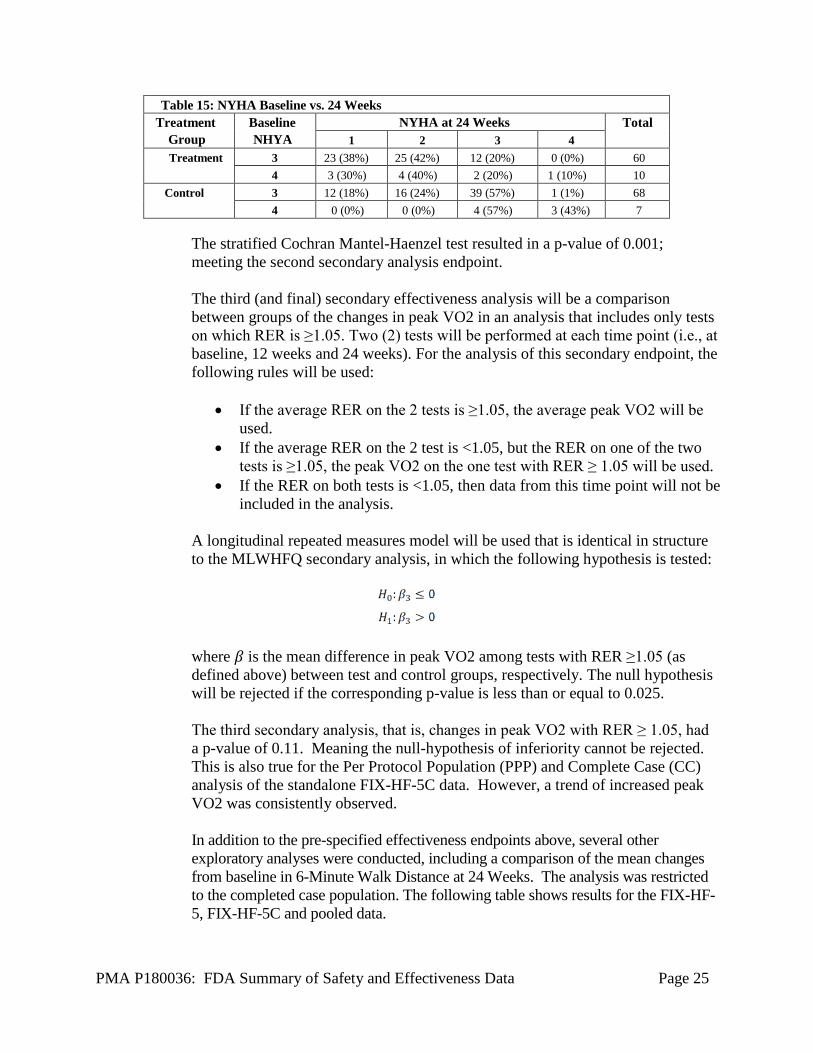
PMA P180036: FDA Summary of Safety and Effectiveness Data Page 25
Table 15: NYHA Baseline vs. 24 Weeks Treatment
Group Baseline NHYA
NYHA at 24 Weeks Total 1 2 3 4
Treatment 3 23 (38%) 25 (42%) 12 (20%) 0 (0%) 60 4 3 (30%) 4 (40%) 2 (20%) 1 (10%) 10
Control 3 12 (18%) 16 (24%) 39 (57%) 1 (1%) 68 4 0 (0%) 0 (0%) 4 (57%) 3 (43%) 7
The stratified Cochran Mantel-Haenzel test resulted in a p-value of 0.001; meeting the second secondary analysis endpoint. The third (and final) secondary effectiveness analysis will be a comparison between groups of the changes in peak VO2 in an analysis that includes only tests on which RER is ≥1.05. Two (2) tests will be performed at each time point (i.e., at baseline, 12 weeks and 24 weeks). For the analysis of this secondary endpoint, the following rules will be used:
• If the average RER on the 2 tests is ≥1.05, the average peak VO2 will be used.
• If the average RER on the 2 test is <1.05, but the RER on one of the two tests is ≥1.05, the peak VO2 on the one test with RER ≥ 1.05 will be used.
• If the RER on both tests is <1.05, then data from this time point will not be included in the analysis.
A longitudinal repeated measures model will be used that is identical in structure to the MLWHFQ secondary analysis, in which the following hypothesis is tested:
where 𝛽𝛽 is the mean difference in peak VO2 among tests with RER ≥1.05 (as defined above) between test and control groups, respectively. The null hypothesis will be rejected if the corresponding p-value is less than or equal to 0.025. The third secondary analysis, that is, changes in peak VO2 with RER ≥ 1.05, had a p-value of 0.11. Meaning the null-hypothesis of inferiority cannot be rejected. This is also true for the Per Protocol Population (PPP) and Complete Case (CC) analysis of the standalone FIX-HF-5C data. However, a trend of increased peak VO2 was consistently observed.
In addition to the pre-specified effectiveness endpoints above, several other exploratory analyses were conducted, including a comparison of the mean changes from baseline in 6-Minute Walk Distance at 24 Weeks. The analysis was restricted to the completed case population. The following table shows results for the FIX-HF-5, FIX-HF-5C and pooled data.
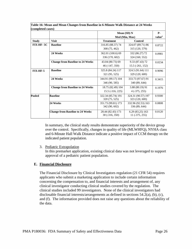
PMA P180036: FDA Summary of Safety and Effectiveness Data Page 26
Table 16: Mean and Mean Changes from Baseline in 6-Minute Walk Distance at 24 Weeks (completed cases) Mean (SD) N
Med (Min, Max) P-
value1
Study Visit Treatment Control FIX-HF- 5C Baseline 316.85 (88.37) 74
308 (75, 462) 324.07 (89.71) 86
315 (120, 579) 0.8722
24 Weeks 362.01 (100.6) 69 336 (170, 602)
332 (86.27) 72 324 (160, 552)
0.0981
Change from Baseline to 24 Weeks 43.04 (80.73) 69 46 (-147, 350)
9.33 (87.43) 72 15.5 (-261, 152)
0.0234
FIX-HF-5 Baseline 325.8 (84.24) 117 321 (95, 525)
324.5 (91.64) 111 329 (120, 600)
0.9096
24 Weeks 344.91 (99.17) 104 346 (90, 585)
333.73 (97.67) 91 340 (89, 644)
0.3415
Change from Baseline to 24 Weeks 18.75 (82.49) 104 15.5 (-316, 225)
3.88 (80.19) 91 4 (-375, 255)
0.1976
Pooled
Baseline
322.34 (85.74) 191 320 (75, 525)
324.31 (90.57) 197 323 (120, 600)
0.9300
24 Weeks
351.73 (99.81) 173 342 (90, 602)
332.96 (92.53) 163 336 (89, 644)
0.0808
Change from Baseline to 24 Weeks
28.44 (82.43) 173 30 (-316, 350)
6.29 (83.25) 163 11 (-375, 255)
0.0120
In summary, the clinical study results demonstrate superiority of the device group over the control. Specifically, changes in quality of life (MLWHFQ), NYHA class and 6-Minute Hall Walk Distance indicate a positive impact of CCM therapy on the indicated patient population.
3. Pediatric Extrapolation
In this premarket application, existing clinical data was not leveraged to support approval of a pediatric patient population.
E. Financial Disclosure
The Financial Disclosure by Clinical Investigators regulation (21 CFR 54) requires applicants who submit a marketing application to include certain information concerning the compensation to, and financial interests and arrangement of, any clinical investigator conducting clinical studies covered by the regulation. The clinical studies included 99 investigators. None of the clinical investigators had disclosable financial interests/arrangements as defined in sections 54.2(a), (b), (c), and (f). The information provided does not raise any questions about the reliability of the data.
PMA P180036: FDA Summary of Safety and Effectiveness Data Page 27
XI. SUMMARY OF SUPPLEMENTAL CLINICAL INFORMATION
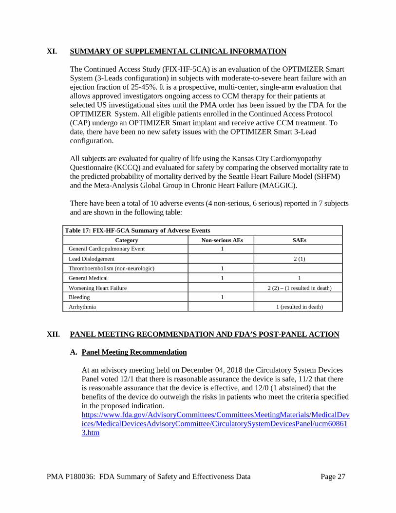
The Continued Access Study (FIX-HF-5CA) is an evaluation of the OPTIMIZER Smart System (3-Leads configuration) in subjects with moderate-to-severe heart failure with an ejection fraction of 25-45%. It is a prospective, multi-center, single-arm evaluation that allows approved investigators ongoing access to CCM therapy for their patients at selected US investigational sites until the PMA order has been issued by the FDA for the OPTIMIZER System. All eligible patients enrolled in the Continued Access Protocol (CAP) undergo an OPTIMIZER Smart implant and receive active CCM treatment. To date, there have been no new safety issues with the OPTIMIZER Smart 3-Lead configuration. All subjects are evaluated for quality of life using the Kansas City Cardiomyopathy Questionnaire (KCCQ) and evaluated for safety by comparing the observed mortality rate to the predicted probability of mortality derived by the Seattle Heart Failure Model (SHFM) and the Meta-Analysis Global Group in Chronic Heart Failure (MAGGIC). There have been a total of 10 adverse events (4 non-serious, 6 serious) reported in 7 subjects and are shown in the following table:
Table 17: FIX-HF-5CA Summary of Adverse Events Category Non-serious AEs SAEs
General Cardiopulmonary Event 1
Lead Dislodgement 2 (1) Thromboembolism (non-neurologic) 1
General Medical 1 1
Worsening Heart Failure 2 (2) – (1 resulted in death) Bleeding 1
Arrhythmia 1 (resulted in death)
XII. PANEL MEETING RECOMMENDATION AND FDA’S POST-PANEL ACTION
A. Panel Meeting Recommendation
At an advisory meeting held on December 04, 2018 the Circulatory System Devices Panel voted 12/1 that there is reasonable assurance the device is safe, 11/2 that there is reasonable assurance that the device is effective, and 12/0 (1 abstained) that the benefits of the device do outweigh the risks in patients who meet the criteria specified in the proposed indication. https://www.fda.gov/AdvisoryCommittees/CommitteesMeetingMaterials/MedicalDevices/MedicalDevicesAdvisoryCommittee/CirculatorySystemDevicesPanel/ucm608613.htm
PMA P180036: FDA Summary of Safety and Effectiveness Data Page 28
B. FDA’s Post-Panel Action
FDA agreed with the Panel’s recommendations. Specifically, the Indications for Use were revised to be reflective of the clinical assessements to remove NYHA class IV patients and improved excerice tolerance. Additionally, the Panel discussed expectations of a post approval study to rule out placebo effects and aim to more precisely identify the group of patients that most benefits from the device since the overall benefit for the group studied was considered to be marginal.
XIII. CONCLUSIONS DRAWN FROM PRECLINICAL AND CLINICAL STUDIES A. Effectiveness Conclusions
Even though the primary effectivness endpoint (change in peak V02) met its pre-specified endpoint the clinical significance was questioned; primarily because the observed treatment difference was due to a decline from baseline in the control arm. The treatment arm, depending on analysis method, either showed a decline in peak VO2 or a marginal increase; making claims of increased exercise tolerance not justifiable. Two subjective endpoints, Quality of life per the MLWHFQ, and the 6 minute hall walk, did show an improvement. However, the confidence intervals were somewhat wide; possibly due to the relatively small sample size and unblinded nature of the trial (control group did not receive a device). The latter raised the question among panel members if the positive outcomes for the subjective endpoints could be due to a placebo effect. Improvement in NYHA class was observed in the treatment group over the control group. These improvements were statistically significant and are clinically meaningful. In conclusion, the treatment group showed a reduction in heart failure symptoms and the device has a positive impact on patients with moderate heart failure.
B. Safety Conclusions
The risks of the device are based on nonclinical laboratory and animal studies as well as data collected in a clinical studies conducted to support PMA approval as described above. Risks associated with the OPTIMIZER Smart system are similar to those associated with ICDs and pacemakers; which are well documented in the literature. The IDE studies were relatively small (~ 327 implanted patients) and short (6 months). Therefore, long term complications such as lead fractures, lead insulation breaches, and delayed infections were not really seen but are known to occur. There was a relatively high number of lead dislodgements which are also known to occur
PMA P180036: FDA Summary of Safety and Effectiveness Data Page 29
but may have been higher due to the additional lead utlized in the OPTIMIZER Smart system. The increased lead count may result in higher long term lead complication rates as well considering that most of the patients receiving a OPTIMIZER Smart system will also have an ICD.
C. Benefit-Risk Determination
The IDE studies demonstrated that (1) there is a reduction in NYHA class, (2) there is an improvement of quality of life as based upon the MLWHFQ scores, (3) there is an increase in distance walked during the 6MHW test. The studies demonstrate very small improvements in peak VO2 for the treatment groups but stronger declines in pVO2 for the control groups. Given that the risks are well known and well understood, and the benefits are subjective (6MHW, MLWHFQ) the benefits marginally outweigh the risks; an opinion shared by the Panel. 1. Patient Perspectives
This submission did not include specific information on patient perspectives for this device.
In conclusion, given the available information above, the data support that for the indication for use of the device the probable benefits outweigh the probable risks.
D. Overall Conclusions
The data in this application support the reasonable assurance of safety and effectiveness of this device when used in accordance with the indications for use.
XIV. CDRH DECISION
CDRH issued an approval order on March 21, 2019. The final conditions of approval cited in the approval order are described below.
The applicant will conduct a post-approval study (PAS) to provide long term safety and effectiveness data for the OPTIMIZER Smart System. The study is a prospective, multi-center, non-randomized, single arm open label study. As per protocol dated March 19, 2019, the goal is to enroll approximately 620 patients such that 500 implanted patients reach the 36 months follow up.
The study as the following safety endpoints: 1. The composite of device- or procedure- related complication incident free
rate at 12 months post-index implantation procedure exceeds 75%. Procedure related complications through the end of 30 days following the index procedure and OPTIMIZER device related complications occurring through the end of 1-year following the index procedure shall contribute to the composite complication rate.
PMA P180036: FDA Summary of Safety and Effectiveness Data Page 30
2. Comparison of the observed mortality rate to the predicted mortality for
the patient group according to the Seattle Heart Failure Model (SHFM) as per protocol follow up schedule.
3. An assessment of the OPTIMIZER device related complications and
observations occurring during the 3-year period following the index procedure.
The study has the following effectiveness endpoints: 1. Monitor changes in Quality of Live (QoL), as measured by the Minnesota
Living with Heart Failure Questionnaire (MLWHFQ), from baseline to per protocol defined time intervals following index procedure.
2. Clinical assessment of New York Heart Association (NYHA) functional
class. A one-class change in functional class at per protocol defined time intervals shall be considered clinically significant.
3. Clinical assessment of changes in left ventricular ejection fraction (LVEF)
and end-systolic volume (ESV) from baseline at per protocol defined time intervals.
4. Clinical assessment of changes in NT-proBNP from baseline at per
protocol defined time intervals. 5. Clinical assessment of changes in QRS duration from baseline at per
protocol defined time intervals.
Interim analyses will be conducted throughout the study. These analyses will be conducted for reporting purposes only without statistical inferences or intention to modify the study. The study sample size shall be based on the primary safety endpoint. All other endpoints will be reported. All testing will be done at a nominal one-sided alpha level of 0.025 without adjustment for multiplicity as the purpose of these analyses is for reporting purposes only and not intended for labeling.
The applicant’s manufacturing facilities have been inspected and found to be in compliance with the device Quality System (QS) regulation (21 CFR 820).
XV. APPROVAL SPECIFICATIONS
Directions for use: See device labeling. Hazards to Health from Use of the Device: See Indications, Contraindications, Warnings, Precautions, and Adverse Events in the device labeling.
PMA P180036: FDA Summary of Safety and Effectiveness Data Page 31
Post-approval Requirements and Restrictions: See approval order.
XVI. REFERENCES
[1] Linde et. al., “Cardiac resynchronization therapy (CRT): Clinical trials, guidelines, and target populations”, Heart Rhythm, Vol 9, No 8S, August Supplement 2012.


![Summary of Safety and Effectivness (SSED)Template · 2020. 9. 1. · crebbp fgf12 inpp4b . mpl [exon 10] pdgfra [exons 12, 18] ros1 [exons 31, 36-38, 40] u2af1 . brca1 {introns 2,](https://img.pdfslide.us/doc/110x75/60aebbf62ac8c7702f4b432e/summary-of-safety-and-effectivness-ssedtemplate-2020-9-1-crebbp-fgf12-inpp4b.jpg)