Embed Size (px)
Citation preview
www.elsevier.com/locate/gca
Geochimica et Cosmochimica Acta 70 (2006) 6118–6135
Sulfates on Mars: A systematic Raman spectroscopic studyof hydration states of magnesium sulfates
Alian Wang a,*, John J. Freeman a, Bradley L. Jolliff a, I-Ming Chou b
a Department of Earth and Planetary Sciences and McDonnell Center for Space Sciences, Washington University, St. Louis, MO 63130, USAb U.S. Geological Survey, 954 National Center, Reston, VA 20192, USA
Received 7 February 2006; accepted in revised form 31 May 2006
Abstract
The martian orbital and landed surface missions, OMEGA on Mar Express and the two Mars Explorations Rovers, respectively, haveyielded evidence pointing to the presence of magnesium sulfates on the martian surface. In situ identification of the hydration states ofmagnesium sulfates, as well as the hydration states of other Ca- and Fe- sulfates, will be crucial in future landed missions on Mars inorder to advance our knowledge of the hydrologic history of Mars as well as the potential for hosting life on Mars. Raman spectroscopyis a technique well-suited for landed missions on the martian surface. In this paper, we report a systematic study of the Raman spectra ofthe hydrates of magnesium sulfate. Characteristic and distinct Raman spectral patterns were observed for each of the 11 distinct hydratesof magnesium sulfates, crystalline and non-crystalline. The unique Raman spectral features along with the general tendency of the shift ofthe position of the sulfate m1 band towards higher wavenumbers with a decrease in the degree of hydration allow in situ identification ofthese hydrated magnesium sulfates from the raw Raman spectra of mixtures. Using these Raman spectral features, we have started thestudy of the stability field of hydrated magnesium sulfates and the pathways of their transformations at various temperature and relativehumidity conditions. In particular we report on the Raman spectrum of an amorphous hydrate of magnesium sulfate (MgSO4Æ2H2O)that may have specific relevance for the martian surface.� 2006 Elsevier Inc. All rights reserved.
1. Introduction
The sulfur enrichment and the compositional correla-tions of Mg and S within martian surface materials werefirst found in X-ray fluorescence data during Viking mis-sions (Clark et al., 1982). Both observations were con-firmed by the APXS (Alpha Particle X-ray Spectrometer)experiments on the Sojourner rover of the Mars Pathfindermission (Rieder et al., 1997; McSween et al., 1999) and onthe Spirit and the Opportunity rovers of the Mars Explora-tion Rovers (MER) missions (Gellert et al., 2004, 2006;Rieder et al., 2004). The correlations between Ca and S,and Fe and S have also been found in various regions atthe Gusev landing site (Haskin et al., 2005; Ming et al.,
0016-7037/$ - see front matter � 2006 Elsevier Inc. All rights reserved.
doi:10.1016/j.gca.2006.05.022
* Corresponding author. Fax: +1 314 935 7361.E-mail address: [email protected] (A. Wang).
2006; Wang et al., 2006a). Chemical correlations fromthese landed missions imply that sulfate salts are importantconstituents in martian surface and subsurface materials.There is only one direct identification of sulfate, as amineral, from the landed missions on Mars, jarosite[KFe3+(SO4)2(OH)6], which was identified throughMossbauer spectrometer analysis (Klingelhofer et al.,2004) by the Opportunity rover at Meridiani Planum.
By comparison, a variety of sulfates has been identifiedthrough the results of the Mars Express orbital mission.The OMEGA spectrometer (Observatiore pour la Mineral-ogie, l’Eau, les Glacies, et l’Activitie) observed characteris-tic near infrared (NIR) peaks of sulfates at variouslocations on Mars (Bibring et al., 2005), including Meridi-ani Planum where the Opportunity rover landed (Arvidsonet al., 2005). The types of sulfates definitively identifiedfrom OMEGA spectra were kieserite (MgSO4ÆH2O), gyp-sum (CaSO4Æ2H2O), and bassanite (2CaSO4ÆH2O). Another
Hydration states of Mg-sulfates on Mars 6119
type of OMEGA spectrum was assigned to polyhydratedsulfates and the sulfates with different cations, for whichthe spectra of epsomite (MgSO4Æ7H2O), copiapite(Fe2þFe3þ
4ðSO4Þ6ðOHÞ2 � 20H2O), and halotrichite(Fe2+Al2(SO4)4Æ22H2O) would provide matches (Gendrinet al., 2005). A few percent of adsorbed water was also as-signed on the basis of observations that (1) 3 lm absorp-tion bands are observed in every spectrum obtained byOMEGA; and (2) the 3-lm band intensity is positively cor-related with the high albedo areas on Mars.
The neutron spectrometer (NS) component of the gam-ma-ray spectrometer suite (GRS) on board the Mars Odys-sey spacecraft detected high concentrations of hydrogen intwo broad equatorial regions in the neighborhood of Ara-bia Terra (centered at �5�N, +25�E) and Medusae Fossae(centered at �15�N, +180�E), which cover respectively theMeridiani Planum and Gusev Crater sites (Boynton et al.,2002; Feldman et al., 2004). The relative maxima in hydro-gen abundances correlate topographically with mid- andlow-altitude areas. Because water–ice should not be stableunder the current conditions at the surface and near surfaceof these equatorial areas, the hydrogen is assumed to residein hydrated minerals although ground ice cannot totally beruled out (Boynton et al., 2002; Feldman et al., 2004, 2005).
Mars apparently had large amounts of surface water inthe past that could plausibly have produced evaporitedeposits, including water-bearing minerals such as thehydrated sulfates (Carr, 1996; Hynek and Phillips, 2001,2003; Carr and Head, 2003; Squyres et al., 2004).
Sulfur-bearing materials associated with the sulfur cyclemust have played an important role in the evolution andhydrologic history of Mars. Large amounts of sulfur mayhave originated from volcanic outgassing, in the form ofH2S or SO2, which could react with oxidizing atmosphericcomponents to form [SO3]2�, [HSO4]�, or [H2SO4], andthen fall onto the martian surface. During a warm andwet era, these gaseous species would have dissolved inwater to form acidic aqueous solutions. The solutionswould be neutralized by reacting with igneous minerals,which release Mg, Ca, Fe, and Al cations. At high degreesof alteration, sulfides, as minor phases in igneous mineralassemblages (as seen in martian meteorites), would alsobe oxidized and S released into solution.
Sulfur-rich solutions might also originate from hydro-thermal alteration associated with convection cells drivenby intrusive igneous activity, especially in areas nearlong-lived volcanic provinces. Igneous activity would haveoccurred throughout Noachian time and may have extend-ed to geologically recent times, judging from relativelyyoung ages of some of the martian basaltic meteorites.Such solutions might also be mixed and carried laterallyby surface or ground waters, and eventually deposited inevaporite sulfates. Sulfur-bearing phases at different stagesof the cycle and phase transitions among them wouldprovide energy sources for metabolic processes of bio-spe-cies, as seen on Earth (Tosca et al., 2004; Golden et al.,2005).
Planetary Raman spectroscopy is a powerful tool forin situ identification of minerals and biogenic speciesexpected (potentially) to be encountered in planetary sur-face exploration (Wdowiak et al., 1994; Wang et al.,1995; Haskin et al., 1997; Wynn-Williams and Edwards,2000). A laser Raman spectroscopy (LRS) experimentcan characterize, structurally and compositionally, manyclasses of minerals and inorganic compounds such as sili-cates, carbonates, sulfates, phosphates, oxides, sulfides,and oxyhydroxides, as well as clathrates of CO2 andCH4. Additionally, Raman spectroscopy is useful for char-acterizing the organic species produced in biogenic or non-biogenic processes where it can distinguish functionalgroups in organics materials containing H–O, H–N, H–C, N–O, C–O, C–N, and P–O bonds (Nakamoto, 1997).All species in the sulfur cycle on Earth have a diagnosticRaman spectrum that would enable in situ identification.A Raman instrument suitable for martian surface studiesis the Mars Microbeam Raman Spectrometer (MMRS)that we have developed which combines a microbeam Ra-man detection capability with a line-scanning capability(Haskin et al., 1997; Wang et al., 1998, 2003). This systempermits trace constituents in a target to be detected (Wanget al., 2004b) in addition to the major and minor phases.The line scanning capability of the MMRS can be usedto obtain information on the distributions and relative pro-portions of species and on grain sizes and the rock textures(Wang et al., 1999a); thus, the mineralogical informationcan be correlated directly with morphological microscopicimaging (Wang et al., 1999b; Kuebler et al., 2003). In addi-tion, remote Raman technology has been developed in re-cent years, which shows the effective detections of variousmineral phases from a distance of �10 m (Sharma et al.,2002; Misra et al., 2006).
Among all species involved in S-cycling, hydrated sul-fate minerals bear special interest due to the recent discov-eries by the Mars Exploration Rover missions and MarsExpress OMEGA. There is no systematic Raman spectro-scopic study to date in the literature on the full range ofhydrated magnesium and iron sulfates (LaFont and Vinh,1966; Chio et al., 2004, 2005; Wang et al., 2005; Sharmaet al., 2006). We report in this paper, our first study of Ra-man experiments on the full range of hydrated Mg-sulfates.
2. Importance of studying the hydration states of sulfates
The highest concentration of Mg-sulfates found by theSpirit rover (inferred from the APXS data) at Gusev thusfar has been in the subsurface regolith at The Boroughstrench (Wang et al., 2005b; Wang et al., 2006a). The distri-bution of sulfates in subsurface regolith within this trenchis heterogeneous. By first-order approximation, sulfatesmake about 22 wt% of subsurface regolith at the most sul-fur-rich spot in the trench, and approximately 85 mole % ofthese sulfates consists of Mg-sulfate. Assuming all theseMg-sulfates are in the form of kieserite as seen by OMEGAfrom other locations on Mars including Meridiani Planum,
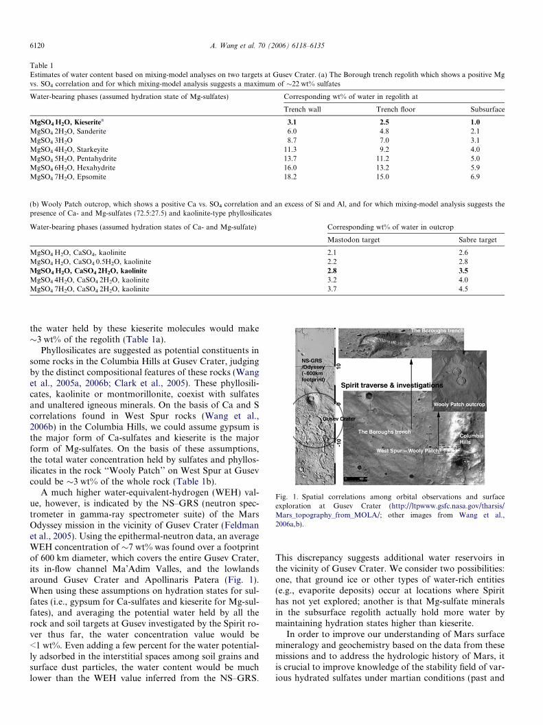
Table 1Estimates of water content based on mixing-model analyses on two targets at Gusev Crater. (a) The Borough trench regolith which shows a positive Mgvs. SO4 correlation and for which mixing-model analysis suggests a maximum of �22 wt% sulfates
Water-bearing phases (assumed hydration state of Mg-sulfates) Corresponding wt% of water in regolith at
Trench wall Trench floor Subsurface
MgSO4ÆH2O, Kieseritea 3.1 2.5 1.0
MgSO4Æ2H2O, Sanderite 6.0 4.8 2.1MgSO4Æ3H2O 8.7 7.0 3.1MgSO4Æ4H2O, Starkeyite 11.3 9.2 4.0MgSO4Æ5H2O, Pentahydrite 13.7 11.2 5.0MgSO4Æ6H2O, Hexahydrite 16.0 13.2 5.9MgSO4Æ7H2O, Epsomite 18.2 15.0 6.9
(b) Wooly Patch outcrop, which shows a positive Ca vs. SO4 correlation and an excess of Si and Al, and for which mixing-model analysis suggests thepresence of Ca- and Mg-sulfates (72.5:27.5) and kaolinite-type phyllosilicates
Water-bearing phases (assumed hydration states of Ca- and Mg-sulfate) Corresponding wt% of water in outcrop
Mastodon target Sabre target
MgSO4ÆH2O, CaSO4, kaolinite 2.1 2.6MgSO4ÆH2O, CaSO4Æ0.5H2O, kaolinite 2.2 2.8MgSO4ÆH2O, CaSO4Æ2H2O, kaolinite 2.8 3.5
MgSO4Æ4H2O, CaSO4Æ2H2O, kaolinite 3.2 4.0MgSO4Æ7H2O, CaSO4Æ2H2O, kaolinite 3.7 4.5
Fig. 1. Spatial correlations among orbital observations and surfaceexploration at Gusev Crater (http://ltpwww.gsfc.nasa.gov/tharsis/Mars_topography_from_MOLA/; other images from Wang et al.,2006a,b).
6120 A. Wang et al. 70 (2006) 6118–6135
the water held by these kieserite molecules would make�3 wt% of the regolith (Table 1a).
Phyllosilicates are suggested as potential constituents insome rocks in the Columbia Hills at Gusev Crater, judgingby the distinct compositional features of these rocks (Wanget al., 2005a, 2006b; Clark et al., 2005). These phyllosili-cates, kaolinite or montmorillonite, coexist with sulfatesand unaltered igneous minerals. On the basis of Ca and Scorrelations found in West Spur rocks (Wang et al.,2006b) in the Columbia Hills, we could assume gypsum isthe major form of Ca-sulfates and kieserite is the majorform of Mg-sulfates. On the basis of these assumptions,the total water concentration held by sulfates and phyllos-ilicates in the rock ‘‘Wooly Patch’’ on West Spur at Gusevcould be �3 wt% of the whole rock (Table 1b).
A much higher water-equivalent-hydrogen (WEH) val-ue, however, is indicated by the NS–GRS (neutron spec-trometer in gamma-ray spectrometer suite) of the MarsOdyssey mission in the vicinity of Gusev Crater (Feldmanet al., 2005). Using the epithermal-neutron data, an averageWEH concentration of �7 wt% was found over a footprintof 600 km diameter, which covers the entire Gusev Crater,its in-flow channel Ma’Adim Valles, and the lowlandsaround Gusev Crater and Apollinaris Patera (Fig. 1).When using these assumptions on hydration states for sul-fates (i.e., gypsum for Ca-sulfates and kieserite for Mg-sul-fates), and averaging the potential water held by all therock and soil targets at Gusev investigated by the Spirit ro-ver thus far, the water concentration value would be<1 wt%. Even adding a few percent for the water potential-ly adsorbed in the interstitial spaces among soil grains andsurface dust particles, the water content would be muchlower than the WEH value inferred from the NS–GRS.
This discrepancy suggests additional water reservoirs inthe vicinity of Gusev Crater. We consider two possibilities:one, that ground ice or other types of water-rich entities(e.g., evaporite deposits) occur at locations where Spirithas not yet explored; another is that Mg-sulfate mineralsin the subsurface regolith actually hold more water bymaintaining hydration states higher than kieserite.
In order to improve our understanding of Mars surfacemineralogy and geochemistry based on the data from thesemissions and to address the hydrologic history of Mars, itis crucial to improve knowledge of the stability field of var-ious hydrated sulfates under martian conditions (past and
Hydration states of Mg-sulfates on Mars 6121
present), and the pathways and the rates of phase transfor-mations. These data will also help to provide insight to thegeochemical processes and evolution during the diurnal cy-cle, the obliquity cycles, and throughout martian geologicaltime during which the surface has been modified by volca-nic, impact, aqueous, and eolian processes.
Several studies concerning the hydrated sulfates havebeen published in the past few years. Among them, Vani-man et al. (2004, 2005) reported that structurally amor-phous Mg-sulfates can be formed rapidly (in a few hoursor a day) from hexahydrite (MgSO4Æ6H2O) under low rela-tive humidity (0.3–0.4% RH) or with a vacuum at 1 torr(132 Pa) total pressure. They suggest the number of struc-tural waters held by this amorphous phase can vary from2 to 1.2. Because of the large relative-humidity swing dur-ing the current diurnal cycle on the surface of Mars, vary-ing from <1% RH (�290 K) at mid-day summer to near100% RH (�180 K) during the cold early morning, kiese-rite at the surface would convert to hexahydrite (and eps-omite) at high RH and then dehydrate to form hydratedamorphous Mg-sulfate, whose re-hydration is extremelysluggish at night-time temperatures. Therefore, the hydrat-ed amorphous phase, not kieserite, would be the favoredMg-sulfate at the martian surface today.
Chipera et al. (2005) demonstrated that besides hexahy-drite and epsomite, other hydrated phases of Mg-sulfate,especially starkeyite (MgSO4Æ4H2O), can be stable andco-exist with kieserite under martian surface conditions.The phase transformations between these different hydrat-ed phases of Mg-sulfates are characterized by sluggishkinetics, path dependence, and the presence of metastableintermediate states of hydration.
Chou and Seal determined the epsomite–hexahydriteequilibria (Chou and Seal, 2003) and starkeyite–hexahy-drite equilibria (Chou et al., 2005) as a function of bothtemperature and relative humidity using the humidity-buff-
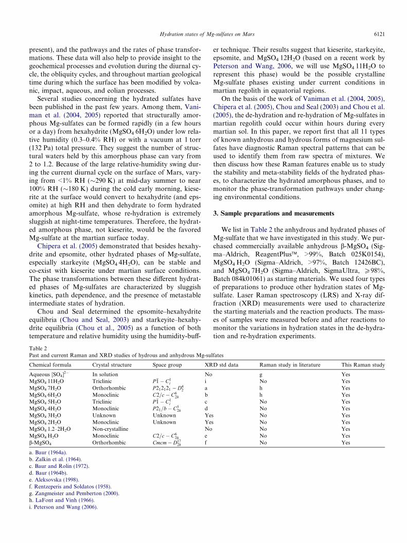
Table 2Past and current Raman and XRD studies of hydrous and anhydrous Mg-sul
Chemical formula Crystal structure Space group XR
Aqueous [SO4]2� In solution NoMgSO4Æ11H2O Triclinic P�1� C1
i iMgSO4Æ7H2O Orthorhombic P212121 � D4
2 aMgSO4Æ6H2O Monoclinic C2=c� C6
2h bMgSO4Æ5H2O Triclinic P�1� C1
i cMgSO4Æ4H2O Monoclinic P21=b� C5
2h dMgSO4Æ3H2O Unknown Unknown YesMgSO4Æ2H2O Monoclinic Unknown YesMgSO4Æ1.2–2H2O Non-crystalline NoMgSO4ÆH2O Monoclinic C2=c� C6
2h eb-MgSO4 Orthorhombic Cmcm� D17
2h f
a. Baur (1964a).b. Zalkin et al. (1964).c. Baur and Rolin (1972).d. Baur (1964b).e. Aleksovska (1998).f. Rentzeperis and Soldatos (1958).g. Zangmeister and Pemberton (2000).h. LaFont and Vinh (1966).i. Peterson and Wang (2006).
er technique. Their results suggest that kieserite, starkeyite,epsomite, and MgSO4Æ12H2O (based on a recent work byPeterson and Wang, 2006, we will use MgSO4Æ11H2O torepresent this phase) would be the possible crystallineMg-sulfate phases existing under current conditions inmartian regolith in equatorial regions.
On the basis of the work of Vaniman et al. (2004, 2005),Chipera et al. (2005), Chou and Seal (2003) and Chou et al.(2005), the de-hydration and re-hydration of Mg-sulfates inmartian regolith could occur within hours during everymartian sol. In this paper, we report first that all 11 typesof known anhydrous and hydrous forms of magnesium sul-fates have diagnostic Raman spectral patterns that can beused to identify them from raw spectra of mixtures. Wethen discuss how these Raman features enable us to studythe stability and meta-stability fields of the hydrated phas-es, to characterize the hydrated amorphous phases, and tomonitor the phase-transformation pathways under chang-ing environmental conditions.
3. Sample preparations and measurements
We list in Table 2 the anhydrous and hydrated phases ofMg-sulfate that we have investigated in this study. We pur-chased commercially available anhydrous b-MgSO4 (Sig-ma–Aldrich, ReagentPlus�, >99%, Batch 025K0154),MgSO4ÆH2O (Sigma–Aldrich, >97%, Batch 12426BC),and MgSO4Æ7H2O (Sigma–Aldrich, SigmaUltra, P98%,Batch 084k01061) as starting materials. We used four typesof preparations to produce other hydration states of Mg-sulfate. Laser Raman spectroscopy (LRS) and X-ray dif-fraction (XRD) measurements were used to characterizethe starting materials and the reaction products. The mass-es of samples were measured before and after reactions tomonitor the variations in hydration states in the de-hydra-tion and re-hydration experiments.
fates
D std data Raman study in literature This Raman study
g YesNo Yesh Yesh YesNo YesNo YesNo YesNo YesNo YesNo YesNo Yes
6122 A. Wang et al. 70 (2006) 6118–6135
3.1. Direct crystallization from saturated solutions
The methods we used are based on those described byEmons et al. (1990) and involve crystallization from satu-rated MgSO4 solution, oversaturated methanolic MgSO4
solution, and mixtures of saturated MgSO4 and MgCl2solutions. Pure epsomite and hexahydrite were produced.From the preparations targeted on Mg-sulfates with 2–5structural water molecules, however, we only obtained mix-tures of Mg-sulfates with various hydration states. Never-theless, using Raman microbeam analysis, single-phaseRaman spectra of most hydration states of the Mg-sulfateswere obtained from individual crystals within these mix-tures. The assignments of these Raman spectra were laterconfirmed by coupled LRS and XRD measurements onthe pure samples prepared using two other methods.
3.2. Heating solid samples at fixed temperatures
Baking epsomite at different temperatures in an openoven has produced homogenous kieserite (at 95 �C) andstarkeyite (at 40 �C) samples, with sufficient quantities forXRD verification of LRS spectral assignments. The tem-perature of the oven was held at the set point to within±1 �C, whereas the water-vapor pressure in the oven wasdetermined by the relative humidity of the laboratorywhich varied from 30% to 55% RH depending on theseason.
3.3. Vacuum desiccation of solid samples
A vacuum desiccator kept at room temperature(21 ± 1 �C) was used to convert epsomite and hexahydriteinto amorphous MgSO4, which was reported in the exper-iments of Vaniman et al. (2004, 2005). The vacuum in thedesiccator was kept at about 0.5 torr (�67 Pa) for twoexperiments of 5 and 15 days duration. LRS and XRDmeasurements were made on the samples in sealed contain-ers immediately after removal from the vacuum.
3.4. Using humidity buffer solutions to convert the hydration
states
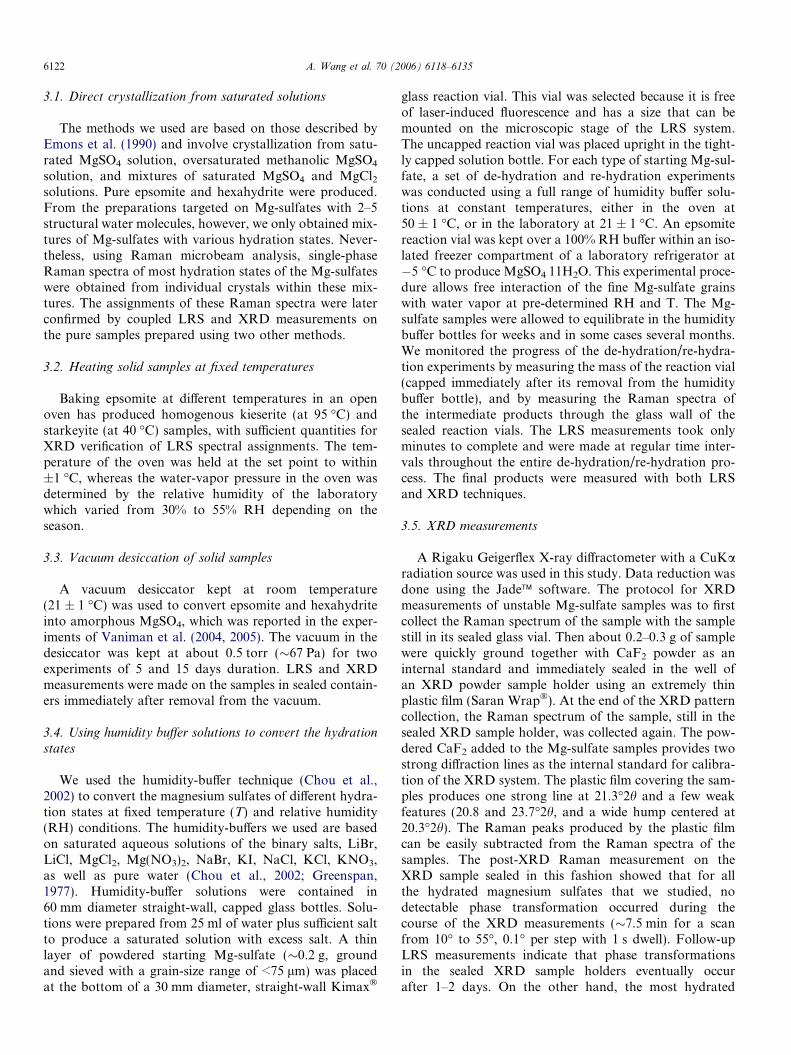
We used the humidity-buffer technique (Chou et al.,2002) to convert the magnesium sulfates of different hydra-tion states at fixed temperature (T) and relative humidity(RH) conditions. The humidity-buffers we used are basedon saturated aqueous solutions of the binary salts, LiBr,LiCl, MgCl2, Mg(NO3)2, NaBr, KI, NaCl, KCl, KNO3,as well as pure water (Chou et al., 2002; Greenspan,1977). Humidity-buffer solutions were contained in60 mm diameter straight-wall, capped glass bottles. Solu-tions were prepared from 25 ml of water plus sufficient saltto produce a saturated solution with excess salt. A thinlayer of powdered starting Mg-sulfate (�0.2 g, groundand sieved with a grain-size range of <75 lm) was placedat the bottom of a 30 mm diameter, straight-wall Kimax�
glass reaction vial. This vial was selected because it is freeof laser-induced fluorescence and has a size that can bemounted on the microscopic stage of the LRS system.The uncapped reaction vial was placed upright in the tight-ly capped solution bottle. For each type of starting Mg-sul-fate, a set of de-hydration and re-hydration experimentswas conducted using a full range of humidity buffer solu-tions at constant temperatures, either in the oven at50 ± 1 �C, or in the laboratory at 21 ± 1 �C. An epsomitereaction vial was kept over a 100% RH buffer within an iso-lated freezer compartment of a laboratory refrigerator at�5 �C to produce MgSO4Æ11H2O. This experimental proce-dure allows free interaction of the fine Mg-sulfate grainswith water vapor at pre-determined RH and T. The Mg-sulfate samples were allowed to equilibrate in the humiditybuffer bottles for weeks and in some cases several months.We monitored the progress of the de-hydration/re-hydra-tion experiments by measuring the mass of the reaction vial(capped immediately after its removal from the humiditybuffer bottle), and by measuring the Raman spectra ofthe intermediate products through the glass wall of thesealed reaction vials. The LRS measurements took onlyminutes to complete and were made at regular time inter-vals throughout the entire de-hydration/re-hydration pro-cess. The final products were measured with both LRSand XRD techniques.
3.5. XRD measurements
A Rigaku Geigerflex X-ray diffractometer with a CuKaradiation source was used in this study. Data reduction wasdone using the Jade� software. The protocol for XRDmeasurements of unstable Mg-sulfate samples was to firstcollect the Raman spectrum of the sample with the samplestill in its sealed glass vial. Then about 0.2–0.3 g of samplewere quickly ground together with CaF2 powder as aninternal standard and immediately sealed in the well ofan XRD powder sample holder using an extremely thinplastic film (Saran Wrap�). At the end of the XRD patterncollection, the Raman spectrum of the sample, still in thesealed XRD sample holder, was collected again. The pow-dered CaF2 added to the Mg-sulfate samples provides twostrong diffraction lines as the internal standard for calibra-tion of the XRD system. The plastic film covering the sam-ples produces one strong line at 21.3�2h and a few weakfeatures (20.8 and 23.7�2h, and a wide hump centered at20.3�2h). The Raman peaks produced by the plastic filmcan be easily subtracted from the Raman spectra of thesamples. The post-XRD Raman measurement on theXRD sample sealed in this fashion showed that for allthe hydrated magnesium sulfates that we studied, nodetectable phase transformation occurred during thecourse of the XRD measurements (�7.5 min for a scanfrom 10� to 55�, 0.1� per step with 1 s dwell). Follow-upLRS measurements indicate that phase transformationsin the sealed XRD sample holders eventually occurafter 1–2 days. On the other hand, the most hydrated
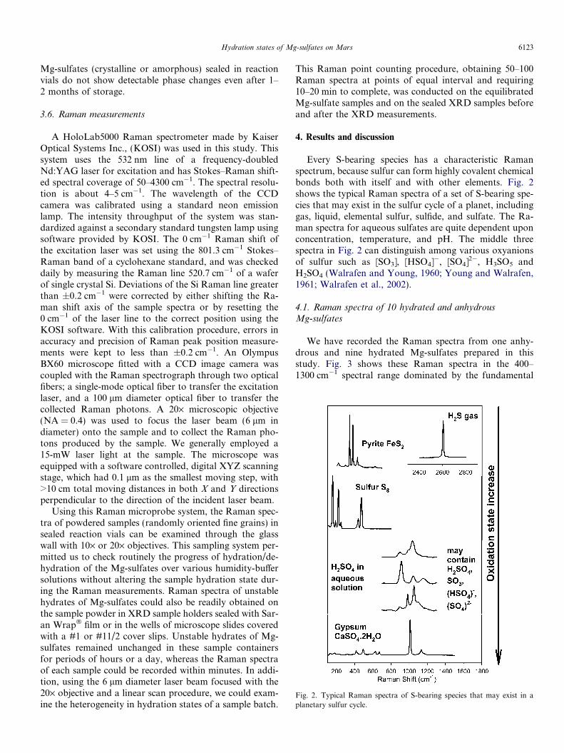
Fig. 2. Typical Raman spectra of S-bearing species that may exist in aplanetary sulfur cycle.
Hydration states of Mg-sulfates on Mars 6123
Mg-sulfates (crystalline or amorphous) sealed in reactionvials do not show detectable phase changes even after 1–2 months of storage.
3.6. Raman measurements
A HoloLab5000 Raman spectrometer made by KaiserOptical Systems Inc., (KOSI) was used in this study. Thissystem uses the 532 nm line of a frequency-doubledNd:YAG laser for excitation and has Stokes–Raman shift-ed spectral coverage of 50–4300 cm�1. The spectral resolu-tion is about 4–5 cm�1. The wavelength of the CCDcamera was calibrated using a standard neon emissionlamp. The intensity throughput of the system was stan-dardized against a secondary standard tungsten lamp usingsoftware provided by KOSI. The 0 cm�1 Raman shift ofthe excitation laser was set using the 801.3 cm�1 Stokes–Raman band of a cyclohexane standard, and was checkeddaily by measuring the Raman line 520.7 cm�1 of a waferof single crystal Si. Deviations of the Si Raman line greaterthan ±0.2 cm�1 were corrected by either shifting the Ra-man shift axis of the sample spectra or by resetting the0 cm�1 of the laser line to the correct position using theKOSI software. With this calibration procedure, errors inaccuracy and precision of Raman peak position measure-ments were kept to less than ±0.2 cm�1. An OlympusBX60 microscope fitted with a CCD image camera wascoupled with the Raman spectrograph through two opticalfibers; a single-mode optical fiber to transfer the excitationlaser, and a 100 lm diameter optical fiber to transfer thecollected Raman photons. A 20· microscopic objective(NA = 0.4) was used to focus the laser beam (6 lm indiameter) onto the sample and to collect the Raman pho-tons produced by the sample. We generally employed a15-mW laser light at the sample. The microscope wasequipped with a software controlled, digital XYZ scanningstage, which had 0.1 lm as the smallest moving step, with>10 cm total moving distances in both X and Y directionsperpendicular to the direction of the incident laser beam.
Using this Raman microprobe system, the Raman spec-tra of powdered samples (randomly oriented fine grains) insealed reaction vials can be examined through the glasswall with 10· or 20· objectives. This sampling system per-mitted us to check routinely the progress of hydration/de-hydration of the Mg-sulfates over various humidity-buffersolutions without altering the sample hydration state dur-ing the Raman measurements. Raman spectra of unstablehydrates of Mg-sulfates could also be readily obtained onthe sample powder in XRD sample holders sealed with Sar-an Wrap� film or in the wells of microscope slides coveredwith a #1 or #11/2 cover slips. Unstable hydrates of Mg-sulfates remained unchanged in these sample containersfor periods of hours or a day, whereas the Raman spectraof each sample could be recorded within minutes. In addi-tion, using the 6 lm diameter laser beam focused with the20· objective and a linear scan procedure, we could exam-ine the heterogeneity in hydration states of a sample batch.
This Raman point counting procedure, obtaining 50–100Raman spectra at points of equal interval and requiring10–20 min to complete, was conducted on the equilibratedMg-sulfate samples and on the sealed XRD samples beforeand after the XRD measurements.
4. Results and discussion
Every S-bearing species has a characteristic Ramanspectrum, because sulfur can form highly covalent chemicalbonds both with itself and with other elements. Fig. 2shows the typical Raman spectra of a set of S-bearing spe-cies that may exist in the sulfur cycle of a planet, includinggas, liquid, elemental sulfur, sulfide, and sulfate. The Ra-man spectra for aqueous sulfates are quite dependent uponconcentration, temperature, and pH. The middle threespectra in Fig. 2 can distinguish among various oxyanionsof sulfur such as [SO3], [HSO4]�, [SO4]2�, H3SO5 andH2SO4 (Walrafen and Young, 1960; Young and Walrafen,1961; Walrafen et al., 2002).
4.1. Raman spectra of 10 hydrated and anhydrousMg-sulfates
We have recorded the Raman spectra from one anhy-drous and nine hydrated Mg-sulfates prepared in thisstudy. Fig. 3 shows these Raman spectra in the 400–1300 cm�1 spectral range dominated by the fundamental

Fig. 4. Raman spectra of 10 hydrated and anhydrous Mg-sulfates in thespectral region of water OH stretching vibrational modes. *, Tentativespectral assignment.
Fig. 3. Raman spectra of 10 hydrated and an anhydrous Mg-sulfates inthe spectral region of SO4 fundamental vibrational modes. *, Tentativespectral assignment; #, peak of kieserite as impurity.
6124 A. Wang et al. 70 (2006) 6118–6135
vibrational modes of the SO4 tetrahedra Fig. 4 shows the2500–4000 cm�1 spectral range where the Raman peaksof structural water dominate. Table 3 lists the positionsof the major Raman peaks of these Mg-sulfates.
Except for the MgSO4 aqueous solution, MgSO4Æ11H2O,and MgSO4Æ3H2O, the Raman spectra assignments of theother seven Mg-sulfates in Figs. 3 and 4 have been con-firmed by XRD measurements on the same samples usingpublished values for the XRD powder diffraction patterns.The spectrum of MgSO4 aqueous solution is obtaineddirectly from a saturated solution within a liquid cell andthe spectrum matches the data for neutral pH aqueous solu-tions of SO4
�2 obtained by Nakamoto (1997). We found nopublished X-ray diffraction pattern for MgSO4Æ12H2O thatcan be used to verify the Raman identification. Our assign-ment is based on the fact that (1) our sample was preparedaccording to the conditions shown in the phase diagram ofHogenboom et al. (1995) (i.e., the hydration of MgSO4 Æ7H2O in equilibrium with ice at �5 �C); (2) the productshows a mass increase from the original mass of MgSO4Æ
7H2O that is consistant with an increased degree of hydra-tion; and (3) the LRS measurements on microscopicallysmall crystals of several batches suggest a mixture ofMgSO4Æ7H2O and a new phase. The spectrum of this newphase is tentatively assigned to MgSO4Æ11H2O (Figs. 3and 4).
The Raman spectrum with a Raman peak at1023.8 cm�1 is also tentatively assigned to MgSO4Æ3H2O.The sample from which this spectrum was obtained wasonly produced once from hexahydrite powder baked inan oven at 60 �C in ambient-laboratory relative humidity.Raman point-counting measurements suggest that thissample is a mixture. Among numerous Raman spectrafrom this sample, we see Raman peaks of hexahydrite witha minor amount of kieserite, starkeyite, and sanderite, plusthe Raman spectrum of a new phase with a unique Ramanspectrum showing a m1 peak at 1023.8 cm�1 and a distinctspectral pattern. Compared with the Raman m1 peak posi-tions of other hydrated Mg-sulfates whose identities were
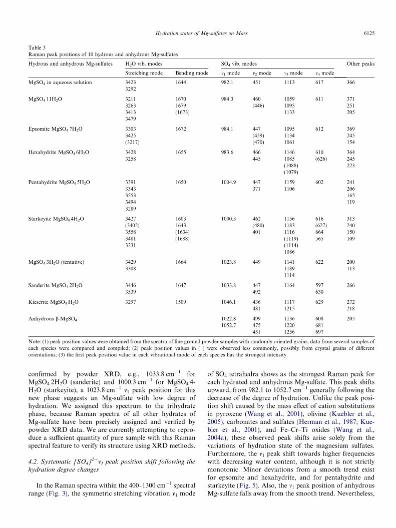
Table 3Raman peak positions of 10 hydrous and anhydrous Mg-sulfates
Hydrous and anhydrous Mg-sulfates H2O vib. modes SO4 vib. modes Other peaks
Stretching mode Bending mode m1 mode m2 mode m3 mode m4 mode
MgSO4 in aqueous solution 3423 1644 982.1 451 1113 617 3663292
MgSO4Æ11H2O 3211 1670 984.3 460 1059 611 3713263 1679 (446) 1095 2513413 (1673) 1133 2053479
Epsomite MgSO4Æ7H2O 3303 1672 984.1 447 1095 612 3693425 (459) 1134 245(3217) (470) 1061 154
Hexahydrite MgSO4Æ6H2O 3428 1655 983.6 466 1146 610 3643258 445 1085 (626) 245
(1088) 223(1079)
Pentahydrite MgSO4Æ5H2O 3391 1650 1004.9 447 1159 602 2413343 371 1106 2063553 1653494 1193289
Starkeyite MgSO4Æ4H2O 3427 1603 1000.3 462 1156 616 313(3402) 1643 (480) 1183 (627) 2403558 (1634) 401 1116 664 1503481 (1688) (1119) 565 1093331 (1114)
1086
MgSO4Æ3H2O (tentative) 3429 1664 1023.8 449 1141 622 2003308 1189 113
1114
Sanderite MgSO4Æ2H2O 3446 1647 1033.8 447 1164 597 2663539 492 630
Kieserite MgSO4ÆH2O 3297 1509 1046.1 436 1117 629 272481 1215 218
Anhydrous b-MgSO4 1022.8 499 1136 608 2051052.7 475 1220 681
451 1256 697
Note: (1) peak position values were obtained from the spectra of fine ground powder samples with randomly oriented grains, data from several samples ofeach species were compared and compiled; (2) peak position values in ( ) were observed less commonly, possibly from crystal grains of differentorientations; (3) the first peak position value in each vibrational mode of each species has the strongest intensity.
Hydration states of Mg-sulfates on Mars 6125
confirmed by powder XRD, e.g., 1033.8 cm�1 forMgSO4Æ2H2O (sanderite) and 1000.3 cm�1 for MgSO4Æ4-H2O (starkeyite), a 1023.8 cm�1 m1 peak position for thisnew phase suggests an Mg-sulfate with low degree ofhydration. We assigned this spectrum to the trihydratephase, because Raman spectra of all other hydrates ofMg-sulfate have been precisely assigned and verified bypowder XRD data. We are currently attempting to repro-duce a sufficient quantity of pure sample with this Ramanspectral feature to verify its structure using XRD methods.
4.2. Systematic [SO4]2�m1 peak position shift following the
hydration degree changes
In the Raman spectra within the 400–1300 cm�1 spectralrange (Fig. 3), the symmetric stretching vibration m1 mode
of SO4 tetrahedra shows as the strongest Raman peak foreach hydrated and anhydrous Mg-sulfate. This peak shiftsupward, from 982.1 to 1052.7 cm�1 generally following thedecrease of the degree of hydration. Unlike the peak posi-tion shift caused by the mass effect of cation substitutionsin pyroxene (Wang et al., 2001), olivine (Kuebler et al.,2005), carbonates and sulfates (Herman et al., 1987; Kue-bler et al., 2001), and Fe–Cr–Ti oxides (Wang et al.,2004a), these observed peak shifts arise solely from thevariations of hydration state of the magnesium sulfates.Furthermore, the m1 peak shift towards higher frequencieswith decreasing water content, although it is not strictlymonotonic. Minor deviations from a smooth trend existfor epsomite and hexahydrite, and for pentahydrite andstarkeyite (Fig. 5). Also, the m1 peak position of anhydrousMg-sulfate falls away from the smooth trend. Nevertheless,
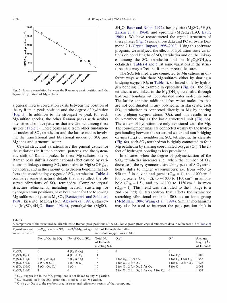
Fig. 5. Inverse correlation between the Raman m1 peak position and thedegree of hydration of Mg-sulfates.
6126 A. Wang et al. 70 (2006) 6118–6135
a general inverse correlation exists between the position ofthe m1 Raman peak position and the degree of hydration(Fig. 5). In addition to the strongest m1 peak for eachMg-sulfate species, the other Raman peaks with weakerintensities also have patterns that are distinct among thesespecies (Table 3). These peaks arise from other fundamen-tal modes of SO4 tetrahedra and the lattice modes involv-ing the translational and librational modes of SO4 andMg ions and structural water.
Crystal structural variations are the general causes forthe variations in Raman spectral patterns and the system-atic shift of Raman peaks. In these Mg-sulfates, the m1
Raman peak shift is a combinational effect caused by vari-ations in linkages among SO4 tetrahedra to MgOn(OH2)6-n
octahedra, and in the amount of hydrogen bonding that af-fects the coordinating oxygen of SO4 tetrahedra. Table 4compares some structural details that may affect the ob-served vibrations of SO4 tetrahedra. Complete crystalstructure refinements, including neutron scattering forhydrogen atom positions, have been made for the followingMg-sulfates: anhydrous MgSO4 (Rentzeperis and Soldatos,1958), kieserite (MgSO4ÆH2O, Aleksovska, 1998), starkey-ite (MgSO4Æ4H2O, Baur, 1964b), pentahydrite (MgSO4Æ
Table 4A comparison of the structural details related to Raman peak positions of the
Mg-sulfates withknown structure
S–Onb bonds in SO4 S–Obb–Mg linkage No. of H-bond
individual oxyg
No. of Onb in SO4 No. of Ob in SO4 Total No.of H-bondsaffecting SO4
MgSO4 0 4 (Oi & Oii) 0MgSO4ÆH2O 0 4 (O1 & O2) 1MgSO4Æ4H2O 2 (Oiii & Oiv) 2 (Oi & Oii) 8MgSO4Æ5H2O 2 (O3 & O4) 2 (O1 & O2) 8MgSO4Æ6H2O 3 (O1, O3, O4) 1 (O2) 10MgSO4Æ7H2O 4 0 10
a Onb, oxygen ion in the SO4 group that is not linked to any Mg cation.b Ob, oxygen ion in the SO4 group that is linked to an Mg cation.c O1,2,3,4 or Oi,ii,iii,iv, the symbols used in structural refinement results of tha
5H2O, Baur and Rolin, 1972), hexahydrite (MgSO4Æ6H2O,Zalkin et al., 1964), and epsomite (MgSO4Æ7H2O, Baur,1964a). We have reconstructed the crystal structures ofthese phases (Fig. 6) using those data and PC software Dia-mond 2.1 (Crystal Impact, 1998–2002). Using this softwareprogram, we analyzed the effects of hydration state varia-tions on bond lengths of SO4 tetrahedra and on the linkag-es among the SO4 tetrahedra and the MgOn(OH2)6-n
octahedra. Tables 4 and 5 list some variations in the struc-tures that may affect the Raman spectral features.
The SO4 tetrahedra are connected to Mg cations in dif-ferent ways within these Mg-sulfates, either by sharing abridging oxygen (Ob in Table 4), or linked only by hydro-gen bonding. For example in epsomite (Fig. 6a), the SO4
tetrahedra are linked to the Mg(OH2)6 octahedra throughhydrogen bonding with coordinated water molecules only.The lattice contains additional free water molecules thatare not coordinated in any polyhedra. In starkeyite, eachSO4 tetrahedron is connected directly to Mg by sharingtwo bridging oxygen atoms (Ob), and this results in afour-member ring as the basic structural unit (Fig. 6b).The waters of hydration are only associated with the Mg.The four-member rings are connected weakly by the hydro-gen bonding between the structural water and non-bridgingoxygen (Onb) on neighboring SO4 tetrahedra. In kieserite(Fig. 6c), each SO4 tetrahedron is tightly connected to fourMg octahedra by sharing coordinated oxygen (Ob). The ef-fect of hydrogen bonding is less prominent.
In silicates, when the degree of polymerization of theSiO4 tetrahedra increases (i.e., when the number of Onb
decreases), the m1 symmetric stretching peak of SiO4 tetra-hedra shifts to higher wavenumbers: i.e. from �860 to950 cm�1 in olivine and garnet (Onb = 4), to �1000 cm�1
for pyroxene (Onb = 2), to �1000 to 1100 cm�1 in amphi-bole (Onb = 1.5), and to �1100 to 1150 cm�1 in mica(Onb = 1). This trend was attributed to the linkage to a2nd (or 3rd) Si tetrahedron that affects the symmetricstretching vibrational mode of SiO4 as an ionic group(McMillan, 1984; Wang et al., 1994). Similar mechanismsmay also be used to interpret the peak-position shift in
SO4 ionic group (from crystal refinement data in references a–f of Table 2)
s that affecten ions in SO4
Onba Ob
b Averagelength (A)of H-bonds
0 01 for O2
c 1.8063 for Oiii, 3 for Oiv. 1 for Oi, 1 for Oii. 1.9552 for O3, 3 for O4. 1 for O1, 2 for O2. 1.9232 for O1, 2 for O3, 3 for O4. 3 for O2 2.0612 for O1, 2 for O2, 3 for O3, 3 for O4. 0 1.834
t compound.

Fig. 6. Crystal Structures of three hydrated Mg-sulfates: (a) epsomite; note that SO4 tetrahedra are only linked to Mg(OH2)6 through hydrogen bondingand note the existence of free H2O molecules; (b) starkeyite; note the four-member ring made by two SO4 tetrahedra and two MgO2(OH2)4 octahedra; and(c) kieserite; note that each oxygen ion in the SO4 group is connected directly to a Mg cation.
Hydration states of Mg-sulfates on Mars 6127
hydrated Mg-sulfates of this study: the lower the number ofOnb in the SO4 tetrahedron, the higher the position of the m1
peak (kieserite has Onb = 0 and a Raman peak at1046.1 cm�1, Tables 3 and 4). We note that the analogy be-tween connected SiO4 tetrahedra in the silicates and theconnected SO4 tetrahedra and Mg-polyhedra in Mg-sulfat-es is not strictly equivalent because the strength of thebonding in S–Ob–Mg is much weaker than the covalentbonding in Si–Ob-Si. Owing to the lower electronegativityof Mg compared to Si, the extent of the peak shift is there-fore much smaller in Mg-sulfates (�70 cm�1) than in sili-cates (�300 cm�1).
In addition to the effect of S–Ob–Mg bonds, we thinkthe hydrogen bonding to coordinated oxygen in SO4 tetra-hedra plays a concomitant role in producing the systematicshift of m1 Raman peak positions in these hydrated Mg-sul-fates. The normal effect of hydrogen bonding to the oxygenof an M–O bond is to shift the vibrational peak of M–Obond to lower wavenumbers. Regarding this point, we notethat the number of total H-bonds that affect the coordinat-ed oxygen ions in SO4 tetrahedra increases following the in-crease of the hydration degree from one in kieserite to tenin epsomite (Table 4). This increase is accompanied by aconsistent tendency of the m1 Raman peak position ofSO4 tetrahedra to shift downward, e.g., the structure ofepsomite and hexahydrite have the highest number (10)of H-bonding that affect the coordinated oxygen in SO4 tet-rahedra per formula unit and the m1 Raman peak positions(984.1 and 983.6 cm�1) at the very low end of the full range(Tables 3 and 4).
4.3. Water-band shape variations and sub-peak position
changes
The broad Raman band in the 2500–4000 cm�1 spectralregion (Fig. 4) consists of the m1 symmetric stretching, theweaker m3 asymmetric stretching, and the 1st overtone ofthe m2 bending mode of the water molecule. Among the
10 hydrated and anhydrous magnesium sulfates, we ob-serve changes in the positions of the maximum and in theoverall band shape following the change of hydrationstates. For each species except anhydrous MgSO4 andkieserite, the broad water band is made of several sub-peaks, and the number of sub-peaks and their positionschange following the number of structural water moleculesin hydrated Mg-sulfates. Table 3 lists the positions of thesesub-peaks.
In five known hydrated magnesium sulfate structures(Baur, 1964a,b; Zalkin et al., 1964; Baur and Rolin, 1972;Aleksovska, 1998), each of the structural waters occupiesa distinct crystallographic site. For example, starkeyitehas four distinct crystallographic sites for its four structuralwater molecules (Ow1, Ow2, Ow3, Ow4 in Table 5, the nota-tion follows that used in the original structural refinement),while pentahydrite has five, hexahydrite has six, epsomitehas seven, and kieserite has only one. From the structuralrefinements using neutron diffraction data, it appears thatthe shape of each structural water molecule can be severelydistorted from the shape of a free water molecule, appar-ently affected by the site symmetry. The two O–H bonds(O–H1 and O–H2 columns of Table 5) in one water mole-cule can have different bond lengths. For example, a watermolecule with O7w as the central oxygen in hexahydrite hastwo O–H bond lengths of LO–H1 = 0.447 A and LO–
H2 = 1.093A. Although Table 5 lists only the changes inO–H bond length, similar changes in H–O–H bond anglesare also present. The change in the molecular shape wouldin turn result in a set of shifted Raman peaks in the 2500–4000 cm�1 spectral region. When the molecular shape dis-tortions are less severe, the individual molecule retains itsbasic symmetry but average bond lengths vary from onesite to another (cf., starkeyite and pentahydrite in Table5). In theses cases, the individual peaks from water mole-cules of distinct crystallographic sites would occur neareach other and would sometimes overlap to become abroad spectral band with distinct sub-peak structure, e.g.,
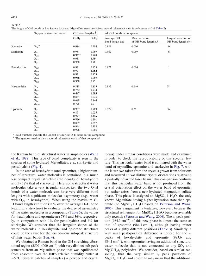
Table 5The length of OH bonds in five known hydrated Mg-sulfate structures (from crystal refinement data in references a–f of Table 2)
Oxygen in structural water OH bond length (A) All OH bonds in compound
O–H1 O–H2 Average OHbond length (A)
Max. variationof OH bond length (A)
Largest variation ofOH bond length (%)
Kieserite O3wb 0.984 0.984 0.984 0.000 0
Starkeyite Ow1 0.951 0.969 0.962 0.059 6Ow2 0.931a 0.968Ow3 0.951 0.99
Ow4 0.958 0.98
Pentahydrite OW5 0.97 0.975 0.972 0.014 1OW6 0.971 0.982
OW7 0.97 0.973OW8 0.968 0.969OW9 0.968 0.97
Hexahydrite O5w 0.818 0.835 0.832 0.646 78O6w 0.752 0.974O7w 0.447 1.093
O8w 0.834 1.025O9w 0.688 0.844O10w 0.775 0.9
Epsomite Ow1 0.957 0.989 0.979 0.35 36Ow2 0.87 1.035Ow3 0.977 1.216
Ow4 0.866 1.101Ow5 0.869 0.897Ow6 0.944 0.988Ow7 0.996 1.006
a Bold numbers indicate the longest or shortest O–H bond in the compound.b The symbols used in the structural refinement work of that compound.
6128 A. Wang et al. 70 (2006) 6118–6135
the Raman band of structural water in amphiboles (Wanget al., 1988). This type of band complexity is seen in thespectra of some hydrated Mg-sulfates, e.g., starkeyite andpentahydrite (Fig. 4).
In the case of hexahydrite (and epsomite), a higher num-ber of structural water molecules is contained in a muchless compact crystal structure (the density of hexahydriteis only 12% that of starkeyite). Here, some structural watermolecules take a very irregular shape, i.e., the two O–Hbonds of a water molecule can have very different bondlengths with significant molecular asymmetry (e.g., waterwith O7w in hexahydrite). When using the maximum O–H bond length variation (in % over the average O–H bondlength in a structure) to evaluate the degree of asymmetryof the water molecules in a compound (Table 5), the valuesfor hexahydrite and epsomite are 78% and 36%, respective-ly, in comparison with 1% for pentahydrite and 6% forstarkeyite. We consider that the irregular shapes of thewater molecules in hexahydrite and epsomite structurescould be the cause for the less obvious sub-peak structureof their water bands (Fig. 4).
We obtained a Raman band in the OH stretching vibra-tional region (2500–4000 cm�1) with very distinct sub-peakstructure from an Mg-sulfate phase that was equilibratedfrom epsomite over the 100% relative humidity buffer at�5 �C. Several batches of samples (in powder and crystal
forms) under similar conditions were made and examinedin order to check the reproducibility of this spectral fea-ture. This particular water band is compared with the waterband of crystalline epsomite and starkeyite in Fig. 7, withthe latter two taken from the crystals grown from solutionsand measured at two distinct crystal orientations relative toa partially polarized laser beam. This comparison confirmsthat this particular water band is not produced from thecrystal orientation effect on the water band of epsomite,but rather arises from a new hydrated magnesium sulfatephase. This phase is assigned to MgSO4Æ11H2O, the onlyknown Mg sulfate having higher hydration state than eps-omite (or MgSO4Æ11H2O based on Peterson and Wang,2006). This assignment is tentative, however, because thestructural refinement for MgSO4Æ11H2O becomes availableonly recently (Peterson and Wang, 2006). The m1 peak posi-tion (984.3 cm�1) of this new phase is almost the same asthat of epsomite (984.1 cm�1), although having minorpeaks at slightly different positions (Table 3). Similarly, avery small peak-position difference is noticed for the m1
peaks of hexahydrite and epsomite (983.6 and984.1 cm�1), with epsomite having an additional structuralwater molecule that is not connected to any SO4 andMg(OH2)6 polyhedra. We consider, based on similar rea-soning, that the very similar m1 peak positions ofMgSO4Æ11H2O and epsomite may mean that the additional
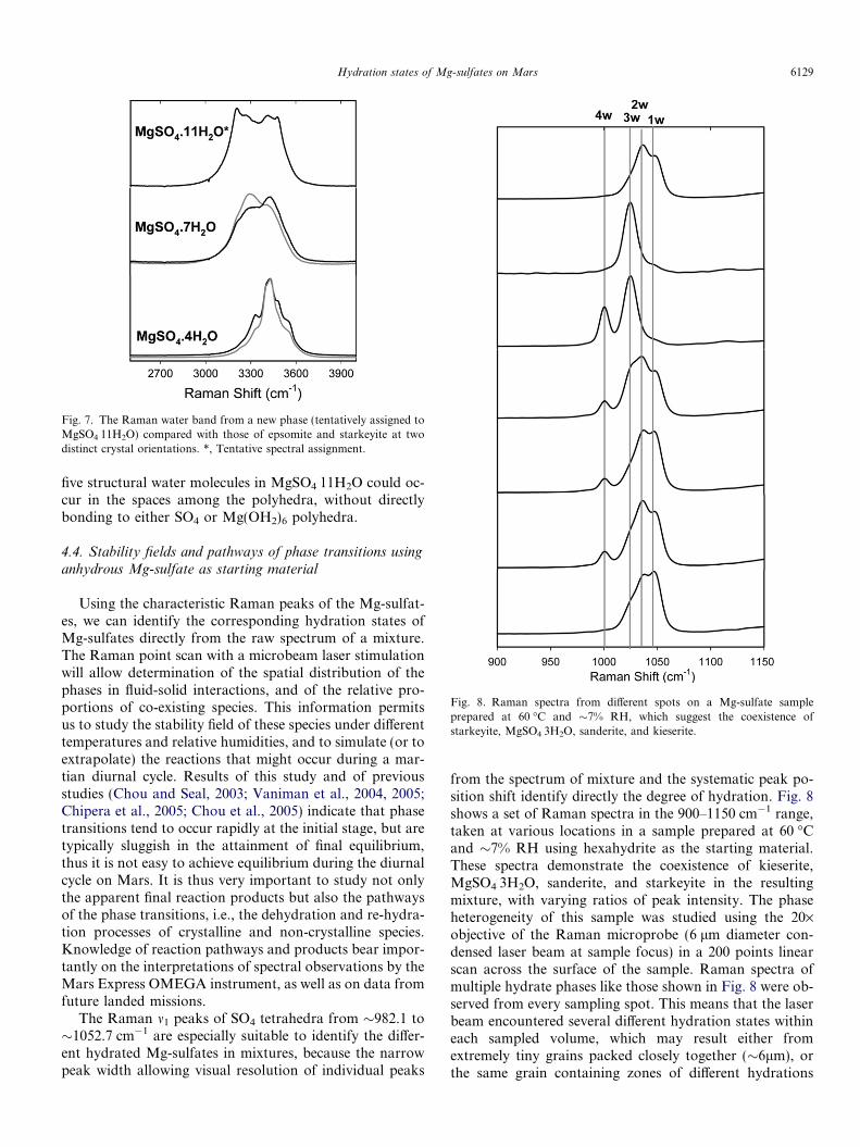
Fig. 7. The Raman water band from a new phase (tentatively assigned toMgSO4Æ11H2O) compared with those of epsomite and starkeyite at twodistinct crystal orientations. *, Tentative spectral assignment.
Fig. 8. Raman spectra from different spots on a Mg-sulfate sampleprepared at 60 �C and �7% RH, which suggest the coexistence ofstarkeyite, MgSO4Æ3H2O, sanderite, and kieserite.
Hydration states of Mg-sulfates on Mars 6129
five structural water molecules in MgSO4Æ11H2O could oc-cur in the spaces among the polyhedra, without directlybonding to either SO4 or Mg(OH2)6 polyhedra.
4.4. Stability fields and pathways of phase transitions using
anhydrous Mg-sulfate as starting material
Using the characteristic Raman peaks of the Mg-sulfat-es, we can identify the corresponding hydration states ofMg-sulfates directly from the raw spectrum of a mixture.The Raman point scan with a microbeam laser stimulationwill allow determination of the spatial distribution of thephases in fluid-solid interactions, and of the relative pro-portions of co-existing species. This information permitsus to study the stability field of these species under differenttemperatures and relative humidities, and to simulate (or toextrapolate) the reactions that might occur during a mar-tian diurnal cycle. Results of this study and of previousstudies (Chou and Seal, 2003; Vaniman et al., 2004, 2005;Chipera et al., 2005; Chou et al., 2005) indicate that phasetransitions tend to occur rapidly at the initial stage, but aretypically sluggish in the attainment of final equilibrium,thus it is not easy to achieve equilibrium during the diurnalcycle on Mars. It is thus very important to study not onlythe apparent final reaction products but also the pathwaysof the phase transitions, i.e., the dehydration and re-hydra-tion processes of crystalline and non-crystalline species.Knowledge of reaction pathways and products bear impor-tantly on the interpretations of spectral observations by theMars Express OMEGA instrument, as well as on data fromfuture landed missions.
The Raman m1 peaks of SO4 tetrahedra from �982.1 to�1052.7 cm�1 are especially suitable to identify the differ-ent hydrated Mg-sulfates in mixtures, because the narrowpeak width allowing visual resolution of individual peaks
from the spectrum of mixture and the systematic peak po-sition shift identify directly the degree of hydration. Fig. 8shows a set of Raman spectra in the 900–1150 cm�1 range,taken at various locations in a sample prepared at 60 �Cand �7% RH using hexahydrite as the starting material.These spectra demonstrate the coexistence of kieserite,MgSO4Æ3H2O, sanderite, and starkeyite in the resultingmixture, with varying ratios of peak intensity. The phaseheterogeneity of this sample was studied using the 20·objective of the Raman microprobe (6 lm diameter con-densed laser beam at sample focus) in a 200 points linearscan across the surface of the sample. Raman spectra ofmultiple hydrate phases like those shown in Fig. 8 were ob-served from every sampling spot. This means that the laserbeam encountered several different hydration states withineach sampled volume, which may result either fromextremely tiny grains packed closely together (�6lm), orthe same grain containing zones of different hydrations
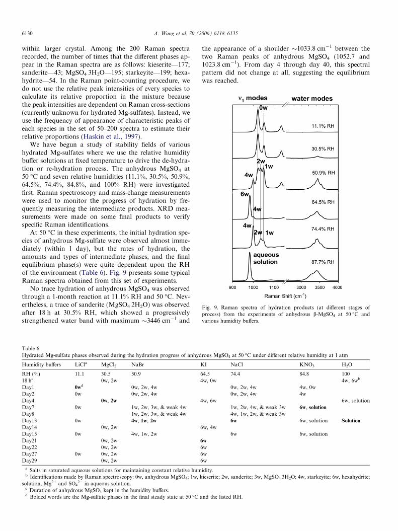
Fig. 9. Raman spectra of hydration products (at different stages ofprocess) from the experiments of anhydrous b-MgSO4 at 50 �C andvarious humidity buffers.
6130 A. Wang et al. 70 (2006) 6118–6135
within larger crystal. Among the 200 Raman spectrarecorded, the number of times that the different phases ap-pear in the Raman spectra are as follows: kieserite—177;sanderite—43; MgSO4Æ3H2O—195; starkeyite—199; hexa-hydrite—54. In the Raman point-counting procedure, wedo not use the relative peak intensities of every species tocalculate its relative proportion in the mixture becausethe peak intensities are dependent on Raman cross-sections(currently unknown for hydrated Mg-sulfates). Instead, weuse the frequency of appearance of characteristic peaks ofeach species in the set of 50–200 spectra to estimate theirrelative proportions (Haskin et al., 1997).
We have begun a study of stability fields of varioushydrated Mg-sulfates where we use the relative humiditybuffer solutions at fixed temperature to drive the de-hydra-tion or re-hydration process. The anhydrous MgSO4 at50 �C and seven relative humidities (11.1%, 30.5%, 50.9%,64.5%, 74.4%, 84.8%, and 100% RH) were investigatedfirst. Raman spectroscopy and mass-change measurementswere used to monitor the progress of hydration by fre-quently measuring the intermediate products. XRD mea-surements were made on some final products to verifyspecific Raman identifications.
At 50 �C in these experiments, the initial hydration spe-cies of anhydrous Mg-sulfate were observed almost imme-diately (within 1 day), but the rates of hydration, theamounts and types of intermediate phases, and the finalequilibrium phase(s) were quite dependent upon the RHof the environment (Table 6). Fig. 9 presents some typicalRaman spectra obtained from this set of experiments.
No trace hydration of anhydrous MgSO4 was observedthrough a 1-month reaction at 11.1% RH and 50 �C. Nev-ertheless, a trace of sanderite (MgSO4Æ2H2O) was observedafter 18 h at 30.5% RH, which showed a progressivelystrengthened water band with maximum �3446 cm�1 and
Table 6Hydrated Mg-sulfate phases observed during the hydration progress of anhyd
Humidity buffers LiCla MgCl2 NaBr
RH (%) 11.1 30.5 50.918 hc 0w, 2wDay1 0wd 0w, 2w, 4wDay2 0w 0w, 2w, 4wDay4 0w, 2w
Day7 0w 1w, 2w, 3w, & weak 4wDay8 1w, 2w, 3w, & weak 4wDay13 0w 4w, 1w, 2w
Day14 0w, 2wDay15 0w 4w, 1w, 2wDay21 0w, 2wDay22 0w, 2wDay27 0w 0w, 2wDay29 0w, 2w
a Salts in saturated aqueous solutions for maintaining constant relative humb Identifications made by Raman spectroscopy: 0w, anhydrous MgSO4; 1w, k
solution, Mg2+ and SO42� in aqueous solution.
c Duration of anhydrous MgSO4 kept in the humidity buffers.d Bolded words are the Mg-sulfate phases in the final steady state at 50 �C a
the appearance of a shoulder �1033.8 cm�1 between thetwo Raman peaks of anhydrous MgSO4 (1052.7 and1023.8 cm�1). From day 4 through day 40, this spectralpattern did not change at all, suggesting the equilibriumwas reached.
rous MgSO4 at 50 �C under different relative humidity at 1 atm
KI NaCl KNO3 H2O
64.5 74.4 84.8 1004w, 0w 4w, 6wb
0w, 2w, 4w 4w, 0w0w, 2w, 4w 4w
4w, 6w 6w, solution1w, 2w, 4w, & weak 3w 6w, solution
4w, 1w, 2w, & weak 3w6w 6w, solution Solution
6w, 4w6w 6w, solution
6w
6w6w6w
idity.ieserite; 2w, sanderite; 3w, MgSO4Æ3H2O; 4w, starkeyite; 6w, hexahydrite;
nd the listed RH.
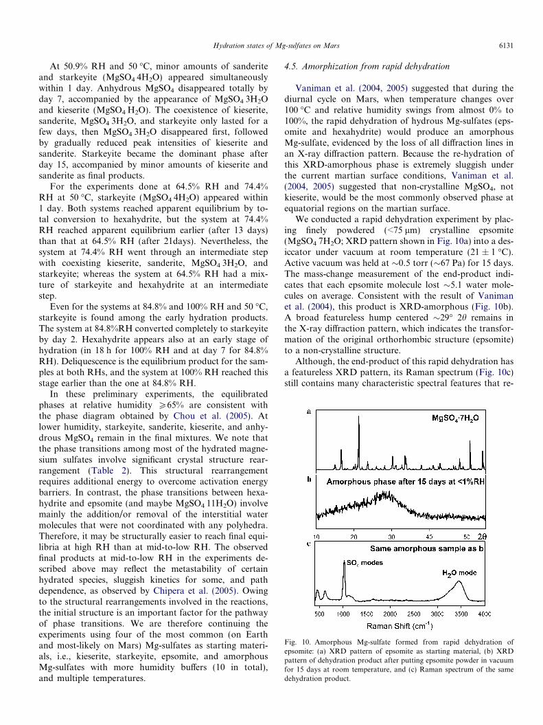
Fig. 10. Amorphous Mg-sulfate formed from rapid dehydration ofepsomite: (a) XRD pattern of epsomite as starting material, (b) XRDpattern of dehydration product after putting epsomite powder in vacuumfor 15 days at room temperature, and (c) Raman spectrum of the samedehydration product.
Hydration states of Mg-sulfates on Mars 6131
At 50.9% RH and 50 �C, minor amounts of sanderiteand starkeyite (MgSO4Æ4H2O) appeared simultaneouslywithin 1 day. Anhydrous MgSO4 disappeared totally byday 7, accompanied by the appearance of MgSO4Æ3H2Oand kieserite (MgSO4ÆH2O). The coexistence of kieserite,sanderite, MgSO4Æ3H2O, and starkeyite only lasted for afew days, then MgSO4Æ3H2O disappeared first, followedby gradually reduced peak intensities of kieserite andsanderite. Starkeyite became the dominant phase afterday 15, accompanied by minor amounts of kieserite andsanderite as final products.
For the experiments done at 64.5% RH and 74.4%RH at 50 �C, starkeyite (MgSO4Æ4H2O) appeared within1 day. Both systems reached apparent equilibrium by to-tal conversion to hexahydrite, but the system at 74.4%RH reached apparent equilibrium earlier (after 13 days)than that at 64.5% RH (after 21days). Nevertheless, thesystem at 74.4% RH went through an intermediate stepwith coexisting kieserite, sanderite, MgSO4Æ3H2O, andstarkeyite; whereas the system at 64.5% RH had a mix-ture of starkeyite and hexahydrite at an intermediatestep.
Even for the systems at 84.8% and 100% RH and 50 �C,starkeyite is found among the early hydration products.The system at 84.8%RH converted completely to starkeyiteby day 2. Hexahydrite appears also at an early stage ofhydration (in 18 h for 100% RH and at day 7 for 84.8%RH). Deliquescence is the equilibrium product for the sam-ples at both RHs, and the system at 100% RH reached thisstage earlier than the one at 84.8% RH.
In these preliminary experiments, the equilibratedphases at relative humidity P65% are consistent withthe phase diagram obtained by Chou et al. (2005). Atlower humidity, starkeyite, sanderite, kieserite, and anhy-drous MgSO4 remain in the final mixtures. We note thatthe phase transitions among most of the hydrated magne-sium sulfates involve significant crystal structure rear-rangement (Table 2). This structural rearrangementrequires additional energy to overcome activation energybarriers. In contrast, the phase transitions between hexa-hydrite and epsomite (and maybe MgSO4Æ11H2O) involvemainly the addition/or removal of the interstitial watermolecules that were not coordinated with any polyhedra.Therefore, it may be structurally easier to reach final equi-libria at high RH than at mid-to-low RH. The observedfinal products at mid-to-low RH in the experiments de-scribed above may reflect the metastability of certainhydrated species, sluggish kinetics for some, and pathdependence, as observed by Chipera et al. (2005). Owingto the structural rearrangements involved in the reactions,the initial structure is an important factor for the pathwayof phase transitions. We are therefore continuing theexperiments using four of the most common (on Earthand most-likely on Mars) Mg-sulfates as starting materi-als, i.e., kieserite, starkeyite, epsomite, and amorphousMg-sulfates with more humidity buffers (10 in total),and multiple temperatures.
4.5. Amorphization from rapid dehydration
Vaniman et al. (2004, 2005) suggested that during thediurnal cycle on Mars, when temperature changes over100 �C and relative humidity swings from almost 0% to100%, the rapid dehydration of hydrous Mg-sulfates (eps-omite and hexahydrite) would produce an amorphousMg-sulfate, evidenced by the loss of all diffraction lines inan X-ray diffraction pattern. Because the re-hydration ofthis XRD-amorphous phase is extremely sluggish underthe current martian surface conditions, Vaniman et al.(2004, 2005) suggested that non-crystalline MgSO4, notkieserite, would be the most commonly observed phase atequatorial regions on the martian surface.
We conducted a rapid dehydration experiment by plac-ing finely powdered (<75 lm) crystalline epsomite(MgSO4Æ7H2O; XRD pattern shown in Fig. 10a) into a des-iccator under vacuum at room temperature (21 ± 1 �C).Active vacuum was held at �0.5 torr (�67 Pa) for 15 days.The mass-change measurement of the end-product indi-cates that each epsomite molecule lost �5.1 water mole-cules on average. Consistent with the result of Vanimanet al. (2004), this product is XRD-amorphous (Fig. 10b).A broad featureless hump centered �29� 2h remains inthe X-ray diffraction pattern, which indicates the transfor-mation of the original orthorhombic structure (epsomite)to a non-crystalline structure.
Although, the end-product of this rapid dehydration hasa featureless XRD pattern, its Raman spectrum (Fig. 10c)still contains many characteristic spectral features that re-

Fig. 11. (a) Comparison of the Raman spectra of an amorphous Mg-sulfate with starkeyite and kieserite; (b) Comparison of the Raman m1 peakpositions of amorphous Mg-sulfates formed after rapid dehydration ofepsomite for various durations.
6132 A. Wang et al. 70 (2006) 6118–6135
veal its hydration state and low-crystallinity features. Inparticular, the broadening of the Raman bands and result-ing loss of fine spectral details (which are normally associ-ated with the spectrum of a crystalline material) clearlyindicates an amorphous material was formed by rapiddehydration.
Unlike XRD, the translational symmetry in a structureis not a necessary condition to produce Raman and infra-red spectra. Gases, liquids, and glasses all have characteris-tic vibrational spectral peaks, because they are produced bythe vibrations of chemical bonds. Nevertheless, the struc-tural distortion or amorphization in solids such as glasses,polymers, and disordered crystalline lattices, arising duringrapid changes in chemical composition or shocks due tosudden changes in pressure and/or temperature, willchange the peak parameters in their vibrational spectra.Thus the crystalline and amorphous forms of a chemicallyidentical species can generally be distinguished. Typically,Raman peak width is the first spectral parameter to beaffected by the loss of crystallinity. The loss of translationalsymmetry would affect the environmental symmetry of asite where an anionic group resides (e.g., SO4 group in sul-fate). Slight structural distortion (bond lengths and bondangles) of the SO4 group could be introduced, which wouldtranslate into a m1 peak position slightly different from thatof an SO4 group in a regular crystalline structure. The con-tributions from many irregularly distorted SO4 groupswould produce a broad Raman peak, i.e., an envelope ofmany peaks with slightly different peak positions. Thehigher the degree of amorphization, the broader the Ra-man peak width.
The Raman spectrum of the end-product of rapid dehy-dration from epsomite shows a prominent water band in the2500–4000 cm�1 spectral region (Fig. 11a), with a maxi-mum centered at 3460 cm�1 and a shoulder at�3290 cm�1, which clearly indicates a hydrated species. Inthe fundamental vibration region (950–1150 cm�1), thisphase shows a m1 peak with much wider peak width thanany crystalline hydrated or anhydrous Mg-sulfates, includ-ing MgSO4 in an aqueous solution (Fig. 11a). This broad-ened peak width is indicative for a heavily disorderednon-crystalline structure. The maximum of the broad peakfor a 15-day dehydration product at room temperature iscentered at �1030 cm�1. This peak position (Fig. 11a) liesbetween the m1 peaks of starkeyite (MgSO4Æ4H2O,1000.3 cm�1) and kieserite (MgSO4ÆH2O, 1046.1 cm�1),and suggests a hydration state within the range of MgSO4Æ3-H2O (1023.8 cm�1) and MgSO4Æ2H2O (1033.8 cm�1). Theassignment of hydration degree based on the Raman peakposition is consistent for the most part with the result ofmass-change measurements, i.e., a product with an averagehydration degree of MgSO4Æ1.9H2O. The loss in crystallin-ity of epsomite by evacuation at ambient temperature canoccur very quickly. Fig. 11b shows that the loss of crystal-linity began within the first 30 min, and a shift upward ofthe m1 peak position of SO4 was observed during the pro-gress of dehydration, evidenced by the decreasing intensity
of the epsomite m1 peak at�984.1 cm�1. These observationssuggest that this peak-position variation of non-crystallineMg-sulfates can also be used to evaluate their degree ofhydration.
The conversion of hexahydrite to the amorphous dihy-drate was also observed after a similar dehydration pro-cess at room temperature, where a loss of fourmolecules of water per molecule of sulfate was observed.Conversely, no weight loss was observed nor did anychanges occur in the Raman spectra of either crystallinekieserite and starkeyite samples subjected to the same vac-uum dehydration at room temperature. In the crystalstructures of kieserite and starkeyite, the water moleculesare more tightly coordinated with Mg cations, and theMg polyhedra are tightly connected with SO4 tetrahedraby sharing the bridging oxygen (Ob), which make theirstructures very stable towards vacuum dehydration. Incomparison, the crystal structures of hexahydrite and eps-omite are more loosely packed, evidenced by the largervolume per formula unit (2.5–2.8 times of kieserite when
Hydration states of Mg-sulfates on Mars 6133
normalized for Z = 4). The water molecules in epsomiteand hexahydrite are associated with the Mg polyhedraand are only linked to SO4 tetrahedra by hydrogen bond-ing, with the 7th water molecule not coordinated to anyof the polyhedra in epsomite. The rapid loss of water mol-ecules from these two higher hydrates would thus causethe crystal framework to collapse producing the observedamorphous phases.
5. Conclusions
Magnesium sulfates are found on Mars by orbital re-mote sensing and landed surface exploration missions. Re-ports of the data from these missions, especially those fromthe Mars Exploration Rovers, the GRS on Mars Odyssey,and OMEGA on Mars Express, suggest that magnesiumsulfates with degrees of hydration higher than kieseritemay exist at the martian surface in equatorial regions.
In this study, we demonstrated that crystalline Mg-sul-fates with nine different hydration states can be readily dis-tinguished using the systematic peak-position shift of theirRaman m1 mode and the specific sub-peak structures intheir Raman water bands. In addition, [SO4]2�, [HSO4]�
and SO3 in aqueous solutions, SO2 and H2S as gases, aswell as elemental sulfur, sulfides, and sulfates that may par-ticipate in the martian sulfur cycle, all have diagnostic Ra-man spectra and thus can be characterized duringhydration, dehydration, oxidation, and reduction process-es, in mixtures, and at intermediate steps during phasetransitions. Amorphous Mg-sulfates produced in rapiddehydration of epsomite and hexahydrite yield characteris-tic Raman spectra that reveal their hydration states anddisordered structures.
The systematic Raman spectroscopic study of hydratedMg-sulfates presented in this paper provides the essentialbasis for using planetary Raman spectroscopy to character-ize, in situ, the hydration state and variation in crystallinity,and to monitor the phase transitions likely to occur (nowor in the past) when the martian surface conditions change,as well as to understand the pathways of phase transitionsas a function of initial structures, and of temperature andrelative humidity excursions.
Studies of the phase stability fields and the phase transi-tion pathways of Mg-sulfates (crystalline or non-crystal-line), and the in situ characterization of their hydrationstates are extremely important in future planetary surfaceexploration. The combination of these two types of infor-mation will enable assessments of the current geochemicalprocesses active at the martian surface as well as an im-proved understanding of martian hydrologic history, thecycles of water and sulfur, and the potential for past orpresent habitability.
Acknowledgments
Ben Greenhagen and Peter Scully helped with the sam-ple preparation and characterization in the early parts of
this study. Bill Feldman provided the assessment of theGRS data in the vicinity of Gusev Crater. Dave Vanimangave constructive advice on sample preparation. Bjorn My-sen, Robert Seal, Harvey Belkin, Robert Downs, and ananonymous reviewer provided constructive reviews. Weare thankful for the assistance of each of these individuals.This work is supported by NASA Grants NAG5-12684(Mars Fundamental Research) and NAG5-10703 (Plane-tary Instrument Definition and Development). The use of
trade, product, industry, or firm name in this report is for
descriptive purpose only and does not constitute endorsement
by us or by the U.S. Government.
Associate editor: Bjorn Mysen
References
Aleksovska, S., 1998. Calculation of structural parameters in isostructuralseries: the kieserite group. Acta Cryst. B54, 564–567.
Arvidson, R.E., Poulet, F., Bibring, J.-P., Wolff, M., Gendrin, A., Morris,R.C., Freeman, J.J., Langevin, Y., Mangold, N., Bellucci, G., 2005.Spectral reflectance and morphologic correlations in eastern TerraMeridiani, Mars. Science V307, 591–1593 (10.1126/science. 1109509).
Baur, W.H., 1964a. On the crystal chemistry of salt hydrates. IV. Therefinement of the crystal structure of MgSO4Æ7H2O (epsomite). Acta
Cryst. 17, 1361–1369.Baur, W.H., 1964b. On the crystal chemistry of salt hydrates. II. A
neutron diffraction study of MgSO4Æ4H2O. Acta Cryst. 17, 863–869.Baur, W.H., Rolin, J.L., 1972. Salt hydrates. IX. The comparison of the
crystal structure of magnesium sulfate pentahydrate with coppersulfate pentahydrate and magnesium chromate pentahydrate. Acta
Cryst. B28, 1448–1455.Bibring, J.-P., Langevin, Y., Gendrin, A., Gondet, B., Poulet, F., Berthe,
M., Soufflot, A., Arvidson, R.E., Mangold, N., Mustard, J.F.,Drossart, P., 2005. Mars surface diversity as observed by theOMEGA/Mars Express investigation. Science V307, 1576–1581(10.1126/science. 1108806).
Boynton, W.V., Feldman, W.C., Squyres, S.W., Prettyman, T.H.,Bruckner, J., Evans, L.G., Reedy, R.C., Starr, R., Arnold, J.R.,Drake, D.M., Englert, P.A.J., Metzger, A.E., Mitrofanov, I.,Trombka, J.I., d’Uston, C., Wanke, H., Gasnault, O., Hamara,D.K., Janes, R.L.M.D.M., Maurice, S., Mikheeva, L., Taylor, G.J.,Tokar, R., Shinohara, C., 2002. Distribution of hydrogen in thenear surface of Mars: evidence for subsurfac ice deposits. Science
297, 81–85.Carr, M., 1996. Water on Mars. Oxford University Press, Oxford.Carr, M., Head, J., 2003. Oceans on Mars: an assessment of the
observational evidence and possible fate. J. Geophys. Res. 108.doi:10.1029/2002JE00196.
Chio, C.H., Sharma, S.K., Muenow, D.W., 2004. Raman spectroscopicstudies of gypsum between 33 and 374 K. Am. Miner. 89, 390–395.
Chio, C.H., Sharma, S.K., Muenow, D.W., 2005. Micro-Raman studies ofhydrous ferrous sulfates and jarosites. Spectrochim. Acta A 61, 2428–2433.
Chipera, S.J., Vaniman, D.T., Bish, D.L., Carey, J.W., Feldman, W.C.,2005. Experimental stability and transformation kinetics of magnesiumsulfate hydrates that may be present on Mars. LPSC XXXVI, AbstractNo. 1497.
Chou, I.-M., Seal II, R.R., Piatak, N., 2005. Determination of hexahy-drite–starkeyite equilibria by the humidity-buffer technique at0.1 MPa: implications for the martian H2O cycle. GSA meeting.
Chou, I.-M., Seal II, R.R., 2003. Determination of epsomite–hexahydriteequilibria by the humidity-buffer technique at 0.1 MPa with implica-tions for phase equilibria in the system MgSO4–H2O. Astrobiology 3,619–629.
6134 A. Wang et al. 70 (2006) 6118–6135
Chou, I.-M., Seal II, R.R., Hemingway, B.S., 2002. Determination ofmelanterite–rozenite and chalcanthite–bonattite equilibria by humiditymeasurements at 0.1 MPa. Amer. Miner. 87, 108–114.
Clark, B.C., Baird, A.K., Weldon, R.J., Tsusaki, D.M., Schnabel, L.,Candelaria, M.P., 1982. Chemical composition of Martian fines. J.
Geophys. Res. 87, 10059–10067.Clark, B.C., Richter, L., Gellert, R., Farrand, W., Ming, D., Morris, D.,
Yen, A., 2005. First low-iron materials on Mars and possibility of amajor montmorillonite component. AGU Abstract, P12A-04.
Crystal Impact, 1998–2002. Copyright, GbR, Bonn, Germany.Emons, H.H., Ziegenbalg, G., Naumann, R., Paulik, F., 1990. Thermal
decomposition of the magnesium sulphate hydrates under quasi-isothermal and quasi-isobaric conditions. J. Therm. Anal. 36, 1265–1279.
Feldman, W.C., Prettyman, T.H., Maurice, S., Plaut, J.J., Bish, D.L.,Vaniman, D.T., Mellon, M.T., Metzger, A.E., Squyres, S.W., Karun-atillake, S., Boynton, W.V., Elphic, R.C., Funsten, H.O., Lawrence,D.J., Tokar, R.L., 2004. The global distribution of near-surfacehydrogen on Mars. J. Geophys. Res. 109 (E09006). doi:10.1029/2003JE00216.
Feldman, W.C., Prettyman, T.H., Maurice, S., Nelli, S., Elphic, R.,Funsten, H.O., Gasnault, O., Lawrence, D.J., Murphy, J.R., Tokar,R.L., Vaniman, D.T., 2005. Topographic control of hydrogen depositsat low latitude to mid latitude of Mars. J. Geophys. Res. 110.doi:10.1029/2005JE00245.
Gellert, R., Rieder, R., Bruckner, J., Clark, B.C., Dreibus, G., Kling-elhofer, G., Lugmair, G., Ming, D.W., Wanke, H., Yen, A., Zipfel, J.,Squyres, S.W., 2006. The alpha particle X-ray spectrometer (APXS)results from Gusev Crater and calibration report. J. Geophys. Res.
V111, E02S05.Gellert, R., Rieder, R., Anderson, R.C., Bruckner, J., Clark, B.C.,
Dreibus, G., Economou, T., Klingelhofer, G., Lugmair, G.W., Ming,D.W., Squyres, S.W., d’Uston, C., Wanke, H., Yen, A., Zipfel, J.,2004. Chemistry of rocks and soils in Gusev Crater from the alphaparticle X-ray spectrometer. Science 305, 829–832.
Gendrin, A., Mangold, N., Bibring, J.-P., Langevin, Y., Gondet, B.,Poulet, F., Bonnello, G., Quantin, C., Mustard, J.F., Arvidson, R.E.,Mouelic, S.L., 2005. Sulfates in martian layered terrains: the OMEGA/Mars Express view. Science V307, 1587–1591 (10.1126/science.1109087).
Golden, D.C., Ming, D.W., Morris, R.V., Mertzman, S.A., 2005.Laboratory-simulated acid-sulfate weathering of basaltic materials:implications for formation of sulfates at Meridiani Planum and Gusevcrater, Mars. J. Geophys. Res. 110, E12S07. doi:10.1029/2005JE002451.
Greenspan, L., 1977. Humidity fixed points of binary saturated aqueoussolution. J. Res. Natl. Bureau of Standards—A. Phys. Chem. 81A (1),89–96.
Haskin, L.A., Wang, A., Jolliff, B.L., Korotev, R.L., Rockow, K.M.,Viskupic, K.M., 1997. Laser Raman spectroscopic determinationof mineral proportions in rocks on planetary surface. Twenty-eighth Lunar and Planetary Science Conference, Part 2, pp. 523–524.
Haskin, L.A., Wang, A., Jolliff, B.L., McSween, H.Y., Clark, B.C., DesMarais, D.J., McLennan, S.M., Tosca, N.J., Hurowitz, J.A., Farmer,J.D., Yen, A., Squyres, S.W., Arvidson, R.E., Klingelhofer, G.,Schroder, C., de Souza, J., Paulo, A., Morris, R.V., Ming, D.W.,Gellert, R., Zipfel, J., Bruckner, J., Bell, I., James, F., Herkenhoff, K.,Christensen, P.R., Ruff, S., Blaney, D., Gorevan, S., Cabrol, N.A.,Crumpler, L., Grant, J., Soderblom, L., 2005. Water alteration ofrocks and soils from the Spirit rover site, Gusev Crater, Mars. Nature
436, 66–69.Herman, R.G., Bogdon, C.E., Sommer, A.J., Simpson, D.R., 1987.
Discrimination among carbonate minerals by Raman spectroscopyusing the laser microprobe. Appl. Spectrosc. 41, 437–440.
Hogenboom, D.L., Kargel, J.S., Ganasan, J.P., Lee, L., 1995. Magnesiumsulfate–water to 400 MPa using a novel piezometer; densities, phaseequilibria, and planetological implications. Icarus 115, 258–277.
Hynek, B., Phillips, R., 2001. Evidence for extensive denudation of theMartian highlands. Geology 29, 407–410.
Hynek, B., Phillips, R., 2003. New data reveal mature, integrated drainagesystems on Mars indicative of past precipitation. Geology 31, 757–760.
Klingelhofer, G.R.D.S., De Souza Jr., P.A., Yen, A., Gellert, R., Evlanov,E.N., Zubkov, B., Foh, J., Bonnes, U., Kankeleit, E., Gutlich, P.,Ming, D.W., Renz, F., Wdowiak, T., Squyres, S.W., Arvidson, R.E.,Morris, R.V., Bernhardt, B., Schroder, C., 2004. Jarosite and hematiteat Meridiani Planum from Opportunity’s Mossbauer spectrometer.
Science 306 (5702), 1740–1745, 03 DEC 2004.Kuebler, K.E., Jolliff, B.L., Wang, A., Haskin, L.A., 2005. Extracting
olivine (Fo–Fa) compositions from Raman spectral peak positions.XXXVI Lunar & Planetary Sciences Conference, Abstract No. 2068.
Kuebler, K.E., Wang, A., Abbott, K., Haskin, L.A., 2001. Can we detectcarbonate and sulfate minerals on the surface of Mars by Ramanspectroscopy? XXXII LPSC, Abstract No. 1889.
Kuebler, K.E., Wang, A., Haskin, L.A., Jolliff, B.L., 2003. A Study ofolivine alteration to iddingsite using Raman spectroscopy. XXXIVLPSC, Abstract No. 1953.
LaFont, R., Vinh, L.D., 1966. Effect Raman d’un monocristal de sulfatede magnesoium hexahydrate. C.R. Acad. Sci. Paris ser. B t.262, 49–51.
McMillan, P., 1984. Structural studies of silicate glasses and melts—applications and limitations of Raman spectroscopy. Amer. Miner. 69,622–644.
McSween Jr., H.Y., Murchie, S.L., Crisp, J.A., Bridges, N.T., Anderson,R.C., Bell III, J.F., Britt, D.T., Brueckner, J., Dreibus, G., Economou,T., Ghosh, A., Golombek, M.P., Greenwood, J.P., Johnson, J.R.,Moore, H.J., Morris, R.V., Parker, T.J., Rieder, R., Singer, R.B.,Waenke, H., 1999. Chemical, multispectral, and textural constraints onthe composition and origin of rocks at the Mars Pathfinder landingsite. J. Geophys. Res. 104 (no. E4), 8679–8715 (19990425).
Ming, D.W., Mittlefehldt, D.W., Morris, R.V., Golden, D.C., Gellert, R.,Yen, A., Clark, B.C., Squyres, S.W., Farrand, W.H., Ruff, S.W.,Arvidson, R.A., Klingelhofer, G., Rodionov, D.S., Schroder, C., deSouza Jr., P.A., Wang, A., 2006. Geochemical and mineralogicalindicators for aqueous processes in the Columbia Hills of GusevCrater Mars. J. Geophys. Res. V111, E02S12.
Misra, A.K., Sharma, S.K., Chi Hong Chio, Lucey, P.G., 2006. Detectionof water and water bearing minerals from 10 m distance under brightcondition using remote Raman system. Lunar and Planetary ScienceConference, XXXVII, Abstract No. 2155.
Nakamoto, K., 1997. Infrared and Raman Spectra of Inorganic and
Coordination Compounds, fifth ed. John Wiley & Sons, New York.Peterson, R.C., Wang, R., 2006. Crystal moulds on Mars explained by a
possible new mineral species and how incongruent melting of thishydrated magnesium sulfate could explain outwash features seen onthe Martian surface. Geology, in press.
Rentzeperis, P.J., Soldatos, C.T., 1958. The crystal structure of theanhydrous magnesium sulphate. Acta Cryst. 11, 686–688.
Rieder, R., Economou, T., Waenke, H., Turkevich, A., Crisp, J.,Brueckner, J., Dreibus, G., McSween Jr., H.Y., 1997. The chemicalcomposition of Martian soil and rocks returned by the Mobile AlphaProton X-ray Spectrometer; preliminary results from the X-ray mode.
Science 278 (5344 (19971205)), 1771–1774.Rieder, R.C., Dreibus, G., Economou, T., Klingelhofer, G., Lugmair,
G.W., Ming, D.W., Squyres, S.W., D’Uston, C., Wanke, H., Yen, A.,Zipfel, J., Gellert, R., Anderson, R.C., Bruckner, J., 2004. Chemistryof rocks and soils at Meridiani Planum from the Alpha Particle X-raySpectrometer. Science 306 (5702), 1746–1749 (03 December 2004).
Sharma, S.K., Chio, C.H., Muenow, D.W., 2006. Raman spectroscopicinvestigation of ferrous sulfate hydrates, Lunar and Planetary ScienceConference, XXXVII, Abstract No. 2069.
Sharma, S.K., Angel, S.M., Ghosh, M., Hubble, H.W., Lucey, P.G., 2002.A remote pulsed-laser Raman spectroscopy system for mineral analysison planetary surfaces to 66 meters. Appl. Spectrosc. 56, 699–705.
Squyres, S.W., Grotzinger, J.P., Arvidson, R.E., Bell III, J.F., Calvin, W.,Christensen, P.R., Clark, B.C., Crisp, J.A., Farrand, W.H., Herkenhoff,K.E., Johnson, J.R., Klingelhofer, G., Knoll, A.H., McLennan, S.M.,

Hydration states of Mg-sulfates on Mars 6135
McSween, Morris, R.V., Rice Jr., J.W., Rieder, R., Soderblom, L.A., . Insitu evidence for an ancient aqueous environment at Meridiani Planum,Mars. Science 3–6 (5702), 1709–1714.
Tosca, N.S., Mclennan, S.M., Lindsley, D., Schoonen, M., 2004. Acid-sulfate weathering of synthetic Martian basalt: the acid-fog modelrevisited. J. Geophys. Res. 109, E05003. doi:10.1029/2003JE002218.
Vaniman, D.T., Bish, D.L., Chipera, S.J., Flalips, C.I., Carey, J.W.,Feldman, W.C., 2004. Magnesium sulphate salts and the history ofwater on Mars. Nature (V431), 663–665.
Vaniman, D.T., Chipera, S.J., Bish, D.L., Carey, J.W., Feldman, W.C.,2005. Martian relevance of dehydration and rehydration in the Mg-sulfate System. XXXVI Lunars and Planetary Science Conference,Abstract No. 1486.
Walrafen, G.E., Yang, W.H., Chu, C., 2002. High-temperature Ramaninvestigation of concentrated sulfuric acid mixtures: measurement ofH-bond DH values between H3O+ or H5O2+ and HSO4
�. J. Chem.
Phys. A. 106 (43), 10162–10173.Walrafen, G.E., Young, T.F., 1960. Structure of oleum. Trans. Faraday
Soc. 56, 1419–1425.Wang, A., Dhamelincourt, P., Turrell, G., 1988. Raman and infrared
spectroscopic investigation of the cation distributions in amphiboles. J.
Mol. Struct. 175, 183–188.Wang, A., Han, J., Guo, L., Yu, J., Zeng, P., 1994. A database of standard
Raman spectra of mineral and related inorganic crystals. Appl.
Spectrosc. 48, p959–p968.Wang, A., Haskin, A.L., Cortez, E., 1998. A Raman spectroscopic sensor
for in situ mineral characterization on planetary surface. Appl.
Spectrosc. 52, 477–487.Wang, A., Haskin, L.A., Korotev, R.L., Jolliff, B.L., de Souza Jr., P.,
Kusack, A.G., and the Athena Science Team. 2005a. Evidence ofphyllosilicate in Wooly Patch— An altered rock encountered on theSpirit rover traverse. LPSC XXXVI, Abstract No. 2327.
Wang, A., Haskin, L.A., Lane, A.L., Wdowiak, T.J., Squyres, S.W.,Wilson, R.J., Hovland, L.E., Manatt, k.S., Raouf, N., Smith, C.D.,2003. Development of the Mars Microbeam Raman Spectrometer(MMRS). J. Geophys. Res. 108 (E1). doi:10.1029/2002JE00190.
Wang, A., Haskin, L.A., Squyres, S.W., Arvidson, R., Crumpler, L.,Gellert, R., Hurowitz, J., Schroder, C., Tosca, N., Herkenhoff, K.,Jolliff, B.L., 2005b. Sulfate deposition in regolith exposed in trencheson the plains between the Spirit landing site and Columbia Hills inGusev Crater, Mars. LPSC XXXVI, Abstract No. 2236.
Wang, A., Haskin, L.A., Squyres, S.W., Arvidson, R., Jolliff, B.L.,Crumpler, L., Gellert, R., Schroder, C., Herkenhoff, K., Hurowitz, J.,Tosca, N., Farrand, W., Anderson, R., Knudson, A.T., 2006a. Sulfatedeposition in subsurface regolith in Gusev Crater, Mars. J. Geophys.
Res. 111, E02S17.
Wang, A., Jolliff, B., Haskin, L.A., 1995. ‘‘Raman spectroscopy as amethod for mineral identification on lunar robotic explorationmissions. J. Geophys. Res. 100, p21189–p21199.
Wang, A., Jolliff, B.L., Haskin, L.A., 1999a. Raman spectroscopiccharacterization of a Martian SNC meteorite: Zagami. J. Geophys.
Res. 104, 8509–8519.Wang, A., Jolliff, B.L., Haskin, L.A., 1999b. Raman spectroscopic
characterization of a highly weathered basalt: igneous mineralogy,alteration products, and a microorganism. J. Geophys. Res. 104,27067–27077.
Wang, A., Jolliff, B.L., Haskin, L.A., Kuebler, K.E., Viskupic, K.M.,2001. Characterization and comparison of structural and composi-tional features of planetary quadrilateral pyroxenes by Ramanspectroscopy. Amer. Mineral. 86, p790–p806.
Wang, A., Korotev, R.L., Jolliff, B.L., Haskin, L.A., Crumpler, L.,Farrand, B., Herkenhoff, K., de Souza Jr., P., Kusack, A.G.,Hurowitz, J., Tosca, N.J., 2006b. Evidence of phyllosilicate in WoolyPatch, an altered rock encountered at West Spur, Columbia Hills bySpirit rover. J. Geophys. Res. V111, E02S16.
Wang, A., Kuebler, K.E., Jolliff, B.L., Haskin, L.A., 2004a. Ramanspectroscopy of Fe–Ti–Cr-oxides, case study: martian meteoriteEETA79001. Amer. Miner. 89, 665–680.
Wang, A., Kuebler, K.E., Jolliff, B.L., Haskin, L.A., 2004b. Mineralogy ofa Martian meteorite as determined by Raman spectroscopy. J. Raman
Spectrosc. 35, 504–514.Wang, F., Zhang, Y.H., KLi, S.H., Wang, L.Y., Zhao, L.J., 2005. A
strategy for single supersaturated droplet analysis: confocal Ramaninvestigations on the complicated hygroscopic properties of individ-ual MgSO4 droplets on the quartz substrate. Anal. Chem. 77, 7148–7155.
Wdowiak, T.J., Agresti, D.G., Mirov, S.B., 1994. A laser Ramanspectrometer system suitable for incorporation into lander spacecraft.XXVI LPSC, pp. 1473–1474.
Wynn-Williams, D.D., Edwards, H.G.M., 2000. Proximal analysis ofregolith habitats and protective biomolecules in situ by laser Ramanspectroscopy: overview of terrestrial Antarctic habitats and Marsanalogs. Icarus 144, 486–503.
Young, T.F., Walrafen, G.E., 1961. Raman spectra of concentratedaqueous solutions of sulfuric acid. Trans. Faraday Soc. 57, 34–39.
Zalkin, A., Ruben, H., Templeton, D.H., 1964. The crystal structure andhydrogen bonding of magnesium sulfate hexahydrate. Acta Cryst. 17,235–240.
Zangmeister, C.D., Pemberton, J.E., 2000. Raman spectroscopy andatomic force microscopy of the reaction of sulfuric acid and sodiumchloride. J. Am. Chem. Soc. 122, 12289–12296.