Embed Size (px)
Citation preview
Multimodal coherent anti-Stokes Raman spectroscopic imaging with a fiberoptical parametric oscillator
Yan-Hua Zhai,1 Christiane Goulart,1 Jay E. Sharping,1,a� Huifeng Wei,2 Su Chen,2
Weijun Tong,2 Mikhail N. Slipchenko,3 Delong Zhang,3 and Ji-Xin Cheng3,a�
1University of California–Merced, Merced, California 95344, USA2Yangtze Optical Fibre and Cable Co. Ltd. R&D Centre, Wuhan 430073, People’s Republic of China3Department of Chemistry, Weldon School of Biomedical Engineering, Purdue University, West Lafayette,Indiana 47907, USA
�Received 29 December 2010; accepted 19 April 2011; published online 10 May 2011�
We report on multimodal coherent anti-Stokes Raman scattering �CARS� imaging with a sourcecomposed of a femtosecond fiber laser and a photonic crystal fiber �PCF�-based optical parametricoscillator �FOPO�. By switching between two PCFs with different zero dispersion wavelengths, atunable signal beam from the FOPO covering the range from 840 to 930 nm was produced. Bycombining the femtosecond fiber laser and the FOPO output, simultaneous CARS imaging of amyelin sheath and two-photon excitation fluorescence imaging of a labeled axons in rat spinal cordhave been demonstrated at the speed of 20 �s per pixel. © 2011 American Institute of Physics.�doi:10.1063/1.3589356�
Coherent anti-Stokes Raman scattering �CARS� micros-copy is a powerful tool for high-speed vibrational imaging ofcells, tissues, and drug delivery systems.1,2 An additional ad-vantage of CARS microscopy is that other linear and nonlin-ear imaging modalities such as confocal Raman microscopyand multiphoton fluorescence microscopy can be combinedwith CARS on a single microscopy platform.3–6 Despite its,the technical complexity of CARS microscopy and the highcost of laser systems have hindered its widespread adapta-tion. Developing laser sources is an integral part of the evo-lution of CARS microscopy toward the ultimate goal of pro-viding a reliable, inexpensive, and user friendly microscopysystem.
First-generation CARS microscopes used two tempo-rally synchronized femtosecond �fs� or picosecond �ps�pulsed lasers.1 Second-generation CARS systems used opti-cal parametric oscillators �OPOs� pumped with fs or pssolid state lasers to generate multiple inherently synchro-nized pulse trains without the need for electronicsynchronization.7,8 Fiber-based laser systems are attractingattention because they provide advantages in terms of sizeand complexity.9–11 Notable progress is being made on de-veloping all-fiber laser sources for CARS, including: super-continuum generation in photonic crystal fiber �PCF� �Ref.10� and a two-color Er:fiber laser system.11 The averagepowers for these systems is limited to �10 mW for theStokes beam, and a larger power from the pump beam isnecessary to achieve high speed CARS imaging.
In this letter we report the application of a fiber-laserpumped, wavelength-tunable, pulsed fiber optical parametricoscillator �FOPO� �Refs. 12–15� to multimodal CARS mi-croscopy. Our FOPO resembles a state-of-the-art ��2� OPOexcept that the crystal is replaced by a PCF and the FOPOoperates through four-wave mixing �FWM� mediated by ��3�
nonlinearity of glass. The fiber’s dispersive properties arechosen to phase match the FWM process leading to a wide
spectral bandwidth of operation. One distinct advantage ofFOPOs over ��2� OPOs is the ability to generate output atwavelengths near to that of the pump laser, which reducesthe number of frequency conversion steps required for thegeneration of wavelengths for CARS microscopy. Further-more, we anticipate that the fiber-based gain medium can beintegrated with other fiber components, leading to a compactall-fiber FOPO system.
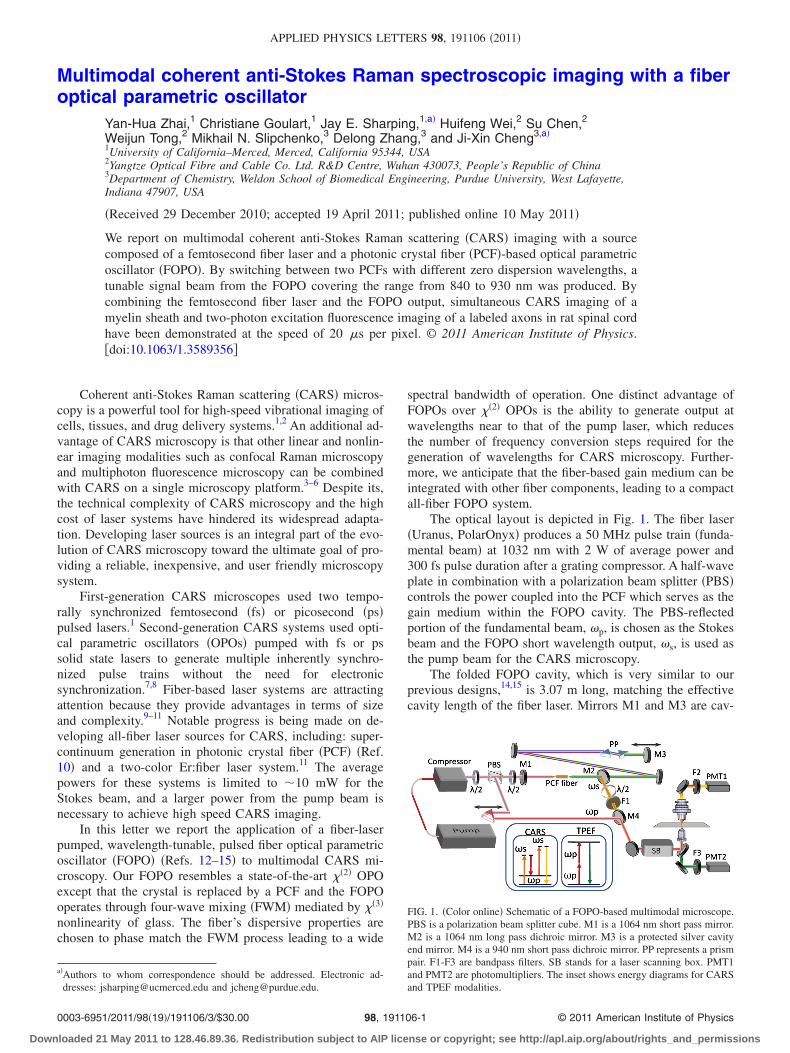
The optical layout is depicted in Fig. 1. The fiber laser�Uranus, PolarOnyx� produces a 50 MHz pulse train �funda-mental beam� at 1032 nm with 2 W of average power and300 fs pulse duration after a grating compressor. A half-waveplate in combination with a polarization beam splitter �PBS�controls the power coupled into the PCF which serves as thegain medium within the FOPO cavity. The PBS-reflectedportion of the fundamental beam, �p, is chosen as the Stokesbeam and the FOPO short wavelength output, �s, is used asthe pump beam for the CARS microscopy.
The folded FOPO cavity, which is very similar to ourprevious designs,14,15 is 3.07 m long, matching the effectivecavity length of the fiber laser. Mirrors M1 and M3 are cav-
a�Authors to whom correspondence should be addressed. Electronic ad-dresses: [email protected] and [email protected].
FIG. 1. �Color online� Schematic of a FOPO-based multimodal microscope.PBS is a polarization beam splitter cube. M1 is a 1064 nm short pass mirror.M2 is a 1064 nm long pass dichroic mirror. M3 is a protected silver cavityend mirror. M4 is a 940 nm short pass dichroic mirror. PP represents a prismpair. F1-F3 are bandpass filters. SB stands for a laser scanning box. PMT1and PMT2 are photomultipliers. The inset shows energy diagrams for CARSand TPEF modalities.
APPLIED PHYSICS LETTERS 98, 191106 �2011�
0003-6951/2011/98�19�/191106/3/$30.00 © 2011 American Institute of Physics98, 191106-1
Downloaded 21 May 2011 to 128.46.89.36. Redistribution subject to AIP license or copyright; see http://apl.aip.org/about/rights_and_permissions
ity end mirrors and M3 is mounted on a translation stage forfine tuning of the cavity length and output wavelength. Theclosely spaced prism pair �PP� is inserted inside the cavity toenhance the positive dispersion. The 1032 nm beam from thefiber laser is coupled into the PCF through an aspheric lens�C230TM-B, Thorlabs�. Collimation of the output from thePCF is done using a similar aspheric lens with a C-coating.The output coupler is a long pass dichroic mirror, M2, �LongPass 1064, Precision Photonics� which delivers the shortwavelength beam, �s, out of the cavity. A half-wave plateplaced before the FOPO cavity is used to match the 1032 nmfundamental-beam polarization with the PCF’s principleaxes. Another half-wave plate is placed in the �s beam to setthe beam polarization parallel to that of the 1032 nm, �pbeam. A bandpass filter F1 �850/90 or 880/40, Chroma� re-moves the weak broadband supercontinuum from the �sbeam.
The two beams, �s and �p, are temporally overlappedand collinearly combined using M4 �940 dcsp, Omega�. Theoverlapped beams are then directed into a laser scanning in-verted microscope �FV300/IX71, Olympus�. We use a 60�NA=1.2 water-immersed objective with improved near in-frared �NIR� transmission �UPLSAPO60XWIR, Olympus� tofocus the laser into the sample. The forward CARS signal iscollected with an air condenser �NA=0.55� and detected byan external photomultiplier tube PMT1 �R3896, Hamamatsu�after propagating through bandpass filters �D695/70 or 740/140 nm, Chroma�. The two-photon excitation fluorescence�TPEF� signal is collected by the objective, separated fromexcitation beams with a dichroic mirror �670 dcxr, Chroma�and detected with PMT2 �R7422–40, Hamamatsu� afterpassing the bandpass filter �520/40 nm, Chroma�. A spec-trometer is mounted on the side port provides confocal Ra-man analysis of the objects.6
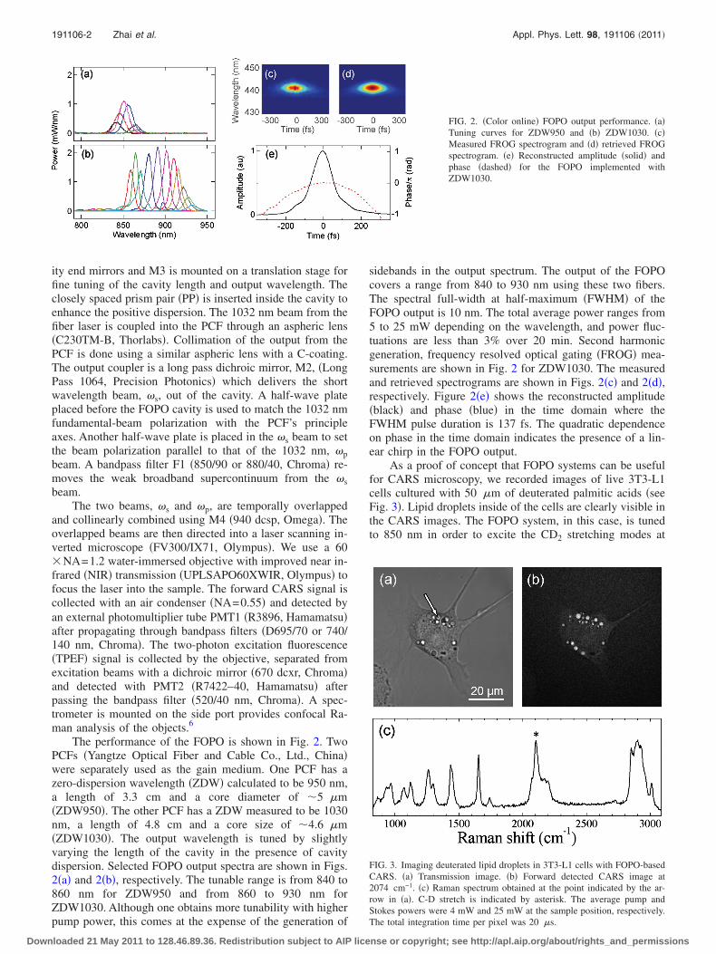
The performance of the FOPO is shown in Fig. 2. TwoPCFs �Yangtze Optical Fiber and Cable Co., Ltd., China�were separately used as the gain medium. One PCF has azero-dispersion wavelength �ZDW� calculated to be 950 nm,a length of 3.3 cm and a core diameter of �5 �m�ZDW950�. The other PCF has a ZDW measured to be 1030nm, a length of 4.8 cm and a core size of �4.6 �m�ZDW1030�. The output wavelength is tuned by slightlyvarying the length of the cavity in the presence of cavitydispersion. Selected FOPO output spectra are shown in Figs.2�a� and 2�b�, respectively. The tunable range is from 840 to860 nm for ZDW950 and from 860 to 930 nm forZDW1030. Although one obtains more tunability with higherpump power, this comes at the expense of the generation of
sidebands in the output spectrum. The output of the FOPOcovers a range from 840 to 930 nm using these two fibers.The spectral full-width at half-maximum �FWHM� of theFOPO output is 10 nm. The total average power ranges from5 to 25 mW depending on the wavelength, and power fluc-tuations are less than 3% over 20 min. Second harmonicgeneration, frequency resolved optical gating �FROG� mea-surements are shown in Fig. 2 for ZDW1030. The measuredand retrieved spectrograms are shown in Figs. 2�c� and 2�d�,respectively. Figure 2�e� shows the reconstructed amplitude�black� and phase �blue� in the time domain where theFWHM pulse duration is 137 fs. The quadratic dependenceon phase in the time domain indicates the presence of a lin-ear chirp in the FOPO output.
As a proof of concept that FOPO systems can be usefulfor CARS microscopy, we recorded images of live 3T3-L1cells cultured with 50 �m of deuterated palmitic acids �seeFig. 3�. Lipid droplets inside of the cells are clearly visible inthe CARS images. The FOPO system, in this case, is tunedto 850 nm in order to excite the CD2 stretching modes at
FIG. 2. �Color online� FOPO output performance. �a�Tuning curves for ZDW950 and �b� ZDW1030. �c�Measured FROG spectrogram and �d� retrieved FROGspectrogram. �e� Reconstructed amplitude �solid� andphase �dashed� for the FOPO implemented withZDW1030.
FIG. 3. Imaging deuterated lipid droplets in 3T3-L1 cells with FOPO-basedCARS. �a� Transmission image. �b� Forward detected CARS image at2074 cm−1. �c� Raman spectrum obtained at the point indicated by the ar-row in �a�. C-D stretch is indicated by asterisk. The average pump andStokes powers were 4 mW and 25 mW at the sample position, respectively.The total integration time per pixel was 20 �s.
191106-2 Zhai et al. Appl. Phys. Lett. 98, 191106 �2011�
Downloaded 21 May 2011 to 128.46.89.36. Redistribution subject to AIP license or copyright; see http://apl.aip.org/about/rights_and_permissions
2100 cm−1. A typical transmission, and the correspondingCARS, images are shown in Figs. 3�a� and 3�b�, respectively.To verify that the signal is from CARS, we perform the samemeasurement with the pump and Stokes beams separated intime and with the beam frequencies tuned away from theRaman resonance. Confocal Raman microspectroscopicanalysis of the lipid droplets reveal a distinctive peak near2100 cm−1 corresponding to the CD2 stretching vibration�see Fig. 3�c��.
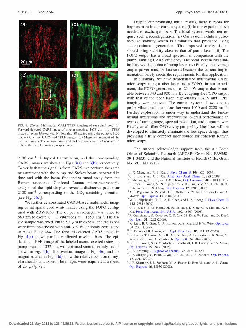
We further demonstrated CARS-based multimodal imag-ing of rat spinal cord white matter using the FOPO config-ured with ZDW1030. The output wavelength was tuned to880 nm to excite CvC vibrations at �1650 cm−1. The tis-sue sample was fixed, cut to 50 �m thickness, and the axonswere immuno-labeled with anti-NF-160 antibody conjugatedto Alexa Fluor 488. The forward-detected CARS image inFig. 4�a� shows parallelly aligned myelin fibers. The epi-detected TPEF image of the labeled axons, excited using thepump beam at 1032 nm, was obtained simultaneously and isshown in Fig. 4�b�. The overlaid image in Fig. 4�c� and themagnified area in Fig. 4�d� show the relative position of my-elin sheaths and axons. The images were acquired at a speedof 20 �s /pixel.
Despite our promising initial results, there is room forimprovement in our current system. �i� In our experiment weneeded to exchange fibers. The ideal system would not re-quire such a reconfiguration. �ii� Our system exhibits pulse-to-pulse stability which is similar to that produced usingsupercontinuum generation. The improved cavity designshould bring stability close to that of pump laser. �iii� TheFOPO output has a broad spectrum in comparison with thepump, limiting CARS efficiency. The ideal system has simi-lar bandwidths to that of pump laser. �iv� Finally, the averageoutput power must be increased because the current imple-mentation barely meets the requirements for this application.
In summary, we have demonstrated multimodal CARSmicroscopy using a fiber laser and a FOPO. In our experi-ment, the FOPO generates up to 25 mW output that is tun-able between 840 and 930 nm. By coupling the FOPO outputwith that of the fiber laser, high-quality CARS and TPEFimaging were realized. The current system allows one toprobe vibrational transitions between 1050 and 2220 cm−1.Further exploration is under way to understand the funda-mental limitations and improve the overall performance interms of tuning range, spectral resolution, and output power.Finally, an all-fiber OPO cavity pumped by fiber laser will bedeveloped to ultimately eliminate the free space design, thusproviding a truly compact laser source for coherent Ramanmicroscopy.
The authors acknowledge support from the Air ForceOffice of Scientific Research �AFOSR; Grant No. FA9550-09-1-0483�, and the National Institute of Health �NIH; GrantNo. R01 EB 7243�.
1J. X. Cheng and X. S. Xie, J. Phys. Chem. B 108, 827 �2004�.2C. L. Evans and X. S. Xie, Annu. Rev. Anal. Chem. 1, 883 �2008�.3H.-W. Wang, T. T. Le, and J.-X. Cheng, Opt. Commun. 281, 1813 �2008�.4H. Chen, H. Wang, M. N. Slipchenko, Y. K. Jung, Y. Z. Shi, J. Zhu, K. K.Buhman, and J.-X. Cheng, Opt. Express 17, 1282 �2009�.
5A. F. Pegoraro, A. Ridsdale, D. J. Moffatt, Y. W. Jia, J. P. Pezacki, and A.Stolow, Opt. Express 17, 2984 �2009�.
6M. N. Slipchenko, T. T. Le, H. Chen, and J.-X. Cheng, J. Phys. Chem. B113, 7681 �2009�.
7C. L. Evans, E. O. Potma, M. Puoris’haag, D. Cote, C. P. Lin, and X. S.Xie, Proc. Natl. Acad. Sci. U.S.A. 102, 16807 �2005�.
8F. Ganikhanov, S. Carrasco, X. S. Xie, M. Katz, W. Seitz, and D. Kopf,Opt. Lett. 31, 1292 �2006�.
9K. Kieu, B. G. Saar, G. R. Holtom, X. S. Xie, and F. W. Wise, Opt. Lett.34, 2051 �2009�.
10H. Kano and H. Hamaguchi, Appl. Phys. Lett. 86, 121113 �2005�.11G. Krauss, T. Hanke, A. Sell, D. Traeutlein, A. Leitenstorfer, R. Selm, M.
Winterhalder, and A. Zumbusch, Opt. Lett. 34, 2847 �2009�.12G. K. L. Wong, S. G. Murdoch, R. Leonhardt, J. D. Harvey, and V. Marie,
Opt. Express 15, 2947 �2007�.13J. E. Sharping, J. Lightwave Technol. 26, 2184 �2008�.14J. E. Sharping, C. Pailo, C. Gu, L. Kiani, and J. R. Sanborn, Opt. Express
18, 3911 �2010�.15J. E. Sharping, J. R. Sanborn, M. A. Foster, D. Broaddus, and A. L. Gaeta,
Opt. Express 16, 18050 �2008�.
FIG. 4. �Color� Multimodal CARS/TPEF imaging of rat spinal cord. �a�Forward detected CARS image of myelin sheath at 1673 cm−1. �b� TPEFimage of axons labeled with NF160/alex488 excited using the pump at 1032nm. �c� Overlaid CARS and TPEF images. �d� Magnified segment of theoverlaid images. The average pump and Stokes powers were 3.3 mW and 15mW at the sample position, respectively.
191106-3 Zhai et al. Appl. Phys. Lett. 98, 191106 �2011�
Downloaded 21 May 2011 to 128.46.89.36. Redistribution subject to AIP license or copyright; see http://apl.aip.org/about/rights_and_permissions