Embed Size (px)
Citation preview
Successful replacement of
animal models:
production of antibodies
Interdisciplinary Technical Journal Clubs on Laboratory Animal Science
Assunta Senatore
May 3rd 2016

Historical overview concerning methods for antibody production
Based on animal immunization:
Monoclonal antibody technology
Recombinant binders:
Immunoglobulin derivatives: Fab/scFv libraries
Non immunoglobulin binders: DARPins
Derived from in vitro selection techniques
Outline
3R
: Re
pla
ce
me
nt

Immunoglobulin structure
Naturally produced immunoglobulins or antibodies are
macromolecular Y-shaped proteins of approximately 150 kDa.
In the animal, antibodies are produced primarily by plasma
cells, a type of terminally differentiated B lymphocyte upon
activation of the immune system.
They can be generated to selectively target a specific antigen
binding partner and are universal weapons against
pathogenic threats.
• Diagnostic
• Research
• Therapy

Polyclonal antibodies
Serum contains mixed population of different types of antibodies (polyclonal) targeting the antigen in different
epitopes.
• Prone to batch-to-batch variability.
• Produce large amounts of non-specific antibodies, which can create background signal in some
applications.
• Multiple epitopes make it important to check immunogen sequence for cross-reactivity.
• Not useful for probing specific domains of antigen because antiserum will usually recognize many
domains
As antibodies producing plasma cells from animals cannot be grown in tissue culture, they cannot be used as
an in vitro source of antibodies.
• .

Mouse monoclonal technology:
From immunization to Hybridoma generation

Mouse monoclonal technology:
From Hybridoma screening to antibody production

Myeloma cell lines used as fusion partners
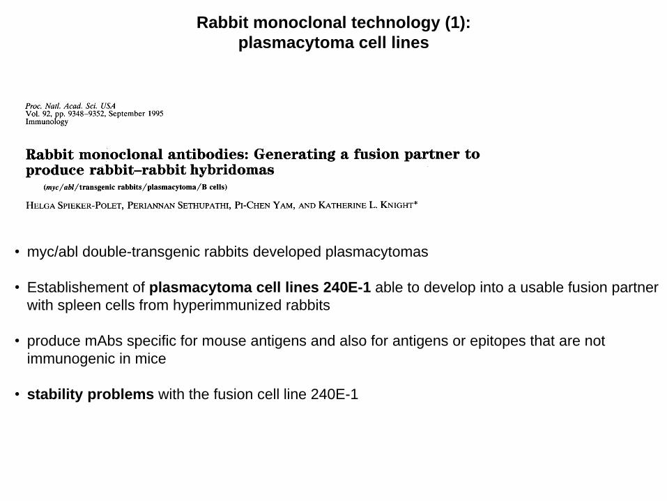
• myc/abl double-transgenic rabbits developed plasmacytomas
• Establishement of plasmacytoma cell lines 240E-1 able to develop into a usable fusion partner
with spleen cells from hyperimmunized rabbits
• produce mAbs specific for mouse antigens and also for antigens or epitopes that are not
immunogenic in mice
• stability problems with the fusion cell line 240E-1
Rabbit monoclonal technology (1):
plasmacytoma cell lines

• After multiple rounds of subcloning and
selection, a new cell line 240E-W, was
identified which expressed better fusion
efficiency and stability
Rabbit monoclonal technology (1):
plasmacytoma cell lines
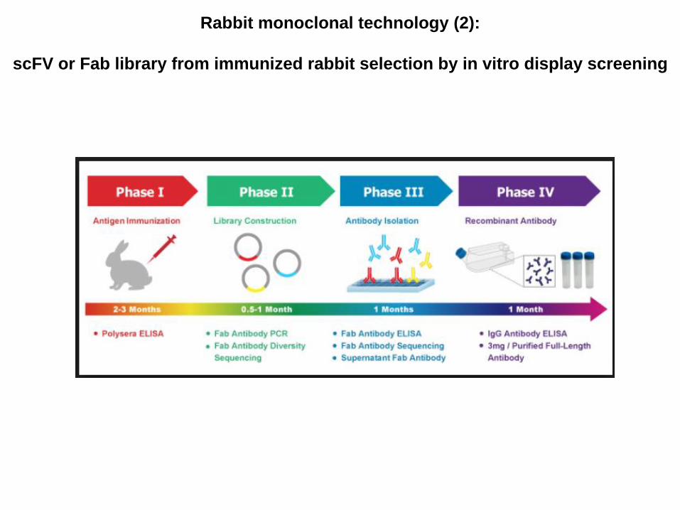
Rabbit monoclonal technology (2):
scFV or Fab library from immunized rabbit selection by in vitro display screening

Method based on a combination of B-Cell Cloning and B-Cell
PCR technology which is highly effective in isolating a large
number of rabbit B-cell clones secreting sufficient monoclonal
antigen specific IgG.
Peripheral B cells could be a preferred source for very good
antibodies in terms of affinity maturation, since the emigration of
the matured ASCs/plasma blasts from the spleen is an affinity
driven process
Rabbit monoclonal technology (3):
Rabbit B cell from peripheral blood

When developing therapeutic mAbs, the choice of IgG
subclass is important, especially in oncology
Going for therapy:
Humanization and and modulation of the Fc effector function

At present, he majority of mAbs approved for therapeutic applications are produced in:
• Chinese hamster ovary cells (12 out of 28),
• SP2/0 (7/28) and NS0 (5/28) mouse cell lines,
• hybridomas (2/28).
The remaining two are antigen-binding fragments (Fabs) that are produced periplasmically
in Escherichia coli.
Advantages of mammalian cell expression systems:
endotoxin-free
high-level expression and stability,
Recently developed for the display of functional glycosylated immunoglobulin on the cell
surface.
However, the selection of stable antibody-producing cell lines is very time consuming and
results in higher costs relative to microbial expression systems that involve much faster
growth rates and thus lower capital investment.
Disadvantages of mammalian cell systems for mAb production
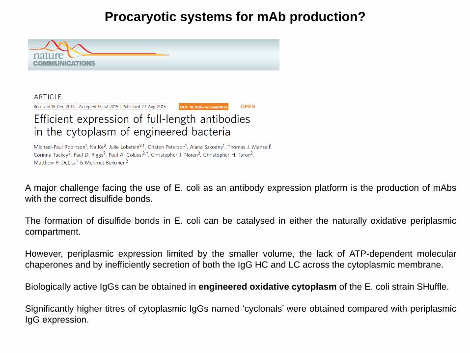
A major challenge facing the use of E. coli as an antibody expression platform is the production of mAbs
with the correct disulfide bonds.
The formation of disulfide bonds in E. coli can be catalysed in either the naturally oxidative periplasmic
compartment.
However, periplasmic expression limited by the smaller volume, the lack of ATP-dependent molecular
chaperones and by inefficiently secretion of both the IgG HC and LC across the cytoplasmic membrane.
Biologically active IgGs can be obtained in engineered oxidative cytoplasm of the E. coli strain SHuffle.
Significantly higher titres of cytoplasmic IgGs named ‘cyclonals’ were obtained compared with periplasmic
IgG expression.
Procaryotic systems for mAb production?

The genes encoding HC (VH–CH1–CH2–CH3) and LC (VL–CL)
of anti-MBP were assembled into a synthetic, bicistronic operon
under the control of a T7/lac promoter in plasmid pET21b.
Test wild-type (Reducing) E. coli B strain vs the isogenic
suppressor strain MB1731 (Oxi), whose cytoplasmic reductive
pathways have been diminished, allowing the formation of
disulfide bonds in the cytoplasm.
Cyclonal production is enhanced in Shuffle strain (Oxi + cDsbC)
by expression of DsbC, an oxidoreductase chaperone capable
of enhancing oxidative protein folding both in its native
periplasmic compartment and when expressed cytoplasmically.
Cytoplasmic expression of mouse anti-MBP cyclonals in SHuffle
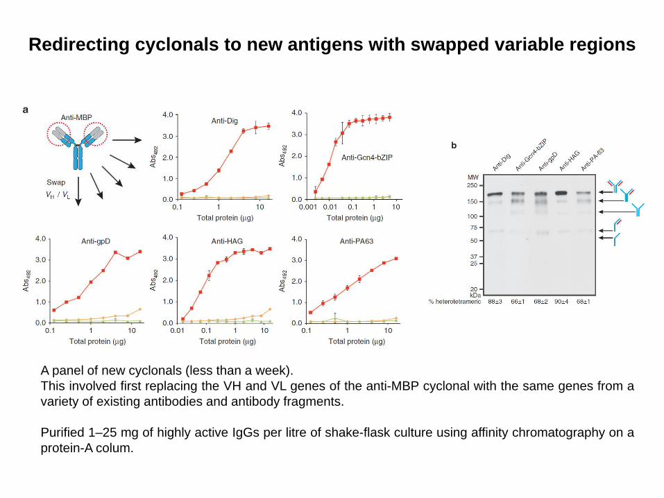
A panel of new cyclonals (less than a week).
This involved first replacing the VH and VL genes of the anti-MBP cyclonal with the same genes from a
variety of existing antibodies and antibody fragments.
Purified 1–25 mg of highly active IgGs per litre of shake-flask culture using affinity chromatography on a
protein-A colum.
Redirecting cyclonals to new antigens with swapped variable regions

Remodelling the Fc domain of cyclonals for binding to FcγRs.
IgGs lacking glycosylation in their Fc domain, such as those produced in E. coli, are completely
unable to bind to FcgRs, and do not induce FcγR-mediated effector functions.
Aglycosylated Fc with E382V/M428I mutation binds to FcγRI and is efficiently produced by
cyclonals
Antigen-binding activity of chimeric anti-PA-63 cyclonal with either Wt or
mutated Fc domain (cyclonal Fc(E382V/M428I)). Glycosylated IgGs with WT
Fc were purified from hybridoma cultures and included as positive control

Comparison of the accumulation of the anti-MBP
IgG in the mFab/hFc format following expression in
the cytoplasm and periplasm
Comparison of cytoplasmic versus periplasmic IgG expression
Periplasm
Cytoplasm
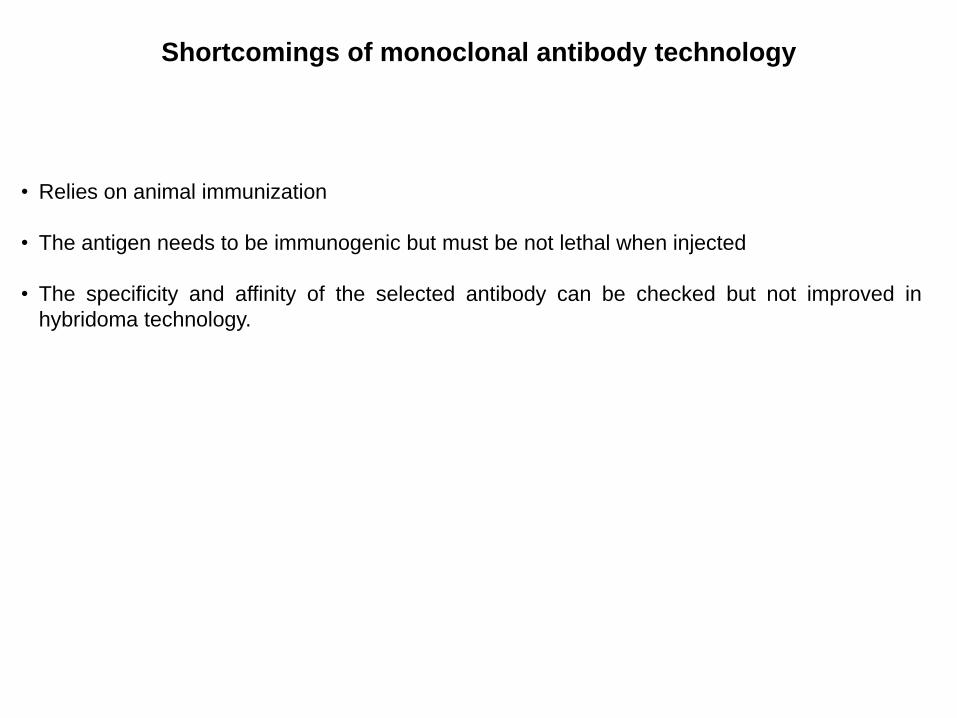
Shortcomings of monoclonal antibody technology
• Relies on animal immunization
• The antigen needs to be immunogenic but must be not lethal when injected
• The specificity and affinity of the selected antibody can be checked but not improved in
hybridoma technology.

Generating recombinant antibodies and antibody mimetics
A large library of potentially interesting
antibodies/mimetics is created, from which binders
with desirable specificity and affinity can be selected
against a specific antigen.
Prerequisite for in vitro selection process:
Link between the genotype (DNA sequence) and
the phenotype (protein)

Construction of antibodies libraries
The repertoire of antibody light and heavy chain
mRNA from B cells is amplified via PCR using a
set of specific primers covering all the V gene
families.
The antibody fragments are generated by
random combination of VL and VH chain genes.
Most commonly Fab and scFv can be cloned
and displayed on phage/ribosome libraries.
Natural Synthetic
Naive Immune
Antibody libraries
Modular Consensus Frameworks and
randomized CDRs:
All the sequences are exactly known
The diversity of the library is crucial for successful isolation of specific binders.
Knappik A. Et al., J. Mol. Biol. (2000) 296, 57-86

Phage display derived antibodies:
The bacteriophage bio-technology
His tag
pIII mediated
infection
Phage display format: Fab fragment has its
CH1 domain fused to the pIII coat protein of
the phage via an Amber codon. Using a bacteria
strain that does not read the Amber codon as a
Stop allows the fusion between the two proteins.

Phage selection: biopanning

Generation of scFv for protein tyrosine phosphatase 1B (PTP1B)
PTP1B plays a pivotal role in insulin and leptin signaling.
PTP1B is oxidized by ROS in response to stimuli (insulin).
Elements of the catalytic cleft are exposed on the surface and the enzyme is inactive
(the substrate binding is inhibited).
This reversible oxidation presents new unique binding surface.
The goal was to generate scFV targeting oxidation-specific epitopes of PTP1B that
could be expressed in cells as intrabodies to block PTP1B function.
CASA mutation mimicking the
conformation of oxidized PTP1B
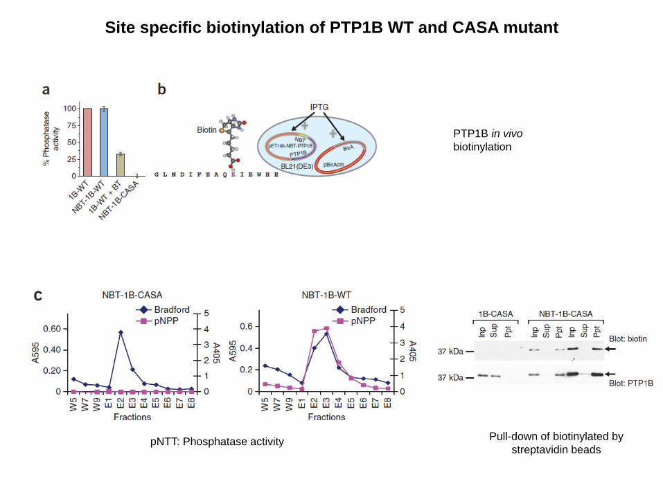
Site specific biotinylation of PTP1B WT and CASA mutant
PTP1B in vivo
biotinylation
pNTT: Phosphatase activity
Pull-down of biotinylated by
streptavidin beads

Schematic representation of the panning steps:
In-solution panning to enrich PTP1B-OX-specific scFvs
Solution-based in vitro selection strategy crucial
to preserve specific interaction with a particular
conformation of the antigen
scFv phage library derived
from PTP1B-CASA immunized
chickens

Phage output/input ratio and library diversity

Screening of scFvs specific to PTP1B-OX
Conditions were established for reversible oxidation of
PTP1B by H2O2 in vitro
Standard screening such as ELISA are not sufficient to assess the function of selected
scFV to inhibit PTP1B-OX reactivation.
Screening based on phosphatase assay in solution

Screening of scFvs specific to PTP1B-OX
Specific binding of scFv45 to oxidized PTP1B in vitro
scFV45-His-tag
+
PTP1B-R
scFV45-His-tag
+
PTP1B-OX
scFV45-His-tag
+
PTP1B-CASA

• Selection in the context of host environment cannot be avoided
• Loos of potential candidates due to growth disadvantage /toxicity
• Risk to lose the best binders (with picomolar affinity) if elution is not efficient
• Diversification step to evolve the antibody is possible but laborious:
mutator strains: it can create unwanted mutations in the plasmid and host
genome
switch to diversification step in vitro (maturation libraries or mutagenesis by
PCR) and subsequent new selection by phage display (laborious)
• Phage display is relatively demanding and time consuming technology
Limitations of the phage display techonology

Principle of ribosome display:
Entirely in vitro cell free transcription, translation and selection system
Selection of very large libraries which do not need to be transformed into cells.
Co-translational
folding of proteins
can be improved by
supplementing PDI
Library No stop codons
Low temperature
and high Mg2+ to
stabilize the complex
By EDTA
Selection in solution is
better. Bound ribosomal
complex are then
captured via tagged
antigen
During PCR, the
promoter and
the RBS are
reintroduced
*

Characteristic of the constructs for ribosome display

Ribosome display to in vitro selection and evolution of scFvs from a large
synthetic library (Human Combinatorial Antibody Library; HuCAL) against bovine insulin
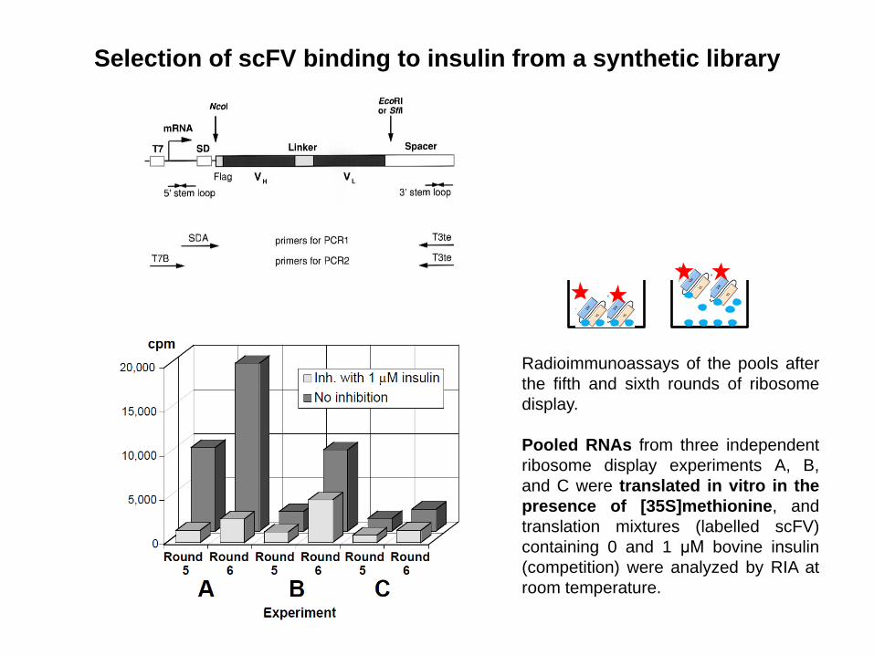
Radioimmunoassays of the pools after
the fifth and sixth rounds of ribosome
display.
Pooled RNAs from three independent
ribosome display experiments A, B,
and C were translated in vitro in the
presence of [35S]methionine, and
translation mixtures (labelled scFV)
containing 0 and 1 μM bovine insulin
(competition) were analyzed by RIA at
room temperature.
Selection of scFV binding to insulin from a synthetic library

Alignment of the amino acid sequences of VH and VL of the scFvs binders: All binders have mutation compared to the original HuCAL sequences
VH
VL

The necessity of noncovalent interdomain interactions for functional
domain assembly impairs the thermodynamic stability of Fabs and scFvs
Naturally evolved heavy chain antibodies (hcAbs) from
camels: the functional antigen-binding unit of hcAbs
consists of one single variable domain (VHH domain;
nanobody).
VHHs have a size of ∼13–14 kD and have evolved
biochemical features:
the substitution of hydrophobic with hydrophilic residues
in framework regions increases stability and solubility
This allows robust, heterologous expression in bacterial
hosts and functional expression in eukaryotic cells.
Immunoglobulins and their derivatives
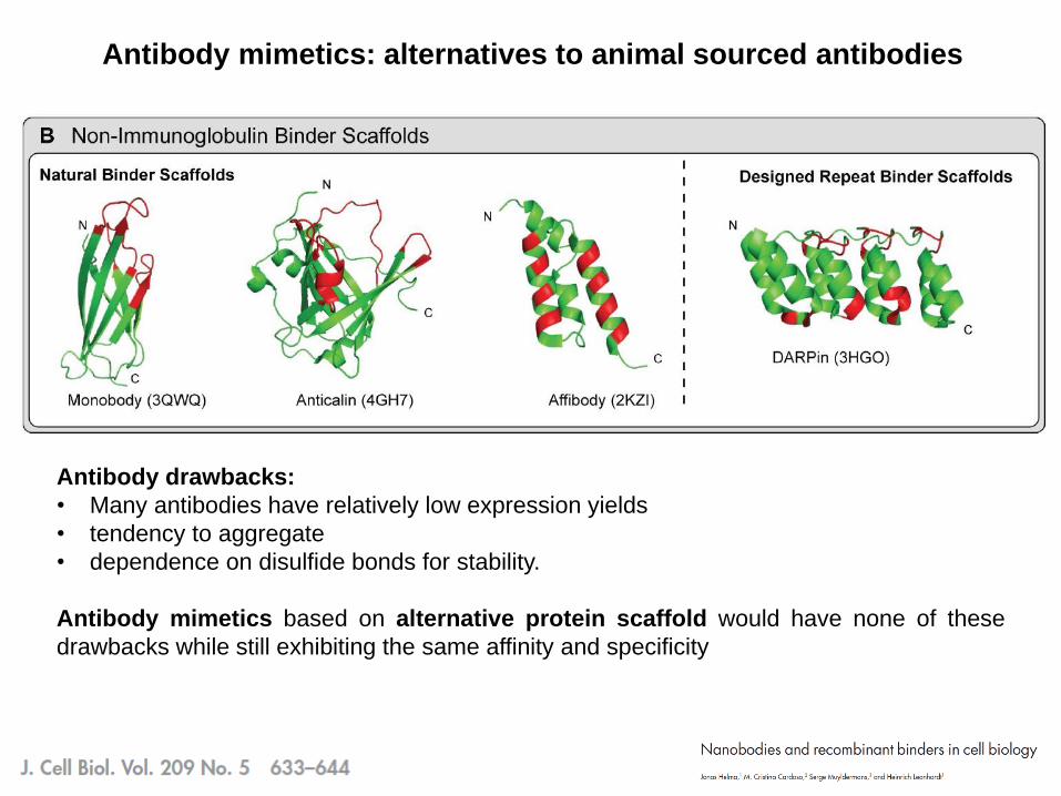
Antibody mimetics: alternatives to animal sourced antibodies
Antibody drawbacks:
• Many antibodies have relatively low expression yields
• tendency to aggregate
• dependence on disulfide bonds for stability.
Antibody mimetics based on alternative protein scaffold would have none of these
drawbacks while still exhibiting the same affinity and specificity

replicate the virtues of antibodies and address their shortcomings
DARPins

• Repeat proteins: consecutive homologous structural units (repeats), which stack to form an
elongated protein domain with a continuous hydrophobic core
• They occur throughout all phyla and mediate protein-protein interactions in the nucleus or
cytoplasm, or while anchored to the membrane or when secreted into the extracellular space
• AR are built from stacked, 33 amino acid repeats, each forming a β-turn that is followed
by two antiparallel α-helices and a loop reaching the β-turn of the next repeat.
• The β-turn and the first α-helix mediate the interactions with the target, and different numbers
of adjacent repeats are involved in binding.
• The reported target binding affinities of natural AR proteins are in the low nanomolar range
Ankyrin repeat proteins

Design of the DARPins library
Based on a consensus strategy:
Underlying assumption: residues important for maintaining the fold will be more conserved
and thus show up prominently in an alignment.
A consensus framework was built and surface residues were identified that might
potentially interact with the target—based on analogy of complexes of natural ankyrin
repeat proteins with their targets.
These residues were randomized, avoiding the residues Cys (to eliminate disulfide
formation), Pro, and Gly (as some of the residues are located in a helix). This restriction was
achieved by using trinucleotide building blocks during library generation.
(DARPin) library thus comprises fixed and variable positions
The fixed positions reflect structurally important framework positions, whereas the six
variable positions per repeat module reflect non-conserved, surface-exposed residues that
can be potentially engaged in interactions with the target.
The theoretical diversities of the DARPin libraries are 5.2 × 10¹⁵ or 3.8 × 10²³ for two-module
or three-module binders, respectively.

• DARPins proteins use both β-turns and a randomized surface
and, because of their modular architecture, the interaction
surface can be adapted by adding more repeat modules
• Concave shape of the binding site which binds structural
epitopes on the target protein surface
• Next generation of DARPins (Loop DARPins) with extended
epitope-binding properties: introduction of an elongated loop
mimicking convex paratope
• With LoopDARPin library, binders with an affinity of 30 pM
could be isolated with only a single round of ribosome display
directly from the original library.
• N- and C-capping repeat flanking the binding modules are
essential for DARPins to fold in E. Coli.
• High stability, no aggregation even at high concentration,
expressed at very high yield in soluble form in the cytoplasm of
E. coli (up to 200 mg per liter of shake-flask culture).
• Purified by IMAC
Properties of the DARPins

Expression, purification and SPR analysis of selected AR proteins
expressed in high amounts in soluble
form and free of cysteines

ELISAs with selected AR proteins
Specificity of MBP binders
Competition ELISA
Specificity comparison of
an MBP, a JNK2 and a p38 binder
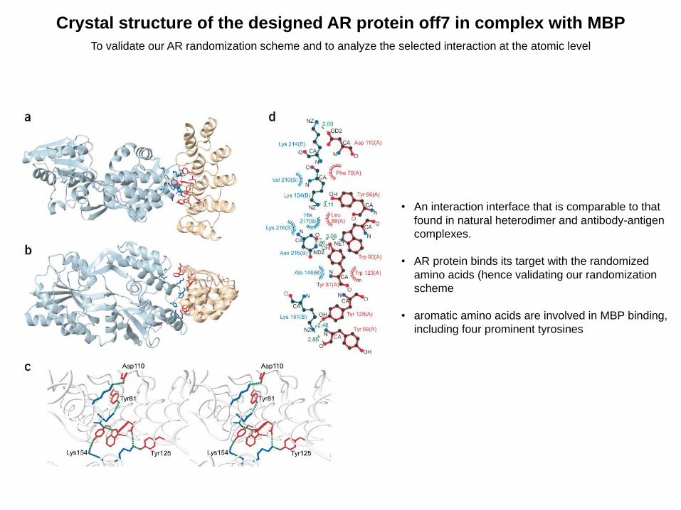
Crystal structure of the designed AR protein off7 in complex with MBP
To validate our AR randomization scheme and to analyze the selected interaction at the atomic level
• An interaction interface that is comparable to that
found in natural heterodimer and antibody-antigen
complexes.
• AR protein binds its target with the randomized
amino acids (hence validating our randomization
scheme
• aromatic amino acids are involved in MBP binding,
including four prominent tyrosines

Ribosome display selected DARPins against either the unphosphorylated or the phosphorylated form
of the MAPK ERK2 (ERK2 or pERK2).
Selections were carried out with N2C and N3C libraries consisting of two or three randomized ankyrin
repeat modules between an N- terminal and a C-terminal capping repeat, respectively.
To be able to select for phosphorylation-specific DARPins, the selection pressure was increased by
introducing a prepanning step, using the non desired ERK2 form.
Antigens: fusion proteins with an N-term avi tag for in vivo biotinylation followed by the respective
MAPK and a C- terminal His tag for purification (avi-MAPK-His6).
Activated MAPKs required the coexpression of upstream kinases:
pLK1_ERK1+MEK1R4F and pLK1_ERK2+MEK1R4F in pAT222 vector.
pBirAcm (Avidity) was used for in vivo biotinylation of MAPKs

Binding specificity analysis of selected DARPins by ELISA
Test MAPK family members with marked
sequence homologies (sequence identity of
>40% over the highly conserved catalytic core).
All tested DARPins were highly specific for their
cognate antigens ERK2 and pERK2 and did not
interact with the inactive or active form of any
other MAPK
Binding of the selected DARPins to ERK2 was
compared with that to ERK1, (85% sequence
identity).
Tested DARPins did not discriminate between
ERK1 and ERK2, but retained the
phosphorylation status specificity observed for
ERK2 and also for ERK1.

Crystal structures of phosphorylation status-specific DARPins E40 and pE59 in
complex with ERK2 and phosphorylated ERK2
To Validate the selection strategy and explain the binding specificity on the atomic level
The binding region of DARPins includes the activation loop, the MAPK insertion, α-helix G, and partially α-helix 1L12.
The interaction of pE59 with pERK2 relies on contact formation with identical regions and residues as identified for the
E40/ERK2 complex.
Thus, specificity of DARPins E40 and pE59 results from binding to discriminating structural elements, which have
changed in spatial conformation, but not from interaction with different amino acid residues on the target.
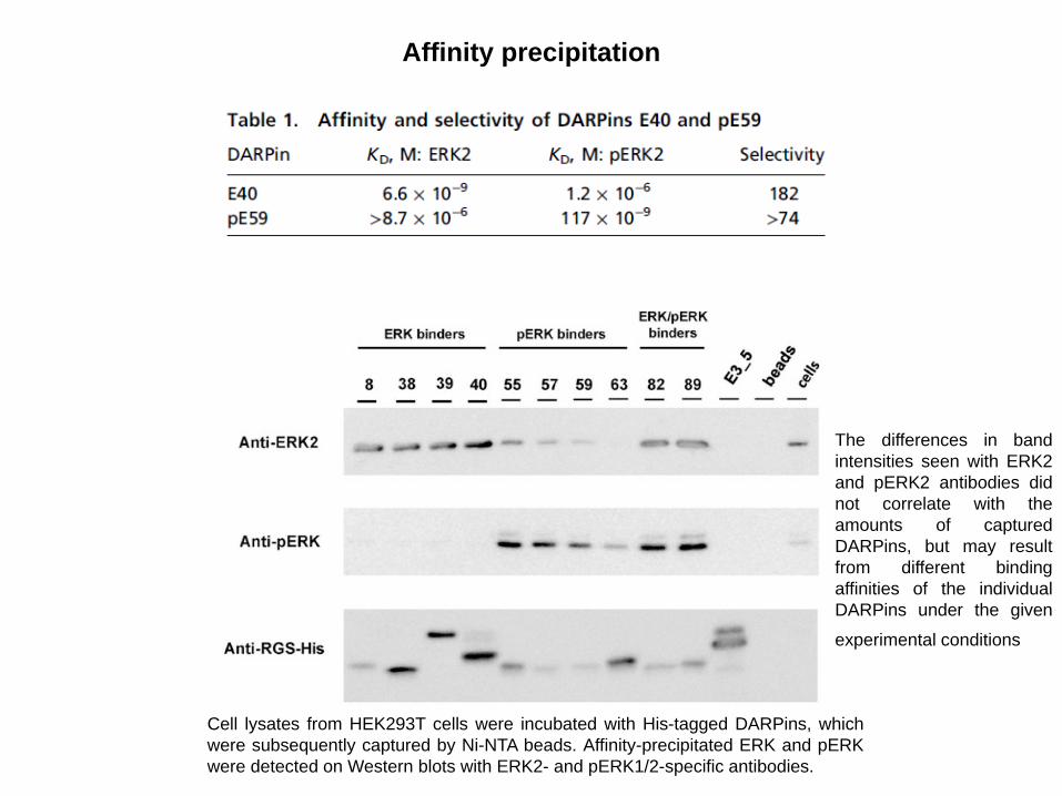
Affinity precipitation
Cell lysates from HEK293T cells were incubated with His-tagged DARPins, which
were subsequently captured by Ni-NTA beads. Affinity-precipitated ERK and pERK
were detected on Western blots with ERK2- and pERK1/2-specific antibodies.
The differences in band
intensities seen with ERK2
and pERK2 antibodies did
not correlate with the
amounts of captured
DARPins, but may result
from different binding
affinities of the individual
DARPins under the given
experimental conditions

Specificity comparison of selected DARPins in living cells by
Bioluminescence resonance energy transfer (BRET2) assays
DARPins fused to the N and C termini of either Renilla luciferase
(Rluc) or a variant of green fluorescent protein (termed GFP2).

Functionality of selected DARPins in living cells
To determine whether the
BRET2 DARPin reporters
GFP2-E40 and -pE59 retain
their specificity for ERK or
pERK, ERK phosphorylation
in COS-7 cells was either
stimulated by addition of
FBS or diminished by the
ERK pathway inhibitor
PD98509, which inhibits the
upstream kinase MEK.
Both sensors and intracellular inhibitors can be
obtained by selecting DARPins to the native targets.

DARPins are suited for functional studies as intracellular protein-specific
reagents (intrabodies), because they do not require stabilizing disulfide bonds and
because they can thus fold in the cytoplasm, where they neither aggregate nor are
degraded.
Biosensor based on the DARPin scaffold that responds specifically to
active doubly phosphorylated ERK (pERK)

Sites for Dye Labeling in the Structure of DARPin pE59
DARPin binding to the target is detected by attachment of a bright solvatochromatic
fluorophore, which has emissive properties that are dependent on the solvent environment.
When positioned appropriately in the binding protein, the exposure of the dye to a
hydrophobic environment, which forms upon target binding within the new protein-protein
interaction interface, causes a change in fluorescence intensity and/or lmax.
The dyes can be excited at long wavelengths (>580 nm) to avoid cell damage and diminish
cellular autofluorescence.
Functional pE59 mutants were
covalently derivatized with a
diverse set of merocyanine dyes.
pE59-C123m87 was selected and
it is referred to as pE59RFD
biosensor

Quantifying Activation of Endogenous ERK in Living Cells
Only the altered conformation of the
active kinase is detected, pointing to a
concept applicable to many molecular
species undergoing conformational
changes in the cell.
Useful chemical tools in studying
subtle changes of protein dynamics in
living cells.
The pE59RFD biosensor was tested in
NIH 3T3 mouse embryo fibroblasts
(MEFs) stably expressing YPet, a yellow
fluorescent protein derivative for
ratiometric imaging.
Kummer L. et al., Chemistry & Biology 20, 847–856, June 20, 2013

Bispecific DARPins to induce apoptosis in HER2-addicted tumor cells.
All signaling from HER2 complexes is obstructed, leading to a pan-HER inhibition.
Human epidermal growth factor
receptor-2 (HER2/ErbB2):
receptor tyrosine kinase without
a known natural ligand, directly
linked to the growth of
malignancies from various
tissues.
Anti-HER2 DARPins create a
trap for HER2 in which the
receptor is bent over and
kinases are unable to interact.
Jost C. et al., Structure 2013 Nov 5;21(11):1979-91
Monovalent DARPins
High tissue penetration: easier reaching of targets outside the blood circulation.
Absence of effector function: ideally suited for neutralization of soluble targets and
undesirable side-effects emerging from binding membrane-associated variants of the target
are minimized
Adjustable pharmacokinetics (PK): Unmodified DARPins offer fast PK with predicted half-lives
in the range of hours, which can be used to rapidly remove unwanted molecules from the
blood stream. The half-life can be prolonged by fusion to PEG or serum protein binding
molecules.
Allosteric Inhibition as they bind their target proteins on a conformational epitope.
New administration routes: routes in which very large amounts of drug is needed.
Conjugated DARPins
Deliver active moieties to sites of disease tissue (in oncology DARPins are used to deliver
toxins to tumors or in inflammation where DARPins inhibit cytokines in inflamed sites).
Multispecificity: DARPins with different specificities can be fused allowing the combination
of various functions in one molecule (to hijack a transcytosis receptor, binding a target in a
disease tissue and recruit effector molecules in that tissue etc).
Advantages of Therapeutic DARPin

Therapeutic DARPins
Immunogenicity?
High stability and no aggregation tendency: prerequisites for low immunogenicity.
Possible immunological tolerance: the abundance of the anykrin protein itself in the
erythrocytes suggests that ankyrin protein fragments are constantly in the circulation.

Thank you for your attention!