Embed Size (px)
Citation preview

Substrate impact on the low-temperature growth of GaN thin films by plasma-assistedatomic layer depositionSeda Kizir, Ali Haider, and Necmi Biyikli
Citation: Journal of Vacuum Science & Technology A 34, 041511 (2016); doi: 10.1116/1.4953463View online: https://doi.org/10.1116/1.4953463View Table of Contents: http://avs.scitation.org/toc/jva/34/4Published by the American Vacuum Society
Articles you may be interested inSubstrate temperature influence on the properties of GaN thin films grown by hollow-cathode plasma-assistedatomic layer depositionJournal of Vacuum Science & Technology A: Vacuum, Surfaces, and Films 34, 01A125 (2016);10.1116/1.4936230
Atomic layer deposition of GaN at low temperaturesJournal of Vacuum Science & Technology A: Vacuum, Surfaces, and Films 30, 01A124 (2012);10.1116/1.3664102
Comparison of trimethylgallium and triethylgallium as “Ga” source materials for the growth of ultrathin GaN filmson Si (100) substrates via hollow-cathode plasma-assisted atomic layer depositionJournal of Vacuum Science & Technology A: Vacuum, Surfaces, and Films 34, 01A137 (2016);10.1116/1.4937725
Demonstration of flexible thin film transistors with GaN channelsApplied Physics Letters 109, 233504 (2016); 10.1063/1.4971837
Low-temperature self-limiting atomic layer deposition of wurtzite InN on Si(100)AIP Advances 6, 045203 (2016); 10.1063/1.4946786
Low temperature thin film transistors with hollow cathode plasma-assisted atomic layer deposition based GaNchannelsApplied Physics Letters 104, 243505 (2014); 10.1063/1.4884061
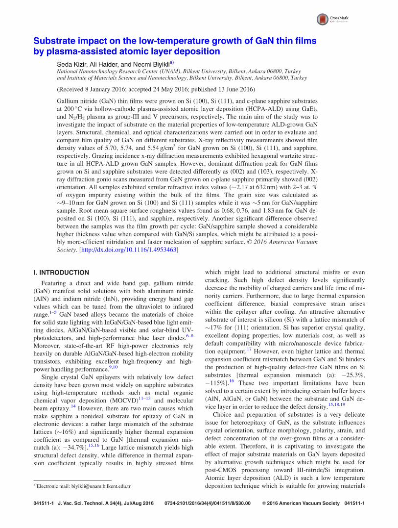
Substrate impact on the low-temperature growth of GaN thin filmsby plasma-assisted atomic layer deposition
Seda Kizir, Ali Haider, and Necmi Biyiklia)
National Nanotechnology Research Center (UNAM), Bilkent University, Bilkent, Ankara 06800, Turkeyand Institute of Materials Science and Nanotechnology, Bilkent University, Bilkent, Ankara 06800, Turkey
(Received 8 January 2016; accepted 24 May 2016; published 13 June 2016)
Gallium nitride (GaN) thin films were grown on Si (100), Si (111), and c-plane sapphire substrates
at 200 �C via hollow-cathode plasma-assisted atomic layer deposition (HCPA-ALD) using GaEt3and N2/H2 plasma as group-III and V precursors, respectively. The main aim of the study was to
investigate the impact of substrate on the material properties of low-temperature ALD-grown GaN
layers. Structural, chemical, and optical characterizations were carried out in order to evaluate and
compare film quality of GaN on different substrates. X-ray reflectivity measurements showed film
density values of 5.70, 5.74, and 5.54 g/cm3 for GaN grown on Si (100), Si (111), and sapphire,
respectively. Grazing incidence x-ray diffraction measurements exhibited hexagonal wurtzite struc-
ture in all HCPA-ALD grown GaN samples. However, dominant diffraction peak for GaN films
grown on Si and sapphire substrates were detected differently as (002) and (103), respectively. X-
ray diffraction gonio scans measured from GaN grown on c-plane sapphire primarily showed (002)
orientation. All samples exhibited similar refractive index values (�2.17 at 632 nm) with 2–3 at. %
of oxygen impurity existing within the bulk of the films. The grain size was calculated as
�9–10 nm for GaN grown on Si (100) and Si (111) samples while it was �5 nm for GaN/sapphire
sample. Root-mean-square surface roughness values found as 0.68, 0.76, and 1.83 nm for GaN de-
posited on Si (100), Si (111), and sapphire, respectively. Another significant difference observed
between the samples was the film growth per cycle: GaN/sapphire sample showed a considerable
higher thickness value when compared with GaN/Si samples, which might be attributed to a possi-
bly more-efficient nitridation and faster nucleation of sapphire surface. VC 2016 American VacuumSociety. [http://dx.doi.org/10.1116/1.4953463]
I. INTRODUCTION
Featuring a direct and wide band gap, gallium nitride
(GaN) manifest solid solutions with both aluminum nitride
(AlN) and indium nitride (InN), providing energy band gap
values which can be tuned from the ultraviolet to infrared
range.1–5 GaN-based alloys became the materials of choice
for solid state lighting with InGaN/GaN-based blue light emit-
ting diodes, AlGaN/GaN-based visible and solar-blind UV-
photodetectors, and high-performance blue laser diodes.6–8
Moreover, state-of-the-art RF high-power electronics rely
heavily on durable AlGaN/GaN-based high-electron mobility
transistors, exhibiting excellent high-frequency and high-
power handling performance.9,10
Single crystal GaN epilayers with relatively low defect
density have been grown most widely on sapphire substrates
using high-temperature methods such as metal organic
chemical vapor deposition (MOCVD)11–13 and molecular
beam epitaxy.14 However, there are two main causes which
make sapphire a nonideal substrate for epitaxy of GaN in
electronic devices: a rather large mismatch of the substrate
lattices (�16%) and significantly higher thermal expansion
coefficient as compared to GaN [thermal expansion mis-
match (a): �34.7%].15,16 Large lattice mismatch yields high
structural defect density, while difference in thermal expan-
sion coefficient typically results in highly stressed films
which might lead to additional structural misfits or even
cracking. Such high defect density levels significantly
decrease the mobility of charged carriers and life time of mi-
nority carriers. Furthermore, due to large thermal expansion
coefficient difference, biaxial compressive strain arises
within the epilayer after cooling. An attractive alternative
substrate of interest is silicon (Si) with a lattice mismatch of
�17% for h111i orientation. Si has superior crystal quality,
excellent doping properties, low materials cost, as well as
default compatibility with micro/nanoscale device fabrica-
tion equipment.17 However, even higher lattice and thermal
expansion coefficient mismatch between GaN and Si hinders
the production of high-quality defect-free GaN films on Si
substrates [thermal expansion mismatch (a): �25.3%,
�115%].16 These two important limitations have been
solved to a certain extent by introducing certain buffer layers
(AlN, AlGaN, or GaN) between the substrate and GaN de-
vice layer in order to reduce the defect density.15,18,19
Choice and preparation of substrates is a very delicate
issue for heteroepitaxy of GaN, as the substrate influences
crystal orientation, surface morphology, polarity, strain, and
defect concentration of the over-grown films at a consider-
able extent. Therefore, it is captivating to investigate the
effect of major substrate materials on GaN layers deposited
by alternative growth techniques which might be used for
post-CMOS processing toward III-nitride/Si integration.
Atomic layer deposition (ALD) is such a low temperature
deposition technique which is suitable for growing materialsa)Electronic mail: [email protected]
041511-1 J. Vac. Sci. Technol. A 34(4), Jul/Aug 2016 0734-2101/2016/34(4)/041511/8/$30.00 VC 2016 American Vacuum Society 041511-1

with excellent uniformity and ultimate conformality. Growth
mechanism of ALD is based on surface reactions which
occur at the available sites of the surface and show self-
terminating character after all available chemisorption sites
are occupied. This “self-terminating” behavior of ALD
allows to control the film thickness at sub-Angstrom level.
Plasma assisted ALD (PA-ALD) is a modified form of ALD
in which energetic radicals assist in accelerating the self-
limiting ALD reactions. PA-ALD offers additional merits
over thermal ALD such as reduced growth temperatures and
improved material properties.20,21
In the initial reports of ALD-grown GaN films, film
growth was achieved at higher temperatures (500–750 �C)
by using GaCl3 and NH3 precursor materials.22 Follow up
work for low-temperature (as low as 200 �C) growth of crys-
talline GaN using PA-ALD has been successfully demon-
strated by several groups utilizing GaMe3 or GaEt3 in
conjunction with N2/H2 or NH3 plasma as Ga and nitrogen
precursor sources, respectively.5,23–26 However, a compara-
tive study investigating the influence of different substrates
on the material properties of GaN grown using low-
temperature PA-ALD is lacking. Recently, the effect of dif-
ferent substrates on material properties of PA-ALD grown
AlN thin films has been investigated, where they demon-
strate that Si (111) is a better substrate for AlN growth.27 In
this work, we have studied and compared the properties of
GaN thin films grown on Si (100), Si (111), and c-plane sap-
phire substrates by using hollow-cathode plasma-assisted
ALD (HCPA-ALD) at 200 �C.
II. EXPERIMENT
A. Film growth
GaN thin film deposition is performed in Fiji F200-LL
ALD reactor (Ultratech/CambridgeNanotech, Inc., Cambridge,
MA) with a modified configuration of stainless-steel hollow-
cathode plasma source (Meaglow, Ltd., Canada). Hollow
cathode plasma (HCP) source has been found superior to
quartz-based inductively coupled plasma source as our former
studies on ALD-grown GaN confirmed a significantly higher
layer quality with considerably lower impurity incorporation.5
Moreover, HCP source possesses advantages such as high
plasma density and rapid on/off cycles for pulsed growth.28,29
Prior to growth experiments, Si (111), Si (100), and sapphire
substrates were cleaned sequentially using isopropanol, ace-
tone, methanol, and deionized water in an ultrasonic bath. In
order to remove the native oxide layer on the substrate surface,
additional diluted HF (2%) cleaning process was applied for 2
min. Afterward, samples were loaded immediately to the reac-
tor, and the process base pressure was adjusted to 150 mTorr.
To prepare an enhanced nitridated surface in order to eliminate
possible nucleation delays and to enhance the crystalline qual-
ity, a nitridation process step was utilized before the ALD
cycles by applying 60 s of N2/H2 plasma and subsequently 60
s of N2 plasma.30 GaEt3 was used as organometallic precursor
for Ga with a pulse length of 0.5 s. As nitrogen half-cycle,
N2/H2 plasma exposure is applied with 50/50 sccm flow rate
for 60 s under 300 W rf-plasma power. Ten seconds purge
intervals were used in between the metal–organic precursor
pulse and N2/H2 plasma exposures to evacuate the excess pre-
cursor molecules and reaction byproducts. Ar is used as the
carrier and plasma gas with 30 and 100 sscm flow rates for
GaEt3 and N2/H2 flows, respectively. A total of 1000 growth
cycles were carried out at a substrate temperature of 200 �C.
B. Film characterization
Both x-ray reflectivity (XRR) and grazing-incidence x-ray
diffraction (GIXRD) measurements were performed with
PANalytical X’Pert PRO MRD diffractometer (PANalytical
B. V., Netherlands) by using Cu Ka radiation. GIXRD spec-
tra are collected at 0.1� step size and 10 s of counting time in
the 2� range of 20�–80�. Omega-2� scan is performed from
0� to 2.5� for XRR measurements. X’pert pro MPD
(PANalytical, Netherlands) system is used for x-ray diffrac-
tion (XRD) measurements with Bragg Brentano geometry.
Gonio scan has been performed with a step size of 0.0016�
in between 33.5� and 35� using a counting time of 10 s.
Interplanar spacing (dhkl) values were evaluated from peak
positions using the well-known Bragg’s law. Lattice parame-
ter c was calculated by substituting dhkl values in Eq. (1),
which relates the interplanar spacing (dhkl), miller indices
(hkl), and lattice parameters (a and c) for hexagonal crystals
1
d2¼ 4
3
h2 þ hk þ k2
a2
� �þ l2
c2: (1)
Ellipsometric measurements were recorded in 300–
1000 nm wavelength interval for three different angles of inci-
dence (65�–70�–75�) by variable-angle spectroscopic ellip-
someter (V-VASE, J. A. Woollam Co., Inc., NE) and a
rotating analyzer. Optical constants and film thicknesses of
the samples were extracted from the measured ellipsometer
data by applying a fitting procedure (WVASE 32 software) with
Cauchy dispersion function and Tauc Lorentz oscillator. The
absorption coefficient
a kð Þ ¼ 4pk kð Þk
(2)
was calculated from k(k) values determined from the ellips-
ometry data. Optical band gap (Eg) is expressed by the fol-
lowing equation for direct band gap materials,31 which can
be analytically extracted via extrapolation of the linear part
of the absorption spectrum to (aE)2¼ 0
aE ¼ AðE� EgÞ1=2: (3)
In order to determine the elemental composition and
chemical bonding states of the film, x-ray photoelectron
spectroscopy was implemented using a Thermo Scientific K-
Alpha spectrometer (Thermo Fisher Scientific, MA) with
monochromatized Al Ka source. For depth profiling, GaN
thin films were etched with an energy and spot size of 1 kV
and 400 lm Ar beam, respectively. Surface morphologies of
the GaN thin films were analyzed using high-resolution
041511-2 Kizir, Haider, and Biyikli: Substrate impact on the low-temperature growth 041511-2
J. Vac. Sci. Technol. A, Vol. 34, No. 4, Jul/Aug 2016

scanning electron microscopy (HR-SEM, FEI, Hillsboro, OR
NNL 600i), and atomic force microscopy (Asylum Research
MFP-3D) was operated in tapping mode.
III. RESULTS AND DISCUSSION
A. Structural and optical characterizations
XRR measurements showed characteristic Kiessig fringes
of three GaN samples grown on different substrates in Fig. 1(a).
Periodicity and amplitude of oscillations provides informa-
tion about film thickness and film density, respectively. In
order to extract this information, X-pert reflectivity software
was used to fit the experimental data by using four and
three-layer models, i.e., Ga2O3/GaN/SiO2/Si and Ga2O3/
GaN/Al2O3 for samples grown on Si and sapphire substrates,
respectively. Figure 1(b) shows the measured versus simu-
lated XRR data for GaN/Si(111) sample, confirming the
close agreement between experimental and calculated data.
X-rays, when impinged on a flat surface with small (graz-
ing) angle, undergo total reflection below a critical angle
(�c). X-ray intensity drops abruptly after exceeding the criti-
cal angle as shown for GaN/Si(111), GaN/Si(100), and GaN/
sapphire, as shown in Fig. 1(c). Critical angle is a specific
material property depending primarily on the film density.
When the second derivative of the XRR intensity d2I/d2� is
plotted versus � [Fig. 1(d)], threshold of x-ray penetration is
located at the first minima of this curve. For GaN/Si (100)
and GaN/sapphire samples, the critical angle was extracted
as �0.32�, while for GaN/Si(111) sample, this value is found
as �0.33�. Motamedi et al.24 recently reported very similar
values in their GaN thin films grown by PA-ALD revealing
critical angles of 0.32�, while this value is around 0.36� for
single crystalline bulk GaN. Density values extracted from
simulations reveal that GaN/Si(111) exhibit the highest den-
sity value of 5.74 g/cm3, while GaN deposited on Si (100)
and sapphire possesses density values of 5.70 and 5.54 g/
cm3, respectively. The density value of GaN/Si (111) is rela-
tively closer to the bulk GaN density value of 6.15 g/cm3.
For extracting the thickness of GaN films, a Ga2O3 layer was
added on top of GaN in XRR fitting software and thickness
values of two layers were summed-up (dGaNþ dGa2O3) to
obtain final thickness of the films. Thickness values came
out as �29, �32, and �34 nm for GaN deposited on Si
(111), Si (100), and sapphire substrates, respectively. In
addition, interlayer roughness values in between GaN and
Ga2O3 layers was found as 1.1, 2.4, and 5.3 nm for GaN de-
posited on the same sequence of substrates. Higher interlayer
roughness obtained from GaN grown on sapphire led to con-
siderably higher fit parameters for data fitting which implies
that thickness and density estimation of GaN grown on sap-
phire cannot be trusted with absolute certainty.
Optical properties, including refractive index dispersion
curves and optical band gap, were determined using spectro-
scopic ellipsometric measurements and related data analysis.
Figure 2(a) shows the comparison of refractive index values
of GaN films deposited on different substrates. The refrac-
tive index values at 632 nm of GaN on Si (111), Si (100),
and sapphire were measured as 2.16, 2.17, and 2.18, respec-
tively. Refractive index value around 2.4 is reported for sin-
gle crystal hexagonal wurtzite GaN, which is notably higher
than the values obtained in our films.32 In the case of poly-
crystalline hexagonal wurtzite GaN thin films, refractive
FIG. 1. (Color online) XRR results for GaN thin films grown on Si (111), Si (100), and sapphire substrates; (a) characteristic Kiessig fringes, (b) measured vs
simulated data for GaN/Si(111), (c) measured angular dependence around critical angle, and (d) critical angle extraction.
041511-3 Kizir, Haider, and Biyikli: Substrate impact on the low-temperature growth 041511-3
JVST A - Vacuum, Surfaces, and Films

index values between 2.10 and 2.17 were reported in litera-
ture, which resembles the data obtained in this study.2,5
Spectroscopic ellipsometer data fitting reveals thickness of
GaN grown on Si (100), Si (111), and sapphire as 33, 35,
and 43 nm, respectively. These thickness values correspond
to growth per cycle (GPC) values of 0.33, 0.35, and 0.43 A/
cycle for GaN grown on same sequence of substrates. As
stated in Sec. II, Si substrates were treated with HF prior to
GaN growth, and the ellipsometric spectra were fitted using
a two-layer model [i.e., Cauchy/Si (0.5 mm)]. It was
assumed that the thickness of oxide layer on Si is negligible
at GaN/Si interface for ellipsometric data fittings. On the
other hand, when performing XRR fitting, SiO2 layer was
taken into account, and data were fitted using a four-layer
model (i.e., Ga2O3/GaN/SiO2/Si). Since a default Ga2O3
layer is not defined in the database of the ellipsometer WVASE
32 software, we have used the conventional Cauchy model
to determine the layer thickness. The different models used
in two different measurement techniques (XRR and ellips-
ometry) might have led to the non-negligible difference in
estimated film thickness.
It is reported in the literature that the initial nitridation
process using N2 plasma species induces the formation of a
thin AlN layer on the surface of sapphire.23 Higher film
thickness values extracted in the case of GaN grown on
sapphire might be the result of a more-efficient and faster
nucleation of GaN on this AlN monolayer. This implies that
GPC was most probably higher in the initial phase of GaN
growth on sapphire and then it might have reduced to a
lower steady state value. Optical band gap values were cal-
culated from (ah�)2 vs h� plot of the films. Extrapolating the
straight line segment of the plot to abscissa provides the
value of Eg value, which came out to be 3.44, 3.45, and
3.61 eV for GaN grown on Si (100), Si (111), and sapphire
[Fig. 2(b)]. The optical band gap values of GaN grown on
both Si substrates are reasonably close to the bulk GaN value
(�3.4 eV). On the other hand, GaN/sapphire sample features
a higher optical band edge which might originate from the
smaller grain/crystallite structure to be discussed later in this
section.
In order to identify the crystalline nature of the GaN thin
films, GIXRD measurements were performed. GIXRD meas-
urements revealed single-phase hexagonal wurtzite polycrys-
talline character of GaN irrespective of the substrate utilized
(Fig. 3). Hexagonal wurtzite reflections of (010), (002),
(110), (103), and (112) were manifested by GIXRD patterns
of GaN grown on Si substrates. No other phase mixing was
detected (i.e., cubic). In GIXRD, sample is exposed to x-rays
at grazing angle of incidence in order to avoid intense signal
from substrate and obtain a stronger signal from the thin
film.33 In contrast to h-2h scans, GIXRD detects the tilted
planes which are present in polycrystalline materials.
However, by its inherent configuration, GIXRD is rather
insensitive to planes which are parallel to the substrate sur-
face. Although displaying quite similar diffraction patterns,
GaN/Si samples show a considerable difference in the peak
intensity values: (002) peak of GaN/Si(111) appeared to be
higher compared to GaN/Si(100). Full width at half maxi-
mum (FWHM) values obtained from (002) reflection of GaN
film deposited on Si(111) and Si(100) was measured as 2484
and 2808 arc sec, respectively. On the other hand, (010),
(110), and (112) reflections disappeared from GIXRD pat-
tern of the GaN film deposited on c-plane sapphire substrate,
which might be due to insufficient penetration of x-rays
inside the GaN layer grown on sapphire. The intensities of
(100) and (002) reflections drastically decreased while the
intensity of (103) refection showed a significant increase.
XRD gonio scans have been conducted on GaN grown on Si
(111), Si (100), and c-plane sapphire. However, due to low
thickness of the grown films, strong background signal from
Si does not allow us to determine the bulk orientation of
these films. However, we were able to obtain the gonio-scan
of GaN/sapphire samples. Regular powder diffraction setup
(X’pert pro multipurpose diffractometer) is used for obtain-
ing gonio (Bragg Brentano geometry) scans from GaN/sap-
phire sample, which primarily showed (002) orientation with
a FWHM of 17.34 arc min [Fig. 3(b)] which is even nar-
rower than the previously reported values for the PA-ALD-
grown GaN on sapphire substrates.24
GaN directly grown (without any buffer layers) on Si at
higher temperatures shows poor crystalline quality in the
presence of a mixture of cubic and hexagonal phases. Si has
a tendency to form an amorphous SixNy layer during the
FIG. 2. (Color online) (a) Spectral refractive index values for GaN/Si(111),
GaN/Si(100), and GaN/sapphire samples. (b) Calculated optical band gap
values of GaN grown on different substrates.
041511-4 Kizir, Haider, and Biyikli: Substrate impact on the low-temperature growth 041511-4
J. Vac. Sci. Technol. A, Vol. 34, No. 4, Jul/Aug 2016

nucleation steps which gives rise to poor crystalline quality
of GaN epilayers grown at higher temperatures.15,17 That
effect was mitigated by introducing a thin SiC or AlN layer
on Si substrates before the growth of GaN. In the case of low-
temperature GaN growth, SixNy layer formation might not be
the prevailing factor to influence the crystalline quality due to
the polycrystalline film character with relatively small crystal-
lite sizes. Also, difference in the thermal expansion coefficient
between substrate material and GaN would not affect the film
properties significantly as growth takes place at substantially
reduced temperatures, 200 �C in the present case of HCPA-
ALD. Apparently, improved crystalline quality and higher
film density in the case of GaN grown on Si (111) might be
due to the hexagonal nature of the specific Si (111) plane. In
the literature, higher crystalline quality of the PE-ALD grown
AlN was observed on Si (111), and it was mainly attributed to
the hexagonal structure of Si (111) substrate.27
The lattice parameter c was determined using the (002) reflec-
tion of hexagonal phase from diffraction patterns of GaN grown
on different substrates. First interplanar spacing values (dhkl)
were calculated using Bragg’s law and these values were then
substituted in Eq. (1), which provides a relationship between
interplanar spacing (dhkl), miller indices, and lattice parameters
for hexagonal crystals. The c-axis lattice parameter was found as
the following: “cGaN/Si(111) ¼ 5.183 A,” “cGaN/Si(100)¼ 5.185 A,”
and “cGaN/sapphire¼ 5.175 A.” C axis lattice parameter values of
GaN/Si samples are in good agreement with the bulk GaN
value (c¼ 5.185 A).34 However, the variation in the c axis
lattice parameter of GaN/sapphire with respect to their nomi-
nally strain-free counterpart possibly indicates the presence
of a minor strain component in the PA-ALD-grown films.
HR-SEM measurements were conducted to observe the
surface morphology and determine the grain sizes of GaN
grown on Si (100), Si (111), and sapphire. Figure 4 shows
the surface morphology of GaN grown on different sub-
strates. Average size of polycrystalline grains was calculated
using IMAGE J (image analysis software). The average grain
size was calculated as �9–10 nm for GaN grown on Si (100)
and Si (111) samples while it was �5 nm for GaN/sapphire
sample. AFM measurements (not shown here) were per-
formed on GaN deposited on Si (111), Si (100), and sapphire
substrates. root mean square (RMS) surface roughness val-
ues were found as 0.69, 0.77, and 1.83 nm for GaN deposited
on the same sequence of substrates.
B. Chemical characterization
GaN thin films deposited on different substrates were ana-
lyzed for elemental composition and chemical bonding states
of the film from the sample surface and the bulk using XPS.
Survey scans from GaN deposited on Si (100), Si (111), and
sapphire revealed the presence of gallium, nitrogen, oxygen,
and carbon with Ga 3d, N 1s, O 1s, and C 1s peaks. Survey
scans from the surface of the film have been charge-
corrected by referencing the C 1s peak at 284.8 eV. GaN thin
films were etched with Arþ ions in order to obtain XPS sur-
vey scan from the bulk of the films. The survey scans from
the bulk of the film were charge-corrected by referencing the
Ar 2p peak at 242 eV. Elemental concentrations obtained
from the surface and bulk are provided in Table I. It is evi-
dent that the very surface of GaN films was contaminated
FIG. 3. (Color online) (a) GIXRD patterns of GaN thin films grown on Si
(111), Si (100), and sapphire substrates; (b) XRD gonio scan of GaN/sap-
phire sample.
FIG. 4. SEM images of (a) GaN/Si (111), (b) GaN/Si (100), and (c) GaN/sapphire.
041511-5 Kizir, Haider, and Biyikli: Substrate impact on the low-temperature growth 041511-5
JVST A - Vacuum, Surfaces, and Films

with oxygen (up to 12 at. %) and carbon (up to 28 at. %).
Adventitious carbon and oxygen found on the surface of the
film arise from postdeposition atmospheric exposure. There
was no evidence of measurable carbon in the bulk of the
films which shows that the surface reaction between GaEt3and N2/H2 plasma led to effective removal of carbon ligands.
On the other hand, there is trace of oxygen (2–3 at. %) in the
bulk of GaN films regardless of the substrate used. Probable
oxygen source might include oxygen and water vapor impur-
ities present in either plasma gases or organometallic precur-
sors. It might also originate from trapped oxygen/water
vapor inside the multilayer coatings on the inner walls of the
ALD vacuum reactor. Another possibility might be the
room-temperature diffusion of surface oxygen after atmos-
pheric oxidation. Survey scans from the surface and bulk of
GaN grown on Si (100), Si (111), and sapphire reveal non-
stoichiometric films with higher atomic percentages for
nitrogen. We have used monochromatic Al Ka source for
XPS scans which provides better resolution. However, it
also makes the analysis of survey spectra more complicated
as N 1s core level peak overlaps with Ga Auger peak.35
Therefore, survey scans recorded using monochromatic Al
Ka source overestimate the nitrogen atomic percentage and
provides inaccurate information about the stoichiometry of
the film.
In order to provide a more reliable explanation for this
overestimation which leads to a nearly (1:2) unrealistic stoi-
chiometry, we decided to take into account only the relative
weights of Ga-N and N-Ga peaks obtained in Ga 3d and N
1s high resolution scans for PA-ALD and MOCVD-grown
GaN films. This way, we omit the contributions coming
from Ga Auger and Ga-Ga peaks for nitrogen and gallium
concentrations, respectively. As already depicted in Table II
within the manuscript, approximately %39.1 of N 1s signal
belongs directly to nitrogen contribution (N-Ga peak) for
GaN/Si(111) sample, which corresponds to an effective
nitrogen concentration of 24.8% when multiplied with the
extracted 63.3% concentration (Table I) within the bulk of
the film. Similarly, the effective Ga concentration is calcu-
lated as 29.9% after taking into account that only 86.4% of
Ga 3d peak contributes to the stoichiometry. Hence, the PA-
ALD grown GaN film exhibits a relative stoichiometry of
(1:0.83). In order to eliminate the preferential etching effect,
we should also correct the stoichiometry via a comparison
with a reference sample exhibiting ideal stoichiometry.
Using the atomic concentrations given in Table I, Ga-N, and
N-Ga peak weights for MOCVD-grown sample, we obtain a
relative stoichiometry of (1:0.81). As we can safely assume
that commercial MOCVD-grown epitaxial GaN sample has
near-ideal stoichiometry (1:1), PA-ALD-grown GaN films
depicts a stoichiometry of (1:1.025), corresponding to only a
slightly N-rich film with 2.5% excess nitrogen concentration
with respect to epitaxial GaN film.
Figure 5 shows Ga 3d and N 1s high resolution XPS scans
from bulk of the PA-ALD-grown GaN/Si(111) and an
MOCVD-grown epitaxial GaN on sapphire to investigate the
individual peaks. At low energy resolution, XPS provides
qualitative and quantitative information on the elements
present in the material whereas high-energy resolution XPS
(HR-XPS) provides information on the chemical state and
bonding of those elements. Ga 3d scans [Figs. 5(a) and 5(c)]
obtained from both PA-ALD and MOCVD-grown GaN sam-
ples were fitted by two subpeaks. The main subpeak reveals
the existence of Ga-N bond in the bulk of the film with peak
position at 19.47 and 18.97 eV for GaN/Si (111) and
MOCVD grown GaN, respectively.5,35 Both the Ga 3d spec-
tra are dominated by Ga–N main peak. In the literature,
Ga–O component is observed at �1.2 eV higher binding
energy while Ga-Ga component was detected at �1 eV lower
binding energy compared to main Ga-N peak binding
energy.36,37 We were not able to identify any contribution
from Ga-O species in the bulk of the film. Ga-Ga bonding
state is observed as a second subpeak at 18.24 and 18.05 eV
in Ga 3d HR-XPS spectra of PA-ALD and MOCVD-grown
GaN, respectively.5,36 Uncoordinated Ga is believed to
appear inside the film after Ar ion etching due to the accu-
mulation of metallic Ga at the sample surface.38 FWHM of
main Ga-N subpeak was recorded as 1.37 eV from MOCVD-
grown GaN, while it increases to 1.79 eV for HCPA-ALD
grown GaN on Si (111). FWHM obtained from the Ga-N
subpeak of MOCVD grown GaN is slightly higher than the
literature reported GaN value of 1.1 eV, which might be
ascribed to increased defect density or localized difference
in charge state of the low-temperature grown ALD sample.30
N 1s HR-XPS spectra [Figs. 5(b) and 5(d)] were fitted
using three components. First subpeak in N 1s HR-XPS
spectra corresponds to N-Ga bond (BE¼ 396.93, 397.58 for
PA-ALD and MOCVD-grown GaN, respectively),36,39 and
the other two subpeaks correspond to Ga-LMM auger
peaks.35 The slight shift in binding energies of Ga–N and
N–Ga bonds in HCPA-ALD and MOCVD-grown GaN sam-
ples might arise due to differential charging, material
defects, or nitrogen vacancies inside the ALD-grown GaN
TABLE I. Elemental at. % of GaN grown on different substrates as extracted
from XPS measurements.
Substrate C at. % O at. % Ga at. % N at. %
Sapphire (surface) 27.94 12.09 21.91 38.06
Sapphire (bulk) — 3.00 31.22 65.78
Si (100) (surface) 19.26 12.55 22.29 45.90
Si (100) (bulk) — 2.98 31.75 65.27
Si (111) (surface) 20.33 12.69 23.13 43.86
Si (111) (bulk) — 2.07 34.60 63.33
TABLE II. Relative percentages of different bonding species present in PA-
ALD grown GaN/Si(111) and MOCVD-grown GaN on sapphire substrates.
Sample Peak
Relative %
[GaN/Si(111)]
Relative %
(MOCVD grown GaN)
Ga 3d Ga-N 86.39 87.06
Ga-Ga 13.61 12.94
N 1s N-Ga 39.05 44.59
Ga-LMM 56.58 51.62
Ga-LMM 4.37 3.78
041511-6 Kizir, Haider, and Biyikli: Substrate impact on the low-temperature growth 041511-6
J. Vac. Sci. Technol. A, Vol. 34, No. 4, Jul/Aug 2016

film. FWHM of main N-Ga peak was recorded as 1.06 and
1.53 eV for MOCVD grown GaN and HCPA-ALD grown
GaN, respectively. FWHM of N-Ga peak for MOCVD
grown GaN is slightly higher than the literature reported
value (0.85 eV) for bulk GaN. Relative percentages of differ-
ent bonding species were calculated for both samples based
on the area under the corresponding peaks and are provided
in Table II. Relative percentages of different bonding species
in GaN/Si (111) and MOCVD-grown GaN are close to each
other which indicates that stoichiometry of GaN grown on Si
(111) resembles with epitaxial GaN sample, confirming the
stoichiometry calculation based on the Ga-N and N-Ga sub-
peaks carried out above.35 Peak positions of Ga 3d and N 1s
HR-scans (not shown here) obtained from bulk of GaN
grown on Si (100) and sapphire matches well with the scans
obtained from bulk of GaN grown on Si (111).In Fig. 6,
GaN/Si(111) depth profile analysis is provided, which indi-
cates the elemental composition variation between GaN–air
and GaN–Si interface. Results reveal that atomic percentages
of gallium and nitrogen remained fairly constant along the
etching direction throughout the film thickness. Carbon dis-
appears after the very first etching cycle while oxygen stabil-
izes to around �2–3 at. % in the bulk of the film.
IV. SUMMARY AND CONCLUSION
We have presented our efforts for understanding the sub-
strate effect on GaN thin films grown using low-temperature
HCPA-ALD. GaN thin films were deposited on Si (111), Si
(100), and c-plane sapphire substrates at a growth temperature
of 200 �C using GaEt3 precursor and N2/H2 plasma as Ga and
nitrogen source, respectively. Unlike high-temperature hetero-
epitaxy, neither lattice mismatch nor thermal expansion coef-
ficient showed significant importance for low-temperature
HCPA-ALD grown GaN. XRR measurements showed film
density values of 5.70, 5.74, and 5.54 g/cm3 for GaN grown
on Si (100), Si (111), and sapphire, respectively. All HCPA-
ALD grown GaN samples showed hexagonal polycrystalline
wurtzite structure. Dominant GIXRD diffraction peaks for
GaN films grown on Si and sapphire substrates were detected
FIG. 5. (Color online) High-resolution XPS scans of Ga 3d and N 1s obtained from bulk of GaN layers grown [(a) and (b)] by low-temperature PA-ALD on Si
(111) and [(c) and (d)] by high-temperature MOCVD on sapphire substrate.
FIG. 6. (Color online) Depth profile experiment performed on Ga/Si (111)
and the thickness evolution of the related elemental concentrations.
041511-7 Kizir, Haider, and Biyikli: Substrate impact on the low-temperature growth 041511-7
JVST A - Vacuum, Surfaces, and Films

as (002) and (103), respectively. A better crystalline quality
and higher film density obtained from GaN grown on Si (111)
is attributed to the hexagonal nature of Si (111) substrate sur-
face. Grain size of the films was determined to be �9–10 nm
for GaN grown on Si (100) and Si (111) samples while it was
�5 nm for GaN/sapphire sample. RMS surface roughness val-
ues were found as 0.68, 0.76, and 1.83 nm for GaN deposited
on Si (111), Si (100), and sapphire. All samples exhibited
similar refractive index values (�2.17 at 632 nm) with
2%–3% of oxygen impurity existing within the bulk of the
films and with no detectable carbon incorporation. Optical
band gap values of 3.44, 3.45, and 3.61 eV were recorded for
GaN grown on Si (100), Si (111), and sapphire substrates,
respectively. Higher growth rate observed for GaN grown on
sapphire was attributed to a more-effective nitridation and
faster nucleation process on the sapphire surface.
1P. F. Yang, S. R. Jian, Y. S. Lai, C. S. Yang, and R. S. Chen, J. Alloys
Compd. 463, 533 (2008).2A. Haider, S. Kizir, C. Ozgit-Akgun, E. Goldenberg, S. A. Leghari, A. K.
Okyay, and N. Biyikli, J. Mater. Chem. C 3, 9620 (2015).3H. Na, S. Takado, S. Sawada, M. Kurouchi, T. Akagi, H. Naoi, T. Araki,
and Y. Nanishi, J. Cryst. Growth 300, 177 (2007).4X. Gao, C. Liu, C. Yin, L. Sun, D. Tao, C. Yang, and B. Man, J. Magn.
Magn. Mater. 343, 65 (2013).5C. Ozgit-Akgun, E. Goldenberg, A. K. Okyay, and N. Biyikli, J. Mater.
Chem. C 2, 2123 (2014).6T. Wang, J. Bai, and S. Sakai, J. Cryst. Growth 224, 5 (2001).7H. Y. Chung, K. Y. Woo, S. J. Kim, and T. G. Kim, Opt. Commun. 331,
282 (2014).8M. L. Lee, J. K. Sheu, Y. K. Su, S. J. Chang, W. C. Lai, and G. C. Chi,
IEEE Electron Device Lett. 25, 593 (2004).9A. El Fatimy et al., J. Appl. Phys. 107, 2 (2010).
10R. Dimitrov, A. Mitchell, L. Wittmer, O. Ambacher, M. Stutzmann, J.
Hilsenbeck, and W. Rieger, Jpn. J. Appl. Phys., Part 1 38, 4962 (1999).11J. N. Dai et al., J. Electron. Mater. 38, 1938 (2009).12T.-Y. Tsai, S.-L. Ou, M.-T. Hung, D.-S. Wuu, and R.-H. Horng,
J. Electrochem. Soc. 158, H1172 (2011).13B. L. Liu, M. Lachab, A. Jia, A. Yoshikawaa, and K. Takahashi, J. Cryst.
Growth 234, 637 (2002).
14D. Doppalapudi, E. Iliopoulos, S. N. Basu, and T. D. Moustakas, J. Appl.
Phys. 85, 3582 (1999).15S. A. Kukushkin, A. V. Osipov, V. N. Bessolov, B. K. Medvedev, V. K.
Nevolin, and K. A. Tcarik, Rev. Adv. Mater. 17, 1 (2008).16D. Zhu, D. J. Wallis, and C. J. Humphreys, Rep. Prog. Phys. 76, 106501
(2013).17S. Pal and C. Jacob, Bull. Mater. Sci. 27, 501 (2004).18C.-A. Chang et al., Jpn. J. Appl. Phys., Part 1 45, 2516 (2006).19E. Arslan, M. K. Ozturk, A. Teke, S. Ozcelik, and E. Ozbay, J. Phys. D:
Appl. Phys. 41, 155317 (2008).20R. L. Puurunen, J. Appl. Phys. 97, 121301 (2005).21H. B. Profijt, S. E. Potts, M. C. M. van de Sanden, and W. M. M. Kessels,
J. Vac. Sci. Technol., A 29, 050801 (2011).22K. E. C. M. Krishnan, R. G. Jahn, and W. F. von Jaskowsky, AIAA J. 15,
1217 (1977).23P. Motamedi and K. Cadien, RSC Adv. 5, 57865 (2015).24P. Motamedi, N. Dalili, and K. C. Cadien, J. Mater. Chem. C 3, 7428
(2015).25M. Alevli, N. Gungor, A. Haider, S. Kizir, S. A. Leghari, and N. Biyikli,
J. Vac. Sci. Technol., A 34, 01A125 (2016).26M. Alevli, A. Haider, S. Kizir, S. A. Leghari, and N. Biyikli, J. Vac. Sci.
Technol., A 34, 01A137 (2016).27S. Banerjee, A. A. I. Aarnink, R. Van de Kruijs, A. Y. Kovalgin, and J.
Schmitz, Phys. Status Solidi 12, 1036 (2015).28K. S. A. Butcher, D. Alexandrov, P. Terziyska, V. Georgiev, D.
Georgieva, and P. W. Binsted, Phys. Status Solidi 209, 41 (2012).29O. H. Kim, D. Kim, and T. J. Anderson, J. Vac. Sci. Technol., A 27, 923
(2009).30N. Nepal, V. R. Anderson, J. K. Hite, and C. R. Eddy, Thin Solid Films
589, 47 (2015).31E. Rosencher, Optoelectronics (Cambridge University, 2002), p. 304.32C. Liu, S. Stepanov, A. Gott, P. A. Shields, E. Zhirnov, W. N. Wang, E.
Steimetz, and J. T. Zettler, Phys. Status Solidi C 3, 1884 (2006).33M. Yasaka, Rigaku J. 26, 1 (2010).34S. Strite, J. Vac. Sci. Technol., B 10, 1237 (1992).35S. S. Kushvaha, M. S. Kumar, A. K. Shukla, B. S. Yadav, D. K. Singh, M.
Jewariya, S. R. Ragam, and K. K. Maurya, RSC Adv. 5, 87818 (2015).36D. Li, M. Sumiya, S. Fuke, D. Yang, D. Que, Y. Suzuki, and Y. Fukuda,
J. Appl. Phys. 90, 4219 (2001).37M. Petravic, V. A. Coleman, K.-J. Kim, B. Kim, and G. Li, J. Vac. Sci.
Technol., A 23, 1340 (2005).38K. S. A. Butcher, Afifuddin, T. L. Tansley, N. Brack, P. J. Pigram, H.
Timmers, K. E. Prince, and R. G. Elliman, Appl. Surf. Sci. 230, 18 (2004).39M. R. Coan, J. H. Woo, D. Johnson, I. R. Gatabi, and H. R. Harris,
J. Appl. Phys. 112, 4 (2012).
041511-8 Kizir, Haider, and Biyikli: Substrate impact on the low-temperature growth 041511-8
J. Vac. Sci. Technol. A, Vol. 34, No. 4, Jul/Aug 2016