Embed Size (px)
Citation preview
fmicb-09-00030 January 23, 2018 Time: 16:12 # 1
ORIGINAL RESEARCHpublished: 24 January 2018
doi: 10.3389/fmicb.2018.00030
Edited by:Satoshi Tsuneda,
Waseda University, Japan
Reviewed by:Nicole Frankenberg-Dinkel,
Kaiserslautern Universityof Technology, Germany
Diane McDougald,University of New South Wales,
AustraliaScott Rice,
Singapore Centre on EnvironmentalLife Sciences Engineering, Singapore
*Correspondence:Thomas K. [email protected]
†Present address:Yunho Lee,
Department of Life Science,Chung-Ang University, Seoul,
South Korea
‡These authors have contributedequally to this work.
Specialty section:This article was submitted to
Microbial Physiology and Metabolism,a section of the journal
Frontiers in Microbiology
Received: 08 November 2017Accepted: 08 January 2018Published: 24 January 2018
Citation:Lee Y, Song S, Sheng L, Zhu L,
Kim J-S and Wood TK (2018)Substrate Binding Protein DppA1
of ABC Transporter DppBCDFIncreases Biofilm Formation
in Pseudomonas aeruginosa byInhibiting Pf5 Prophage Lysis.
Front. Microbiol. 9:30.doi: 10.3389/fmicb.2018.00030
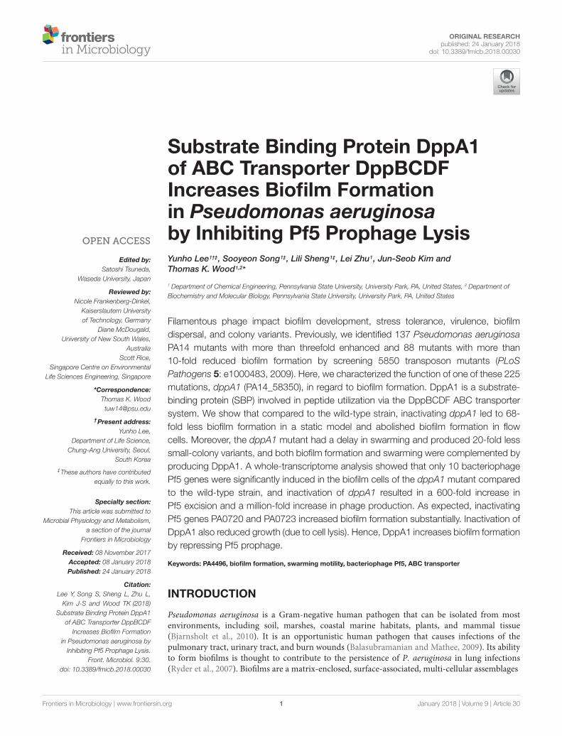
Substrate Binding Protein DppA1of ABC Transporter DppBCDFIncreases Biofilm Formationin Pseudomonas aeruginosaby Inhibiting Pf5 Prophage LysisYunho Lee1†‡, Sooyeon Song1‡, Lili Sheng1‡, Lei Zhu1, Jun-Seob Kim andThomas K. Wood1,2*
1 Department of Chemical Engineering, Pennsylvania State University, University Park, PA, United States, 2 Department ofBiochemistry and Molecular Biology, Pennsylvania State University, University Park, PA, United States
Filamentous phage impact biofilm development, stress tolerance, virulence, biofilmdispersal, and colony variants. Previously, we identified 137 Pseudomonas aeruginosaPA14 mutants with more than threefold enhanced and 88 mutants with more than10-fold reduced biofilm formation by screening 5850 transposon mutants (PLoSPathogens 5: e1000483, 2009). Here, we characterized the function of one of these 225mutations, dppA1 (PA14_58350), in regard to biofilm formation. DppA1 is a substrate-binding protein (SBP) involved in peptide utilization via the DppBCDF ABC transportersystem. We show that compared to the wild-type strain, inactivating dppA1 led to 68-fold less biofilm formation in a static model and abolished biofilm formation in flowcells. Moreover, the dppA1 mutant had a delay in swarming and produced 20-fold lesssmall-colony variants, and both biofilm formation and swarming were complemented byproducing DppA1. A whole-transcriptome analysis showed that only 10 bacteriophagePf5 genes were significantly induced in the biofilm cells of the dppA1 mutant comparedto the wild-type strain, and inactivation of dppA1 resulted in a 600-fold increase inPf5 excision and a million-fold increase in phage production. As expected, inactivatingPf5 genes PA0720 and PA0723 increased biofilm formation substantially. Inactivation ofDppA1 also reduced growth (due to cell lysis). Hence, DppA1 increases biofilm formationby repressing Pf5 prophage.
Keywords: PA4496, biofilm formation, swarming motility, bacteriophage Pf5, ABC transporter
INTRODUCTION
Pseudomonas aeruginosa is a Gram-negative human pathogen that can be isolated from mostenvironments, including soil, marshes, coastal marine habitats, plants, and mammal tissue(Bjarnsholt et al., 2010). It is an opportunistic human pathogen that causes infections of thepulmonary tract, urinary tract, and burn wounds (Balasubramanian and Mathee, 2009). Its abilityto form biofilms is thought to contribute to the persistence of P. aeruginosa in lung infections(Ryder et al., 2007). Biofilms are a matrix-enclosed, surface-associated, multi-cellular assemblages
Frontiers in Microbiology | www.frontiersin.org 1 January 2018 | Volume 9 | Article 30
fmicb-09-00030 January 23, 2018 Time: 16:12 # 2
Lee et al. DppA1 Reduces Pf5 Prophage Lysis
that protect cells in hostile environments (Friedman and Kolter,2004) such as phagocytosis (Hoiby et al., 2010). Biofilms areformed through the production of extracellular matrix composedof water, exopolysaccharides (EPS), nucleic acids, proteins, andlipids (Flemming and Wingender, 2010).
Pseudomonas aeruginosa is one of the most-studied microbeswith respect to the genetic regulation taking place throughoutthe process of biofilm formation (Petrova and Sauer, 2009;Martínez-Gil et al., 2010); however, this regulation is notcompletely elucidated. For example, the genetic mechanismfor matrix formation has not been fully elucidated (Franklinet al., 2011), and the complex role of the matrix is onlybeginning to be understood; for example, the interactionbetween the Pel polymer and extracellular DNA in P. aeruginosa(Jennings et al., 2015). The molecular mechanisms thatregulate biofilm formation differ between strains, while somefeatures are conserved in biofilm formation such as theformation of extracellular matrix (López et al., 2010). Theformation and maintenance of P. aeruginosa PA14 (henceforthPA14) biofilm critically depends upon the production of twodistinct EPS, alginate and Pel (Friedman and Kolter, 2004).Pel is a polymer of N-acetyl-β-D-glucosamine (GlcNAc)and N-acetyl-β-D-galactosamine (GalNAc) (Jennings et al.,2015), and pel mutations dramatically decrease biofilmformation as well as the pellicle that forms at the air-liquidinterface (Friedman and Kolter, 2004). The regulation ofthe pel operon involves tyrosine phosphatase TpbA (Uedaand Wood, 2009) and the Las quorum sensing system(Sakuragi and Kolter, 2007).
In many Gram-negative bacteria, ATP binding cassette (ABC)transporters are used to transport substrates across the innerand outer membrane (Chung et al., 2009) using the energyderived from the hydrolysis of ATP (Moussatova et al., 2008).Transport systems including ABC transporters often recognizediverse substrates, such as ions, amino acids, and antibiotics(Moussatova et al., 2008), as well as materials required forbiofilm formation (O’May et al., 2009; Iwai et al., 2010). InP. aeruginosa, ABC transport system DppBCDF is responsiblefor the uptake of dipeptides and tripeptides, which areused as nitrogen sources. The specificity of DppBCDF iscontrolled by its 5 substrate-binding proteins (SBPs) DppA1-A5 (PA14_58350, PA14_58360, PA14_58390, PA14_58420, andPA14_70200 in P. aeruginosa PA14, respectively), which functionin the periplasmic space (Pletzer et al., 2014). ABC transportersfunction in DNA replication, protein degradation, membranefusion, antibiotic efflux, signal transduction, and chemotaxis(Mai-Prochnow et al., 2015).
Filamentous bacteriophage such as f1, fd, and M13 ofEscherichia coli are lysogens that, upon excision, are packagedas single-stranded phages that have a long filamentous shape;in Pseudomonas spp. they are designated as Pf phage (Pf1to Pf6) (Mai-Prochnow et al., 2015). PA14 contains only Pf5which is most closely related to Pf4 of P. aeruginosa PAO1(63% homology) and somewhat related to Pf1 (Mooij et al.,2007). Specifically, Pf5 of PA14 is missing five genes of Pf4 ofPAO1 (PA0715, PA0716, PA0728, and PA0729), and has threeadditional genes (PA14_49030, PA14_49010, and PA14_ 49020)
(Mooij et al., 2007). Pf5 is thought not lead to phage particles(Mooij et al., 2007).
In P. aeruginosa biofilms, Pf1-related genes are highlyexpressed (up to 84-fold) suggesting that phage inductionmight be important for gene transfer within biofilms (Whiteleyet al., 2001) and may be important as a stress response(Hui et al., 2014). Furthermore, there is a repeatable patternof cell death and lysis in biofilms (Webb et al., 2003).Subsequently, Pf4 prophage were found to be essential forautolysis (programmed cell death) in P. aeruginosa PAO1 asdeletion of the prophage prevented cells from undergoing celldeath and from developing hollow regions in biofilms (Riceet al., 2009). Filamentous phage may also be secreted bythe host without lysis (Hui et al., 2014). In P. aeruginosa,filamentous phage may also lead to the emergence of small-colony variants that might function in multicellular biofilmdevelopment (Webb et al., 2004). However, others have foundthat the prophage of PA14 and many clinical isolates do notaffect small-colony variants which arise in biofilms (Mooij et al.,2007). In addition, Pf4 prophage facilitate biofilm formationby promoting the formation of liquid crystals of phage andP. aeruginosa PAO1 matrix polymers (Secor et al., 2015). Hence,filamentous bacteriophage are important for P. aeruginosabiofilm formation and may be involved in the production ofsmall-colony variants.
Our goal here was to characterize the manner in which theSBP DppA1 (PA14_58350 in PA14 and PA4496 in P. aeruginosaPAO1), promotes biofilm formation in P. aeruginosa; thisprotein was identified via screen of 5850 transposon mutantsthat affected biofilm formation (Ueda and Wood, 2009). Wefound that inactivation of dppA1 abolished biofilm formationin flow devices and delayed swarming. Using a whole-transcriptome analysis, we also determined that deletion ofdppA1 induced bacteriophage Pf5 genes during biofilm formationand led to a million-fold increase in lytic phage particles.Hence, DppA1 regulates Pf5 prophage to reduce biofilmformation.
MATERIALS AND METHODS
Bacterial Strains, Plasmids, Primers, andGrowth ConditionsStrains and plasmids used in this study are listed in Table 1.PA14 and its isogenic mutants were obtained from the HarvardMedical School (Liberati et al., 2006). Transposon insertion ofthe dppA1 mutant was verified using a two-round PCR protocol1.Briefly, a PCR product was amplified from chromosomal DNAusing primer pair GB 3a and Arb1D (Table 2); the product ofthis reaction was used as a template for the second reaction withprimer pair GB 2a and Arb2A which was sequenced with twoprimers GB 2a and GB 4a (Table 2). The transposon insertionsin dppA2, dppA3, dppA4, dppA5, and dppC were verified usingflanking PCR with primer pairs Gb 4a/dppA2 R, Gb 4a/dppA3R, dppA4 F/dppA4 R, Gb 4a/dppA5 R, and Gb 4a/dppC R,
1http://ausubellab.mgh.harvard.edu/cgi-bin/pa14/productionmethods.cgi
Frontiers in Microbiology | www.frontiersin.org 2 January 2018 | Volume 9 | Article 30
fmicb-09-00030 January 23, 2018 Time: 16:12 # 3
Lee et al. DppA1 Reduces Pf5 Prophage Lysis
TABLE 1 | Strains and plasmids used in this study.
Strain and plasmids Genotype or description Reference
P. aeruginosa
PA14 Wild-type strain Liberati et al., 2006
PA14_58350 (PA4496, dppA1) PA14_58350 � Mar2xT7, GmR Liberati et al., 2006
PA14_58360 (PA4497, dppA2) PA14_58360 � Mar2xT7, GmR Liberati et al., 2006
PA14_58390 (PA4500, dppA3) PA14_58390 � Mar2xT7, GmR Liberati et al., 2006
PA14_58420 (PA4502, dppA4) PA14_58420 � Mar2xT7, GmR Liberati et al., 2006
PA14_70200 (PA5317, dppA5) PA14_70200 � Mar2xT7, GmR Liberati et al., 2006
PA14_58450 (PA4504, dppC) PA14_58450 � Mar2xT7, GmR Liberati et al., 2006
PA14_13660 (PA3885, tpbA) PA14_13660 � Mar2xT7, GmR Liberati et al., 2006
PA14_24480 (PA3064, pelA) PA14_24480 � Mar2xT7, GmR Liberati et al., 2006
PA14_19120 (PA3477, rhlR) PA14_19120 � Mar2xT7, GmR Liberati et al., 2006
PA14_33700 (PA2396, pvdF ) PA14_33700 � Mar2xT7, GmR Liberati et al., 2006
PA14_49000 (PA0717) PA14_49000 � Mar2xT7, GmR Liberati et al., 2006
PA14_48990 (PA0718) PA14_48990 � Mar2xT7, GmR Liberati et al., 2006
PA14_48970 (PA0720) PA14_48970 � Mar2xT7, GmR Liberati et al., 2006
PA14_48940 (PA0723) PA14_48940 � Mar2xT7, GmR Liberati et al., 2006
PA14_48920 (PA0725) PA14_48920 � Mar2xT7, GmR Liberati et al., 2006
PA14_48890 (PA0727) PA14_48890 � Mar2xT7, GmR Liberati et al., 2006
E. coli
HB101/pRK2013 pro leu thi lacY StrR endol− recA− r− m− with pRK2013, KmR Ueda and Wood, 2009
TG1 K12, lac–pro supE thi hsdD5 (F’ traD36 proA+B+ lacIq lacZ M15 Ueda and Wood, 2009
Plasmids
pMQ70 CarR, ApR, PBAD, expression vector Shanks et al., 2006
pMQ-dppA1 CarR, ApR, PBAD:: dppA1, complementation vector This study
GmR and CarR indicate gentamicin, and carbenicillin resistance, respectively.
Table 2). P. aeruginosa and E. coli were routinely grown inLuria–Bertani (LB) medium at 37◦C with aeration (250 rpm).Gentamicin (15 µg/mL) was used for growth of the P. aeruginosatransposon mutants, and carbenicillin (300 µg/mL) was usedto maintain pMQ70 vector (Shanks et al., 2006). The specificgrowth rate was calculated using turbidity at 600 nm from 0.05to 0.7.
Complementation of dppA1To complement the dppA1 mutation, DppA1 was produced usingthe pBAD promoter in pMQ70 (Shanks et al., 2006). dppA1was amplified using Pfu DNA polymerase with primers DppA1-OE1 and DppA1-OE2 (Table 2). The PCR product was clonedinto the NheI and HindIII cloning sites of pMQ70, creatingpMQ-dppA1. The plasmid was confirmed by sequencing andtransformed by conjugation (Ueda and Wood, 2009) using helperstrain HB101/pRK2013.
Biofilm AssayBiofilm formation was examined in quiescent culturesusing 96-well polystyrene plates and crystal violet staining(Ueda and Wood, 2009). Overnight-grown cultures ofP. aeruginosa were diluted to a turbidity of 0.05 at 600 nmwith fresh LB medium, then 300 µL of diluted bacterialculture were incubated in 96-well polystyrene plates for 4,8, and 24 h. Six wells were used for each strain, and twoindependent cultures were used for each experiment. The
tpbA and pelA mutants were used as a positive controland a negative control, respectively (Ueda and Wood,2009).
Biofilm formation in flow cells was examined by dilutingovernight cultures to a turbidity of 0.05 in 5% LB mediumand pumping this culture through the flow-cell (BST modelFC81, Biosurface Technologies, Bozeman, MT, United States)at 10 mL/h for 2 h (Ueda and Wood, 2010). Fresh 5% LBmedium was added for 72 h, then the biofilms were stainedwith SYTO9 dye (5 µL/mL) for 20 min and were visualizedusing a TCS SP5 scanning confocal laser microscope (LeicaMicrosystems, Wetzlar, Germany). Nine random positionswere chosen, and 25 images were taken for each position.Simulated three-dimensional images were obtained usingIMARIS software (BITplane, Zurich, Switzerland), and thebiomass, surface coverage, mean thickness, and roughnesscoefficient were determined using COMSTAT image-processingsoftware (Heydorn et al., 2000).
Small-Colony VariantsSmall-colony variants were examined as described previously(Mooij et al., 2007). Approximately 2.5 × 10⁸ bacteriafrom overnight cultures were inoculated into glass tubescontaining 5 mL of fresh LB medium and were incubatedunder static conditions for 2 to 5 days. Cells were removedfrom the tubes vortexed vigorously followed by gentlesonication (3W, 15 s) of 1 mL samples to dissolve bacterial
Frontiers in Microbiology | www.frontiersin.org 3 January 2018 | Volume 9 | Article 30
fmicb-09-00030 January 23, 2018 Time: 16:12 # 4
Lee et al. DppA1 Reduces Pf5 Prophage Lysis
TABLE 2 | Primers used for DNA sequencing, complementation, phage excision,and quantitative real-time, reverse-transcription PCR (qRT-PCR) in this study.
Primer name Primer sequence (listed 5′–3′)
DNA sequencing
GB 2a TGTCAACTGGGTTCGTGCCTTCATCCG
GB 3a TACAGTTTACGAACCGAACAGGC
GB 4a GACCGAGATAGGGTTGAGTG
Arb1D GGCCAGGCCTGCAGATGATGNNNNNNNNNNGTAT
Arb2A GGCCAGGCCTGCAGATGATG
dppA2 R ATCTGCTCGTGGAAGATTCG
dppA3 R GTAGCGGCTCAGGATGAAAG
dppA4 F AACCTCGCTCTGGACAAGAA
dppA4 R AACGGATTGATCACGTAGCC
dppA5 R CAGATGTTCAGCTCGGGAAG
dppC R CATCAGGTTGATCGCCAGTA
Complementaion
DppA1-OE1 GCCCCCGCTAGCAAGAAGGAGATATACCATGCGTAGAAACGCCGTCATCCGC
DppA1-OE2 GCCCCCAAGCTTCTAGTGGTGGTGGTGGTGGTGCTTGCCGACGCTGACCTCGTAGAACG
Page excision
Pf5-F CAGGTAGAGCAGGATGTCG
Pf5-R GCGTTGAACAGGAGGAAATGG
Pf5RF-F AACAGTGAATTGCGGACAAGG
Pf5RF-R ACGGTGGAAACATCCTGGC
Pf5-q-F2 GGTGCTCTGGAATCCGGGTGTTC
Pf5-q-R2 GTGGTCGAGGTCGGGAGTCATGG
rplU-F AGGTTACCGCTGAAGTGGTTT
rplU-R CCGGTGATCTTGATTTCAGTG
qRT-PCR
PA0718-RT1 AGTCCCTACTACCTGCACCAAAC
PA0718-RT2 CGTACACAACGTGCCAGTACTTC
PA0722-RT1 CAACAGGCCTACCTGATTCC
PA0722-RT2 GGCTTTACGAAGAAGTGACG
aggregates completely. The sonicated cultures were dilutedwith PBS and plated on Congo-Red agar plates, whichwere incubated for 24 h. Two independent cultures wereused for each strain along with three replicates for eachculture.
Prophage ExcisionExcision was detected by PCR and by qPCR. Total DNA wasextracted using the Ultra Clean Microbial DNA Isolation Kit(MO BIO Laboratories, Inc., Carlsbad, CA, United States). Forthe PCR method, excision was detected by amplifying the PA14genome using primers PF5-F and PF5-R (these primers onlygive a band when the prophage has excised) (Table 2) andby amplifying circular prophage using primers PF5RF-F andPF5RF-R (these primers only give a band when the prophagehas circularized) (Table 2 and Supplementary Figure S1). Forthe qPCR method, total DNA (80 ng) was used with the PF5-q-F2 and PF5-q-R2 primers (Table 2) and the ABI PowerSYBR R© Green RNA-to-CTTM 1-Step Kit (Applied Biosystems,
Carlsbad, CA, United States); these results were confirmed byusing the Step OnePlusTM Real-Time PCR System (AppliedBiosystems, Foster City, CA, United States). Two independentcultures were used for each strain along with four replicates foreach culture.
Plaque AssayThe plaques were quantified by using a modified top-layer agarapproach (Eisenstark, 1967). The bacterial lawns of the wild-type PA14 were prepared using early exponential-phase cells (108
cells/mL) in LB media. For the phage stock solution, PA14 andthe dppA1 mutant were washed with fresh LB media (to removeany phage), inoculated (1%) into 25 mL of fresh LB media, andincubated with shaking for 8 h. Supernatants from the PA14 andthe dppA1 mutant were harvested by centrifugation (7,000 rpmfor 15 min), diluted from 1/100 to 1/104, and mixed with 0.1 mLof wild-type bacteria (108 cells/mL) in 4 mL of soft agar. Thesoft agar was poured onto the agar surface of a warm base plate.Plaques were observed after incubating for 6, 12, and 24 h. Thisplaque assay was performed with six independent cultures foreach strain with three replicates for each culture.
Live/Dead AssayThe percentage of live cells were determined using theLIVE/DEAD BacLight Bacterial Viability Kits L7012 (MolecularProbes, Inc., Eugene, OR, United States). The samples wereharvested at the same turbidity at 600 nm after 3 to 5 h. Thefluorescence emission spectrum (excitation 470 nm, emission490 to 700 nm) of each cell suspension was measured using anInfinite M200 Pro plate reader (Tecan, Switzerland). The ratiowas calculated of the integrated intensity of the portion of eachspectrum from 510 to 540 nm (green, live cells) to that from620 to 650 nm (red, dead cells) for each bacterial suspension.Isopropanol-treated cells (30 min) were used as the dead cellnegative control.
Motility AssaySwarming motility was examined with BM-2 plates (62 mMpotassium phosphate, 2 mM MgSO4, 10 mM FeSO4, 0.1%Casamino acids, 0.4% glucose, and 0.5% Bacto agar) (Overhageet al., 2008) after growth for 8 and 16 h. Three plates were testedfor each culture, and two independent cultures were used. TherhlR mutant was used as a negative control (Köhler et al., 2000).
EPS AssayExopolysaccharides production was quantified via Congo redstaining (Ma and Wood, 2009). One mL of overnight-grownculture was washed with 1 mL of 1% tryptone (T-broth),resuspended in 1 mL T-broth, and sonicated three times at3 W for 10 s. Bacterial suspensions in T-broth (500 µL)were incubated with 40 µg/mL Congo red with vigorousshaking (250 rpm) for 2 h. At that time, the absorbanceof the supernatants was measured at 490 nm using aspectrophotometer. The tpbA and pelA mutants were used aspositive control and negative controls, respectively (Ueda andWood, 2009).
Frontiers in Microbiology | www.frontiersin.org 4 January 2018 | Volume 9 | Article 30
fmicb-09-00030 January 23, 2018 Time: 16:12 # 5
Lee et al. DppA1 Reduces Pf5 Prophage Lysis
Pyoverdine AssayProduction of pyoverdine was determined as describedpreviously (Ren et al., 2005). Overnight cultures in LB brothwere diluted to a turbidity of 0.05 at 600 nm in 25 mL ofminimal succinate medium (6 g/L K2HPO4, 3 g/L KH2PO4,1 g/L (NH4)2SO4, 0.2 g/L MgSO4·7H2O, and 9.15 g/L sodiumsuccinate), and were incubated with aeration (250 rpm) for 4, 6,and 8 h. The absorbance at 405 nm of the supernatants was usedfor quantifying pyoverdine production which was normalized bycell turbidity.
Pyocyanin AssayProduction of pyocyanin was quantified by the absorbance at520 nm in acidic solution (Essar et al., 1990) and was normalizedby cell turbidity. A 800 µL sample of culture grown in LB brothfor 24 h was extracted with 480 µL of chloroform and then re-extracted into 800 µL of 0.2 N HCl to give a pink to deep redsolution.
Whole-Transcriptome AnalysisThe P. aeruginosa genome array (Affymetrix, P/N 510596) wasused to investigate differential gene expression in biofilm cellsbetween wild-type PA14 and the dppA1 mutant. After incubatingin 250 mL LB medium with 10 g of glass wool for 7 h with aeration(250 rpm), biofilm cells were harvested from the glass wool,and the cell pellets were resuspended in RNAlater (Ambion Inc.,Austin, TX). Total RNA was isolated using the RNeasy Mini Kit(Qiagen Inc., Valencia, CA, United States) (Ren et al., 2004a) anda bead beater (Biospec, Bartlesville, OK, United States). cDNAsynthesis, fragmentation, and hybridizations were performedas described previously (González Barrios et al., 2006). Ifthe gene with the larger transcription rate did not have aconsistent transcription rate based on the 13 probe pairs (p-value less than 0.05), these genes were discarded. A genewas considered differentially expressed when the p-value forcomparing two chips was lower than 0.05 (to assure that thechange in gene expression was statistically significant and thatfalse positives arise less than 5%) and if their fold change ishigher than standard deviation for the whole genome (Renet al., 2004b) (1.8-fold). The expression data have been submittedto the NCBI Gene Expression Omnibus (GSE24638). TheSTRING online database was used to construct a protein-protein interaction network among the proteins (Szklarczyk et al.,2015).
Quantitative Real Time, ReverseTranscription-PCR (qRT-PCR)Quantitative real time, reverse transcription-PCR was performedusing the StepOnePlusTM Real-Time PCR System (AppliedBiosystems, Foster City, CA, United States). Expressionlevels were measured using total RNA isolated frombiofilm cells under the same conditions as the microarrayexperiment (independent samples were used). The primersfor qRT-PCR are listed in Table 2. The values werenormalized using a housekeeping gene rplU (Kuchma et al.,2007).
RESULTS
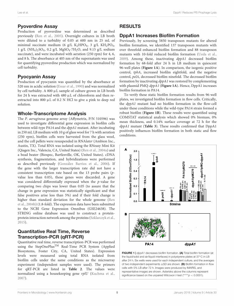
DppA1 Increases Biofilm FormationPreviously, by screening 5850 transposon mutants for alteredbiofilm formation, we identified 137 transposon mutants withover threefold enhanced biofilm formation and 88 transposonmutants with 10-fold reduced biofilm formation (Ueda et al.,2009). Among these, inactivating dppA1 decreased biofilmformation by 68-fold after 24 h in LB medium in quiescent96-well plates (Figure 1A). In comparison, the isogenic positivecontrol, tpbA, increased biofilm eightfold, and the negativecontrol, pelA, decreased biofilm ninefold. The decreased biofilmformation by inactivating dppA1 was restored by complementingwith plasmid PMQ-dppA1 (Figure 1A). Hence, DppA1 increasesbiofilm formation in PA14.
To verify these static biofilm formation results from 96-wellplates, we investigated biofilm formation in flow cells. Critically,the dppA1 mutant had no biofilm formation in the flow-cellunder these conditions while the wild-type PA14 strain formed arobust biofilm (Figure 1B). These results were quantified usingCOMSTAT statistical analysis which showed 0% biomass, 0%mean thickness, and 0.14% surface coverage at 72 h for thedppA1 mutant (Table 3). These results confirmed that DppA1positively influences biofilm formation in both static and flowconditions.
FIGURE 1 | dppA1 decreases biofilm formation. (A) Total biofilm formation (atthe liquid/solid and air/liquid interfaces) in polystyrene plates at 37◦C in LBafter 24 h. Six wells were used for each independent culture, and the averagesof two independent experiments ±SD are shown. (B) Biofilm formation in flowcells with 5% LB after 72 h. Images were produced by IMARIS, andrepresentative images are shown. Asterisks above the columns representsignificance based on the unpaired Wilcoxon t-test (∗∗∗∗p < 0.0001).
Frontiers in Microbiology | www.frontiersin.org 5 January 2018 | Volume 9 | Article 30
fmicb-09-00030 January 23, 2018 Time: 16:12 # 6
Lee et al. DppA1 Reduces Pf5 Prophage Lysis
TABLE 3 | COMSTAT analysis for biofilms of PA14 and the dppA1 mutant in flowcells.
COMSTAT values PA14 dppA1
Biomass (µm3/µm2) 7 ± 4 0 ± 0
Surface coverage (%) 7 ± 4 0.14 ± 0.02
Mean thickness (µm) 14 ± 9 0 ± 0
Roughness coefficient 1.4 ± 0.2 2 ± 0
Biofilms were evaluated after 72 h of development in 5% LB medium at 37◦C.
We also investigated whether the effect on biofilm formationwas specific for DppA1 since there are four other substratebinding proteins for dipeptides associated with ABC transporterDppBCDF. We found that the mutations in the other substratebinding protein dppA2, dppA3, dppA4, and dppA5 have noeffect on biofilm formation, i.e., these mutants have thesame biofilm level as the wild-type strain. Furthermore, wefound that there was no effect on biofilm formation uponmutating the permease DppC. Therefore, the dramaticbiofilm reduction phenotype is specific for inactivatingDppA1.
Small-Colony VariantsSince DppA1 increases biofilm formation dramatically, we alsoinvestigated whether DppA1 increases the formation of small-colony variants. Inactivating dppA1 reduces the production ofsmall-colony variants by 20-fold (40% for wild-type vs. 2% forthe dppA1 mutant, images of colonies not shown). Productionof DppA1 from pMQ-dppA1 increased the number of small-colony variants to about half that of the wild-type strain; hence,
the large decrease in small-colony variants could be partiallycomplemented.
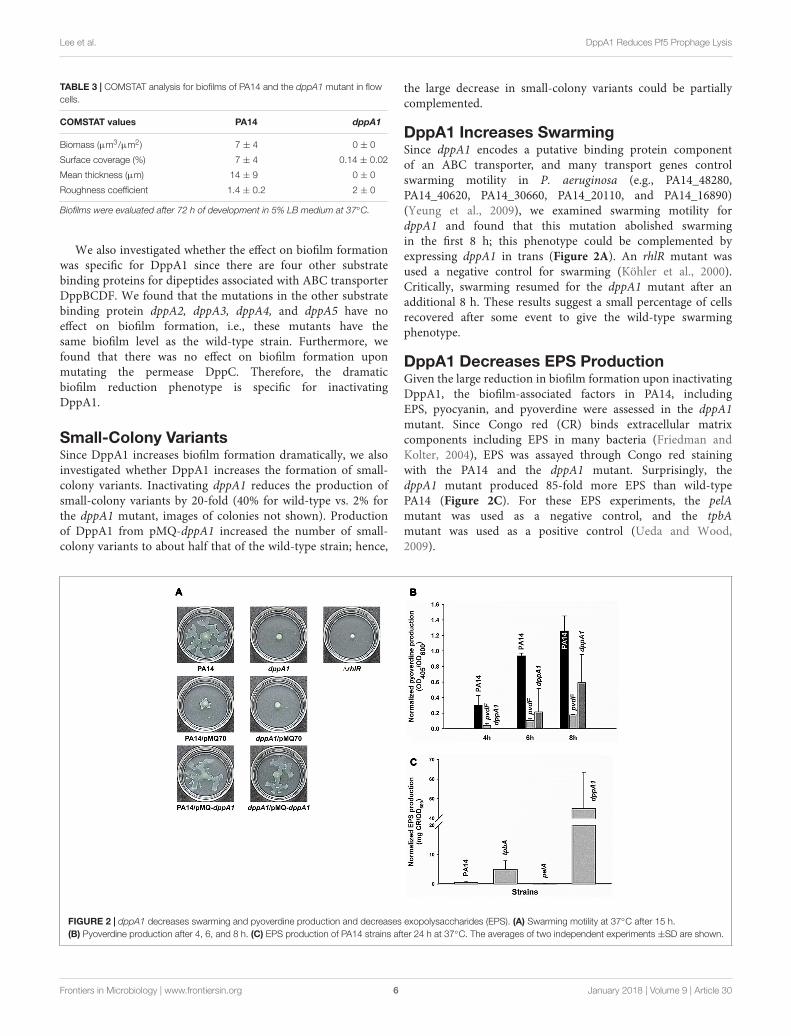
DppA1 Increases SwarmingSince dppA1 encodes a putative binding protein componentof an ABC transporter, and many transport genes controlswarming motility in P. aeruginosa (e.g., PA14_48280,PA14_40620, PA14_30660, PA14_20110, and PA14_16890)(Yeung et al., 2009), we examined swarming motility fordppA1 and found that this mutation abolished swarmingin the first 8 h; this phenotype could be complemented byexpressing dppA1 in trans (Figure 2A). An rhlR mutant wasused a negative control for swarming (Köhler et al., 2000).Critically, swarming resumed for the dppA1 mutant after anadditional 8 h. These results suggest a small percentage of cellsrecovered after some event to give the wild-type swarmingphenotype.
DppA1 Decreases EPS ProductionGiven the large reduction in biofilm formation upon inactivatingDppA1, the biofilm-associated factors in PA14, includingEPS, pyocyanin, and pyoverdine were assessed in the dppA1mutant. Since Congo red (CR) binds extracellular matrixcomponents including EPS in many bacteria (Friedman andKolter, 2004), EPS was assayed through Congo red stainingwith the PA14 and the dppA1 mutant. Surprisingly, thedppA1 mutant produced 85-fold more EPS than wild-typePA14 (Figure 2C). For these EPS experiments, the pelAmutant was used as a negative control, and the tpbAmutant was used as a positive control (Ueda and Wood,2009).
FIGURE 2 | dppA1 decreases swarming and pyoverdine production and decreases exopolysaccharides (EPS). (A) Swarming motility at 37◦C after 15 h.(B) Pyoverdine production after 4, 6, and 8 h. (C) EPS production of PA14 strains after 24 h at 37◦C. The averages of two independent experiments ±SD are shown.
Frontiers in Microbiology | www.frontiersin.org 6 January 2018 | Volume 9 | Article 30

fmicb-09-00030 January 23, 2018 Time: 16:12 # 7
Lee et al. DppA1 Reduces Pf5 Prophage Lysis
Moreover, since pyocyanin is a redox-active pigment secretedby P. aeruginosa that affects the structure of biofilms (Dietrichet al., 2008), and iron serves as a signal in P. aeruginosabiofilm development through pyoverdine iron acquisition(Banin et al., 2005), we investigated whether pyocyanin andpyoverdine production is altered by in the dppA1 mutant.The phzM/phzS (Mavrodi et al., 2001) and pvdF mutants(Banin et al., 2005) were used as negative controls forpyocyanin and pyoverdine production, respectively. We foundthere was no difference in pyocyanin production betweenPA14 and the dppA1 mutant LB medium at 10 h and24 h (data not shown), while the dppA1 mutant decreasedpyoverdine production twofold compared to PA14 at 8 h(Figure 2B).
DppA1 Represses Bacteriophage Pf5 inBiofilm CellsTo determine how the dppA1 mutation affects biofilm formation,a whole-transcriptome analysis was performed to comparegene expression in early biofilm formation (7 h) for thedppA1 mutant relative to the wild-type (grown on glass wool).Surprisingly, since dppA1 is not near the Pf5 locus, thewhole-transcriptome data showed that inactivation of dppA1significantly induced 10 bacteriophage Pf5 genes (Table 4). Toconfirm the induction of the prophage Pf5 genes, expressionof PA0718 and PA0722 were determined by qRT-PCR. Usingtotal RNA isolated from biofilm cells of PA14 and dppA1, wefound that PA0718 and PA0722 were induced 30 ± 4-foldand 41 ± 4-fold in the dppA1 mutant compared to PA14,respectively.
Based on these results; i.e., that inactivation of dppA1induced bacteriophage Pf5 and reduced biofilm formation,we hypothesized that deletion of individual bacteriophagePf5 genes should increase biofilm formation. In agreementwith this hypothesis, we found that the PA0723 andPA0725 mutants exhibited 4 ± 1- and 3 ± 1-fold increasein biofilm formation in LB medium at 4 h, respectively(Figure 3). Therefore, dppA1 represses, either directly orindirectly, expression of Pf5 genes which results in increasedbiofilm.
FIGURE 3 | dppA1 influences biofilm formation through bacteriophage Pf5.Relative biofilm formation in polystyrene plates at 37◦C in LB after 4 and 8 hdue to inactivating bacteriophage Pf5 genes. Six wells were used for eachculture. The averages of two independent experiments ±SD are shown.Asterisks above the columns represent significance based on the unpairedWilcoxon t-test relative to PA14 for each time (∗∗∗∗p < 0.0001, ∗∗∗p < 0.001,∗∗p < 0.01, and ∗p < 0.05).
DppA1 Decreases Pf5 ExcisionSince the dppA1 deletion increases transcription of the Pf5phage locus, we checked for prophage excision in this strainrelative to the wild-type strain by quantifying the presence ofre-circularized prophage via PCR for a pair of primers thatonly give a PCR band if the circularized prophage is formed(Supplementary Figure S1). As expected, we found via PCRthat the dppA1 deletion led to a 10-fold increase in excised,circularized prophage compared to the wild-type strain as wellas a corresponding 10-fold increase in the chromosomal deletionarea.
To corroborate and better quantify these PCR results, weperformed qPCR with rplU as the housekeeping gene and foundthat the dppA1 deletion had 600 ± 1-fold greater Pf5 phageexcision from the chromosome compared to the wild-type strain(0.1% vs. 0.00017%) (Supplementary Table S1). Hence, DppA1
TABLE 4 | Summary of the differentially expressed biofilm genes related to bacteriophage Pf5 for the dppA1 mutant vs. wild-type PA14 in LB medium for 7 h at 37◦C.
PAO1 ID PA14 ID Gene name Fold change Description
PA0717 PA14_49000 7.0 Hypothetical protein
PA0718 PA14_48990 12.1 Hypothetical protein
PA0719 PA14_48980 7.5 Hypothetical protein
PA0720 PA14_48970 6.5 Single-stranded binding protein
PA0721 PA14_48960 10.6 ABC transporter permease
PA0722 PA14_48950 9.2 Hypothetical protein
PA0723 PA14_48940 coaB 2.8 Coat protein B
PA0725 PA14_48920 5.7 Head virion protein G6P
PA0726 PA14_48910 zot 3.2 Zona occludens toxin
PA0727 PA14_48890 2.1 Hypothetical protein
Raw data for the two DNA microarrays are available using GEO series accession number GSE24638.
Frontiers in Microbiology | www.frontiersin.org 7 January 2018 | Volume 9 | Article 30
fmicb-09-00030 January 23, 2018 Time: 16:12 # 8
Lee et al. DppA1 Reduces Pf5 Prophage Lysis
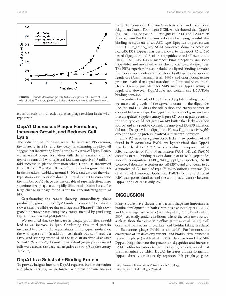
FIGURE 4 | dppA1 decreases growth. Cells were grown in LB broth at 37◦Cwith shaking. The averages of two independent experiments ±SD are shown.
either directly or indirectly represses phage excision in the wild-type strain.
DppA1 Decreases Plaque Formation,Increases Growth, and Reduces CellLysisThe induction of Pf5 phage genes, the increased Pf5 excision,the increase in EPS, and the delay in swarming motility, allsuggest that inactivating DppA1 results in active cell lysis. Hence,we measured plaque formation with the supernatants of thedppA1 mutant and wild-type and found an explosive 1.7 million-fold increase in plaque formation when DppA1 is inactivated(1.5± 0.3× 10⁸ vs. 0.9± 0.5× 10² pfu/mL) after growth for 8 hin rich medium (turbidity around 3). Note that we used the wild-type strain as is routinely done (Hui et al., 2014) to enumeratethe number of Pf5 phage that are capable of superinfection; thesesuperinfective phage arise rapidly (Rice et al., 2009); hence, thelarge change in phage found is for the superinfecting form ofPf5.
Corroborating the results showing extraordinary phageproduction, growth of the dppA1 mutant is initially dramaticallyslower than the wild-type due to phage lysis (Figure 4). This slow-growth phenotype was completely complemented by producingDppA1 from plasmid pMQ-dppA1.
We reasoned that the increase in plaque production shouldlead to an increase in lysis. Confirming this, total proteinincreased twofold in the supernatants of the dppA1 mutant vs.the wild-type strain. In addition, cell death was confirmed viaLive/Dead staining where all of the wild-strain were alive after5 h but 50% of the dppA1 mutant were dead (isopropanol-treatedcells were used as the dead cell negative control) (SupplementaryTable S2).
DppA1 Is a Substrate-Binding ProteinTo provide insights into how DppA1 regulates biofilm formationand phage excision, we performed a protein domain analysis
using the Conserved Domain Search Service2 and Basic LocalAlignment Search Tool3 from NCBI, which showed that DppA1(537 aa, PA14_58350 in P. aeruginosa PA14 and PA4496 inP. aeruginosa PAO1) contains a domain belonging to substrate-binding component of an ABC-type dipeptide import systemPBP2 (PBP2_DppA_like, NCBI conserved domains accessionno. cd08493). DppA1 has been shown to transport 72 of 246tested dipeptides and 3 of 14 tripeptides tested (Pletzer et al.,2014). The PBP2 family members bind dipeptides and sometripeptides and are involved in chemotaxis toward dipeptides.The PBP2 superfamily also includes the ligand-binding domainsfrom ionotropic glutamate receptors, LysR-type transcriptionalregulators (Anantharaman et al., 2001), and unorthodox sensorproteins involved in signal transduction (Tam and Saier, 1993).Hence, there is precedent for SBPs such as DppA1 acting asregulators. However, DppA1does not contain any DNA/RNAbinding domains.
To confirm the role of DppA1 as a dipeptide binding protein,we measured growth of the dppA1 mutant on the dipeptidesPhe-Pro and Gly-Glu as the sole carbon and energy sources. Incontrast to the wildtype, the dppA1 mutant cannot grow on thesetwo dipeptides (Supplementary Figure S2). As a negative control,the wild-type could not grow on M9 buffer that lacks a carbonsource, and as a positive control, the unrelated PA4499 mutationdid not affect growth on dipeptides. Hence, DppA1 is a bona fidedipeptide binding protein involved in their transportation.
Since Pf5 in P. aeruginosa PA14 lacks a few proteins of Pf4found in P. aeruginosa PAO1, we hypothesized that DppA1may be related to PA0716, which is also a component of anABC transporter of Pf4 in P. aeruginosa PAO1 (441 aa); PA0176contains an ATP-binding cassette domain of nickel/oligopeptidesspecific transporters (ABC_NikE_OppD_transporters, NCBIconserved domains accession no. cd03257), and also seems to bea putative AbiEii toxin of type IV toxin/antitoxin systems (Dyet al., 2014). However, DppA1 and PA0716 belong to differentABC transporter families, and the amino acid identity betweenDppA1 and PA0716 is only 7%.
DISCUSSION
Many studies have shown that bacteriophage are important inbiofilm development in both Gram-positive (Stanley et al., 2003)and Gram-negative bacteria (Whiteley et al., 2001; Domka et al.,2007), especially under conditions where the cells are stressed,such as those that exist in biofilms (Domka et al., 2007). Celldeath and lysis occur in biofilms, and biofilm killing is relatedto filamentous phage (Webb et al., 2003). Furthermore, theemergence of small-colony variants and biofilm development isrelated to phage (Webb et al., 2004). Here we found that SBPDppA1 helps facilitate the growth on dipeptides and increasesPA14 biofilm formation 68-fold. Critically, we determined thatthe mechanism by which DppA1 increases biofilm formation:DppA1 directly or indirectly represses Pf5 prophage genes
2https://www.ncbi.nlm.nih.gov/Structure/cdd/wrpsb.cgi3https://blast.ncbi.nlm.nih.gov/Blast.cgi
Frontiers in Microbiology | www.frontiersin.org 8 January 2018 | Volume 9 | Article 30
fmicb-09-00030 January 23, 2018 Time: 16:12 # 9
Lee et al. DppA1 Reduces Pf5 Prophage Lysis
which prevents cell lysis and a million-fold production of activephage.
Although the importance of Pf phage in P. aeruginosabiofilm formation is well-documented, such as the formation ofextracellular liquid crystals by Pf4 with alginate and extracellularDNA (Secor et al., 2015) and the hyper-mutability of Pf4 duringbiofilm evolution (McElroy et al., 2014), our results shed light onhow this phage is regulated, through the unexpected mechanismof a SBP DppA1. Clearly DppA1 helps to limit phage lysis andthereby increases biofilm production by reducing excess phageproduction; this appears to be at least part of the unknownmechanism by which Pf phage generate diversity in biofilms thatwas alluded to previously (Hui et al., 2014).
Furthermore, the mechanism we found here for DppA1 forcontrolling phage production and biofilm formation is distinct.For example, in contrast to our results where excessive phageproduction is deleterious for biofilm formation, previous workhas shown that explosive cell lysis via an endolysin from crypticprophage is beneficial for biofilm formation since it producesextracellular DNA that helps to initiate biofilm production(Turnbull et al., 2016). Therefore, phage production and phage-related genes must be regulated well to control biofilm formation.
Further work is clearly required to determine the mechanismby which DppA1 represses Pf5 prophage genes, either directlyor indirectly, through dipeptide transport. However, it istempting to speculate that the dipeptides transported byDppA1 in the periplasm are relayed as a signal to the Pf5phage integrase/excisionase (PA14_48880) or some other phageregulator to limit phage lysis when nutrients are plentiful(extracellular proteins are readily degraded by the extracellularproteases of PA14). In contrast, when nutrients are depleted, the
dipeptides transported by DppA1 are reduced, and Pf5 phage areproduced to drive cell evolution and to create voids in biofilmsto facilitate dispersal (inactivation of dppA1 would mimic a nopeptide condition). Such biofilm voids and dispersal have beenshown to be dependent on the related phage, Pf4 (Rice et al.,2009), and biofilm dispersal has long been known to be governedby changes in nutrient levels (Sauer et al., 2004). Therefore,the dipeptide concentrations in the cells, governed by DppA1transport, may serve as a simple indicator of external nutrientlevels and control biofilm development.
AUTHOR CONTRIBUTIONS
TW conceived the project. YL, SS, LS, LZ, and J-SK conductedthe experiments. YL, SS, and LS analyzed the data. YL, SS, andTW co-wrote the manuscript.
ACKNOWLEDGMENTS
This research was supported by the NIH (R01 GM089999) andfunds derived from the Biotechnology Endowed Professorship atthe Pennsylvania State University.
SUPPLEMENTARY MATERIAL
The Supplementary Material for this article can be foundonline at: https://www.frontiersin.org/articles/10.3389/fmicb.2018.00030/full#supplementary-material
REFERENCESAnantharaman, V., Koonin, E. V., and Aravind, L. (2001). Regulatory potential,
phyletic distribution and evolution of ancient, intracellular small-molecule-binding domains11Edited by F. Cohen. J. Mol. Biol. 307, 1271–1292. doi: 10.1006/jmbi.2001.4508
Balasubramanian, D., and Mathee, K. (2009). Comparative transcriptome analysesof Pseudomonas aeruginosa. Hum. Genomics 3, 349–361. doi: 10.1186/1479-7364-3-4-361
Banin, E., Vasil, M. L., and Greenberg, E. P. (2005). Iron and Pseudomonasaeruginosa biofilm formation. Proc. Natl. Acad. Sci. U.S.A. 102, 11076–11081.doi: 10.1073/pnas.0504266102
Bjarnsholt, T., Tolker-Nielsen, T., Hoiby, N., and Givskov, M. (2010).Interference of Pseudomonas aeruginosa signalling and biofilm formation forinfection control. Expert Rev. Mol. Med. 12:e11. doi: 10.1017/S1462399410001420
Chung, C. W., You, J., Kim, K., Moon, Y., Kim, H., and Ahn, J. H. (2009). Exportof recombinant proteins in Escherichia coli using ABC transporter with anattached lipase ABC transporter recognition domain (LARD). Microb. Cell Fact.8:11. doi: 10.1186/1475-2859-8-11
Dietrich, L. E., Teal, T. K., Price-Whelan, A., and Newman, D. K. (2008). Redox-active antibiotics control gene expression and community behavior in divergentbacteria. Science 321, 1203–1206. doi: 10.1126/science.1160619
Domka, J., Lee, J., Bansal, T., and Wood, T. K. (2007). Temporal gene-expressionin Escherichia coli K-12 biofilms. Environ. Microbiol. 9, 332–346. doi: 10.1111/j.1462-2920.2006.01143.x
Dy, R. L., Przybilski, R., Semeijn, K., Salmond, G. P. C., and Fineran, P. C. (2014).A widespread bacteriophage abortive infection system functions through a Type
IV toxin–antitoxin mechanism. Nucleic Acids Res. 42, 4590–4605. doi: 10.1093/nar/gkt1419
Eisenstark, A. (1967). Bacteriophage Techniques: Methods in Virology. New York,NY: Academic Press.
Essar, D. W., Eberly, L., Hadero, A., and Crawford, I. P. (1990). Identificationand characterization of genes for a second anthranilate synthase inPseudomonas aeruginosa: interchangeability of the two anthranilate synthasesand evolutionary implications. J. Bacteriol. 172, 884–900. doi: 10.1128/jb.172.2.884-900.1990
Flemming, H. C., and Wingender, J. (2010). The biofilm matrix. Nat. Rev. Microbiol.8, 623–633. doi: 10.1038/nrmicro2415
Franklin, M., Nivens, D., Weadge, J., and Howell, P. (2011). Biosynthesis of thePseudomonas aeruginosa extracellular polysaccharides, Alginate, Pel, and Psl.Front. Microbiol. 2:167. doi: 10.3389/fmicb.2011.00167
Friedman, L., and Kolter, R. (2004). Genes involved in matrix formation inPseudomonas aeruginosa PA14 biofilms. Mol. Microbiol. 51, 675–690. doi: 10.1046/j.1365-2958.2003.03877.x
González Barrios, A. F., Zuo, R., Hashimoto, Y., Yang, L., Bentley, W. E., and Wood,T. K. (2006). Autoinducer 2 controls biofilm formation in Escherichia colithrough a novel motility quorum-sensing regulator (MqsR, B3022). J. Bacteriol.188, 305–316. doi: 10.1128/JB.188.1.305-316.2006
Heydorn, A., Nielsen, A. T., Hentzer, M., Sternberg, C., Givskov, M., Ersboll,B. K., et al. (2000). Quantification of biofilm structures by the novel computerprogram COMSTAT. Microbiology 146(Pt 10), 2395–2407. doi: 10.1099/00221287-146-10-2395
Hoiby, N., Bjarnsholt, T., Givskov, M., Molin, S., and Ciofu, O. (2010). Antibioticresistance of bacterial biofilms. Int. J. Antimicrob. Agents 35, 322–332.doi: 10.1016/j.ijantimicag.2009.12.011
Frontiers in Microbiology | www.frontiersin.org 9 January 2018 | Volume 9 | Article 30
fmicb-09-00030 January 23, 2018 Time: 16:12 # 10
Lee et al. DppA1 Reduces Pf5 Prophage Lysis
Hui, J. G. K., Mai-Prochnow, A., Kjelleberg, S., McDougald, D., and Rice,S. A. (2014). Environmental cues and genes involved in establishment of thesuperinfective Pf4 phage of Pseudomonas aeruginosa. Front. Microbiol. 5:654.doi: 10.3389/fmicb.2014.00654
Iwai, S., Doi, K., Fujino, Y., Nakazono, T., Fukuda, K., Motomura, Y., et al. (2010).Silica deposition and phenotypic changes to Thermus thermophilus cultivatedin the presence of supersaturated silicia. ISME J. 4, 809–816. doi: 10.1038/ismej.2010.12
Jennings, L. K., Storek, K. M., Ledvina, H. E., Coulon, C., Marmont, L. S.,Sadovskaya, I., et al. (2015). Pel is a cationic exopolysaccharide that cross-links extracellular DNA in the Pseudomonas aeruginosa biofilm matrix.Proc. Natl. Acad. Sci. U.S.A. 112, 11353–11358. doi: 10.1073/pnas.1503058112
Köhler, T., Curty, L. K., Barja, F., van Delden, C., and Pechere, J. C.(2000). Swarming of Pseudomonas aeruginosa is dependent on cell-to-cell signaling and requires flagella and pili. J. Bacteriol. 182, 5990–5996.doi: 10.1128/JB.182.21.5990-5996.2000
Kuchma, S. L., Brothers, K. M., Merritt, J. H., Liberati, N. T., Ausubel, F. M.,and O’Toole, G. A. (2007). BifA, a cyclic-Di-GMP phosphodiesterase,inversely regulates biofilm formation and swarming motility byPseudomonas aeruginosa PA14. J. Bacteriol. 189, 8165–8178. doi: 10.1128/JB.00586-07
Liberati, N. T., Urbach, J. M., Miyata, S., Lee, D. G., Drenkard, E., Wu, G.,et al. (2006). An ordered, nonredundant library of Pseudomonas aeruginosastrain PA14 transposon insertion mutants. Proc. Natl. Acad. Sci. U.S.A. 103,2833–2838. doi: 10.1073/pnas.0511100103
López, D., Vlamakis, H., and Kolter, R. (2010). Biofilms. Cold Spring Harb. Perspect.Biol. 2:a000398. doi: 10.1101/cshperspect.a000398
Ma, Q., and Wood, T. K. (2009). OmpA influences Escherichia coli biofilmformation by repressing cellulose production through the CpxRA two-component system. Environ. Microbiol. 11, 2735–2746. doi: 10.1111/j.1462-2920.2009.02000.x
Mai-Prochnow, A., Hui, J. G. K., Kjelleberg, S., Rakonjac, J., McDougald, D., andRice, S. A. (2015). ‘Big things in small packages: the genetics of filamentousphage and effects on fitness of their host’. FEMS Microbiol. Rev. 39, 465–487.doi: 10.1093/femsre/fuu007
Martínez-Gil, M., Yousef-Coronado, F., and Espinosa-Urgel, M. (2010). LapF,the second largest Pseudomonas putida protein, contributes to plant rootcolonization and determines biofilm architecture. Mol. Microbiol. 77, 549–561.doi: 10.1111/j.1365-2958.2010.07249.x
Mavrodi, D. V., Bonsall, R. F., Delaney, S. M., Soule, M. J., Phillips, G., andThomashow, L. S. (2001). Functional analysis of genes for biosynthesis ofpyocyanin and phenazine-1-carboxamide from Pseudomonas aeruginosaPAO1. J. Bacteriol. 183, 6454–6465. doi: 10.1128/JB.183.21.6454-6465.2001
McElroy, K. E., Hui, J. G. K., Woo, J. K. K., Luk, A. W. S., Webb, J. S., Kjelleberg, S.,et al. (2014). Strain-specific parallel evolution drives short-term diversificationduring Pseudomonas aeruginosa biofilm formation. Proc. Natl. Acad. Sci. U.S.A.111, E1419–E1427. doi: 10.1073/pnas.1314340111
Mooij, M. J., Drenkard, E., Llamas, M. A., Vandenbroucke-Grauls, C. M. J. E.,Savelkoul, P. H. M., Ausubel, F. M., et al. (2007). Characterizationof the integrated filamentous phage Pf5 and its involvement in small-colony formation. Microbiology 153, 1790–1798. doi: 10.1099/mic.0.2006/003533-0
Moussatova, A., Kandt, C., O’Mara, M. L., and Tieleman, D. P. (2008). ATP-binding cassette transporters in Escherichia coli. Biochim. Biophys. Acta 1778,1757–1771. doi: 10.1016/j.bbamem.2008.06.009
O’May, G. A., Jacobsen, S. M., Longwell, M., Stoodley, P., Mobley, H. L., andShirtliff, M. E. (2009). The high-affinity phosphate transporter Pst in Proteusmirabilis HI4320 and its importance in biofilm formation. Microbiology 155,1523–1535. doi: 10.1099/mic.0.026500-0
Overhage, J., Bains, M., Brazas, M. D., and Hancock, R. E. W. (2008). Swarmingof Pseudomonas aeruginosa is a complex adaptation leading to increasedproduction of virulence factors and antibiotic resistance. J. Bacteriol. 190,2671–2679. doi: 10.1128/Jb.01659-07
Petrova, O. E., and Sauer, K. (2009). A novel signaling network essentialfor regulating Pseudomonas aeruginosa biofilm development. PLOS Pathog.5:e1000668. doi: 10.1371/journal.ppat.1000668
Pletzer, D., Lafon, C., Braun, Y., Köhler, T., Page, M. G. P., Mourez, M., et al.(2014). High-throughput screening of dipeptide utilization mediated by theABC transporter DppBCDF and its substrate-binding proteins DppA1-A5 inPseudomonas aeruginosa. PLOS ONE 9:e111311. doi: 10.1371/journal.pone.0111311
Ren, D., Bedzyk, L. A., Thomas, S. M., Ye, R. W., and Wood, T. K. (2004a). Geneexpression in Escherichia coli biofilms. Appl. Microbiol. Biotechnol. 64, 515–524.doi: 10.1007/s00253-003-1517-y
Ren, D., Bedzyk, L. A., Ye, R. W., Thomas, S. M., and Wood, T. K. (2004b).Differential gene expression shows natural brominated furanones interfere withthe autoinducer-2 bacterial signaling system of Escherichia coli. Biotechnol.Bioeng. 88, 630–642. doi: 10.1002/bit.20259
Ren, D., Zuo, R., and Wood, T. K. (2005). Quorum-sensing antagonist(5Z)-4-bromo-5-(bromomethylene)-3-butyl-2(5H)-furanone influencessiderophore biosynthesis in Pseudomonas putida and Pseudomonasaeruginosa. Appl. Microbiol. Biotechnol. 66, 689–695. doi: 10.1007/s00253-004-1691-6
Rice, S. A., Tan, C. H., Mikkelsen, P. J., Kung, V., Woo, J., Tay, M., et al.(2009). The biofilm life cycle and virulence of Pseudomonas aeruginosa aredependent on a filamentous prophage. ISME J. 3, 271–282. doi: 10.1038/ismej.2008.109
Ryder, C., Byrd, M., and Wozniak, D. J. (2007). Role of polysaccharides inPseudomonas aeruginosa biofilm development. Curr. Opin. Microbiol. 10,644–648. doi: 10.1016/j.mib.2007.09.010
Sakuragi, Y., and Kolter, R. (2007). Quorum-sensing regulation of the biofilmmatrix genes (pel) of Pseudomonas aeruginosa. J. Bacteriol. 189, 5383–5386.doi: 10.1128/JB.00137-07
Sauer, K., Cullen, M. C., Rickard, A. H., Zeef, L. A. H., Davies, D. G.,and Gilbert, P. (2004). Characterization of nutrient-induced dispersion inPseudomonas aeruginosa PAO1 biofilm. J. Bacteriol. 186, 7312–7326. doi: 10.1128/JB.186.21.7312-7326.2004
Secor, P. R., Sweere, Johanna, M., Michaels, L. A., Malkovskiy, A. V.,Lazzareschi, D., et al. (2015). Filamentous bacteriophage promote biofilmassembly and function. Cell Host Microbe 18, 549–559. doi: 10.1016/j.chom.2015.10.013
Shanks, R. M., Caiazza, N. C., Hinsa, S. M., Toutain, C. M., and O’Toole, G. A.(2006). Saccharomyces cerevisiae-based molecular tool kit for manipulation ofgenes from gram-negative bacteria. Appl. Environ. Microbiol. 72, 5027–5036.doi: 10.1128/AEM.00682-06
Stanley, N. R., Britton, R. A., Grossman, A. D., and Lazazzera, B. A. (2003).Identification of catabolite repression as a physiological regulator of biofilmformation by Bacillus subtilis by use of DNA microarrays. J. Bacteriol. 185,1951–1957. doi: 10.1128/JB.185.6.1951-1957.2003
Szklarczyk, D., Franceschini, A., Wyder, S., Forslund, K., Heller, D., Huerta-Cepas, J., et al. (2015). STRING v10: protein-protein interaction networks,integrated over the tree of life. Nucleic Acids Res. 43, D447–D452. doi: 10.1093/nar/gku1003
Tam, R., and Saier, M. H. (1993). Structural, functional, and evolutionaryrelationships among extracellular solute-binding receptors of bacteria.Microbiol. Rev. 57, 320–346.
Turnbull, L., Toyofuku, M., Hynen, A. L., Kurosawa, M., Pessi, G., Petty, N. K.,et al. (2016). Explosive cell lysis as a mechanism for the biogenesis ofbacterial membrane vesicles and biofilms. Nat. Commun. 7:11220. doi: 10.1038/ncomms11220
Ueda, A., Attila, C., Whiteley, M., and Wood, T. K. (2009). Uracil influencesquorum sensing and biofilm formation in Pseudomonas aeruginosa andfluorouracil is an antagonist. Microb. Biotechnol. 2, 62–74. doi: 10.1111/j.1751-7915.2008.00060.x
Ueda, A., and Wood, T. K. (2009). Connecting quorum sensing, c-di-GMP, pelpolysaccharide, and biofilm formation in Pseudomonas aeruginosa throughtyrosine phosphatase TpbA (PA3885). PLOS Pathog. 5:e1000483. doi: 10.1371/journal.ppat.1000483
Ueda, A., and Wood, T. K. (2010). Tyrosine phosphatase TpbA of Pseudomonasaeruginosa controls extracellular DNA via cyclic diguanylic acid concentrations.Environ. Microbiol. Rep. 2, 449–455. doi: 10.1111/j.1758-2229.2010.00171.x
Webb, J. S., Lau, M., and Kjelleberg, S. (2004). Bacteriophage and phenotypicvariation in Pseudomonas aeruginosa biofilm development. J. Bacteriol. 186,8066–8073. doi: 10.1128/JB.186.23.8066-8073.2004
Frontiers in Microbiology | www.frontiersin.org 10 January 2018 | Volume 9 | Article 30
fmicb-09-00030 January 23, 2018 Time: 16:12 # 11
Lee et al. DppA1 Reduces Pf5 Prophage Lysis
Webb, J. S., Thompson, L. S., James, S., Charlton, T., Tolker-Nielsen, T., Koch, B.,et al. (2003). Cell death in Pseudomonas aeruginosa biofilm development.J. Bacteriol. 185, 4585–4592. doi: 10.1128/JB.185.15.4585-4592.2003
Whiteley, M., Bangera, M. G., Bumgarner, R. E., Parsek, M. R., Teitzel, G. M.,Lory, S., et al. (2001). Gene expression in Pseudomonas aeruginosa biofilms.Nature 413, 860–864. doi: 10.1038/3510162735101627
Yeung, A. T., Torfs, E. C., Jamshidi, F., Bains, M., Wiegand, I., Hancock, R. E., et al.(2009). Swarming of Pseudomonas aeruginosa is controlled by a broad spectrumof transcriptional regulators, including MetR. J. Bacteriol. 191, 5592–5602.doi: 10.1128/JB.00157-09
Conflict of Interest Statement: The authors declare that the research wasconducted in the absence of any commercial or financial relationships that couldbe construed as a potential conflict of interest.
Copyright © 2018 Lee, Song, Sheng, Zhu, Kim and Wood. This is an open-accessarticle distributed under the terms of the Creative Commons Attribution License(CC BY). The use, distribution or reproduction in other forums is permitted, providedthe original author(s) or licensor are credited and that the original publication in thisjournal is cited, in accordance with accepted academic practice. No use, distributionor reproduction is permitted which does not comply with these terms.
Frontiers in Microbiology | www.frontiersin.org 11 January 2018 | Volume 9 | Article 30