Embed Size (px)
Citation preview
1
Evolution of substrate specificity for the bile salt transporter ABST (SLC10A2)
Daniël A. Lionarons*, James L. Boyer*† and Shi-Ying Cai*
*Department of Internal Medicine and Liver Center, Yale University School of Medicine,
New Haven, CT 06520; and †Mount Desert Island Biological Laboratory, Salisbury Cove,
ME 04672
Running footline: Asbt gained substrate specificity for bile salts in evolution
Address correspondence to: Shi-Ying Cai, Ph.D., Liver Center, Yale University School of
Medicine, 333 Cedar Street, 1080 LMP, New Haven, CT 06520. Tel: (203)785-3150; Fax:
(203)785-7273; E-mail: [email protected], or James L. Boyer, M.D. Ensign Professor of
Medicine, Emeritus Director, Liver Center, Yale University School of Medicine, 333 Cedar
Street, 1080 LMP, New Haven, CT 06520-8019. Phone: (203)785-5279; Fax: (203)785-
7273; email: [email protected]
Abbreviations used in this paper: ASBT/Asbt, apical sodium-dependent bile salt
transporter; FXR/Fxr, Farnesoid X receptor; hASBT, human ASBT; lpAsbt, lamprey Asbt;
skAsbt, skate Asbt; TCA, taurocholic acid.
by guest, on June 16, 2018w
ww
.jlr.orgD
ownloaded from
2
ABSTRACT
The apical Na+-dependent bile salt transporter (ASBT/SLC10A2) is essential for maintaining
the enterohepatic circulation of bile salts. It is not known when Slc10a2 evolved as a bile salt
transporter, or how it adapted to substantial changes in bile salt structure during evolution.
We characterized ASBT orthologs from two primitive vertebrates, the lamprey which utilizes
early 5α-bile alcohols; and the skate which utilizes structurally different 5β-bile alcohols; and
compared substrate specificity with ASBT from humans which utilize modern 5β-bile acids.
Everted gut sacs of skate but not the more primitive lamprey transported 3H-taurocholic acid
(TCA), a modern 5β-bile acid. However, molecular cloning identified ASBT orthologs from
both species. Cell-based assays using recombinant ASBT/Asbt’s indicate that lamprey Asbt
has high affinity for 5α-bile alcohols, low affinity for 5β-bile alcohols, and lacks affinity for
TCA; whereas skate Asbt showed high affinity for 5α- and 5β-bile alcohols, but low affinity
for TCA. In contrast, human ASBT demonstrated high affinity for all three bile salt types.
These findings suggest that ASBT evolved from the earliest vertebrates by gaining affinity for
modern bile salts while retaining affinity for older bile salts. Also, our results indicate that the
bile salt enterohepatic circulation is conserved throughout vertebrate evolution.
Supplementary Keywords: sodium-dependent transporter, enterohepatic circulation, bile
acids, bile alcohols, Isbt, taurocholic acid, lamprey, petromyzon marinus, skate, leucoraja
erinacea
by guest, on June 16, 2018w
ww
.jlr.orgD
ownloaded from
3
INTRODUCTION
Bile alcohols and bile acids are the end products of cholesterol metabolism. Most bile
alcohols and bile acids are conjugated with sulfate, taurine or glycine at the terminal carbon
of the side chain, and are isolated as bile salts(1). Bile salts play critical physiological roles in
vertebrates. They facilitate lipid absorption, inhibit microbe growth in the biliary tract and
intestine, and function as signaling molecules that regulate energy expenditure and
carbohydrate and lipid metabolism(2). The bile salt pool is maintained in an enterohepatic
circulation by bile salt transporters in the distal ileum and the liver(3). Central to this process
is the apical sodium (Na+)-dependent bile salt transporter (ASBT/SLC10A2), located on the
luminal membrane in the distal ileum and proximal tubule of the kidney in humans and
rodents(4). ASBT maintains the enterohepatic and renal-hepatic circulation of bile salts by
facilitating their re-absorption from the intestinal lumen and renal tubules. Dysfunction of
ASBT/Asbt interrupts the bile salt enterohepatic circulation, reduces the bile salt pool size by
80% in mice, and leads to bile salt malabsorption, diarrhea and steatorrhea in humans, where
reduced plasma levels of cholesterol are also observed(5;6). Thus there is considerable
pharmaceutical interest in ASBT inhibition as a potential target for drug discovery for the
treatment of hypercholesterolemia and diabetes mellitus type 2(7;8). Furthermore, an ASBT
inhibitor has been demonstrated to be beneficial for patients with chronic idiopathic
constipation(9;10). In contrast to what is known for mammalian species, very little is known
about the presence or function of Asbt in other vertebrates.
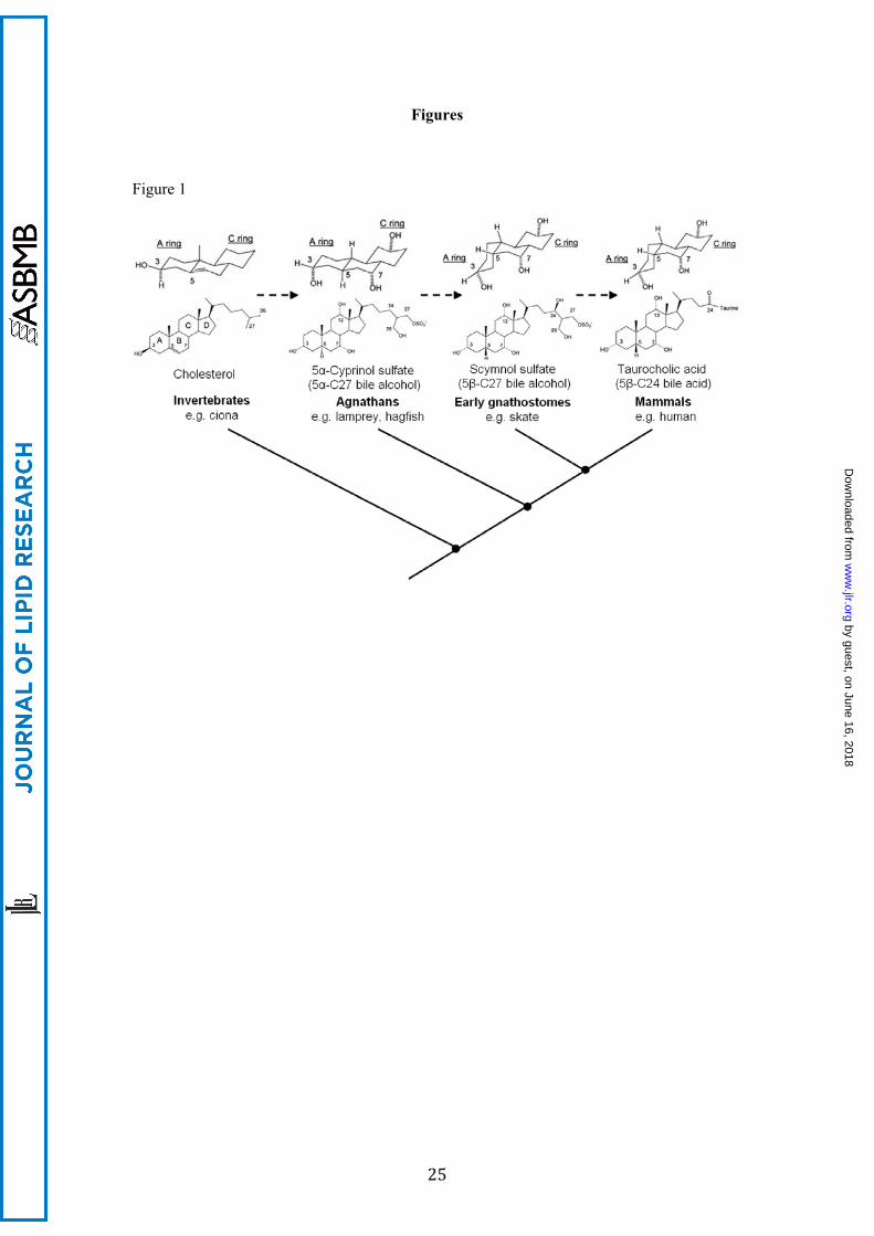
Bile salts demonstrate considerable structural variation within different vertebrate
phyla(1;11) (Fig. 1). The enzymatic pathway that converts cholesterol into bile salts is
complex and requires a minimum of 5 enzymes in primitive vertebrates and up to 16 enzymes
in humans(12-14). The most primitive vertebrates (agnathans or jawless fish) use early
evolving C27 sulfated bile alcohols with a C-5 hydrogen at α configuration (i.e. 5α), which is
an overall planar structure to the four conjoined steroid rings, one that is similar to that of
cholesterol(1;11). Evolutionarily “intermediate” 5-C27 sulfated bile alcohols in which the
by guest, on June 16, 2018w
ww
.jlr.orgD
ownloaded from
4
four rings of the steroid possess a tilted structure are mostly detected in jawed cartilaginous
fishes (early gnathostomes). More complex vertebrates including mammals primarily use
later evolving bile salts, which are 5-C24 bile acid conjugates with a bent ring juncture and a
shortened side chain containing a carboxylic acid. Because of these structural changes, bile
salt composition has been proposed as a complementing biochemical trait to characterize
evolutionary relationships among species(1).
The major structural variation of bile salts seen over the course of vertebrate
evolution raises the question as to how the various transporters that move bile salts into and
out of cells located in the intestine and liver have adapted to these changes. Specifically, do
non-mammalian Slc10a2’s in more primitive species function as Na+-dependent bile salt
transporters and how have they adapted structurally and functionally with the changing
shapes of bile salts? Insight into the structure/function relationship of SLC10A/Slc10a
members has been advanced by solving the crystal structure of a distant ASBT homolog from
the bacterium Neisseria meningitidis(15). Still, the structural determinants of ASBT/Asbt for
its bile salt substrates remain unclear because of low homology in the substrate binding
pocket between ASBT/Asbt and this bacterial homolog. In particular, it is not known which
residues directly bind bile salts when ASBT is configured in an outward direction to accept
substrates for uptake.
In this report, we have characterized two ASBT orthologs at the molecular and
functional level that represent early stages in the vertebrate lineage. First, we identified an
ASBT ortholog in the sea lamprey (Petromyzon marinus), representing agnathans, the most
primitive vertebrate class that diverged from a more complex lineage ~500 million years ago
and whose bile consists of some of the earliest 5α-C27 bile alcohols. Second, we identified an
ASBT ortholog in the little skate (Leucoraja erinacea), an early gnathostome that diverged
~300 million years ago, whose bile salts are 5β-C27 bile alcohols (“intermediate” bile salts).
Finally, we compared our findings with the structure and substrate specificity of ASBT from
humans, whose bile contains the later evolving 5β-C24 bile acids. Our phylogenetic and
experimental findings support the concept that ASBT emerged at the very beginning of
by guest, on June 16, 2018w
ww
.jlr.orgD
ownloaded from
5
vertebrate evolution with a limited ability to transport bile salts. As vertebrate evolution
progressed, the substrate specificity of ASBT/Asbt for bile salts expanded while at the same
time retaining its ability to transport the earlier evolved forms. These findings also indicate
that the enterohepatic circulation of bile salts is a conserved function throughout vertebrate
evolution.
MATERIALS AND METHODS
Chemicals
Unless otherwise stated, all chemicals were from Sigma (St. Louis, MO). 3H-taurocholic acid
(TCA, activity 5.0 Ci/mmol) and 3H-estrone-3-sulfate (activity 57.3 Ci/mmol) were purchased
from PerkinElmer (Waltham, MA). 5α-petromyzonol sulfate (PZS) was from Toronto
Research Chemicals. 5α-cyprinol sulfate was kindly provided by Dr. Lee Hagey (UCSD, San
Diego, CA). Taurodehydrocholic acid was from Calbiochem (San Diego, CA) and bilirubin-
ditaurate was from Frontier Scientific (Logan, Utah). Oligonucleotides and DNA sequencing
were provided by the Keck Biotechnology Resource Laboratory at Yale University.
Fluorescence dye labeled DNA probes were made by Integrated DNA Technologies
(Coralville, IA).
Animals
All animal experiments were performed at the Mount Desert Island Biological Laboratory
(MDIBL) in Salisbury Cove, ME. Animal experiments were approved by the Institutional
Animal Care and Use Committee and in concordance with the Public Health Service Policy
on Humane Care and Use of Laboratory Animals. Larval lampreys were acquired from Acme
Lamprey Co. (Harrison, ME). Adult lampreys were caught while migrating upstream in the
Kennebunk River, Maine, in May-June 2011. Larval and adult lampreys were kept in dark-
adapted freshwater tanks at 11°C. Skates were collected in June-July from Gulf of Maine, off
Biddeford, and maintained in seawater tanks at 15°C.
by guest, on June 16, 2018w
ww
.jlr.orgD
ownloaded from
6
Everted gut sac 3H-TCA uptake assay
All animals were anesthetized with Tricaine before sacrifice. The intestine was removed
proximally at the liver or bile duct junction and distally at the start of the rectum. Proximal
and distal everted gut sacs were prepared as described by Lack and Weiner(16). Gut sacs
were washed 4 times in either lamprey Ringer’s solution (130 mM NaCl, 2.1 mM KCl,
1.8mM MgCl2, 2.6 mM CaCl2, 1mM NaHCO3, 4 mM D-glucose, 4 mM HEPES,
tetramethylammonium hydroxide to pH 7.4) or elasmobranch Ringer’s solution, prepared as
described previously(17). Next, all sacs were submerged in their respective solutions
supplemented with 50 µM 3H-TCA (1 mCi/mmol), lightly gassed with ambient air and
incubated at 15°C water bath for 30, 60 or 120 minutes with gentle manual agitation.
Following incubation, sacs were washed 4 times in ice-cold solution, homogenized and lysed
in 0.5% Triton X-100 PBS. Lysate was centrifuged at 18,000 G for 10 minutes and
supernatant collected for measurement of protein and radioactivity. Protein concentration was
determined according to the Bradford method, using a commercial kit (Bio-Rad).
Radioactivity was measured in a Tri-Carb 2100TR liquid scintillation counter (Packard), and
data was normalized to total cell protein.
RNA extraction and quantification
Total RNA was isolated using TRIzol reagent (Invitrogen, Carlsbad, CA) and purified using a
kit (RNeasy Clean-up Kit, Qiagen, Valencia, CA). Two μg of total RNA from each sample
was reverse transcribed into cDNA using a kit from Roche (Indianapolis, IN). TaqMan real-
time RT-PCR was performed on an ABI 7500 Sequence Detection System (Applied
Biosystems, Carlsbad, CA). The specific primers and probes are listed in Table S1
(Supplementary Information). Because the expression of the housekeeping gene β-actin
varied significantly in the broad range of tissues that were examined, we normalized mRNA
expression to 1 µg of total RNA. Results were expressed in copy number, where the cloned
constructs were used to establish a standard curve.
by guest, on June 16, 2018w
ww
.jlr.orgD
ownloaded from
7
Molecular cloning
To clone lamprey Asbt (lpAsbt), we first retrieved sequence fragments from the lamprey
genome (www.ensembl.org) by ortholog searching using human ASBT (hASBT) protein
sequence as query. We then designed primers and amplified a 600 bp fragment from lamprey
intestine using RT-PCR. DNA sequencing and phylogenetic analysis confirmed that this
fragment encoded a portion of lpAsbt. A full-length lpAsbt was obtained by 5´- and 3´-
RACE PCR using a kit from Clontech. To clone skate Asbt (skAsbt), we first acquired a
DNA fragment by RT-PCR using degenerate primers that matched two conserved regions of
ASBT/Asbt’s. The full-length skAsbt was also obtained by RACE PCR. To obtain hASBT,
we directly amplified the coding region from Caco-2 cells and inserted it into a pcDNA3
vector, and confirmed sequence identity with GenBank data. To functionally characterize
lpAsbt and skAsbt, both were also subcloned into pcDNA3 vectors. For both lpAsbt and
skAsbt, at least 6 full-length clones were sequenced and one clone with identical sequence to
the lamprey genome or original RACE-PCR products was picked for further experiments. In
addition, we made lpAsbt-FLAG and skAsbt-FLAG constructs in pcDNA3 vectors to tag
these two proteins at the C-terminus for purposes of Western blotting and immunofluorescent
labeling. The primers are listed in Table S1.
Phylogenetic analysis
Sequence alignment was performed with the ClustalW2 algorithm assuming the Gonnet
replacement matrix(18). We inferred phylogeny with Bayesian Markov Chain Monte Carlo
(MCMC) analysis using MrBayes v3.2.0 software(19). The analysis was performed assuming
the Jones model for amino acid replacement, an equal rates gamma distribution with 4
categories and run for 300,000 generations with 1 cold chain and 3 heated chains. Posterior
probabilities were calculated by sampling every 100 generations and discarding the first 500
samples as “burn-in”. The phylogenetic tree was rooted using SLC17A5 as an out-group and
visualized with FigTree v1.3.1.
by guest, on June 16, 2018w
ww
.jlr.orgD
ownloaded from
8
COS-7 cell based 3H-TCA uptake assay
COS-7 cells were maintained at low passage number in growth medium (DMEM with 10%
FBS, 50 U/ml penicillin and 50 µg/ml streptomycin, all from Invitrogen). When cells reached
80% confluence, they were transfected with either pcDNA3 (control), pcDNA3-lpAsbt,
pcDNA3-skAsbt or pcDNA3-hASBT using Fugene HD or X-tremeGENE 9 transfection
reagent (Roche). Forty hours post-transfection, cells were subjected to the uptake assay as
previously described(20). Transport activity was normalized to total cell protein. Kinetic
constants are expressed as value ± SE, and were calculated by non-linear fitting of data to the
Michaelis-Menten equation, using least squares (Graphpad Prism 5, Graphpad Software).
ASBT-farnesoid X receptor α (FXR/NR1H4) luciferase reporter assay for bile salt
transport
A dual-luciferase gene reporter assay (Promega, Madison, WI) was utilized to assess the
ability of conjugated bile salts to be transported into cells transfected with ASBT/Asbt’s.
HEK293T cells were maintained in growth medium. When cells reached 80% confluence, the
culture medium was changed to DMEM supplemented with 0.5% charcoal-stripped FBS and
co-transfected with 50 ng pcDNA3 (control) or 4 ng pcDNA3-lpAsbt and 46 ng pcDNA3 or
50 ng pcDNA3-skAsbt or 50 ng pcDNA3-hASBT, along with 50 ng pCMX-hFXRα, 37.5 ng
pCMX-hRXRα, 125 ng pGL3-hIBABP and 1.5 ng phRL-CMV in triplicates, using 3l
Lipofectamine 2000 (Invitrogen) for each well in 24-well plates. Twenty four hours post-
transfection, cells were treated with bile salts for an additional 24 hours in 0.5% charcoal-
stripped FBS DMEM. Passive lysis buffer (Promega) was used to prepare cell lysate, and
luminescence was detected in a Synergy2 Microplate Reader (BioTek). Firefly luciferase
readings were normalized to Renila luciferase, the internal control. To validate the bile salt
transport function of lpAsbt, cells were treated with lipid extract (1:1000 dilution) isolated
from adult lamprey liver.
by guest, on June 16, 2018w
ww
.jlr.orgD
ownloaded from
9
Statistical analysis
Statistical analysis was performed with Graphpad Prism 5 (Graphpad Software). Unpaired
two-tailed t-test was used to detect differences between two groups and one-way analysis of
variance was used to detect differences between more than two groups, followed by Tukey’s
post-hoc test for pairwise comparison. P < 0.05 was considered to be statistically significant.
RESULTS
An intestinal Na+-dependent transport system for the modern bile salt TCA is absent in
lamprey in contrast to the little skate
Previous work by our group demonstrated that bile salts are reabsorbed from the intestine of
the little skate, suggesting that an active transport system is present for bile salts(21).
However, it was not known if this transport system is Na+-dependent, as is human and rodent
ASBT/Asbt. To address this question, 3H-TCA uptake was assessed in everted gut sacs
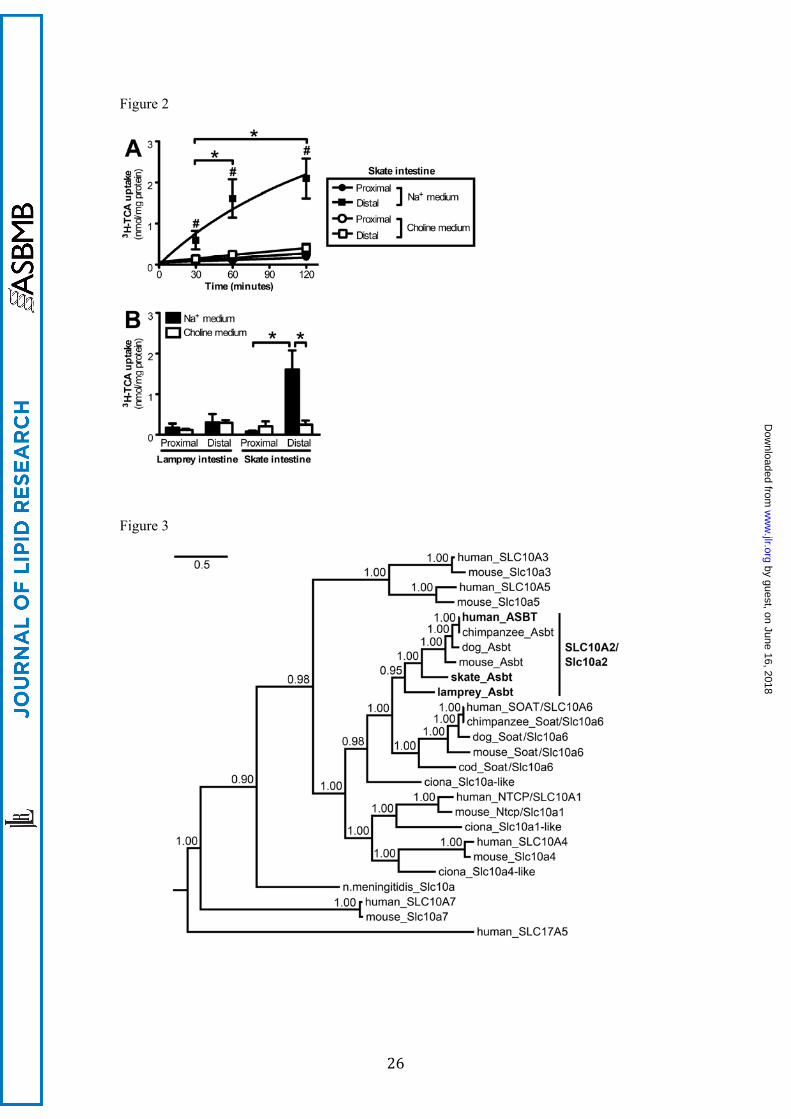
isolated from the little skate(16). As demonstrated in Figure 2A, a significant time-dependent
uptake of 3H-TCA was observed in the distal but not proximal intestine. When Na+ was
replaced by choline in the medium, uptake of 3H-TCA was abolished, indicating that a Na+-
dependent bile salt transporter was present in the distal intestine of skate. A similar transport
experiment was then carried out in adult lamprey, and in contrast to skate intestine, neither
the proximal nor distal intestine of the lamprey showed significant Na+-dependent uptake of
3H-TCA (Figure 2B). This negative result suggests that either an ASBT ortholog has not
evolved in lamprey or its substrate specificity is limited and does not accommodate the
modern bile salt TCA.
ASBT orthologs are identified in the distal intestine of both lamprey and skate
A search of the lamprey genome revealed DNA sequences with the potential to encode
portions of an ASBT ortholog. Subsequent RT-PCR and RACE PCR identified a full-length
lpAsbt transcript which encodes 363 amino acids with a 132 bp 5´-untranslated region (UTR)
by guest, on June 16, 2018w
ww
.jlr.orgD
ownloaded from
10
and 1.7 kb 3´-UTR (GenBank accession number JX014266). To identify skAsbt, we
performed RT-PCR using degenerate primers, followed by RACE PCR. We amplified a full-
length skAsbt transcript 2.3 kb in size, encoding for 393 amino acids with 189 bp at the 5´-
UTR and 918 bp at the 3´-UTR (GenBank accession number JX014267). LpAsbt and skAsbt
share 58% and 64% amino acid identity to hASBT, respectively (Table S2). Phylogenetic
analysis placed lpAsbt and skAsbt as the most primitive of known ASBT/SLC10A2 orthologs
(Figure 3). BLAST search of genome of the sea squirt (Ciona intestinalis), an invertebrate
which is believed to share the last common ancestor with vertebrates, produced 16 putative
protein sequences that could be members of the Slc10 protein family. However, phylogenetic
analysis did not identify a potential ortholog of SLC10A2 (Figure 3 and Figure S1). The ciona
sequence that was most closely related was placed on the common branch of Slc10a2 and
Slc10a6 subfamilies. Based on these findings, we propose that a distinct Asbt/Slc10a2 first
emerged from an ancient Slc10a2/a6-like gene near the beginning of vertebrate evolution,
presumably from a gene duplication between ciona and lamprey evolution, at a time that
coincides with the extensive utilization of cholesterol, the emergence of bile salts, the
development of a biliary system and the ability to produce bile.
Alignment of experimentally verified sequences of ASBT/Asbt’s revealed that the
proposed transmembrane domains are well conserved, including the Na+-binding core and the
substrate binding pocket based on the recently solved crystal structure of a distant ASBT
homolog in N. meningitidis(15) (Fig. S2 and S3). In contrast, the sequences of the N-
terminus and C-terminus show great interspecies variation. For example, while a N-
glycosylation site in hASBT Asn-10 is well conserved(22), computer software predicts an
additional N-glycosylation site at Asn-22 in skAsbt (Fig. S2).
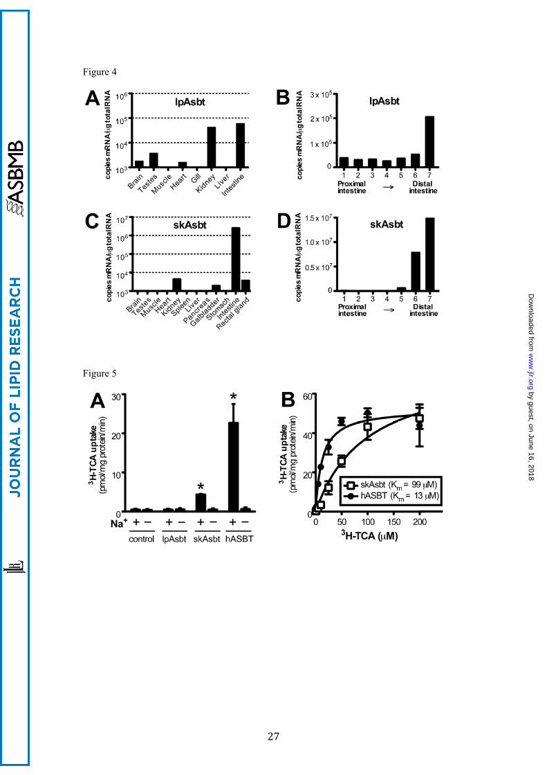
To determine the tissue distribution of lpAsbt and skAsbt, we performed real-time
RT-PCR. As shown in Figure 4A, lpAsbt mRNA was expressed primarily in the kidney and
intestine; less in testes, heart and brain; and was undetectable in muscle, gill and liver. A
similar expression pattern was found in lamprey larva. SkAsbt mRNA was highly expressed
in the intestine with lower levels in the kidney, gallbladder and rectal gland; it was absent
by guest, on June 16, 2018w
ww
.jlr.orgD
ownloaded from
11
from other tissues (Fig. 4C). In addition, we analyzed mRNA distribution along segments of
intestine, which demonstrated that both lpAsbt and skAsbt are most abundant in the distal
intestine (Fig. 4B and 4D). This was particularly evident in skate with a greater than 300 fold
expression in the distal compared to the proximal intestine. These expression profiles are
similar in human and rodents, and correspond with the known bile salt recycling function of
ASBT.
Affinity for the modern bile salt TCA is absent in lpAsbt, low in skAsbt and high in
hASBT
To functionally characterize lpAsbt and skAsbt, and to compare their substrate specificity to
hASBT, we subcloned these three genes into a pcDNA3 plasmid vector and transfected them
into COS-7 cells for expression. As shown in Figure 5A, skAsbt and hASBT demonstrated
Na+-dependent uptake of 10 µM 3H-TCA that increased significantly over vector control. In
contrast, lpAsbt did not show any specific transport activity for 3H-TCA (10-100 µM) with or
without Na+ in the media, despite confirmation by Western blot and immunofluorescent
labeling that the lpAsbt-FLAG fusion protein was expressed on the plasma membrane of
transfected cells (Fig. S4). These results are consistent with the negative results of 3H-TCA
transport in the everted gut sac experiments (Fig. 2B) and suggest that either lpAsbt is not a
functional bile salt transporter, or it is functionally inactive for the modern 5β-C24 bile salt
TCA.
Michaelis-Menten analysis of 3H-TCA transport kinetics indicated that skAsbt has a
Vmax of 75 ± 9 pmol/mg protein/min with a Km of 99 ± 24 µM, whereas hASBT has a Vmax of
51 ± 3 pmol/mg protein/min with a significantly lower Km of 13 ± 2 µM (Fig. 5B). Of note,
the Vmax was derived from data normalized to total protein in transfected cells – not to
transporter protein – which prevents direct comparison between absolute transport rates and
subsequently comparison of Vmax. However, Km is independent of absolute transport rate, and
can be compared to determine differences in affinity. The findings of lack of TCA affinity for
lpAsbt, and low affinity for skAsbt, albeit lower than for hASBT, suggests that structural
by guest, on June 16, 2018w
ww
.jlr.orgD
ownloaded from
12
changes in Asbt have evolved that allowed accommodation of this new bile acid structure
sometime between the development of agnathans and early gnathostomes, whereas TCA
affinity was optimized further between early gnathostome and mammalian evolution.
Bile salt substrate affinity is limited to early evolving bile salts for lpAsbt, early and
“intermediate” bile salts for skAsbt; whereas hASBT has high affinity for all bile salt
forms
To test if lpAsbt transports bile salts, we utilized a luciferase-based ASBT-FXR
reporter assay. Because conjugated bile salts require a specific transporter (e.g. ASBT) to
cross the cell membrane, and they are ligands for human FXR, transactivation of FXR reflects
the ability of ASBT to transport a given bile salt into the transfected cells. In this experiment,
expression constructs of lpAsbt, skAsbt, hASBT, or empty vector were co-transfected with
FXR reporter constructs into HEK293T cells. The transfected cells were then treated with 5α-
PZS, scymnol sulfate, and TCA, the three major bile salts in lamprey, skate and human,
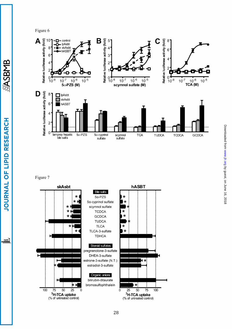
respectively. Interestingly, lpAsbt co-transfection transactivated FXR after 5α-PZS treatment,
indicating lpAsbt does transport this endogenous bile salt (Fig. 6A). SkAsbt and hASBT also
demonstrated transport activity for 5α-PZS. When 5β-C27 scymnol sulfate was tested, lpAsbt
showed significant activity only at 25 µM or higher concentrations, while skAsbt and hASBT
demonstrated activity with nanomolar concentrations (Fig. 6B). When cells were treated with
5β-C24 TCA, lpAsbt did not show any transport activity, skAsbt showed low activity,
whereas hASBT showed high activity (Fig. 6C). To further confirm that lpAsbt, skAsbt, and
hASBT have differential selectivity for bile salts, we tested additional early and late evolving
bile salts with the ASBT-FXR reporter assay. As indicated by Figure 6D, lpAsbt can transport
lamprey bile salts from its liver lipid extract and 5α-cyprinol sulfate (1 μM), another 5α-C27
bile alcohol, but not modern 5β-C24 bile salts, including tauroursodeoxycholic acid
(TUDCA), taurochenodeoxycholic acid (TCDCA), TCA, glycocholic acid and
glycoursodeoxycholic acid. Interestingly, glycochenodeoxycholic acid (GCDCA) is an
exception and apparently was transported by lpAsbt. skAsbt transported scymnol sulfate in
by guest, on June 16, 2018w
ww
.jlr.orgD
ownloaded from
13
addition to all the lpAsbt’s substrates, but again demonstrated low affinity for most modern
bile salts (Fig. 6D). In contrast, all tested bile salts were effectively transported by hASBT.
Collectively, these findings indicate that lpAsbt’s affinity for bile salts is largely confined to
the 5α-C27 early evolving “ancient” bile salts, skAsbt can effectively transport both the
“ancient” and evolutionarily “intermediate” bile salts, while hASBT effectively transports
bile salts with the entire spectrum of structures.
Substrate specificity of skAsbt is confined to bile salts, similar to hASBT.
To further assess skAsbt substrate specificity, we tested an array of bile salts, steroid sulfates,
and organic anions in assays competing with uptake of 3H-TCA. In general, more bile salt
structures inhibited hASBT 3H-TCA uptake more effectively than with skAsbt (Fig. 7).
Specifically, the early evolved bile salts (5α-PZS, 5α-cyprinol sulfate), “intermediate” bile
salts (5β-C27 scymnol sulfate) and modern 5β-C24 bile acids (TCDCA, GCDCA,
taurolithocholic acid [TLCA], and TLCA-3-sulfate) significantly reduced 3H-TCA transport
activity in both skAsbt and hASBT. Interestingly, tauroursodeoxycholic acid (TUDCA)
effectively reduced 3H-TCA transport activity in hASBT but not in skAsbt, consistent with
results from the ASBT-FXR reporter assay. Taurodehydrocholic acid (TDHCA) had no
effect on 3H-TCA transport activity with either skAsbt or hASBT. We then determined the
affinity of skAsbt for 5β-C27 scymnol sulfate, the major endogenous bile salt in skate, in a
competition assay for 3H-TCA uptake. Scymnol sulfate has a Ki of 42 ± 12 µM, whereas the
Km for 3H-TCA was calculated as 87 ± 38 µM (Figure S5).
In addition, we tested whether or not steroid sulfates could inhibit hASBT and skAsbt
transport of 3H-TCA. These molecules are structurally similar to bile salts and are transported
by SOAT/SLC10A6(23), a close paralog of ASBT (Fig. 3). Neither pregnenolone-3-sulfate
nor dehydroepiandrosterone (DHEA)-3-sulfate inhibited uptake activity of hASBT and
skAsbt for 3H-TCA. Estradiol-3-sulfate weakly inhibited skAsbt but not hASBT, whereas
estrone-3-sulfate weakly inhibited hASBT but not skAsbt. However, 3H-estrone-3-sulfate (25
μM) was not transported by lpAsbt, skAsbt or hASBT. Also neither skAsbt nor hASBT
by guest, on June 16, 2018w
ww
.jlr.orgD
ownloaded from
14
showed significant affinity for several other organic anions, including bromosulfophtalein and
bilirubin-ditaurate. Taken together, these competition experiments suggest that even as the
substrate specificity of these transporters expanded for bile salts, this expansion did not
extend to the closely related family of steroid sulfates.
DISCUSSION
In order to explore the evolutionary development of ASBT/SLC10A2 and its
substrate specificity, we functionally characterized this intestinal bile salt transport system in
the sea lamprey and the little skate, two species that represent early evolving members of
vertebrate evolution, and compared these with the human system. Our results demonstrate
that primitive ASBT/SLC10A2 orthologs in lamprey and skate have the ability to transport
bile salts (Fig. 2, 5, and 7). Like mammalian ASBT/Asbt, the substrate specificity of lpAsbt
and skAsbt appears confined to bile salts, as they did not demonstrate affinity for structurally
related steroid sulfates and other organic anions (Fig. 7). The mRNA tissue distribution of
both lpAsbt and skAsbt was most abundant in the distal intestine and kidney (Fig. 4),
consistent with the development of transporters to reabsorb bile salts from the intestinal
lumen, an essential function for establishing an enterohepatic circulation. This molecular
evidence indicates that the enterohepatic circulation of bile salts was present at the earliest
beginnings of vertebrate evolution. Since an ASBT ortholog was not identified in the sea
squirt (Fig. 2 and Fig. S1), a late non-vertebrate, we propose that ASBT/Asbt is a bile salt
transporter that evolved at the beginning of vertebrate evolution.
We also demonstrated that skAsbt transports bile salts in a Na+-dependent manner.
At this time, we were not able to directly determine whether bile salt transport by lpAsbt is
Na+-dependent since appropriate radiolabeled bile salt substrates are not available. However,
protein sequence alignment reveals that amino acids directly involved in Na+-binding for N.
meningitidis Slc10a (Asbt-like) are completely conserved in lpAsbt, skAsbt, mammalian
by guest, on June 16, 2018w
ww
.jlr.orgD
ownloaded from
15
ASBT/Asbt’s and even Na+-dependent paralogs NTCP and SOAT (Fig. S2)(15), suggesting
that lpAsbt is likely to be a Na+-dependent transporter.
In this study we assessed transport activity and substrate specificity with 3H-TCA
uptake assays in intact intestinal tissue (Fig. 2) and cell-based assays as well as in ASBT-FXR
reporter assays using recombinant expressed transporters (Fig. 5 and 6). Collectively, these
assays demonstrate that lpAsbt has a bile salt substrate specificity with high affinity only for
5α-PZS (the endogenous bile salt of lamprey) and 5α-cyprinol sulfate, two early evolving
“ancient” 5α bile alcohols, while the more evolutionarily advanced “intermediate” 5β-C27
bile alcohol scymnol sulfate was a low affinity substrate. In contrast, the modern 5β-C24 bile
acids TCA and TCDCA were not transported. In the case of skAsbt, not only was 5β-C27
scymnol sulfate (its endogenous bile salt) a high affinity substrate but so were the “ancient”
5α-bile alcohols. The later evolving 5β-C24 bile acids such as TCA and TCDCA showed only
low affinity for skAsbt. Surprisingly, all the structural forms of bile salts were high affinity
substrates for hASBT, even though the major endogenous bile salts in humans are 5β-C24
bile acids. Altogether, these results suggest that ASBT expanded its substrate specificity
whenever a novel class of bile salts emerged in evolution(1;11).
This broad substrate affinity for human ASBT is unusual, as most orthologs generally
lose affinity for their earlier substrates when they acquire affinity for novel substrates during
evolution(24-26), a phenomenon termed “ligand-receptor” co-evolution. ASBT/Asbt has
apparently retained affinity for old substrates even as it gained affinity for more modern bile
salts. We speculate that modern ASBT/Asbt’s retain its broad substrate specificity in order to
recover as many different species of bile salts as possible. In mammals, 16 different
enzymatic reactions are required to convert cholesterol to 5β-C24 bile acids. As each step is
not 100% efficient, small amounts of intermediate bile salts will be produced and excreted in
to bile(27). In order to most efficiently maintain the bile salt pool size, these “accidental but
functional by-products of bile salt synthesis” are also reclaimed. We speculate that other
transporters involved in the enterohepatic circulation of bile salts might also retain specificity
by guest, on June 16, 2018w
ww
.jlr.orgD
ownloaded from
16
for a wide variety of bile salt substrates structures, although this hypothesis has not yet been
tested.
Our phylogenetic analysis indicates that the recently solved crystal structure of N.
meningitidis Slc10a (formally called nmASBT) is not a member of the SLC10A2 gene family
(Figure 3). Although it transports TCA, it is not known if it can transport other bile salts or
other molecules. In addition, N. meningitidis Slc10a shares lower identity to hASBT than
does SLC10A3, SLC10A4, SLC10A5 and SOAT/SLC10A6 (Table S2). Since the latter four
solute carriers do not transport TCA, this raises a question as to the specificity of the substrate
binding pocket of N. meningitidis Slc10a for bile salts. Because lpAsbt, skAsbt and hASBT
share much higher sequence identity and demonstrate differential selectivity for bile salts, it
should be possible to identify specific amino acids that are responsible for the expansion of
mammalian ASBT/Asbt substrate specificity using further sequence analyses and mutation
experiments. Doing so will help to determine ASBT/Asbt’s evolutionary path, while at the
same time provide insight into the structure-function determinants of hASBT.
In conclusion, the present study demonstrates that Asbt is a functional bile salt
transporter in the most primitive vertebrates, occurring together with the emergence of bile
salts and the ability of organisms to form bile. This study establishes that ASBT/Asbt and the
enterohepatic circulation of bile salts were present at the beginning of vertebrate evolution,
giving vertebrates the transport ability essential to regulate both bile salt and lipid
homeostasis. Our findings also suggest that as ASBT evolved, it gained substrate specificity
to novel bile salt structures while retaining affinity for its old substrates.
by guest, on June 16, 2018w
ww
.jlr.orgD
ownloaded from
17
ACKNOWLEDGEMENTS
We thank Dr. Lee Hagey (UCSD, San Diego, CA) for generously providing 5α-cyprinol
sulfate and for critical review and editing of this manuscript. Dr. Carol Soroka and Shuhua
Xu in our laboratory have provided technical support. Also, we are grateful to Professor
Ulrich Beuers (University of Amsterdam) for advice and constructive discussions. This work
was supported by NIH grants P30 DK34989 and R37 DK25636 (J.L.B.) and student
fellowship grant 10-06s from the Dutch Digestive Foundation (D.A.L.).
by guest, on June 16, 2018w
ww
.jlr.orgD
ownloaded from
18
Reference List
1. Hofmann, A. F., L. R. Hagey, and M. D. Krasowski. 2010. Bile salts of vertebrates:
structural variation and possible evolutionary significance. J. Lipid Res. 51: 226-246.
2. Trauner, M., T. Claudel, P. Fickert, T. Moustafa, and M. Wagner. 2010. Bile acids as
regulators of hepatic lipid and glucose metabolism. Dig. Dis. 28: 220-224.
3. Dawson, P. A., T. Lan, and A. Rao. 2009. Bile acid transporters. J. Lipid Res. 50: 2340-
2357.
4. Dawson, P. A. 2011. Role of the intestinal bile acid transporters in bile acid and drug
disposition. Handb. Exp. Pharmacol. 201: 169-203.
5. Dawson, P. A., J. Haywood, A. L. Craddock, M. Wilson, M. Tietjen, K. Kluckman, N.
Maeda, and J. S. Parks. 2003. Targeted deletion of the ileal bile acid transporter
eliminates enterohepatic cycling of bile acids in mice. J. Biol. Chem. 278: 33920-
33927.
6. Oelkers, P., L.C. Kirby, J. E. Heubi, and P. A. Dawson. 1997. Primary bile acid
malabsorption caused by mutations in the ileal sodium-dependent bile acid transporter
gene (SLC10A2). J. Clin. Invest 99: 1880-1887.
7. Lewis, M. C., L. E. Brieaddy, and C. Root. 1995. Effects of 2164U90 on ileal bile acid
absorption and serum cholesterol in rats and mice. J. Lipid Res. 36: 1098–1105.
8. Bhat, B. G., S. R. Rapp, J. A. Beaudry, N. Napawan, D. N. Butteiger, K. A. Hall, C. L.
Null, Y. Luo, and B. T. Keller. 2003 . Inhibition of ileal bile acid transport and reduced
atherosclerosis in apoE2/2 mice by SC-435. J. Lipid Res. 44: 1614–1621.
9. Chey, W. D., M. Camilleri, L. Chang, L. Rikner, and H. Graffner. 2011. A randomized
placebo-controlled phase IIb trial of a3309, a bile acid transporter inhibitor, for chronic
idiopathic constipation. Am. J. Gastroenterol. 106: 1803-1812.
by guest, on June 16, 2018w
ww
.jlr.orgD
ownloaded from
19
10. Simren, M., A. Bajor, P. G. Gillberg, M. Rudling, and H. Abrahamsson. 2011.
Randomised clinical trial: The ileal bile acid transporter inhibitor A3309 vs. placebo in
patients with chronic idiopathic constipation--a double-blind study. Aliment.
Pharmacol. Ther. 34: 41-50.
11. Haslewood, G. A. 1967. Bile salt evolution. J. Lipid Res. 8: 535-550.
12. Russell, D. W. 2009. Fifty years of advances in bile acid synthesis and metabolism. J.
Lipid Res. 50 Suppl: S120-S125.
13. Norlin, M., and K. Wikvall. 2007. Enzymes in the conversion of cholesterol into bile
acids. Curr. Mol. Med. 7: 199-218.
14. Hagey, L. R., P. R. Moller, A.F. Hofmann, and M.D. Krasowski. 2010. Diversity of
bile salts in fish and amphibians: evolution of a complex biochemical pathway. Physiol
Biochem. Zool. 83: 308-321.
15. Hu, N. J., S. Iwata, A. D. Cameron, and D. Drew. 2011. Crystal structure of a bacterial
homologue of the bile acid sodium symporter ASBT. Nature 478: 408-411.
16. Lack, L., and I. M. Weiner. 1961. In vitro absorption of bile salts by small intestine of
rats and guinea pigs. Am. J. Physiol 200: 313-317.
17. Fricker, G., G. Hugentobler, P. J. Meier, G. Kurz, and J. L. Boyer. 1987. Identification
of a single sinusoidal bile salt uptake system in skate liver. Am. J. Physiol 253: G816-
G822.
18. Larkin, M. A., G. Blackshields, N.P. Brown, R. Chenna, P. A. McGettigan, H.
McWilliam, F. Valentin, I. M. Wallace, A. Wilm, R. Lopez, J.D. Thompson, T. J.
Gibson, and D. G. Higgins. 2007. Clustal W and Clustal X version 2.0. Bioinformatics.
23: 2947-2948.
by guest, on June 16, 2018w
ww
.jlr.orgD
ownloaded from
20
19. Ronquist, F., and J. P. Huelsenbeck. 2003. MrBayes 3: Bayesian phylogenetic inference
under mixed models. Bioinformatics. 19: 1572-1574.
20. Wong, M. H., P. Oelkers, A. L. Craddock, and P. A. Dawson. 1994. Expression cloning
and characterization of the hamster ileal sodium-dependent bile acid transporter. J. Biol.
Chem. 269: 1340-1347.
21. Fricker, G., R. Wossner, J. Drewe, R. Fricker, and J. L. Boyer. 1997. Enterohepatic
circulation of scymnol sulfate in an elasmobranch, the little skate (Raja erinacea). Am.
J. Physiol 273: G1023-G1030.
22. Zhang, E. Y., M. A. Phelps, A. Banerjee, C. M. Khantwal, C. Chang, F. Helsper, and P.
W. Swaan. 2004. Topology scanning and putative three-dimensional structure of the
extracellular binding domains of the apical sodium-dependent bile acid transporter
(SLC10A2). Biochemistry 43: 11380-11392.
23. Geyer, J., B. Doring, K. Meerkamp, B. Ugele, N. Bakhiya, C. F. Fernandes, J. R.
Godoy, H. Glatt, and E. Petzinger. 2007. Cloning and functional characterization of
human sodium-dependent organic anion transporter (SLC10A6). J. Biol. Chem. 282:
19728-19741.
24. Moyle, W. R., R. K. Campbell, R. V. Myers, M. P Bernard, Y. Han, and X. Wang.
1994. Co-evolution of ligand-receptor pairs. Nature 368: 251-255.
25. Park, Y., Y. J. Kim, and M. E. Adams. 2002. Identification of G protein-coupled
receptors for Drosophila PRXamide peptides, CCAP, corazonin, and AKH supports a
theory of ligand-receptor coevolution. Proc. Natl. Acad. Sci. U. S. A 99: 11423-11428.
26. Baker, M. E. 2011. Origin and diversification of steroids: co-evolution of enzymes and
nuclear receptors. Mol. Cell Endocrinol. 334: 14-20.
by guest, on June 16, 2018w
ww
.jlr.orgD
ownloaded from
21
27. Griffiths, W. J., and J. Sjovall. 2010. Bile acids: analysis in biological fluids and
tissues. J. Lipid Res. 51: 23-41.
by guest, on June 16, 2018w
ww
.jlr.orgD
ownloaded from
22
Figure legends
Figure 1. The structural change of bile salts during vertebrate evolution. A simplified
phylogenetic tree of vertebrate classes is shown, with respective major bile salt class indicated
above, where dashed lines represent the molecular evolution of bile salts. Bile salts are
metabolites of cholesterol, a versatile molecule that is already seen in invertebrates that do not
use bile salts. Agnathans (jawless fish) are the earliest vertebrate class that primarily use
“ancient” 5α-C27 sulfated bile alcohols with an overall planar structure as cholesterol, such as
5α-cyprinol sulfate. Of note, the bile salt spectrum of lamprey comprises a mixture of C27-
and C24-5α sulfated bile alcohols that includes C24 5α-Petromyzonol sulfate (5α-PZS), a bile
alcohol similar to 5α-cyprinol sulfate but with a shortened side-chain. The evolutionarily
more advanced early gnatosthomes (jawed cartilaginous fish) mostly use 5-C27 sulfated bile
alcohols with a tilted structure of the steroid rings, such as scymnol sulfate, the major bile salt
of skate. As evolution further progressed, more complex vertebrates including mammals
mostly use 5-C24 bile acids with a shortened side-chain containing a carboxylic acid, such
as taurocholic acid, the major bile salt in humans.
Figure 2. Uptake of 3H-TCA into skate and lamprey intestine. (A) Everted gut sacs prepared
from the proximal and distal skate intestine were incubated for 30, 60 or 120 minutes in
medium supplemented with 50 μM 3H-TCA containing Na+ or choline (Na+-void). Values
represent means (n = 3-4) ± SD. *, P <0.05; #, P < 0.01 versus all other groups at the same
time point. (B) Uptake of 50 μM 3H-TCA in everted gut sacs prepared from proximal or distal
intestine of skate and lamprey, incubated for 60 minutes in Na+ or choline (Na+-void)
medium. Values represent means (n = 3-4) ± SD. *, P < 0.05.
Figure 3. Phylogeny of the Slc10a family shows that ortholog genes identified in lamprey
and skate are the most primitive members of the ASBT/SLC10A2 subfamily. Phylogeny was
by guest, on June 16, 2018w
ww
.jlr.orgD
ownloaded from
23
inferred using Bayesian MCMC analysis. Posterior probabilities are indicated at nodes.
Branch length is expressed as number of expected substitutions per site. Accession numbers
of protein sequences used are listed in Table S3.
Figure 4. Messenger RNA tissue distribution of lpAsbt and skAsbt by quantitative real-time
RT-PCR. A broad variety of tissues was tested for mRNA expression of lpAsbt (A) and
skAsbt (C). Expression along the intestine was determined by dividing the intestine in 7
segments of equal length (panels B and D). Segment 1 starts at the intestinal connection to the
bile duct/liver and segment 7 ends at the start of the rectum. Expression below 1000 copies
mRNA/μg total RNA was considered background and is not shown. All values represent one
animal and are expressed as means from triplicate measurements.
Figure 5. 3H-TCA uptake assay in transfected COS-7 cells. (A) Uptake of 10 μM 3H-TCA in
cells transiently transfected with vector without insert (control), skAsbt, lpAsbt or hASBT.
Cells were incubated for 10 minutes in medium with Na+ (+) or medium where Na+ was
substituted with choline (–). *, P < 0.05 versus choline. (B) Kinetics of 3H-TCA uptake in
cells transfected with skAsbt or hASBT. Cells were incubated for 10 minutes and background
uptake levels derived from cells transfected with vector control were subtracted. lpAsbt did
not show increased uptake over background (vector control) at 10 μM and 100 μM and was
excluded. Data was normalized to total cell protein. Kinetic parameters were obtained by non-
linear curve fitting using the Michaelis-Menten equation. All values represent at least three
independent experiments and are expressed as means ± SD.
Figure 6. ASBT-FXR luciferase reporter assay in co-transfected HEK293T cells. Cells were
co-transfected with FXR reporter constructs plus either vector without insert (control),
lpAsbt, skAsbt or hASBT for 24 hours and subjected to bile salt treatment for additional 24
hours. Dose-response curve of cells treated with 5α-petromyzonol sulfate (5α-PZS), the
endogenous bile salt of lamprey (A); with 5β-scymnol sulfate, the endogenous bile salt of
by guest, on June 16, 2018w
ww
.jlr.orgD
ownloaded from
24
skate (B); and with taurocholic acid (TCA), the endogenous bile salt of humans(C).
Luciferase readings of dose-response curves are relative to cells transfected with vector
control treated with 10-8 M bile salts. (D) Cells transfected with vector control, lpAsbt, skAsbt
or hASBT were treated with 1 μM indicated bile salt or ~1 μM bile salts from adult lamprey
liver. Luciferase readings of this analysis are relative to cells transfected with vector control.
All values represent at least three independent experiments and are expressed as means ± SD.
Figure 7. Competition assay of 3H-TCA transport. COS-7 cells transfected with skAsbt or
hASBT were incubated for 10 minutes in Na+ medium supplemented with 10 μM 3H-TCA
without competitor (untreated control) or with 100 μM competitor. Background levels derived
from cells transfected with vector control were subtracted and uptake measurements were
normalized for protein concentration of the lysate.*, P < 0.05 for untreated control versus
competitor. All values represent at least three independent experiments and are expressed as
means ± SD.
by guest, on June 16, 2018w
ww
.jlr.orgD
ownloaded from