Embed Size (px)
Citation preview

Research ArticleSubjective Cognitive Impairment, DepressiveSymptoms, and Fatigue after a TIA or Transient NeurologicalAttack: A Prospective Study
Frank G. van Rooij,1 Nicole O. Plaizier,2 Sarah E. Vermeer,3 Bozena M. Góraj,4
Peter J. Koudstaal,5 Edo Richard,1 Frank-Erik de Leeuw,1 Roy P. C. Kessels,2,6
and Ewoud J. van Dijk1
1Department of Neurology, Donders Institute for Brain, Cognition and Behaviour, Centre for Neuroscience,Radboud University Medical Center, P.O. Box 9101, 6500 HB Nijmegen, Netherlands2Department of Medical Psychology, Donders Institute for Brain, Cognition and Behaviour, Centre for Neuroscience,Radboud University Medical Center, P.O. Box 9101, 6500 HB Nijmegen, Netherlands3Department of Neurology, Rijnstate Hospital, P.O. Box 9555, 6800 TA Arnhem, Netherlands4Department of Radiology, Donders Institute for Brain, Cognition and Behaviour, Centre for Neuroscience,Radboud University Medical Center, P.O. Box 9101, 6500 HB Nijmegen, Netherlands5Department of Neurology, Erasmus Medical Center, P.O. Box 2040, 3000 CA Rotterdam, Netherlands6Donders Institute for Brain, Cognition and Behaviour, Centre for Cognition, Radboud University, P.O. Box 9101,6500 HB Nijmegen, Netherlands
Correspondence should be addressed to Ewoud J. van Dijk; [email protected]
Received 9 July 2017; Revised 30 September 2017; Accepted 8 October 2017; Published 19 November 2017
Academic Editor: Gabriella Santangelo
Copyright © 2017 Frank G. van Rooij et al. This is an open access article distributed under the Creative Commons AttributionLicense, which permits unrestricted use, distribution, and reproduction in any medium, provided the original work isproperly cited.
Introduction. Subjective cognitive impairment (SCI), depressive symptoms, and fatigue are common after stroke and are associatedwith reduced quality of life. We prospectively investigated their prevalence and course after a transient ischemic attack (TIA) ornonfocal transient neurological attack (TNA) and the association with diffusion-weighted imaging (DWI) lesions. Methods. TheCognitive Failures Questionnaire, Hospital Anxiety and Depression Scale, and Subjective Fatigue subscale from the ChecklistIndividual Strength were used to assess subjective complaints shortly after TIA or TNA and six months later. With repeatedmeasure analysis, the associations between DWI lesion presence or clinical diagnosis (TIA or TNA) and subjective complaintsover time were determined. Results. We included 103 patients (28 DWI positive). At baseline, SCI and fatigue were less severe inDWI positive than in DWI negative patients, whereas at follow-up, there were no differences. SCI (p = 0 02) and fatigue (p =0 01) increased in severity only in DWI positive patients. There were no differences between TIA and TNA. Conclusions.Subjective complaints are highly prevalent in TIA and TNA patients. The short-term prognosis is not different between DWI-positive and DWI negative patients, but SCI and fatigue increase in severity within six months after the event when an initialDWI lesion is present.
1. Introduction
Subjective cognitive impairment (SCI), depressive symp-toms, and fatigue are highly prevalent after stroke and arerelated to stroke severity [1–4]. Although by definition thesymptoms of a transient ischemic attack (TIA) subside
completely within 24 hours [5], subjective cognitive com-plaints, depressive symptoms, and fatigue often persist inthese patients as well [6–8].
Diagnosing TIA is notoriously difficult [9]. Patients oftenreport attacks of atypical or nonfocal neurological symptoms.In the absence of an alternative diagnosis, these episodes are
HindawiBehavioural NeurologyVolume 2017, Article ID 5181024, 7 pageshttps://doi.org/10.1155/2017/5181024
referred to as transient neurological attack (TNA) [10]. Inone-third of TIA patients, diffusion-weighted imaging(DWI) shows signs of acute ischemia beyond the pointof symptom resolution, ascertaining a cerebrovascular eti-ology of the attack [11]. Acute DWI lesions are howeveralso present in more than 20% of clinically diagnosedTNA patients [12].
Previous studies on subjective complaints after short-lasting attacks of neurological symptoms have focused solelyon TIA, did not take DWI findings into account, and werecross-sectional in nature [6–8]. Since subjective complaintsare associated with a reduced quality of life and are possibleharbingers of forthcoming cognitive decline, it is importantto understand their prevalence and determinants [13, 14].
We prospectively investigated the prevalence, severity,and course of SCI, depressive symptoms, anxiety, and fatiguein a cohort of TIA and TNA patients and determined therelation with event type and DWI results. We hypothesizedthat patients with acute DWI lesions would report anincrease in severity of complaints in the months after the ini-tial event. Since TIA and TNA have a comparable prevalenceof DWI positivity, we expected to find no differences betweenthese patient categories [12].
2. Methods
2.1. Study Design and Patients. This study was part of theprospective Cohort study ON Neuroimaging, Etiology, andCognitive consequences of Transient neurological attacks(CONNECT), the details of which have been previouslyreported [15]. Consecutive stroke-free patients aged≥ 45years referred to a specialized outpatient TIA clinic within7 days after an event of acute onset neurological symptomslasting< 24 hours were included. Baseline measurementstook place within seven days after the qualifying event, andfollow-up was performed six months later. All baselineassessments were performed before the final diagnosis wasdiscussed with the patient. The Medical Review Ethics Com-mittee region Arnhem-Nijmegen approved the study, andwritten informed consent was obtained from all participants.
2.2. Classification of Qualifying Event. Based on a detaileddescription of the signs and symptoms of the event includinga structured assessment of the presence or absence of eigh-teen specific predefined symptoms (Table 1), three special-ized stroke neurologists adjudicated each event as TIA,TNA, or a specified other diagnosis, using previously deter-mined definitions of TIA and TNA [5, 10]. Events with bothfocal and nonfocal symptoms were classified as TIA. Qualify-ing neurologists were blinded to results from MRI, and incase of disagreement, a consensus meeting was held.
2.3. Brain Imaging. Brain MRI was performed within sevendays after the qualifying event on a 1.5 Tesla MagnetomScanner (Siemens, Erlangen, Germany) and included DWI,FLAIR, T1-, T2-, and T2∗-weighted sequences. Two experi-enced raters individually evaluated the severity of white mat-ter hyperintensities, the presence of (silent) territorialinfarcts, lacunes, and microbleeds [16, 17]. DWI was visually
assessed for signs of acute infarction. Raters were unaware ofclinical information, and a consensus meeting was held incase of disagreement.
2.4. Subjective Cognitive Impairment. At baseline and fol-low-up, the presence and severity of SCI in the previousmonth was assessed with a 15-item semistructured interviewbased on the Cognitive Failures Questionnaire (CFQ) [18].Items concerning remembering, word finding, planning,concentration, and slowness of thought were given a widerscore range (0–3) than other items (0-1). SCI was consid-ered present if ≥1 moderate problem (score≥ 2) on an itemwith a score range of 0 to 3 or a score of 1 on a dichotomousitem was reported [19]. Trained examiners, unaware of clin-ical diagnosis and DWI status, administered all semistruc-tured interviews.
2.5. Other Measurements. At both baseline and follow-up, theHospital Anxiety and Depression Scale (HADS) was admin-istered to measure the severity of symptoms of depressionand anxiety [20]. Relevant symptoms of depression or anxi-ety were defined as a value of >7 on the subscales [21].Fatigue was assessed with the subscale Subjective Fatigue ofthe Checklist Individual Strength (CIS20R-fatigue) with ascore> 35 considered indicative for severe fatigue [22]. Levelof education was classified using seven categories (1= lessthan primary school; 7 = academic degree) [23]. Vascular riskfactors were assessed at baseline. Incident vascular events(stroke, TIA, and myocardial infarction) between baselineand follow-up were assessed with a standardized, structuredquestionnaire. All questionnaires were handed out at thebaseline or follow-up visit. In case of limited time, these werefilled in at home and returned within one week.
2.6. Statistical Analysis.Only patients who completed follow-up were included. Baseline characteristics were comparedbetween patients with complete and incomplete follow-up,between TIA and TNA between patients, and patients withand without DWI lesion, using Student’s t-test, χ2 test, orMann–Whitney U test as appropriate.
Table 1: Predefined focal and nonfocal neurological symptoms.
Focal Nonfocal
HemiparesisDecreased consciousness or
unconsciousness
Hemihypesthesia Confusion
Dysphasia Amnesia
Dysarthria Unsteadiness
Hemianopia Nonrotatory dizziness
Transient monocularblindness
Positive visual phenomena
Hemiataxia Paresthesias
Diplopia Bilateral weakness of arms or legs
Vertigo Unwell feelings∗
Symptoms should have sudden onset, rapid clearance, and duration of<24 hours. ∗Referable to the nervous system if the referring physicianconsidered TIA but patients were unable to specify further.
2 Behavioural Neurology
Differences in prevalence of SCI, symptoms of depressionor anxiety, and severe fatigue between groups (clinicaldiagnosis and DWI status) and time points were analyzed withMcNemar’s test. Subsequently, the effect of DWI lesion andclinical diagnosis on change in subjective outcomes over timewas determined with repeated measures analyses of variance.Associations between SCI, fatigue, and depressive and anxietysymptoms were assessed with multivariate regression analysis.
Age, sex, and level of education were regarded as poten-tial confounders and adjusted for in all repeated measuresanalyses. Alpha was set at 0.05 and statistical analyses wereperformed with IBM SPSS Statistics version 24.0 (IBM Corp.,Armonk, NY).
3. Results
CONNECT included 150 patients, 87 of whom were diag-nosed as TIA, 56 as TNA, and 7 with a specific diagnosis.Patients in the last category were excluded from further
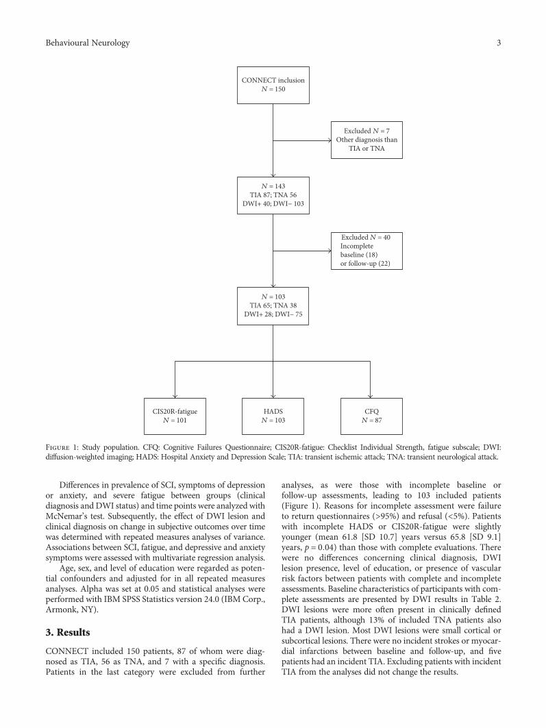
analyses, as were those with incomplete baseline orfollow-up assessments, leading to 103 included patients(Figure 1). Reasons for incomplete assessment were failureto return questionnaires (>95%) and refusal (<5%). Patientswith incomplete HADS or CIS20R-fatigue were slightlyyounger (mean 61.8 [SD 10.7] years versus 65.8 [SD 9.1]years, p = 0 04) than those with complete evaluations. Therewere no differences concerning clinical diagnosis, DWIlesion presence, level of education, or presence of vascularrisk factors between patients with complete and incompleteassessments. Baseline characteristics of participants with com-plete assessments are presented by DWI results in Table 2.DWI lesions were more often present in clinically definedTIA patients, although 13% of included TNA patients alsohad a DWI lesion. Most DWI lesions were small cortical orsubcortical lesions. There were no incident strokes or myocar-dial infarctions between baseline and follow-up, and fivepatients had an incident TIA. Excluding patients with incidentTIA from the analyses did not change the results.
CONNECT inclusionN = 150
CIS20R-fatigueN = 101
HADSN = 103
CFQN = 87
Excluded N = 7Other diagnosis than
TIA or TNA
Excluded N = 40 Incompletebaseline (18)or follow-up (22)
N = 143TIA 87; TNA 56
DWI+ 40; DWI− 103
N = 103TIA 65; TNA 38
DWI+ 28; DWI− 75
Figure 1: Study population. CFQ: Cognitive Failures Questionnaire; CIS20R-fatigue: Checklist Individual Strength, fatigue subscale; DWI:diffusion-weighted imaging; HADS: Hospital Anxiety and Depression Scale; TIA: transient ischemic attack; TNA: transient neurological attack.
3Behavioural Neurology
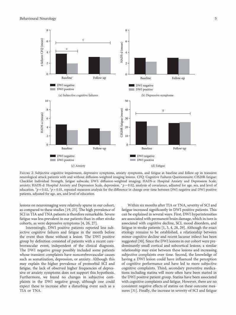
3.1. Subjective Cognitive Impairment (n = 87). The overallprevalence of SCI in TIA and TNApatients was 82% at baselineand 77% at follow-up and was not significantly differentbetween time points, DWI status, or clinical diagnosis. At base-line, themean number of subjective cognitive failures was lowerin patients with a DWI lesion than in those without (mean (SD)1.83 (1.75) versus 3.77 (3.27), p = 0 01), while at follow-up, thisincreased to 3.00 (2.70) in the first group and decreased slightlyto 3.14 (3.17) in the latter (p = 0 73) (Figure 2(a)). Repeatedmeasures analysis (adjusted for age, sex, and level of education)showed that change over time in the number of subjective cog-nitive failures was significantly different between DWI positiveand DWI negative patients (p = 0 01). There was no differencein SCI between TIA and TNA patients.
3.2. Depressive Symptoms and Anxiety (n = 103). Relevantdepressive symptoms (HADS-depression subscore> 7) werepresent in 8% of all patients at baseline and 9% at follow-up. The prevalence of relevant anxiety symptoms (HADS-anxiety subscore> 7) was 15% at baseline and 11% atfollow-up. Depressive and anxiety symptoms did not differbetween DWI negative and DWI positive patients or betweenTIA and TNA patient groups (Figures 2(b) and 2(c)).
3.3. Fatigue (n = 101). Severe fatigue (CIS20R-fatiguescore> 35) was present in 23% of patients at baseline and in19% six months later. Prevalence of severe fatigue did not
differ between DWI positive and DWI negative patients orbetween TIA and TNA patients.
Mean (SD) CIS20R-fatigue score was 25.0 (12.7) at base-line and 24.8 (11.5) at follow-up. Baseline scores were lowerin DWI positive patients (21.0 (12.7) compared to 26.6(12.4) for those without; p = 0 07) but equal at follow-up(24.9 (11.6) in the DWI positive group versus 24.7 (11.6) inthe DWI negative group; p = 0 86). Patients with and withoutDWI lesions differed with respect to change in CIS20R-fatigue scores (p = 0 01) (Figure 2(d)). The clinical diagnosiswas unrelated to severity or change over time of fatigue.
At both baseline and follow-up, SCI was associated withhigher CIS20R-fatigue scores (p = 0 001), but not with symp-toms of depression or anxiety.
4. Discussion
Subjective complaints, especially SCI and fatigue, are highlyprevalent in TIA and TNA patients both directly before theevent and after six months. The initial qualifying diagnosiswas unrelated to the presence, severity, and course of subjec-tive complaints. Patients with signs of acute ischemia onDWI reported less severe SCI and fatigue in the month beforethe TIA or TNA than those without such a lesion. In thisgroup of patients, severity subsequently increased in the sixmonths after the event to a level equal to that of DWI nega-tive patients.
Some methodological issues need to be considered wheninterpreting these results. First, given the loss to follow-up,selection bias might have occurred. Patients with missingfollow-up HADS and CIS20R-fatigue assessments were onaverage slightly younger than participants. Since especiallyfatigue is more often reported in older patients, this mighthave resulted in an overestimation of its prevalence [24].Alternatively, patients with incomplete assessments mighthave dropped out because of complaints, resulting in anunderestimation. Other demographic variables however didnot differ between patients with and without complete assess-ments, and we adjusted for age in our analyses. Therefore, wefeel that selection bias has not largely influenced our results.The relatively small patient numbers in our study howeverlimit statistical power, and our results need to be replicatedin a larger cohort. Secondly, we used questionnaires to obtaininformation on the presence of SCI, depression, anxiety, andfatigue. These screening instruments, although validated,indicate whether patients experience dysfunction whenactively asked about it. This differs from spontaneouslyreported complaints and may explain the high frequency ofSCI observed in our cohort. The CFQ handles a strict cutofffor SCI, making it a sensitive but perhaps not very specificscreening instrument. Thirdly, our study did not include acontrol group, limiting the interpretation of an added effectof TIA or TNA on subjective complaints.
The prevalence of SCI in our study is comparable to thatin both stroke patients and those with evidence of small ves-sel disease on neuroimaging, using the same screeninginstrument [2, 19, 25]. Patients in those studies were on aver-age older, had more often suffered stroke instead of TIA, andwere tested several years after the initial event. Also, vascular
Table 2: Baseline patient characteristics stratified by DWI result(n = 103).
DWI+(n= 28)
DWI−(n= 75)
p∗
Women 8 (29) 29 (39) 0.34
Age, mean (SD) 66.7 (8.5) 65.6 (9.3) 0.60
Level of education, median (IQR) 5 (3) 5 (2) 0.10
TIA 23 (82) 42 (56) 0.01
Hypertension 25 (89) 60 (80) 0.27
Dyslipidemia 18 (64) 53 (71) 0.53
Diabetes mellitus 2 (7) 7 (9) 0.73
Atrial fibrillation 3 (11) 10 (13) 0.72
Smoking 13 (46) 17 (23) 0.02
Diffusion-weighted imaginglesions, total n
45 N/A N/A
Lesion type
Small cortical 26 (58)
Small subcortical 14 (31)
Territorial 5 (11)
Fazekas score, median (IQR) 1 (1) 1 (1) 0.30
Lacunes 5 (18) 12 (16) 0.82
Territorial infarcts 2 (7) 8 (11) 0.59
Microbleeds(available for 72 patients)
1 (6) 6 (11) 0.49
Values are n (%) unless stated otherwise. ∗For difference using Student’s t-test, χ2 test, or Mann–Whitney U test as appropriate. DWI: diffusion-weighted imaging; IQR: interquartile range; TIA: transient ischemic attack;N/A: not applicable.
4 Behavioural Neurology
lesions on neuroimaging were relatively sparse in our cohort,as compared to these studies [19, 25]. The high prevalence ofSCI in TIA and TNA patients is therefore remarkable. Severefatigue was less prevalent in our patients than in other strokecohorts, as were depressive symptoms [4, 26, 27].
Interestingly, DWI positive patients reported less sub-jective cognitive failures and fatigue in the month beforethe event than those without a lesion. The DWI positivegroup by definition consisted of patients with a recent cere-brovascular event, independent of the clinical diagnosis.The DWI negative group possibly included some patientswhose transient complaints have noncerebrovascular causessuch as somatization, depression, or anxiety. Although thismay explain the higher prevalence of premorbid SCI andfatigue, the lack of observed higher frequencies of depres-sive or anxiety symptoms does not support this hypothesis.Furthermore, we found no changes in subjective com-plaints in the DWI negative group, although one couldexpect these to increase after a disturbing event such as aTIA or TNA.
Within six months after TIA or TNA, severity of SCI andfatigue increased significantly in DWI positive patients. Thiscan be explained in several ways. First, DWI hyperintensitiesare associated with permanent brain damage, which in turn isassociated with cognitive decline, SCI, mood disorders, andfatigue in stroke patients [1, 3, 4, 28, 29]. Although the exactetiology remains to be established, a relationship betweenminor cognitive decline and recent lacunar infarct has beensuggested [30]. Since the DWI lesions in our cohort were pre-dominantly small cortical and subcortical lesions, a similarrelationship may exist between these lesions and increasingsubjective complaints over time. Second, the knowledge ofhaving a DWI lesion could have influenced the perceptionof cognitive performance and have led to more subjectivecognitive complaints. Third, secondary preventive medica-tions including statins will more often have been started inthe DWI positive patient group. Statins have been associatedwith cognitive complaints and fatigue. However, there are noconsistent negative effects of statins on these outcome mea-sures [31]. Finally, the increase in severity of SCI and fatigue
8
6
4
2
0
†
⁎
Baseline Follow-up
DWI negativeDWI positive
n fa
ilure
s CFQ
(mea
n)
(a) Subjective cognitive failures
8
6
4
2
0Baseline Follow-up
DWI negativeDWI positive
HA
DS-
d (m
ean)
(b) Depressive symptoms
10
8
6
4
2
0
DWI negativeDWI positive
Baseline Follow-up
HA
DS-
a (m
ean)
(c) Anxiety
50
‡40
30
20
10
0
DWI negativeDWI positive
Baseline Follow-up
CIS2
0R-fa
tigue
(mea
n)
(d) Fatigue
Figure 2: Subjective cognitive impairment, depressive symptoms, anxiety symptoms, and fatigue at baseline and follow-up in transientneurological attack patients with and without diffusion-weighted imaging lesions. CFQ: Cognitive Failures Questionnaire; CIS20R-fatigue:Checklist Individual Strength, fatigue subscale; DWI: diffusion-weighted imaging; HADS-a: Hospital Anxiety and Depression Scale,anxiety; HADS-d: Hospital Anxiety and Depression Scale, depression. ∗p = 0 02, analysis of covariance, adjusted for age, sex, and level ofeducation. †p = 0 02, ‡p = 0 01, repeated measures analysis for the difference in change over time between DWI negative and DWI positivepatients, adjusted for age, sex, and level of education.
5Behavioural Neurology
observed in the DWI positive patient group could be merely acorrection of an unexplained baseline difference, that is,regression towards the mean. However, no changes in preva-lence or severity of these complaints were found in the DWInegative group, countering this explanation.
Fatigue was associated with subjective cognitive dysfunc-tion. Possibly, both fatigue and SCI, as measured in ourstudy, are expressions of the same underlying sense ofunwell-being. We found no association between depressivesymptoms and subjective cognitive dysfunction, suggestingthat the increased severity of SCI observed in DWI positivepatients was not influenced by mood changes.
5. Conclusions
Subjective complaints are highly prevalent in TIA and TNApatients. Larger sampled studies with longer follow-up needto determine whether subjective complaints last beyond sixmonths after TIA or TNA and assess the course over timewith respect to DWI lesion presence and the association withcognitive performance. Furthermore, the etiology of subjec-tive cognitive impairment and fatigue after short-lastingcerebral ischemia and the association with radiologicalmarkers of cerebrovascular damage such as lacunes and cere-bral atrophy should be subject to further research. Ourresults nevertheless add to the growing notion that TIA andTNA are more than just transient attacks but are associatedwith ongoing deficits and problems. This can be used toinform patients on the potential long-term prognosis of theirTIA or TNA.
Conflicts of Interest
The authors declare that there is no conflict of interestregarding the publication of this article.
Acknowledgments
This study was supported by a fellowship from theNetherlands Brain Foundation received by Dr. EwoudJ. van Dijk (Grant F2009(1)-16).
References
[1] F. Lamb, J. Anderson, M. Saling, and H. Dewey, “Predictors ofsubjective cognitive complaint in postacute older adult strokepatients,” Archives of Physical Medicine and Rehabilitation,vol. 94, no. 9, pp. 1747–1752, 2013.
[2] N. A. Maaijwee, P. Schaapsmeerders, L. C. Rutten-Jacobs et al.,“Subjective cognitive failures after stroke in young adults:prevalent but not related to cognitive impairment,” Journalof Neurology, vol. 261, no. 7, pp. 1300–1308, 2014.
[3] N. El Husseini, L. B. Goldstein, E. D. Peterson et al., “Depres-sion and antidepressant use after stroke and transient ischemicattack,” Stroke, vol. 43, no. 6, pp. 1609–1616, 2012.
[4] A. Lerdal, L. N. Bakken, S. E. Kouwenhoven et al., “Poststrokefatigue—a review,” Journal of Pain and Symptom Manage-ment, vol. 38, no. 6, pp. 928–949, 2009.
[5] National Institute of Neurological Disorders and StrokeCommittee, “Special report from the National Institute of
Neurological Disorders and Stroke. Classification of cerebro-vascular diseases III,” Stroke, vol. 21, no. 4, pp. 637–676,1990.
[6] M. Fens, C. M. van Heugten, G. H. Beusmans et al., “Not astransient: patients with transient ischaemic attack or minorstroke experience cognitive and communication problems;an exploratory study,” The European Journal of GeneralPractice, vol. 19, no. 1, pp. 11–16, 2013.
[7] H. J. Luijendijk, B. H. Stricker, R. G. Wieberdink et al., “Tran-sient ischemic attack and incident depression,” Stroke, vol. 42,no. 7, pp. 1857–1861, 2011.
[8] C. Winward, C. Sackley, Z. Metha, and P. M. Rothwell, “Apopulation-based study of the prevalence of fatigue after tran-sient ischemic attack and minor stroke,” Stroke, vol. 40, no. 3,pp. 757–761, 2009.
[9] J. Castle, M. Mlynash, K. Lee et al., “Agreement regardingdiagnosis of transient ischemic attack fairly low amongstroke-trained neurologists,” Stroke, vol. 41, no. 7, pp. 1367–1370, 2010.
[10] M. J. Bos, M. J. van Rijn, J. C. Witteman, A. Hofman, P. J.Koudstaal, and M. M. Breteler, “Incidence and prognosis oftransient neurological attacks,” JAMA, vol. 298, no. 24,pp. 2877–2885, 2007.
[11] M. Brazzelli, F. M. Chappell, H. Miranda et al., “Diffusion-weighted imaging and diagnosis of transient ischemic attack,”Annals of Neurology, vol. 75, no. 1, pp. 67–76, 2014.
[12] F. G. van Rooij, S. E. Vermeer, B. M. Goraj et al., “Diffusion-weighted imaging in transient neurological attacks,” Annalsof Neurology, vol. 78, no. 6, pp. 1005–1010, 2015.
[13] P. Montejo, M. Montenegro, M. A. Fernandez, andF. Maestu, “Memory complaints in the elderly: quality oflife and daily living activities. A population based study,”Archives of Gerontology and Geriatrics, vol. 54, no. 2,pp. 298–304, 2012.
[14] R. J. Kryscio, E. L. Abner, G. E. Cooper et al., “Self-reportedmemory complaints: implications from a longitudinal cohortwith autopsies,” Neurology, vol. 83, no. 15, pp. 1359–1365, 2014.
[15] F. G. van Rooij, A. M. Tuladhar, R. P. Kessels et al., “Cohortstudy ON Neuroimaging, Etiology and Cognitive conse-quences of Transient neurological attacks (CONNECT): studyrationale and protocol,” BMC Neurology, vol. 15, no. 1, p. 36,2015.
[16] F. Fazekas, J. B. Chawluk, A. Alavi, H. I. Hurtig, and R. A.Zimmerman, “MR signal abnormalities at 1.5 T in Alzheimer’sdementia and normal aging,” American Journal of Roentgenol-ogy, vol. 149, no. 2, pp. 351–356, 1987.
[17] J. M. Wardlaw, E. E. Smith, G. J. Biessels et al., “Neuroimagingstandards for research into small vessel disease and its contri-bution to ageing and neurodegeneration,” Lancet Neurology,vol. 12, no. 8, pp. 822–838, 2013.
[18] D. E. Broadbent, P. F. Cooper, P. FitzGerald, and K. R. Parkes,“The Cognitive Failures Questionnaire (CFQ) and its corre-lates,” The British Journal of Clinical Psychology, vol. 21,no. 1, pp. 1–16, 1982.
[19] J. C. de Groot, F. E. de Leeuw, M. Oudkerk, A. Hofman,J. Jolles, and M. M. Breteler, “Cerebral white matter lesionsand subjective cognitive dysfunction: the Rotterdam ScanStudy,” Neurology, vol. 56, no. 11, pp. 1539–1545, 2001.
[20] A. S. Zigmond and R. P. Snaith, “The Hospital Anxiety andDepression Scale,” Acta Psychiatrica Scandinavica, vol. 67,no. 6, pp. 361–370, 1983.
6 Behavioural Neurology
[21] I. Bjelland, A. A. Dahl, T. T. Haug, and D. Neckelmann, “Thevalidity of the Hospital Anxiety and Depression Scale. Anupdated literature review,” Journal of Psychosomatic Research,vol. 52, no. 2, pp. 69–77, 2002.
[22] J. H. Vercoulen, C. M. Swanink, J. F. Fennis, J. M. Galama,J. W. van der Meer, and G. Bleijenberg, “Dimensional assess-ment of chronic fatigue syndrome,” Journal of PsychosomaticResearch, vol. 38, no. 5, pp. 383–392, 1994.
[23] J. Hochstenbach, T. Mulder, J. van Limbeek, R. Donders, andH. Schoonderwaldt, “Cognitive decline following stroke: acomprehensive study of cognitive decline following stroke∗,”Journal of Clinical and Experimental Neuropsychology,vol. 20, no. 4, pp. 503–517, 1998.
[24] J. H. Loge, O. Ekeberg, and S. Kaasa, “Fatigue in the generalNorwegian population: normative data and associations,”Journal of Psychosomatic Research, vol. 45, no. 1, pp. 53–65,1998.
[25] A. G. van Norden, W. F. Fick, K. F. de Laat et al., “Subjectivecognitive failures and hippocampal volume in elderly withwhite matter lesions,” Neurology, vol. 71, no. 15, pp. 1152–1159, 2008.
[26] N. A. Maaijwee, R. M. Arntz, L. C. Rutten-Jacobs et al., “Post-stroke fatigue and its association with poor functional outcomeafter stroke in young adults,” Journal of Neurology, Neurosur-gery & Psychiatry, vol. 86, no. 10, pp. 1120–1126, 2015.
[27] L. Snaphaan, S. van derWerf, K. Kanselaar, and F. E. de Leeuw,“Post-stroke depressive symptoms are associated with post-stroke characteristics,” Cerebrovascular Diseases, vol. 28,no. 6, pp. 551–557, 2009.
[28] C. Oppenheim, C. Lamy, E. Touze et al., “Do transient ische-mic attacks with diffusion-weighted imaging abnormalitiescorrespond to brain infarctions?,” American Journal of Neuro-radiology, vol. 27, no. 8, pp. 1782–1787, 2006.
[29] S. T. Pendlebury and P. M. Rothwell, “Prevalence, incidence,and factors associated with pre-stroke and post-stroke demen-tia: a systematic review and meta-analysis,” Lancet Neurology,vol. 8, no. 11, pp. 1006–1018, 2009.
[30] L. Blanco-Rojas, A. Arboix, D. Canovas, M. Grau-Olivares,J. C. Oliva Morera, and O. Parra, “Cognitive profile in patientswith a first-ever lacunar infarct with and without silentlacunes: a comparative study,” BMC Neurology, vol. 13,p. 203, 2013.
[31] C. S. Desai, S. S. Martin, and R. S. Blumenthal, “Non-cardio-vascular effects associated with statins,” BMJ, vol. 349, articleg3743, 2014.
7Behavioural Neurology

Submit your manuscripts athttps://www.hindawi.com
Stem CellsInternational
Hindawi Publishing Corporationhttp://www.hindawi.com Volume 2014
Hindawi Publishing Corporationhttp://www.hindawi.com Volume 2014
MEDIATORSINFLAMMATION
of
Hindawi Publishing Corporationhttp://www.hindawi.com Volume 2014
Behavioural Neurology
EndocrinologyInternational Journal of
Hindawi Publishing Corporationhttp://www.hindawi.com Volume 2014
Hindawi Publishing Corporationhttp://www.hindawi.com Volume 2014
Disease Markers
Hindawi Publishing Corporationhttp://www.hindawi.com Volume 2014
BioMed Research International
OncologyJournal of
Hindawi Publishing Corporationhttp://www.hindawi.com Volume 2014
Hindawi Publishing Corporationhttp://www.hindawi.com Volume 2014
Oxidative Medicine and Cellular Longevity
Hindawi Publishing Corporationhttp://www.hindawi.com Volume 2014
PPAR Research
The Scientific World JournalHindawi Publishing Corporation http://www.hindawi.com Volume 2014
Immunology ResearchHindawi Publishing Corporationhttp://www.hindawi.com Volume 2014
Journal of
ObesityJournal of
Hindawi Publishing Corporationhttp://www.hindawi.com Volume 2014
Hindawi Publishing Corporationhttp://www.hindawi.com Volume 2014
Computational and Mathematical Methods in Medicine
OphthalmologyJournal of
Hindawi Publishing Corporationhttp://www.hindawi.com Volume 2014
Diabetes ResearchJournal of
Hindawi Publishing Corporationhttp://www.hindawi.com Volume 2014
Hindawi Publishing Corporationhttp://www.hindawi.com Volume 2014
Research and TreatmentAIDS
Hindawi Publishing Corporationhttp://www.hindawi.com Volume 2014
Gastroenterology Research and Practice
Hindawi Publishing Corporationhttp://www.hindawi.com Volume 2014
Parkinson’s Disease
Evidence-Based Complementary and Alternative Medicine
Volume 2014Hindawi Publishing Corporationhttp://www.hindawi.com
![Subjective Anxiety and Stress—A Systematic Revie · depressive states has been established (for reviews see [10,11]). Magnesium plays a key role in the activity of psychoneuroendocrine](https://img.pdfslide.us/doc/110x75/5f593574ba0fe865c63f2204/subjective-anxiety-and-stressaa-systematic-depressive-states-has-been-established.jpg)
![Welcome [isctm.org]...Chairs: Richard Keefe, Stefan Leucht Cognitive Impairment in Major Depressive Disorder as a Target for Drug Development Chairs: Thomas Laughren, Catherine Harmer](https://img.pdfslide.us/doc/110x75/5f43f9c530d4eb30167c99dd/welcome-isctmorg-chairs-richard-keefe-stefan-leucht-cognitive-impairment.jpg)