Embed Size (px)
Citation preview
STUDY OF DISEASE AND PHYSIOLOGY IN
THE 1979 HOMING STUDY HATCHERY STOCKS--A SUPPLEMENT
TO IMPRINTING SALMON AND STEELHEAD TROUT FOR HOMING 1979
BY SLATICK GILBREATH AND WALCH
by
Anthony J Novotny
and
Waldo S Zaugg
Annual Report of Research Financed by
Bonneville Power Administration (Contract DE-A179-79BP10682)
and
NOAA National Marine Fisheries Service
Northwest and Alaska Fisheries Center Coastal Zone and Estuarine Studies Division
2725 Montlake Boulevard East Seattle Washington 98112
September 1981
CONTENTS
Page
I NTRODUCTIONbullbullbullbullbullbullbullbullbullbullbullbullbullbullbullbullbullbullbullbullbullbullbullbullbullbullbullbullbullbullbullbullbull bull bullbull bull bull bull bull bull bull bull bull bull bull bull bull bull bull bull bull bull bull bull bull bull bull 1
ETHons ANI) MATERIALS 3
IIatchery Sampling 3
Sampling for Physiologybullbullbullbullbullbullbullbullbullbullbullbullbullbullbullbullbullbullbullbullbullbullbullbullbullbullbullbullbullbullbullbullbullbullbullbullbullbullbullbullbullbullbull 4
Plasma Electrolytes bull bull 4
Gill Na + -K + ATPase bullbullbullbullbullbullbullbullbullbullbullbullbullbullbullbullbullbullbull bull -bullbullbullbullbullbullbullbullbullbullbullbullbullbull 4
Disease Sampling 5
Life History of Hatchery Juveniles bullbullbullbullbullbullbullbullbullbullbullbullbullbullbullbullbullbullbullbullbullbullbullbullbullbullbull 5
Blood Sample Collectionbullbullbullbullbullbullbullbullbullbullbullbullbullbullbullbullbullbullbullbullbullbullbullbullbullbullbullbullbullbullbullbullbullbullbullbullbullbull 5
Viral As says bullbullbull 7
Histopathologybullbullbullbullbullbullbullbullbullbullbullbullbull 7
Bacteriological Assays 11 bullbullbullbullbull bullbullbull bullbullbullbull bullbull bull bullbullbull bull bullbull bull bull bullbull bull bullbull bullbull bullbullbull 7
RESULTS AND DISCUSSION OF HATCHERY STEELHEAD SURVEyS bullbullbullbullbullbullbullbullbullbullbullbullbullbullbullbullbullbullbull 9
Chelan Hatchery (Transferred to Leavenworth Hatchery) Steelhead 9
Gill Na+ -K+ ATPase 9
Plasma Electrolytes 9
Hematology bullbullbullbullbullbullbullbullbullbullbullbullbullbullbullbullbullbullbullbullbullbullbullbullbullbullbullbullbullbullbull -bullbullbullbullbullbullbull - bull bull bull bull bull bull bull bull bull bullbull 13
Viral Screening bullbullbull 17
Indirect Fluorescent Antibody Test for Bacterial Kidney
Disease bullbullbullbullbullbullbullbullbullbullbullbullbullbullbullbullbullbullbullbullbullbullbullbullbull bullbullbullbullbullbullbull 17
Hi stopa thology bull bullbullbullbullbullbullbullbullbullbullbullbullbullbullbullbullbullbullbullbullbullbullbullbullbullbullbullbullbull bull bull bull bull bull bull bull bull bull bull bull bull bull bull bullbull 17
Seawater Adaptation bullbullbullbullbullbullbullbullbullbullbullbullbullbullbullbullbullbullbullbullbullbullbullbullbullbullbullbullbullbull 19
Wells Hatchery (Transferred to Winthrop Hatchery) Steelhead bullbullbullbullbullbullbull 19
Gill Na+-K+ ATPase 19
Plasma Electrolytes 21
Hematology bull bull bull bull bull bull bull bull bull bull bull bull bull bull bull bull bull bull bull bull bull bull bull bull bull bull bull bull bull bull bull bull bull bull bull bull bull bullbull 21
Viral Sc reening bull bull bull bull bull bull bull bull 21
Indirect Fluorescent Antibody Test for Bacerial Kidney
Disease bullbullbullbullbullbullbullbullbull 21
Histopathology bullbullbullbullbullbullbullbullbullbullbullbullbullbullbullbullbullbullbullbullbull 23
Seawater Adaptation bullbullbullbullbullbullbullbullbullbullbullbullbullbullbullbullbullbullbullbullbullbullbullbullbullbullbullbullbullbullbullbullbullbullbullbullbull 23
Tucannon Hatchery Steelhead bullbullbullbullbullbullbullbullbullbullbullbullbullbullbullbullbullbullbullbullbullbullbullbullbullbullbullbullbullbullbullbullbullbullbullbullbullbullbullbull 23
Gill Na+-K+ ATPase 23
Plasma Electrolytes bullbullbullbullbullbullbullbullbullbullbullbullbullbullbullbullbullbullbullbullbullbullbullbullbullbullbullbull 23
Hematologybullbullbullbullbullbullbullbullbullbullbullbullbullbullbullbullbullbullbullbullbullbullbullbullbullbullbullbullbullbullbullbullbullbullbullbullbull 25
Viral Screening bullbullbullbullbullbullbullbullbullbullbullbullbullbullbullbullbullbullbullbullbullbullbullbullbullbullbull 25
Indirect Fluorescent Antibody Test for Bacterial Kidney
Disease bullbullbullbull 25
Histopa thology bull bull 25
Seawater Adaptation bullbullbullbullbullbullbullbullbullbullbullbullbullbullbullbullbullbullbullbullbullbullbullbullbullbullbullbullbullbullbullbullbullbullbullbullbullbullbull 28
Summary bull bull bull bull bull bull bull bull bull bull bull bull bull bull bull bull bull bull bull bull bull bull bull bull bull bull bull bull bull bull bull bull bull bull bull bull bull bull bull bull 29
RESULTS AND DISCUSSION OF HATCHERY CHINOOK SALMON SURVEyS bullbullbullbullbullbullbullbullbullbullbullbullbullbullbull 34
Carson Hatchery Spring Chinook Salmon bullbullbullbullbullbullbullbullbullbullbullbullbullbullbullbullbullbullbullbullbullbullbullbullbullbullbullbullbullbull 34
Gill Na+-K+ ATPase bullbullbullbullbullbullbullbull 34
Plasma Electrolytes bull 36
Hematology bull bull bull bull bull bull bull bull bull bull bull bull bull bull bull bull bull 36
Indirect Fluorescent Antibody Test for Bacterial Kidney
Disease bullbullbullbullbullbull 40
Histopathology (See Appendix B) bullbullbullbullbullbullbullbullbullbullbullbullbullbullbullbullbullbullbullbullbullbullbullbullbullbullbullbullbullbullbull 40
Seawater Adaptation bullbullbullbullbullbullbullbullbullbullbullbullbullbullbullbullbullbullbullbullbullbullbullbullbullbullbullbullbullbullbullbullbullbullbullbullbullbullbullbull 42
Big White Salmon Hatchery Fall Chinook Salmon bullbullbullbullbullbullbullbullbullbullbullbullbullbullbullbullbullbullbullbullbullbull 42
Gill Na+-K+ ATPase bullbullbullbullbullbullbullbullbullbullbullbullbullbullbullbullbullbullbullbullbullbullbullbullbullbullbullbullbullbullbullbullbullbullbullbullbullbullbullbullbullbullbull 42
Plasma Electrolytes bullbullbullbullbullbullbullbullbullbull 45
Hematology bullbullbullbullbullbullbullbullbullbullbullbullbullbullbullbullbullbullbullbullbullbullbullbullbullbullbullbull 45
Viral Screening bullbullbullbullbullbullbullbullbullbullbullbullbullbullbullbullbullbullbullbullbullbullbullbullbullbullbullbullbullbullbullbullbullbullbullbullbullbullbullbullbullbull 45
Indirect Fluorescent Antibody Test for Bacterial Kidney
Disease bullbullbullbullbullbullbullbullbullbullbullbullbullbullbullbullbullbullbullbullbullbullbullbull 45
Histopathologybullbullbullbullbullbullbullbullbullbullbullbullbullbullbullbullbullbullbullbullbullbullbullbullbullbullbullbullbullbullbullbullbullbullbullbullbullbullbullbullbullbullbullbullbullbullbull 47
Seawater Adaptationbullbullbullbullbullbullbullbullbullbullbullbullbullbullbullbullbullbullbull
47
Summarybullbullbullbullbull 49
CONCLUS IONS bull bull bull bull bull bull bull bull bull bull bull bull bull bull bull bull bull bull bull bull bull bull bull bull bull bull bull bull bull bull bull bull bull bull bull bull bull bull bull bull bull bull bull bull bull bull bull bull bull bull bull bull bull bull bull bull bull bull bullbull 50
Steelhead bullbullbull ~ bull bull bull bull bull bull bull bull bull bull bull 50
Chinook Salmon bullbullbullbullbullbullbullbullbullbullbullbullbullbullbullbullbullbullbullbullbullbullbullbullbullbullbullbullbullbullbullbullbullbullbullbullbullbullbull 51
LITERATlJRE CITED 53
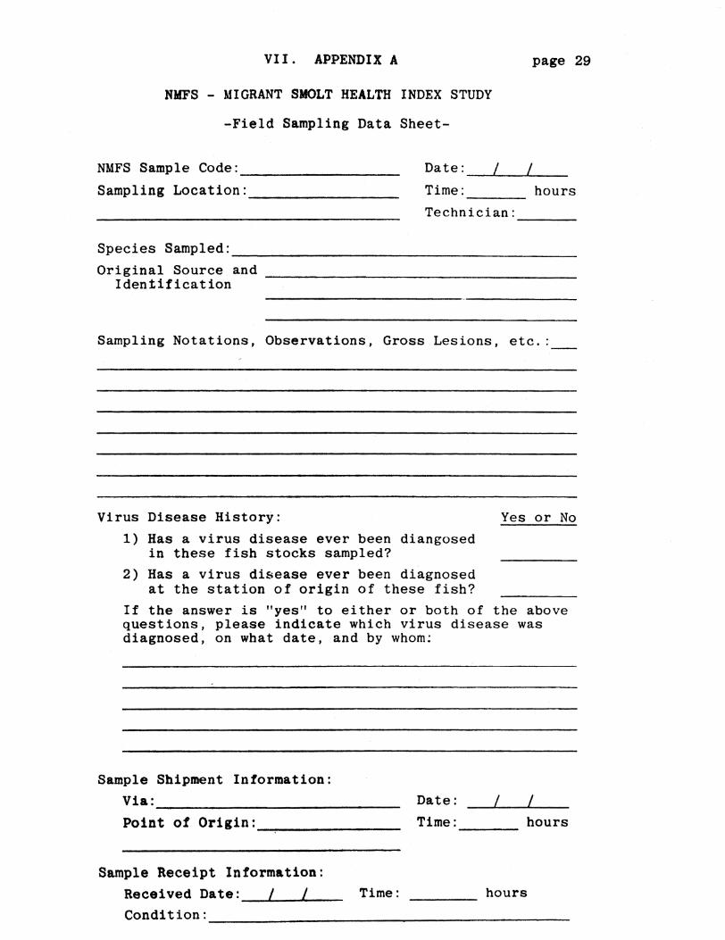
APPENDIX A
APPENDIX B
-
INTRODUCTION
The National Marine Fisheries Service (NMFS) under contract to the
Bonneville Power Administration is conducting research on imprinting
salmon and steelhead for homing (Slatick et a1 1979 1980 Novotny and
Zaugg 1979) The studies were begun with little background knowledge of
the effects of disease or certain physiological functions on imprinting and
homing in salmonids Consequently work aimed at filling this void was
begun by the authors in 1978 (Novotny and Zaugg 1979) and continued in
1979
In 1979 we examined random samples of normal populations of homing
test fish at the hatcheries to determine the physiological readiness to
migrate and adapt to seawater and general fish health At the Manchester
Marine Experimental Station Manchester Washington we determined the
survival of samples of the test fish maintained in marine net-pens after
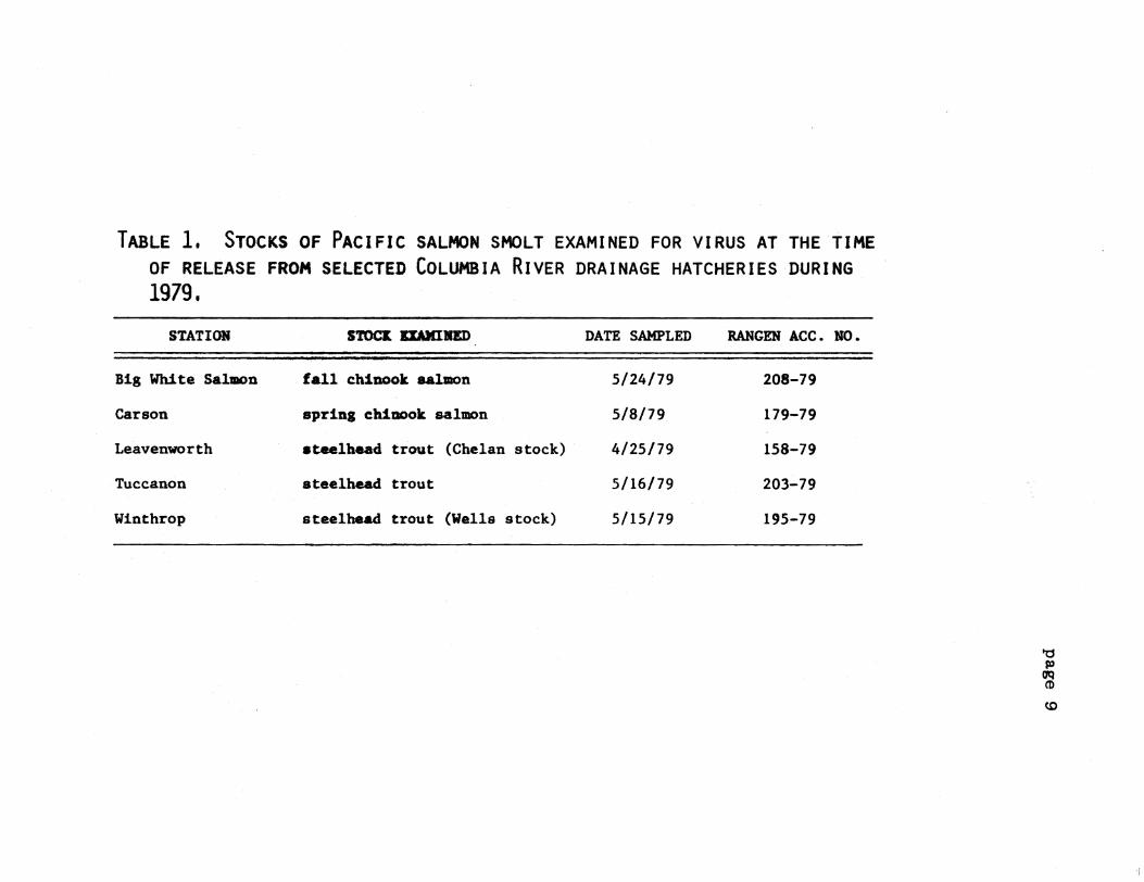
release from the hatcheries Hatcheries and stocks sampled are listed in
Table 1
The data collected from random samples were as follows
1 Physiology
Gill Na+-K+ ATPase Abnormally low values could be indications
that the fish were either in pre- or post-smolt condition or had been
stressed in some way
Plasma elec trolytes bull Lower than normal values of Na or Cl could
indicate immediate problems of osmoregulation when the fish were introduced
to seawater high values may indicate some dehydration due to stress
Increases in levels of K can indicate kidney failure or nitrogen
supersaturation stresses
Table 1--Hatcheries and stocks sampled in the 1979 homing studies
a a a Hatchery Date- Date of- Date of-Pathology arrived at viral blood Date transferred ATPase release
Hatchery Species tag no Manchester assay sampling to seawater pens profile dates
Chelan- Steelhead 6101-6160 42579 42679 42679 42779 Yes 42679 Leavenworth
Wells- Steelhead 3001-3060 51079 51179 51179 51279 No 5979 Winthrop
Tucannon Steelhead 6801-6860 51479 51679 51679 51679 Yes 51779
Carson Spring 6401-6460 50179 50479 50479 50379 Yes 50879 chinook
Big White Salmon Fall chinook 6301-6360 51879 51979 51979 52279 Yes 52179
~ Held in fresh water at Manchester for sampling nrior to transfer to seawater pens
1 )~ -j J bull ) J J ) ) J
Hematocrits and hemoglobins Values below or above normal ranges
usually indicate anemia or dehydration which can reflect nutritional
disease or physiological changes
2 Fish health
The incidence of diseases during freshwater rearing as reported in
hatchery records describing the treatment of fishwere examined
The extent of latent bacterial kidney disease (BKD) as determined by
indirect fluorescent antibody technique and the presence (or absence) of
certain pathogenic viruses were of particular interest
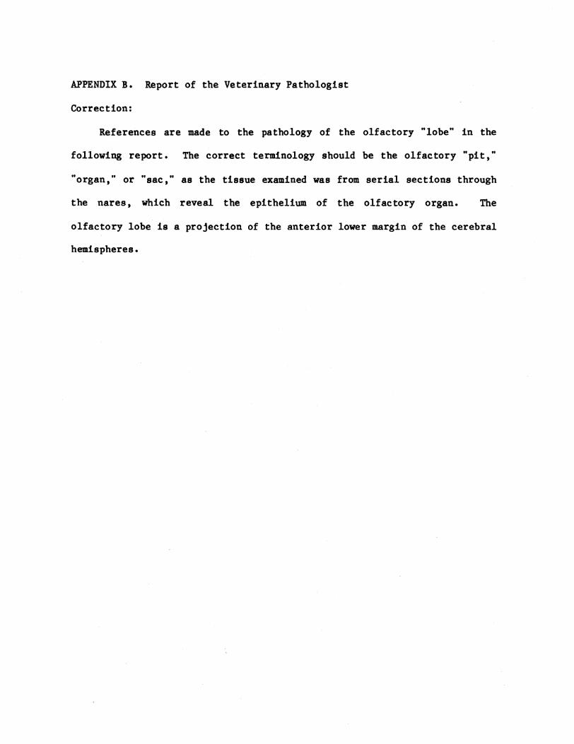
A histological determination of significant lesions or abnormalities
in tissue from the gill eye liver kidney thyroid brain and olfactory
sac was undertaken
3 Survival in seawater net-pens
Periodic assessments of survival and growth were made and the major
causes of mortality were determined
These surveys were conducted to provide a documentation of the health
and physiological (smolt) condition of the populations of fish involved in
the tests especially at the time of imprinting and release When the
marked adult fish return the data analyzed from the health and physiology
surveys should provide us with information that would indicate any adverse
influence on survival Low survival in the marine net-pens caused by poor
health or a low percentage of fish transformed through the smolting stages
could bias any attempts to relate returns to imprinting
METHODS AND MATERIALS
Hatchery Sampling
The sampling of fish from the hatchery stocks for health profiles was
based on a combination of statistics and economics Random sampling from
3
populations ranging as high as 100000 or more showed that a population
with a disease incidence of 5 or greater can be detected from a sample of
60 fish (Ossiander and Wedemeyer 1973) Health survey samples of 60 fish
were taken at the hatcheries and held in circular tanks in fresh water at
Manchester In most cases the tissue and blood samples were collected
within 24 hours after arrival at Manchester
Sampling for Physiology
Plasma Electrolytes
Sodium potassium and chloride ion levels in plasma were determined
for the Wells Dam Hatchery steelhead Salmo gairdneri and Big Creek
Hatchery fall chinook salmon Oncorhynchus tshawytscha near the time of
release Profiles of plasma electrolytes of the Tucannon and Chelan
Hatchery steelhead and Carson Hatchery spring chinook salmon were
determined in fresh water and during seawater culture at Manchester
Plasma sodium and potassium values were determined by atomic absorption
spectrometry and chlorides with a chloridometer
Gill Na+-K+ ATPase
During 1979 selected stocks of fall and spring chinook salmon and
steelhead trout being reared for release at state and federal hatcheries in
the Columbia River drainage were monitored for changes in gill Na+-K+
ATPase to evaluate the state of smoltification at release
From tagged releases we determined the relationship between the state
of smoltification at release and length of migration time from the hatchery
to the estuary
At approximately 2-week intervals during the spring and summer of
1979 30 fish were removed by dip net from representative ponds or raceways
at Tucannon Carson Leavenworth (Chelan) and Big White Salmon Hatcheries
4
Steelhead from the Wells Dam rearing ponds were sampled only at release
Ten groups of three fish each were anesthestized or killed by a blow on the
head at each sampling After weights andor fork lengths were determined
approximately equal quantities of gill filaments were removed from the gill
arches of each of the three fish in the group (total weight of gill
filaments-Ol to 02 g) and processed as described in our previous report
(Novotny and Zaugg 1979)
Disease Sampling
Life History of Hatchery Juveniles
Husbandry techniques disease and environmental history may have
deleterious effects on fish health and smolt quality (Wedemeyer et a1shy
1979J Folmar and Dickhoff 1979) Many chemotherapeutic compounds used in
the treatment of parasitic and bacterial diseases of fish may affect
smoltification (Lorz and McPherson 1976) and subclinical infections may be
exacerbated by the stress of seawater entry
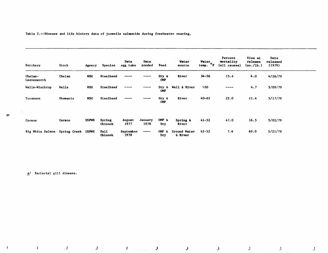
The information (Table 2) was obtained from hatchery management and is
self-explanatory Where information was not obtained the entries have
been left blank
Blood Sample Collection
The fish were lightly anesthetized in an aerated 120000 solution of
MS-222 In the larger fish blood was sampled from the caudal arch with a
1 cc heparinized syringe and a 25 gauge hypodermic needle Small fish were
bled by severing the caudal peduncle and collecting the blood in
heparinized capillary tubes
Blood samples taken for hematocrits (packed cell volume) were
5
Table 2--Disease and life history data of juvenile salmonids during freshwater rearing
Hatchery Stock Agency Species Date
egg take Date
ponded Feed Water
source Water
temp OF
Percent mortality
(all causes)
Size at release
(nolb)
Date released
(1979)
Chelan-Leavenworth
Wells-Winthrop
Tucannon
Chelan
Wells
Skamania
WDG
WDG
WDG
Steelhead
Steelhead
Steelhead
Dry amp OMP
Dry amp OMP
Dry amp OMP
River
Well amp River
River
34-56
50
40-62
154
220
40
47
114
42679
50979
51779
0
Carson
Big White Salmon
Carson
Spring Creek
USFWS
USFWS
Spring Chinook
Fall Chinook
August 1977
September 1978
January 1978
OMP amp Dry
OMP amp Dry
Spring amp River
Ground Water amp River
41-52
42-52
470
76
165
690
50279
52179
~ Bacterial gill disease
_J ) ~ J ) J ) I
J --
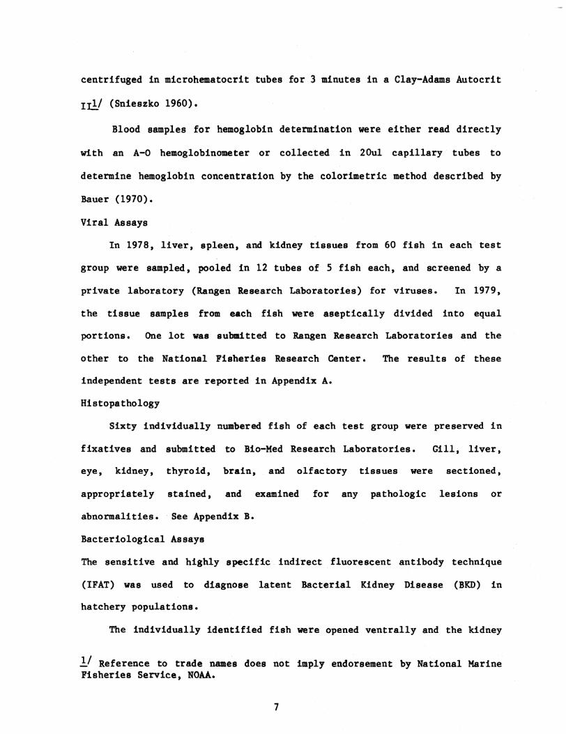
centrifuged in microhematocrit tubes for 3 minutes in a Clay-Adams Autocrit
II (Snieszko 1960)
Blood samples for hemoglobin determination were either read directly
with an A-O hemoglobinometer or collected in 20ul capillary tubes to
determine hemoglobin concentration by the colorimetric method described by
Bauer (1970)
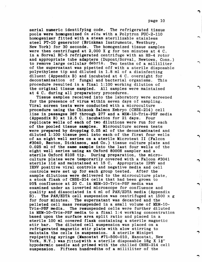
Viral Assays
In 1978 liver spleen and kidney tissues from 60 fish in each test
group were sampled pooled in 12 tubes of 5 fish each and screened by a
private laboratory (Rangen Research Laboratories) for viruses In 1979
the tissue samples from each fish were aseptically divided into equal
portions One lot was submitted to Rangen Research Laboratories and the
other to the National Fisheries Research Center The results of these
independent tests are reported in Appendix A
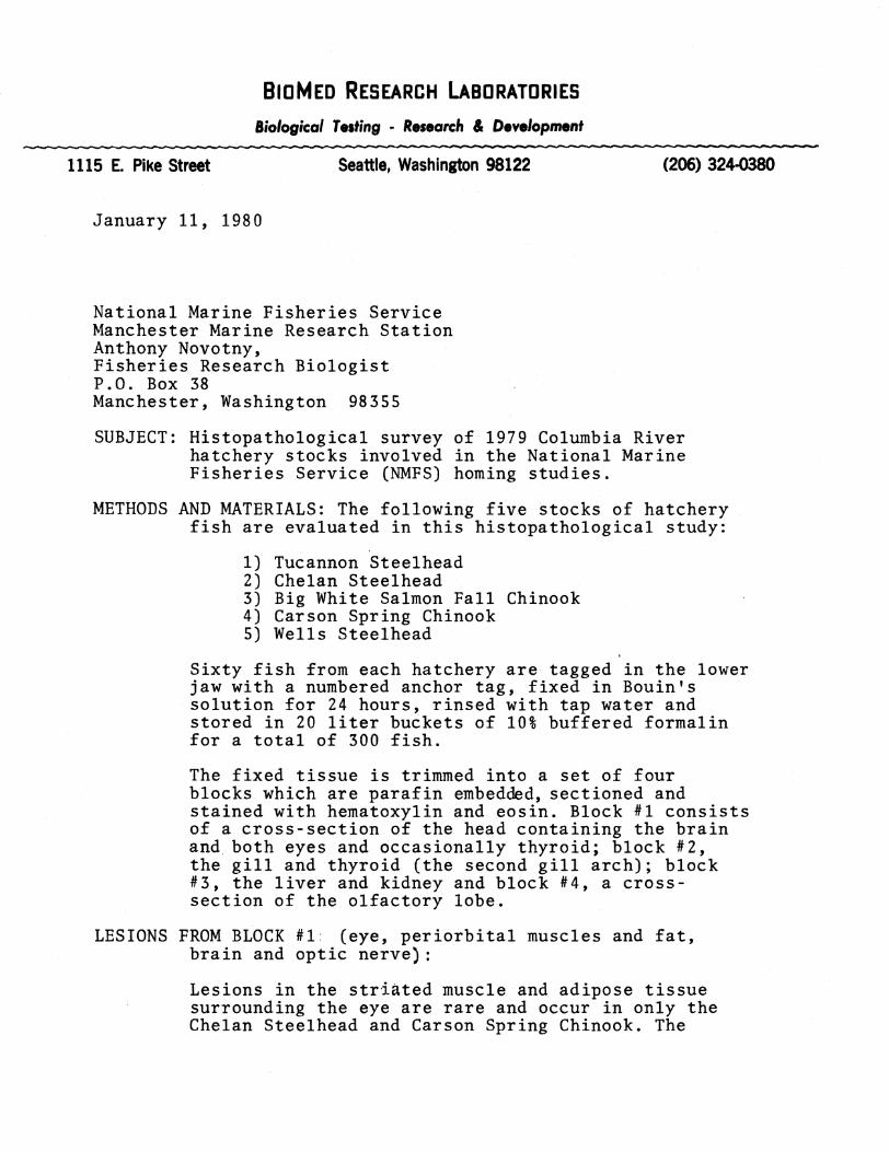
Histopathology
Sixty individually numbered fish of each test group were preserved in
fixatives and submitted to Bio-Med Research Laboratories Gill liver
eye kidney thyroid brain and olfactory tissues were sectioned
appropriately stained and examined for any pathologic lesions or
abnormalities bull See Appendix B
Bacteriological Assays
The sensitive and highly specific indirect fluorescent antibody technique
(IFAT) was used to diagnose latent Bacterial Kidney Disease (BKD) in
hatchery populations
The individually identified fish were opened ventrally and the kidney
l Reference to trade names does not imply endorsement by National Marine Fisheries Service NOAA
7
exposed Thin smears of anterior and posterior kidney tissue were made on
multi-spot slides after piercing the kidney with a sterile inoculation
loop The slides were air-dried and fixed in reagent grade acetone for 10
minutes The acetone fixed slides were stored at -20oC until they were
examined Prior to the sampling season 40 pos tive control slides were
prepared in the same manner and stored at -20oC The control slides were
prepared from a clean kidney lesion from a spring chinook salmon from
Carson National Salmon Hatchery that was tested and confirmed to have high
numbers of BKD organisms
The IFAT for BKD was originally described by Bullock and Stuckey
(1975) and later modified by G W Camenisch (unpublished report) of the
US Fish and Wildlife Service (FWS) Eastern Fish Disease Laboratory The
complete procedure used in this study is described in our previous report
(Novotny and Zaugg 1979)
All dead and dying fish in the seawater pens were collected daily
Each fish was opened from the vent external and internal lesions noted
and the procedures for culturing vibriosis and other gram negative bacteria
(Novotny Harrell and Nyegaard 1975) were followed
The postmortems were classified as follows
1 Negative (cause of death not determined)
2 BKD (from lesions)
3 Vibrio anguillarum--serotypes 775 1669 or 7244
4 Vibrio sp
S ERM (enteric redmouth)
6 Furunculosis
7 Aeromonas hydrophilia (ex liquefaciens)
8
RESULTS AND DISCUSSION OF HATCHERY STEEtHEAD SURVEYS
Chelan Hatchery (Transferred to Leavenworth Hatchery) Steelhead
Gill Na+-K+ ATPase
Since the phenomenon of elevation in gill sodium potassium stimulated
ATPase (Na+-K+ ATPase) activity was first reported to be associated
with parr-smolt transformation in steelhead (Zaugg and Wagner 1973) and in
in coho salmon ~ kisutch (Zaugg and McLain 1970) numerous experiments
have been conducted to verify these results and extend observations to
other species As a result it has been conclusively shown that the rise
in gill Na+-K+ ATPase activity is one of the many physiological changes
which occur at the time of parr-smolt transformation
The average gill Na+-K+ ATPase activity of Chelan Hatchery
steelhead sampled in 1979 at Leavenworth Hatchery was not substantially
different from 1978 (Table 3) Gill Na+-K+ ATPase showed only a small
rise in late April (Figure 1) with a peak mean value of 94 The absence
of a greater increase in activity may have resulted from water temperatures
which remained at the upper limit (13degC) for good smoltification during
late April and May (Zaugg et al 1972) The average fork length (208 cm)
was similar to 1978 (210 em) but the average weight (981 g) was up from
1978 (794 g)
Plasma Electrolytes
A compilation of data on rainbow trout by Miles and Smith (1968) and
Hickman et a1 (1964) suggests expected normal or near normal plasma
electrolyte values in fresh water of 130 to 172 meq (milliequivalents)1
for Na+ 14 to 60 meql for K+ and 111 to 155 meql of Cl-
Table 4 lists some known values for steelhead trout from available
published literature including data for the Dworshak Hatchery steelhead
9
Tabl 3--Floh health and phylololieal data for the 1979 tal atudy fish ed a eoariaon with the 1978 homnl study fhh~
8-day 8t seawater try 111 ATPe
Peak freshwter 8ill Be_to1081ltamp1 data
Plasma electrolytes )1 (HelI)
cgtf bUilt IU detectable I the kidney FAT
aet1VltY-1IIO~10 pl prhlt
ATPase aetivttyshyules P1118 prh
(taken at -helter pOe arrlYti)- - _Ra___ Cl ____It
Stoek and ~ecJ_ ~ Allterior Eitherl
lo_st~r1C)~IIo~h__b~t~__1It__~~ _ X_ SD Date
Actv1ty X
Ii_toedt _1 (I)
_aJobln loot i SD ft i SD i SD
Chelan-Leavenworth steelhead
(1979) (1978)
0 133
17 167
0 567
17 867
112 142
218 286
166 195
36 43
4-23-79 5-0]-78
94+22 i-5
4911 4]3
9 89
10 60
1540 1650
+22 ~148
10 58
1248 llO9
+21 ~172
10 60
25 11
+18 ~8
ileUs 5teelhead
(1979) (1978)
17 167
17 200
0 467
]4 834
IIot bullbullled 71 137 109 28
5-19-79 5-03-78
165-+92 17--0
SO8 556
96 114
39 58
1382 lSO3
+200 tIl 1
20 58
1329 1079
+112 1206
S9 58
161 25
+22 t2 bull6
Tucannon steelhead
(l979) (1978)
17 83
0 33
0 100
17 216
180 109
277 2]4
2211 176
31 46
5-08-79 5-08-78 5-22-78
259-+9 18--2 11 7
530 485
92 97
58 60
1407 1595
+113plusmntS
59 59
1270 1316
+87plusmn6 5
58 60
29 24
+17 26
Caron ( 1979) 100 33 200 333 258 382 n7 34 (up to 204+54 367 52 10 1456 plusmn42 10 1341 21 10 37 ~3 sPrlng chinook relebullbulle)
5-01-79
0
Bll hite sal faU c1l111OO1lt
(1979) 0 83 0 83 IIot lOPled 5-lt19-79 135 436 71 53 l703 156 53 24 14
al bl EI
Fro IIovotny ZampullI 1979 10 days for the 1978 holnR atudy taken upon arrival at Hanchuter
noh
J -l -J ) ) J ) J ) ) )
505 (+55)
March April May June 1979
+
bull F reshwate ATPase
--- --- Seawater ATPase
Hatchery release date
t
5
+ 200 fish transferred to seawater
+
after 208 days in seawater
Fish held in freshwater past normal release
o L-1~O----~20------3~Or-----9L-----~19~----2~9r-----~9----~1~9----~29~--~8
I
to z
Figure l--Gill Na+-~ ATPase activity (means and standard deviations) of the Chelan Hatchery steelhead in fresh water and seawater
11
+ +Table 4--A summary of plasma Na K and C1 values in stee1head trout (from published sources)
Condition Na+ C1 K+ Reference
June-July (55 Laboratory test
g fish) Freshwater Saltwater
x X
= 162 = 170
range 140-160 137-185
X = 60 Houston (1959)
(after 36 hours)
March-May (13-15 cm fish) Laboratory tests Freshwater (range of mean values)
102-149 105-161 Conte and Wagner (1965)
Spring 1975 Dworshak Hatchery (at release)
X = 1342 range 128-138
Newcomb (1978)
N
Captured at Little Goose Dam (downstream from Dworshak)
x = 1342 range 128-141
Laboratory tests (cont ro1 groups-Spring)
Mean values range fro~ 159 to 169 133 to 138 26 to 43
Newcomb (1978)
Individual values range from 155 to 182 128 to 144 23 to 52
) -l J ) ) J ) J ) ) J
(Newcomb 1978) Newcombs data are extensive represent reasonably large
sample sizes (15 to 25) and are probably good approximations of Columbia
River steelhead
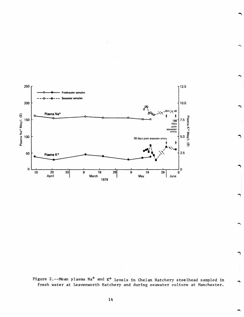
The mean plasma Na+ and K+ values of the Chelan Hatchery steelhead
were within normal limits (Table 3 Figure 2) at the time of release The
Na+ levels increased in seawater but returned to normal (in the
survivors) after 1 week
Hematology
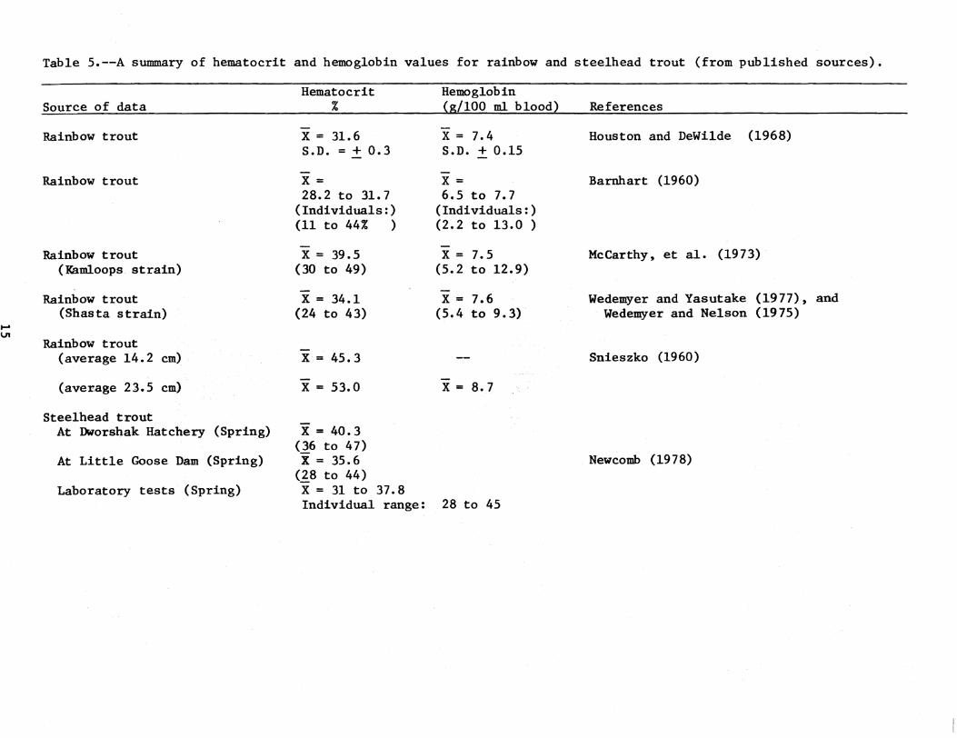
There is considerable hematological data in the literature for rainbow
trout less for steelhead trout From the data summarized in Table 5 it
may be possible to estimate the range of hematocrit and hemoglobin values
for healthy steelhead The lo~er limit of mean hematocrit should not fall
below 30 and mean hemoglobin values below 6 would certainly be suspect
Upper levels are more difficult to define Snieszko (1960) reports mean
hematocrits of 53 and mean hemoglobin levels of 87 g100 ml of blood in
rainbo~ trout of a size comparable to large steelhead smolts Although our
values on steelhead trout (Table 3) were much closer to Snieszkos Newcomb
(1978) reported mean hematocrit levels in steelhead similar to that found
by other researchers working on rainbow trout (Table 5) A number of
authors (McCarthy et a1 1973 Wedemeyer and Nelson 1975 Wedemeyer and
Yasutake 1977) repeatedly suggest that the hematocrit levels of clinically
healthy rainbow trout should be between 24 and 43 with hemoglobins
ranging from 54 to 93 g100 ml blood and these values will be used as
the expected range for individual fish for the purposes of this report
The summarized data of the hematocrit and hemoglobin values for the
Chelan Hatchery steelhead are presented in Figure 3 There was no
difference in mean hemoglobin between 1978 and 1979 (89 g100 ml) nor in
13
250 125
0 bull Freshwater samples
--0------ Seawater samples
200 100
~ 0-
Plasma Na+ ~~~-~~ ~ 0shy
lt) -0 ~ lt)---() Qj150 75 I)-= 188 IJ 3daysCIl Q)
post Aseawater+ +
entryZ s 100 50 g
30 days post seawater entry ~ co t t0 sect
~-~ 50 ) 25~
V
0 0 10 20 30 9 19 29 9 19 29 8
April March May June 1979
Figure 2--Mean plasma Na+ and ~ levels in Chelan Hatchery steelhead sampled in fresh water at Leavenworth Hatchery and during seawater culture at Manchester
14
Table 5--A summary of hematocrit and hemoglobin values for rainbow and steelhead trout (from published sources)
Hematocrit Hemoglobin Source of data (gIOO m1 blood) References
Rainbow trout x = 316 x = 74 Houston and DeWilde (1968) SD = + 03 SD + 015
Rainbow trout x = x = Barnhart (1960) 282 to 31 7 65 to 77
(Individuals) (Individuals) (11 to 44 ) (22 to 130 )
Rainbow trout x = 395 x = 75 McCarthy~ et al (1973) (Kamloops strain) (30 to 49) (52 to 129)
Rainbow trout x = 341 x = 76 Wedemyer and Yasutake (1977)~ and (Shasta strain) (24 to 43) (54 to 93) Wedemyer and Nelson (1975)
Rainbow trout (average 142 cm) x = 453 Snieszko (1960)
(average 235 cm) x = 530 x = 87
Steelhead trout At Dworshak Hatchery (Spring) x = 403
(36 to 47) At Little Goose Dam (Spring) X = 356 Newcomb (1978)
(~8 to 44) Laboratory tests (Spring) X = 31 to 378
Individual range 28 to 45
JI
6101 - 6160 April 26 1979 Steelhead Chelan
40 n= 60 x=49835 s= 77
30
c 25 0 ~ 208 E l z 15
10
5
0 1
Percent () Hematocrit 40
n= 60 x= 88635 s= 158
30
c
25 0
8 ~
E l z 15
10
5
0 2 4 6 8 10 12 14 20
Hemoglobin - gdcl blood
Figure 3--Frequency histogram for hematocrit and hemoglobin values for the Chelan Hatchery steelhe~ in 1979 Number of fish sampled (n) mean hemoglobin and hematocrit values (X) and standard devia~ion (s) are also given
16
the range of values Mean hematocrit in 1979 (498) was slightly higher
than in 1978 (434) and 85 of the fish had hematocrits above the
expected maximum of 43 (for rainbow trout) in 1979 compared to 50 in
1978 None of the hematocr1ts or hemoglobins fell below the minimum
expected values
Viral Screening
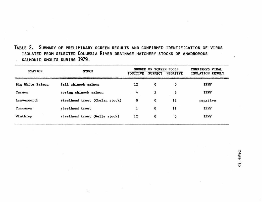
Both Rangen Research Laboratories and the National Fisheries Research
Center (USFWS) indicated that no IPN virus was present in the Chelan
(Leavenworth) Hatchery steelhead
Indirect Fluorescent Antibody Test for Bacterial Kidney Disease
One posterior kidney smear (17) was found to have a few BKD organisms
in the Chelan (Leavenworth) Hatchery stee1head
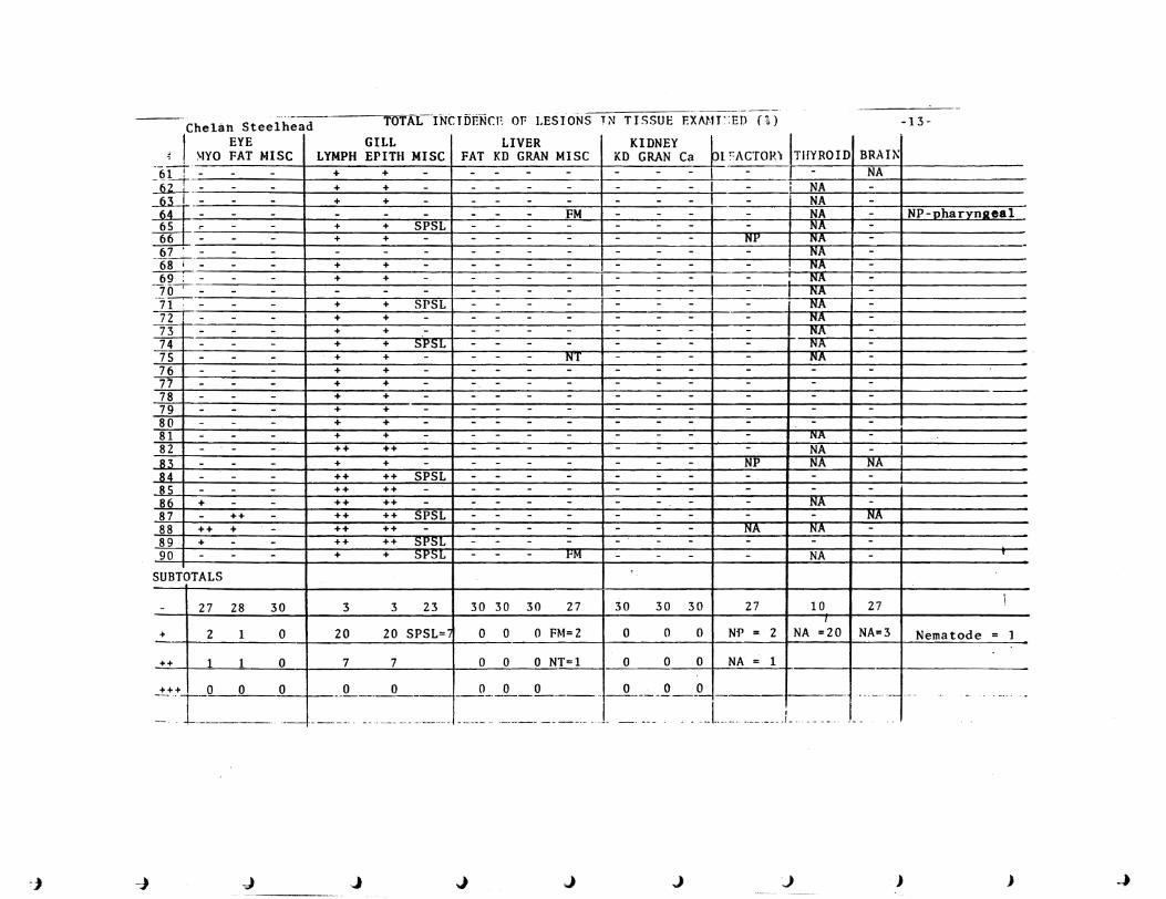
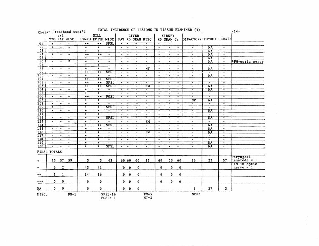
Histopathology
A detailed report on the examination and interpretation of selected
tissue sections from the random samples is presented in Appendix B A
summary of the pathological conditions observed their severity and their
frequency of occurrence is presented in Table 6 The severity is ranked
as I--recognizible (least severe) II--intermediate and III--severe
Note that the incidence of rank II and III severity was low for all
conditions encountered (Table 6)
The major pathological conditions encountered in the Chelan
(Leavenworth) Hdatchery stee1head were of lymphocyte infiltration and
epithelial hypertrophy in gill tissue and the presence of sporozoan
parasites
Records (Table 2) indicate the total mortality in the hatchery was
15
17
Table 6--Ptholo~tcal coodlt1oul oltaervd 1n the twina etod _d their percenta of incidence
lad_c (I)
Tu- ala-lAa_nb IIIIU -----raWIlle Canoa c1 etu fll Itt~1 pdD dl~1tI WI C t _1t
iJ iJ iJ ~ ~ I II III bull II III bull II III 11 III e- II III ~
Or~an ptnolollY =shy
17
Retrobulbar fat ledon 0 0 0 0 33 17 0 50 0 0 0 0 0 0 0 0 150 0 0 150 Acute focal bettormage 17 0 0 17 0 0 0 0 0 0 0 0 0
Skeletal uscle leloll8 0 0 0 0 100 17 0 117 0 0 0 0 0 0 0 0 133 0 150
0 0 0 0 0 0 0
Deal _nonvelar cell infiltration 0 0 0 0 17 0 0 17 0 0 0 0 0 0 0 0 0 0 0 0
Retrobulbar pyolranulo_cou tnfla_cion 0 0 0 0 17 0 0 0 0 0 0 0 0 0 0 0 200 00 200 Retrobulbar nuclear loftltractoa 0 0 0 0 0 0 0 0 0 0 0 0 0 0 0 0 67 0 0 67
G1ll ~creased nu1lbers of lyaphocytes 81 7 183 0 1000 717 233 0 950 800 0 0 800 950 17 0 967 983 0 0 983
poundpltneU41 cell proUferation 783200 17 1000 683 267 0 950 913 17 o 1000 950 50 o 1000 933 67 o 100
Ly~htic telangiectasis of C01)dary lampMllae 50 0 0 50 0 0 0 0 0 0 0 0 0 0 0 0 0 0 0 0 Solitary basophilic as In econ~ry lae11ae 67 0 0 67 0 0 0 0 0 0 0 0 0 0 0 0 0 0 0 0 SoUury eosiJlOph1Uc in bullbullcondary 1_11ee 33 0 0 33 0 0 0 0 0 0 0 0 0 0 0 0 0 0 0 0
lie_tude parasice 1ft secondary 1_11ae 17 0 0 17 0 0 0 0 0 0 0 0 0 0 0 omiddot 0 0 0 0 rocal granuloma tn lecoaclary 111bullbull 0 0 0 0 17 0 0 17 0 0 0 0 0 0 0 0 0 0 0 0 SlOrooaa paralte tn eeoary 111 0 0 0 0 267 0 0 267 0 0 0 0 0 0 0 0 0 0 0 0 Focal 111ft_tion at 1_11 bbullbullbull 0 0 0 0 0 0 0 0 17 0 0 17 0 0 0 0 17 0 0 17 Vascular telusiectbullbullia of the econdary 1_I1ae 0 0 0 0 0 0 0 0 17 0 middot0 17 0 0 0 0 0 0 0 0
Hucoputu1ent 11111 0 0 0 0 0 0 0 0 17 0 0 17 0 0 0 0 0 0 0 0
General laflaaat iOG 0 0 0 0 0 0 0 0 17 0 0 17 0 0 0 0 0 0 0 0
Liver
00
Focal mononuclear cell infiltration 33 0 0 33 L3 0 0 83 17 0 0 17345 0 0 345 241 0 0 241
Sporozoan parasite 17 0 0 17 0 0 0 0 0 0 0 0 0 0 0 0
JIonsuppuratlve triaditt 0 0 0 0 33 0 0 33 0 0 0 0 0 0 0 0 0 34Increased parenchyl fat 0 0 0 0 33 0 0 33 0 0 0 0 34 0
LelliOnA typically asaociated with bacterial kidney disea 0 0 0 0 0 0 0 0 0 0 0 0 0 0 0 0 52 0 0 52 0 0 0 0 0 0 0 0 0 0 0 0 0 69 0 0 69Htcrogranulomas 0 0 0
Capsular parasttlc aranuloa 0 0 0 0 0 0 0 0 0 0 0 0 0 0 0 0 17 0 0 17
KLdltY Us Lons typlcal1 y associated with bacterial kidney d18case 0 0 0 0 0 0 0 0 0 0 0 0 0 0 0 0 88 0 0 88 Microgranuloraas 0 0 0 0 0 0 0 0 0 0 0 0 0 0 0 0 18 0 0 18 Pyo~ranuloma8 nephritis 0 0 0 0 0 0 0 0 0 0 0 0 0 0 0 0 18 0 0 18 Pyogranulomatous periuretelitis 0 0 0 0 0 0 0 0 0 0 0 0 0 0 0 0 18 0 0 18 Dilated tubule with giant bacteria 0 0 0 0 0 0 0 0 0 0 0 0 0 0 0 0 18 0 0 18 Focal tubular degenerative aiant cells 0 0 0 0 0 0 0 0 0 0 0 0 0 0 0 0 18 0 0 18
01 factory sac Ciliated protozoan parasite 533 0 0 533 0 0 0 0 0 0 0 0 0 0 0 0 0 0 0 0 Mmatode parasite 33 0 0 33 0 0 0 0 0 0 0 0 0 0 0 0 0 0 0 0 PyogranulolllAtous inflammation of the olfactory sac 17 0 0 17 51 0 0 51 0 0 0 0 0 0 0 0 254 0 0 254 Acute focal hellOrrhage 0 0 omiddot 0 0 0 0 0 0 0 0 0 0 0 0 0 17 0 0 17 Infltion of t~e olfactory 8_~c _ __9___L _lL___0 __-1 __--1 o 17 _I_ _0 17 0 0 0 __ 9 17 0 0 17
----------- ------shyThyroid
Perifollicular thyrofd1ti 0 0 0 0 0 0 0 0 0 0 0 0 0 0 0 0 62 0 0 62
flrdn MOnonuclear menlgeal infiltration 17 0 0 17 0 0 0 0 53 0 0 53 0 0 0 0 19 0 0 19 Ence(hal1 t is 0 0 0 0 ) 0 0 0 0 0 0 0 0 0 0 0 38 0 0 38
Other ~ryngeal sporozoan parasite 17 0 0 17 0 0 0 0 0 0 0 0 0 0 0 0 0 0 0 0
Pha-rynleal nematode parasite 0 0 0 0 17 0 0 0 0 0 0 0 0 0 0 0 0 0 0 0
~f I - Recognlzable (least severe) 1 I - IntenMdiate
III - Severe
) )J -J- J J J J -J -J j
Seawater Adaptation
At the time of introduction to seawater the test group contained
mostly transitional and smolted fish with 7 classified as precocious
males These precocious fish had all died by 82 days of seawater
residence Cumulative losses due to osmoregulatory dysfunction during the
first 30 days in seawater were about 28 in spite of 48 of the fish being
smolted at seawater entry Reversion from an apparent smolt or
transitional stage to a transitional or parr stage did take place after 82
days in seawater This suggests that those fish which were judged to be
smo1ts based upon external characteristics were not physiologically true
amolts or that high seawater temperatures may have forced reversion
Vibrio Strain 775 was the bacterial pathogen most commonly isolated
from moribund fish The 30-day survival was 59 and the survival after
completion of the tests (207 days) was only 8 (Table 7)
Wells Hatchery (Transferred to Winthrop Hatchery) Steelhead
Gill Na+-K+ ATPase
In 1978 Na+-K+ ATPase activities were determined on representative
samples of Wells Dam Hatchery (rearing pond) steelhead sampled at Winthrop
Hatchery on 3 May These fish had average gill Na+-K+ ATPase activity
of 170 (plusmn 51) with values ranging from 116 to 266 The sampled fish
averaged 225 cm fork length and 1022 g body weight and were judged to be
in a good smolted condition
In 1979 the average length and weight of samples transferred to
Manchester were 217 em and 970 g (respectively) on 11 May 1979 The
average g11l Na+-K+ ATPase activity from Wells Hatchery fish at
Winthrop Hatchery on 19 May was 165 (plusmn 92) with values ranging from 82
to 371
19
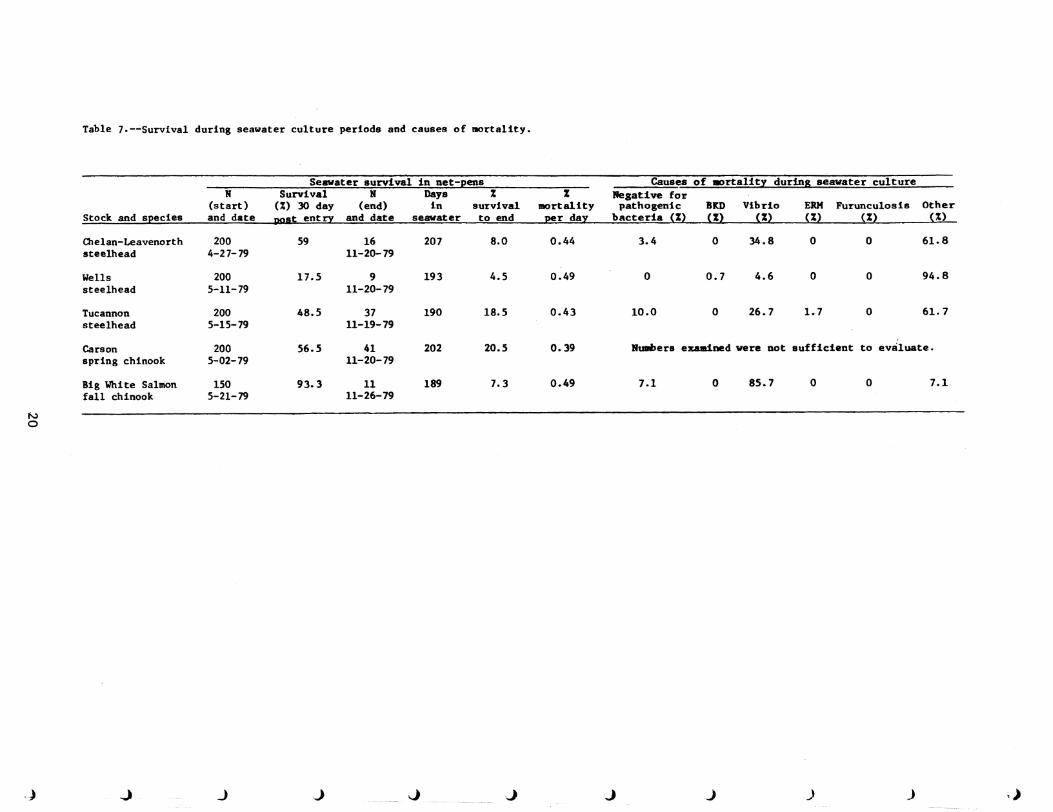
Table 7--Survival during seawater culture periods and causes of mortality
Stock and species
H (start) and date
Seawater survival in net-pens Survival N Days
()30 day (end) in survival post entry and date seawater to end
mortality per day
Causa of rtality during seawater culture Negative for pathogenic BKD Vibrio ERH Furunculosis Other
bacteria () () () el) () ()
Chelan-Leavenorth steelhead
200 4-27-79
59 16 11-20-79
207 80 044 34 0 348 0 0 618
Wells steelhead
200 5-11-79
175 9 11-20-79
193 45 049 0 07 46 0 0 948
Tucannon steelhead
200 5-15-79
485 37 11-19-79
190 185 043 100 0 267 17 0 61 7
Carson spring chinook
200 5-02-79
565 41 11-20-79
202 205 039 Numbers examined were not sufficient to evaluate
Big White Salmon fall chinook
150 5-21-79
933 1l 11-26-79
189 73 049 71 0 857 0 0 71
J) 0
)) J ) ) J J J j J J
Plasma Electrolytes
As in 1978 there were noticeable differences in the plasma
electrolytes of the Wells-Winthrop Hatchery steelhead when compared to the
other steelhead stocks in these studies (Table 3) There was only one
sample taken (at the time of release) but this stock again had the lowest
mean Na+ and K+ values even lower than in 1978 (Table 3)
There were no differences in transportation techniques or water
quality in 1979 between stocks thus no obvious stresses were involved
In 1979 plasma chloride was also measured In 1978 the mean plasma
chloride level (l08 meql) was below the expected value of III megl for
heal thy rainbow trout In 1979 none of the chloride values (Y 133
meql) were below normal
Hematology
The hematocrit and hemoglobin data for the Wells Hatchery steelhead
are presented in Figure 4 In 1978 the Wells Hatchery steelhead had the
highest mean hematocrit and hemoglobin values of any of the steelhead
stocks that we studied (Table 3) In 1979 849 were above the expected
maximum (43) hematocrit level and 517 above the maximum expected 93
g100 ml blood hemoglobin value None of the hematocrit or hemoglobin
values were below the expected minimums
Viral Screening
Rangen Research Laboratories reported IPN virus in 12 out of 12 pooled
samples in the Wells Hatchery steelhead and the National Fisheries
Research Center (USFWS) reported all samples negative for IPN virus
Indirect Fluorescent Antibody Test for Bacterial Kidney Disease
Two out of 60 fish s_pled (33) from the Wells (Winthrop) Hatchery
steelhead were found to have light BKD infections in anterior or posterior
kidney
21
i
3001 - 3060 May 11 1979 Steelhead Wells
40 n=53 lt=507735 s= 687
30
c 25CII 0
20 ~
Percent () Hematocrit 40
n= 60 x= 96135 s= 114
E ~ 152
10
5
a
30
-5 25 0 ~
20 E ~
2 15
10
5
0 12 20
Hemoglobin - gdcl blood
Figure 4--Frequency histogram for hematocrit and hemoglobin values for the tJells (Winthrop) Hatchery steelhead in 1979 Number of fish sampled (n) mean hemoglobin and hematocrit values (X) and standard deviations (s) are also given
22
Histopathology
The major pathological conditions encountered in the Wells (Winthrop)
Hatchery steelhead were increased numbers of lymphocytes and epithelial
hypertrophy in gill tissue (Table 6)
Seawater Adaptation
At entry to seawater Wells-Winthrop Hatchery steelhead were primarily
of transitional stage fish based upon external characteristics The
cumulative mortality within 30 days of seawater residence attributable to
osmoregulatory dysfunction was 73 Vibriosis accounted for most of the
other mortalities for the remainder of the study The presence of
precocious males was not a problem in this test group The survival to
test completion (193 days) was only 5 (Table 7)
Tucannon Hatchery Steelhead
Gill Na+-K+ ATPase
The gill Na+-K+ ATPase profile of summer-run steelhead from the
Tucannon Hatchery in fresh water was qualitatively similar to that observed
in 1978 with a distinct peak in enzyme activity in early May (Figure 5)
followed by a sharp decline A typical pulse of Na+-K+ ATPase activity
was observed when these fish were transferred to seawater at Manchester
Li tt1e change occurred until the fourth day when act ivi ty began to rise
(Figure 5) Fish held at the hatchery and sampled just 5 days after
release of the main group averaged 172 cm fork length and weighed 431 g
This was down from the average lengths and weights of 195 cm and
659 g respectively at the same time in 1978
Plasma Electrolytes
The summary data for plasma electrolytes at the time of release are
listed in Table 3 The mean values for Na K and Cl of the Tucannon
23
Hatchery release date
25 t x = 270 after 30 days and
bull Freshwater ATPase 369 at 188 days
- - - --- - - - Seawater ATPase
~lt I
O~1~O----~2~O----~3plusmnOr---~9----~19~--~29~----~9-----1~9----~29 March April May
1979
Figure 5--Gill Na+-~ ATPase activity (means and standard deviations) of the Tucannon Hatchery steelhead in fresh~ater and seawater
24
Hatchery steelhead fall within the expected ranges for rainbow trout
There was little difference in the mean Cl and K values between 1978
and 1979 (Table 3) In 1978 433 of the Tucannon Hatchery samples were
below the minimum range reported for K in rainbow trout
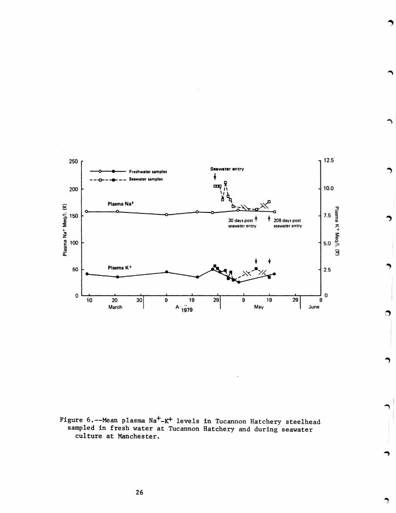
The plasma Na and K profiles of the Tucannon Hatchery steelhead in
both fresh and seawater appeared to be within normal ranges (Figure 6)
There was a typical rise in Na and K followed by a drop wi thin the first
week after transfer to the seawater pens and a return to normal in the
surviving fish after this initial stress period
Hematology
The summarized data of the hematocrit and hemoglobin values for the
Tucannon Hatchery steelhead are presented in Figure 7 and Table 3 The
mean hematocrit was slightly higher in 1979 than in 1978 and the mean
hemoglobin value was slightly lower than the 1978 mean
Viral Screening
The National Fisheries Research Center (USFWS) reported all Tucannon
Hatchery steelheadsamples tested as negative for IPN virus Rangen
Research Laboratories reported IPN virus in 1 out of 12 pooled samples
tested
Indirect Fluorescent Antibody Test for Bacterial Kidney Disease
Only 1 out of 60 Tucannon Hatchery steelhead sampled (17) was found
to have BKD organisms in an anterior kidney smear
Histopathology
The major pathological conditions encountered in the Tucannon Hatchery
steelhead were lymphocytic infiltration and epithelial hypertrophy in gill
tissue and a 53 incidence of ciliated protozoan parasites in the
olfactory sac (Table 6) Total mortality during rearing in the hatchery
25
-
~
~
125250
bull Seawater entry---lt) Freshwater samples __ ~ _ ___ _ _ Seawater samples +
i 100200 CDI
~ Plasma Na+
-0g A) 0-
b4=Q~ -00 ~ or 150 -ltgt- 75 IIgt
3i 30 days post + +208 days post III
IE seawater entry seawater entry + +
z CIgt
100 50 ~
c
a ~ t t
Plasma K+50 25
o L-l~0----~20------3~Or-----9~----1~9----~29~----~9~--~1~9~--~2~9~--~80 k~ A~~ M~ ~M
Figure 6--Mean plasma Na+-~ levels in Tucannon Hatchery steelhead sampled in fresh water at Tucannon Hatchery and during seawater
culture at Manchester
26
6801-6860 May 16 1979 Steel head Tucannon
40 n= 60 x= 529735 s= 766
30
i 25 j 0 E z 15
10
5
0
Percent () Hematocrit 40
n=60 x= 91835 s= 132
30
oS j 25 0 E z 15
bullo L-L-L__
Hemoglobin - gdcl blood
Figure 7--Frequency histogram for hematocrit and hemoglobin values for the Tucannon Hatchery steelhead in 1979 Number of fish sampled (n) mean hemoglobi~ and hematocrtt values (X) and s~andard deshyviations (s) are also given
27
was 22
Seawater Adaptation
At seawater entry 66 of the Tucannon Hatchery steelhead were judged
to be smolts Within 30 days of seawater residence 51 of the population
had died Osmoregulatory dysfunction accounted for 19 of the initial
mortality Vibriosis was the pathogen most commonly isolated from moribund
fish A large number of fish (25) were unaccounted for at the end of
testing and the loss may have been due to escape from the net-pen At
completion of the experiment (188 days) 185 of the fish had survived
The overall survival may have been closer to 40 if the losses from the
net-pen are included
I I
28
Summary
The compilation of the 1978 and 1979 data and preliminary analysis of
the 1980 data suggest that the generally high mean hematocrit and
hemoglobin values in northwest steelhead stocks may reflect a normal
hematological condition for these anadromous strains of the rainbow trout
or they may be associated with smoltification
The pathologist did not find histological lesions typically associated
wi th BKD in the kidney or liver tissue of the three steelhead stocks
examined Although the more sensitive IFAT tests of kidney tissue smears
from the same specimens did reveal the presence of BKD organisims the
incidence of the disease was extremely low There were no known
mortalities due to BKD in any of the steelhead stocks sampled during
seawater culture except the Wells Winthrop Hatchery stock Lesions
symptomatic of BKD were found in 07 of the mortalities examined but the
disease was not confirmed by IFAT
Analysis of the pathologists data indicate that lesions of the eye
were much reduced in comparison to 1978 Histopathological conditions
observed in the three steelhead stocks were restricted to a few organ
systems and may not significantly effect homing response or survival
However there were several conditions that appeared in all three stocks
and it may be of interest to summarize the probable causes
The pathological conditions in gill tissue of the 3 steelheadstocks
were predominately lymphocytic infiltration and epithelial hypertrophy
The incidence was higher in 1979 especially in the Tucannon Hatchery fish
These observations are probably indicative of exposure to antigens
including pathogenic and non-pathogenic microorganisms irritants or a
29
mild form of nutritional gill disease Second in frequency of occurrence
was the presence of sporozoan parasites in the gills of Chelan-Leavenworth
Ha tchery steelhead Pathological conditions in other tissues were minor
with the exception of a high incidence of ciliated protozoan parasites in
the olfactory rosettes or sacs of the Tucannon Hatchery steelhead
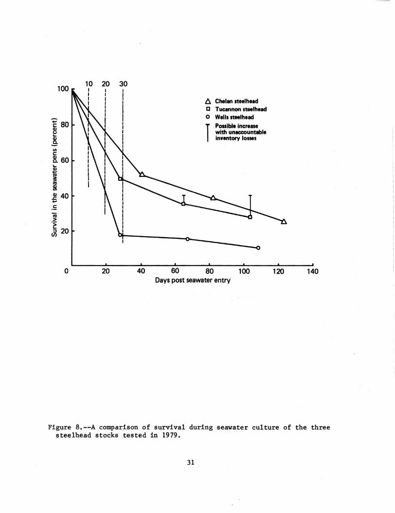
Figure 8 compares the survival of all three steelhead stocks during
seawater culture The survival wi thin the first 10 to 30 days after
transfer to the seawater pens was highest in the Chelan-Leavenworth
Hatchery stock and lowest in the Wells-Winthrop Hatchery stock This trend
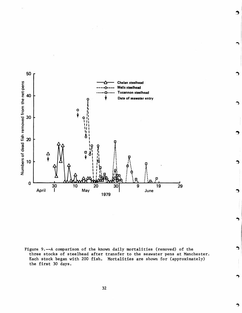
is again apparent in Figure 9 which compares the known (removed) daily
mortalities for the first 4 weeks after transfer to the seawater pens The
largest numbers of dead fish were removed from the pen of the
Wells-Winthrop Hatchery steelhead Over 72 of the mortalities that
occurred within the first 4 weeks were removed within 1 week after transfer
to seawater
Figure 10 is a comparison (for all three stocks) of the average fork
lengths (at release) of steelhead that could be separated into three states
of development based on visual observation These were (1) heavy parr
marks present (2) fish in transition to molting with parr marks still
discernible although faint and (3) silvery smolts parr marks absent
Although the average length of the Wells-Winthrop Hatchery steelhead was
much higher than the Tucannon Hatchery stock only 28 of the
Wells-Winthrop Hatchery stock had the visual appearance of smolts versus
66 for the Tucannon Hatchery stock The Chelan-Leavenworth Hatchery
steelhead were in between with 48 considered to be visibly smolted
However the large average size of the Chelan-Leavenworth Hatchery smolts
~I
I
30
100 20 30
-~ 80 ~ ~-
ll Chelan steelhead C Tucannon steelhead o Wells steelhead
1Pooible increase with unaccountable inventory losses
o 20 40 60 80 100 120 140 Days post seawater entry
Figure 8--A comparison of survival during seawater culture of the three steelhead stocks tested in 1979
31
50
11 --tr-- Chelan steel head c QJ ----0---- Wells steelheadC I ~ __0- Tucannon steel head ~40 t Date of seawater entryQJ
s ~
E ~ 0 o -0 30 t QQJgt I I I 0 I I I E I IQJ ~ II I
s
A20
I -0 It QJ 0 ~
D 11 I ~
0 - to Pshy11 ~ t I QJ 10 c I 6E I bull ~ I I Z I I I~ Q
I I II I I I C I~R
0 30 9 19 29
April I May June 1979
~
I
I
Figure 9--A comparison of the known daily mortalities (removed) of the three stocks of steelhead after transfer to the seawater pens at Manchester Each stock began with 200 fish Mortalities are shown for (approximately) the first 30 days
~
32
I
100 A Chelan steel head (X 2265 mm) c Tucannon steelhead (I 1661 mm)
- o Wells steelhead ur- 2109 mm)
1 parr ~ 80 2middot in transition to smolting ~ Q) 3 bull visibly smolted
9shy gto ~ 60 10 () 2 3Q)
iii N
s ~ Q)
40 5 C ca s Q)
Qj 20 ~ 1---_--1
o 140 160 180 200 220 240
Average fork length (mm)
Figure 10--Relationship of the percentage of abundance of the three visible physiological stages at the time of release to the average size of each state (for all three steelhead stocks studied in 1979)
33
probably contributed to the greater initial survival after transfer to
seawater
The clinical health status of the Wells-Winthrop Hatchery steelhead
was satisfactory The high initial mortality after transfer to seawater
(in comparison to the other steelhead stocks) suggests however a severe
osmoregulatory dysfunction We cannot directly compare this type of
immediate stress with a supposed transition through the Columbia River
estuary because culturing samples of these hatchery test groups in net-pens
in seawater is an artificial situation and is recognized as such Lower
survival may not be indicative of what is occurring in nature as (1) the
fish are transferred directly from fresh to 280 00 seawater without
conditioning in estuarine water (as presumably might be the case in
nature) (2) they are fed an artificial diet and (3) they are contained in
net-pens and stressed by frequent (monthly) measurement activities
Nevertheless one can assume that if the survival in the net-pens was
high the fish should be able to withstand the normal transition rigors in
the wild and that the tests may be a relative measure of seawater
adaptability between treatments or stocks In comparison to the other two
stocks the Wells-Winthrop Hatchery fish were much less likely to survive
any early osmoregulatory stresses
RESULTS AND DISCUSSION OF HATCHERY CHINOOK SALMON SURVEYS
Carson Hatchery Spring Chinook Salmon
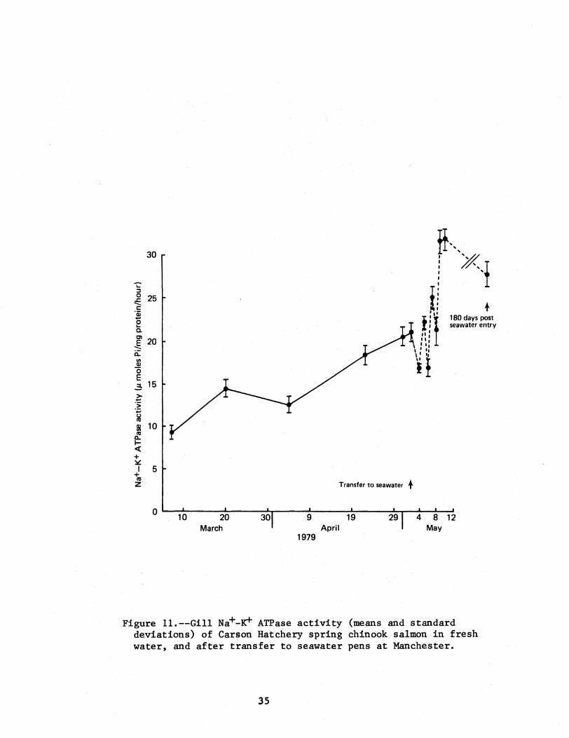
Gill Na+-K+ ATPase
Figure 11 is a graph of the gill Na+-K+ ATPase activity in 1979
The enzyme values were somewhat higher in 1979 than in 1978 The time
during which activity increased appeared to be the same in both years
34
H30
i Xi
Transfer to seawater
Ii t II 180 days post seawater entry
III f
it
+
0~-1~0------2LO-----3~0~----~9------1L9------2~9~-4L-~8~12
March April May 1979
Figure 11--Gill Na+-~ ATPase activity (means and standard deviations) of Carson Hatchery spring chinook salmon in fresh water and after transfer to seawater pens at Manchester
35
I ~
-1
i i
i
)
Plasma Electrolytes
There is little published data on normal plasma electrolyte levels in
hatchery chinook salmon Table 8 is a summary of the mean plasma Na Cl
and K values from chinook salmon that we previously examined The
exceptionally high K values in the Kalama Hatchery spring chinook salmon
may be due to hemolysis that occurred after sampling
There is a slight decrease of plasma Na in the Carson Hatchery spring
chinook salmon from 158 meq1 in March until the fish were released in
early May (Figure 12) The mean plasma Na value of 1456 meq1 at this
time (Table 3) is within the range of means encountered in other chinook
salmon samplings (Table 8) As expected plasma Na values rose abruptly
after transferring the fish to seawater (Figure 12) but quickly returned
to normal levels The mean chloride level of 1341 meql at release is
higher than any previously observed mean levels in any of the species
studied in 1978 or 1979 The mean plasma K value of 37 meql was also the
highest for any species studied in the past two years J with the exception
of the coho salmon There were fluctuations in the mean K levels after
transfer to seawater for the first week and then a leveling off in the
surviving fish to 3 to 35 meql
Hematology
Unpublished data from salmon diet studies in Oregon indicate expected
mean hematocrits for spring chinook salmon ranging from 242 to 380 and
35 to 39 for fall chinook salmon Published data on small fall chinook
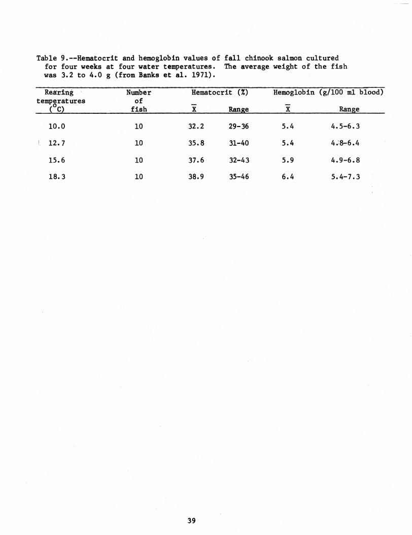
salmon fingerlings (Banks et al 1971) indicate that hematocrit and
hemoglobin values increase as the water temperature increases (Table 9)
36
Table 8--Mean values of plasma Nat CIt and K from other samplings of hatchery chinook salmon
37
Sample Ntlliequivaleritsl
1978 Kalama Falls Hatchery spring chinook salmon (at release) 137 116 119
1978 Kooskia Hatchery spring chinook salmon ~at release) 114 104
1978 Leavenworth Hatchery spring chinook (at release) 150 108 17
1979 Leavenworth Hatchery spring chinook
March At release (late April) June
158 149 148
129 30 125 08 130 23
These were abnormally high potassium values and may have been due to some hemolysis of the samples
250 125
-(0)--- Freshwater samples __~ _ _ _ _ _ _ Seawater samples
200 100
~
E a-150 III
E + co Z co
100 i co 0
50
Plasma Na+ ltgt Cgt
Plasma K+
~_______~~_- ______o
- fa -------shy
bull 1M -------
or75 3
7+
s 50 ~
~
25
o~----~~----~----~----~----~----~----~~----------Jo20 30 9 19 29 9 19 29
March April May 1979
I ~
I
I
Figure 12--Mean plasma Na+ and K+ levels in Carson Hatchery spring chinook salmon in fresh water at Carson Hatchery and during seawater culture at Manchester
~I
38
Table 9--Hematocrit and hemoglobin values of fall chinook salmon cultured for four weeks at four water temperatures The average weight of the fish was 32 to 40 g (from Banks et al 1971)
Reiirtng Number Hematocrit () Hemoglobin (g100 m1 blood) temperatures
(oe) of
fish X Range X Range
100 10 322 29-36 54 45-63
127 10 358 31~40 54 48-64
156 10 376 32-43 59 49-68
183 10 389 35-46 64 54-73
39
I ~i
The mean values of fall and spring chinook salmon sampled in 1978 (for
all studies) ranged from (1) hematocrits - 367 to 594 and (2)
hemoglobins - 52 to 89 g Hb100ml blood Hematocrit values below 28 in
Pacific salmon may be the beginning stages of a number of problems and
should signal a cautionary warning
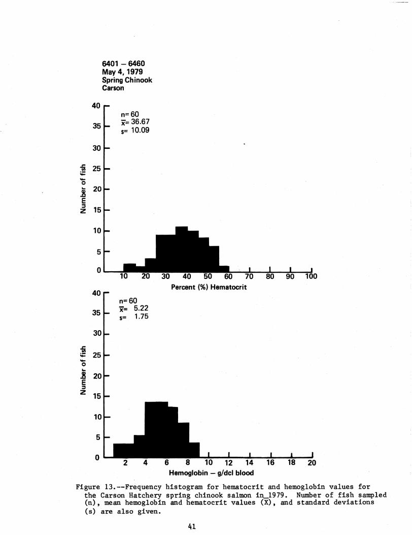
Although the mean hematocrits and hemoglobins of the Carson Hatchery
stock were well within the expected limits 183 of the fish had
hematocrits below 28 (Figure 13) Most of the low hemoglobin values
laquo 30 g HblOO ml blood) were associated with the low hematocrits
Bacterial kidney disease organisms were present in all samples with
hematocrits below 25
Indirect Fluorescent Antibody Test for Bacterial Kidney Disease
The Carson Hatchery spring chinook salmon had the highest incidence of
BKD (as determined by FAT) for any of the homing stocks screened (Table
3) The total incidence was 333 with 25 of these classified as level
III (severe) All fish with hematocrits below 25 were positive for BKD
However not all BKD infected fish had low hematocrits Latent infections
of BKD may require several years to develop into an active form capable of
killing fish in the marine environment (Ellis et al 1978) On the basis
of the number of fish with heavy intensities of BKD infection in our
subsamp1e we would antiCipate some ocean mortality from BKD
Histopathology (See Appendix B)
The Carson spring chinook salmon had a high incidence of epithelial
hyperplasia and lymphocytic infiltration in the g11ls (Table 6) This
inflammatory response is probably the result of exposure to a number of
pathogenic andor non-pathogenic organisms The relatively high incidence
40
40
35
30
c 25III Ot 0 20 E z = 15
10
5
0
40
35
c III
Ot 25 0 E = z 15
5
o
6401 - 6460 May 41979 Spring Chinook Carson
n=60 x= 3667 5= 1009
Percent () Hematocrit
n=60 x= 522 5= 175
Hemoglobin - gdcl blood
Figure 13--Frequency histogram for hematocrit and hemoglobin values for the Carson Hatchery spring chinook salmon iIL1979 Number of fish sampled (n) mean hemoglobin and hematocrit values (X) and standard deviations (s) are also given
41
of focal mononuclear cells in the liver is also indicative of possible I
I
~
I
I
~i
~
antigenic stimulation This is supported by the confirmation of bacterial
kidney disease in this stock In addition the Carson Hatchery spring
chinook salmon had the highest incidence (25) of granulomatous
inflammation of the olfactory sac which may be associated with
gramulomatous lesions of the liver and kidney typical of BKD
Seawater Adaptation
At the time of seawater entry the Carson Hatchery spring chinook
salmon were visually characterized as primarily transitional (55) and
smolted (39) fish The mean weight of the smolted fish was 258 g and the
mean weight of the population sample was 230 g
In early summer 13 precocious males were observed in the surviving
population If this represented the maximum knumber in the original
population there would be a minimum loss of 65 from precocious
maturation
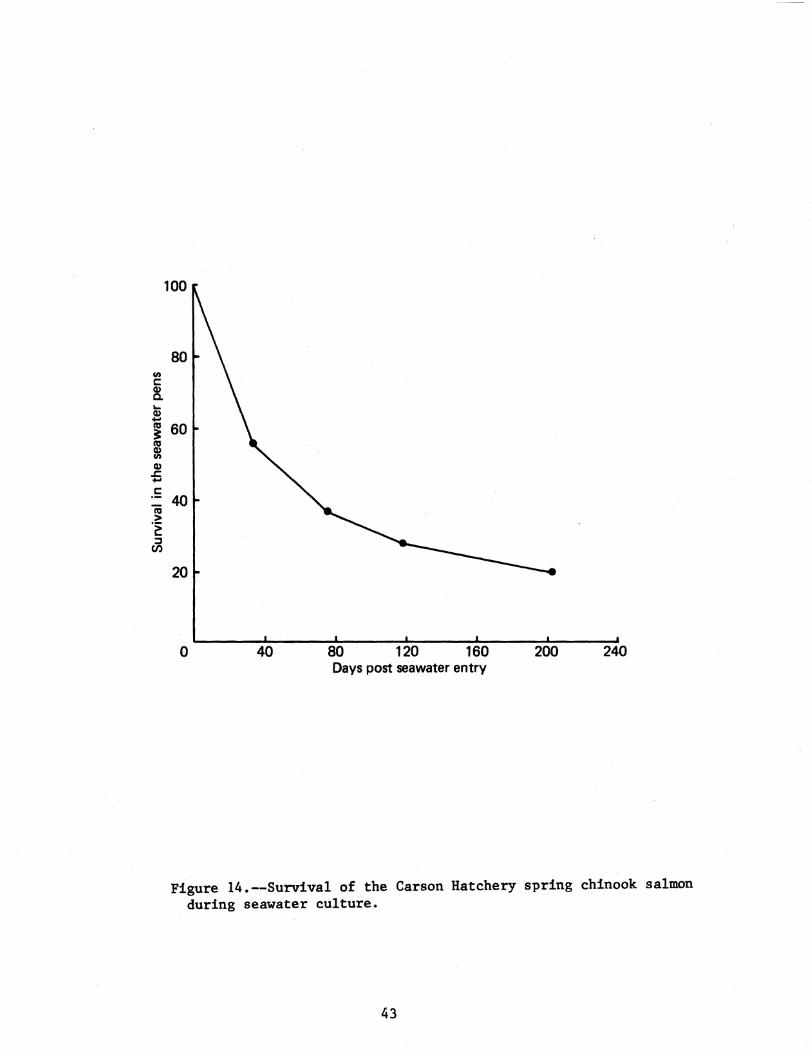
Initial losses due to osmoregulatory dysfunction were minimal (6)
Further losses occurring during the third and fourth week after seawater
entry were probably due to seawater diseases The survival during
seawater culture is shown in Figure 14 and indicates that approximately
60 of the fish were able to survive the first 30 days
Big White Salmon Hatchery Fall Chinook Salmon
Gill Na+-K+ ATPase
Fall chinook salmon used in the homing experiments were tagged and
moved from the Spring Creek Hatchery to holding ponds on the Big White
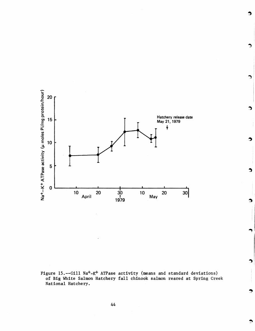
Salmon River Gill Na+-K+ ATPase activities are plotted in Figure 15
Gill Na+-K+ ATPase in the homing study fish showed an increase in
42
100
80
20
o 40 80 120 160 200 240 Days post seawater entry
Figure 14--Survival of the Carson Hatchery spring chinook salmon during seawater culture
43
-5 ~
20E c ~
-o ~
C- Hatchery release date Ol 15 E May 211979
c t en ~ o E 10 3shygt t gtji () CO 5 ~ rf Ishyet + ~ 0 ~----~------~-------+------~------~------~I + 10 20 30 10 20 30
CO April I MayZ 1979
) I
Figure 15--Gill Na+-~ ATPase activity (means and standard deviations) of Big White Salmon Hatchery fall chinook salmon reared at Spring Creek National Hatchery
44
activity at approximately the same time as fish held at Spring Creek
Hatchery though the magnitude of that increase was less Colder water at
the Big White SalDlon River and slower growth were factors that probably
affected the gill Na+-K+ ATPase activity
Plasma Electrolytes
The mean plasma Na levels of 1703 meql from the Big White Salmon
Hatchery fall chinook salmon at the time of release were the highest of any
of the species of salmonids sampled in 1978 or 1979 The mean K level of
24 meql was within normal limits for chinook salmon Only small amounts
of plasma are available from fall chinook salmon and the volumes were not
sufficient to include chloride analyses No samples were taken after
transfer to seawater
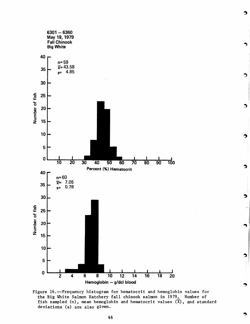
Hematology
The mean hematocrit and hemoglobin values (Figure 16) of the Big White
Salmon Hatchery fall chinook salmon were in the upper level ranges for fall
and spring chinook salmon sampled in 1978 None of the hematocrits were
below 28 and the hematological data suggested a healthy stock of fish
Viral Screening
The National Fisheries Research Center (USPWS) reported all of the Big
White Salmon Hatchery fall chinook samples negative for IPN virus Rangen
Research Laboratories reported 4 out of 12 pooled samples as positive for
IPN virus
Indirect Fluorescent Antibody Test for Bacterial Kidney Disease
There was an 83 incidence of BKD in the BIg White Salmon Hatchery
fall chinook sa11l0n all associated with posterior kidney However two
(out of five) of the fish had moderate (level II) infections This could
represent an eventual loss from BKD after release
45
6301 - 6360 May 19 1979 Fall Chinook Big White
40 n=59 gtlt=435835 485 5=
30
~ en 25 ij 4shy0 20 E z ~
15
10
5
0
Percent () Hematocrit 40
n=60 x= 705
35 078s=
30
~ en ~ 25 4-0
20 E
Hemoglobin - gdcl blood
~
z 15
10
5
0
II
~
~
~
I II
1
~
Figure 16--Frequency histogram for hematocrit and hemoglobin values for
the Big White Salmon Hatchery fall chinook salmon in 1979 Number of fish sampled (n) mean hemoglobin and hematocrit values (X) and standard deviations (s) are also given
46
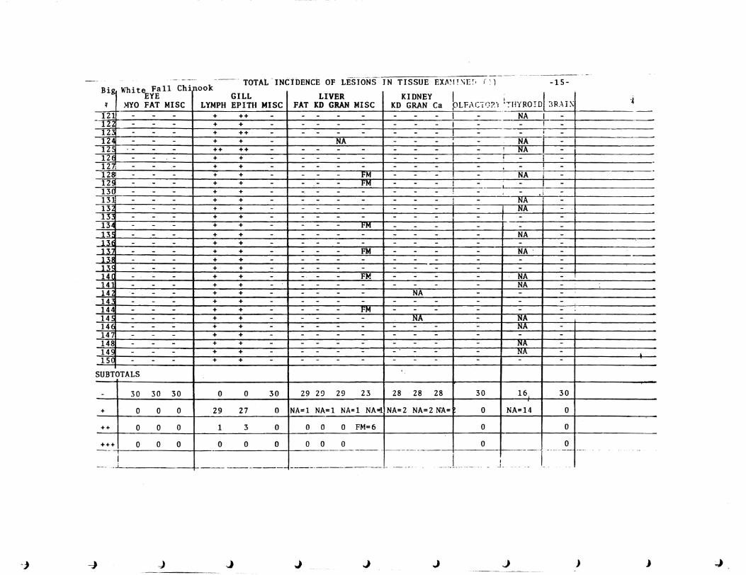
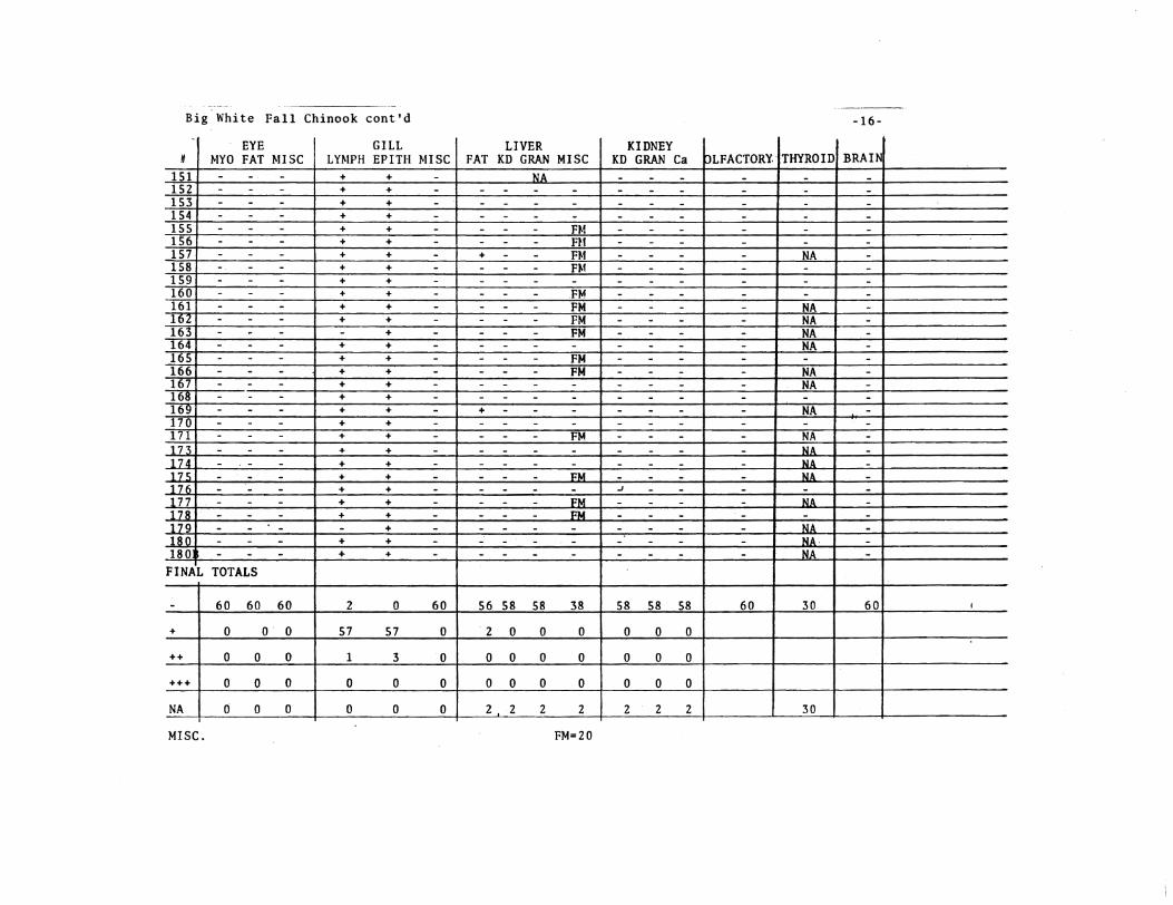
Histopathology
As reported for all homing test groups in 1979 the Big White Salmon
fall chinook salmon had a high incidence of epithelial cell proliferation
and lymphocytic infiltration in the gills (Table 6) This stock suffered
mortalities from enteric red mouth disease (Yersinia ruckerii) after they
were transferred to the Big White Salmon Hatchery rearing ponds and this
may have contributed to the development of these lesions The high
incidence of focal mononuclear cells in the liver is indicative of possible
antigenic stimulation and is consistant with the confirmation of bacterial
kidney disease by IFAT
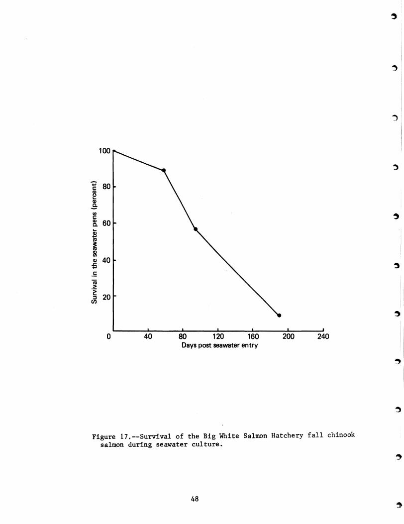
Seawater Adaptation
The mean weight of the Big White Salmon Hatchery fall chinook salmon
sample was 76 g when introduced to seawater and only 15 of the test
group were visually characterized as smolts (mean weight of 97 g) The
majority of the remaining fish appeared to be in the transitional stage
The immediate mortality from osmoregulatory stress was slight and the
survival during the first 30 days in seawater was over 90 (Figure 17)
The mortality increased dramatically after 60 days in seawater and was due
primarily to seawater diseases No precocious males were observed in this
test group
47
100
Figure 17--Survival of the Big White Salmon Hatchery fall chinook salmon during seawater culture
48
Summary
We began our first examination of the olfactory or sac in 1979 since
this tissue may be the source of cuing for any olfactory imprinting We
were not able to measure responses to olfactory stimuli in fish with
obvious damage to olfactory sac epithelial tissue but we have been able to
demonstrate that this sensory receptQr can be the site of pathological
conditions in normal populations
For example as mentioned in the section on steelhead ciliated
protozoans in the olfactory sac were found in 53 of the Tucannon
steelhead Histological evidence of any type of olfactory sac pathology
was neglible in the two other steelhead stocks and no pathology was found
in the Big White Salmon fall chinook salmon No parasites were found in
the olfactory sacs of the Carson Spring chinook salmon but evidence of
some type of inflammatory lesion was found in over 25 of the Carson fish
The pathologist suggests that the lesions found in both the kidney and
olfactory sac are highly indicative of bacterial kidney disease and that
the nares may be a portal of entry for the organism (Appendix B) As a
result of this assessment we examined tissue smears of the olfactory sac
by IFAT of the Leavenworth and Carson Hatchery spring chinook salmon from
the following year class BKDorganisms were found in many samples
including some in the Class II and III intensity Until we have sufficient
tag return data for any of the 1979 homing study releases of fish it will
be difficult to assess any problems that may have been due to pathology of
the olfactory sac
In 1979 the general health status of the Big White Salmon Hatchery
fall chinook salmon stocks was good in comparison to the Carson Hatchery
49
I ~
spring chinook salmon There were no problems with hematology in the fall
chinook salmon but over 18 of the spring chinook salmon had low
hematocrits and hemoglobins Bacterial kidney disease organisms were found
in every kidney sample of fish with hematocrits below 25 Histological
examination of the gills and other organ tissues suggest a serious problem
exists with BKD (Appendix B) in the Carson fish Although the Big White
Salmon Hatchery fall chinook salmon had been exposed to some microscopic
organisms (Appendix B) this was probably Enteric Redmouth Disease and at
the time of release most of the fish were clinically healthy There was no
evidence of abnormal indicators of smoltification in either stock but the
short-term (30 day) survival of the Carson fish in the seawater net-pens
was poor In general a combination of health status evaluation factors
plus a high incidence of precocious males indicate that losses in the
Carson Hatchery spring chinook salmon are going to occur prior to or during
the first winter after release
CONCLUSIONS
Steelhead
1 The general health of the three steelhead stocks in the 1979 studies
was good and there were no indications of any pathology that would impair
survival or imprinting wi th two possible exceptions the presence of
sporozoan parasites in the g111s of over 25 of the Chelan Hatchery fish
and protozoan parasites in the olfactory sacs of over 50 of the Tucannon
Hatchery fish Unfortunately t we cannot evaluate the impact of such
parasitic infestations at this time
2 Observations of external appearances to determine the extent of
smoltification indicated that less than 33 of the Wells-Winthrop Hatchery
50
steelhead were smolted at the time of transfer to seawater (no gill
Na+-K+ ATPase profiles were available for this stock) Both the Chelan
and Tucannon Hatchery steelhead were transferred shortly after peak g111
Na+-K+ ATPase activi ties and had a higher percentage of visible smolts
than the Wells-Winthrop fish This indicates that the Tucannon and Chelan
fish were better prepared for the transition to seawater
3 The survival of the Tucannon and Chelan Hatchery steelhead during the
first 30 days after transfer to the seawater pens was two to three times
greater than that of the Wells-Winthrop stock even though the average size
of the Wells-Winthrop fish was larger This indicates that the expected
survival of themiddot Wells-Winthrop Hatchery steelhead will be less than
survival from the Chelan and Tucannon Hatcheries
Chinook Salmon
1 lbe incidence of latent BKD in the Big White Salmon Hatchery fall
chinook salmon was low basic hematology was normal and the pathology of
examined organ tissue reflects the probable exposure to ERM and BKD The
recovery from ERK was apparently successful and the fish were healthy when
transferred
2 Approximately 20 of the Carson Hatchery spring chinook salmon were
anemic and BKD organisms were found in every fish with hematocrits below
25 BKD was found in about 33 of the Carson Hatchery fish and the
intensity of infection was heavy in 25 of the infected fish The presence
of granulomatous lesions typical of BKD in 25 of the olfactory sacs also
reflects the serious nature of this disease in the Carson fish and may
well affect imprinting as well as survival
51
3 The g11l Na+-K+ ATPase profiles were normal for both the Carson and
Big White Salmon chinook salmon and the major releases were made just
after the peaks of Na+-K+ ATPase activity indicating good preparation
for seawater adaptation
4 About 90 of the Big White Salmon fish survived the first 30 days in l I
the seawater pens Slightly over 50 of the Carson fish survived the first
30 days in the seawater pens This indicates that the chances for survival
of the Big White Salmon Hatchery fall chinook salmon are excellent and the
chances for the Carson Hatchery spring chinook salmon are below normal
52
LITERATURE CITED
BANKS J L L G FOWLER and I W ELLIOTT 1971 Effects of rearing temperature on growth body form and
hematology of fall chinook fingerlings Progressive Fish-Culturist 33(1)20-26
BARNHART R A 1969 Effects of certain variables on hematological characteristics
of rainbow trout Trans Amer Fish SOc 98(3)411-418
BAUER J D 1970 Hemoglobin In Gradwohls Clinical Laboratory Methods and
Diagnosis S Frankel S Reitman and A C Sonnenwirth editors C V Mosby Co St Louis 7th Ed Vol 1 p 403
BULLOCK G L and H M STUCKEY 1975 Fluorescent antibody identification and detection of the
corynebacterium causing kidney disease of salmonids J Fish Res Bd Can 32(11)2224-2227
CONTE F P and H H WAGNER 1965 Development of osmotic and ionic regulation in steelhead trout
SalDlo gairdneri Compo Biochem Physiol ~603-620
ELLIS R W A J NOVOTNY and L W HARRELL 1978 Case report of kidney disease in a wild chinook salmon
Oncorhynchus tshawytscha in the sea Journ Wildlife Dis 14120-123
FOLMAR L C and W W DICKHOFF 1979 Plasma thyroxine and gill Na+-K+ ATPase changes during
seawater acclimation of coho salmon Oncorhynchus kisutch Compo Biochea Physiol 63A(2) 329-332
HICKMAN C P JR R A McNABB J S NELSON E D VAN BREEMEN and D COMFORT
1964 Effect of cold accliaation on electrolyte distribution in rainbow trout (Sa10 gairdneri) Can Journ Zoo 42577-597
HOUSTON A H 1959 Osmoregulatory adaptation of steelhead trout (Salmo gairdneri
Richardson) to seawater Can J Zool ~729-747
HOUSTON A H and M A DEWILDE 1968 Hematological correlations in the rainbow trout (Salmo
galrdnerl) J Fish Res Bd Can 25173-176
53
1ORZ H W and B P McPHERSON 1976 Effects of copper or zinc in fresh water on the adaptation to
seawater and ATPase activity and the effects of copper on migratory disposition of coho salmon J Fish Res Bd Can 33(9)2023-2030
McCARTHY D H J P STEVENSON and M S ROBERTS 1973 Some blood parameters of the rainbow trout (Salmo gairdneri)
J Fish BioI 51-8
MILES H M and L S SMITH 1968 Ionic regulation in migrating juvenile coho salmon
Oncorhynchus kisutch Comp Biochem Physiol 26381-398
NEWCOMB T W 1978 Some physiological effects of total gas pressure supersaturated
water on juvenile Pacific salmon and steelhead trout a laboratory and field study PhD theSis University of Washington Seattle 165 p
NOVOTNY A J L W HARRELL and C Nyegaard 1975 Vibrosis a common disease of Pacific salmon cultured in marine
waters of Washington Wash State Univ Ext Bull 663 8 p
NOVOTNY Anthony J and WALDO S ZAUGG 1979 Study of disease and physiology in the 1978 homing study
hatchery stocks--a supplement to Imprinting salmon and steelhead trout for homing by Slatick Novotny and Gilbreath January 1979 Processed
OSSlANDER F J and G WEDEMEYER 1973 Computer program for sample sizes required to determine disease
incidence in fish populations J Fish Res Bd Can 30 1383-1384
SLATICK E A J NOVOTNY and L G GILBREATH 1979 Imprinting salmon and steelhead trout for homing WAFC NMFS
Processed 23 p
SLATICK E L G GILBREATH and K A WALCH 1980 Imprinting salmon and steelhead trout for homing~ NWAFC NMFS
Processed 44 p
SNIESZKO S F 1960 Microhematocrit as a tool in fishery research and management
US Dept Int Fish and Wildlife Serv Spec Sci Rep Fish No 341 15 p
WEDEMEYER G A and N C NELSON 1975 Statistical methods for estimating normal blood chemistry
ranges and variance in rainbow trout (Salmo gairdneri) Shasta strain J Fish Res Bd Can 32551-554
)
I
54
WEDEMEYER G A and W T YASUTAKE 1977 Clinical methods for the assessment of the effects of
environmental stress on fish health US Dept Int USFWS Tech Paper No 89 18 p
WEDEMEYER GA R L SAUNDERS AND C C CLARKE 1980 Environmental factors affecting marine survival of anadromous
salmonids Mar Fish Rev 42(6)1-14
ZAUGG W S and L R McLAIN 1970 Adenosinetriphosphatase activity in gills of salmonids
seasonal variations and salt water influence in coho salmon Oncorhynchus kisutch eomp Biochem Physio1 ~587-596
ZAUGG W S B L ADAMS and L R McLAIN 1972 Stee1head migration potential temperature effects as indicated
by gill adenosine triphosphatase activities Science 176415-416
ZAUGG W S and H H WAGNER 1973 Gill ATPase activity related to parr-smolt transformation and
migration in stee1head trout (Sa1mo gairdneri) influence of photoperiod and temperature Compo Biochem Physio1 45955-965
55
CONTRACT No 9-79 (EFFECTIVE APRIL 1 1979 TO MARCH 1 1980) USDCNOAA PURCHASE ORDER No 79-ABB-00276
THE SURVEILLANCE OF VIRUS DISEASES I NSELECTED HATCHERY STOCKS OF SAU10N AND STEELHEAD SrOL
IN THE COLUMBIA RIVER BASIN DURING 1979
FINAL REPORT PREPARED JUNE 1 1980
FOR
NATIONAL MARINE FISHERIES SERVICE NORTHWEST AND ALASKA FISHERIES CENTER 2725 MONTLAKE BOULEVARD EAST SEATTLE WASHINGTON 98112
By
RANGEN RESEARCH ROUTE 1 Box 26l HAGERMAN IDAHO 83332
T
-
RANGEN RESEARCH J A DIVISION OF RANGEN J INC J CONSIDERS THIS REPORT AS SATISFACTION IN FULL OF ALL OBLIGATIONS UNDER CONTRACT NO 9-79 (NOAA PO NO 79-ABB-00276)WE HAVE APPRECIATED THE OPPORTUNITY OF BEING OF SERVICE TO YOUR ORGANIZATION AND HOPE THAT WE WILL HAVE THE OPPORTUNITY TO DO SO AGAIN IN THE FUTURE
ANY QUESTIONS WITH REGARD TO THE CONTENTS OF THIS AND ANY RELATED REPORTS MADE UNDER CONTRACT NO 9-79 SHOULD BE DIRECTED TO
Dr Robert A Busch Director Rangen Research Route 1 Box 264 Hagerman Idaho 83332
(208) 837-6192
i
TABLE OF CONTENTS
I Introduction 1
II Materials and Methods 6
I I I Resu ts 14
IV Discussion 20
V Conclusions 24
VI Li terature Ci ted bull 25
VII Appendices 27
ii
LIST OF TABLES
1 Stocks of Pacific salmon and steelhead trout smolt examined for virus at the time of release from
selected Columbia River drainage hatcheries during 1979 9
2 Summary of preliminary screen results and confirmed identification of virus isolated from selected
Columbia River drainage hatchery stocks of anadromous salmonid smolts during 1979 15
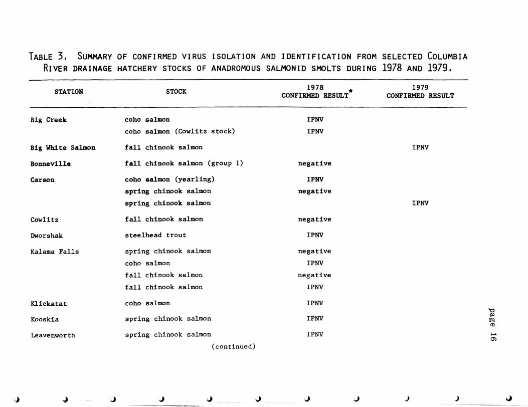
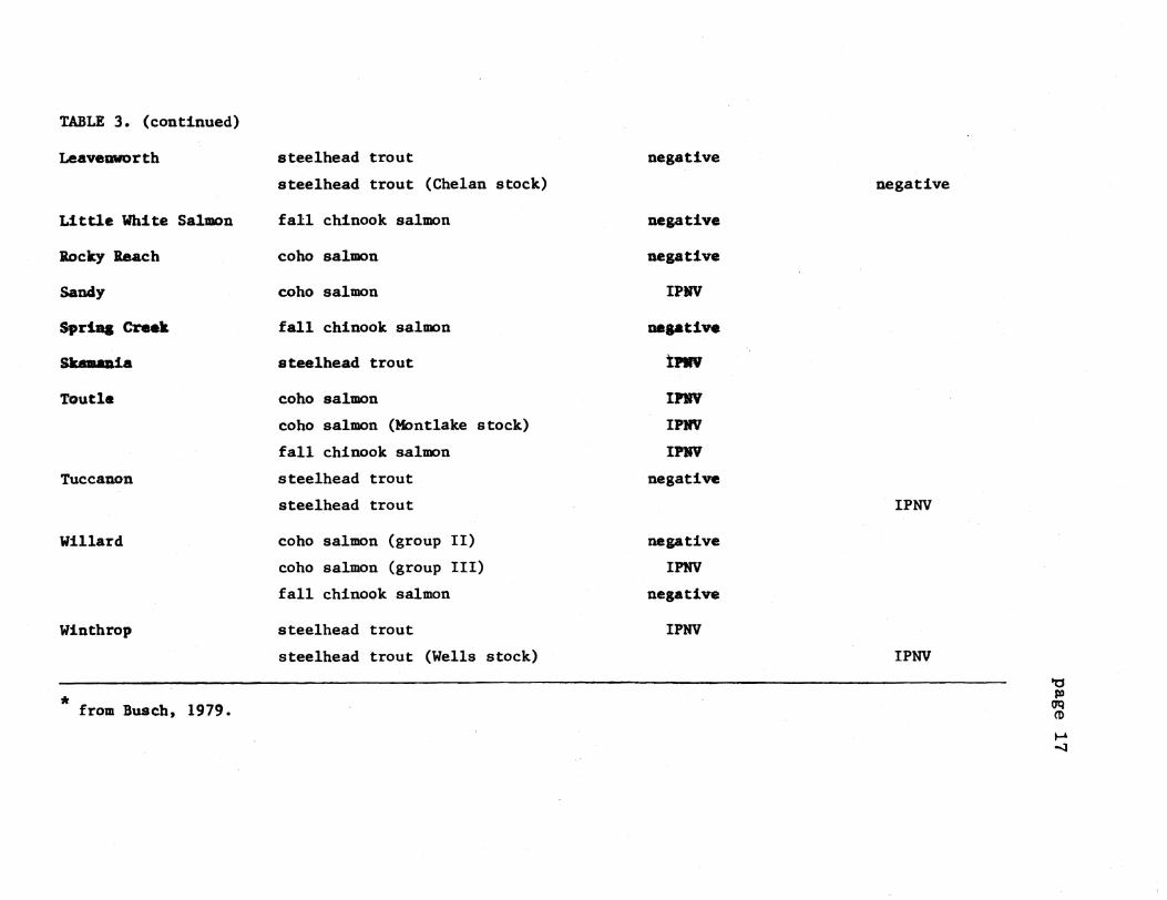
3 Summary of confirmed cirus isolation and identification from selected Columbia River
drainage hatchery stocks of anadromous salmonid smolts during 1978 and 1979 16
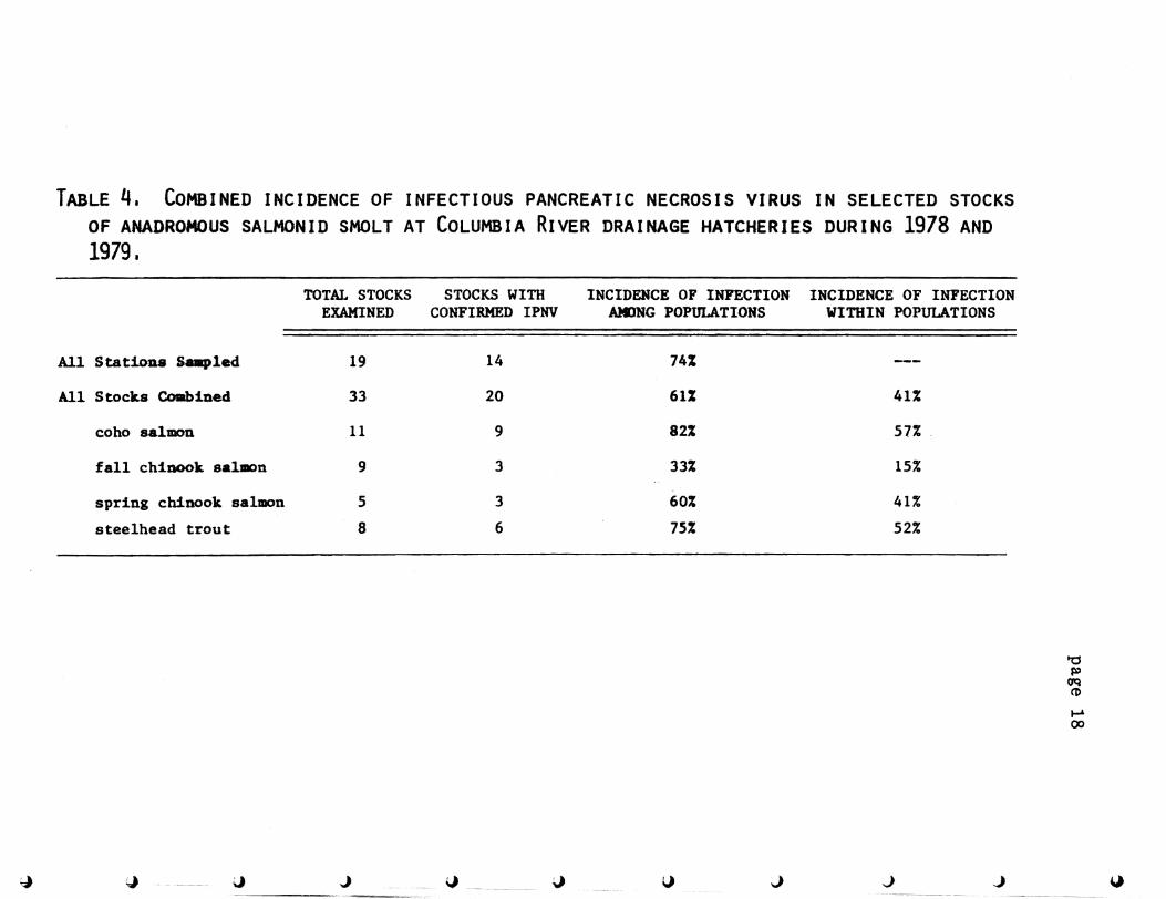
4 Combined incidence of infectious pancreatic necrosis virus in selected stocks of anadromous salmonid
smolt at Columbia River drainage hatcheries during 1978 and 1979 18 5 Summary of confirmed viral and serum neutralization
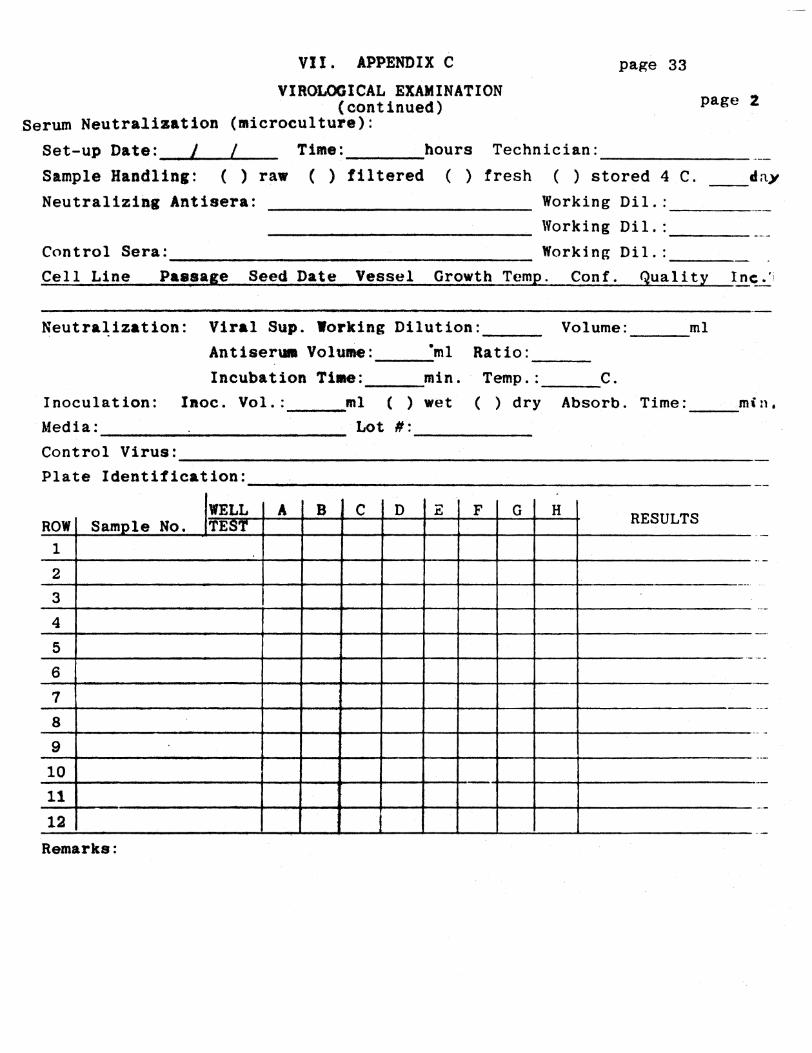
results from five selected Columbia River drainage hatchery stocks of anadromous salmonid smolts during 1979 Os bull bull bull bull bull bull bull bull bull bull bull bull bull bull bull bull bull bull bull bull bull bull bull bull bull bull bull bull bull bull bull bull bull bull bull bull bull bullbull 19
-
iii
LIST OF FIGURES
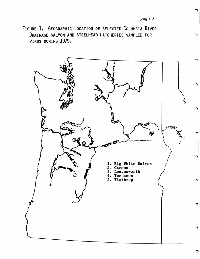
1 Geographic location of selected Columbia River drainage salmon and steelhead hatcheries sampled
for virus during 1979 8
I
I
I
page 1
I INTRODUCTION
On the springo 1978 the National Marine Fisheries Service (NMFS) Manchester Washington laboratory began an annual study to evaluate various factors pertinent to the successful smoltification ocean survival and adult return of selected anadromous salmonid fish stocks of hatchery origin in the Columbia River basin A portion of this study was devoted to ascertaining the general health profile of each stock at the time of smoltification and immediately prior to hatchery release and natural outshymigration into saltwater The purpose of this general health profile was to determine the occurrence and incidence of selected infectious diseases known to be potentially important to the growth and survival of salmonid fishes in general The health profile data acquired is then used in the evaluation and interpretation of other data obtained with regard to the relative success of saltwater adaptation ocean survival and hatchery return potential
Migrant hatchery flah stocks were examined for selected viral bacterial and parasitic disease agents using a wide variety of techniques as described in the parent NMFS project report Summary results of the first year of this study pertaining to 1978-79 brood year stocks have been reported by Busch (1979) CZESD and ETSD (1979) Novotny and Zaugg (1979) This report concerns itself only with the work contracted by NKFS to Rangen Research under USDC NOAI Purchase Order No 79-ABB-00276 (Rangen Contract No 9-79) and pertains to that portion of the general health profile dealing with the screening of selected populations for the presence of important viral pathogens known to infect salmonid fish in North America Included among these pathogens are infectious pincreatic necrosis virus (IPNV) infectious hematopoietic necrosis virus (IHNV) Herpe8v~rus saZmonis and any additional replicating agents capable of inducing specific cytopathic effects (CPE) under the given conditions of surveillance
The virus disease most commonly associated with Pacific salmon and steelhead trout stocks in the Pacific Northwest is infectious hematopoietic necrosis virus IHNV was first recognized in 1951 in sockeye salmon (OnchorhynchuB nerka) at Leavenworth National Fish Hatchery and kokanee salmon a land-locked sockeye salmon at Winthrop National Fish Hatchery The virus was isolated for the first time from sockeye salmon in 1958 (Wood 1974) The disease is known to cause significant mortality in hatchery populations of Pacific salmon and steelhead trout as well as other salmonid species all a10ftg the Pacific coast of North America from California to Alaska and is considered endemic to maDY watersbeds ieluding the Columbia River Basin
IHRV infectioa character1stically results in a peracute to acute course ot dl resulting in high levels of mortality in cbinook al8aoa (OnahorhynchuB tshawytscha) and an acute to subacute disease in sockeye salmon The
~I
j
page 2
disease is characterized as its name implies by destruction of the bematopoietic tissues resulting in an acute condition of hemorrhage anemia and often severe mortality among fry and fingerling fish under hatchery conditions Infection seems to be primarily vertical in nature with the disease being transmitted with the els and reproductive fluids of asymptoatically infected returning broodstock The coho salmon (Onahorhynahu8 kieutah) appears to be more resistant to IHNV infection than the other species of Pacific salmon but can function as an asymptomatic carrier in the maintenshyance and dissemination of the disease (Wolf 1972)
Survivors of an epizootic infection of IHNV are known to carry the virus in an eclipse phase of intracellular infection typical of the rhabdovirus group of which it is a member Duringtnisasymptomatic carrier state the virus is not detectable by routine methods of surveillance and is presumed to be non-infectious This period of subclinical infection includes the time of physiological transition to saltwater known as smoltification The latent effects of its presence upon smoltification ocean survival and adult return are not known at this time However the detectable infectious virus has been shown to reappear immediately prior to spawning in infected populations of returning adults and is readily transmitted to the progeny at this time
The early epizootiology of suspected viral diseases including IHNV in the Columbia River drainage is reviewed by Parisot et al (1965) Several extensive surveys to determine the incidence and distribution of the virus have been conducted since that time in selected stocks of Columbia River trout and salmon In 1972 Amend and Wood reported that no IHNV could be found in Columbia River stocks of Pacific salmon returning to 15 selected hatcheries in the state of Washington These findings were based uponthe extensive sampling of 130 to 150 ovarian fluid samples taken from each population at the time of spawning Tebbit and McMichael (1973) found no evidence of IHNV in 10 Columbia River salmon stocks returning to hatcheries in the state of Oregon during 1971 and 1972 However in 1973 they reported the confirmed isolation of IHNV from an adult spring chinook salmon stock returning to Oregons Pelton Dam Holding Facility Numerous additional studies have been undertaken by various state and federal agencies in subsequent years but have often failed to be comprehensive in deSign and execution and their results are often not readily available
Even though IHNV has not been found to be a majorproblem for Columbia River stocks of chinook salmon in general it does continue to pose a threat to this specie as well as sockeye salmon under hatchery conditions The
real occurrence and incidence of IHNV is still not well defined due to problems in the detection of asymptomatic carrier states of infection during the eclipse stage of the virus Knowledge is also lacking on the possible
page 3
residual effects of the carrier state infection on successtul smoltitication saltwater survival and adult return The disease retUinsendemic to the watershed and has been increasing in its known host range geographical distribution and physical tolerances such as temperature IHNV bas recently been found to be pathogenic for rainbow trout (Satmo gaipdnspi) fry fingerlings and yearling fish at 145 C (58 F) in the Snake River drainage of the upper Columbia River basin It has also been found in a subclinical infectious carrier stage in rainbow trout broodstock in this same area (Morton and Busch 1980)
Another salmonid virus infectious pancreatic necrosis virus (IPNV) has not often been associated with or considered a problem in Pacific salmon stocks However IPNV is known to be endemic in the Columbia river drainage where it is primarily recognized as a reovirus disease of trouts and chars It is also known to be capable of infecting Atlantic coho and chinook salmon (Fish Health Section 1974)
IPNV is considered to be the same disease originallydescribed by MGonigle (1941) in brook trout (SaZveZinus ontinaZis) and Atlantic salmon (SaZmo salap) in hatcheries in the Canadian maritime provinces and later confirmed byMacKelvie and Artsob (1969) The pathogenesis of IPNV in Atlantic salmon has recently been described by Swanson and Gillespie (1979) The disease was first isolated in the western United States in 1963 by Parisot et al
IPNV disease is typically characterized by a peracute to acute course of infection and mortality of fry and fingerling fish or a subacute to chronic infection of larger fish up to and including yearling sizes The infectious agent is readily transmitted horizontally through the water between fish by means of infected feces and urine from clinically diseased or asymptomatically infected carrier fish It is also readily transmitted in a vertical manner from asymptomatically infected adult fish to their progeny via infected reproductive fluids and eggs Survivors of an epizootic infection often remain asymptoshymatic carriers for life and continually shed the infectious virus into the water a condition which has recently been modeled on a Chinook salaoft cell line by Hedrick et al (1978) and established in rainbow trout under laboratory conditions by Reno at a1 (1978) The disease has been shown to be transmitted in the natural environment to susceptible stocks and maintained in wild and feral populations for extended periods of time with the incidence of infection gradually decreasing over time when no new introduction of the virus is made (Yamamoto 1975) Sonstegard (1970) also showed long term survival of IPNV in the gastrointestinal tracts of selected non-salmonid fishes picivorous birds and aquatic invertebrates
Until 1968 IPNV had not been known to occur in any of the Pacific salmon species However in July of that year Wolf and Pettijohn (1970) isolated the virus from coho
page 4
salmon fingerlings at Lamar National Fish Hatchery in Pennsylvania The eggs of the infected stock had been taken from a spawning population of landlocked adult salmon in Lake Michigan The fish had been checked for virus as fry and were determined to be free of the infectshyion Bowever t IPNV was enzootic among hatchery stocks of trout at the time and viru8 was soon isolated from the fingerllD salmon durin an epizootic of furunculosis disease (Apomona salmoniaida) No mortality could be attributed to the preseDce of the virus and evidence indicated that infection was due to horizontal rather than vertical transmission
Amend and Wood (1112) 8urveed 15 Columbia River stocks of Pacific salmon returning to hatcheries in the state of Washington in the fall of 1970 selecting only tor IHNV on primary screen and confirmatory cultures Consequently no IPNV was reported In 1973 Tebbit and McMichael reported OD the surveillance of Columbia River stocks of Pacific salmon returning to selected hatcheries in Oregon Their comprehensive design included the examination of visceral tissues and ovarian fluids from SO adult females from each of four discrete populations Adult fish and progeny fry were also bled and the sera titered for specific neutralizing antibodies against the common viral diseases IPNV virus was isolated from two of twelve five-fish tissue pools from adult coho salmon returning to the Bonneville Hatchery in 1971 and also from their progeny fry in thirty or thirty ten-fish pools indicating verttcal transmission It is interesting to note that McMichael (1974) was only able to isolate virus from the fry progeny at 30 and 60 days of age post hatch and that these same fish as fingerlings when sampled at 90 and 210 days post hatcb no longer yielded detectable levels of viru but did delROnstrate specific neutralizing antibodies against IPNV with titers in excess of 1200 as confirming evidence of prior exposure Attempts at horizontal transmi8sion of the virus under hatchery conditions were unsuccessful at 15 C
A report in tbe FAO Aquaculture Bulletin (1973) indicated that McMichaels cobo salmon isolate of IPNV was unlike the standard ATCC UR-299 (American Type Culture Collection) IPNV trout isolate and more typical of French isolates of IPNV that are known to lose 99 of their infectivity in a single freezethaw cycle
When Tebbit and McMichael (1973) continued their sampling program they found returning adult populations and their progeny fry to be negative for IPNV at all of the selected hatcheries sampled in 1972 and 1973 However t
they were able to demonstrate specific IPNV neutralizing antibodies in the sera of 270 adult fall chinook and coho salmon The overall incidence was 74 in the returning coho and 92 in the returning fall chinook populations In 1972 they were also able to isolate IPNV from adult coho salmon returning to Cascade Hatchery but the virus
page 5
could Dot be demonstrated in the fry progeny or in the adult returns the following year Tebbit and McMichael (1973) also reported finding IPNV in adult chinook salmon returniag to the Pelton Dam Holding Facility in 1973 after the adult returns and progeny fry were found to be free of the virus in the 1971 and 1972 brood years
IPNV was isolated from ateelhead trout being reared at Idaho Power Companys iacria Springs Steelhead Hatchery in the Snake River CaDlOD of Southern Idaho near Buhl in 1974 IPNV is known to be endemic to the commercial rainbow trout hatcheries in tbe local area and its appearance at the Niagria Springs statioR was Dot surprising The virus has since reappeared at the atation periodically and has been associated with significant mortality
In 1174 Wood indicated that he had yet to make any positive IPNV iaolatioDs from Columbia River stocks in Washington state However in 1975 Tebbit reviewed McMichaels initial surveillance results and continued the program Again lookincmiddotat visceral tissue samples ovarian fluids and progenyfrJ Tebbit was able to demonstrate IPNV in six of twelve five-fish tissue pools from adult coho salmon returning to the Cascade hatchery during the 1972 broodyear but he could not demonstrate vertical transmission to the progeny fry That same year Tebbit also isolated IPNV frOll a population of spring chinook salmon fry at the Pelton Dam Holding Facility that were the progeny of an adult stock that had been previouslydiagnosed with a confirmed asymptomatic infection of IHNV (Tebbit 1975)
In 1967 Wolf and Quimby were first able to demonstrate the development of specific viral neutralizing antibodies in the serum of asymptomatically infected IPNV carrier fish Presence of such specific antibodies are considered as presumptive serological evidence of prior or concomitant exposure to the virus During the 1973 broodyear Tebbit was able to detect IPNV specific neutralizing antibodies in the sera of adult coho salmon returning to both the Sandy Hatchery and the Bonaeville Hatchery None of these seropositive populatioa or their progeny fry yielded a confirmed viral isolation Tebbit continued surveillance during the 1974 brood year but failed to isolate virus from any of the adult returns sampled but IPNV specificneutralizing antibodies were found in a stock of landlocked spring chinook salmon in the Detroit River impoundment on the North Santiam River in Oregon An endemic infection of IPNV had previously been demonstrated in wild cutthroat trout populations in theae same waters
Based upon the findings of both IPNV and IHNV in stocks of anadromous salaonida in the Columbia River basin the state of Oregon established a management policy prohibitingthe transport of any Columbia River stocks of fish to a coastal river system for fear of disseminating an endemic viral disease problem
In 1975 Mulcahy and Sanders reported isolating IPNV from
I
page 6
spring chinook salmon at the Oregon Fish and Wildlife Commissions Corvallis Research Laboratory More recently in the first year of this study Busch (1979) was able to isolate IPNV from 16 of 28 stocks of Pacific salmon and steelhead trout smolts at 12 of 18 Columbia River hatcheries sampled The incidence of infection among 11 coho salmon stocks sampled was 82 Of five steelhead trout stocks examined 75 were found to be infected with IPNV Four spring chinook salmon stocks had an incidence of IPNV of 60 and eight fall chinook salmon stocks had an incidence of IPNV of 33 All infections appeared to be asymptomatic carrier states and could not be associated with anysignificant mortality
It has been shown time and again that IPNV can indeed infect Pacific salmoa aad steelhead trout It is becoming recognized thatIPNV may be all too common as an asymptoshymatic infection in a wide variety of discrete spawning populations in the Columbia River basin and that these infections may be easily missed or not clinically diagnosed in certain types of samples taken at certain stages in the life history of the Pacific salmon and steelhead trout It still remains to be shown whether or not the presence of an endemic infection of IPNV has a detrimental effect upon the population Mortality directly attributed to the presence of the virus under hatchery conditions has yet to be demonstrated for Pacific salmon stocks but is known to occur with steelhead trout populations
The last of three recognized salmonid viruses to be potentially found in the Columbia River basin is Herpesvirus 8almonis This virus or other closely related viruses are known to infect rainbow trout and sockeye salmon at all stages in their life history and has been implicated in mortality of both fry and adult fish The virus was first described in Japan as being endemic among certain stocks of sockeye salmon It has only been diagnosed once in the United States as an asymptomatic infection of rainbow trout broodstock at the Winthrop National Fish Hatchery The virus still remains a fairly unknown entity with regard to the general health of Pacific salmon and steelhead trout stocks as its true host range geographical distribution and general ecology and epidemiology has yet to be determined (Wolf et ale 1975)
II MATERIALS AND METHODS
Procedures of viral surveillance were designed to fit within the overall field sampling program and budget of the parent study and at the same time give optimum sensitivity and accuracy with regard to detecting the presence of any of the major salmonid viruses found within the Columbia River basin Techniques for both surveillance of viral diseases including primary isolation and confirmed serological identification and surveillance of specific
page 7
viral neutralizing humoral antibodies were used A total of 5 stocks of chinook salmon coho salmon and
steelhead trout from 5 different Columbia River basin hatcheries (Figure 1 and Table 1) were checked for the presence of virus at the time of smoltification and hatchery release during 1979 Field sampling was conducted by NMFS personnel according to procedures established by and with field sampling kits provided by Rangen Research (Appendix A) Hatchery stocks being sampled were first transported in a live haul container back to the NMFS Laboratory at Manchester Washington They were then maintained in freshwater flow-through systems (Beaver Creek water supply)for one to two days prior to lethal sampling The field sampling diluent provided was formulated (see Appendix B) to provide optimum survival of any infectious virus under transport conditions while inhibiting the growth of any microbial contaminants It was provided in sterile graduated polycarbonate screw-cap tissue culture grade centrifuge tubes (Corning 25310 Corning Glass Works Corning New York)The use of the graduated tube allowed for the calculation of accurate sample dilution factors based upon the displaceshyment method as 02 g of tissue was pooled from each of five fish into 9 ml of field sampling diluent and a total of twleve five-fish pools collected from the randomly selected 60 fish sample
Serum samples were obtained by lightly anesthetizing 60 fish from each stock in an aerated solution of 120000 tricaine methanesulfonate (Ayerst Laboratories New York)In most cases blood was sampled from the caudal arch by means of a 10 cc heparinized syringe fitted with a 25 G hypodermic needle Small fish were bled by severing the caudal peduncle All whole blood samples were immediatelytransfered to heparinized micro-capillary tubes plugged at one end and centrifuged for three minutes in a ClayshyAdams Autocrit II centrifuge The serum was then transshyfered to a second micro-capillary tube plugged at both ends and placed sequentially in a numbered tube rack
Following completioR of all sampling procedures and the Field Sampling Data Sheet (Appendix A) the complete refrigerated field kit and samples were returned to the Rangen Research Laboratory in Hagerman Idaho via Greyhound NBO Package Express Service The average time in transit was 20 hours and the temperature of the samples upon receipt was oonsistently between 6 and 8 C A duplicate set of tissues samples were taken from the same fish as per the directions of Dr Dan Mulcaby and sent to the National Fisheries Research Center in Seattle Washington for confirmatory viral analysis by a comparable methodology
Upon receipt into the Rangen Laboratory the receiving temperature of the tissue samples was recorded The raw serum samples were stored at 4 C for a maximum time of five days prior to titration and serum neutralization The twelve tissue sample tubes were assigned a group accession number and each tube in the group given a
I page 8
FIGURE 1 GEOGRAPHIC LOCATION OF SELECTED COLUMBIA RIVER DRAINAGE SALMON AND STEELHEAD HATCHERIES SAMPLED FOR VIRUS DURING 1979
1 Big White Salmon 2 Carson 3 Leavenworth 4 Tuccanon 5 Winthrop
TABLE 1 STOCKS OF PACIFIC SALMON SHOLT EXAMINED FOR VIRUS AT THE TIME OF RELEASE FROM SELECTED COLUMBIA RIVER DRAINAGE HATCHERIES DURING 1979
STATION S1Oa UAlIIKID DATE SAMPLED RANGEN ACC NO
Big White Salmon fall eh1nook salmon 52479 208-79
Carson prins chinook salmon 5879 179-79
Leavenworth ateelbead trout (Chelan stock) 42579 158-79
Tuccanon steelhead trout 51679 203-79
Winthrop steelhead trout (Wells stock) 51579 195-79
d fI)
oq CD
(0
page 10
serial numeric identifying code The refrigerated tissue pools were homogenized in situ with a Polytron PUC-2-110 homogenizer fitted with a steam sterilizable stainless steel PT-IO generator (Brinkman Instruments Westbury New York) for 30 seconds The homogenized tissue samples were then centrifuged at 2000 X g for ten minutes at 4 C in a Sorval RC-5 refrigerated centrifuge with an HS-4 rotor and appropriate tube adapters (DupontSorval Newtown Conn) to remove large cellular debris Two tenths of a milliliter of the supernatant was pipetted off with a sterile disposable polyethylene tip and diluted in 18 ml of a disinfecting diluent (Appendix B) and incubated at 4 C overnight for decontamination of fungal and bacterial organisms This procedure resulted in a final 1100 working dilution of the original tissue sampled All samples were maintained at 4 C during all preparatory procedures
Tissue samples received into the laboratory were screened for the presence of virus within seven days of sampling Viral screen tests were conducted with a microculture procedure using the Chinook Salmon Embryo (CHSE-214) cell line in passages 267 through 277 and a MEM-10-Tris-PSF media (Appendix B) at 125 C incubation for 21 days Four replicate wells of each of two dilutions were run for each of the pooled tissue samples Microculture screen tests were prepared by dropping 005 ml of the decontaminated and diluted 1100 tissue pool into eachof the first four wells of an eight well series on a sterile Microtest II (Falcon 3040 Becton Dickinson and Co) tissue culture plate and 0025 ml of the same sample into the last four wells of the eight well series using an Oxford 8000H sampler and a sterile Oxford 810S tip During preparation the microshyculture plates were temporarily covered with a Falcon 3041 sterile lid and maintained at 15 C Appropriate IPNV and IHNV positive viral controls and negative media and cell controls were set up for each group tested After the sample dilutions were delivered to the microculture plate a stock flask of CHSE-214 cells that had been grown to 95 confluence at 20 C in MEM-10-Tris-PSF media was examined under an inverted microscope for confluence and quality and dissociated in 4 ml of PANEDTA media (Appendix B) The PANEDTA cell suspension was centrifuged at 1500 x g for four minutes The supernatant was decanted and the pelleted cell mass resuspended in a small volume of MEM-10shyTris-PSF media The resuspended cells were further diluted in MEM-10-Tris-PSF media to a final 14 working concentration based upon the surface area split ratio and placed in a sterile 100 ml covered flask containing a sterile magneticstir bar The diluted cell suspension was placed on a refrigerated magnetic stir plate with slow stirring to maintain the cells in suspension A sterile Minipet repipetting syringe (Manostat 71-500-010 Manostat New York NY) was ittedwith a sterile disposable 18g X Ii hypodermic needle and primed with the chilled CHSE-214 cell suspension Fifteen hundredths of a milliliter of the
page 11
diluted CHSE-214 cell suspension was pipet ted into each well on the inoculated microculture test plate The plate was immediately sealed with Falcon 3044 pressure sensitive film and incubated at 125 C The suspended CHSE-214 cells were allowed to settle down through the sample material in the well adsorb infectious virus present and attach to the bottom of the well
All viral screen tests were incubated at 125 C for 21 days A~l wells of both dilutions of both test and control series were periodically read under an inverted microscope for evidence of specific cytopathic effect (CPE) cytotoxicity and microbial contamination The daily observations were recorded on the Virological Examination Report Sheet (Appendix C) If more than four wells in an eight well series were found to be cytotoxic the original decontaminated sample was diluted 12 with additional disinfecting diluent to a final working dilution of 1200 and re-run on the microculture screen procedure as described above If more tban four wells in an eight well test series were found to be contaminated the original sample was filter sterilized through a sterile 045u membrane fil ter and re-run on the microculture screen procedure
At the end of the 21 day incubation period the 96 wells on each plate (8 wells for each of 12 pooled tissue samples in the lot) were classified as being either positive (definite CPE) questionable (possible CPE or cytotoxicity) or negative (no evidence of CPE or cytotoxicity) and pooled into one of three tubes according to its classification After the supernatant medias had been aspirated and pooled from the wells of the microculture screen plates for further testing all wells of the plate were stained with a 1 alcoholic solution of crystal violet and dried as a permanent record of the screen results The presence or absence of infectious virus in each of the three classified screen pools was confirmed and identifshyied by means of a microculture serum neutralization procedure One quarter of a milliliter of sterile field sampling diluent was placed in each of the first three wells of an eight well series on a sterile microculture plate Twenty five thousandths of a milliliter of EFDL 149 Polyshyvalent IPNV Antisera (Eastern Fish Disease Laboratory Kearneysville West Virginia) diluted 1100 in sterile field sampling diluent was pipet ted into the next two wells of each eight wellseries Twenty five thousandths of a milliliter of EFDL 150 IHNV antisera diluted 1100 in sterile field sampling diluent was pipetted into the next two wells of each eight well series and finally 0025 ml of EFDL 100 normal rabbit sera diluted to a final 1100 working concentration in sterile field sampling diluent was pipetted into the last well of each eight well series Each of the three classified and pooled screen aspirates were then serially diluted out to 10-3 by pipetting 0025 ml of the material into the first well of an eight well series mixing with the diluent present and transfering 0025 ml
page 12
into the second well mIxIng and transfering 0025 ml into the third well and mixing Twenty five thousandths of a milliliter of the 10-3 dilution of the sample in the third well of each series was then transfered to each of the remaining five wells in the series containing the various antiseras Appropriate positive IPNV and IHNV controls as well as negative media and cell controls were prepared in the same manner The prepared microculture serum neutralshyization plates were then temporarily covered with a sterile Falcon 3041 lid and incubated at 15 C for 60 minutes to affect appropriate neutralization of any virus present CHSE-214 cells were then prepared to a 1t2 dilution based upon surface area as described above and 015 ml of the diluted cell suspension pipetted into the last five wells of each eight well test series on the plate The plate was immediately sealed with film and incubated at 125 C for five days
The serum neutralization results were read under an inverted microscope at the end of the five day incubation period The results were recorded on the Virological Examination Report Sheet (Appendix C) Destruction of the cell monolayer with characteristic CPE in well four five and eight of a test series indicated the confirmed presence of IHNV in that sample pool Destruction of the cell monolayer with characteristic CPE in wells six seven and eight indicated the confirmed presence of IPNV in that sample pool Destruction of the cell monolayer with characteristic CPE in wells four five six seven and eight could indicate the presence of Herpesvirus satmonis a mixture of viral agents partial neutralization of a particular strain of a virus or possibly a previously undescribed viral agent in which case additional procedureswould have to be applied to confirm identity All wells demonstrating CPE in an eight well sample series on the serum neutralization plate were pooled and lyophilized in a stabilizer as a reference stock culture All wells of the serum neutralization plate were then stained with a 1 alcoholic solution of crystal violet and dried as a permanent record of the results
Sixty individual raw refrigerated serum samples from each stock of fish were screened for the presence of virus specific neutralizing antibodies by means of a microculture modified procedure using the CHSE-214 cell line in passages267 through 277 and a MEM-10-Tris-PSF media (Appendix B) A virulent IPNV stock culture originally isolated in 1978 from Leavenworth spring chinook salmon as Rangen Path 138shy
78-3 (Busch 1979) was used in all serum neutralization procedures
The microculture neutralizing serum titration procedure was conducted by pipetting 0025 ml of sterile field sampling diluent (Appendix B) into each well of an eight well series on a sterile Microtest II (Falcon 3040) tissue culture plate for each serum sample to be titered using an Oxford 8000H pipette and a sterile Oxford 8108 tip During
I Imiddot
page 13
preparation the microculture plates were temporarily covered with a Falcon 3041 sterile plastic lid and maintained at 15 C Calibrated 0025 microtiter loops (1-220-33 microdiluters Cooke Laboratory Products Alexandria Virginia) were flame sterilized wetted in sterile 09$ Hanks balanced salt solution blotted on sterile towels and loaded with individual raw serum samples from the plugged heparinized microcapillary tubes The 0025 ml of raw sera was then serially diluted through the eight well series of diluent on the microculture plate to give a two-fold series of dilutions from 12 to 1256 Appropriate known positive and negative trout and rabbit serum controls were included wi th each test Following dilution all wells on the plate were then inoculated with 0025 WI of a field sampling diluent suspension containing 10 TC1D50 units of Rangen Path 138-78-3 Leavenworth spring chinook salmon IPNV virus previously grown out on CHSE-214 cells covered and incubated at 15 C for 30 minutes and 4 C for an additional 30 minutes to effect serum neutralization
During the 60 minute serum neutralization period a stock flask of CHSE-214 cells that had been previously grown out to 95 confluence at 20 C in MEM-10-Tris-PSF was examined under the inverted microscope for confluence and quality and dissociated in 4 ml of PANEDTA media (Appendix B) The PANEDTA cell suspension was centrifuged at 1500 x g for four minutes The supernatant was decanted and the pelleted cell mass resuspended in a small volume of MEM-10-Tris-PSF media The resuspended cells were further diluted in MEM-10-Tris-PSF media to a final 12 working dilution based upon surface area split ratio and placedin a 100 ml covered sterile flask containing a sterile magnetic stir bar The diluted cell suspension was placed on a magnetic stir plate with slow stirring to maintain the cells in suspension A sterile Minipet repipetting syringe (Manostat 71-500-101 Manostat New York NY) was fitted with a sterile disposble 18g X Ii hypodermic needle and primed with the chilled CHSE-214 cell suspension Fifteen hundredths of a milliliter of the diluted CHSE-214 cell suspension was pipetted into each well on the incubated serum neutralization plates The plates were immediately sealed with Falcon 3044 pressure sensitive film and incubated at 15 C for seven days All wells of both the test and control dilution series were read under the inverted microscope periodically for evidence of specific cytopathic effect (CPE) cytotoxicity and microbial contamination Observations were recorded as the highest serum dilution demonstrating complete viral neutralization and a total lack of any specific CPE All wells of the plate were stained with a 1 alcoholic solution of crystal violet and dried as a permanent record of the serum neutralization results
page 14
III RESULTS
A total of 300 Pacific salmon and steelhead trout smolts representing five discrete anadromous stocks at five Columbia River basin hatcheries were tested for the presence of infectious viruses during 1979 The results of these tests are summarized in Table 2 for each of the hatchery stocks examined
Neither infectious hematopoietic necrosis virus (IHNV) nor Herpesvirus saZmonis were found during the course of this study InfectioQs pancreatic necrosis virus (IPNV) was confirmed at four of five hatcheries sampled and in four of five stocks of smolted fish examined IPNV was isolated from two of three populations of steelhead trout examined with all twelve five-fish pools of tissue from the Winthrop steelhead trout stock being positive but only one five-fish pool of tissue from the Tuccanon steelhead trout stock being positive All sixty fish sampled from the Chelan stock of steelhead trout from Leavenworth Hatchery were negative for virus In addition all twelve five-fish pools of tissue from the Big White Salmon stock of fall chinook salmon as well as the Carson stock of spring chinook salmon were found to be positive for IPNV
Because of the statistically insignificant numbers of stocks included for surveillance in the 1979 study further analysis of this data by itself is not justified However recognizing that the year class of stocks examined for virus in 1978 and 1979 are largely unique and discrete spawning populations the data from both years has been combined and summarized in Table 3
Since the year class stocks checked for virus during the 1978 and 1979 studies can be combined to increase sample size and statistical significance Table 4 is used to summarize the combined incidence of IPNV infection among and within populations by specie It is shown that among populations by species IPNV was most common in coho salmon smolt (82 incidence among populations sampled) and least common in fall chinook salmon smolt (33 incidence among populations sampled) The IPNV carrier incidence of infection within populations by species was determined on the basis of confirmed viral isolation from five-fish tissue pools within a sixty-fish lot for each population On this basis the IPNV carrier incidence wi thin populations sampled was the highest for coho salmon smolt at 57 and lowest for fall chinook salmon smelt at 15
None of the confirmed IPNV isolations could be associated with any significant mortality or loss within the populatshyions examined
The results of serum neutralization testing a~e summarized is Table 5 IPNV specific neutralizing antibodies were detected in 244 of the sera from spring chinook salmon smolt at Carson Hatcherywhile IPNV was isolated from 33
TABLE 2 SUMMARY OF PRELIMINARY SCREEN RESULTS AND CONFIRMED IDENTIFICATION OF VIRUS ISOLATED FROM SELECTED COLUMBIA RIVER DRAINAGE HATCHERY STOCKS OF ANADROMOUS SALMONID SMOLTS DURING 1979
STATION SroCK NUMBER OF SCREEN POOLS POSITIVE SUSPECT NEGATIVE
CONFIRMED VIRALISOLATION RESULT
Big White Salman fall chinook ulman 12 0 0 IPNV
Carson sprina chinook salmon 4 5 3 IPRV
Leavenworth steelbead trout (Chelan stock) 0 0 12 negative
Tuccanon steelbead trout 1 0 11 IPNV lt
Winthrop stee1head trout (Wells stock) 12 0 0 IPNV
0 SIl
(JQ (t)
I- CJ1
TABLE 3 SUMMARY OF CONFIRMED VIRUS ISOLATION AND IDENTIFICATION FROM SELECTED COLUMBIA RIVER DRAINAGE HATCHERY STOCKS OF ANADROMOUS SALMONID SMOLTS DURING 1978 AND 1979
STATION STOCK 1978 it
CONFIRMED RESULT 1979
CONFIRMED RESULT
Big Creek coho salmon IPNV
coho salmon (Cowlitz stock) IPNV
Big White Salmon fall chinook salmon IPNV
Bonneville fall chinook salmon (group 1) negative
Carson coho salmon (yearling) IPNV
spring chinook salmon negative
spring chinook salmon IPNV
Cowlitz fall chinook salmon negative
Dworshak steelhead trout IPNV
Kalama Falls spring chinook salmon negative
coho salmon IPNV
fall chinook salmon negative
fall chinook salmon IPNV
Klickatat
Kooskia
coho salmon
spring chinook salmon
IPNV
IPNV tj ~
(JQ ro
Leavenworth spring chinook salmon
(continued)
IPNV ~ (J)
-J J J ) ) ) ) j j ) u
TABLE 3 (continued)
Leavenworth
Li t tle Whi te Salmon
llocky leach
Sanely
S9riaa Creek
914
Toutle
Tuccanon
Willard
Winthrop
from Busch 1979
s teelhead tro u t
steelhead trout (Chelan stock)
fall chinook salmon
coho salmon
coho salmon
fall chinook salmon
steelhead trout
coho salmon
coho salmon (MOntlake stock)
fall chinook salmon
stee1head trout
steelhead trout
coho salmon (group II)
coho salmon (group III)
fall chinook salmon
steelhead trout
steelhead trout (Wells stock)
negative
_sative
negative
IPNV
neaaUve
t1fi
IPBV
IPlIV
IPlW
negative
negative
IPRV
negative
IPNV
negative
IPNV
IPNV
0 Iraquo
OQ (I)
~ J
TABLE 4 COMBINED INCIDENCE OF INFECTIOUS PANCREATIC NECROSIS VIRUS IN SELECTED STOCKS OF ANADROHOUS SALMONID SMOLT AT COLUMBIA RIVER DRAINAGE HATCHERIES DURING 1978 AND 1979
TOTAL STOCKS EXAMINED
STOCKS WITH CONFIRMED IPNV
INCIDENCE OF INFECTION AMONG POPULATIONS
INCIDENCE OF INFECTION WITHIN POPULATIONS
All Statioas Saaplecl 19 14 74
All Stocks Combined 33 20 61 41
coho salmon 11 9 82 57
fall chinook salmon 9 3 33 15
spring chinook salmon
steelhead trout
5
8
3
6
60
75
41
52
00 ~
aq CD
~ 00
~ j u J IJ a u J J J u
TABLE 5 SUMMARY OF CONFIRMED VIRAL AND SERUM NEUTRALIZATION RESULTS FROM FIVE SELECTED COLUMBIA RIVER DRAINAGE HATCHERY STOCKS OF ANADROMOUS SALMONID SMeLTS DURING 1979
tHCIDDICEmiddotmiddot OF MEAN CONFIRMED TOTAL SERA TOTAL SERA SElWPOSITIVE TITERSTATION STOCK VIRAL ISOLATION EXAMINED IPNV POSITIVE SAIPLISRESULT
Big White Salmon fall chinook salmon IPNV --------- no serum samples taken ---------shy
Carson spring chinook salmon IPNV 41 1 244 14
Leavenworth steelhead trout (Chelan stock) negative --------- no serum samples taken ----------
Tuccanon steelhead trout IPNV 56 17 3036 136
Winthrop steelhead trout (Wells stock) IPNV 38 o 000 negative
~ PJ
(lQ CD
~ lt0
i 3
page 20
of the homologous five-fish tissue pools tested Steelhead trout smolts sampled at Tuccanon Hatchery had a 3036 incidence of seropositive IPNV specific neutralizing antibodies among 56 individual sera while only 833 of the homologous five-fish tissue pools were positive for IPNV isolation
Even though the Wells stocks of steelhead trout at Winthrop Hatchery had a 100$ incidence of IPNV isolations among the twelve five-fish tissue pools sampled no IPNV specific neutralizing antibodies could be found among the 38 homologous individual sera tested As no sera were made available to the Big White Salmon or Leavenworth stocks no serum neutralization tests on these two stocks were run
IV DISCUSSION
The failure to isolate either Herpesvirus saZmonis or infectious hematopoietic necrosis virus (IHNV) during the course of this study either during 1978 or 1979 is not unusual when considered within the given limitations of the experimental design applied Herpesvirus BaZmoniB or other closely related salmonid viruses have yet to be isolated from coho salmon chinook salmon or steelhead trout in the United States In fact the virus has only been isolated once in North America from a Columbia River basin salmonid stock and has notmiddot been seen since Due to the fact that our knowledge of the ecology and epidemiology of the disease is still limited it can only be said that the virus was not detected within the limitations of the experimental design applied Possibly by broadening the scope of the study by iDc1uding conditions known to be optimum for the recovery of the virus at all stages in the life history of Pacific salmon and steelhead trout as well as the application of DOre sensitive techniques of serologishycal surveillance such as detection of specific neutralizingfactors in the sera or other body fluids or the detection of specific antigens or antibodies in the various body tissues and fluids by means of enzyme linked immunoadsorbent assay (ELISA) or counterimmunoelectrophoresis (eIE) procedures could also give a better understanding of the ecology of the disease and its impact on anadromous stocks of salmonids
Infectious hematopoietic necrosis virus (IHNV) is known to be endemic to the Columbia River basin but is usually considered a disease of fry and fingerling chinook salmon and steelhead trout Coho salmon appear resistant but are known to function as asymptomatic carriers IHNV is also known to enter into a non-infectious eclipse phase of infection in post-epizootic or convalescent populations and as such cannot be isolated and identified by routine methods of culture as applied in this study In order to obtain a better understanding of the ecology epidemiology and overall impact of IHNV on the anadromous salmonid stocks
page 21
of the Columbia River basin surveillance programs should include testing of ovarian fluids from adult spawming populations as well as progeny fry at the swim-up stage as these are the two stages in the life history of the fish when the virus is known to exist in the infectious form Techniques of chemical induction or serological surveillance using serum neutralization ELISA and CIE procedures may be effective in detecting virus at these and other stages when the virus may be present in the non-infectious eclipse form
Extensive recent isolations of IHNV from clinically diseased rainbow trout fingerlings and yearling fish at 145 C (58 F) (Morton and Busch 1980) may indicate that strains of IHNV in the Columbia River basin are significantly extending their host geographic and temperature tolerance ranges and again points out the fact that the salmon ids viruses are in a continuing dynamic state of adaptation Descriptions and accepted facts of ecology and epidemiology based upon historical information may not necessarily continue to hold true in the future as the viral pathogens adapt to new hosts and environments
Isolation of infectious pancreatic necrosis virus (IPNV) from the stocks examined and in particularthe repeated finding of a relatively high incidence of the virus among the populations in comparison to some previous reports of surveillance again seems unusual upon initial examination as was pointed out in last years study However when one takes into consideration that IPNV is 1) known to be endemic to large areas of the Columbia River basin 2) known to infect both Pacific salmon and steelhead trout under both experimental and natural conditions and 3) has been detected in at least ten discrete populations of Pacific salmon and steelhead at seven different stations since 1971 and prior to this study the results take on a more consistent image It is also noted in this study and in several previous surveillance studies that IPNV has been isolated only from visceral tissues of infected returning adult stocks and their 30 to 60 day old progeny fry and not from reproductive fluid samples However our current understanding of the epidemiology of IPNV in anadromous stocks of Columbia River salmon and steelhead trout seems to be primarily based upon studies which relied primarily upon ovarian reproductive fluid samples and were specifically designed to monitor for IHNV When McMichael (1974) and Tebbit (1975) sampled both reproductive fluids and pooled visceral tissues from returning adult salmon populations they were able to isolate IPNV from four discrete populations However all four IPNV isolations were obtained exclusively from the pooled visceral tissue samples while all of the paired homologous reproductive fluids samples remained negative for the virus (Tebbit 1979 personal cOlIIDunication) This observation would indicate that surveillance studies based solely upon
page 22
reproductive fluid samples while being well suited to the detection of IHNV may not accurately reflect the true incidence or occurrence of IPNV in a population of asymptoshymatic adult carriers
It should also be noted that McMichael (1974) consistently failed to isolate IPNV from known infected fry populations of Pacific salmon after 60 days of age post feeding Therefore additional surveillance programs should be designed to sample fry and fingerling sized fish at 30 to 60 days of age post feeding and at smoltification prior to hatchery release in order to detect the presence of IPNV accurately
IPNV as well as many other diseases of viral etiology are known to be stress mediated as well as being species and age specific The period of smoltification in the life history of an anadromous salmonid fish is recognized as a time of major physiological change and stress to the animal particularly under hatchery conditions of intensive culture and nutrition It is also a time when hormonal balances are in a state of flux and these too have been correlated wi th the occurrence of certain diseases Therefore it is reasonable to assume that this period of smoltification and physiological stress particularly when coupled with major changes in hormonal balances and behavior could well exacerbate a preexisting subclinical infection of IPNV or even increase the relative susceptibility of an uninfected population to infection from an endemic source common to the hatchery or watershed To the best of our knowledge this is the first surveillance study to report on results obtained specifically from populations undergoing smoltifishycation
Another consideration for discussion is the demonstrated low incidence of IPNV to be found among the asymptomaticly infected populations Tebbit and McMichael (1974) reported only two of twelve five-fish tissue pools taken from adult coho salmon returning to Bonneville Hatchery in 1971 were found to be positive for IPNV Only six of twelve five-fish pools of tissue frommiddot adult coho salmon returning to Cascade Hatchery in 1972 were positive for IPNV (Tebbit 1975) In last years study of 1978 broodyear fish seven of the 16 populations found to be infected with IPNV demonstrated virus in less than half of the five-fish tissue pools examined on initial screening This year in 1979 broodshyyear fish two of the four populations found to be infected with IPNV demonstrated the virus in fewer than half of the five-fish tissue pools examined on initial screening These findings would indicate that a full GO-fish sample based upon hypergeometric sampling statistics may be necessary in order to consistently detect the virus Spot checks of fewer than 60 fish are apt to miss detecting an infection of low incidence as many seem to be
A final consideration in the surveillance of IPNV in the Pacific salmon and steelhead trout stocks of the Columbia River basin is the demonstrated variability
page 23
in the occurrence of the virus between different year classes of a particular stock During extensive sampling between 1971 and 1974 McMichael (1974) and Tebbit (1975) consistentshyly failed to isolate IPNV from the same hatchery stocks during subsequent years even though neutralizing antibodies indicating prior exposure were found to be present at times Therefore it appears reasonable to assume that the demonstrated presence or absence of IPNV in a particular year class of a given hatchery stock of anadromous salmon or steelhead trout may not necessarily mean that all other year classes of that same stock will be similiarly infected or free of the disease As was shown in Table 3 the 1978 year class of Carson spring chinook salmon were negativefor IPNV however the 1979 year class was found to be positive The same holds true for the Tuccanon steelhead trout stocks examined in 1978 and 1979
It should also be noted that paired five-fish pooledtissue samples sent to Dr Dan Mulcahy at the National Fisheries Research Center in Seattle were all reported as being negative for virus (see Appendix D) As Dr Mulcahy indicated in his letter this additional testing did nothing to clear up the question It is suggested that additional intensive testing of 120 to 150 individual fish from one or two selected sites be undertaken by two or more laboratories using the identical cell lines protocols and procedures as used in this study by Rangen Research A possible exchange of personnel may also be needed to insure complete and accurate duplication Additional methods such as ELISA CIA FAT etc may also be considered
The serum neutralization results obtained in this study indicate that this technique may be useful in additional surveillance studies However more sensitive serological techniques such as ELISA and CIA may be able to give higher titers and more definitive results Samples also need to be handled and processed more rapidly to insure minimum loss of acitvity during storage
Based upon these observations including the relatively low incidencemiddotof IPNV both among and within populations of anadromous Pacific salmon and steelhead trout in the Columbia River basin the serological findings of McMichael (1974) and Tebbit (1975) utilizing neutralizing antibody techniques and the variability of clinical infection between the different year classes and life history stages of a particular stock it is suggested that future attempts at viral surveillance include both sampling of visceral tissues as well as reproductive fluids from spawning populations Progeny fry and fingerlings should be sampled at 30 to 60 days of age post feeding and smolted fish should be sampled just prior to hatchery release Attempts at isolation of virus during the various stages of the life history should be coupled with techniques of serological surveillance based upon serum neutralization ELISA and CIE procedures All year classes of a particular stock should be examined over the course of the study
page 24
Any additional studies of a viral surveillance nature undertaken in the future may also want to include procedures for the detection of piscine erythrocytic necrosis (PEN) virus disease particularly if returning adult salmon and steelhead trout populations are included in the sampling
The demonstrated presence of IPNV in smolted stocks of Pacific salmon and steelhead trout in the Columbia River basin should be of concern but not alarm to resource management agencies IPNV has yet to be associated with any significant mortality in stocks of Pacific salmon under hatchery conditions and is not an altogether common cause of mortality in stocks of steelhead trout with the possible exception of one station in Southern Idaho The disease does however hold the potential for significant losses of steelhead trout under hatchery conditions and signifishycant harm to this species The sublethal impact of the virus on Pacific salmon throughout their life history particularly with regard to ocean survival and adult return has yet to be determined
V CONCLUSIONS
1 Infectious pancreatiC necrosis virus (IPNV) was isolated from four of five stocks of Pacific salmon
and steelhead trout smolts at four of five Columbia River basin hatcheries during surveillance of 1979 brood yearstocks
2 The presence of infectious pancreatic necrosis virus (IPNV) in stocks of Pacific salmon and steelhead trout
smolts at selected Columbia River basin hatcheries in 1979 was not associated with any significant mortality
3 Infectious hematopOietic necrosis virus (IHNV) was not isolated from any of the five stocks of Pacific
salmon and steelhead trout smolts at five Columbia River basin hatcheries in 1979
4 Heppesvipu8 saZmonis was not isolated from any of the five stocks of Pacific salmon and steelhead
trout smolts at five Columbia River basin hatcheries in 1979
5 Additional funded in-depth studies are needed to resolve questions concerning the ecology and epidemioshy
logy of infectious pancreatic necrosis virus (IPNV) in hatchery stocks of Pacific salmon and steelhead trout in the Columbia River basin
page 25
VI LITERATURE CITED
Amend DF and JW Wood 1972 Survey for infectious hematopoietic necrosis (IHN) virus in Washington salmon Prog Fish-Cult 34(3)143-147
Busch RA 1979 The surveillance of virus diseases in selected hatchery stocks of salmon and steelhead smolt in the Columbia River basin during 1978 Rangen Res Cont 2-78 Final Report Hagerman Id 30p
Coastal Zone and Estuarine Studies Division and Technical Services Division 1979 Fitness of hatchery reared Pacific salmon for ocean survival Report for FY-78 NOAA NMFS Seattle Wa 91p
Fish Health Section 1974 Suggested procedures for the detection and identification of certain infectious diseases of fishes Am Fish Soc Washington DC 99p
Food and Agriculture Organization of the United Nations 1973 Fish disease and their control salmonid virus in Oregon (USA) FAO Aquacult Bull 5(3-4)14
Hedrick RP JC Leong and JL Fryer 1978 Persistant infections in salmonid fish cells with infectious pancreatic necrosis virus (IPNV) J Fish Dis 1297-308
MacKelvie RM and H Artsob 1969 Infectious pancreatic necrosis virus in yound salmonids of the Canadian Maritime Provinces J Fish Res Bd Can 263259-3262
McMichael JS 1974 The isolation comparison and attenuation of several viruses infecting Oregon salmonids PhD Diss Oregon St Univ Corvallis 116p
MGonigle RW 1941 Acute catarrhal enteritis of salmonid fingerlings Trans Am Fish Soc 70297-303
Morton AW and RA Busch 1980 Current status of IHN virus disease in the Hagerman Valley of Idaho Proceed Fourth Biennial Workshop FHSjAFS June 23-26 1980 Seattle Wa
Mulcahy D and JE Sanders 1975 Distribution of fish viruses in Oregon Proc 26th Ann NW Fish Cult Conf Otter Rock Oregon p 12-13
Novotny AJ and WB Zaugg 1979 Study of disease and physiology in the 1978 homing study hatchery stocks A supplement to Imprinting salmon and steelhead trout for homing by Slatick Novotny and Gilbreath Jan 1979 NOAA NMFS Seattle Wa 51p
Parisot TJ WT Yasutake and V Bressler 1963 A new geographic and host record for infectious pancreatic
page 26
necrosis Trans Am Fish Soc 92(1)63-66 Parisot TJ WT Yasutake and GW Klontz 1965 Virus
diseases of the salmonidae in Western United States I etiology and epizootiology Ann NY Acad Aci 126(1) 502-519
Rucker RR WJ Whipple SR Parvin and CA Evans 1953 A contagious disease of salmon possibly of virus origin USFWS Fish Bull 76(54)35-46
Sonstegard RA 1970 Descriptive and epizootiological studies of infectious pancreatic necrosis of salmonids and lymphosarcoma of Esox masquinongy PhD Diss Univ of Guelph Ontario
Reno PW S Darley and M Savan 1978 Infectious pancreatic necrosis experimental induction of a carrier state in trout J Fish Res Bd Can 351451-1456
Swanson RN and JH Gillespie 1979 Pathogenesis of infectious pancreatic necrosis in Atlantic salmon (Salmo solar) J Fish Res Bd Can 36587-591
Tebbit G and J McMichael 1973 The occurrence and distribution of fish viruses in anadromous salmonid fished from Oregon Proc 24th Ann NW Fish Cult Conf Wemrne Oregon p 8-10
Tebbit GL 1975 Viruses infecting salmonid fishes from Oregon A the occurrence and distribution of infectious pancreatic necrosis virus PhD Diss Oregon St Univ Corvallis Or 81p
Wingfield WH 1968 Characterization of the Oregon sockeye salmon virus PhD Diss Oregon St Univ Corvallis Or
Wolf K and LL Pettijohn 1970 Infectious pancreatic necrosis virus isolated from coho salmon fingerlings Prog Fish-Cult 32(1)17-18
Wolf K 1972 Advances in fish virology a review 1966shy1971 p 305-331 In LE Mawdesley-Thomas Diseases of fish Acad Press New York
Wolf K T Sano and T Kimura 1975 Herpesvirus disease of salmonids Fish Dis Leaflet No 44 USFWS Washington DC 8p
Wood JW 1974 Diseases of Pacific salmon their preventshyion and cure (2nd ed) Wash Dept Fish Olympia 82p
Yamamoto T 1975 Frequency of detection and curvival of infectious pancreatic necrosis virus in a carrier population of brook trout (Salvelinus fontinalis) in a lake J Fish Res Bd Can 32(4)568-570
ERIES DIVISION N In
VII APPENDIX A page 27
RANGEN RESEARCH HATCHERY TROUT AND SALMON DIETS bull LIVE TROUT
FISH PATHOLOGY bull DISEASE CERTIFICATION bull CONTRACT REseARCH
ROUTE ONE bull TELEPHONE (208) 837middot4464 HAGERMAN IDAHO 83332
RANGE
NMFS - MIGRANT SMOLT HEALTH INDEX STUDY
-Field Sampling Procedures-
A Field Sampling Kit - each field sampling kit consists of 13 graduated plastic screw-cap tubes of sterile viral sampling and transport diluent (12 sample tubes and 1 replacement tube) 1 sample tube shipping rack 1 preaddressed shipping label for return of field sampling kit and samples 1 instruction and field sampling data sheet 1 insulated shipping contatner This kit is sufficient for sampling a single lot of 60 fish for virus screening Field kits should be stored at 4 C prior to use and at no time should they be frozen or held above 15 C Gel packs of refrigerant should be beld in the freezer prior tc packing for return sbipment
B samplin~ Procedure - 60 fish should be randomly selecte from a defined lot and divided into 12 5-fish pools The fish should be sacrificed by a blow to the head and aseptically opened to expose the kidney and viscera with sterile dissection instruments Care must be taken during dissection not to cut into the gastro-intestinal tract or otherwise contaminate the internal tissues Tissues for viral assay should be quantitatively sampled with the modified Russion Tissue Forceps provided The forceps should be dipped in 70 isopropyl alcohol and wiped clean to disinfect between 5-fish pools Tissues from each fish are sampled by taking one (1) full forcep of material and depositing it in the sterile viral sampling diluent This tissue volume is critical and the tissue should only fill the cups of the forceps no more no less Tissues to be sampled from each of the 5 fish in the pool in order of sampling are the liver (being careful to avoid the gall bladder and the introduction of bile into the sample) the spleen (being carefull to sample as little as possible of associated adipose or fat tissue) and the kidney After all three tissues from each of the five fish in tbe pool are sampled and placed in the tube of sampling diluent the tube is capped tightly and placed securely in the tube rack Samples and diluent should be kept cool and out of the sun during the entire sampling procedure
C Shipping - When all of the samples are taken and the field sampling data sbeet has been completed all materials are placed back into the insulated shipping container together with sufficient frozen gel pack refrigerant to last for 48 hours The preaddres~ed shipping label is placed on the outside of the container
VII APPENDIX A page 28
Field Sampling Procedures page 2
and the container sent as soon as possible after sampling the single lot Shipment should be by either Grayhound Package Express or United Parcel Service Avoid shipping over weekends or holidays If necessary hold samples at 4 C in a refrigerator and ship on the following Sunday or Monday
D If there are any questions or problems contact
Dr Robert A Busch Rangen Research Station Route 1 Box 264 Hagerman Idaho 83332
Office (208) 837-6192 Home (208) 837-6370
VII APPENDIX A page 29
NMFS - MIGRANT SMOLT HEALTH INDEX STUDY
-Field Sampling Data Sheet-
NMFS Sample Code ___________________ Date ___~~____
Sampling Location _________ Time ---- hours
Technician ----Species Sampled _________________________________________
Original Source and ______________________________________ Identification
Sampling Notations Observations Gross Lesions etc
Virus Disease History Yes or No
1) Has a virus disease ever been diangosed in these fish stocks sampled
2) Has a virus disease ever been diagnosed at the station of origin of these fish
If the answer is yes to either or both of the above questions please indicate which virus disease was diagnosed on what date and by whom
Sample Shipment Information Via --------------------------------- Date I I
Point of Origin__~----------- Time ----- hours
Sample Receipt Information
Received Date I I Time hours Condition ____________________________________________
I
~
VI APPENDIX B page 30
Formulation of Medias and Reagents
A Field Samplini Diluent
Dulbeccos PBS (lOX stock) Gibco 408 100 ml
TC Grade Water 800 ml Gentamicin (50 mgml stock)
SheringCorp 4 ml Amphotericin B (250 ugml stock)
Gibco 529L 4 ml Phenol Red (05 stock)
Gibco 510 4 ml Adjust pH to 72 with sterile 1N NaOH
Adjust final volume to 1 liter with TC grade water
B Decontamination Diluent
Dulbeccos PBS (lOX stock) Gibco 408 100 ml
TC Grade Water 800 ml Gentamicin (50 mgml stock)
Schering Corp 20 ml Amphotericin B (250 ugml stock)
Gibco 529L 2 ml Phenol Red (05 stock)
Gibco 510 4 ml Adjust pH to 72 with sterile 1N NaOH Adjust final volume to 1 liter with TIC
grade water
C MEM-10-Tris-PSF Tissue Culture Media EagleEarle MEM (Auto-Pow)
Gibco 410-1700 4701 g TIC Grade Water 4213 ml Autoclave at 121 C for 15 minutes Cool and aspetically add
Fetal Calf Serum (mycoplasma and virus free) Gibco 614 50 ml
I-Glutamine (200 nM) Gibco 503 5 ml
Sodium Bicarbonate Solution (75 stock) Gibco 508 5 ml
Tricine Buffer (1M Tris) Gibco 573 5 ml
Antibiotic-Antimycotic (100X stock) Penicillin 10000 Uml
page 31
Streptomycin 10000 ugml Amphotericin B 250 usml
Gibco 600-5240 5 ml Calcium Chloride (101 stock) 1 ml Adjust pH to 72 with sterile 1N NaOH Adjust final voluae to 500 ml with TIC
grade water
D PANEDTA Dissociation Media Versene (15000 solution)
Gibco 670-5040 100 ml Pancreatin (4X NF reconstituted to
25 in 20 ml sterile TIC water) Gibco R13-5720-L 4 1111
Phenol Red (05 stock)Gibco 510 04 ml
VII APPENDIX C ~
(
~
i
I - I
page 32
( ) Diagnosis VIROLOGICAL EXAMINATION File No ( ) Certification FISH PATHOLOGY LABORATORY ( ) Surveil) ance HANGEN RESEARCH HATeBEHY Ace No
C)arnpling Date Time hours Tp~hnician_______
Receiving Condit-ion ( ) live ( ) fresh No SIdamp e ( ) frozen ( ) refrigerated Lot
HoJding condition ____hours degc Morb Comment Mort
ASYE TiHSIW Sampled ()- whole fish ( ) mix(~d viscera ( ) liver __
( ) spleen ( ) p~loric caeca ( ) kidney ( ) ovarianflUids ( ) seminal fluids ( ) other
p Iing Lo t I ish roo1 1)10 P~Q 1 s Lo t F i_S_h_I_P_O_(_)l-+_N_O__P_O_O_l_S_
Ino(1I1ant Preparation Pool Tis wt Btl f ft~r Sup vol penstrep rnyeostat ~(n tami ci n filtpr fin a 1
Ii g -- rnl ml ml stock tnl ---------- _---__ sLocllt -_1111 ------ pr(~p 045 II I a- f--- -_- 1----- _---- ----_- -- --- --- --_----- ------- -shy
---- ------ f-------- f---------- f--------- ------- f--shy
-- --
f---shy
----f---shy
Pr(~p Incubation degc ---hours Inoculation Date I Time
(~chnician ---- lnoe Vol 0025 005 01 02 ml ( ) wpt ( ) ( I
( ) single ( ) duplicate ( ) mierot i t0r ( ) n1lJ I t i We ] ] ( ) GO mm d i ~I oAbsorbtion Time min C
Cell Line Passage Seed Date Vessel Growth Temp Conf Quality Inc 1 n
------ -------- Control Virus ---------__------- -- -shyLot
shyRESULTS
Date Day I
---- -
-1-shy
t- - -shyr-
f-shy
-E - I--r-- -- - -shy
----f--f-- - --
-f--- f-- - - -- -- shy
---II _gt- --1--- - -- -- -shy
--
~fshyI
VII APPENDIX C page 33
VIROLOGICAL EXAMINATION page 2(continued)Serum Neutralization (microeulture)
Set-up Date__--__ Tilfte____hours Technician --------Sample Handlinl () raw ( ) filtered ( ) fresh ( ) stored 4 C __day
_________________________ Working Di1 _______Neutralizing Antisera ________________________ Working Di1 _______
Control Sera ___________________________________ Working Di1 _____
Ce11_L=1n~e---Pasaagle~~SeedD_at-e_Vessel=--GrowthTcmpoundp_Co=n~f_QJtuality~I n c
Neutralization Viral Sup Working Dilution--- Volume ---m1
Antiserua Volume ------ml Ratio -----Incubation TiMe min Temp C
Inoculation Inoe Vol ml () wet ( ) dry Absorb Time -----mi n
Media ________~______________ Lot ___________
Control Virus
Plate Identification ---------------------------------------------------
----------------------------------------------WELL A B C D E F G H
ROW Sample No RESULTS ITEST
1
2 --shy
3
4
5 --shy
6
7
8
9
10
11 -
12
Remarks
VII APPENDIX D page 34
National Fisheries Research Center - Seattle Bldg 204 Naal Support Activity
Seattle XashinSlOn 98115
June 28 1979
Mr John A Miller us Fish and Wildlife Service Olympia Area Office 2625 Parkmont Lane Olympia Washington 98502
Dear John
As mentioned in our telephone conversation today we have comshypleted the virus testing of five Columbia River basin hatcheries Mr Tony Novotny NMFS collected the fish and live-hauled them to his facility He supplied organ samples as 5 fish pools from each group Each organ was split and half sent to us and half to Dr Bob Busch Ranger Research Hatchery Idaho One group was of such small size that splitting organs was not possible Organs from separate test groups were supplied Steve Leak USFWS obtained samples of one group at the time Novotny picked up the fish
The sites and species tested and the results obtained are given here
Species Location Results Busch Mulcahy Leek
Steelhead Wells Rearing Pond + NT (held at Winthrop H)
Steelhead Leavenworth H NT (Chelan H stock)
Steelhead Tucannon H NT
Sp Chinook Carson H +
Fall Chinook Big White H + NT
Not tested
As you can see this testing does not clear anything up I would recommend selecting one site for a more intensive examination of about 150 individual fish Both the usual detection rn~he~~~~~ fluorescent antibody should be used Additionally a
Sat Enug) and You Serve A mericCl
i
~
~
VII APPENDIX D page 35
Page 2 June 28 1979
be returned to the lab for placement in salt water and any morshytalities examined
Please feel free to call me for any further assistance
Sincerely
Dan Mulcahy PhD Research Virologist
cc Steve Leek Bob Busch Tony Novotny
APPENDIX B Report of the Veterinary Pathologist
Correction
References are made to the pathology of the olfactory lobe in the
following report The correct terminology should be the olfactory pit
organ or sac as the tissue examined was from serial sections through
the nares which reveal the epithelium of the olfactory organ The
olfactory lobe is a projection of the anterior lower margin of the cerebral
hemispheres
BIOMED RESEARCH LABORATORIES
8iological Teing Research amp Development
1115 E Pike Street Seattle Washington 98122 (206) 324-0380
January 11 1980
National Marine Fisheries Service Manchester Marine Research Station Anthony Novotny Fisheries Research Biologist PO Box 38 Manchester Washington 98355
SUBJECT Histopathological survey of 1979 Columbia River hatchery stocks involved in the National Marine Fisheries Service (NMFS) homing studies
METHODS AND MATERIALS The following five stocks of hatchery fish are evaluated in this histopathological study
1) Tucannon Steelhead 2) Chelan Steelhead 3) Big White Salmon Fall Chinook 4) Carson Spring Chinook 5) Wells Steelhead
Sixty fish from each hatchery are tagged in the lower jaw with a numbered anchor tag fixed in Bouins solution for 24 hours rinsed with tap water and stored in 20 liter buckets of 10 buffered formalin for a total of 300 fish
The fixed tissue is trimmed into a set of four blocks which are parafin embedded sectioned and stained with hematoxylin and eosin Block 1 consists of a cross-section of the head containing the brain and both eyes and occasionally thyroid block 2 the gill and thyroid (the second gill arch) block 3 the liver and kidney and block 4 a crossshysection of the olfactory lobe
LESIONS FROM BLOCK 1 (eye periorbital muscles and fat brain and optic nerve)
Lesions in the striated muscle and adipose tissue surrounding the eye are rare and occur in only the Chelan Steelhead and Carson Spring Chinook The
-2shy
evaluation of the muscle lesions is subjective and it is difficult to tell fixation artifacts from very mild degenerative changes The typical (+) lesion is a mild one in which there is an irregular loss of myofibrills within the muscle fiber and a mild granushylarity of the sarcoplasm The moderate (++) lesions are slightly more pronounced and in some instance have a few mononuclear cells between the muscle fibers
The retrobulbar fat lesions vary from a mild infiltshyration of mononuclear cells to necrosis of the adipose tissue to actual inflammation which is usually a pyogranulomatous retrobulbar panniculitis (especiallyin the Carson Spring Chinook) This retrobulbar panniculitis is associated with a pyogranulomatous inflammation of the olfactory lobe in a high percent of the cases No intraocular lesions are presentin any of the fish
One fish has an acute focal area of hemorrhage in the periocular muscles which appears to be the result of trauma immediately prior to death One fish has a microfocal accumulation of mononuclear cells in the optic nerve five have a mononuclear infiltration of the meninges and two have encephalitis One fish has a sporazoan parasite in the pharyngeal epithelium and another has a nematode adhearing to the wall of the pharynx
LESIONS FROM BLoCK 2 (gill and thyroid) In almost all of the hatchery ftshthere isa slight (+) proliferation of the gill epithelium with subepithelial infiltration of a few lymphocytes This is most pronounced where the gill filament joins the gill arch In moderate cases (++) this lymphoid infiltrate increases to several cells in thickness No severe (+++) cases of lymphoid infiltration are present in these five groups of fish The gill epithelium proliferationtends to parallel the lymphoid infiltration in severity The epithelial proliferation is most pronounced on the tips of the gill filament In moderate (++) cases lamellar hypertrophy is presentwith some proliferation from the bases of the secondary lamellae In marked (+++) cases this proliferation results in short club-like lamellae which are fused at the base In the fish examined none have lesions that have progressed to the point of complete fusion between adjacent gill lamellae
-3shy
OTHER SPORADIC LESIONS PRESENT ARE Lymphatic telangiectasis(Fl) of the secondary lamellae in three of the Tucannon Steelhead Vascular telangiectasis of the secondary lamellae in one of the Wells Steelhead The lymphatic telangiectasis represents an acute terminal change related to impaired lymphatic return and probably results when the fish is not euthanized rapidly The vascular telangiectasis is a chronic lesion associated with physical or chemical trauma to the gills This is commonly found in farmed fish after grading pond transfer or when they are exposed to metabolic waste highlevels of feed particlesin the water or chemical pollution It results from the rupture of the pilaster cells which normally join the dorsal surface of the secondary lamellae to the ventral This results in a dilation of the lamellar capillary pooling of blood thromboses and fibrosis
Solitary basophilic masses in the secondary lamellae are present in 67 of the Tucannon Steelhead These bodies appear to be an intracellular or subepithelial microsporidian protozoan parasite (F2)
Solitary eosinophilic masses in the secondary lamellae are present in 33 of the Tucannon Steelhead These bodies are individual gill epithelial cells which are undergoing necrosis (FI amp3) The significanceof these lesions is unknown
One of the Tucannon Steelhead has a nematode parasite in the gills one of the Chelan Steelhead has a focal granuloma of undetermined etiology in the secondarylamellae one Carson Spring Chinook has a mixed inflammatory lesion at the base of the second gill arch and one of the Wells Steelhead has a mucopurulentinflammation of the gill surface
A sporazoan parasite is present in the gill epithelium of 266 of the Chelan Steelhead (F 3) The majority of the thyroids examined have normal thyroid follicles with normal low cuboidal epithelium and eosinophilic colloid secretion Because of the diffuse distribution of the thyroid follicles in telosts thyroid tissue was not found in all of the tissue sections There was no evidence of thyroid neoplasia or degeneration however two Carson Spring Chinook had a perifollicularthyroiditis which appeared to be a secondary infection and not a primary thyroid lesion
-4shy
LESIONS OF BLOCK 3 (liver and kidney) Microfocal accumulations of mononuclear cells in the hepatic parenchyma is present in the fish from all five hatcheries however the percentage of fish with the lesions varied considerably
Tucannon Steelhead 33 Chelan Steelhead 83 Big White Salmon Fall Chinook 345 Carson Spring Chinook 241 Wells Steelhead 1 7
Although no necrosis is associated with the focal accumulation of mononuclear cells some of the larger foci show a tendency towards a granuloma formation The Carson Spring Chinook have 69 of the fish with these microgranulomas and an additional 52 with well developed lesions comparable with those produced by ~orynebalt_terium 3gt__ (KD) One of the Chelan Steelhead as a nonsuppurative triadi tis of unknown etiology and
one of the Tucannon Steelhead has a single sporazoan parasitic granuloma (F5) Only the Carson Spring Chinook have any renal lesions Although the abundant hematopoietic tissue make the granulomas more difficult to recognize 88 of the Carson Spring Chinook have lesions characteristic of kidney disease (Corynebacterium p)
LESIONS OF BLOCK 4 The olfactory lobe appears to be an excellent organ to examine in a health screen because of the variety of etiological agents that appear to become established there Ciliated protozoan parasites (F 6 amp7) are present in 535 of the Tucannon Steelhead and 33 of the fish have a nematode parasite present The lot from the Chelan hatchery has nematode parasites in the olfactory lobe in 51 of the fish Most of the hatcheries have a low incidence of inflammatory lesions in the olfactory lobe with no infections in the Chelan Steelhead and Big White Salmon Fall Chinook only one of the Wells Steelhead have a mild inflammation and only one of the Tuccanon Steelhead have a pyogranulomatous inflammation of the olfactory lobe The Carson Spring Chinook have 254 with a pyogranulomatous inflammation of the olfactory lobe 17 with a mild inflammatoryreaction and 17 with acute focal hemorrhages which appeared to be traumatic in orgin The highincidence of the pyogranulomatous olfactory inflammation correlates with the kidney disease lesions and leads one to speculate that the Corynebacterium may enter the host through the olfactory epithelium in some cases
-5shy
Other spontaneous lesions found in the fish are a sporazoan parasite in the pharyngeal epithelium of a single Tucannon Steelhead and pyogranulomatous ureteritis focal tubular degeneration and a focal dilated renal tubule containing large bacteria in three seperate Carson Spring Chinook
DISCUSSION The total incidence of the lesions in the fish examined is reported as the percent of lesion containing tissue in the total number of tissues examined A detailed statistical analysis is not attempted Some of the lesions are grouped in mild (+) moderate (++) and marked (+++) categories Most of the lesions in the fish examined are mild to moderate with severe lesions being rare
The skeletal muscle lesions of the eye are mild and not very common The mild lesions could possibly be an artifact due to inadequate (slow) fixation Some of the more severe lesions may be secondary degeneration due to periorbital inflammation however a few cases are suggestive of a primary muscle degeneration This appears to be the case in the Chelan Steelhead which are from a selenium deficiency area of the state A comparison of tissue selenium levels from the various hatcheries would help evaluate this problem and provide a guide for food supplemenshytation
The gills of the fish have only mild to moderate gill lamellar hypertrophy and lymphocytic infiltration which is expected in hatchery fish Histological evaluation of the gills is subjective because the lymphoid cell population varies in density at different levels in the gill arch Increased lymphoid cells in the gills are a non-specific indicator of exposure to antigens in the water The lamellar epithelium hypertrophy is in response to physical or chemical irritation which is common in hatchery reared fish Other sporatic gill lesions such as lymphatic telangiectasis and vascular telangiectasis of the secondary lamellae solitary basophic and solitary eosinophilic masses of the secondary lamellae and nematode parasites are of low incidence Only the sporazoan parasites in the gill epithelium of 266 of the Chelan Steelhead have a high level of infestation
-6shy
The liver and kidney sections from the Carson Spring Chinook are the only ones that show any charasteric histological lesions of kidney disease These fish also have the highest incidence of pyogranulomatous olfactory lobe inflammation pyogranulomatous retrobulbar inflammation and brain inflammation It may be that one portal of entry for the corynebacshyterium organism is through the olfactory lobe While KD organisms cannot be detected in the H ampE tissues the correlation of these histological lesions with the incidence of positive FA procedures for KD will be important
SUMMARY This is a histopathological survey of sixty fish each from the following Columbia River Hatchery stocks involved in the National Marine Fisheries Service homing studies Tucannon Steelhead Chelan Steelhead Big White Salmon Fall Chinook Carson Spring Chinook and Wells Steelhead In this study particular emphasi~ by reques~ is directed to the eye gillliver kidney olfactory lobe thyroid and brain
Periorbi tal striatedmluscle degeneration is present in the Chelan Steelhead and the Carson Spring Chinook The Chelan Steelhead lesions may be a nutritional myopathy however the Carson Spring Chinook lesions appear to be secondary to a periorbital bacteria inflammation
All of the hatchery fish have mild to moderate gill epithelial hypertrophy and lymphocytic infiltration A few sporatic gill parasites are present however there is a high incidence (266) of sporazoan parasites in the secondary lamellae of the Chelan Steelhead A high number (533) of the Tucannon Steelhead have ciliated protozoan parasites in the olfactory lobe and an occassional nematode parasiteis seen in the Tucannon and Chelan Steelhead
A few foci of mononuclear cells are present in the livers of the fish from all five hatcheries however only Carson Spring Chinook have liver and kidneylesions indicative of kidney disease Marked pyogranulomatous inflammation of the olfactory lobe
-7shy
or periorbital tissue is present in all fish with lesions suggestive of kidney disease The overall health of these fish appears good except for the Carson Spring Chinook which have the highest incidence of disease
Respectfully
Lawrence L Kunz DVM Diplomate American Coll~ge of Veterinary Pathologists
-8shy
INDEX OF DATA TABLES
DATA TABLE DATA TABLE NUMBER PAGE
Totals in (A summary of the incidence of lesions in all six populations of fish) A 10
Tucannon Stee1head Bl (06801 - 06861) B2 12
Chelan Stee1head C1 13 (06101 - 06160) C2 14
Big White Salmon Fal1chinook Dl 15 (06301 - 06360) D2 16
Carson Spring Chinook El 17 (06401 - 06461) E2 18
E3 19
Wells Steelhead Fl 20 (03001 - 03060) F2 21
-9shy
LIST OF CODES USED IN DATA TABLES
AH Acute focal hemorrhage
LTSL Lymphatic telangiectasis of secondary lamellae NPSL Nematode parasite in secondary lamellae SBM Solitary basophilic mass in secondary lamellae SEM Solitary eosinophilic mass in secondary lamellae FM Focal mononuclear cell infiltration SP Sporozoan parasite CP Ciliated protozoan parasite NP Nematode parasite PO Pyogranulomatous inflammation of the olfactory lobe MMI Mononuclear menigeal infiltration SPSL Sporozoan parasite in secondary lamellae FGSL Focal granuloma in secondary lamellae NT Nonsuppurative triaditis ROPG Retrobulbar pyogranulomatous inflammation ROMI Retrobulbar mononuclear infiltration o Inflammation of the olfactory lobe (acute suppurative) PT Perifollicular thyroiditis SLT Secondary lamellae (vascular) telangiectasis
I
rOT INCIDENCE OF LESIONS IN TIS ilE LXMIINED () -10shy
EYE GILL LIVER KID~EY
I MYO FAT t-IISC LYMPH EPITH l-lISe FAT KD GRAN MISe KD GIlt_N Ca PLFACTORY THYROID BRAIN
Tucannon Steel ead Total Inciden e A -+ ++
91l ) 100 0 0 0 0
100 0 0
0 0 115 81 7 783LT5L 5 lS3 20 NPSL1
100 0 0
100 0 0
100 o IM o SP
95 33 17
100 0 0
100 0 0
100 0 0
43 3 r 533
NP 33
100 98 MMJ
3 I
SP inoharshyncal cnith of fish = 71
+++ 0 ) 0 0 17 SBM 6 0 0 0 0 0 0 PO I 7 AH-1 7 SEM 3
Chelan Steelhe d Total Incidence - 883 95 983 5 5 71 7 100 100 100 8S3 100 100 100 949 100 100 Pharyn2eal
nematode = 1 7
+ 10 33FM 17 717 683SPL26f 0 0 0 FM 83 0 0 0 NP 51 HI in optic nerve =17
++ 17 17 233 267FGSLI 0 0 0 NT 34 0 0 0 +++ 0 0 0 0 0 0 0 0 0 0
ill White Salmo Fall Chinook Tota 1 Incidence - 00 100 100 33 0 100 966 100 100 65S 100 100 100 100 100 100 + 0 0 0 95 95 34 0 0 FM345 0 0 0 0 0 0 ++_ +++
0 0
0 0
0 0
17 0
5 0
0 0 0 0
0 0
0 0
0 0
0 0
0 0
0 Omiddot
U 0
-Carson Spring 85 85 733
hinook Total 17 0
Incid ence 983 100 948 931 75 91 2 100 100 712 941 942 pyogranulom
atous + 133 15 ROPG2 983 933 U SZ 69 8H 0 U PO 254 PT 59 IMI 1 9 Uretheritis
17 ++ 17 0 ROt-lI6 0 6 7 0 0 0 0 0 0 o = 1 7 E 3 9 Focal tubular +++ 0 0 0 0 0 0 0 0 0 0 AH = 1 7 IDelleneration z
Focal m1xed 1nflan base of lamellae
FM41 1 7 with iant Ilacteria
17 =1 7
ells Steelhea Total Incidence
- 100 100 100 20 U 966 IOU 100 983 100 100 100 983 100 ~47 + 0 0 SO 983SLT 1 0 0 ---~-++ 0 0 0 0 17 0 0
FM 1 7 0 0 0 o = 1 7 - shy0-0 u
MMI 53 -+++ 0 0 o 0 0 o 0
--~---- -----Mucoputulent gilinflammation = 11
__o ____________0 0 ___L_____ I --- ------ - - - -- -- -
shy
Tlcannon Steelheadi cIE
middot--TotAlINCIDENCE OFLESIONS IN TISSUE EXAMINED () -11shy
GILL I LIVER KIDNEY IYO FAT MISC LYtIPH EPITH MISC FATKD GRAN MISC KD GRAN Ca PLFACTOR) ITHYROID BRAn
--L_ - - - ++ ++ - - - - - - - - - I -_2_ - - -__-
3 - - -
--5_ ----LL ~ _ 8 9
+ + ffiM-SEM - - - - - - CP - - ~P-nh~rvnleal LT~ I
++ + - - - - - - - - - I NA
+ ++ - FM - P -+ + - I ----f --r-11 -~ -~ + - = =~= Nlt ~ + + S8M - Pf + + FM I - CP
10 + + I - CP 11 + ++ I - CP 12 I + + I - CP 13 I ++
14 I + + --smr (1
IS I + + ---sID
16 I + + 17 I
i~ J = = = + +
1-- ---- =l - I = - - I CP
- - I ~A--I--I-- shy20 I + + SP CP NA 21 I + + CP 22 I + + TTSI ~ I + + CP 24 I + + NA 2S + + CP NA
26 I + + 27 I + + I - CP 28 I 2930 I -
SUBTOTALS
++
+-++
++
++ ++
CP
CP NA
I 30 30 30 o o 2S 30 30 30 SP =1 30 30 30 11 i 22 28
+ o o o 2S 24 SBM=4 o 0 o FM=2 o o o CP=19 NA=8 INA=
++ I 0 0 0 5 6 SEM=1 o o o 000
+++ 1- ~- 0 0 -__--~------ o o LTSL- 1 o o o ~_ 0 I J~~~~ _
I __ 1_________ ___ I
_ _ _ _ _ 1
shy
shy
shy
J -) ) J J J J J ) j )
TucGnnon Steelheadcontd TOTAL JNCIDENCF or LESIONS IN TISSUE EXAMINED () -12 -
-3fT --32
33
---
---
---
LYMPH + + +
GILL EPIT MISC
+ -+ -++ -
FAT ---
LIVER KD GRAN - -- -- -
MISC ---
KIDNEY KD GRAN Ca - - -- -- - -
PLFACTOJl)ITIlYROJOI BRAli____ NP ~ rp ~ = 1_ = I
-riP i -
_pa
__ _------ -
----34 - - - ++ ++ - - - - - - - - - I )JA
35 - - - ++ +++ - - - - - - - - - I -36 - - - ++ ++ - - - - - - - - rP r NA I 37 i - - - ++ ++ - - - - - - - - CP I - I ~ - -39 - -~o - -
41 i - -
----
+ + ++ +
+ LTSL + SEM ++ -+ -
----
----
----
----
----
----
----
CP -CP -
T
1 i
----
I l i
NA -
-_shy
------------ ~
_47 - - - + + LTSL - - - - - - - CP I -43 - - - + + - - - - - - - - - l -44 - - - ++ ++ - - - - - - - - - -45 - - - + + NPSL - - - - - - - - NA 46 - - - + + - - - - - - - - CP NA 47 - - - + ++ - NA - - - - NA ---48 - - - + + - - - - - - - - CP -49 - - - + + - - - - - - - - - -50 - - - + + - - - - - - - - - -51 - - - + + - - - - - - - - PO NA 52 AH - - + + - - - - - - - - CP -53 - - - + + - - - - - - - - - NA 54 - - - + + - - - - - - - - - -55 - - - + + - - - - - - - - CP -56 - - - + + - - - - - - - - CP -57 - - - + + - - - - - - - - - -58 - - - + + - - - - - - - - CP NA 59 - - - + + - - - - - - - - - -60 - - - + + - - - - - - - - CP -
FINAL TOTALS
- 59 60 60 0 0 51 60 59 60 57 60 I
60 60 26 44 57
I + 0 0 0 49 47 0 0 0 0 0 0 -
++ 0 0 0 11 12 0 0 0 0 0 0 -+++ 0 0 0 0 1 0 0 ----- NA 1000 00 0 L_______- __ _ _______________ 1 -
0
0
o 0 0---------- -----1-------
_-~---O ~___ 16
- --~-
I 2
-~--
MIse AH 1 LTSL 3 SBM 4 FM NPSL 1 SEM 2 SP
2 1
CP 32 NP 2 PO 1
MMI 1 R~~ar ~F~H ellum
shyshyshyshyshyshy
shy
shyshyshyshyshyshyshyshyshyshyshyshyshyshyshyshyshyshyshyshy
~
Chelan Stee1head I EYE ~IYO FAT MIse LYMPH
TOTAC-INCTDfNfr OF LESIONS
GILL LIVER EPITH MIse FAT KD GRAN MIse
TN TISSUE EXNHED (1)
KIDNEYKD GRAN Ca PL -ACTOR) rIlYROID
-13 -
BRAn
-61T - - - + + - - - - - - - - - r - NA
65 ~ 66 -if 67 -
-6Si -
-------
-------
+ + -+ + -+
+ + -+ + -+
---
SPSL ---
-------
-------
-------
--FM ----
-------
-------
-------
I ----NP --
I middot middot
middot
NA NA NA NA NA NA NA
- NP-uharynlleal
-
~-70 --7-1- 721-
----
----
+ -+ +
+ -+ +
--
SP-S-L -
----
----
----
----
----
----
----
----
NA NA
NA NA
shy73 - - - + + - - - - - - - - - NA shy74 - - - + + 5PSL - - - - - - - - NA 75 - - - + + - - - - NT - - - _NA 76 - - - + + - - - - - - - - - shy77 - - - + + - - - - - - - - - - shy78 - - - + + - - - - - - - - - -79 - - - + + - - - - - - - - -80 - - - + + - - - - - - - - - -81 - - - + + - - - - - - - - - NJ
82 - - - ++ ++ - - - - - - - - NA shy~ - - - + + - - - - - - - - NP NA NA 84 - - - ++ ++ SPSL - - - - - - - - -85 - - - ++ ++ - - - - - - - - - -86 + - - ++ ++ - - - - - - - - - NA 87 - ++ - ++ ++ SPSL - - - - - - - - - NA 88 ++ + - ++ ++ - - - - - - - - NA NA 89 + - - ++ ++ 5PSL - - - - - - - - -90 - - - + + SPSL - - - FM - - - - NA
SUBTOTALS
- 27 28 30 3 3 23 30 30 30 27 30 30 30 27 10 27 I
+ 2 1 0 20 20 SPSL= 0 0 o FM=2 0 0 0 Ni = 2 NA =20 NA=3 Nematode = 1
++ 1 1 0 7 7 0 0 o NT=1 0 0 0 NA = 1
~++ 0 0 0 _~___O____ __O__~_L____ __O____ Q __L __________ ___ _ - -
_____ ~ _______ ~ ________ I________ - _-I___ - __ 0--- _ bullbull ___ bullbull bull - I------- -- ----- ------- --- -------- I___
shyshyshyshy
shy
shyshyshyshy
shyshy
shyshy
shy
shyshyshy
shyshyshy
shyshyshy
shy
j j ) ) )J -J J 1 J J
Che1an Stee1head contd ~E
TOTAL
GILL
INCIDENCE OF LESIONS
LIVER
IN TISSUE
KIDNEY
EXAMINED () -14shy
IT msc LYMPH EPITH MISC FAT KD GRAN MISC KD GRAN Ca DLFACTORyrHYROJD BRAn 91-gzshy
93T -94
95 i
+ + -+ -
-----
-----
++ + + ++ +
++ + + ++ +
~PL
----
-----
-----
-----
-----
-----
-----
-----
-----
II
-NA NA NA NA shy
961 -~ -9-S -991-Hi0shyiOl
------
------
-----
+ + +++ + ++
+ + + ++ + ++
---
SPSL -
SPSL
------
------
------
--NT ---
------
------
------
------
NA -NA -NA -
shyFM-opt1c nerve
102 - - - ++ ++ PL - - - - - - - - -1031 i04T
--
--
--
++ +
++ +
~PJ
---
--
--
FM -
--
--
--
--
NA NA
105 - - - + + - - - - - - - - - -106 - - - ++ ++ FGSL - - - - - - - - -107 - - - + + - - - - - - - - Nt NA IDS - - - + + - - - - - - - - - - shy109 + + - + + ~t~L - - - - - - - - -110 - - - + + - - - - - - - - - NA 111 - - - + + - - - - - - - - - -112 - - - + + ~tL - - - - - - - - ~_A_
ll3 - - - + + - - - - FM - - - - -114 - - - + ++ SPSL - - - - - - - - NA i 115 - - - + ++ - - - - - - - - - NA 116 - - - + + - - - - FM - - - - NA 117 - - - + + - - - - - - - - - -US - - - + + - - - - - - - - - -119 - - - + + - - - - - - - - - NA 120 - - - + + SISL - - - - - - - - NA FINAL TOTALS
- 53 57 59 3 3 43 60 60 60 53 60 60 60 56 23 57 Paryngea1tlematode 1
+ 6 2 43 41 0 0 0 0 0 0 FM 1n opt1C nerve 1
++ 1 1 14 16 0 0 0- 0 0 0
+++ 0 - ----i NA 0
- ~-----
MISC
0
0 --_bull -FM1
0 0 0 0 0 0 0 0- --- --0 0 0 0 0 1 I 37bull___ ___ 1__
----~ --~---------- ---------~------- ----- ---SPSL16 FM5 Np3
----- shy -
t 3
_ _--
FGSL 1 NT2
shyshyshyshy
shy
shyshyshyshyshyshyshyshyshyshyshyshyshyshyshyshyshyshyshyshyshyshy
shy
-C-h~middot k---TOTALINCfbENCE OF LEsioNS Bi~ White Fall lnoo
I EYE I GILL LIVER ~O FAT MISe LYMPH EPI1 FAT KD GRAN MIse
1 + =Hi2
IN TISSUE EXA~1Ji[~1 () -15shy
KIDNEY I i ~ KD GRAN Ca PLFCif)r -IlYROID 3R-I
---- shy - NA I
~
+ +
~Umiddot r--+ NA I -t
I n --NA-r--i-----l~-----129 - -1 ++ ++ I - NA 12~ -=- + + - i 12 1281
+ +
+ + - - -1 - --------w
NA f29 + + FM - ---- shy13d + + ------i-__-+-_______ ~ + + NA I 13~ + + I ---- I - NA 13~ + + D4 p~ -
3 ---
- I -
+
+ +
+
+ +
-m - I - - - - I - - - I
-- - - -- I NA I - I
- shy1371 + + PM NA 1381 + + l31 + + IH + + FM NA 1411 + + NA 1471 + + NA l4i1 + + 1441 + + FM 14i1 + + NA NA 14l1 + + NA 1471 + + 1481 + + NA 1491 + + NA 1St] + +
SUBTOTALS
30 30 30 o o 30 29 29 29 23 28 28 28 30 16 30
+ o o o 29 27 0 INA1 NA=1 NA=l NA-iNA=2 NA=2 NA=2 0 I NA=14 I 0
++ o o o 1 3 0 I 0 0 0 fl= 6 I 0 0
+++ o _1 o o =t~~ _0__~j__~___o____-I~~~-middot _001 _ 0 r~-~t h_O~
shy
shyshy
J -J J j J ) J J ) J ~
Big White Fall Chinook contd -16shy
EYE GILL LIVER KIDNEY t-IYO FAT MISC LYMPH EPITH msc FAT KD GRAN MISC KD GRAN Ca PLFACTORY THYROID BRAIN
151 - - - + + - NA - - - - -152 - - - + + - - - - - - - - - -153 - - - + + - - - - - - - - - -154 - - - + + - - - - - - - - - -ISS - - - + + - - - - FM - - - - -156 - - - + + - - - - HI - - - - -157 - - - + + - + - - FM - - - - NA 158 - - - + + - - - - FM - - - - -159 - - - + + - - - - - - - - - -160 - - - + + - - - - FM - - - - -161 - - - + + - - - - FM - - - - NA 1bl - - - + + - - - - FM - - - - NA 163 - -- - + - - - - FM - - - - NA 164 - - - + + - - - - - - - - - NA shy165 - - - + + - - - - FM - - - - -166 - - - + + - - - - FM - - - - NA shy167 - - - + + - - - - - - - - - NA 168 - - - + + - - - - - - - - - - shy169 - - - + + - + - - - - - - - NA 170 - - - + + - - - - - - - - - -171 - - - + + - - - - FM - - - - NA shy173 - - - + + - - - - - - - - - NA 174 - - - + + - - - - - - - - - NA 171) 176
- - -- - -
+ +
+ +
--
--
--
--
1M -
-- --
--
--
NA
- shy177 - - - + + - - - - FM - - - - NA
HI - - - + + - - - - _EM - - - - -179 - - - - + - - - - - - - - - NA shy180 - - - + + - - - - - - - - - NAmiddot 180 - - - + + - - - - - - - - - NA
FINAL TOTALS
- 60 60 60 2 0 60 56 58 58 38 58 58 58 60 30 60 + 0 o 0 57 57 0 2 0 0 0 0 0 0
++ 0 0 0 1 3 0 0 0 0 0 0 0 0
+++ 0 0 0 0 0 0 0 0 0 0 0 0 0
NA 0 0 0 0 0 0 2~ _2_ L --l_ 2 2 2 30
MISC FM=20
shyshyshyshyshyshyshyshyshyshyshyshyshyshy
shy
shyshy
shyshyshyshyshy
shyshy
Carson Spri1amp Chinook
- --- --~TOfAlmiddotINtJDENCE Of LESIONS
GILL LIVER
IN TISSUE
KIDNEY
EXAMINED () -17shy
--iill -r IISC
- -LYMPH EPITH MISC
+ + -FAT KD GRAN MISe
- - - -KD
-GRAN
-Ca
-PLFACTORt
-THYROID
NA BRAIN
182 - - - + + - - - - FM - - - - -1831 - - - + + - - - - - - - - - -184l - - - + + - - - - PM - - - - -185 - - - + + - - - - - - - - - -1861 - - - + + - - - - - - - - - NA 187 - - - + + - - - - FM - - - - NA 188 1 - - - + + - - - - - - - - - -189 - - - + + - - - - FM - - - - NA
-190 - - - + + - NA - - - - NA 191 - - - + + - - - + - - - - - -192 - - ROPG + + - - - - FM - - - PO - - 1)3 194
--
--
ROPG -
+ +
+ +
--
--
--
--
--
--
--
--
--
--
E
195 ++ + ROPG + + TnflaJT - - - FM + - - PO - MMI 1J6 + + - + + - - - - - - - - - NA 197 198
--
--
-ROPG
+ +
+ +
--
--
--
--
-FM
--
--
--
-PO
- PT - - shy
199 - - - + + - - - - W r-JA - NA 200 - - ROM I + + - - - - - - - - PU -201 - - - + + - - - - - - - - - NA 202 - - - + + - - - - - - - - PO -203 - - ROM I + + - - - - IM - - - PO NA 1
204 - - - + + - - - - - - - - - NA 205 - - - + + - - - - - - - - - PT 206 - - - + + - - - - - - - - - NA NA
207 - - - + + - - - - - - - - - -208 - - - + + - - - - FM - - - - NA
209 - - RaMI + + - - - - - - - - -210 - - - + + - - - - - - - - - -
SUBTOTALS
- 28 28 23 0 0 29 29 29 29 i
19 28 29 29 24 1lt1 26 l
+ 1 2 ROPG=4 30 30 Inflam FM=10 1 NA=1 NA= PO=6 PT=2 middotIM = 1
++ 1 o ROMI=3 0 0 0 0 Nk=l NA=12 Fnceph llitis=2
+++ 0 0 0 0 0 0 --------
__J NA=l -- ----- _----- - --- -_ -- -_ I
____ ____ bull ______J NA=l NA=l --- _---- -_-__-- -----
NA=l I_-_-- ----- --_ - -~-------- -_-- - - -
shyshyshyshyshyshyshyshyshyshyshy
shy
shyshyshyshyshyshy
shyshy
shyshyshyshy
) J j j j J J J ----------~-~-
)-j ~
shy
-------- ----- - --- ------Carson Spring Chinook contd -18shy
EYE GILL LIVER KIDNEY i
I IYO FAT loUSC LYMPH EPITH MISC FAT KD GRAN MISC KD GRAN Ca OLFACTORY THYROID BRAIN 211 - - - + ++ - - - - - - - NA NA-shy
2U - - - + + - - - + - - + - PO 213 - - - + + - - - - - - - - AH - - h~l~ (n~tilha)
214 - - - + ++ - - - - - - - - - NA shy2IS - - - - + + - - - - - - - - - -216 - - ROMI + + - - - - - - - - PO NA 217 + + ROPG + + - - - + - NA PO NA NA 218 - - - + + - - - - - - - - n - NA 219 - - - + ++ - NA - - - - - NA 220 - - - + + - - - - - - - - - - -221 - - - + + - - - - - - - - PO - L-
222 - - - + + - - - - - +It - - - NA - middotnM)tHtyen1~vllla IVU
223 + + ROPG + + - - - - - - - - PO -224 - - - + + - - - +It - - - - - NA - middotli~i~l-iiHA
gc Pshy
22S + + ROPG + + - - - +It - - It - - NA shy ~p)Jlllt RArashyloma 0
~rXoiUlilu12ma-cerH_l_s
226 - - ROPG + + - - - - - - - - PO NA NA 227 - - - + + - - - - - - - - - -228 - - - + + - - - - - - - - - - - shy229 + + ROPG + + - - + - - + - - PO - - -foli~ltyenIHfnIS 230 - - - - + - - - - PM NA - NA 231 - - ROPG + + - - - - FM - - PO -232 - - - + + - - - - - - - _It - NA - iHiPfIf~+aney
Oaler~a
233 - - - + + - - - - -FM - - - - NA shy234 - - - + + - - - - - - - - NA - shy235 + + - -- + + - - - - FM - - - - -
FINAL TOTALS Con inued on next sheEt
--~------ - --- - -- --- -- - --- - - -- -- -- - - - - ----- - I -- shy
shy
shyshy
shyshy
shy
shy
shyshy
shy
-19shycarson prln Chinook contmiddot_d
EYE GILL LIVER KIDNEY N MYO FAT MISe LYMPH EPITH MIse FAT KD GRAN MIse KD GRAN Ca PLFACTORY THYROID BRAIN 23 - - - + + - - - - - - - - - NA 23 - - - + + - - - - - - - - - - NA 23 - - - + + - - - - - - - -jI - NA - tl~lf1 Ltfilhltmiddot
eL~S
23( + + ROPr + + - - + - - + - - PO - NA 14 + + ROPr + ++ - - + - - + - - - NA
FINAL TOTALS
i - 51 51 44 1 0 59 58 55 54 44 52 57 57 42 32 49 Pyogranuloma tOll
+ 8 9 59 56 0 3 4 5 0 PO=15 PT=2 MMI=l Ureteritis=l Focal tubular
++ 1 0 0 4 0 0 0 0 0 01 E=2 degeneration=l DIlated renal
+++ 0 0 0 0 0 0 0 0 0 AH=1 tubule with
fA ~ o ~o o 0 2 2 A ~ 2 - -
gIant bacterIa =1
MIse ROPG=12 ROMI= 4
~Fola1 mixed lilt am 1at Baseot arne lae=l
FM=14
shy
shy
-J ~ j j J j J j ) ) J I
weqs Steelhead TOTAL INCIDENCE OF LESIONS
GILL LIVER
IN TISSUE EXAMINED ()
KIDNEY
-20shy
I I I -241 - _ _ ~C LY~PH EPH MI~C F~T ~D G~ M~SC ~D G~Na PLFATORY TII~OJDi B~AJXn
- 21 2 4 ~
- -- -
--
+ + - - - - -+ + - - - - -
- - - -- - - -
I NA shyNA ----shy
244 - - - - + - - - - - - - - - NA - shy74 laquo - - - + + - - - - - - - - - NA - shy7if - - - + + - - - - - - - - - - 1shy71 71 - - - + - - - - - - - - - NA I MM I 71 A I 249_
- -- -
--
+ - - - - -+ + - - - - -
- - - -- - - -
NA 1shyNA Jshy
-1~ - - - + + - - - - - - - - - - i - ___ 2511 - - - + + - - - - - - - - I - NA shy
252 - - - + - - - - - - - - - NA NA 2 5 3 - - - + + - - - - - - - - - NA - shy254 - - - + + - - - - - - - - - NA shy255 - - - + + - - - - - - - - - NA shy256 - - - + + - - - - - - - - - - shy257 - - - + - - - - - - - - - NA shy258 - - - + + 1gt_LT - - - - - - - - NA NA -shy259 - - - + + - - - - - - - - NA shy260 - - - + + - - - - - - - - - NAmiddotshy261 - - - - + - - - - - - - - - NA shy262 - - - - + - - - - - - - - - NA - 263 - - - + - - - - - - - - - - shy264 - - - - - - - - - - - - - shy265 - - - + + - - - - - - - - - - shy266 - - - - - - - - - - - - - NA shyl67 - - - - + - - - - - - - - 0 - shy261 - - - - + - - - - - - - - - NA shy269 - - - - - - - - - - - - - - shy270 - - - - + - - - - - - - - - - shy
SUBTOTALS
- 30 30 30 8 0 29 30 30 30 30 30 30 30 29 9 27
+ 0 0 0 22 29 SLT=1 0 0 0 0 0 0 0 0=1 NA=21 IMI=1
++ 0 0 0 0 1 0 0 0 0 0 0 0 NAZ
H+ 0 0 0 0 0 0 0 0 0 0 0 0
- --------- --- shy----1 --- _1- __________________ -- ___________ - ----- - -- - -- -- -- -- _L -------- -shy
Wells Steelhead -21shy
EYE GILL LIVER KIDNEY n bull 1gtIYO FAT msc LYMPH EPITH MISC FAT KD GRAN MISe KD GRAN Ca OLFACTORY THYROID BRAIN
271 - - - - + - - - - - - - - - NA 27 - - - - + - - - - - - - - - -2n - - - - + - - - - - - - - - ~_A
27 - - - + + - - - - - - - - NA MMl Gill inflam27 - - - + + - - - - - - - - - NA Ishy
~7 - - - - + - - - - - - - - - NA 27 - - - + + - - - - - - - - - NA 27~ - - - + + - - - - - - - - - NA MMI 27 - - - + + - - - - - - - - - NA shy28( 28
- -- -
--
+ +
+ +
--
--
--
--
--
--
--
--
--
NA NA shy
2K 28
- -- -
--
+ +
+ +
--
--
--
--
--
--
--
--
--
NA NA
28 - - - + + - - - - FM - - - - NA shy~8 - - - + + - - - - - - - - - NA shy28( 28
- -- -
--
+ +
+ +
--
--
--
--
--
--
--
--
--
NA NA
28~ - - - + + - - - - - - - - - NA shy28lt 29( 29
- -- -- -
---
+ +
+ + +
---
---
---
---
---
---
---
---
---
NA NA NA
~~shy
29 - - - + + - - - - - - - - - NA 29 29 29 29
- -- -- -- -
----
+ + +
+ + + +
----
----
----
----
----
----
----
----
----
NA NA NA NA shy
29 - - - + + - - - - - - - - - - shy29 - - - + + - - - - - - - - - -29 - - - + + - - - - - - - - - -30 - - - + + - - - - - - - - NA
FINAL TOTALS
- 60 60 60 12 0 58 60 60 60 59 60 60 60 59 13J 54
+ 0 0 0 48 S9 5LT=l 0 0 0 0 0 0 mil =3
++ 0 0 0 0 1~~H~Y 0 0 0 0 0 0
+++ 0 0 0 0 o InflaIT =1 0 0 0 0 0 0
NA 0 0 0 0 0 0 0 0 0 0 0 - 47 3
MISC FM=l ~ 0=1
shyshyshy
shyshy
shy
shyshy
shyshy
shyshyshyshyshy
shyshyshy
~ ) J ) j j J j J J j I
-22shy
Fig I-Lymphatic telangiectasis () and a solitaryeosinophilic mass of the secondary lamellae (arrow)
Fig 2-Solitary basophilic mass in the secondary lamellae of the gill
-23shy
Fig 3-Solitary eosinophilic mass in the gill secondarylamellae (arrow)
j
1
Fig 4-A sporazoan parasite in the gill secondary lamellae
-24shy
Fig S-Hepatic sporazoan parasitic granuloma
Fig 6-Ciliated protozoan parasite in the olfactory lobe
~2S~
II
i
Fig 7~Ciliated protozoan parasite in the olfactory lobe
CONTENTS
Page
I NTRODUCTIONbullbullbullbullbullbullbullbullbullbullbullbullbullbullbullbullbullbullbullbullbullbullbullbullbullbullbullbullbullbullbullbullbull bull bullbull bull bull bull bull bull bull bull bull bull bull bull bull bull bull bull bull bull bull bull bull bull bull 1
ETHons ANI) MATERIALS 3
IIatchery Sampling 3
Sampling for Physiologybullbullbullbullbullbullbullbullbullbullbullbullbullbullbullbullbullbullbullbullbullbullbullbullbullbullbullbullbullbullbullbullbullbullbullbullbullbullbullbullbullbullbull 4
Plasma Electrolytes bull bull 4
Gill Na + -K + ATPase bullbullbullbullbullbullbullbullbullbullbullbullbullbullbullbullbullbullbull bull -bullbullbullbullbullbullbullbullbullbullbullbullbullbull 4
Disease Sampling 5
Life History of Hatchery Juveniles bullbullbullbullbullbullbullbullbullbullbullbullbullbullbullbullbullbullbullbullbullbullbullbullbullbullbull 5
Blood Sample Collectionbullbullbullbullbullbullbullbullbullbullbullbullbullbullbullbullbullbullbullbullbullbullbullbullbullbullbullbullbullbullbullbullbullbullbullbullbullbull 5
Viral As says bullbullbull 7
Histopathologybullbullbullbullbullbullbullbullbullbullbullbullbull 7
Bacteriological Assays 11 bullbullbullbullbull bullbullbull bullbullbullbull bullbull bull bullbullbull bull bullbull bull bull bullbull bull bullbull bullbull bullbullbull 7
RESULTS AND DISCUSSION OF HATCHERY STEELHEAD SURVEyS bullbullbullbullbullbullbullbullbullbullbullbullbullbullbullbullbullbullbull 9
Chelan Hatchery (Transferred to Leavenworth Hatchery) Steelhead 9
Gill Na+ -K+ ATPase 9
Plasma Electrolytes 9
Hematology bullbullbullbullbullbullbullbullbullbullbullbullbullbullbullbullbullbullbullbullbullbullbullbullbullbullbullbullbullbullbull -bullbullbullbullbullbullbull - bull bull bull bull bull bull bull bull bull bullbull 13
Viral Screening bullbullbull 17
Indirect Fluorescent Antibody Test for Bacterial Kidney
Disease bullbullbullbullbullbullbullbullbullbullbullbullbullbullbullbullbullbullbullbullbullbullbullbullbull bullbullbullbullbullbullbull 17
Hi stopa thology bull bullbullbullbullbullbullbullbullbullbullbullbullbullbullbullbullbullbullbullbullbullbullbullbullbullbullbullbullbull bull bull bull bull bull bull bull bull bull bull bull bull bull bull bullbull 17
Seawater Adaptation bullbullbullbullbullbullbullbullbullbullbullbullbullbullbullbullbullbullbullbullbullbullbullbullbullbullbullbullbullbull 19
Wells Hatchery (Transferred to Winthrop Hatchery) Steelhead bullbullbullbullbullbullbull 19
Gill Na+-K+ ATPase 19
Plasma Electrolytes 21
Hematology bull bull bull bull bull bull bull bull bull bull bull bull bull bull bull bull bull bull bull bull bull bull bull bull bull bull bull bull bull bull bull bull bull bull bull bull bull bullbull 21
Viral Sc reening bull bull bull bull bull bull bull bull 21
Indirect Fluorescent Antibody Test for Bacerial Kidney
Disease bullbullbullbullbullbullbullbullbull 21
Histopathology bullbullbullbullbullbullbullbullbullbullbullbullbullbullbullbullbullbullbullbullbull 23
Seawater Adaptation bullbullbullbullbullbullbullbullbullbullbullbullbullbullbullbullbullbullbullbullbullbullbullbullbullbullbullbullbullbullbullbullbullbullbullbullbull 23
Tucannon Hatchery Steelhead bullbullbullbullbullbullbullbullbullbullbullbullbullbullbullbullbullbullbullbullbullbullbullbullbullbullbullbullbullbullbullbullbullbullbullbullbullbullbullbull 23
Gill Na+-K+ ATPase 23
Plasma Electrolytes bullbullbullbullbullbullbullbullbullbullbullbullbullbullbullbullbullbullbullbullbullbullbullbullbullbullbullbull 23
Hematologybullbullbullbullbullbullbullbullbullbullbullbullbullbullbullbullbullbullbullbullbullbullbullbullbullbullbullbullbullbullbullbullbullbullbullbullbull 25
Viral Screening bullbullbullbullbullbullbullbullbullbullbullbullbullbullbullbullbullbullbullbullbullbullbullbullbullbullbull 25
Indirect Fluorescent Antibody Test for Bacterial Kidney
Disease bullbullbullbull 25
Histopa thology bull bull 25
Seawater Adaptation bullbullbullbullbullbullbullbullbullbullbullbullbullbullbullbullbullbullbullbullbullbullbullbullbullbullbullbullbullbullbullbullbullbullbullbullbullbullbull 28
Summary bull bull bull bull bull bull bull bull bull bull bull bull bull bull bull bull bull bull bull bull bull bull bull bull bull bull bull bull bull bull bull bull bull bull bull bull bull bull bull bull 29
RESULTS AND DISCUSSION OF HATCHERY CHINOOK SALMON SURVEyS bullbullbullbullbullbullbullbullbullbullbullbullbullbullbull 34
Carson Hatchery Spring Chinook Salmon bullbullbullbullbullbullbullbullbullbullbullbullbullbullbullbullbullbullbullbullbullbullbullbullbullbullbullbullbullbull 34
Gill Na+-K+ ATPase bullbullbullbullbullbullbullbull 34
Plasma Electrolytes bull 36
Hematology bull bull bull bull bull bull bull bull bull bull bull bull bull bull bull bull bull 36
Indirect Fluorescent Antibody Test for Bacterial Kidney
Disease bullbullbullbullbullbull 40
Histopathology (See Appendix B) bullbullbullbullbullbullbullbullbullbullbullbullbullbullbullbullbullbullbullbullbullbullbullbullbullbullbullbullbullbullbull 40
Seawater Adaptation bullbullbullbullbullbullbullbullbullbullbullbullbullbullbullbullbullbullbullbullbullbullbullbullbullbullbullbullbullbullbullbullbullbullbullbullbullbullbullbull 42
Big White Salmon Hatchery Fall Chinook Salmon bullbullbullbullbullbullbullbullbullbullbullbullbullbullbullbullbullbullbullbullbullbull 42
Gill Na+-K+ ATPase bullbullbullbullbullbullbullbullbullbullbullbullbullbullbullbullbullbullbullbullbullbullbullbullbullbullbullbullbullbullbullbullbullbullbullbullbullbullbullbullbullbullbull 42
Plasma Electrolytes bullbullbullbullbullbullbullbullbullbull 45
Hematology bullbullbullbullbullbullbullbullbullbullbullbullbullbullbullbullbullbullbullbullbullbullbullbullbullbullbullbull 45
Viral Screening bullbullbullbullbullbullbullbullbullbullbullbullbullbullbullbullbullbullbullbullbullbullbullbullbullbullbullbullbullbullbullbullbullbullbullbullbullbullbullbullbullbull 45
Indirect Fluorescent Antibody Test for Bacterial Kidney
Disease bullbullbullbullbullbullbullbullbullbullbullbullbullbullbullbullbullbullbullbullbullbullbullbull 45
Histopathologybullbullbullbullbullbullbullbullbullbullbullbullbullbullbullbullbullbullbullbullbullbullbullbullbullbullbullbullbullbullbullbullbullbullbullbullbullbullbullbullbullbullbullbullbullbullbull 47
Seawater Adaptationbullbullbullbullbullbullbullbullbullbullbullbullbullbullbullbullbullbullbull
47
Summarybullbullbullbullbull 49
CONCLUS IONS bull bull bull bull bull bull bull bull bull bull bull bull bull bull bull bull bull bull bull bull bull bull bull bull bull bull bull bull bull bull bull bull bull bull bull bull bull bull bull bull bull bull bull bull bull bull bull bull bull bull bull bull bull bull bull bull bull bull bullbull 50
Steelhead bullbullbull ~ bull bull bull bull bull bull bull bull bull bull bull 50
Chinook Salmon bullbullbullbullbullbullbullbullbullbullbullbullbullbullbullbullbullbullbullbullbullbullbullbullbullbullbullbullbullbullbullbullbullbullbullbullbullbullbull 51
LITERATlJRE CITED 53
APPENDIX A
APPENDIX B
-
INTRODUCTION
The National Marine Fisheries Service (NMFS) under contract to the
Bonneville Power Administration is conducting research on imprinting
salmon and steelhead for homing (Slatick et a1 1979 1980 Novotny and
Zaugg 1979) The studies were begun with little background knowledge of
the effects of disease or certain physiological functions on imprinting and
homing in salmonids Consequently work aimed at filling this void was
begun by the authors in 1978 (Novotny and Zaugg 1979) and continued in
1979
In 1979 we examined random samples of normal populations of homing
test fish at the hatcheries to determine the physiological readiness to
migrate and adapt to seawater and general fish health At the Manchester
Marine Experimental Station Manchester Washington we determined the
survival of samples of the test fish maintained in marine net-pens after
release from the hatcheries Hatcheries and stocks sampled are listed in
Table 1
The data collected from random samples were as follows
1 Physiology
Gill Na+-K+ ATPase Abnormally low values could be indications
that the fish were either in pre- or post-smolt condition or had been
stressed in some way
Plasma elec trolytes bull Lower than normal values of Na or Cl could
indicate immediate problems of osmoregulation when the fish were introduced
to seawater high values may indicate some dehydration due to stress
Increases in levels of K can indicate kidney failure or nitrogen
supersaturation stresses
Table 1--Hatcheries and stocks sampled in the 1979 homing studies
a a a Hatchery Date- Date of- Date of-Pathology arrived at viral blood Date transferred ATPase release
Hatchery Species tag no Manchester assay sampling to seawater pens profile dates
Chelan- Steelhead 6101-6160 42579 42679 42679 42779 Yes 42679 Leavenworth
Wells- Steelhead 3001-3060 51079 51179 51179 51279 No 5979 Winthrop
Tucannon Steelhead 6801-6860 51479 51679 51679 51679 Yes 51779
Carson Spring 6401-6460 50179 50479 50479 50379 Yes 50879 chinook
Big White Salmon Fall chinook 6301-6360 51879 51979 51979 52279 Yes 52179
~ Held in fresh water at Manchester for sampling nrior to transfer to seawater pens
1 )~ -j J bull ) J J ) ) J
Hematocrits and hemoglobins Values below or above normal ranges
usually indicate anemia or dehydration which can reflect nutritional
disease or physiological changes
2 Fish health
The incidence of diseases during freshwater rearing as reported in
hatchery records describing the treatment of fishwere examined
The extent of latent bacterial kidney disease (BKD) as determined by
indirect fluorescent antibody technique and the presence (or absence) of
certain pathogenic viruses were of particular interest
A histological determination of significant lesions or abnormalities
in tissue from the gill eye liver kidney thyroid brain and olfactory
sac was undertaken
3 Survival in seawater net-pens
Periodic assessments of survival and growth were made and the major
causes of mortality were determined
These surveys were conducted to provide a documentation of the health
and physiological (smolt) condition of the populations of fish involved in
the tests especially at the time of imprinting and release When the
marked adult fish return the data analyzed from the health and physiology
surveys should provide us with information that would indicate any adverse
influence on survival Low survival in the marine net-pens caused by poor
health or a low percentage of fish transformed through the smolting stages
could bias any attempts to relate returns to imprinting
METHODS AND MATERIALS
Hatchery Sampling
The sampling of fish from the hatchery stocks for health profiles was
based on a combination of statistics and economics Random sampling from
3
populations ranging as high as 100000 or more showed that a population
with a disease incidence of 5 or greater can be detected from a sample of
60 fish (Ossiander and Wedemeyer 1973) Health survey samples of 60 fish
were taken at the hatcheries and held in circular tanks in fresh water at
Manchester In most cases the tissue and blood samples were collected
within 24 hours after arrival at Manchester
Sampling for Physiology
Plasma Electrolytes
Sodium potassium and chloride ion levels in plasma were determined
for the Wells Dam Hatchery steelhead Salmo gairdneri and Big Creek
Hatchery fall chinook salmon Oncorhynchus tshawytscha near the time of
release Profiles of plasma electrolytes of the Tucannon and Chelan
Hatchery steelhead and Carson Hatchery spring chinook salmon were
determined in fresh water and during seawater culture at Manchester
Plasma sodium and potassium values were determined by atomic absorption
spectrometry and chlorides with a chloridometer
Gill Na+-K+ ATPase
During 1979 selected stocks of fall and spring chinook salmon and
steelhead trout being reared for release at state and federal hatcheries in
the Columbia River drainage were monitored for changes in gill Na+-K+
ATPase to evaluate the state of smoltification at release
From tagged releases we determined the relationship between the state
of smoltification at release and length of migration time from the hatchery
to the estuary
At approximately 2-week intervals during the spring and summer of
1979 30 fish were removed by dip net from representative ponds or raceways
at Tucannon Carson Leavenworth (Chelan) and Big White Salmon Hatcheries
4
Steelhead from the Wells Dam rearing ponds were sampled only at release
Ten groups of three fish each were anesthestized or killed by a blow on the
head at each sampling After weights andor fork lengths were determined
approximately equal quantities of gill filaments were removed from the gill
arches of each of the three fish in the group (total weight of gill
filaments-Ol to 02 g) and processed as described in our previous report
(Novotny and Zaugg 1979)
Disease Sampling
Life History of Hatchery Juveniles
Husbandry techniques disease and environmental history may have
deleterious effects on fish health and smolt quality (Wedemeyer et a1shy
1979J Folmar and Dickhoff 1979) Many chemotherapeutic compounds used in
the treatment of parasitic and bacterial diseases of fish may affect
smoltification (Lorz and McPherson 1976) and subclinical infections may be
exacerbated by the stress of seawater entry
The information (Table 2) was obtained from hatchery management and is
self-explanatory Where information was not obtained the entries have
been left blank
Blood Sample Collection
The fish were lightly anesthetized in an aerated 120000 solution of
MS-222 In the larger fish blood was sampled from the caudal arch with a
1 cc heparinized syringe and a 25 gauge hypodermic needle Small fish were
bled by severing the caudal peduncle and collecting the blood in
heparinized capillary tubes
Blood samples taken for hematocrits (packed cell volume) were
5
Table 2--Disease and life history data of juvenile salmonids during freshwater rearing
Hatchery Stock Agency Species Date
egg take Date
ponded Feed Water
source Water
temp OF
Percent mortality
(all causes)
Size at release
(nolb)
Date released
(1979)
Chelan-Leavenworth
Wells-Winthrop
Tucannon
Chelan
Wells
Skamania
WDG
WDG
WDG
Steelhead
Steelhead
Steelhead
Dry amp OMP
Dry amp OMP
Dry amp OMP
River
Well amp River
River
34-56
50
40-62
154
220
40
47
114
42679
50979
51779
0
Carson
Big White Salmon
Carson
Spring Creek
USFWS
USFWS
Spring Chinook
Fall Chinook
August 1977
September 1978
January 1978
OMP amp Dry
OMP amp Dry
Spring amp River
Ground Water amp River
41-52
42-52
470
76
165
690
50279
52179
~ Bacterial gill disease
_J ) ~ J ) J ) I
J --
centrifuged in microhematocrit tubes for 3 minutes in a Clay-Adams Autocrit
II (Snieszko 1960)
Blood samples for hemoglobin determination were either read directly
with an A-O hemoglobinometer or collected in 20ul capillary tubes to
determine hemoglobin concentration by the colorimetric method described by
Bauer (1970)
Viral Assays
In 1978 liver spleen and kidney tissues from 60 fish in each test
group were sampled pooled in 12 tubes of 5 fish each and screened by a
private laboratory (Rangen Research Laboratories) for viruses In 1979
the tissue samples from each fish were aseptically divided into equal
portions One lot was submitted to Rangen Research Laboratories and the
other to the National Fisheries Research Center The results of these
independent tests are reported in Appendix A
Histopathology
Sixty individually numbered fish of each test group were preserved in
fixatives and submitted to Bio-Med Research Laboratories Gill liver
eye kidney thyroid brain and olfactory tissues were sectioned
appropriately stained and examined for any pathologic lesions or
abnormalities bull See Appendix B
Bacteriological Assays
The sensitive and highly specific indirect fluorescent antibody technique
(IFAT) was used to diagnose latent Bacterial Kidney Disease (BKD) in
hatchery populations
The individually identified fish were opened ventrally and the kidney
l Reference to trade names does not imply endorsement by National Marine Fisheries Service NOAA
7
exposed Thin smears of anterior and posterior kidney tissue were made on
multi-spot slides after piercing the kidney with a sterile inoculation
loop The slides were air-dried and fixed in reagent grade acetone for 10
minutes The acetone fixed slides were stored at -20oC until they were
examined Prior to the sampling season 40 pos tive control slides were
prepared in the same manner and stored at -20oC The control slides were
prepared from a clean kidney lesion from a spring chinook salmon from
Carson National Salmon Hatchery that was tested and confirmed to have high
numbers of BKD organisms
The IFAT for BKD was originally described by Bullock and Stuckey
(1975) and later modified by G W Camenisch (unpublished report) of the
US Fish and Wildlife Service (FWS) Eastern Fish Disease Laboratory The
complete procedure used in this study is described in our previous report
(Novotny and Zaugg 1979)
All dead and dying fish in the seawater pens were collected daily
Each fish was opened from the vent external and internal lesions noted
and the procedures for culturing vibriosis and other gram negative bacteria
(Novotny Harrell and Nyegaard 1975) were followed
The postmortems were classified as follows
1 Negative (cause of death not determined)
2 BKD (from lesions)
3 Vibrio anguillarum--serotypes 775 1669 or 7244
4 Vibrio sp
S ERM (enteric redmouth)
6 Furunculosis
7 Aeromonas hydrophilia (ex liquefaciens)
8
RESULTS AND DISCUSSION OF HATCHERY STEEtHEAD SURVEYS
Chelan Hatchery (Transferred to Leavenworth Hatchery) Steelhead
Gill Na+-K+ ATPase
Since the phenomenon of elevation in gill sodium potassium stimulated
ATPase (Na+-K+ ATPase) activity was first reported to be associated
with parr-smolt transformation in steelhead (Zaugg and Wagner 1973) and in
in coho salmon ~ kisutch (Zaugg and McLain 1970) numerous experiments
have been conducted to verify these results and extend observations to
other species As a result it has been conclusively shown that the rise
in gill Na+-K+ ATPase activity is one of the many physiological changes
which occur at the time of parr-smolt transformation
The average gill Na+-K+ ATPase activity of Chelan Hatchery
steelhead sampled in 1979 at Leavenworth Hatchery was not substantially
different from 1978 (Table 3) Gill Na+-K+ ATPase showed only a small
rise in late April (Figure 1) with a peak mean value of 94 The absence
of a greater increase in activity may have resulted from water temperatures
which remained at the upper limit (13degC) for good smoltification during
late April and May (Zaugg et al 1972) The average fork length (208 cm)
was similar to 1978 (210 em) but the average weight (981 g) was up from
1978 (794 g)
Plasma Electrolytes
A compilation of data on rainbow trout by Miles and Smith (1968) and
Hickman et a1 (1964) suggests expected normal or near normal plasma
electrolyte values in fresh water of 130 to 172 meq (milliequivalents)1
for Na+ 14 to 60 meql for K+ and 111 to 155 meql of Cl-
Table 4 lists some known values for steelhead trout from available
published literature including data for the Dworshak Hatchery steelhead
9
Tabl 3--Floh health and phylololieal data for the 1979 tal atudy fish ed a eoariaon with the 1978 homnl study fhh~
8-day 8t seawater try 111 ATPe
Peak freshwter 8ill Be_to1081ltamp1 data
Plasma electrolytes )1 (HelI)
cgtf bUilt IU detectable I the kidney FAT
aet1VltY-1IIO~10 pl prhlt
ATPase aetivttyshyules P1118 prh
(taken at -helter pOe arrlYti)- - _Ra___ Cl ____It
Stoek and ~ecJ_ ~ Allterior Eitherl
lo_st~r1C)~IIo~h__b~t~__1It__~~ _ X_ SD Date
Actv1ty X
Ii_toedt _1 (I)
_aJobln loot i SD ft i SD i SD
Chelan-Leavenworth steelhead
(1979) (1978)
0 133
17 167
0 567
17 867
112 142
218 286
166 195
36 43
4-23-79 5-0]-78
94+22 i-5
4911 4]3
9 89
10 60
1540 1650
+22 ~148
10 58
1248 llO9
+21 ~172
10 60
25 11
+18 ~8
ileUs 5teelhead
(1979) (1978)
17 167
17 200
0 467
]4 834
IIot bullbullled 71 137 109 28
5-19-79 5-03-78
165-+92 17--0
SO8 556
96 114
39 58
1382 lSO3
+200 tIl 1
20 58
1329 1079
+112 1206
S9 58
161 25
+22 t2 bull6
Tucannon steelhead
(l979) (1978)
17 83
0 33
0 100
17 216
180 109
277 2]4
2211 176
31 46
5-08-79 5-08-78 5-22-78
259-+9 18--2 11 7
530 485
92 97
58 60
1407 1595
+113plusmntS
59 59
1270 1316
+87plusmn6 5
58 60
29 24
+17 26
Caron ( 1979) 100 33 200 333 258 382 n7 34 (up to 204+54 367 52 10 1456 plusmn42 10 1341 21 10 37 ~3 sPrlng chinook relebullbulle)
5-01-79
0
Bll hite sal faU c1l111OO1lt
(1979) 0 83 0 83 IIot lOPled 5-lt19-79 135 436 71 53 l703 156 53 24 14
al bl EI
Fro IIovotny ZampullI 1979 10 days for the 1978 holnR atudy taken upon arrival at Hanchuter
noh
J -l -J ) ) J ) J ) ) )
505 (+55)
March April May June 1979
+
bull F reshwate ATPase
--- --- Seawater ATPase
Hatchery release date
t
5
+ 200 fish transferred to seawater
+
after 208 days in seawater
Fish held in freshwater past normal release
o L-1~O----~20------3~Or-----9L-----~19~----2~9r-----~9----~1~9----~29~--~8
I
to z
Figure l--Gill Na+-~ ATPase activity (means and standard deviations) of the Chelan Hatchery steelhead in fresh water and seawater
11
+ +Table 4--A summary of plasma Na K and C1 values in stee1head trout (from published sources)
Condition Na+ C1 K+ Reference
June-July (55 Laboratory test
g fish) Freshwater Saltwater
x X
= 162 = 170
range 140-160 137-185
X = 60 Houston (1959)
(after 36 hours)
March-May (13-15 cm fish) Laboratory tests Freshwater (range of mean values)
102-149 105-161 Conte and Wagner (1965)
Spring 1975 Dworshak Hatchery (at release)
X = 1342 range 128-138
Newcomb (1978)
N
Captured at Little Goose Dam (downstream from Dworshak)
x = 1342 range 128-141
Laboratory tests (cont ro1 groups-Spring)
Mean values range fro~ 159 to 169 133 to 138 26 to 43
Newcomb (1978)
Individual values range from 155 to 182 128 to 144 23 to 52
) -l J ) ) J ) J ) ) J
(Newcomb 1978) Newcombs data are extensive represent reasonably large
sample sizes (15 to 25) and are probably good approximations of Columbia
River steelhead
The mean plasma Na+ and K+ values of the Chelan Hatchery steelhead
were within normal limits (Table 3 Figure 2) at the time of release The
Na+ levels increased in seawater but returned to normal (in the
survivors) after 1 week
Hematology
There is considerable hematological data in the literature for rainbow
trout less for steelhead trout From the data summarized in Table 5 it
may be possible to estimate the range of hematocrit and hemoglobin values
for healthy steelhead The lo~er limit of mean hematocrit should not fall
below 30 and mean hemoglobin values below 6 would certainly be suspect
Upper levels are more difficult to define Snieszko (1960) reports mean
hematocrits of 53 and mean hemoglobin levels of 87 g100 ml of blood in
rainbo~ trout of a size comparable to large steelhead smolts Although our
values on steelhead trout (Table 3) were much closer to Snieszkos Newcomb
(1978) reported mean hematocrit levels in steelhead similar to that found
by other researchers working on rainbow trout (Table 5) A number of
authors (McCarthy et a1 1973 Wedemeyer and Nelson 1975 Wedemeyer and
Yasutake 1977) repeatedly suggest that the hematocrit levels of clinically
healthy rainbow trout should be between 24 and 43 with hemoglobins
ranging from 54 to 93 g100 ml blood and these values will be used as
the expected range for individual fish for the purposes of this report
The summarized data of the hematocrit and hemoglobin values for the
Chelan Hatchery steelhead are presented in Figure 3 There was no
difference in mean hemoglobin between 1978 and 1979 (89 g100 ml) nor in
13
250 125
0 bull Freshwater samples
--0------ Seawater samples
200 100
~ 0-
Plasma Na+ ~~~-~~ ~ 0shy
lt) -0 ~ lt)---() Qj150 75 I)-= 188 IJ 3daysCIl Q)
post Aseawater+ +
entryZ s 100 50 g
30 days post seawater entry ~ co t t0 sect
~-~ 50 ) 25~
V
0 0 10 20 30 9 19 29 9 19 29 8
April March May June 1979
Figure 2--Mean plasma Na+ and ~ levels in Chelan Hatchery steelhead sampled in fresh water at Leavenworth Hatchery and during seawater culture at Manchester
14
Table 5--A summary of hematocrit and hemoglobin values for rainbow and steelhead trout (from published sources)
Hematocrit Hemoglobin Source of data (gIOO m1 blood) References
Rainbow trout x = 316 x = 74 Houston and DeWilde (1968) SD = + 03 SD + 015
Rainbow trout x = x = Barnhart (1960) 282 to 31 7 65 to 77
(Individuals) (Individuals) (11 to 44 ) (22 to 130 )
Rainbow trout x = 395 x = 75 McCarthy~ et al (1973) (Kamloops strain) (30 to 49) (52 to 129)
Rainbow trout x = 341 x = 76 Wedemyer and Yasutake (1977)~ and (Shasta strain) (24 to 43) (54 to 93) Wedemyer and Nelson (1975)
Rainbow trout (average 142 cm) x = 453 Snieszko (1960)
(average 235 cm) x = 530 x = 87
Steelhead trout At Dworshak Hatchery (Spring) x = 403
(36 to 47) At Little Goose Dam (Spring) X = 356 Newcomb (1978)
(~8 to 44) Laboratory tests (Spring) X = 31 to 378
Individual range 28 to 45
JI
6101 - 6160 April 26 1979 Steelhead Chelan
40 n= 60 x=49835 s= 77
30
c 25 0 ~ 208 E l z 15
10
5
0 1
Percent () Hematocrit 40
n= 60 x= 88635 s= 158
30
c
25 0
8 ~
E l z 15
10
5
0 2 4 6 8 10 12 14 20
Hemoglobin - gdcl blood
Figure 3--Frequency histogram for hematocrit and hemoglobin values for the Chelan Hatchery steelhe~ in 1979 Number of fish sampled (n) mean hemoglobin and hematocrit values (X) and standard devia~ion (s) are also given
16
the range of values Mean hematocrit in 1979 (498) was slightly higher
than in 1978 (434) and 85 of the fish had hematocrits above the
expected maximum of 43 (for rainbow trout) in 1979 compared to 50 in
1978 None of the hematocr1ts or hemoglobins fell below the minimum
expected values
Viral Screening
Both Rangen Research Laboratories and the National Fisheries Research
Center (USFWS) indicated that no IPN virus was present in the Chelan
(Leavenworth) Hatchery steelhead
Indirect Fluorescent Antibody Test for Bacterial Kidney Disease
One posterior kidney smear (17) was found to have a few BKD organisms
in the Chelan (Leavenworth) Hatchery stee1head
Histopathology
A detailed report on the examination and interpretation of selected
tissue sections from the random samples is presented in Appendix B A
summary of the pathological conditions observed their severity and their
frequency of occurrence is presented in Table 6 The severity is ranked
as I--recognizible (least severe) II--intermediate and III--severe
Note that the incidence of rank II and III severity was low for all
conditions encountered (Table 6)
The major pathological conditions encountered in the Chelan
(Leavenworth) Hdatchery stee1head were of lymphocyte infiltration and
epithelial hypertrophy in gill tissue and the presence of sporozoan
parasites
Records (Table 2) indicate the total mortality in the hatchery was
15
17
Table 6--Ptholo~tcal coodlt1oul oltaervd 1n the twina etod _d their percenta of incidence
lad_c (I)
Tu- ala-lAa_nb IIIIU -----raWIlle Canoa c1 etu fll Itt~1 pdD dl~1tI WI C t _1t
iJ iJ iJ ~ ~ I II III bull II III bull II III 11 III e- II III ~
Or~an ptnolollY =shy
17
Retrobulbar fat ledon 0 0 0 0 33 17 0 50 0 0 0 0 0 0 0 0 150 0 0 150 Acute focal bettormage 17 0 0 17 0 0 0 0 0 0 0 0 0
Skeletal uscle leloll8 0 0 0 0 100 17 0 117 0 0 0 0 0 0 0 0 133 0 150
0 0 0 0 0 0 0
Deal _nonvelar cell infiltration 0 0 0 0 17 0 0 17 0 0 0 0 0 0 0 0 0 0 0 0
Retrobulbar pyolranulo_cou tnfla_cion 0 0 0 0 17 0 0 0 0 0 0 0 0 0 0 0 200 00 200 Retrobulbar nuclear loftltractoa 0 0 0 0 0 0 0 0 0 0 0 0 0 0 0 0 67 0 0 67
G1ll ~creased nu1lbers of lyaphocytes 81 7 183 0 1000 717 233 0 950 800 0 0 800 950 17 0 967 983 0 0 983
poundpltneU41 cell proUferation 783200 17 1000 683 267 0 950 913 17 o 1000 950 50 o 1000 933 67 o 100
Ly~htic telangiectasis of C01)dary lampMllae 50 0 0 50 0 0 0 0 0 0 0 0 0 0 0 0 0 0 0 0 Solitary basophilic as In econ~ry lae11ae 67 0 0 67 0 0 0 0 0 0 0 0 0 0 0 0 0 0 0 0 SoUury eosiJlOph1Uc in bullbullcondary 1_11ee 33 0 0 33 0 0 0 0 0 0 0 0 0 0 0 0 0 0 0 0
lie_tude parasice 1ft secondary 1_11ae 17 0 0 17 0 0 0 0 0 0 0 0 0 0 0 omiddot 0 0 0 0 rocal granuloma tn lecoaclary 111bullbull 0 0 0 0 17 0 0 17 0 0 0 0 0 0 0 0 0 0 0 0 SlOrooaa paralte tn eeoary 111 0 0 0 0 267 0 0 267 0 0 0 0 0 0 0 0 0 0 0 0 Focal 111ft_tion at 1_11 bbullbullbull 0 0 0 0 0 0 0 0 17 0 0 17 0 0 0 0 17 0 0 17 Vascular telusiectbullbullia of the econdary 1_I1ae 0 0 0 0 0 0 0 0 17 0 middot0 17 0 0 0 0 0 0 0 0
Hucoputu1ent 11111 0 0 0 0 0 0 0 0 17 0 0 17 0 0 0 0 0 0 0 0
General laflaaat iOG 0 0 0 0 0 0 0 0 17 0 0 17 0 0 0 0 0 0 0 0
Liver
00
Focal mononuclear cell infiltration 33 0 0 33 L3 0 0 83 17 0 0 17345 0 0 345 241 0 0 241
Sporozoan parasite 17 0 0 17 0 0 0 0 0 0 0 0 0 0 0 0
JIonsuppuratlve triaditt 0 0 0 0 33 0 0 33 0 0 0 0 0 0 0 0 0 34Increased parenchyl fat 0 0 0 0 33 0 0 33 0 0 0 0 34 0
LelliOnA typically asaociated with bacterial kidney disea 0 0 0 0 0 0 0 0 0 0 0 0 0 0 0 0 52 0 0 52 0 0 0 0 0 0 0 0 0 0 0 0 0 69 0 0 69Htcrogranulomas 0 0 0
Capsular parasttlc aranuloa 0 0 0 0 0 0 0 0 0 0 0 0 0 0 0 0 17 0 0 17
KLdltY Us Lons typlcal1 y associated with bacterial kidney d18case 0 0 0 0 0 0 0 0 0 0 0 0 0 0 0 0 88 0 0 88 Microgranuloraas 0 0 0 0 0 0 0 0 0 0 0 0 0 0 0 0 18 0 0 18 Pyo~ranuloma8 nephritis 0 0 0 0 0 0 0 0 0 0 0 0 0 0 0 0 18 0 0 18 Pyogranulomatous periuretelitis 0 0 0 0 0 0 0 0 0 0 0 0 0 0 0 0 18 0 0 18 Dilated tubule with giant bacteria 0 0 0 0 0 0 0 0 0 0 0 0 0 0 0 0 18 0 0 18 Focal tubular degenerative aiant cells 0 0 0 0 0 0 0 0 0 0 0 0 0 0 0 0 18 0 0 18
01 factory sac Ciliated protozoan parasite 533 0 0 533 0 0 0 0 0 0 0 0 0 0 0 0 0 0 0 0 Mmatode parasite 33 0 0 33 0 0 0 0 0 0 0 0 0 0 0 0 0 0 0 0 PyogranulolllAtous inflammation of the olfactory sac 17 0 0 17 51 0 0 51 0 0 0 0 0 0 0 0 254 0 0 254 Acute focal hellOrrhage 0 0 omiddot 0 0 0 0 0 0 0 0 0 0 0 0 0 17 0 0 17 Infltion of t~e olfactory 8_~c _ __9___L _lL___0 __-1 __--1 o 17 _I_ _0 17 0 0 0 __ 9 17 0 0 17
----------- ------shyThyroid
Perifollicular thyrofd1ti 0 0 0 0 0 0 0 0 0 0 0 0 0 0 0 0 62 0 0 62
flrdn MOnonuclear menlgeal infiltration 17 0 0 17 0 0 0 0 53 0 0 53 0 0 0 0 19 0 0 19 Ence(hal1 t is 0 0 0 0 ) 0 0 0 0 0 0 0 0 0 0 0 38 0 0 38
Other ~ryngeal sporozoan parasite 17 0 0 17 0 0 0 0 0 0 0 0 0 0 0 0 0 0 0 0
Pha-rynleal nematode parasite 0 0 0 0 17 0 0 0 0 0 0 0 0 0 0 0 0 0 0 0
~f I - Recognlzable (least severe) 1 I - IntenMdiate
III - Severe
) )J -J- J J J J -J -J j
Seawater Adaptation
At the time of introduction to seawater the test group contained
mostly transitional and smolted fish with 7 classified as precocious
males These precocious fish had all died by 82 days of seawater
residence Cumulative losses due to osmoregulatory dysfunction during the
first 30 days in seawater were about 28 in spite of 48 of the fish being
smolted at seawater entry Reversion from an apparent smolt or
transitional stage to a transitional or parr stage did take place after 82
days in seawater This suggests that those fish which were judged to be
smo1ts based upon external characteristics were not physiologically true
amolts or that high seawater temperatures may have forced reversion
Vibrio Strain 775 was the bacterial pathogen most commonly isolated
from moribund fish The 30-day survival was 59 and the survival after
completion of the tests (207 days) was only 8 (Table 7)
Wells Hatchery (Transferred to Winthrop Hatchery) Steelhead
Gill Na+-K+ ATPase
In 1978 Na+-K+ ATPase activities were determined on representative
samples of Wells Dam Hatchery (rearing pond) steelhead sampled at Winthrop
Hatchery on 3 May These fish had average gill Na+-K+ ATPase activity
of 170 (plusmn 51) with values ranging from 116 to 266 The sampled fish
averaged 225 cm fork length and 1022 g body weight and were judged to be
in a good smolted condition
In 1979 the average length and weight of samples transferred to
Manchester were 217 em and 970 g (respectively) on 11 May 1979 The
average g11l Na+-K+ ATPase activity from Wells Hatchery fish at
Winthrop Hatchery on 19 May was 165 (plusmn 92) with values ranging from 82
to 371
19
Table 7--Survival during seawater culture periods and causes of mortality
Stock and species
H (start) and date
Seawater survival in net-pens Survival N Days
()30 day (end) in survival post entry and date seawater to end
mortality per day
Causa of rtality during seawater culture Negative for pathogenic BKD Vibrio ERH Furunculosis Other
bacteria () () () el) () ()
Chelan-Leavenorth steelhead
200 4-27-79
59 16 11-20-79
207 80 044 34 0 348 0 0 618
Wells steelhead
200 5-11-79
175 9 11-20-79
193 45 049 0 07 46 0 0 948
Tucannon steelhead
200 5-15-79
485 37 11-19-79
190 185 043 100 0 267 17 0 61 7
Carson spring chinook
200 5-02-79
565 41 11-20-79
202 205 039 Numbers examined were not sufficient to evaluate
Big White Salmon fall chinook
150 5-21-79
933 1l 11-26-79
189 73 049 71 0 857 0 0 71
J) 0
)) J ) ) J J J j J J
Plasma Electrolytes
As in 1978 there were noticeable differences in the plasma
electrolytes of the Wells-Winthrop Hatchery steelhead when compared to the
other steelhead stocks in these studies (Table 3) There was only one
sample taken (at the time of release) but this stock again had the lowest
mean Na+ and K+ values even lower than in 1978 (Table 3)
There were no differences in transportation techniques or water
quality in 1979 between stocks thus no obvious stresses were involved
In 1979 plasma chloride was also measured In 1978 the mean plasma
chloride level (l08 meql) was below the expected value of III megl for
heal thy rainbow trout In 1979 none of the chloride values (Y 133
meql) were below normal
Hematology
The hematocrit and hemoglobin data for the Wells Hatchery steelhead
are presented in Figure 4 In 1978 the Wells Hatchery steelhead had the
highest mean hematocrit and hemoglobin values of any of the steelhead
stocks that we studied (Table 3) In 1979 849 were above the expected
maximum (43) hematocrit level and 517 above the maximum expected 93
g100 ml blood hemoglobin value None of the hematocrit or hemoglobin
values were below the expected minimums
Viral Screening
Rangen Research Laboratories reported IPN virus in 12 out of 12 pooled
samples in the Wells Hatchery steelhead and the National Fisheries
Research Center (USFWS) reported all samples negative for IPN virus
Indirect Fluorescent Antibody Test for Bacterial Kidney Disease
Two out of 60 fish s_pled (33) from the Wells (Winthrop) Hatchery
steelhead were found to have light BKD infections in anterior or posterior
kidney
21
i
3001 - 3060 May 11 1979 Steelhead Wells
40 n=53 lt=507735 s= 687
30
c 25CII 0
20 ~
Percent () Hematocrit 40
n= 60 x= 96135 s= 114
E ~ 152
10
5
a
30
-5 25 0 ~
20 E ~
2 15
10
5
0 12 20
Hemoglobin - gdcl blood
Figure 4--Frequency histogram for hematocrit and hemoglobin values for the tJells (Winthrop) Hatchery steelhead in 1979 Number of fish sampled (n) mean hemoglobin and hematocrit values (X) and standard deviations (s) are also given
22
Histopathology
The major pathological conditions encountered in the Wells (Winthrop)
Hatchery steelhead were increased numbers of lymphocytes and epithelial
hypertrophy in gill tissue (Table 6)
Seawater Adaptation
At entry to seawater Wells-Winthrop Hatchery steelhead were primarily
of transitional stage fish based upon external characteristics The
cumulative mortality within 30 days of seawater residence attributable to
osmoregulatory dysfunction was 73 Vibriosis accounted for most of the
other mortalities for the remainder of the study The presence of
precocious males was not a problem in this test group The survival to
test completion (193 days) was only 5 (Table 7)
Tucannon Hatchery Steelhead
Gill Na+-K+ ATPase
The gill Na+-K+ ATPase profile of summer-run steelhead from the
Tucannon Hatchery in fresh water was qualitatively similar to that observed
in 1978 with a distinct peak in enzyme activity in early May (Figure 5)
followed by a sharp decline A typical pulse of Na+-K+ ATPase activity
was observed when these fish were transferred to seawater at Manchester
Li tt1e change occurred until the fourth day when act ivi ty began to rise
(Figure 5) Fish held at the hatchery and sampled just 5 days after
release of the main group averaged 172 cm fork length and weighed 431 g
This was down from the average lengths and weights of 195 cm and
659 g respectively at the same time in 1978
Plasma Electrolytes
The summary data for plasma electrolytes at the time of release are
listed in Table 3 The mean values for Na K and Cl of the Tucannon
23
Hatchery release date
25 t x = 270 after 30 days and
bull Freshwater ATPase 369 at 188 days
- - - --- - - - Seawater ATPase
~lt I
O~1~O----~2~O----~3plusmnOr---~9----~19~--~29~----~9-----1~9----~29 March April May
1979
Figure 5--Gill Na+-~ ATPase activity (means and standard deviations) of the Tucannon Hatchery steelhead in fresh~ater and seawater
24
Hatchery steelhead fall within the expected ranges for rainbow trout
There was little difference in the mean Cl and K values between 1978
and 1979 (Table 3) In 1978 433 of the Tucannon Hatchery samples were
below the minimum range reported for K in rainbow trout
The plasma Na and K profiles of the Tucannon Hatchery steelhead in
both fresh and seawater appeared to be within normal ranges (Figure 6)
There was a typical rise in Na and K followed by a drop wi thin the first
week after transfer to the seawater pens and a return to normal in the
surviving fish after this initial stress period
Hematology
The summarized data of the hematocrit and hemoglobin values for the
Tucannon Hatchery steelhead are presented in Figure 7 and Table 3 The
mean hematocrit was slightly higher in 1979 than in 1978 and the mean
hemoglobin value was slightly lower than the 1978 mean
Viral Screening
The National Fisheries Research Center (USFWS) reported all Tucannon
Hatchery steelheadsamples tested as negative for IPN virus Rangen
Research Laboratories reported IPN virus in 1 out of 12 pooled samples
tested
Indirect Fluorescent Antibody Test for Bacterial Kidney Disease
Only 1 out of 60 Tucannon Hatchery steelhead sampled (17) was found
to have BKD organisms in an anterior kidney smear
Histopathology
The major pathological conditions encountered in the Tucannon Hatchery
steelhead were lymphocytic infiltration and epithelial hypertrophy in gill
tissue and a 53 incidence of ciliated protozoan parasites in the
olfactory sac (Table 6) Total mortality during rearing in the hatchery
25
-
~
~
125250
bull Seawater entry---lt) Freshwater samples __ ~ _ ___ _ _ Seawater samples +
i 100200 CDI
~ Plasma Na+
-0g A) 0-
b4=Q~ -00 ~ or 150 -ltgt- 75 IIgt
3i 30 days post + +208 days post III
IE seawater entry seawater entry + +
z CIgt
100 50 ~
c
a ~ t t
Plasma K+50 25
o L-l~0----~20------3~Or-----9~----1~9----~29~----~9~--~1~9~--~2~9~--~80 k~ A~~ M~ ~M
Figure 6--Mean plasma Na+-~ levels in Tucannon Hatchery steelhead sampled in fresh water at Tucannon Hatchery and during seawater
culture at Manchester
26
6801-6860 May 16 1979 Steel head Tucannon
40 n= 60 x= 529735 s= 766
30
i 25 j 0 E z 15
10
5
0
Percent () Hematocrit 40
n=60 x= 91835 s= 132
30
oS j 25 0 E z 15
bullo L-L-L__
Hemoglobin - gdcl blood
Figure 7--Frequency histogram for hematocrit and hemoglobin values for the Tucannon Hatchery steelhead in 1979 Number of fish sampled (n) mean hemoglobi~ and hematocrtt values (X) and s~andard deshyviations (s) are also given
27
was 22
Seawater Adaptation
At seawater entry 66 of the Tucannon Hatchery steelhead were judged
to be smolts Within 30 days of seawater residence 51 of the population
had died Osmoregulatory dysfunction accounted for 19 of the initial
mortality Vibriosis was the pathogen most commonly isolated from moribund
fish A large number of fish (25) were unaccounted for at the end of
testing and the loss may have been due to escape from the net-pen At
completion of the experiment (188 days) 185 of the fish had survived
The overall survival may have been closer to 40 if the losses from the
net-pen are included
I I
28
Summary
The compilation of the 1978 and 1979 data and preliminary analysis of
the 1980 data suggest that the generally high mean hematocrit and
hemoglobin values in northwest steelhead stocks may reflect a normal
hematological condition for these anadromous strains of the rainbow trout
or they may be associated with smoltification
The pathologist did not find histological lesions typically associated
wi th BKD in the kidney or liver tissue of the three steelhead stocks
examined Although the more sensitive IFAT tests of kidney tissue smears
from the same specimens did reveal the presence of BKD organisims the
incidence of the disease was extremely low There were no known
mortalities due to BKD in any of the steelhead stocks sampled during
seawater culture except the Wells Winthrop Hatchery stock Lesions
symptomatic of BKD were found in 07 of the mortalities examined but the
disease was not confirmed by IFAT
Analysis of the pathologists data indicate that lesions of the eye
were much reduced in comparison to 1978 Histopathological conditions
observed in the three steelhead stocks were restricted to a few organ
systems and may not significantly effect homing response or survival
However there were several conditions that appeared in all three stocks
and it may be of interest to summarize the probable causes
The pathological conditions in gill tissue of the 3 steelheadstocks
were predominately lymphocytic infiltration and epithelial hypertrophy
The incidence was higher in 1979 especially in the Tucannon Hatchery fish
These observations are probably indicative of exposure to antigens
including pathogenic and non-pathogenic microorganisms irritants or a
29
mild form of nutritional gill disease Second in frequency of occurrence
was the presence of sporozoan parasites in the gills of Chelan-Leavenworth
Ha tchery steelhead Pathological conditions in other tissues were minor
with the exception of a high incidence of ciliated protozoan parasites in
the olfactory rosettes or sacs of the Tucannon Hatchery steelhead
Figure 8 compares the survival of all three steelhead stocks during
seawater culture The survival wi thin the first 10 to 30 days after
transfer to the seawater pens was highest in the Chelan-Leavenworth
Hatchery stock and lowest in the Wells-Winthrop Hatchery stock This trend
is again apparent in Figure 9 which compares the known (removed) daily
mortalities for the first 4 weeks after transfer to the seawater pens The
largest numbers of dead fish were removed from the pen of the
Wells-Winthrop Hatchery steelhead Over 72 of the mortalities that
occurred within the first 4 weeks were removed within 1 week after transfer
to seawater
Figure 10 is a comparison (for all three stocks) of the average fork
lengths (at release) of steelhead that could be separated into three states
of development based on visual observation These were (1) heavy parr
marks present (2) fish in transition to molting with parr marks still
discernible although faint and (3) silvery smolts parr marks absent
Although the average length of the Wells-Winthrop Hatchery steelhead was
much higher than the Tucannon Hatchery stock only 28 of the
Wells-Winthrop Hatchery stock had the visual appearance of smolts versus
66 for the Tucannon Hatchery stock The Chelan-Leavenworth Hatchery
steelhead were in between with 48 considered to be visibly smolted
However the large average size of the Chelan-Leavenworth Hatchery smolts
~I
I
30
100 20 30
-~ 80 ~ ~-
ll Chelan steelhead C Tucannon steelhead o Wells steelhead
1Pooible increase with unaccountable inventory losses
o 20 40 60 80 100 120 140 Days post seawater entry
Figure 8--A comparison of survival during seawater culture of the three steelhead stocks tested in 1979
31
50
11 --tr-- Chelan steel head c QJ ----0---- Wells steelheadC I ~ __0- Tucannon steel head ~40 t Date of seawater entryQJ
s ~
E ~ 0 o -0 30 t QQJgt I I I 0 I I I E I IQJ ~ II I
s
A20
I -0 It QJ 0 ~
D 11 I ~
0 - to Pshy11 ~ t I QJ 10 c I 6E I bull ~ I I Z I I I~ Q
I I II I I I C I~R
0 30 9 19 29
April I May June 1979
~
I
I
Figure 9--A comparison of the known daily mortalities (removed) of the three stocks of steelhead after transfer to the seawater pens at Manchester Each stock began with 200 fish Mortalities are shown for (approximately) the first 30 days
~
32
I
100 A Chelan steel head (X 2265 mm) c Tucannon steelhead (I 1661 mm)
- o Wells steelhead ur- 2109 mm)
1 parr ~ 80 2middot in transition to smolting ~ Q) 3 bull visibly smolted
9shy gto ~ 60 10 () 2 3Q)
iii N
s ~ Q)
40 5 C ca s Q)
Qj 20 ~ 1---_--1
o 140 160 180 200 220 240
Average fork length (mm)
Figure 10--Relationship of the percentage of abundance of the three visible physiological stages at the time of release to the average size of each state (for all three steelhead stocks studied in 1979)
33
probably contributed to the greater initial survival after transfer to
seawater
The clinical health status of the Wells-Winthrop Hatchery steelhead
was satisfactory The high initial mortality after transfer to seawater
(in comparison to the other steelhead stocks) suggests however a severe
osmoregulatory dysfunction We cannot directly compare this type of
immediate stress with a supposed transition through the Columbia River
estuary because culturing samples of these hatchery test groups in net-pens
in seawater is an artificial situation and is recognized as such Lower
survival may not be indicative of what is occurring in nature as (1) the
fish are transferred directly from fresh to 280 00 seawater without
conditioning in estuarine water (as presumably might be the case in
nature) (2) they are fed an artificial diet and (3) they are contained in
net-pens and stressed by frequent (monthly) measurement activities
Nevertheless one can assume that if the survival in the net-pens was
high the fish should be able to withstand the normal transition rigors in
the wild and that the tests may be a relative measure of seawater
adaptability between treatments or stocks In comparison to the other two
stocks the Wells-Winthrop Hatchery fish were much less likely to survive
any early osmoregulatory stresses
RESULTS AND DISCUSSION OF HATCHERY CHINOOK SALMON SURVEYS
Carson Hatchery Spring Chinook Salmon
Gill Na+-K+ ATPase
Figure 11 is a graph of the gill Na+-K+ ATPase activity in 1979
The enzyme values were somewhat higher in 1979 than in 1978 The time
during which activity increased appeared to be the same in both years
34
H30
i Xi
Transfer to seawater
Ii t II 180 days post seawater entry
III f
it
+
0~-1~0------2LO-----3~0~----~9------1L9------2~9~-4L-~8~12
March April May 1979
Figure 11--Gill Na+-~ ATPase activity (means and standard deviations) of Carson Hatchery spring chinook salmon in fresh water and after transfer to seawater pens at Manchester
35
I ~
-1
i i
i
)
Plasma Electrolytes
There is little published data on normal plasma electrolyte levels in
hatchery chinook salmon Table 8 is a summary of the mean plasma Na Cl
and K values from chinook salmon that we previously examined The
exceptionally high K values in the Kalama Hatchery spring chinook salmon
may be due to hemolysis that occurred after sampling
There is a slight decrease of plasma Na in the Carson Hatchery spring
chinook salmon from 158 meq1 in March until the fish were released in
early May (Figure 12) The mean plasma Na value of 1456 meq1 at this
time (Table 3) is within the range of means encountered in other chinook
salmon samplings (Table 8) As expected plasma Na values rose abruptly
after transferring the fish to seawater (Figure 12) but quickly returned
to normal levels The mean chloride level of 1341 meql at release is
higher than any previously observed mean levels in any of the species
studied in 1978 or 1979 The mean plasma K value of 37 meql was also the
highest for any species studied in the past two years J with the exception
of the coho salmon There were fluctuations in the mean K levels after
transfer to seawater for the first week and then a leveling off in the
surviving fish to 3 to 35 meql
Hematology
Unpublished data from salmon diet studies in Oregon indicate expected
mean hematocrits for spring chinook salmon ranging from 242 to 380 and
35 to 39 for fall chinook salmon Published data on small fall chinook
salmon fingerlings (Banks et al 1971) indicate that hematocrit and
hemoglobin values increase as the water temperature increases (Table 9)
36
Table 8--Mean values of plasma Nat CIt and K from other samplings of hatchery chinook salmon
37
Sample Ntlliequivaleritsl
1978 Kalama Falls Hatchery spring chinook salmon (at release) 137 116 119
1978 Kooskia Hatchery spring chinook salmon ~at release) 114 104
1978 Leavenworth Hatchery spring chinook (at release) 150 108 17
1979 Leavenworth Hatchery spring chinook
March At release (late April) June
158 149 148
129 30 125 08 130 23
These were abnormally high potassium values and may have been due to some hemolysis of the samples
250 125
-(0)--- Freshwater samples __~ _ _ _ _ _ _ Seawater samples
200 100
~
E a-150 III
E + co Z co
100 i co 0
50
Plasma Na+ ltgt Cgt
Plasma K+
~_______~~_- ______o
- fa -------shy
bull 1M -------
or75 3
7+
s 50 ~
~
25
o~----~~----~----~----~----~----~----~~----------Jo20 30 9 19 29 9 19 29
March April May 1979
I ~
I
I
Figure 12--Mean plasma Na+ and K+ levels in Carson Hatchery spring chinook salmon in fresh water at Carson Hatchery and during seawater culture at Manchester
~I
38
Table 9--Hematocrit and hemoglobin values of fall chinook salmon cultured for four weeks at four water temperatures The average weight of the fish was 32 to 40 g (from Banks et al 1971)
Reiirtng Number Hematocrit () Hemoglobin (g100 m1 blood) temperatures
(oe) of
fish X Range X Range
100 10 322 29-36 54 45-63
127 10 358 31~40 54 48-64
156 10 376 32-43 59 49-68
183 10 389 35-46 64 54-73
39
I ~i
The mean values of fall and spring chinook salmon sampled in 1978 (for
all studies) ranged from (1) hematocrits - 367 to 594 and (2)
hemoglobins - 52 to 89 g Hb100ml blood Hematocrit values below 28 in
Pacific salmon may be the beginning stages of a number of problems and
should signal a cautionary warning
Although the mean hematocrits and hemoglobins of the Carson Hatchery
stock were well within the expected limits 183 of the fish had
hematocrits below 28 (Figure 13) Most of the low hemoglobin values
laquo 30 g HblOO ml blood) were associated with the low hematocrits
Bacterial kidney disease organisms were present in all samples with
hematocrits below 25
Indirect Fluorescent Antibody Test for Bacterial Kidney Disease
The Carson Hatchery spring chinook salmon had the highest incidence of
BKD (as determined by FAT) for any of the homing stocks screened (Table
3) The total incidence was 333 with 25 of these classified as level
III (severe) All fish with hematocrits below 25 were positive for BKD
However not all BKD infected fish had low hematocrits Latent infections
of BKD may require several years to develop into an active form capable of
killing fish in the marine environment (Ellis et al 1978) On the basis
of the number of fish with heavy intensities of BKD infection in our
subsamp1e we would antiCipate some ocean mortality from BKD
Histopathology (See Appendix B)
The Carson spring chinook salmon had a high incidence of epithelial
hyperplasia and lymphocytic infiltration in the g11ls (Table 6) This
inflammatory response is probably the result of exposure to a number of
pathogenic andor non-pathogenic organisms The relatively high incidence
40
40
35
30
c 25III Ot 0 20 E z = 15
10
5
0
40
35
c III
Ot 25 0 E = z 15
5
o
6401 - 6460 May 41979 Spring Chinook Carson
n=60 x= 3667 5= 1009
Percent () Hematocrit
n=60 x= 522 5= 175
Hemoglobin - gdcl blood
Figure 13--Frequency histogram for hematocrit and hemoglobin values for the Carson Hatchery spring chinook salmon iIL1979 Number of fish sampled (n) mean hemoglobin and hematocrit values (X) and standard deviations (s) are also given
41
of focal mononuclear cells in the liver is also indicative of possible I
I
~
I
I
~i
~
antigenic stimulation This is supported by the confirmation of bacterial
kidney disease in this stock In addition the Carson Hatchery spring
chinook salmon had the highest incidence (25) of granulomatous
inflammation of the olfactory sac which may be associated with
gramulomatous lesions of the liver and kidney typical of BKD
Seawater Adaptation
At the time of seawater entry the Carson Hatchery spring chinook
salmon were visually characterized as primarily transitional (55) and
smolted (39) fish The mean weight of the smolted fish was 258 g and the
mean weight of the population sample was 230 g
In early summer 13 precocious males were observed in the surviving
population If this represented the maximum knumber in the original
population there would be a minimum loss of 65 from precocious
maturation
Initial losses due to osmoregulatory dysfunction were minimal (6)
Further losses occurring during the third and fourth week after seawater
entry were probably due to seawater diseases The survival during
seawater culture is shown in Figure 14 and indicates that approximately
60 of the fish were able to survive the first 30 days
Big White Salmon Hatchery Fall Chinook Salmon
Gill Na+-K+ ATPase
Fall chinook salmon used in the homing experiments were tagged and
moved from the Spring Creek Hatchery to holding ponds on the Big White
Salmon River Gill Na+-K+ ATPase activities are plotted in Figure 15
Gill Na+-K+ ATPase in the homing study fish showed an increase in
42
100
80
20
o 40 80 120 160 200 240 Days post seawater entry
Figure 14--Survival of the Carson Hatchery spring chinook salmon during seawater culture
43
-5 ~
20E c ~
-o ~
C- Hatchery release date Ol 15 E May 211979
c t en ~ o E 10 3shygt t gtji () CO 5 ~ rf Ishyet + ~ 0 ~----~------~-------+------~------~------~I + 10 20 30 10 20 30
CO April I MayZ 1979
) I
Figure 15--Gill Na+-~ ATPase activity (means and standard deviations) of Big White Salmon Hatchery fall chinook salmon reared at Spring Creek National Hatchery
44
activity at approximately the same time as fish held at Spring Creek
Hatchery though the magnitude of that increase was less Colder water at
the Big White SalDlon River and slower growth were factors that probably
affected the gill Na+-K+ ATPase activity
Plasma Electrolytes
The mean plasma Na levels of 1703 meql from the Big White Salmon
Hatchery fall chinook salmon at the time of release were the highest of any
of the species of salmonids sampled in 1978 or 1979 The mean K level of
24 meql was within normal limits for chinook salmon Only small amounts
of plasma are available from fall chinook salmon and the volumes were not
sufficient to include chloride analyses No samples were taken after
transfer to seawater
Hematology
The mean hematocrit and hemoglobin values (Figure 16) of the Big White
Salmon Hatchery fall chinook salmon were in the upper level ranges for fall
and spring chinook salmon sampled in 1978 None of the hematocrits were
below 28 and the hematological data suggested a healthy stock of fish
Viral Screening
The National Fisheries Research Center (USPWS) reported all of the Big
White Salmon Hatchery fall chinook samples negative for IPN virus Rangen
Research Laboratories reported 4 out of 12 pooled samples as positive for
IPN virus
Indirect Fluorescent Antibody Test for Bacterial Kidney Disease
There was an 83 incidence of BKD in the BIg White Salmon Hatchery
fall chinook sa11l0n all associated with posterior kidney However two
(out of five) of the fish had moderate (level II) infections This could
represent an eventual loss from BKD after release
45
6301 - 6360 May 19 1979 Fall Chinook Big White
40 n=59 gtlt=435835 485 5=
30
~ en 25 ij 4shy0 20 E z ~
15
10
5
0
Percent () Hematocrit 40
n=60 x= 705
35 078s=
30
~ en ~ 25 4-0
20 E
Hemoglobin - gdcl blood
~
z 15
10
5
0
II
~
~
~
I II
1
~
Figure 16--Frequency histogram for hematocrit and hemoglobin values for
the Big White Salmon Hatchery fall chinook salmon in 1979 Number of fish sampled (n) mean hemoglobin and hematocrit values (X) and standard deviations (s) are also given
46
Histopathology
As reported for all homing test groups in 1979 the Big White Salmon
fall chinook salmon had a high incidence of epithelial cell proliferation
and lymphocytic infiltration in the gills (Table 6) This stock suffered
mortalities from enteric red mouth disease (Yersinia ruckerii) after they
were transferred to the Big White Salmon Hatchery rearing ponds and this
may have contributed to the development of these lesions The high
incidence of focal mononuclear cells in the liver is indicative of possible
antigenic stimulation and is consistant with the confirmation of bacterial
kidney disease by IFAT
Seawater Adaptation
The mean weight of the Big White Salmon Hatchery fall chinook salmon
sample was 76 g when introduced to seawater and only 15 of the test
group were visually characterized as smolts (mean weight of 97 g) The
majority of the remaining fish appeared to be in the transitional stage
The immediate mortality from osmoregulatory stress was slight and the
survival during the first 30 days in seawater was over 90 (Figure 17)
The mortality increased dramatically after 60 days in seawater and was due
primarily to seawater diseases No precocious males were observed in this
test group
47
100
Figure 17--Survival of the Big White Salmon Hatchery fall chinook salmon during seawater culture
48
Summary
We began our first examination of the olfactory or sac in 1979 since
this tissue may be the source of cuing for any olfactory imprinting We
were not able to measure responses to olfactory stimuli in fish with
obvious damage to olfactory sac epithelial tissue but we have been able to
demonstrate that this sensory receptQr can be the site of pathological
conditions in normal populations
For example as mentioned in the section on steelhead ciliated
protozoans in the olfactory sac were found in 53 of the Tucannon
steelhead Histological evidence of any type of olfactory sac pathology
was neglible in the two other steelhead stocks and no pathology was found
in the Big White Salmon fall chinook salmon No parasites were found in
the olfactory sacs of the Carson Spring chinook salmon but evidence of
some type of inflammatory lesion was found in over 25 of the Carson fish
The pathologist suggests that the lesions found in both the kidney and
olfactory sac are highly indicative of bacterial kidney disease and that
the nares may be a portal of entry for the organism (Appendix B) As a
result of this assessment we examined tissue smears of the olfactory sac
by IFAT of the Leavenworth and Carson Hatchery spring chinook salmon from
the following year class BKDorganisms were found in many samples
including some in the Class II and III intensity Until we have sufficient
tag return data for any of the 1979 homing study releases of fish it will
be difficult to assess any problems that may have been due to pathology of
the olfactory sac
In 1979 the general health status of the Big White Salmon Hatchery
fall chinook salmon stocks was good in comparison to the Carson Hatchery
49
I ~
spring chinook salmon There were no problems with hematology in the fall
chinook salmon but over 18 of the spring chinook salmon had low
hematocrits and hemoglobins Bacterial kidney disease organisms were found
in every kidney sample of fish with hematocrits below 25 Histological
examination of the gills and other organ tissues suggest a serious problem
exists with BKD (Appendix B) in the Carson fish Although the Big White
Salmon Hatchery fall chinook salmon had been exposed to some microscopic
organisms (Appendix B) this was probably Enteric Redmouth Disease and at
the time of release most of the fish were clinically healthy There was no
evidence of abnormal indicators of smoltification in either stock but the
short-term (30 day) survival of the Carson fish in the seawater net-pens
was poor In general a combination of health status evaluation factors
plus a high incidence of precocious males indicate that losses in the
Carson Hatchery spring chinook salmon are going to occur prior to or during
the first winter after release
CONCLUSIONS
Steelhead
1 The general health of the three steelhead stocks in the 1979 studies
was good and there were no indications of any pathology that would impair
survival or imprinting wi th two possible exceptions the presence of
sporozoan parasites in the g111s of over 25 of the Chelan Hatchery fish
and protozoan parasites in the olfactory sacs of over 50 of the Tucannon
Hatchery fish Unfortunately t we cannot evaluate the impact of such
parasitic infestations at this time
2 Observations of external appearances to determine the extent of
smoltification indicated that less than 33 of the Wells-Winthrop Hatchery
50
steelhead were smolted at the time of transfer to seawater (no gill
Na+-K+ ATPase profiles were available for this stock) Both the Chelan
and Tucannon Hatchery steelhead were transferred shortly after peak g111
Na+-K+ ATPase activi ties and had a higher percentage of visible smolts
than the Wells-Winthrop fish This indicates that the Tucannon and Chelan
fish were better prepared for the transition to seawater
3 The survival of the Tucannon and Chelan Hatchery steelhead during the
first 30 days after transfer to the seawater pens was two to three times
greater than that of the Wells-Winthrop stock even though the average size
of the Wells-Winthrop fish was larger This indicates that the expected
survival of themiddot Wells-Winthrop Hatchery steelhead will be less than
survival from the Chelan and Tucannon Hatcheries
Chinook Salmon
1 lbe incidence of latent BKD in the Big White Salmon Hatchery fall
chinook salmon was low basic hematology was normal and the pathology of
examined organ tissue reflects the probable exposure to ERM and BKD The
recovery from ERK was apparently successful and the fish were healthy when
transferred
2 Approximately 20 of the Carson Hatchery spring chinook salmon were
anemic and BKD organisms were found in every fish with hematocrits below
25 BKD was found in about 33 of the Carson Hatchery fish and the
intensity of infection was heavy in 25 of the infected fish The presence
of granulomatous lesions typical of BKD in 25 of the olfactory sacs also
reflects the serious nature of this disease in the Carson fish and may
well affect imprinting as well as survival
51
3 The g11l Na+-K+ ATPase profiles were normal for both the Carson and
Big White Salmon chinook salmon and the major releases were made just
after the peaks of Na+-K+ ATPase activity indicating good preparation
for seawater adaptation
4 About 90 of the Big White Salmon fish survived the first 30 days in l I
the seawater pens Slightly over 50 of the Carson fish survived the first
30 days in the seawater pens This indicates that the chances for survival
of the Big White Salmon Hatchery fall chinook salmon are excellent and the
chances for the Carson Hatchery spring chinook salmon are below normal
52
LITERATURE CITED
BANKS J L L G FOWLER and I W ELLIOTT 1971 Effects of rearing temperature on growth body form and
hematology of fall chinook fingerlings Progressive Fish-Culturist 33(1)20-26
BARNHART R A 1969 Effects of certain variables on hematological characteristics
of rainbow trout Trans Amer Fish SOc 98(3)411-418
BAUER J D 1970 Hemoglobin In Gradwohls Clinical Laboratory Methods and
Diagnosis S Frankel S Reitman and A C Sonnenwirth editors C V Mosby Co St Louis 7th Ed Vol 1 p 403
BULLOCK G L and H M STUCKEY 1975 Fluorescent antibody identification and detection of the
corynebacterium causing kidney disease of salmonids J Fish Res Bd Can 32(11)2224-2227
CONTE F P and H H WAGNER 1965 Development of osmotic and ionic regulation in steelhead trout
SalDlo gairdneri Compo Biochem Physiol ~603-620
ELLIS R W A J NOVOTNY and L W HARRELL 1978 Case report of kidney disease in a wild chinook salmon
Oncorhynchus tshawytscha in the sea Journ Wildlife Dis 14120-123
FOLMAR L C and W W DICKHOFF 1979 Plasma thyroxine and gill Na+-K+ ATPase changes during
seawater acclimation of coho salmon Oncorhynchus kisutch Compo Biochea Physiol 63A(2) 329-332
HICKMAN C P JR R A McNABB J S NELSON E D VAN BREEMEN and D COMFORT
1964 Effect of cold accliaation on electrolyte distribution in rainbow trout (Sa10 gairdneri) Can Journ Zoo 42577-597
HOUSTON A H 1959 Osmoregulatory adaptation of steelhead trout (Salmo gairdneri
Richardson) to seawater Can J Zool ~729-747
HOUSTON A H and M A DEWILDE 1968 Hematological correlations in the rainbow trout (Salmo
galrdnerl) J Fish Res Bd Can 25173-176
53
1ORZ H W and B P McPHERSON 1976 Effects of copper or zinc in fresh water on the adaptation to
seawater and ATPase activity and the effects of copper on migratory disposition of coho salmon J Fish Res Bd Can 33(9)2023-2030
McCARTHY D H J P STEVENSON and M S ROBERTS 1973 Some blood parameters of the rainbow trout (Salmo gairdneri)
J Fish BioI 51-8
MILES H M and L S SMITH 1968 Ionic regulation in migrating juvenile coho salmon
Oncorhynchus kisutch Comp Biochem Physiol 26381-398
NEWCOMB T W 1978 Some physiological effects of total gas pressure supersaturated
water on juvenile Pacific salmon and steelhead trout a laboratory and field study PhD theSis University of Washington Seattle 165 p
NOVOTNY A J L W HARRELL and C Nyegaard 1975 Vibrosis a common disease of Pacific salmon cultured in marine
waters of Washington Wash State Univ Ext Bull 663 8 p
NOVOTNY Anthony J and WALDO S ZAUGG 1979 Study of disease and physiology in the 1978 homing study
hatchery stocks--a supplement to Imprinting salmon and steelhead trout for homing by Slatick Novotny and Gilbreath January 1979 Processed
OSSlANDER F J and G WEDEMEYER 1973 Computer program for sample sizes required to determine disease
incidence in fish populations J Fish Res Bd Can 30 1383-1384
SLATICK E A J NOVOTNY and L G GILBREATH 1979 Imprinting salmon and steelhead trout for homing WAFC NMFS
Processed 23 p
SLATICK E L G GILBREATH and K A WALCH 1980 Imprinting salmon and steelhead trout for homing~ NWAFC NMFS
Processed 44 p
SNIESZKO S F 1960 Microhematocrit as a tool in fishery research and management
US Dept Int Fish and Wildlife Serv Spec Sci Rep Fish No 341 15 p
WEDEMEYER G A and N C NELSON 1975 Statistical methods for estimating normal blood chemistry
ranges and variance in rainbow trout (Salmo gairdneri) Shasta strain J Fish Res Bd Can 32551-554
)
I
54
WEDEMEYER G A and W T YASUTAKE 1977 Clinical methods for the assessment of the effects of
environmental stress on fish health US Dept Int USFWS Tech Paper No 89 18 p
WEDEMEYER GA R L SAUNDERS AND C C CLARKE 1980 Environmental factors affecting marine survival of anadromous
salmonids Mar Fish Rev 42(6)1-14
ZAUGG W S and L R McLAIN 1970 Adenosinetriphosphatase activity in gills of salmonids
seasonal variations and salt water influence in coho salmon Oncorhynchus kisutch eomp Biochem Physio1 ~587-596
ZAUGG W S B L ADAMS and L R McLAIN 1972 Stee1head migration potential temperature effects as indicated
by gill adenosine triphosphatase activities Science 176415-416
ZAUGG W S and H H WAGNER 1973 Gill ATPase activity related to parr-smolt transformation and
migration in stee1head trout (Sa1mo gairdneri) influence of photoperiod and temperature Compo Biochem Physio1 45955-965
55
CONTRACT No 9-79 (EFFECTIVE APRIL 1 1979 TO MARCH 1 1980) USDCNOAA PURCHASE ORDER No 79-ABB-00276
THE SURVEILLANCE OF VIRUS DISEASES I NSELECTED HATCHERY STOCKS OF SAU10N AND STEELHEAD SrOL
IN THE COLUMBIA RIVER BASIN DURING 1979
FINAL REPORT PREPARED JUNE 1 1980
FOR
NATIONAL MARINE FISHERIES SERVICE NORTHWEST AND ALASKA FISHERIES CENTER 2725 MONTLAKE BOULEVARD EAST SEATTLE WASHINGTON 98112
By
RANGEN RESEARCH ROUTE 1 Box 26l HAGERMAN IDAHO 83332
T
-
RANGEN RESEARCH J A DIVISION OF RANGEN J INC J CONSIDERS THIS REPORT AS SATISFACTION IN FULL OF ALL OBLIGATIONS UNDER CONTRACT NO 9-79 (NOAA PO NO 79-ABB-00276)WE HAVE APPRECIATED THE OPPORTUNITY OF BEING OF SERVICE TO YOUR ORGANIZATION AND HOPE THAT WE WILL HAVE THE OPPORTUNITY TO DO SO AGAIN IN THE FUTURE
ANY QUESTIONS WITH REGARD TO THE CONTENTS OF THIS AND ANY RELATED REPORTS MADE UNDER CONTRACT NO 9-79 SHOULD BE DIRECTED TO
Dr Robert A Busch Director Rangen Research Route 1 Box 264 Hagerman Idaho 83332
(208) 837-6192
i
TABLE OF CONTENTS
I Introduction 1
II Materials and Methods 6
I I I Resu ts 14
IV Discussion 20
V Conclusions 24
VI Li terature Ci ted bull 25
VII Appendices 27
ii
LIST OF TABLES
1 Stocks of Pacific salmon and steelhead trout smolt examined for virus at the time of release from
selected Columbia River drainage hatcheries during 1979 9
2 Summary of preliminary screen results and confirmed identification of virus isolated from selected
Columbia River drainage hatchery stocks of anadromous salmonid smolts during 1979 15
3 Summary of confirmed cirus isolation and identification from selected Columbia River
drainage hatchery stocks of anadromous salmonid smolts during 1978 and 1979 16
4 Combined incidence of infectious pancreatic necrosis virus in selected stocks of anadromous salmonid
smolt at Columbia River drainage hatcheries during 1978 and 1979 18 5 Summary of confirmed viral and serum neutralization
results from five selected Columbia River drainage hatchery stocks of anadromous salmonid smolts during 1979 Os bull bull bull bull bull bull bull bull bull bull bull bull bull bull bull bull bull bull bull bull bull bull bull bull bull bull bull bull bull bull bull bull bull bull bull bull bull bullbull 19
-
iii
LIST OF FIGURES
1 Geographic location of selected Columbia River drainage salmon and steelhead hatcheries sampled
for virus during 1979 8
I
I
I
page 1
I INTRODUCTION
On the springo 1978 the National Marine Fisheries Service (NMFS) Manchester Washington laboratory began an annual study to evaluate various factors pertinent to the successful smoltification ocean survival and adult return of selected anadromous salmonid fish stocks of hatchery origin in the Columbia River basin A portion of this study was devoted to ascertaining the general health profile of each stock at the time of smoltification and immediately prior to hatchery release and natural outshymigration into saltwater The purpose of this general health profile was to determine the occurrence and incidence of selected infectious diseases known to be potentially important to the growth and survival of salmonid fishes in general The health profile data acquired is then used in the evaluation and interpretation of other data obtained with regard to the relative success of saltwater adaptation ocean survival and hatchery return potential
Migrant hatchery flah stocks were examined for selected viral bacterial and parasitic disease agents using a wide variety of techniques as described in the parent NMFS project report Summary results of the first year of this study pertaining to 1978-79 brood year stocks have been reported by Busch (1979) CZESD and ETSD (1979) Novotny and Zaugg (1979) This report concerns itself only with the work contracted by NKFS to Rangen Research under USDC NOAI Purchase Order No 79-ABB-00276 (Rangen Contract No 9-79) and pertains to that portion of the general health profile dealing with the screening of selected populations for the presence of important viral pathogens known to infect salmonid fish in North America Included among these pathogens are infectious pincreatic necrosis virus (IPNV) infectious hematopoietic necrosis virus (IHNV) Herpe8v~rus saZmonis and any additional replicating agents capable of inducing specific cytopathic effects (CPE) under the given conditions of surveillance
The virus disease most commonly associated with Pacific salmon and steelhead trout stocks in the Pacific Northwest is infectious hematopoietic necrosis virus IHNV was first recognized in 1951 in sockeye salmon (OnchorhynchuB nerka) at Leavenworth National Fish Hatchery and kokanee salmon a land-locked sockeye salmon at Winthrop National Fish Hatchery The virus was isolated for the first time from sockeye salmon in 1958 (Wood 1974) The disease is known to cause significant mortality in hatchery populations of Pacific salmon and steelhead trout as well as other salmonid species all a10ftg the Pacific coast of North America from California to Alaska and is considered endemic to maDY watersbeds ieluding the Columbia River Basin
IHRV infectioa character1stically results in a peracute to acute course ot dl resulting in high levels of mortality in cbinook al8aoa (OnahorhynchuB tshawytscha) and an acute to subacute disease in sockeye salmon The
~I
j
page 2
disease is characterized as its name implies by destruction of the bematopoietic tissues resulting in an acute condition of hemorrhage anemia and often severe mortality among fry and fingerling fish under hatchery conditions Infection seems to be primarily vertical in nature with the disease being transmitted with the els and reproductive fluids of asymptoatically infected returning broodstock The coho salmon (Onahorhynahu8 kieutah) appears to be more resistant to IHNV infection than the other species of Pacific salmon but can function as an asymptomatic carrier in the maintenshyance and dissemination of the disease (Wolf 1972)
Survivors of an epizootic infection of IHNV are known to carry the virus in an eclipse phase of intracellular infection typical of the rhabdovirus group of which it is a member Duringtnisasymptomatic carrier state the virus is not detectable by routine methods of surveillance and is presumed to be non-infectious This period of subclinical infection includes the time of physiological transition to saltwater known as smoltification The latent effects of its presence upon smoltification ocean survival and adult return are not known at this time However the detectable infectious virus has been shown to reappear immediately prior to spawning in infected populations of returning adults and is readily transmitted to the progeny at this time
The early epizootiology of suspected viral diseases including IHNV in the Columbia River drainage is reviewed by Parisot et al (1965) Several extensive surveys to determine the incidence and distribution of the virus have been conducted since that time in selected stocks of Columbia River trout and salmon In 1972 Amend and Wood reported that no IHNV could be found in Columbia River stocks of Pacific salmon returning to 15 selected hatcheries in the state of Washington These findings were based uponthe extensive sampling of 130 to 150 ovarian fluid samples taken from each population at the time of spawning Tebbit and McMichael (1973) found no evidence of IHNV in 10 Columbia River salmon stocks returning to hatcheries in the state of Oregon during 1971 and 1972 However in 1973 they reported the confirmed isolation of IHNV from an adult spring chinook salmon stock returning to Oregons Pelton Dam Holding Facility Numerous additional studies have been undertaken by various state and federal agencies in subsequent years but have often failed to be comprehensive in deSign and execution and their results are often not readily available
Even though IHNV has not been found to be a majorproblem for Columbia River stocks of chinook salmon in general it does continue to pose a threat to this specie as well as sockeye salmon under hatchery conditions The
real occurrence and incidence of IHNV is still not well defined due to problems in the detection of asymptomatic carrier states of infection during the eclipse stage of the virus Knowledge is also lacking on the possible
page 3
residual effects of the carrier state infection on successtul smoltitication saltwater survival and adult return The disease retUinsendemic to the watershed and has been increasing in its known host range geographical distribution and physical tolerances such as temperature IHNV bas recently been found to be pathogenic for rainbow trout (Satmo gaipdnspi) fry fingerlings and yearling fish at 145 C (58 F) in the Snake River drainage of the upper Columbia River basin It has also been found in a subclinical infectious carrier stage in rainbow trout broodstock in this same area (Morton and Busch 1980)
Another salmonid virus infectious pancreatic necrosis virus (IPNV) has not often been associated with or considered a problem in Pacific salmon stocks However IPNV is known to be endemic in the Columbia river drainage where it is primarily recognized as a reovirus disease of trouts and chars It is also known to be capable of infecting Atlantic coho and chinook salmon (Fish Health Section 1974)
IPNV is considered to be the same disease originallydescribed by MGonigle (1941) in brook trout (SaZveZinus ontinaZis) and Atlantic salmon (SaZmo salap) in hatcheries in the Canadian maritime provinces and later confirmed byMacKelvie and Artsob (1969) The pathogenesis of IPNV in Atlantic salmon has recently been described by Swanson and Gillespie (1979) The disease was first isolated in the western United States in 1963 by Parisot et al
IPNV disease is typically characterized by a peracute to acute course of infection and mortality of fry and fingerling fish or a subacute to chronic infection of larger fish up to and including yearling sizes The infectious agent is readily transmitted horizontally through the water between fish by means of infected feces and urine from clinically diseased or asymptomatically infected carrier fish It is also readily transmitted in a vertical manner from asymptomatically infected adult fish to their progeny via infected reproductive fluids and eggs Survivors of an epizootic infection often remain asymptoshymatic carriers for life and continually shed the infectious virus into the water a condition which has recently been modeled on a Chinook salaoft cell line by Hedrick et al (1978) and established in rainbow trout under laboratory conditions by Reno at a1 (1978) The disease has been shown to be transmitted in the natural environment to susceptible stocks and maintained in wild and feral populations for extended periods of time with the incidence of infection gradually decreasing over time when no new introduction of the virus is made (Yamamoto 1975) Sonstegard (1970) also showed long term survival of IPNV in the gastrointestinal tracts of selected non-salmonid fishes picivorous birds and aquatic invertebrates
Until 1968 IPNV had not been known to occur in any of the Pacific salmon species However in July of that year Wolf and Pettijohn (1970) isolated the virus from coho
page 4
salmon fingerlings at Lamar National Fish Hatchery in Pennsylvania The eggs of the infected stock had been taken from a spawning population of landlocked adult salmon in Lake Michigan The fish had been checked for virus as fry and were determined to be free of the infectshyion Bowever t IPNV was enzootic among hatchery stocks of trout at the time and viru8 was soon isolated from the fingerllD salmon durin an epizootic of furunculosis disease (Apomona salmoniaida) No mortality could be attributed to the preseDce of the virus and evidence indicated that infection was due to horizontal rather than vertical transmission
Amend and Wood (1112) 8urveed 15 Columbia River stocks of Pacific salmon returning to hatcheries in the state of Washington in the fall of 1970 selecting only tor IHNV on primary screen and confirmatory cultures Consequently no IPNV was reported In 1973 Tebbit and McMichael reported OD the surveillance of Columbia River stocks of Pacific salmon returning to selected hatcheries in Oregon Their comprehensive design included the examination of visceral tissues and ovarian fluids from SO adult females from each of four discrete populations Adult fish and progeny fry were also bled and the sera titered for specific neutralizing antibodies against the common viral diseases IPNV virus was isolated from two of twelve five-fish tissue pools from adult coho salmon returning to the Bonneville Hatchery in 1971 and also from their progeny fry in thirty or thirty ten-fish pools indicating verttcal transmission It is interesting to note that McMichael (1974) was only able to isolate virus from the fry progeny at 30 and 60 days of age post hatch and that these same fish as fingerlings when sampled at 90 and 210 days post hatcb no longer yielded detectable levels of viru but did delROnstrate specific neutralizing antibodies against IPNV with titers in excess of 1200 as confirming evidence of prior exposure Attempts at horizontal transmi8sion of the virus under hatchery conditions were unsuccessful at 15 C
A report in tbe FAO Aquaculture Bulletin (1973) indicated that McMichaels cobo salmon isolate of IPNV was unlike the standard ATCC UR-299 (American Type Culture Collection) IPNV trout isolate and more typical of French isolates of IPNV that are known to lose 99 of their infectivity in a single freezethaw cycle
When Tebbit and McMichael (1973) continued their sampling program they found returning adult populations and their progeny fry to be negative for IPNV at all of the selected hatcheries sampled in 1972 and 1973 However t
they were able to demonstrate specific IPNV neutralizing antibodies in the sera of 270 adult fall chinook and coho salmon The overall incidence was 74 in the returning coho and 92 in the returning fall chinook populations In 1972 they were also able to isolate IPNV from adult coho salmon returning to Cascade Hatchery but the virus
page 5
could Dot be demonstrated in the fry progeny or in the adult returns the following year Tebbit and McMichael (1973) also reported finding IPNV in adult chinook salmon returniag to the Pelton Dam Holding Facility in 1973 after the adult returns and progeny fry were found to be free of the virus in the 1971 and 1972 brood years
IPNV was isolated from ateelhead trout being reared at Idaho Power Companys iacria Springs Steelhead Hatchery in the Snake River CaDlOD of Southern Idaho near Buhl in 1974 IPNV is known to be endemic to the commercial rainbow trout hatcheries in tbe local area and its appearance at the Niagria Springs statioR was Dot surprising The virus has since reappeared at the atation periodically and has been associated with significant mortality
In 1174 Wood indicated that he had yet to make any positive IPNV iaolatioDs from Columbia River stocks in Washington state However in 1975 Tebbit reviewed McMichaels initial surveillance results and continued the program Again lookincmiddotat visceral tissue samples ovarian fluids and progenyfrJ Tebbit was able to demonstrate IPNV in six of twelve five-fish tissue pools from adult coho salmon returning to the Cascade hatchery during the 1972 broodyear but he could not demonstrate vertical transmission to the progeny fry That same year Tebbit also isolated IPNV frOll a population of spring chinook salmon fry at the Pelton Dam Holding Facility that were the progeny of an adult stock that had been previouslydiagnosed with a confirmed asymptomatic infection of IHNV (Tebbit 1975)
In 1967 Wolf and Quimby were first able to demonstrate the development of specific viral neutralizing antibodies in the serum of asymptomatically infected IPNV carrier fish Presence of such specific antibodies are considered as presumptive serological evidence of prior or concomitant exposure to the virus During the 1973 broodyear Tebbit was able to detect IPNV specific neutralizing antibodies in the sera of adult coho salmon returning to both the Sandy Hatchery and the Bonaeville Hatchery None of these seropositive populatioa or their progeny fry yielded a confirmed viral isolation Tebbit continued surveillance during the 1974 brood year but failed to isolate virus from any of the adult returns sampled but IPNV specificneutralizing antibodies were found in a stock of landlocked spring chinook salmon in the Detroit River impoundment on the North Santiam River in Oregon An endemic infection of IPNV had previously been demonstrated in wild cutthroat trout populations in theae same waters
Based upon the findings of both IPNV and IHNV in stocks of anadromous salaonida in the Columbia River basin the state of Oregon established a management policy prohibitingthe transport of any Columbia River stocks of fish to a coastal river system for fear of disseminating an endemic viral disease problem
In 1975 Mulcahy and Sanders reported isolating IPNV from
I
page 6
spring chinook salmon at the Oregon Fish and Wildlife Commissions Corvallis Research Laboratory More recently in the first year of this study Busch (1979) was able to isolate IPNV from 16 of 28 stocks of Pacific salmon and steelhead trout smolts at 12 of 18 Columbia River hatcheries sampled The incidence of infection among 11 coho salmon stocks sampled was 82 Of five steelhead trout stocks examined 75 were found to be infected with IPNV Four spring chinook salmon stocks had an incidence of IPNV of 60 and eight fall chinook salmon stocks had an incidence of IPNV of 33 All infections appeared to be asymptomatic carrier states and could not be associated with anysignificant mortality
It has been shown time and again that IPNV can indeed infect Pacific salmoa aad steelhead trout It is becoming recognized thatIPNV may be all too common as an asymptoshymatic infection in a wide variety of discrete spawning populations in the Columbia River basin and that these infections may be easily missed or not clinically diagnosed in certain types of samples taken at certain stages in the life history of the Pacific salmon and steelhead trout It still remains to be shown whether or not the presence of an endemic infection of IPNV has a detrimental effect upon the population Mortality directly attributed to the presence of the virus under hatchery conditions has yet to be demonstrated for Pacific salmon stocks but is known to occur with steelhead trout populations
The last of three recognized salmonid viruses to be potentially found in the Columbia River basin is Herpesvirus 8almonis This virus or other closely related viruses are known to infect rainbow trout and sockeye salmon at all stages in their life history and has been implicated in mortality of both fry and adult fish The virus was first described in Japan as being endemic among certain stocks of sockeye salmon It has only been diagnosed once in the United States as an asymptomatic infection of rainbow trout broodstock at the Winthrop National Fish Hatchery The virus still remains a fairly unknown entity with regard to the general health of Pacific salmon and steelhead trout stocks as its true host range geographical distribution and general ecology and epidemiology has yet to be determined (Wolf et ale 1975)
II MATERIALS AND METHODS
Procedures of viral surveillance were designed to fit within the overall field sampling program and budget of the parent study and at the same time give optimum sensitivity and accuracy with regard to detecting the presence of any of the major salmonid viruses found within the Columbia River basin Techniques for both surveillance of viral diseases including primary isolation and confirmed serological identification and surveillance of specific
page 7
viral neutralizing humoral antibodies were used A total of 5 stocks of chinook salmon coho salmon and
steelhead trout from 5 different Columbia River basin hatcheries (Figure 1 and Table 1) were checked for the presence of virus at the time of smoltification and hatchery release during 1979 Field sampling was conducted by NMFS personnel according to procedures established by and with field sampling kits provided by Rangen Research (Appendix A) Hatchery stocks being sampled were first transported in a live haul container back to the NMFS Laboratory at Manchester Washington They were then maintained in freshwater flow-through systems (Beaver Creek water supply)for one to two days prior to lethal sampling The field sampling diluent provided was formulated (see Appendix B) to provide optimum survival of any infectious virus under transport conditions while inhibiting the growth of any microbial contaminants It was provided in sterile graduated polycarbonate screw-cap tissue culture grade centrifuge tubes (Corning 25310 Corning Glass Works Corning New York)The use of the graduated tube allowed for the calculation of accurate sample dilution factors based upon the displaceshyment method as 02 g of tissue was pooled from each of five fish into 9 ml of field sampling diluent and a total of twleve five-fish pools collected from the randomly selected 60 fish sample
Serum samples were obtained by lightly anesthetizing 60 fish from each stock in an aerated solution of 120000 tricaine methanesulfonate (Ayerst Laboratories New York)In most cases blood was sampled from the caudal arch by means of a 10 cc heparinized syringe fitted with a 25 G hypodermic needle Small fish were bled by severing the caudal peduncle All whole blood samples were immediatelytransfered to heparinized micro-capillary tubes plugged at one end and centrifuged for three minutes in a ClayshyAdams Autocrit II centrifuge The serum was then transshyfered to a second micro-capillary tube plugged at both ends and placed sequentially in a numbered tube rack
Following completioR of all sampling procedures and the Field Sampling Data Sheet (Appendix A) the complete refrigerated field kit and samples were returned to the Rangen Research Laboratory in Hagerman Idaho via Greyhound NBO Package Express Service The average time in transit was 20 hours and the temperature of the samples upon receipt was oonsistently between 6 and 8 C A duplicate set of tissues samples were taken from the same fish as per the directions of Dr Dan Mulcaby and sent to the National Fisheries Research Center in Seattle Washington for confirmatory viral analysis by a comparable methodology
Upon receipt into the Rangen Laboratory the receiving temperature of the tissue samples was recorded The raw serum samples were stored at 4 C for a maximum time of five days prior to titration and serum neutralization The twelve tissue sample tubes were assigned a group accession number and each tube in the group given a
I page 8
FIGURE 1 GEOGRAPHIC LOCATION OF SELECTED COLUMBIA RIVER DRAINAGE SALMON AND STEELHEAD HATCHERIES SAMPLED FOR VIRUS DURING 1979
1 Big White Salmon 2 Carson 3 Leavenworth 4 Tuccanon 5 Winthrop
TABLE 1 STOCKS OF PACIFIC SALMON SHOLT EXAMINED FOR VIRUS AT THE TIME OF RELEASE FROM SELECTED COLUMBIA RIVER DRAINAGE HATCHERIES DURING 1979
STATION S1Oa UAlIIKID DATE SAMPLED RANGEN ACC NO
Big White Salmon fall eh1nook salmon 52479 208-79
Carson prins chinook salmon 5879 179-79
Leavenworth ateelbead trout (Chelan stock) 42579 158-79
Tuccanon steelhead trout 51679 203-79
Winthrop steelhead trout (Wells stock) 51579 195-79
d fI)
oq CD
(0
page 10
serial numeric identifying code The refrigerated tissue pools were homogenized in situ with a Polytron PUC-2-110 homogenizer fitted with a steam sterilizable stainless steel PT-IO generator (Brinkman Instruments Westbury New York) for 30 seconds The homogenized tissue samples were then centrifuged at 2000 X g for ten minutes at 4 C in a Sorval RC-5 refrigerated centrifuge with an HS-4 rotor and appropriate tube adapters (DupontSorval Newtown Conn) to remove large cellular debris Two tenths of a milliliter of the supernatant was pipetted off with a sterile disposable polyethylene tip and diluted in 18 ml of a disinfecting diluent (Appendix B) and incubated at 4 C overnight for decontamination of fungal and bacterial organisms This procedure resulted in a final 1100 working dilution of the original tissue sampled All samples were maintained at 4 C during all preparatory procedures
Tissue samples received into the laboratory were screened for the presence of virus within seven days of sampling Viral screen tests were conducted with a microculture procedure using the Chinook Salmon Embryo (CHSE-214) cell line in passages 267 through 277 and a MEM-10-Tris-PSF media (Appendix B) at 125 C incubation for 21 days Four replicate wells of each of two dilutions were run for each of the pooled tissue samples Microculture screen tests were prepared by dropping 005 ml of the decontaminated and diluted 1100 tissue pool into eachof the first four wells of an eight well series on a sterile Microtest II (Falcon 3040 Becton Dickinson and Co) tissue culture plate and 0025 ml of the same sample into the last four wells of the eight well series using an Oxford 8000H sampler and a sterile Oxford 810S tip During preparation the microshyculture plates were temporarily covered with a Falcon 3041 sterile lid and maintained at 15 C Appropriate IPNV and IHNV positive viral controls and negative media and cell controls were set up for each group tested After the sample dilutions were delivered to the microculture plate a stock flask of CHSE-214 cells that had been grown to 95 confluence at 20 C in MEM-10-Tris-PSF media was examined under an inverted microscope for confluence and quality and dissociated in 4 ml of PANEDTA media (Appendix B) The PANEDTA cell suspension was centrifuged at 1500 x g for four minutes The supernatant was decanted and the pelleted cell mass resuspended in a small volume of MEM-10shyTris-PSF media The resuspended cells were further diluted in MEM-10-Tris-PSF media to a final 14 working concentration based upon the surface area split ratio and placed in a sterile 100 ml covered flask containing a sterile magneticstir bar The diluted cell suspension was placed on a refrigerated magnetic stir plate with slow stirring to maintain the cells in suspension A sterile Minipet repipetting syringe (Manostat 71-500-010 Manostat New York NY) was ittedwith a sterile disposable 18g X Ii hypodermic needle and primed with the chilled CHSE-214 cell suspension Fifteen hundredths of a milliliter of the
page 11
diluted CHSE-214 cell suspension was pipet ted into each well on the inoculated microculture test plate The plate was immediately sealed with Falcon 3044 pressure sensitive film and incubated at 125 C The suspended CHSE-214 cells were allowed to settle down through the sample material in the well adsorb infectious virus present and attach to the bottom of the well
All viral screen tests were incubated at 125 C for 21 days A~l wells of both dilutions of both test and control series were periodically read under an inverted microscope for evidence of specific cytopathic effect (CPE) cytotoxicity and microbial contamination The daily observations were recorded on the Virological Examination Report Sheet (Appendix C) If more than four wells in an eight well series were found to be cytotoxic the original decontaminated sample was diluted 12 with additional disinfecting diluent to a final working dilution of 1200 and re-run on the microculture screen procedure as described above If more tban four wells in an eight well test series were found to be contaminated the original sample was filter sterilized through a sterile 045u membrane fil ter and re-run on the microculture screen procedure
At the end of the 21 day incubation period the 96 wells on each plate (8 wells for each of 12 pooled tissue samples in the lot) were classified as being either positive (definite CPE) questionable (possible CPE or cytotoxicity) or negative (no evidence of CPE or cytotoxicity) and pooled into one of three tubes according to its classification After the supernatant medias had been aspirated and pooled from the wells of the microculture screen plates for further testing all wells of the plate were stained with a 1 alcoholic solution of crystal violet and dried as a permanent record of the screen results The presence or absence of infectious virus in each of the three classified screen pools was confirmed and identifshyied by means of a microculture serum neutralization procedure One quarter of a milliliter of sterile field sampling diluent was placed in each of the first three wells of an eight well series on a sterile microculture plate Twenty five thousandths of a milliliter of EFDL 149 Polyshyvalent IPNV Antisera (Eastern Fish Disease Laboratory Kearneysville West Virginia) diluted 1100 in sterile field sampling diluent was pipet ted into the next two wells of each eight wellseries Twenty five thousandths of a milliliter of EFDL 150 IHNV antisera diluted 1100 in sterile field sampling diluent was pipetted into the next two wells of each eight well series and finally 0025 ml of EFDL 100 normal rabbit sera diluted to a final 1100 working concentration in sterile field sampling diluent was pipetted into the last well of each eight well series Each of the three classified and pooled screen aspirates were then serially diluted out to 10-3 by pipetting 0025 ml of the material into the first well of an eight well series mixing with the diluent present and transfering 0025 ml
page 12
into the second well mIxIng and transfering 0025 ml into the third well and mixing Twenty five thousandths of a milliliter of the 10-3 dilution of the sample in the third well of each series was then transfered to each of the remaining five wells in the series containing the various antiseras Appropriate positive IPNV and IHNV controls as well as negative media and cell controls were prepared in the same manner The prepared microculture serum neutralshyization plates were then temporarily covered with a sterile Falcon 3041 lid and incubated at 15 C for 60 minutes to affect appropriate neutralization of any virus present CHSE-214 cells were then prepared to a 1t2 dilution based upon surface area as described above and 015 ml of the diluted cell suspension pipetted into the last five wells of each eight well test series on the plate The plate was immediately sealed with film and incubated at 125 C for five days
The serum neutralization results were read under an inverted microscope at the end of the five day incubation period The results were recorded on the Virological Examination Report Sheet (Appendix C) Destruction of the cell monolayer with characteristic CPE in well four five and eight of a test series indicated the confirmed presence of IHNV in that sample pool Destruction of the cell monolayer with characteristic CPE in wells six seven and eight indicated the confirmed presence of IPNV in that sample pool Destruction of the cell monolayer with characteristic CPE in wells four five six seven and eight could indicate the presence of Herpesvirus satmonis a mixture of viral agents partial neutralization of a particular strain of a virus or possibly a previously undescribed viral agent in which case additional procedureswould have to be applied to confirm identity All wells demonstrating CPE in an eight well sample series on the serum neutralization plate were pooled and lyophilized in a stabilizer as a reference stock culture All wells of the serum neutralization plate were then stained with a 1 alcoholic solution of crystal violet and dried as a permanent record of the results
Sixty individual raw refrigerated serum samples from each stock of fish were screened for the presence of virus specific neutralizing antibodies by means of a microculture modified procedure using the CHSE-214 cell line in passages267 through 277 and a MEM-10-Tris-PSF media (Appendix B) A virulent IPNV stock culture originally isolated in 1978 from Leavenworth spring chinook salmon as Rangen Path 138shy
78-3 (Busch 1979) was used in all serum neutralization procedures
The microculture neutralizing serum titration procedure was conducted by pipetting 0025 ml of sterile field sampling diluent (Appendix B) into each well of an eight well series on a sterile Microtest II (Falcon 3040) tissue culture plate for each serum sample to be titered using an Oxford 8000H pipette and a sterile Oxford 8108 tip During
I Imiddot
page 13
preparation the microculture plates were temporarily covered with a Falcon 3041 sterile plastic lid and maintained at 15 C Calibrated 0025 microtiter loops (1-220-33 microdiluters Cooke Laboratory Products Alexandria Virginia) were flame sterilized wetted in sterile 09$ Hanks balanced salt solution blotted on sterile towels and loaded with individual raw serum samples from the plugged heparinized microcapillary tubes The 0025 ml of raw sera was then serially diluted through the eight well series of diluent on the microculture plate to give a two-fold series of dilutions from 12 to 1256 Appropriate known positive and negative trout and rabbit serum controls were included wi th each test Following dilution all wells on the plate were then inoculated with 0025 WI of a field sampling diluent suspension containing 10 TC1D50 units of Rangen Path 138-78-3 Leavenworth spring chinook salmon IPNV virus previously grown out on CHSE-214 cells covered and incubated at 15 C for 30 minutes and 4 C for an additional 30 minutes to effect serum neutralization
During the 60 minute serum neutralization period a stock flask of CHSE-214 cells that had been previously grown out to 95 confluence at 20 C in MEM-10-Tris-PSF was examined under the inverted microscope for confluence and quality and dissociated in 4 ml of PANEDTA media (Appendix B) The PANEDTA cell suspension was centrifuged at 1500 x g for four minutes The supernatant was decanted and the pelleted cell mass resuspended in a small volume of MEM-10-Tris-PSF media The resuspended cells were further diluted in MEM-10-Tris-PSF media to a final 12 working dilution based upon surface area split ratio and placedin a 100 ml covered sterile flask containing a sterile magnetic stir bar The diluted cell suspension was placed on a magnetic stir plate with slow stirring to maintain the cells in suspension A sterile Minipet repipetting syringe (Manostat 71-500-101 Manostat New York NY) was fitted with a sterile disposble 18g X Ii hypodermic needle and primed with the chilled CHSE-214 cell suspension Fifteen hundredths of a milliliter of the diluted CHSE-214 cell suspension was pipetted into each well on the incubated serum neutralization plates The plates were immediately sealed with Falcon 3044 pressure sensitive film and incubated at 15 C for seven days All wells of both the test and control dilution series were read under the inverted microscope periodically for evidence of specific cytopathic effect (CPE) cytotoxicity and microbial contamination Observations were recorded as the highest serum dilution demonstrating complete viral neutralization and a total lack of any specific CPE All wells of the plate were stained with a 1 alcoholic solution of crystal violet and dried as a permanent record of the serum neutralization results
page 14
III RESULTS
A total of 300 Pacific salmon and steelhead trout smolts representing five discrete anadromous stocks at five Columbia River basin hatcheries were tested for the presence of infectious viruses during 1979 The results of these tests are summarized in Table 2 for each of the hatchery stocks examined
Neither infectious hematopoietic necrosis virus (IHNV) nor Herpesvirus saZmonis were found during the course of this study InfectioQs pancreatic necrosis virus (IPNV) was confirmed at four of five hatcheries sampled and in four of five stocks of smolted fish examined IPNV was isolated from two of three populations of steelhead trout examined with all twelve five-fish pools of tissue from the Winthrop steelhead trout stock being positive but only one five-fish pool of tissue from the Tuccanon steelhead trout stock being positive All sixty fish sampled from the Chelan stock of steelhead trout from Leavenworth Hatchery were negative for virus In addition all twelve five-fish pools of tissue from the Big White Salmon stock of fall chinook salmon as well as the Carson stock of spring chinook salmon were found to be positive for IPNV
Because of the statistically insignificant numbers of stocks included for surveillance in the 1979 study further analysis of this data by itself is not justified However recognizing that the year class of stocks examined for virus in 1978 and 1979 are largely unique and discrete spawning populations the data from both years has been combined and summarized in Table 3
Since the year class stocks checked for virus during the 1978 and 1979 studies can be combined to increase sample size and statistical significance Table 4 is used to summarize the combined incidence of IPNV infection among and within populations by specie It is shown that among populations by species IPNV was most common in coho salmon smolt (82 incidence among populations sampled) and least common in fall chinook salmon smolt (33 incidence among populations sampled) The IPNV carrier incidence of infection within populations by species was determined on the basis of confirmed viral isolation from five-fish tissue pools within a sixty-fish lot for each population On this basis the IPNV carrier incidence wi thin populations sampled was the highest for coho salmon smolt at 57 and lowest for fall chinook salmon smelt at 15
None of the confirmed IPNV isolations could be associated with any significant mortality or loss within the populatshyions examined
The results of serum neutralization testing a~e summarized is Table 5 IPNV specific neutralizing antibodies were detected in 244 of the sera from spring chinook salmon smolt at Carson Hatcherywhile IPNV was isolated from 33
TABLE 2 SUMMARY OF PRELIMINARY SCREEN RESULTS AND CONFIRMED IDENTIFICATION OF VIRUS ISOLATED FROM SELECTED COLUMBIA RIVER DRAINAGE HATCHERY STOCKS OF ANADROMOUS SALMONID SMOLTS DURING 1979
STATION SroCK NUMBER OF SCREEN POOLS POSITIVE SUSPECT NEGATIVE
CONFIRMED VIRALISOLATION RESULT
Big White Salman fall chinook ulman 12 0 0 IPNV
Carson sprina chinook salmon 4 5 3 IPRV
Leavenworth steelbead trout (Chelan stock) 0 0 12 negative
Tuccanon steelbead trout 1 0 11 IPNV lt
Winthrop stee1head trout (Wells stock) 12 0 0 IPNV
0 SIl
(JQ (t)
I- CJ1
TABLE 3 SUMMARY OF CONFIRMED VIRUS ISOLATION AND IDENTIFICATION FROM SELECTED COLUMBIA RIVER DRAINAGE HATCHERY STOCKS OF ANADROMOUS SALMONID SMOLTS DURING 1978 AND 1979
STATION STOCK 1978 it
CONFIRMED RESULT 1979
CONFIRMED RESULT
Big Creek coho salmon IPNV
coho salmon (Cowlitz stock) IPNV
Big White Salmon fall chinook salmon IPNV
Bonneville fall chinook salmon (group 1) negative
Carson coho salmon (yearling) IPNV
spring chinook salmon negative
spring chinook salmon IPNV
Cowlitz fall chinook salmon negative
Dworshak steelhead trout IPNV
Kalama Falls spring chinook salmon negative
coho salmon IPNV
fall chinook salmon negative
fall chinook salmon IPNV
Klickatat
Kooskia
coho salmon
spring chinook salmon
IPNV
IPNV tj ~
(JQ ro
Leavenworth spring chinook salmon
(continued)
IPNV ~ (J)
-J J J ) ) ) ) j j ) u
TABLE 3 (continued)
Leavenworth
Li t tle Whi te Salmon
llocky leach
Sanely
S9riaa Creek
914
Toutle
Tuccanon
Willard
Winthrop
from Busch 1979
s teelhead tro u t
steelhead trout (Chelan stock)
fall chinook salmon
coho salmon
coho salmon
fall chinook salmon
steelhead trout
coho salmon
coho salmon (MOntlake stock)
fall chinook salmon
stee1head trout
steelhead trout
coho salmon (group II)
coho salmon (group III)
fall chinook salmon
steelhead trout
steelhead trout (Wells stock)
negative
_sative
negative
IPNV
neaaUve
t1fi
IPBV
IPlIV
IPlW
negative
negative
IPRV
negative
IPNV
negative
IPNV
IPNV
0 Iraquo
OQ (I)
~ J
TABLE 4 COMBINED INCIDENCE OF INFECTIOUS PANCREATIC NECROSIS VIRUS IN SELECTED STOCKS OF ANADROHOUS SALMONID SMOLT AT COLUMBIA RIVER DRAINAGE HATCHERIES DURING 1978 AND 1979
TOTAL STOCKS EXAMINED
STOCKS WITH CONFIRMED IPNV
INCIDENCE OF INFECTION AMONG POPULATIONS
INCIDENCE OF INFECTION WITHIN POPULATIONS
All Statioas Saaplecl 19 14 74
All Stocks Combined 33 20 61 41
coho salmon 11 9 82 57
fall chinook salmon 9 3 33 15
spring chinook salmon
steelhead trout
5
8
3
6
60
75
41
52
00 ~
aq CD
~ 00
~ j u J IJ a u J J J u
TABLE 5 SUMMARY OF CONFIRMED VIRAL AND SERUM NEUTRALIZATION RESULTS FROM FIVE SELECTED COLUMBIA RIVER DRAINAGE HATCHERY STOCKS OF ANADROMOUS SALMONID SMeLTS DURING 1979
tHCIDDICEmiddotmiddot OF MEAN CONFIRMED TOTAL SERA TOTAL SERA SElWPOSITIVE TITERSTATION STOCK VIRAL ISOLATION EXAMINED IPNV POSITIVE SAIPLISRESULT
Big White Salmon fall chinook salmon IPNV --------- no serum samples taken ---------shy
Carson spring chinook salmon IPNV 41 1 244 14
Leavenworth steelhead trout (Chelan stock) negative --------- no serum samples taken ----------
Tuccanon steelhead trout IPNV 56 17 3036 136
Winthrop steelhead trout (Wells stock) IPNV 38 o 000 negative
~ PJ
(lQ CD
~ lt0
i 3
page 20
of the homologous five-fish tissue pools tested Steelhead trout smolts sampled at Tuccanon Hatchery had a 3036 incidence of seropositive IPNV specific neutralizing antibodies among 56 individual sera while only 833 of the homologous five-fish tissue pools were positive for IPNV isolation
Even though the Wells stocks of steelhead trout at Winthrop Hatchery had a 100$ incidence of IPNV isolations among the twelve five-fish tissue pools sampled no IPNV specific neutralizing antibodies could be found among the 38 homologous individual sera tested As no sera were made available to the Big White Salmon or Leavenworth stocks no serum neutralization tests on these two stocks were run
IV DISCUSSION
The failure to isolate either Herpesvirus saZmonis or infectious hematopoietic necrosis virus (IHNV) during the course of this study either during 1978 or 1979 is not unusual when considered within the given limitations of the experimental design applied Herpesvirus BaZmoniB or other closely related salmonid viruses have yet to be isolated from coho salmon chinook salmon or steelhead trout in the United States In fact the virus has only been isolated once in North America from a Columbia River basin salmonid stock and has notmiddot been seen since Due to the fact that our knowledge of the ecology and epidemiology of the disease is still limited it can only be said that the virus was not detected within the limitations of the experimental design applied Possibly by broadening the scope of the study by iDc1uding conditions known to be optimum for the recovery of the virus at all stages in the life history of Pacific salmon and steelhead trout as well as the application of DOre sensitive techniques of serologishycal surveillance such as detection of specific neutralizingfactors in the sera or other body fluids or the detection of specific antigens or antibodies in the various body tissues and fluids by means of enzyme linked immunoadsorbent assay (ELISA) or counterimmunoelectrophoresis (eIE) procedures could also give a better understanding of the ecology of the disease and its impact on anadromous stocks of salmonids
Infectious hematopoietic necrosis virus (IHNV) is known to be endemic to the Columbia River basin but is usually considered a disease of fry and fingerling chinook salmon and steelhead trout Coho salmon appear resistant but are known to function as asymptomatic carriers IHNV is also known to enter into a non-infectious eclipse phase of infection in post-epizootic or convalescent populations and as such cannot be isolated and identified by routine methods of culture as applied in this study In order to obtain a better understanding of the ecology epidemiology and overall impact of IHNV on the anadromous salmonid stocks
page 21
of the Columbia River basin surveillance programs should include testing of ovarian fluids from adult spawming populations as well as progeny fry at the swim-up stage as these are the two stages in the life history of the fish when the virus is known to exist in the infectious form Techniques of chemical induction or serological surveillance using serum neutralization ELISA and CIE procedures may be effective in detecting virus at these and other stages when the virus may be present in the non-infectious eclipse form
Extensive recent isolations of IHNV from clinically diseased rainbow trout fingerlings and yearling fish at 145 C (58 F) (Morton and Busch 1980) may indicate that strains of IHNV in the Columbia River basin are significantly extending their host geographic and temperature tolerance ranges and again points out the fact that the salmon ids viruses are in a continuing dynamic state of adaptation Descriptions and accepted facts of ecology and epidemiology based upon historical information may not necessarily continue to hold true in the future as the viral pathogens adapt to new hosts and environments
Isolation of infectious pancreatic necrosis virus (IPNV) from the stocks examined and in particularthe repeated finding of a relatively high incidence of the virus among the populations in comparison to some previous reports of surveillance again seems unusual upon initial examination as was pointed out in last years study However when one takes into consideration that IPNV is 1) known to be endemic to large areas of the Columbia River basin 2) known to infect both Pacific salmon and steelhead trout under both experimental and natural conditions and 3) has been detected in at least ten discrete populations of Pacific salmon and steelhead at seven different stations since 1971 and prior to this study the results take on a more consistent image It is also noted in this study and in several previous surveillance studies that IPNV has been isolated only from visceral tissues of infected returning adult stocks and their 30 to 60 day old progeny fry and not from reproductive fluid samples However our current understanding of the epidemiology of IPNV in anadromous stocks of Columbia River salmon and steelhead trout seems to be primarily based upon studies which relied primarily upon ovarian reproductive fluid samples and were specifically designed to monitor for IHNV When McMichael (1974) and Tebbit (1975) sampled both reproductive fluids and pooled visceral tissues from returning adult salmon populations they were able to isolate IPNV from four discrete populations However all four IPNV isolations were obtained exclusively from the pooled visceral tissue samples while all of the paired homologous reproductive fluids samples remained negative for the virus (Tebbit 1979 personal cOlIIDunication) This observation would indicate that surveillance studies based solely upon
page 22
reproductive fluid samples while being well suited to the detection of IHNV may not accurately reflect the true incidence or occurrence of IPNV in a population of asymptoshymatic adult carriers
It should also be noted that McMichael (1974) consistently failed to isolate IPNV from known infected fry populations of Pacific salmon after 60 days of age post feeding Therefore additional surveillance programs should be designed to sample fry and fingerling sized fish at 30 to 60 days of age post feeding and at smoltification prior to hatchery release in order to detect the presence of IPNV accurately
IPNV as well as many other diseases of viral etiology are known to be stress mediated as well as being species and age specific The period of smoltification in the life history of an anadromous salmonid fish is recognized as a time of major physiological change and stress to the animal particularly under hatchery conditions of intensive culture and nutrition It is also a time when hormonal balances are in a state of flux and these too have been correlated wi th the occurrence of certain diseases Therefore it is reasonable to assume that this period of smoltification and physiological stress particularly when coupled with major changes in hormonal balances and behavior could well exacerbate a preexisting subclinical infection of IPNV or even increase the relative susceptibility of an uninfected population to infection from an endemic source common to the hatchery or watershed To the best of our knowledge this is the first surveillance study to report on results obtained specifically from populations undergoing smoltifishycation
Another consideration for discussion is the demonstrated low incidence of IPNV to be found among the asymptomaticly infected populations Tebbit and McMichael (1974) reported only two of twelve five-fish tissue pools taken from adult coho salmon returning to Bonneville Hatchery in 1971 were found to be positive for IPNV Only six of twelve five-fish pools of tissue frommiddot adult coho salmon returning to Cascade Hatchery in 1972 were positive for IPNV (Tebbit 1975) In last years study of 1978 broodyear fish seven of the 16 populations found to be infected with IPNV demonstrated virus in less than half of the five-fish tissue pools examined on initial screening This year in 1979 broodshyyear fish two of the four populations found to be infected with IPNV demonstrated the virus in fewer than half of the five-fish tissue pools examined on initial screening These findings would indicate that a full GO-fish sample based upon hypergeometric sampling statistics may be necessary in order to consistently detect the virus Spot checks of fewer than 60 fish are apt to miss detecting an infection of low incidence as many seem to be
A final consideration in the surveillance of IPNV in the Pacific salmon and steelhead trout stocks of the Columbia River basin is the demonstrated variability
page 23
in the occurrence of the virus between different year classes of a particular stock During extensive sampling between 1971 and 1974 McMichael (1974) and Tebbit (1975) consistentshyly failed to isolate IPNV from the same hatchery stocks during subsequent years even though neutralizing antibodies indicating prior exposure were found to be present at times Therefore it appears reasonable to assume that the demonstrated presence or absence of IPNV in a particular year class of a given hatchery stock of anadromous salmon or steelhead trout may not necessarily mean that all other year classes of that same stock will be similiarly infected or free of the disease As was shown in Table 3 the 1978 year class of Carson spring chinook salmon were negativefor IPNV however the 1979 year class was found to be positive The same holds true for the Tuccanon steelhead trout stocks examined in 1978 and 1979
It should also be noted that paired five-fish pooledtissue samples sent to Dr Dan Mulcahy at the National Fisheries Research Center in Seattle were all reported as being negative for virus (see Appendix D) As Dr Mulcahy indicated in his letter this additional testing did nothing to clear up the question It is suggested that additional intensive testing of 120 to 150 individual fish from one or two selected sites be undertaken by two or more laboratories using the identical cell lines protocols and procedures as used in this study by Rangen Research A possible exchange of personnel may also be needed to insure complete and accurate duplication Additional methods such as ELISA CIA FAT etc may also be considered
The serum neutralization results obtained in this study indicate that this technique may be useful in additional surveillance studies However more sensitive serological techniques such as ELISA and CIA may be able to give higher titers and more definitive results Samples also need to be handled and processed more rapidly to insure minimum loss of acitvity during storage
Based upon these observations including the relatively low incidencemiddotof IPNV both among and within populations of anadromous Pacific salmon and steelhead trout in the Columbia River basin the serological findings of McMichael (1974) and Tebbit (1975) utilizing neutralizing antibody techniques and the variability of clinical infection between the different year classes and life history stages of a particular stock it is suggested that future attempts at viral surveillance include both sampling of visceral tissues as well as reproductive fluids from spawning populations Progeny fry and fingerlings should be sampled at 30 to 60 days of age post feeding and smolted fish should be sampled just prior to hatchery release Attempts at isolation of virus during the various stages of the life history should be coupled with techniques of serological surveillance based upon serum neutralization ELISA and CIE procedures All year classes of a particular stock should be examined over the course of the study
page 24
Any additional studies of a viral surveillance nature undertaken in the future may also want to include procedures for the detection of piscine erythrocytic necrosis (PEN) virus disease particularly if returning adult salmon and steelhead trout populations are included in the sampling
The demonstrated presence of IPNV in smolted stocks of Pacific salmon and steelhead trout in the Columbia River basin should be of concern but not alarm to resource management agencies IPNV has yet to be associated with any significant mortality in stocks of Pacific salmon under hatchery conditions and is not an altogether common cause of mortality in stocks of steelhead trout with the possible exception of one station in Southern Idaho The disease does however hold the potential for significant losses of steelhead trout under hatchery conditions and signifishycant harm to this species The sublethal impact of the virus on Pacific salmon throughout their life history particularly with regard to ocean survival and adult return has yet to be determined
V CONCLUSIONS
1 Infectious pancreatiC necrosis virus (IPNV) was isolated from four of five stocks of Pacific salmon
and steelhead trout smolts at four of five Columbia River basin hatcheries during surveillance of 1979 brood yearstocks
2 The presence of infectious pancreatic necrosis virus (IPNV) in stocks of Pacific salmon and steelhead trout
smolts at selected Columbia River basin hatcheries in 1979 was not associated with any significant mortality
3 Infectious hematopOietic necrosis virus (IHNV) was not isolated from any of the five stocks of Pacific
salmon and steelhead trout smolts at five Columbia River basin hatcheries in 1979
4 Heppesvipu8 saZmonis was not isolated from any of the five stocks of Pacific salmon and steelhead
trout smolts at five Columbia River basin hatcheries in 1979
5 Additional funded in-depth studies are needed to resolve questions concerning the ecology and epidemioshy
logy of infectious pancreatic necrosis virus (IPNV) in hatchery stocks of Pacific salmon and steelhead trout in the Columbia River basin
page 25
VI LITERATURE CITED
Amend DF and JW Wood 1972 Survey for infectious hematopoietic necrosis (IHN) virus in Washington salmon Prog Fish-Cult 34(3)143-147
Busch RA 1979 The surveillance of virus diseases in selected hatchery stocks of salmon and steelhead smolt in the Columbia River basin during 1978 Rangen Res Cont 2-78 Final Report Hagerman Id 30p
Coastal Zone and Estuarine Studies Division and Technical Services Division 1979 Fitness of hatchery reared Pacific salmon for ocean survival Report for FY-78 NOAA NMFS Seattle Wa 91p
Fish Health Section 1974 Suggested procedures for the detection and identification of certain infectious diseases of fishes Am Fish Soc Washington DC 99p
Food and Agriculture Organization of the United Nations 1973 Fish disease and their control salmonid virus in Oregon (USA) FAO Aquacult Bull 5(3-4)14
Hedrick RP JC Leong and JL Fryer 1978 Persistant infections in salmonid fish cells with infectious pancreatic necrosis virus (IPNV) J Fish Dis 1297-308
MacKelvie RM and H Artsob 1969 Infectious pancreatic necrosis virus in yound salmonids of the Canadian Maritime Provinces J Fish Res Bd Can 263259-3262
McMichael JS 1974 The isolation comparison and attenuation of several viruses infecting Oregon salmonids PhD Diss Oregon St Univ Corvallis 116p
MGonigle RW 1941 Acute catarrhal enteritis of salmonid fingerlings Trans Am Fish Soc 70297-303
Morton AW and RA Busch 1980 Current status of IHN virus disease in the Hagerman Valley of Idaho Proceed Fourth Biennial Workshop FHSjAFS June 23-26 1980 Seattle Wa
Mulcahy D and JE Sanders 1975 Distribution of fish viruses in Oregon Proc 26th Ann NW Fish Cult Conf Otter Rock Oregon p 12-13
Novotny AJ and WB Zaugg 1979 Study of disease and physiology in the 1978 homing study hatchery stocks A supplement to Imprinting salmon and steelhead trout for homing by Slatick Novotny and Gilbreath Jan 1979 NOAA NMFS Seattle Wa 51p
Parisot TJ WT Yasutake and V Bressler 1963 A new geographic and host record for infectious pancreatic
page 26
necrosis Trans Am Fish Soc 92(1)63-66 Parisot TJ WT Yasutake and GW Klontz 1965 Virus
diseases of the salmonidae in Western United States I etiology and epizootiology Ann NY Acad Aci 126(1) 502-519
Rucker RR WJ Whipple SR Parvin and CA Evans 1953 A contagious disease of salmon possibly of virus origin USFWS Fish Bull 76(54)35-46
Sonstegard RA 1970 Descriptive and epizootiological studies of infectious pancreatic necrosis of salmonids and lymphosarcoma of Esox masquinongy PhD Diss Univ of Guelph Ontario
Reno PW S Darley and M Savan 1978 Infectious pancreatic necrosis experimental induction of a carrier state in trout J Fish Res Bd Can 351451-1456
Swanson RN and JH Gillespie 1979 Pathogenesis of infectious pancreatic necrosis in Atlantic salmon (Salmo solar) J Fish Res Bd Can 36587-591
Tebbit G and J McMichael 1973 The occurrence and distribution of fish viruses in anadromous salmonid fished from Oregon Proc 24th Ann NW Fish Cult Conf Wemrne Oregon p 8-10
Tebbit GL 1975 Viruses infecting salmonid fishes from Oregon A the occurrence and distribution of infectious pancreatic necrosis virus PhD Diss Oregon St Univ Corvallis Or 81p
Wingfield WH 1968 Characterization of the Oregon sockeye salmon virus PhD Diss Oregon St Univ Corvallis Or
Wolf K and LL Pettijohn 1970 Infectious pancreatic necrosis virus isolated from coho salmon fingerlings Prog Fish-Cult 32(1)17-18
Wolf K 1972 Advances in fish virology a review 1966shy1971 p 305-331 In LE Mawdesley-Thomas Diseases of fish Acad Press New York
Wolf K T Sano and T Kimura 1975 Herpesvirus disease of salmonids Fish Dis Leaflet No 44 USFWS Washington DC 8p
Wood JW 1974 Diseases of Pacific salmon their preventshyion and cure (2nd ed) Wash Dept Fish Olympia 82p
Yamamoto T 1975 Frequency of detection and curvival of infectious pancreatic necrosis virus in a carrier population of brook trout (Salvelinus fontinalis) in a lake J Fish Res Bd Can 32(4)568-570
ERIES DIVISION N In
VII APPENDIX A page 27
RANGEN RESEARCH HATCHERY TROUT AND SALMON DIETS bull LIVE TROUT
FISH PATHOLOGY bull DISEASE CERTIFICATION bull CONTRACT REseARCH
ROUTE ONE bull TELEPHONE (208) 837middot4464 HAGERMAN IDAHO 83332
RANGE
NMFS - MIGRANT SMOLT HEALTH INDEX STUDY
-Field Sampling Procedures-
A Field Sampling Kit - each field sampling kit consists of 13 graduated plastic screw-cap tubes of sterile viral sampling and transport diluent (12 sample tubes and 1 replacement tube) 1 sample tube shipping rack 1 preaddressed shipping label for return of field sampling kit and samples 1 instruction and field sampling data sheet 1 insulated shipping contatner This kit is sufficient for sampling a single lot of 60 fish for virus screening Field kits should be stored at 4 C prior to use and at no time should they be frozen or held above 15 C Gel packs of refrigerant should be beld in the freezer prior tc packing for return sbipment
B samplin~ Procedure - 60 fish should be randomly selecte from a defined lot and divided into 12 5-fish pools The fish should be sacrificed by a blow to the head and aseptically opened to expose the kidney and viscera with sterile dissection instruments Care must be taken during dissection not to cut into the gastro-intestinal tract or otherwise contaminate the internal tissues Tissues for viral assay should be quantitatively sampled with the modified Russion Tissue Forceps provided The forceps should be dipped in 70 isopropyl alcohol and wiped clean to disinfect between 5-fish pools Tissues from each fish are sampled by taking one (1) full forcep of material and depositing it in the sterile viral sampling diluent This tissue volume is critical and the tissue should only fill the cups of the forceps no more no less Tissues to be sampled from each of the 5 fish in the pool in order of sampling are the liver (being careful to avoid the gall bladder and the introduction of bile into the sample) the spleen (being carefull to sample as little as possible of associated adipose or fat tissue) and the kidney After all three tissues from each of the five fish in tbe pool are sampled and placed in the tube of sampling diluent the tube is capped tightly and placed securely in the tube rack Samples and diluent should be kept cool and out of the sun during the entire sampling procedure
C Shipping - When all of the samples are taken and the field sampling data sbeet has been completed all materials are placed back into the insulated shipping container together with sufficient frozen gel pack refrigerant to last for 48 hours The preaddres~ed shipping label is placed on the outside of the container
VII APPENDIX A page 28
Field Sampling Procedures page 2
and the container sent as soon as possible after sampling the single lot Shipment should be by either Grayhound Package Express or United Parcel Service Avoid shipping over weekends or holidays If necessary hold samples at 4 C in a refrigerator and ship on the following Sunday or Monday
D If there are any questions or problems contact
Dr Robert A Busch Rangen Research Station Route 1 Box 264 Hagerman Idaho 83332
Office (208) 837-6192 Home (208) 837-6370
VII APPENDIX A page 29
NMFS - MIGRANT SMOLT HEALTH INDEX STUDY
-Field Sampling Data Sheet-
NMFS Sample Code ___________________ Date ___~~____
Sampling Location _________ Time ---- hours
Technician ----Species Sampled _________________________________________
Original Source and ______________________________________ Identification
Sampling Notations Observations Gross Lesions etc
Virus Disease History Yes or No
1) Has a virus disease ever been diangosed in these fish stocks sampled
2) Has a virus disease ever been diagnosed at the station of origin of these fish
If the answer is yes to either or both of the above questions please indicate which virus disease was diagnosed on what date and by whom
Sample Shipment Information Via --------------------------------- Date I I
Point of Origin__~----------- Time ----- hours
Sample Receipt Information
Received Date I I Time hours Condition ____________________________________________
I
~
VI APPENDIX B page 30
Formulation of Medias and Reagents
A Field Samplini Diluent
Dulbeccos PBS (lOX stock) Gibco 408 100 ml
TC Grade Water 800 ml Gentamicin (50 mgml stock)
SheringCorp 4 ml Amphotericin B (250 ugml stock)
Gibco 529L 4 ml Phenol Red (05 stock)
Gibco 510 4 ml Adjust pH to 72 with sterile 1N NaOH
Adjust final volume to 1 liter with TC grade water
B Decontamination Diluent
Dulbeccos PBS (lOX stock) Gibco 408 100 ml
TC Grade Water 800 ml Gentamicin (50 mgml stock)
Schering Corp 20 ml Amphotericin B (250 ugml stock)
Gibco 529L 2 ml Phenol Red (05 stock)
Gibco 510 4 ml Adjust pH to 72 with sterile 1N NaOH Adjust final volume to 1 liter with TIC
grade water
C MEM-10-Tris-PSF Tissue Culture Media EagleEarle MEM (Auto-Pow)
Gibco 410-1700 4701 g TIC Grade Water 4213 ml Autoclave at 121 C for 15 minutes Cool and aspetically add
Fetal Calf Serum (mycoplasma and virus free) Gibco 614 50 ml
I-Glutamine (200 nM) Gibco 503 5 ml
Sodium Bicarbonate Solution (75 stock) Gibco 508 5 ml
Tricine Buffer (1M Tris) Gibco 573 5 ml
Antibiotic-Antimycotic (100X stock) Penicillin 10000 Uml
page 31
Streptomycin 10000 ugml Amphotericin B 250 usml
Gibco 600-5240 5 ml Calcium Chloride (101 stock) 1 ml Adjust pH to 72 with sterile 1N NaOH Adjust final voluae to 500 ml with TIC
grade water
D PANEDTA Dissociation Media Versene (15000 solution)
Gibco 670-5040 100 ml Pancreatin (4X NF reconstituted to
25 in 20 ml sterile TIC water) Gibco R13-5720-L 4 1111
Phenol Red (05 stock)Gibco 510 04 ml
VII APPENDIX C ~
(
~
i
I - I
page 32
( ) Diagnosis VIROLOGICAL EXAMINATION File No ( ) Certification FISH PATHOLOGY LABORATORY ( ) Surveil) ance HANGEN RESEARCH HATeBEHY Ace No
C)arnpling Date Time hours Tp~hnician_______
Receiving Condit-ion ( ) live ( ) fresh No SIdamp e ( ) frozen ( ) refrigerated Lot
HoJding condition ____hours degc Morb Comment Mort
ASYE TiHSIW Sampled ()- whole fish ( ) mix(~d viscera ( ) liver __
( ) spleen ( ) p~loric caeca ( ) kidney ( ) ovarianflUids ( ) seminal fluids ( ) other
p Iing Lo t I ish roo1 1)10 P~Q 1 s Lo t F i_S_h_I_P_O_(_)l-+_N_O__P_O_O_l_S_
Ino(1I1ant Preparation Pool Tis wt Btl f ft~r Sup vol penstrep rnyeostat ~(n tami ci n filtpr fin a 1
Ii g -- rnl ml ml stock tnl ---------- _---__ sLocllt -_1111 ------ pr(~p 045 II I a- f--- -_- 1----- _---- ----_- -- --- --- --_----- ------- -shy
---- ------ f-------- f---------- f--------- ------- f--shy
-- --
f---shy
----f---shy
Pr(~p Incubation degc ---hours Inoculation Date I Time
(~chnician ---- lnoe Vol 0025 005 01 02 ml ( ) wpt ( ) ( I
( ) single ( ) duplicate ( ) mierot i t0r ( ) n1lJ I t i We ] ] ( ) GO mm d i ~I oAbsorbtion Time min C
Cell Line Passage Seed Date Vessel Growth Temp Conf Quality Inc 1 n
------ -------- Control Virus ---------__------- -- -shyLot
shyRESULTS
Date Day I
---- -
-1-shy
t- - -shyr-
f-shy
-E - I--r-- -- - -shy
----f--f-- - --
-f--- f-- - - -- -- shy
---II _gt- --1--- - -- -- -shy
--
~fshyI
VII APPENDIX C page 33
VIROLOGICAL EXAMINATION page 2(continued)Serum Neutralization (microeulture)
Set-up Date__--__ Tilfte____hours Technician --------Sample Handlinl () raw ( ) filtered ( ) fresh ( ) stored 4 C __day
_________________________ Working Di1 _______Neutralizing Antisera ________________________ Working Di1 _______
Control Sera ___________________________________ Working Di1 _____
Ce11_L=1n~e---Pasaagle~~SeedD_at-e_Vessel=--GrowthTcmpoundp_Co=n~f_QJtuality~I n c
Neutralization Viral Sup Working Dilution--- Volume ---m1
Antiserua Volume ------ml Ratio -----Incubation TiMe min Temp C
Inoculation Inoe Vol ml () wet ( ) dry Absorb Time -----mi n
Media ________~______________ Lot ___________
Control Virus
Plate Identification ---------------------------------------------------
----------------------------------------------WELL A B C D E F G H
ROW Sample No RESULTS ITEST
1
2 --shy
3
4
5 --shy
6
7
8
9
10
11 -
12
Remarks
VII APPENDIX D page 34
National Fisheries Research Center - Seattle Bldg 204 Naal Support Activity
Seattle XashinSlOn 98115
June 28 1979
Mr John A Miller us Fish and Wildlife Service Olympia Area Office 2625 Parkmont Lane Olympia Washington 98502
Dear John
As mentioned in our telephone conversation today we have comshypleted the virus testing of five Columbia River basin hatcheries Mr Tony Novotny NMFS collected the fish and live-hauled them to his facility He supplied organ samples as 5 fish pools from each group Each organ was split and half sent to us and half to Dr Bob Busch Ranger Research Hatchery Idaho One group was of such small size that splitting organs was not possible Organs from separate test groups were supplied Steve Leak USFWS obtained samples of one group at the time Novotny picked up the fish
The sites and species tested and the results obtained are given here
Species Location Results Busch Mulcahy Leek
Steelhead Wells Rearing Pond + NT (held at Winthrop H)
Steelhead Leavenworth H NT (Chelan H stock)
Steelhead Tucannon H NT
Sp Chinook Carson H +
Fall Chinook Big White H + NT
Not tested
As you can see this testing does not clear anything up I would recommend selecting one site for a more intensive examination of about 150 individual fish Both the usual detection rn~he~~~~~ fluorescent antibody should be used Additionally a
Sat Enug) and You Serve A mericCl
i
~
~
VII APPENDIX D page 35
Page 2 June 28 1979
be returned to the lab for placement in salt water and any morshytalities examined
Please feel free to call me for any further assistance
Sincerely
Dan Mulcahy PhD Research Virologist
cc Steve Leek Bob Busch Tony Novotny
APPENDIX B Report of the Veterinary Pathologist
Correction
References are made to the pathology of the olfactory lobe in the
following report The correct terminology should be the olfactory pit
organ or sac as the tissue examined was from serial sections through
the nares which reveal the epithelium of the olfactory organ The
olfactory lobe is a projection of the anterior lower margin of the cerebral
hemispheres
BIOMED RESEARCH LABORATORIES
8iological Teing Research amp Development
1115 E Pike Street Seattle Washington 98122 (206) 324-0380
January 11 1980
National Marine Fisheries Service Manchester Marine Research Station Anthony Novotny Fisheries Research Biologist PO Box 38 Manchester Washington 98355
SUBJECT Histopathological survey of 1979 Columbia River hatchery stocks involved in the National Marine Fisheries Service (NMFS) homing studies
METHODS AND MATERIALS The following five stocks of hatchery fish are evaluated in this histopathological study
1) Tucannon Steelhead 2) Chelan Steelhead 3) Big White Salmon Fall Chinook 4) Carson Spring Chinook 5) Wells Steelhead
Sixty fish from each hatchery are tagged in the lower jaw with a numbered anchor tag fixed in Bouins solution for 24 hours rinsed with tap water and stored in 20 liter buckets of 10 buffered formalin for a total of 300 fish
The fixed tissue is trimmed into a set of four blocks which are parafin embedded sectioned and stained with hematoxylin and eosin Block 1 consists of a cross-section of the head containing the brain and both eyes and occasionally thyroid block 2 the gill and thyroid (the second gill arch) block 3 the liver and kidney and block 4 a crossshysection of the olfactory lobe
LESIONS FROM BLOCK 1 (eye periorbital muscles and fat brain and optic nerve)
Lesions in the striated muscle and adipose tissue surrounding the eye are rare and occur in only the Chelan Steelhead and Carson Spring Chinook The
-2shy
evaluation of the muscle lesions is subjective and it is difficult to tell fixation artifacts from very mild degenerative changes The typical (+) lesion is a mild one in which there is an irregular loss of myofibrills within the muscle fiber and a mild granushylarity of the sarcoplasm The moderate (++) lesions are slightly more pronounced and in some instance have a few mononuclear cells between the muscle fibers
The retrobulbar fat lesions vary from a mild infiltshyration of mononuclear cells to necrosis of the adipose tissue to actual inflammation which is usually a pyogranulomatous retrobulbar panniculitis (especiallyin the Carson Spring Chinook) This retrobulbar panniculitis is associated with a pyogranulomatous inflammation of the olfactory lobe in a high percent of the cases No intraocular lesions are presentin any of the fish
One fish has an acute focal area of hemorrhage in the periocular muscles which appears to be the result of trauma immediately prior to death One fish has a microfocal accumulation of mononuclear cells in the optic nerve five have a mononuclear infiltration of the meninges and two have encephalitis One fish has a sporazoan parasite in the pharyngeal epithelium and another has a nematode adhearing to the wall of the pharynx
LESIONS FROM BLoCK 2 (gill and thyroid) In almost all of the hatchery ftshthere isa slight (+) proliferation of the gill epithelium with subepithelial infiltration of a few lymphocytes This is most pronounced where the gill filament joins the gill arch In moderate cases (++) this lymphoid infiltrate increases to several cells in thickness No severe (+++) cases of lymphoid infiltration are present in these five groups of fish The gill epithelium proliferationtends to parallel the lymphoid infiltration in severity The epithelial proliferation is most pronounced on the tips of the gill filament In moderate (++) cases lamellar hypertrophy is presentwith some proliferation from the bases of the secondary lamellae In marked (+++) cases this proliferation results in short club-like lamellae which are fused at the base In the fish examined none have lesions that have progressed to the point of complete fusion between adjacent gill lamellae
-3shy
OTHER SPORADIC LESIONS PRESENT ARE Lymphatic telangiectasis(Fl) of the secondary lamellae in three of the Tucannon Steelhead Vascular telangiectasis of the secondary lamellae in one of the Wells Steelhead The lymphatic telangiectasis represents an acute terminal change related to impaired lymphatic return and probably results when the fish is not euthanized rapidly The vascular telangiectasis is a chronic lesion associated with physical or chemical trauma to the gills This is commonly found in farmed fish after grading pond transfer or when they are exposed to metabolic waste highlevels of feed particlesin the water or chemical pollution It results from the rupture of the pilaster cells which normally join the dorsal surface of the secondary lamellae to the ventral This results in a dilation of the lamellar capillary pooling of blood thromboses and fibrosis
Solitary basophilic masses in the secondary lamellae are present in 67 of the Tucannon Steelhead These bodies appear to be an intracellular or subepithelial microsporidian protozoan parasite (F2)
Solitary eosinophilic masses in the secondary lamellae are present in 33 of the Tucannon Steelhead These bodies are individual gill epithelial cells which are undergoing necrosis (FI amp3) The significanceof these lesions is unknown
One of the Tucannon Steelhead has a nematode parasite in the gills one of the Chelan Steelhead has a focal granuloma of undetermined etiology in the secondarylamellae one Carson Spring Chinook has a mixed inflammatory lesion at the base of the second gill arch and one of the Wells Steelhead has a mucopurulentinflammation of the gill surface
A sporazoan parasite is present in the gill epithelium of 266 of the Chelan Steelhead (F 3) The majority of the thyroids examined have normal thyroid follicles with normal low cuboidal epithelium and eosinophilic colloid secretion Because of the diffuse distribution of the thyroid follicles in telosts thyroid tissue was not found in all of the tissue sections There was no evidence of thyroid neoplasia or degeneration however two Carson Spring Chinook had a perifollicularthyroiditis which appeared to be a secondary infection and not a primary thyroid lesion
-4shy
LESIONS OF BLOCK 3 (liver and kidney) Microfocal accumulations of mononuclear cells in the hepatic parenchyma is present in the fish from all five hatcheries however the percentage of fish with the lesions varied considerably
Tucannon Steelhead 33 Chelan Steelhead 83 Big White Salmon Fall Chinook 345 Carson Spring Chinook 241 Wells Steelhead 1 7
Although no necrosis is associated with the focal accumulation of mononuclear cells some of the larger foci show a tendency towards a granuloma formation The Carson Spring Chinook have 69 of the fish with these microgranulomas and an additional 52 with well developed lesions comparable with those produced by ~orynebalt_terium 3gt__ (KD) One of the Chelan Steelhead as a nonsuppurative triadi tis of unknown etiology and
one of the Tucannon Steelhead has a single sporazoan parasitic granuloma (F5) Only the Carson Spring Chinook have any renal lesions Although the abundant hematopoietic tissue make the granulomas more difficult to recognize 88 of the Carson Spring Chinook have lesions characteristic of kidney disease (Corynebacterium p)
LESIONS OF BLOCK 4 The olfactory lobe appears to be an excellent organ to examine in a health screen because of the variety of etiological agents that appear to become established there Ciliated protozoan parasites (F 6 amp7) are present in 535 of the Tucannon Steelhead and 33 of the fish have a nematode parasite present The lot from the Chelan hatchery has nematode parasites in the olfactory lobe in 51 of the fish Most of the hatcheries have a low incidence of inflammatory lesions in the olfactory lobe with no infections in the Chelan Steelhead and Big White Salmon Fall Chinook only one of the Wells Steelhead have a mild inflammation and only one of the Tuccanon Steelhead have a pyogranulomatous inflammation of the olfactory lobe The Carson Spring Chinook have 254 with a pyogranulomatous inflammation of the olfactory lobe 17 with a mild inflammatoryreaction and 17 with acute focal hemorrhages which appeared to be traumatic in orgin The highincidence of the pyogranulomatous olfactory inflammation correlates with the kidney disease lesions and leads one to speculate that the Corynebacterium may enter the host through the olfactory epithelium in some cases
-5shy
Other spontaneous lesions found in the fish are a sporazoan parasite in the pharyngeal epithelium of a single Tucannon Steelhead and pyogranulomatous ureteritis focal tubular degeneration and a focal dilated renal tubule containing large bacteria in three seperate Carson Spring Chinook
DISCUSSION The total incidence of the lesions in the fish examined is reported as the percent of lesion containing tissue in the total number of tissues examined A detailed statistical analysis is not attempted Some of the lesions are grouped in mild (+) moderate (++) and marked (+++) categories Most of the lesions in the fish examined are mild to moderate with severe lesions being rare
The skeletal muscle lesions of the eye are mild and not very common The mild lesions could possibly be an artifact due to inadequate (slow) fixation Some of the more severe lesions may be secondary degeneration due to periorbital inflammation however a few cases are suggestive of a primary muscle degeneration This appears to be the case in the Chelan Steelhead which are from a selenium deficiency area of the state A comparison of tissue selenium levels from the various hatcheries would help evaluate this problem and provide a guide for food supplemenshytation
The gills of the fish have only mild to moderate gill lamellar hypertrophy and lymphocytic infiltration which is expected in hatchery fish Histological evaluation of the gills is subjective because the lymphoid cell population varies in density at different levels in the gill arch Increased lymphoid cells in the gills are a non-specific indicator of exposure to antigens in the water The lamellar epithelium hypertrophy is in response to physical or chemical irritation which is common in hatchery reared fish Other sporatic gill lesions such as lymphatic telangiectasis and vascular telangiectasis of the secondary lamellae solitary basophic and solitary eosinophilic masses of the secondary lamellae and nematode parasites are of low incidence Only the sporazoan parasites in the gill epithelium of 266 of the Chelan Steelhead have a high level of infestation
-6shy
The liver and kidney sections from the Carson Spring Chinook are the only ones that show any charasteric histological lesions of kidney disease These fish also have the highest incidence of pyogranulomatous olfactory lobe inflammation pyogranulomatous retrobulbar inflammation and brain inflammation It may be that one portal of entry for the corynebacshyterium organism is through the olfactory lobe While KD organisms cannot be detected in the H ampE tissues the correlation of these histological lesions with the incidence of positive FA procedures for KD will be important
SUMMARY This is a histopathological survey of sixty fish each from the following Columbia River Hatchery stocks involved in the National Marine Fisheries Service homing studies Tucannon Steelhead Chelan Steelhead Big White Salmon Fall Chinook Carson Spring Chinook and Wells Steelhead In this study particular emphasi~ by reques~ is directed to the eye gillliver kidney olfactory lobe thyroid and brain
Periorbi tal striatedmluscle degeneration is present in the Chelan Steelhead and the Carson Spring Chinook The Chelan Steelhead lesions may be a nutritional myopathy however the Carson Spring Chinook lesions appear to be secondary to a periorbital bacteria inflammation
All of the hatchery fish have mild to moderate gill epithelial hypertrophy and lymphocytic infiltration A few sporatic gill parasites are present however there is a high incidence (266) of sporazoan parasites in the secondary lamellae of the Chelan Steelhead A high number (533) of the Tucannon Steelhead have ciliated protozoan parasites in the olfactory lobe and an occassional nematode parasiteis seen in the Tucannon and Chelan Steelhead
A few foci of mononuclear cells are present in the livers of the fish from all five hatcheries however only Carson Spring Chinook have liver and kidneylesions indicative of kidney disease Marked pyogranulomatous inflammation of the olfactory lobe
-7shy
or periorbital tissue is present in all fish with lesions suggestive of kidney disease The overall health of these fish appears good except for the Carson Spring Chinook which have the highest incidence of disease
Respectfully
Lawrence L Kunz DVM Diplomate American Coll~ge of Veterinary Pathologists
-8shy
INDEX OF DATA TABLES
DATA TABLE DATA TABLE NUMBER PAGE
Totals in (A summary of the incidence of lesions in all six populations of fish) A 10
Tucannon Stee1head Bl (06801 - 06861) B2 12
Chelan Stee1head C1 13 (06101 - 06160) C2 14
Big White Salmon Fal1chinook Dl 15 (06301 - 06360) D2 16
Carson Spring Chinook El 17 (06401 - 06461) E2 18
E3 19
Wells Steelhead Fl 20 (03001 - 03060) F2 21
-9shy
LIST OF CODES USED IN DATA TABLES
AH Acute focal hemorrhage
LTSL Lymphatic telangiectasis of secondary lamellae NPSL Nematode parasite in secondary lamellae SBM Solitary basophilic mass in secondary lamellae SEM Solitary eosinophilic mass in secondary lamellae FM Focal mononuclear cell infiltration SP Sporozoan parasite CP Ciliated protozoan parasite NP Nematode parasite PO Pyogranulomatous inflammation of the olfactory lobe MMI Mononuclear menigeal infiltration SPSL Sporozoan parasite in secondary lamellae FGSL Focal granuloma in secondary lamellae NT Nonsuppurative triaditis ROPG Retrobulbar pyogranulomatous inflammation ROMI Retrobulbar mononuclear infiltration o Inflammation of the olfactory lobe (acute suppurative) PT Perifollicular thyroiditis SLT Secondary lamellae (vascular) telangiectasis
I
rOT INCIDENCE OF LESIONS IN TIS ilE LXMIINED () -10shy
EYE GILL LIVER KID~EY
I MYO FAT t-IISC LYMPH EPITH l-lISe FAT KD GRAN MISe KD GIlt_N Ca PLFACTORY THYROID BRAIN
Tucannon Steel ead Total Inciden e A -+ ++
91l ) 100 0 0 0 0
100 0 0
0 0 115 81 7 783LT5L 5 lS3 20 NPSL1
100 0 0
100 0 0
100 o IM o SP
95 33 17
100 0 0
100 0 0
100 0 0
43 3 r 533
NP 33
100 98 MMJ
3 I
SP inoharshyncal cnith of fish = 71
+++ 0 ) 0 0 17 SBM 6 0 0 0 0 0 0 PO I 7 AH-1 7 SEM 3
Chelan Steelhe d Total Incidence - 883 95 983 5 5 71 7 100 100 100 8S3 100 100 100 949 100 100 Pharyn2eal
nematode = 1 7
+ 10 33FM 17 717 683SPL26f 0 0 0 FM 83 0 0 0 NP 51 HI in optic nerve =17
++ 17 17 233 267FGSLI 0 0 0 NT 34 0 0 0 +++ 0 0 0 0 0 0 0 0 0 0
ill White Salmo Fall Chinook Tota 1 Incidence - 00 100 100 33 0 100 966 100 100 65S 100 100 100 100 100 100 + 0 0 0 95 95 34 0 0 FM345 0 0 0 0 0 0 ++_ +++
0 0
0 0
0 0
17 0
5 0
0 0 0 0
0 0
0 0
0 0
0 0
0 0
0 Omiddot
U 0
-Carson Spring 85 85 733
hinook Total 17 0
Incid ence 983 100 948 931 75 91 2 100 100 712 941 942 pyogranulom
atous + 133 15 ROPG2 983 933 U SZ 69 8H 0 U PO 254 PT 59 IMI 1 9 Uretheritis
17 ++ 17 0 ROt-lI6 0 6 7 0 0 0 0 0 0 o = 1 7 E 3 9 Focal tubular +++ 0 0 0 0 0 0 0 0 0 0 AH = 1 7 IDelleneration z
Focal m1xed 1nflan base of lamellae
FM41 1 7 with iant Ilacteria
17 =1 7
ells Steelhea Total Incidence
- 100 100 100 20 U 966 IOU 100 983 100 100 100 983 100 ~47 + 0 0 SO 983SLT 1 0 0 ---~-++ 0 0 0 0 17 0 0
FM 1 7 0 0 0 o = 1 7 - shy0-0 u
MMI 53 -+++ 0 0 o 0 0 o 0
--~---- -----Mucoputulent gilinflammation = 11
__o ____________0 0 ___L_____ I --- ------ - - - -- -- -
shy
Tlcannon Steelheadi cIE
middot--TotAlINCIDENCE OFLESIONS IN TISSUE EXAMINED () -11shy
GILL I LIVER KIDNEY IYO FAT MISC LYtIPH EPITH MISC FATKD GRAN MISC KD GRAN Ca PLFACTOR) ITHYROID BRAn
--L_ - - - ++ ++ - - - - - - - - - I -_2_ - - -__-
3 - - -
--5_ ----LL ~ _ 8 9
+ + ffiM-SEM - - - - - - CP - - ~P-nh~rvnleal LT~ I
++ + - - - - - - - - - I NA
+ ++ - FM - P -+ + - I ----f --r-11 -~ -~ + - = =~= Nlt ~ + + S8M - Pf + + FM I - CP
10 + + I - CP 11 + ++ I - CP 12 I + + I - CP 13 I ++
14 I + + --smr (1
IS I + + ---sID
16 I + + 17 I
i~ J = = = + +
1-- ---- =l - I = - - I CP
- - I ~A--I--I-- shy20 I + + SP CP NA 21 I + + CP 22 I + + TTSI ~ I + + CP 24 I + + NA 2S + + CP NA
26 I + + 27 I + + I - CP 28 I 2930 I -
SUBTOTALS
++
+-++
++
++ ++
CP
CP NA
I 30 30 30 o o 2S 30 30 30 SP =1 30 30 30 11 i 22 28
+ o o o 2S 24 SBM=4 o 0 o FM=2 o o o CP=19 NA=8 INA=
++ I 0 0 0 5 6 SEM=1 o o o 000
+++ 1- ~- 0 0 -__--~------ o o LTSL- 1 o o o ~_ 0 I J~~~~ _
I __ 1_________ ___ I
_ _ _ _ _ 1
shy
shy
shy
J -) ) J J J J J ) j )
TucGnnon Steelheadcontd TOTAL JNCIDENCF or LESIONS IN TISSUE EXAMINED () -12 -
-3fT --32
33
---
---
---
LYMPH + + +
GILL EPIT MISC
+ -+ -++ -
FAT ---
LIVER KD GRAN - -- -- -
MISC ---
KIDNEY KD GRAN Ca - - -- -- - -
PLFACTOJl)ITIlYROJOI BRAli____ NP ~ rp ~ = 1_ = I
-riP i -
_pa
__ _------ -
----34 - - - ++ ++ - - - - - - - - - I )JA
35 - - - ++ +++ - - - - - - - - - I -36 - - - ++ ++ - - - - - - - - rP r NA I 37 i - - - ++ ++ - - - - - - - - CP I - I ~ - -39 - -~o - -
41 i - -
----
+ + ++ +
+ LTSL + SEM ++ -+ -
----
----
----
----
----
----
----
CP -CP -
T
1 i
----
I l i
NA -
-_shy
------------ ~
_47 - - - + + LTSL - - - - - - - CP I -43 - - - + + - - - - - - - - - l -44 - - - ++ ++ - - - - - - - - - -45 - - - + + NPSL - - - - - - - - NA 46 - - - + + - - - - - - - - CP NA 47 - - - + ++ - NA - - - - NA ---48 - - - + + - - - - - - - - CP -49 - - - + + - - - - - - - - - -50 - - - + + - - - - - - - - - -51 - - - + + - - - - - - - - PO NA 52 AH - - + + - - - - - - - - CP -53 - - - + + - - - - - - - - - NA 54 - - - + + - - - - - - - - - -55 - - - + + - - - - - - - - CP -56 - - - + + - - - - - - - - CP -57 - - - + + - - - - - - - - - -58 - - - + + - - - - - - - - CP NA 59 - - - + + - - - - - - - - - -60 - - - + + - - - - - - - - CP -
FINAL TOTALS
- 59 60 60 0 0 51 60 59 60 57 60 I
60 60 26 44 57
I + 0 0 0 49 47 0 0 0 0 0 0 -
++ 0 0 0 11 12 0 0 0 0 0 0 -+++ 0 0 0 0 1 0 0 ----- NA 1000 00 0 L_______- __ _ _______________ 1 -
0
0
o 0 0---------- -----1-------
_-~---O ~___ 16
- --~-
I 2
-~--
MIse AH 1 LTSL 3 SBM 4 FM NPSL 1 SEM 2 SP
2 1
CP 32 NP 2 PO 1
MMI 1 R~~ar ~F~H ellum
shyshyshyshyshyshy
shy
shyshyshyshyshyshyshyshyshyshyshyshyshyshyshyshyshyshyshyshy
~
Chelan Stee1head I EYE ~IYO FAT MIse LYMPH
TOTAC-INCTDfNfr OF LESIONS
GILL LIVER EPITH MIse FAT KD GRAN MIse
TN TISSUE EXNHED (1)
KIDNEYKD GRAN Ca PL -ACTOR) rIlYROID
-13 -
BRAn
-61T - - - + + - - - - - - - - - r - NA
65 ~ 66 -if 67 -
-6Si -
-------
-------
+ + -+ + -+
+ + -+ + -+
---
SPSL ---
-------
-------
-------
--FM ----
-------
-------
-------
I ----NP --
I middot middot
middot
NA NA NA NA NA NA NA
- NP-uharynlleal
-
~-70 --7-1- 721-
----
----
+ -+ +
+ -+ +
--
SP-S-L -
----
----
----
----
----
----
----
----
NA NA
NA NA
shy73 - - - + + - - - - - - - - - NA shy74 - - - + + 5PSL - - - - - - - - NA 75 - - - + + - - - - NT - - - _NA 76 - - - + + - - - - - - - - - shy77 - - - + + - - - - - - - - - - shy78 - - - + + - - - - - - - - - -79 - - - + + - - - - - - - - -80 - - - + + - - - - - - - - - -81 - - - + + - - - - - - - - - NJ
82 - - - ++ ++ - - - - - - - - NA shy~ - - - + + - - - - - - - - NP NA NA 84 - - - ++ ++ SPSL - - - - - - - - -85 - - - ++ ++ - - - - - - - - - -86 + - - ++ ++ - - - - - - - - - NA 87 - ++ - ++ ++ SPSL - - - - - - - - - NA 88 ++ + - ++ ++ - - - - - - - - NA NA 89 + - - ++ ++ 5PSL - - - - - - - - -90 - - - + + SPSL - - - FM - - - - NA
SUBTOTALS
- 27 28 30 3 3 23 30 30 30 27 30 30 30 27 10 27 I
+ 2 1 0 20 20 SPSL= 0 0 o FM=2 0 0 0 Ni = 2 NA =20 NA=3 Nematode = 1
++ 1 1 0 7 7 0 0 o NT=1 0 0 0 NA = 1
~++ 0 0 0 _~___O____ __O__~_L____ __O____ Q __L __________ ___ _ - -
_____ ~ _______ ~ ________ I________ - _-I___ - __ 0--- _ bullbull ___ bullbull bull - I------- -- ----- ------- --- -------- I___
shyshyshyshy
shy
shyshyshyshy
shyshy
shyshy
shy
shyshyshy
shyshyshy
shyshyshy
shy
j j ) ) )J -J J 1 J J
Che1an Stee1head contd ~E
TOTAL
GILL
INCIDENCE OF LESIONS
LIVER
IN TISSUE
KIDNEY
EXAMINED () -14shy
IT msc LYMPH EPITH MISC FAT KD GRAN MISC KD GRAN Ca DLFACTORyrHYROJD BRAn 91-gzshy
93T -94
95 i
+ + -+ -
-----
-----
++ + + ++ +
++ + + ++ +
~PL
----
-----
-----
-----
-----
-----
-----
-----
-----
II
-NA NA NA NA shy
961 -~ -9-S -991-Hi0shyiOl
------
------
-----
+ + +++ + ++
+ + + ++ + ++
---
SPSL -
SPSL
------
------
------
--NT ---
------
------
------
------
NA -NA -NA -
shyFM-opt1c nerve
102 - - - ++ ++ PL - - - - - - - - -1031 i04T
--
--
--
++ +
++ +
~PJ
---
--
--
FM -
--
--
--
--
NA NA
105 - - - + + - - - - - - - - - -106 - - - ++ ++ FGSL - - - - - - - - -107 - - - + + - - - - - - - - Nt NA IDS - - - + + - - - - - - - - - - shy109 + + - + + ~t~L - - - - - - - - -110 - - - + + - - - - - - - - - NA 111 - - - + + - - - - - - - - - -112 - - - + + ~tL - - - - - - - - ~_A_
ll3 - - - + + - - - - FM - - - - -114 - - - + ++ SPSL - - - - - - - - NA i 115 - - - + ++ - - - - - - - - - NA 116 - - - + + - - - - FM - - - - NA 117 - - - + + - - - - - - - - - -US - - - + + - - - - - - - - - -119 - - - + + - - - - - - - - - NA 120 - - - + + SISL - - - - - - - - NA FINAL TOTALS
- 53 57 59 3 3 43 60 60 60 53 60 60 60 56 23 57 Paryngea1tlematode 1
+ 6 2 43 41 0 0 0 0 0 0 FM 1n opt1C nerve 1
++ 1 1 14 16 0 0 0- 0 0 0
+++ 0 - ----i NA 0
- ~-----
MISC
0
0 --_bull -FM1
0 0 0 0 0 0 0 0- --- --0 0 0 0 0 1 I 37bull___ ___ 1__
----~ --~---------- ---------~------- ----- ---SPSL16 FM5 Np3
----- shy -
t 3
_ _--
FGSL 1 NT2
shyshyshyshy
shy
shyshyshyshyshyshyshyshyshyshyshyshyshyshyshyshyshyshyshyshyshyshy
shy
-C-h~middot k---TOTALINCfbENCE OF LEsioNS Bi~ White Fall lnoo
I EYE I GILL LIVER ~O FAT MISe LYMPH EPI1 FAT KD GRAN MIse
1 + =Hi2
IN TISSUE EXA~1Ji[~1 () -15shy
KIDNEY I i ~ KD GRAN Ca PLFCif)r -IlYROID 3R-I
---- shy - NA I
~
+ +
~Umiddot r--+ NA I -t
I n --NA-r--i-----l~-----129 - -1 ++ ++ I - NA 12~ -=- + + - i 12 1281
+ +
+ + - - -1 - --------w
NA f29 + + FM - ---- shy13d + + ------i-__-+-_______ ~ + + NA I 13~ + + I ---- I - NA 13~ + + D4 p~ -
3 ---
- I -
+
+ +
+
+ +
-m - I - - - - I - - - I
-- - - -- I NA I - I
- shy1371 + + PM NA 1381 + + l31 + + IH + + FM NA 1411 + + NA 1471 + + NA l4i1 + + 1441 + + FM 14i1 + + NA NA 14l1 + + NA 1471 + + 1481 + + NA 1491 + + NA 1St] + +
SUBTOTALS
30 30 30 o o 30 29 29 29 23 28 28 28 30 16 30
+ o o o 29 27 0 INA1 NA=1 NA=l NA-iNA=2 NA=2 NA=2 0 I NA=14 I 0
++ o o o 1 3 0 I 0 0 0 fl= 6 I 0 0
+++ o _1 o o =t~~ _0__~j__~___o____-I~~~-middot _001 _ 0 r~-~t h_O~
shy
shyshy
J -J J j J ) J J ) J ~
Big White Fall Chinook contd -16shy
EYE GILL LIVER KIDNEY t-IYO FAT MISC LYMPH EPITH msc FAT KD GRAN MISC KD GRAN Ca PLFACTORY THYROID BRAIN
151 - - - + + - NA - - - - -152 - - - + + - - - - - - - - - -153 - - - + + - - - - - - - - - -154 - - - + + - - - - - - - - - -ISS - - - + + - - - - FM - - - - -156 - - - + + - - - - HI - - - - -157 - - - + + - + - - FM - - - - NA 158 - - - + + - - - - FM - - - - -159 - - - + + - - - - - - - - - -160 - - - + + - - - - FM - - - - -161 - - - + + - - - - FM - - - - NA 1bl - - - + + - - - - FM - - - - NA 163 - -- - + - - - - FM - - - - NA 164 - - - + + - - - - - - - - - NA shy165 - - - + + - - - - FM - - - - -166 - - - + + - - - - FM - - - - NA shy167 - - - + + - - - - - - - - - NA 168 - - - + + - - - - - - - - - - shy169 - - - + + - + - - - - - - - NA 170 - - - + + - - - - - - - - - -171 - - - + + - - - - FM - - - - NA shy173 - - - + + - - - - - - - - - NA 174 - - - + + - - - - - - - - - NA 171) 176
- - -- - -
+ +
+ +
--
--
--
--
1M -
-- --
--
--
NA
- shy177 - - - + + - - - - FM - - - - NA
HI - - - + + - - - - _EM - - - - -179 - - - - + - - - - - - - - - NA shy180 - - - + + - - - - - - - - - NAmiddot 180 - - - + + - - - - - - - - - NA
FINAL TOTALS
- 60 60 60 2 0 60 56 58 58 38 58 58 58 60 30 60 + 0 o 0 57 57 0 2 0 0 0 0 0 0
++ 0 0 0 1 3 0 0 0 0 0 0 0 0
+++ 0 0 0 0 0 0 0 0 0 0 0 0 0
NA 0 0 0 0 0 0 2~ _2_ L --l_ 2 2 2 30
MISC FM=20
shyshyshyshyshyshyshyshyshyshyshyshyshyshy
shy
shyshy
shyshyshyshyshy
shyshy
Carson Spri1amp Chinook
- --- --~TOfAlmiddotINtJDENCE Of LESIONS
GILL LIVER
IN TISSUE
KIDNEY
EXAMINED () -17shy
--iill -r IISC
- -LYMPH EPITH MISC
+ + -FAT KD GRAN MISe
- - - -KD
-GRAN
-Ca
-PLFACTORt
-THYROID
NA BRAIN
182 - - - + + - - - - FM - - - - -1831 - - - + + - - - - - - - - - -184l - - - + + - - - - PM - - - - -185 - - - + + - - - - - - - - - -1861 - - - + + - - - - - - - - - NA 187 - - - + + - - - - FM - - - - NA 188 1 - - - + + - - - - - - - - - -189 - - - + + - - - - FM - - - - NA
-190 - - - + + - NA - - - - NA 191 - - - + + - - - + - - - - - -192 - - ROPG + + - - - - FM - - - PO - - 1)3 194
--
--
ROPG -
+ +
+ +
--
--
--
--
--
--
--
--
--
--
E
195 ++ + ROPG + + TnflaJT - - - FM + - - PO - MMI 1J6 + + - + + - - - - - - - - - NA 197 198
--
--
-ROPG
+ +
+ +
--
--
--
--
-FM
--
--
--
-PO
- PT - - shy
199 - - - + + - - - - W r-JA - NA 200 - - ROM I + + - - - - - - - - PU -201 - - - + + - - - - - - - - - NA 202 - - - + + - - - - - - - - PO -203 - - ROM I + + - - - - IM - - - PO NA 1
204 - - - + + - - - - - - - - - NA 205 - - - + + - - - - - - - - - PT 206 - - - + + - - - - - - - - - NA NA
207 - - - + + - - - - - - - - - -208 - - - + + - - - - FM - - - - NA
209 - - RaMI + + - - - - - - - - -210 - - - + + - - - - - - - - - -
SUBTOTALS
- 28 28 23 0 0 29 29 29 29 i
19 28 29 29 24 1lt1 26 l
+ 1 2 ROPG=4 30 30 Inflam FM=10 1 NA=1 NA= PO=6 PT=2 middotIM = 1
++ 1 o ROMI=3 0 0 0 0 Nk=l NA=12 Fnceph llitis=2
+++ 0 0 0 0 0 0 --------
__J NA=l -- ----- _----- - --- -_ -- -_ I
____ ____ bull ______J NA=l NA=l --- _---- -_-__-- -----
NA=l I_-_-- ----- --_ - -~-------- -_-- - - -
shyshyshyshyshyshyshyshyshyshyshy
shy
shyshyshyshyshyshy
shyshy
shyshyshyshy
) J j j j J J J ----------~-~-
)-j ~
shy
-------- ----- - --- ------Carson Spring Chinook contd -18shy
EYE GILL LIVER KIDNEY i
I IYO FAT loUSC LYMPH EPITH MISC FAT KD GRAN MISC KD GRAN Ca OLFACTORY THYROID BRAIN 211 - - - + ++ - - - - - - - NA NA-shy
2U - - - + + - - - + - - + - PO 213 - - - + + - - - - - - - - AH - - h~l~ (n~tilha)
214 - - - + ++ - - - - - - - - - NA shy2IS - - - - + + - - - - - - - - - -216 - - ROMI + + - - - - - - - - PO NA 217 + + ROPG + + - - - + - NA PO NA NA 218 - - - + + - - - - - - - - n - NA 219 - - - + ++ - NA - - - - - NA 220 - - - + + - - - - - - - - - - -221 - - - + + - - - - - - - - PO - L-
222 - - - + + - - - - - +It - - - NA - middotnM)tHtyen1~vllla IVU
223 + + ROPG + + - - - - - - - - PO -224 - - - + + - - - +It - - - - - NA - middotli~i~l-iiHA
gc Pshy
22S + + ROPG + + - - - +It - - It - - NA shy ~p)Jlllt RArashyloma 0
~rXoiUlilu12ma-cerH_l_s
226 - - ROPG + + - - - - - - - - PO NA NA 227 - - - + + - - - - - - - - - -228 - - - + + - - - - - - - - - - - shy229 + + ROPG + + - - + - - + - - PO - - -foli~ltyenIHfnIS 230 - - - - + - - - - PM NA - NA 231 - - ROPG + + - - - - FM - - PO -232 - - - + + - - - - - - - _It - NA - iHiPfIf~+aney
Oaler~a
233 - - - + + - - - - -FM - - - - NA shy234 - - - + + - - - - - - - - NA - shy235 + + - -- + + - - - - FM - - - - -
FINAL TOTALS Con inued on next sheEt
--~------ - --- - -- --- -- - --- - - -- -- -- - - - - ----- - I -- shy
shy
shyshy
shyshy
shy
shy
shyshy
shy
-19shycarson prln Chinook contmiddot_d
EYE GILL LIVER KIDNEY N MYO FAT MISe LYMPH EPITH MIse FAT KD GRAN MIse KD GRAN Ca PLFACTORY THYROID BRAIN 23 - - - + + - - - - - - - - - NA 23 - - - + + - - - - - - - - - - NA 23 - - - + + - - - - - - - -jI - NA - tl~lf1 Ltfilhltmiddot
eL~S
23( + + ROPr + + - - + - - + - - PO - NA 14 + + ROPr + ++ - - + - - + - - - NA
FINAL TOTALS
i - 51 51 44 1 0 59 58 55 54 44 52 57 57 42 32 49 Pyogranuloma tOll
+ 8 9 59 56 0 3 4 5 0 PO=15 PT=2 MMI=l Ureteritis=l Focal tubular
++ 1 0 0 4 0 0 0 0 0 01 E=2 degeneration=l DIlated renal
+++ 0 0 0 0 0 0 0 0 0 AH=1 tubule with
fA ~ o ~o o 0 2 2 A ~ 2 - -
gIant bacterIa =1
MIse ROPG=12 ROMI= 4
~Fola1 mixed lilt am 1at Baseot arne lae=l
FM=14
shy
shy
-J ~ j j J j J j ) ) J I
weqs Steelhead TOTAL INCIDENCE OF LESIONS
GILL LIVER
IN TISSUE EXAMINED ()
KIDNEY
-20shy
I I I -241 - _ _ ~C LY~PH EPH MI~C F~T ~D G~ M~SC ~D G~Na PLFATORY TII~OJDi B~AJXn
- 21 2 4 ~
- -- -
--
+ + - - - - -+ + - - - - -
- - - -- - - -
I NA shyNA ----shy
244 - - - - + - - - - - - - - - NA - shy74 laquo - - - + + - - - - - - - - - NA - shy7if - - - + + - - - - - - - - - - 1shy71 71 - - - + - - - - - - - - - NA I MM I 71 A I 249_
- -- -
--
+ - - - - -+ + - - - - -
- - - -- - - -
NA 1shyNA Jshy
-1~ - - - + + - - - - - - - - - - i - ___ 2511 - - - + + - - - - - - - - I - NA shy
252 - - - + - - - - - - - - - NA NA 2 5 3 - - - + + - - - - - - - - - NA - shy254 - - - + + - - - - - - - - - NA shy255 - - - + + - - - - - - - - - NA shy256 - - - + + - - - - - - - - - - shy257 - - - + - - - - - - - - - NA shy258 - - - + + 1gt_LT - - - - - - - - NA NA -shy259 - - - + + - - - - - - - - NA shy260 - - - + + - - - - - - - - - NAmiddotshy261 - - - - + - - - - - - - - - NA shy262 - - - - + - - - - - - - - - NA - 263 - - - + - - - - - - - - - - shy264 - - - - - - - - - - - - - shy265 - - - + + - - - - - - - - - - shy266 - - - - - - - - - - - - - NA shyl67 - - - - + - - - - - - - - 0 - shy261 - - - - + - - - - - - - - - NA shy269 - - - - - - - - - - - - - - shy270 - - - - + - - - - - - - - - - shy
SUBTOTALS
- 30 30 30 8 0 29 30 30 30 30 30 30 30 29 9 27
+ 0 0 0 22 29 SLT=1 0 0 0 0 0 0 0 0=1 NA=21 IMI=1
++ 0 0 0 0 1 0 0 0 0 0 0 0 NAZ
H+ 0 0 0 0 0 0 0 0 0 0 0 0
- --------- --- shy----1 --- _1- __________________ -- ___________ - ----- - -- - -- -- -- -- _L -------- -shy
Wells Steelhead -21shy
EYE GILL LIVER KIDNEY n bull 1gtIYO FAT msc LYMPH EPITH MISC FAT KD GRAN MISe KD GRAN Ca OLFACTORY THYROID BRAIN
271 - - - - + - - - - - - - - - NA 27 - - - - + - - - - - - - - - -2n - - - - + - - - - - - - - - ~_A
27 - - - + + - - - - - - - - NA MMl Gill inflam27 - - - + + - - - - - - - - - NA Ishy
~7 - - - - + - - - - - - - - - NA 27 - - - + + - - - - - - - - - NA 27~ - - - + + - - - - - - - - - NA MMI 27 - - - + + - - - - - - - - - NA shy28( 28
- -- -
--
+ +
+ +
--
--
--
--
--
--
--
--
--
NA NA shy
2K 28
- -- -
--
+ +
+ +
--
--
--
--
--
--
--
--
--
NA NA
28 - - - + + - - - - FM - - - - NA shy~8 - - - + + - - - - - - - - - NA shy28( 28
- -- -
--
+ +
+ +
--
--
--
--
--
--
--
--
--
NA NA
28~ - - - + + - - - - - - - - - NA shy28lt 29( 29
- -- -- -
---
+ +
+ + +
---
---
---
---
---
---
---
---
---
NA NA NA
~~shy
29 - - - + + - - - - - - - - - NA 29 29 29 29
- -- -- -- -
----
+ + +
+ + + +
----
----
----
----
----
----
----
----
----
NA NA NA NA shy
29 - - - + + - - - - - - - - - - shy29 - - - + + - - - - - - - - - -29 - - - + + - - - - - - - - - -30 - - - + + - - - - - - - - NA
FINAL TOTALS
- 60 60 60 12 0 58 60 60 60 59 60 60 60 59 13J 54
+ 0 0 0 48 S9 5LT=l 0 0 0 0 0 0 mil =3
++ 0 0 0 0 1~~H~Y 0 0 0 0 0 0
+++ 0 0 0 0 o InflaIT =1 0 0 0 0 0 0
NA 0 0 0 0 0 0 0 0 0 0 0 - 47 3
MISC FM=l ~ 0=1
shyshyshy
shyshy
shy
shyshy
shyshy
shyshyshyshyshy
shyshyshy
~ ) J ) j j J j J J j I
-22shy
Fig I-Lymphatic telangiectasis () and a solitaryeosinophilic mass of the secondary lamellae (arrow)
Fig 2-Solitary basophilic mass in the secondary lamellae of the gill
-23shy
Fig 3-Solitary eosinophilic mass in the gill secondarylamellae (arrow)
j
1
Fig 4-A sporazoan parasite in the gill secondary lamellae
-24shy
Fig S-Hepatic sporazoan parasitic granuloma
Fig 6-Ciliated protozoan parasite in the olfactory lobe
~2S~
II
i
Fig 7~Ciliated protozoan parasite in the olfactory lobe
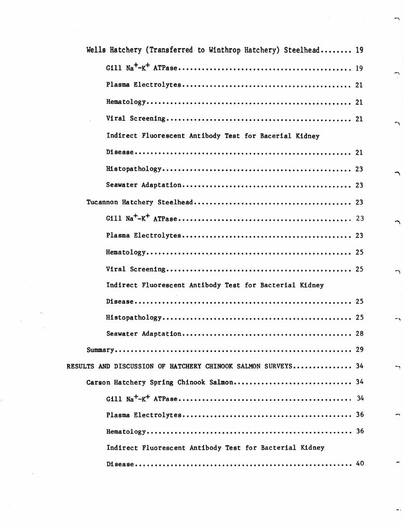
-
INTRODUCTION
The National Marine Fisheries Service (NMFS) under contract to the
Bonneville Power Administration is conducting research on imprinting
salmon and steelhead for homing (Slatick et a1 1979 1980 Novotny and
Zaugg 1979) The studies were begun with little background knowledge of
the effects of disease or certain physiological functions on imprinting and
homing in salmonids Consequently work aimed at filling this void was
begun by the authors in 1978 (Novotny and Zaugg 1979) and continued in
1979
In 1979 we examined random samples of normal populations of homing
test fish at the hatcheries to determine the physiological readiness to
migrate and adapt to seawater and general fish health At the Manchester
Marine Experimental Station Manchester Washington we determined the
survival of samples of the test fish maintained in marine net-pens after
release from the hatcheries Hatcheries and stocks sampled are listed in
Table 1
The data collected from random samples were as follows
1 Physiology
Gill Na+-K+ ATPase Abnormally low values could be indications
that the fish were either in pre- or post-smolt condition or had been
stressed in some way
Plasma elec trolytes bull Lower than normal values of Na or Cl could
indicate immediate problems of osmoregulation when the fish were introduced
to seawater high values may indicate some dehydration due to stress
Increases in levels of K can indicate kidney failure or nitrogen
supersaturation stresses
Table 1--Hatcheries and stocks sampled in the 1979 homing studies
a a a Hatchery Date- Date of- Date of-Pathology arrived at viral blood Date transferred ATPase release
Hatchery Species tag no Manchester assay sampling to seawater pens profile dates
Chelan- Steelhead 6101-6160 42579 42679 42679 42779 Yes 42679 Leavenworth
Wells- Steelhead 3001-3060 51079 51179 51179 51279 No 5979 Winthrop
Tucannon Steelhead 6801-6860 51479 51679 51679 51679 Yes 51779
Carson Spring 6401-6460 50179 50479 50479 50379 Yes 50879 chinook
Big White Salmon Fall chinook 6301-6360 51879 51979 51979 52279 Yes 52179
~ Held in fresh water at Manchester for sampling nrior to transfer to seawater pens
1 )~ -j J bull ) J J ) ) J
Hematocrits and hemoglobins Values below or above normal ranges
usually indicate anemia or dehydration which can reflect nutritional
disease or physiological changes
2 Fish health
The incidence of diseases during freshwater rearing as reported in
hatchery records describing the treatment of fishwere examined
The extent of latent bacterial kidney disease (BKD) as determined by
indirect fluorescent antibody technique and the presence (or absence) of
certain pathogenic viruses were of particular interest
A histological determination of significant lesions or abnormalities
in tissue from the gill eye liver kidney thyroid brain and olfactory
sac was undertaken
3 Survival in seawater net-pens
Periodic assessments of survival and growth were made and the major
causes of mortality were determined
These surveys were conducted to provide a documentation of the health
and physiological (smolt) condition of the populations of fish involved in
the tests especially at the time of imprinting and release When the
marked adult fish return the data analyzed from the health and physiology
surveys should provide us with information that would indicate any adverse
influence on survival Low survival in the marine net-pens caused by poor
health or a low percentage of fish transformed through the smolting stages
could bias any attempts to relate returns to imprinting
METHODS AND MATERIALS
Hatchery Sampling
The sampling of fish from the hatchery stocks for health profiles was
based on a combination of statistics and economics Random sampling from
3
populations ranging as high as 100000 or more showed that a population
with a disease incidence of 5 or greater can be detected from a sample of
60 fish (Ossiander and Wedemeyer 1973) Health survey samples of 60 fish
were taken at the hatcheries and held in circular tanks in fresh water at
Manchester In most cases the tissue and blood samples were collected
within 24 hours after arrival at Manchester
Sampling for Physiology
Plasma Electrolytes
Sodium potassium and chloride ion levels in plasma were determined
for the Wells Dam Hatchery steelhead Salmo gairdneri and Big Creek
Hatchery fall chinook salmon Oncorhynchus tshawytscha near the time of
release Profiles of plasma electrolytes of the Tucannon and Chelan
Hatchery steelhead and Carson Hatchery spring chinook salmon were
determined in fresh water and during seawater culture at Manchester
Plasma sodium and potassium values were determined by atomic absorption
spectrometry and chlorides with a chloridometer
Gill Na+-K+ ATPase
During 1979 selected stocks of fall and spring chinook salmon and
steelhead trout being reared for release at state and federal hatcheries in
the Columbia River drainage were monitored for changes in gill Na+-K+
ATPase to evaluate the state of smoltification at release
From tagged releases we determined the relationship between the state
of smoltification at release and length of migration time from the hatchery
to the estuary
At approximately 2-week intervals during the spring and summer of
1979 30 fish were removed by dip net from representative ponds or raceways
at Tucannon Carson Leavenworth (Chelan) and Big White Salmon Hatcheries
4
Steelhead from the Wells Dam rearing ponds were sampled only at release
Ten groups of three fish each were anesthestized or killed by a blow on the
head at each sampling After weights andor fork lengths were determined
approximately equal quantities of gill filaments were removed from the gill
arches of each of the three fish in the group (total weight of gill
filaments-Ol to 02 g) and processed as described in our previous report
(Novotny and Zaugg 1979)
Disease Sampling
Life History of Hatchery Juveniles
Husbandry techniques disease and environmental history may have
deleterious effects on fish health and smolt quality (Wedemeyer et a1shy
1979J Folmar and Dickhoff 1979) Many chemotherapeutic compounds used in
the treatment of parasitic and bacterial diseases of fish may affect
smoltification (Lorz and McPherson 1976) and subclinical infections may be
exacerbated by the stress of seawater entry
The information (Table 2) was obtained from hatchery management and is
self-explanatory Where information was not obtained the entries have
been left blank
Blood Sample Collection
The fish were lightly anesthetized in an aerated 120000 solution of
MS-222 In the larger fish blood was sampled from the caudal arch with a
1 cc heparinized syringe and a 25 gauge hypodermic needle Small fish were
bled by severing the caudal peduncle and collecting the blood in
heparinized capillary tubes
Blood samples taken for hematocrits (packed cell volume) were
5
Table 2--Disease and life history data of juvenile salmonids during freshwater rearing
Hatchery Stock Agency Species Date
egg take Date
ponded Feed Water
source Water
temp OF
Percent mortality
(all causes)
Size at release
(nolb)
Date released
(1979)
Chelan-Leavenworth
Wells-Winthrop
Tucannon
Chelan
Wells
Skamania
WDG
WDG
WDG
Steelhead
Steelhead
Steelhead
Dry amp OMP
Dry amp OMP
Dry amp OMP
River
Well amp River
River
34-56
50
40-62
154
220
40
47
114
42679
50979
51779
0
Carson
Big White Salmon
Carson
Spring Creek
USFWS
USFWS
Spring Chinook
Fall Chinook
August 1977
September 1978
January 1978
OMP amp Dry
OMP amp Dry
Spring amp River
Ground Water amp River
41-52
42-52
470
76
165
690
50279
52179
~ Bacterial gill disease
_J ) ~ J ) J ) I
J --
centrifuged in microhematocrit tubes for 3 minutes in a Clay-Adams Autocrit
II (Snieszko 1960)
Blood samples for hemoglobin determination were either read directly
with an A-O hemoglobinometer or collected in 20ul capillary tubes to
determine hemoglobin concentration by the colorimetric method described by
Bauer (1970)
Viral Assays
In 1978 liver spleen and kidney tissues from 60 fish in each test
group were sampled pooled in 12 tubes of 5 fish each and screened by a
private laboratory (Rangen Research Laboratories) for viruses In 1979
the tissue samples from each fish were aseptically divided into equal
portions One lot was submitted to Rangen Research Laboratories and the
other to the National Fisheries Research Center The results of these
independent tests are reported in Appendix A
Histopathology
Sixty individually numbered fish of each test group were preserved in
fixatives and submitted to Bio-Med Research Laboratories Gill liver
eye kidney thyroid brain and olfactory tissues were sectioned
appropriately stained and examined for any pathologic lesions or
abnormalities bull See Appendix B
Bacteriological Assays
The sensitive and highly specific indirect fluorescent antibody technique
(IFAT) was used to diagnose latent Bacterial Kidney Disease (BKD) in
hatchery populations
The individually identified fish were opened ventrally and the kidney
l Reference to trade names does not imply endorsement by National Marine Fisheries Service NOAA
7
exposed Thin smears of anterior and posterior kidney tissue were made on
multi-spot slides after piercing the kidney with a sterile inoculation
loop The slides were air-dried and fixed in reagent grade acetone for 10
minutes The acetone fixed slides were stored at -20oC until they were
examined Prior to the sampling season 40 pos tive control slides were
prepared in the same manner and stored at -20oC The control slides were
prepared from a clean kidney lesion from a spring chinook salmon from
Carson National Salmon Hatchery that was tested and confirmed to have high
numbers of BKD organisms
The IFAT for BKD was originally described by Bullock and Stuckey
(1975) and later modified by G W Camenisch (unpublished report) of the
US Fish and Wildlife Service (FWS) Eastern Fish Disease Laboratory The
complete procedure used in this study is described in our previous report
(Novotny and Zaugg 1979)
All dead and dying fish in the seawater pens were collected daily
Each fish was opened from the vent external and internal lesions noted
and the procedures for culturing vibriosis and other gram negative bacteria
(Novotny Harrell and Nyegaard 1975) were followed
The postmortems were classified as follows
1 Negative (cause of death not determined)
2 BKD (from lesions)
3 Vibrio anguillarum--serotypes 775 1669 or 7244
4 Vibrio sp
S ERM (enteric redmouth)
6 Furunculosis
7 Aeromonas hydrophilia (ex liquefaciens)
8
RESULTS AND DISCUSSION OF HATCHERY STEEtHEAD SURVEYS
Chelan Hatchery (Transferred to Leavenworth Hatchery) Steelhead
Gill Na+-K+ ATPase
Since the phenomenon of elevation in gill sodium potassium stimulated
ATPase (Na+-K+ ATPase) activity was first reported to be associated
with parr-smolt transformation in steelhead (Zaugg and Wagner 1973) and in
in coho salmon ~ kisutch (Zaugg and McLain 1970) numerous experiments
have been conducted to verify these results and extend observations to
other species As a result it has been conclusively shown that the rise
in gill Na+-K+ ATPase activity is one of the many physiological changes
which occur at the time of parr-smolt transformation
The average gill Na+-K+ ATPase activity of Chelan Hatchery
steelhead sampled in 1979 at Leavenworth Hatchery was not substantially
different from 1978 (Table 3) Gill Na+-K+ ATPase showed only a small
rise in late April (Figure 1) with a peak mean value of 94 The absence
of a greater increase in activity may have resulted from water temperatures
which remained at the upper limit (13degC) for good smoltification during
late April and May (Zaugg et al 1972) The average fork length (208 cm)
was similar to 1978 (210 em) but the average weight (981 g) was up from
1978 (794 g)
Plasma Electrolytes
A compilation of data on rainbow trout by Miles and Smith (1968) and
Hickman et a1 (1964) suggests expected normal or near normal plasma
electrolyte values in fresh water of 130 to 172 meq (milliequivalents)1
for Na+ 14 to 60 meql for K+ and 111 to 155 meql of Cl-
Table 4 lists some known values for steelhead trout from available
published literature including data for the Dworshak Hatchery steelhead
9
Tabl 3--Floh health and phylololieal data for the 1979 tal atudy fish ed a eoariaon with the 1978 homnl study fhh~
8-day 8t seawater try 111 ATPe
Peak freshwter 8ill Be_to1081ltamp1 data
Plasma electrolytes )1 (HelI)
cgtf bUilt IU detectable I the kidney FAT
aet1VltY-1IIO~10 pl prhlt
ATPase aetivttyshyules P1118 prh
(taken at -helter pOe arrlYti)- - _Ra___ Cl ____It
Stoek and ~ecJ_ ~ Allterior Eitherl
lo_st~r1C)~IIo~h__b~t~__1It__~~ _ X_ SD Date
Actv1ty X
Ii_toedt _1 (I)
_aJobln loot i SD ft i SD i SD
Chelan-Leavenworth steelhead
(1979) (1978)
0 133
17 167
0 567
17 867
112 142
218 286
166 195
36 43
4-23-79 5-0]-78
94+22 i-5
4911 4]3
9 89
10 60
1540 1650
+22 ~148
10 58
1248 llO9
+21 ~172
10 60
25 11
+18 ~8
ileUs 5teelhead
(1979) (1978)
17 167
17 200
0 467
]4 834
IIot bullbullled 71 137 109 28
5-19-79 5-03-78
165-+92 17--0
SO8 556
96 114
39 58
1382 lSO3
+200 tIl 1
20 58
1329 1079
+112 1206
S9 58
161 25
+22 t2 bull6
Tucannon steelhead
(l979) (1978)
17 83
0 33
0 100
17 216
180 109
277 2]4
2211 176
31 46
5-08-79 5-08-78 5-22-78
259-+9 18--2 11 7
530 485
92 97
58 60
1407 1595
+113plusmntS
59 59
1270 1316
+87plusmn6 5
58 60
29 24
+17 26
Caron ( 1979) 100 33 200 333 258 382 n7 34 (up to 204+54 367 52 10 1456 plusmn42 10 1341 21 10 37 ~3 sPrlng chinook relebullbulle)
5-01-79
0
Bll hite sal faU c1l111OO1lt
(1979) 0 83 0 83 IIot lOPled 5-lt19-79 135 436 71 53 l703 156 53 24 14
al bl EI
Fro IIovotny ZampullI 1979 10 days for the 1978 holnR atudy taken upon arrival at Hanchuter
noh
J -l -J ) ) J ) J ) ) )
505 (+55)
March April May June 1979
+
bull F reshwate ATPase
--- --- Seawater ATPase
Hatchery release date
t
5
+ 200 fish transferred to seawater
+
after 208 days in seawater
Fish held in freshwater past normal release
o L-1~O----~20------3~Or-----9L-----~19~----2~9r-----~9----~1~9----~29~--~8
I
to z
Figure l--Gill Na+-~ ATPase activity (means and standard deviations) of the Chelan Hatchery steelhead in fresh water and seawater
11
+ +Table 4--A summary of plasma Na K and C1 values in stee1head trout (from published sources)
Condition Na+ C1 K+ Reference
June-July (55 Laboratory test
g fish) Freshwater Saltwater
x X
= 162 = 170
range 140-160 137-185
X = 60 Houston (1959)
(after 36 hours)
March-May (13-15 cm fish) Laboratory tests Freshwater (range of mean values)
102-149 105-161 Conte and Wagner (1965)
Spring 1975 Dworshak Hatchery (at release)
X = 1342 range 128-138
Newcomb (1978)
N
Captured at Little Goose Dam (downstream from Dworshak)
x = 1342 range 128-141
Laboratory tests (cont ro1 groups-Spring)
Mean values range fro~ 159 to 169 133 to 138 26 to 43
Newcomb (1978)
Individual values range from 155 to 182 128 to 144 23 to 52
) -l J ) ) J ) J ) ) J
(Newcomb 1978) Newcombs data are extensive represent reasonably large
sample sizes (15 to 25) and are probably good approximations of Columbia
River steelhead
The mean plasma Na+ and K+ values of the Chelan Hatchery steelhead
were within normal limits (Table 3 Figure 2) at the time of release The
Na+ levels increased in seawater but returned to normal (in the
survivors) after 1 week
Hematology
There is considerable hematological data in the literature for rainbow
trout less for steelhead trout From the data summarized in Table 5 it
may be possible to estimate the range of hematocrit and hemoglobin values
for healthy steelhead The lo~er limit of mean hematocrit should not fall
below 30 and mean hemoglobin values below 6 would certainly be suspect
Upper levels are more difficult to define Snieszko (1960) reports mean
hematocrits of 53 and mean hemoglobin levels of 87 g100 ml of blood in
rainbo~ trout of a size comparable to large steelhead smolts Although our
values on steelhead trout (Table 3) were much closer to Snieszkos Newcomb
(1978) reported mean hematocrit levels in steelhead similar to that found
by other researchers working on rainbow trout (Table 5) A number of
authors (McCarthy et a1 1973 Wedemeyer and Nelson 1975 Wedemeyer and
Yasutake 1977) repeatedly suggest that the hematocrit levels of clinically
healthy rainbow trout should be between 24 and 43 with hemoglobins
ranging from 54 to 93 g100 ml blood and these values will be used as
the expected range for individual fish for the purposes of this report
The summarized data of the hematocrit and hemoglobin values for the
Chelan Hatchery steelhead are presented in Figure 3 There was no
difference in mean hemoglobin between 1978 and 1979 (89 g100 ml) nor in
13
250 125
0 bull Freshwater samples
--0------ Seawater samples
200 100
~ 0-
Plasma Na+ ~~~-~~ ~ 0shy
lt) -0 ~ lt)---() Qj150 75 I)-= 188 IJ 3daysCIl Q)
post Aseawater+ +
entryZ s 100 50 g
30 days post seawater entry ~ co t t0 sect
~-~ 50 ) 25~
V
0 0 10 20 30 9 19 29 9 19 29 8
April March May June 1979
Figure 2--Mean plasma Na+ and ~ levels in Chelan Hatchery steelhead sampled in fresh water at Leavenworth Hatchery and during seawater culture at Manchester
14
Table 5--A summary of hematocrit and hemoglobin values for rainbow and steelhead trout (from published sources)
Hematocrit Hemoglobin Source of data (gIOO m1 blood) References
Rainbow trout x = 316 x = 74 Houston and DeWilde (1968) SD = + 03 SD + 015
Rainbow trout x = x = Barnhart (1960) 282 to 31 7 65 to 77
(Individuals) (Individuals) (11 to 44 ) (22 to 130 )
Rainbow trout x = 395 x = 75 McCarthy~ et al (1973) (Kamloops strain) (30 to 49) (52 to 129)
Rainbow trout x = 341 x = 76 Wedemyer and Yasutake (1977)~ and (Shasta strain) (24 to 43) (54 to 93) Wedemyer and Nelson (1975)
Rainbow trout (average 142 cm) x = 453 Snieszko (1960)
(average 235 cm) x = 530 x = 87
Steelhead trout At Dworshak Hatchery (Spring) x = 403
(36 to 47) At Little Goose Dam (Spring) X = 356 Newcomb (1978)
(~8 to 44) Laboratory tests (Spring) X = 31 to 378
Individual range 28 to 45
JI
6101 - 6160 April 26 1979 Steelhead Chelan
40 n= 60 x=49835 s= 77
30
c 25 0 ~ 208 E l z 15
10
5
0 1
Percent () Hematocrit 40
n= 60 x= 88635 s= 158
30
c
25 0
8 ~
E l z 15
10
5
0 2 4 6 8 10 12 14 20
Hemoglobin - gdcl blood
Figure 3--Frequency histogram for hematocrit and hemoglobin values for the Chelan Hatchery steelhe~ in 1979 Number of fish sampled (n) mean hemoglobin and hematocrit values (X) and standard devia~ion (s) are also given
16
the range of values Mean hematocrit in 1979 (498) was slightly higher
than in 1978 (434) and 85 of the fish had hematocrits above the
expected maximum of 43 (for rainbow trout) in 1979 compared to 50 in
1978 None of the hematocr1ts or hemoglobins fell below the minimum
expected values
Viral Screening
Both Rangen Research Laboratories and the National Fisheries Research
Center (USFWS) indicated that no IPN virus was present in the Chelan
(Leavenworth) Hatchery steelhead
Indirect Fluorescent Antibody Test for Bacterial Kidney Disease
One posterior kidney smear (17) was found to have a few BKD organisms
in the Chelan (Leavenworth) Hatchery stee1head
Histopathology
A detailed report on the examination and interpretation of selected
tissue sections from the random samples is presented in Appendix B A
summary of the pathological conditions observed their severity and their
frequency of occurrence is presented in Table 6 The severity is ranked
as I--recognizible (least severe) II--intermediate and III--severe
Note that the incidence of rank II and III severity was low for all
conditions encountered (Table 6)
The major pathological conditions encountered in the Chelan
(Leavenworth) Hdatchery stee1head were of lymphocyte infiltration and
epithelial hypertrophy in gill tissue and the presence of sporozoan
parasites
Records (Table 2) indicate the total mortality in the hatchery was
15
17
Table 6--Ptholo~tcal coodlt1oul oltaervd 1n the twina etod _d their percenta of incidence
lad_c (I)
Tu- ala-lAa_nb IIIIU -----raWIlle Canoa c1 etu fll Itt~1 pdD dl~1tI WI C t _1t
iJ iJ iJ ~ ~ I II III bull II III bull II III 11 III e- II III ~
Or~an ptnolollY =shy
17
Retrobulbar fat ledon 0 0 0 0 33 17 0 50 0 0 0 0 0 0 0 0 150 0 0 150 Acute focal bettormage 17 0 0 17 0 0 0 0 0 0 0 0 0
Skeletal uscle leloll8 0 0 0 0 100 17 0 117 0 0 0 0 0 0 0 0 133 0 150
0 0 0 0 0 0 0
Deal _nonvelar cell infiltration 0 0 0 0 17 0 0 17 0 0 0 0 0 0 0 0 0 0 0 0
Retrobulbar pyolranulo_cou tnfla_cion 0 0 0 0 17 0 0 0 0 0 0 0 0 0 0 0 200 00 200 Retrobulbar nuclear loftltractoa 0 0 0 0 0 0 0 0 0 0 0 0 0 0 0 0 67 0 0 67
G1ll ~creased nu1lbers of lyaphocytes 81 7 183 0 1000 717 233 0 950 800 0 0 800 950 17 0 967 983 0 0 983
poundpltneU41 cell proUferation 783200 17 1000 683 267 0 950 913 17 o 1000 950 50 o 1000 933 67 o 100
Ly~htic telangiectasis of C01)dary lampMllae 50 0 0 50 0 0 0 0 0 0 0 0 0 0 0 0 0 0 0 0 Solitary basophilic as In econ~ry lae11ae 67 0 0 67 0 0 0 0 0 0 0 0 0 0 0 0 0 0 0 0 SoUury eosiJlOph1Uc in bullbullcondary 1_11ee 33 0 0 33 0 0 0 0 0 0 0 0 0 0 0 0 0 0 0 0
lie_tude parasice 1ft secondary 1_11ae 17 0 0 17 0 0 0 0 0 0 0 0 0 0 0 omiddot 0 0 0 0 rocal granuloma tn lecoaclary 111bullbull 0 0 0 0 17 0 0 17 0 0 0 0 0 0 0 0 0 0 0 0 SlOrooaa paralte tn eeoary 111 0 0 0 0 267 0 0 267 0 0 0 0 0 0 0 0 0 0 0 0 Focal 111ft_tion at 1_11 bbullbullbull 0 0 0 0 0 0 0 0 17 0 0 17 0 0 0 0 17 0 0 17 Vascular telusiectbullbullia of the econdary 1_I1ae 0 0 0 0 0 0 0 0 17 0 middot0 17 0 0 0 0 0 0 0 0
Hucoputu1ent 11111 0 0 0 0 0 0 0 0 17 0 0 17 0 0 0 0 0 0 0 0
General laflaaat iOG 0 0 0 0 0 0 0 0 17 0 0 17 0 0 0 0 0 0 0 0
Liver
00
Focal mononuclear cell infiltration 33 0 0 33 L3 0 0 83 17 0 0 17345 0 0 345 241 0 0 241
Sporozoan parasite 17 0 0 17 0 0 0 0 0 0 0 0 0 0 0 0
JIonsuppuratlve triaditt 0 0 0 0 33 0 0 33 0 0 0 0 0 0 0 0 0 34Increased parenchyl fat 0 0 0 0 33 0 0 33 0 0 0 0 34 0
LelliOnA typically asaociated with bacterial kidney disea 0 0 0 0 0 0 0 0 0 0 0 0 0 0 0 0 52 0 0 52 0 0 0 0 0 0 0 0 0 0 0 0 0 69 0 0 69Htcrogranulomas 0 0 0
Capsular parasttlc aranuloa 0 0 0 0 0 0 0 0 0 0 0 0 0 0 0 0 17 0 0 17
KLdltY Us Lons typlcal1 y associated with bacterial kidney d18case 0 0 0 0 0 0 0 0 0 0 0 0 0 0 0 0 88 0 0 88 Microgranuloraas 0 0 0 0 0 0 0 0 0 0 0 0 0 0 0 0 18 0 0 18 Pyo~ranuloma8 nephritis 0 0 0 0 0 0 0 0 0 0 0 0 0 0 0 0 18 0 0 18 Pyogranulomatous periuretelitis 0 0 0 0 0 0 0 0 0 0 0 0 0 0 0 0 18 0 0 18 Dilated tubule with giant bacteria 0 0 0 0 0 0 0 0 0 0 0 0 0 0 0 0 18 0 0 18 Focal tubular degenerative aiant cells 0 0 0 0 0 0 0 0 0 0 0 0 0 0 0 0 18 0 0 18
01 factory sac Ciliated protozoan parasite 533 0 0 533 0 0 0 0 0 0 0 0 0 0 0 0 0 0 0 0 Mmatode parasite 33 0 0 33 0 0 0 0 0 0 0 0 0 0 0 0 0 0 0 0 PyogranulolllAtous inflammation of the olfactory sac 17 0 0 17 51 0 0 51 0 0 0 0 0 0 0 0 254 0 0 254 Acute focal hellOrrhage 0 0 omiddot 0 0 0 0 0 0 0 0 0 0 0 0 0 17 0 0 17 Infltion of t~e olfactory 8_~c _ __9___L _lL___0 __-1 __--1 o 17 _I_ _0 17 0 0 0 __ 9 17 0 0 17
----------- ------shyThyroid
Perifollicular thyrofd1ti 0 0 0 0 0 0 0 0 0 0 0 0 0 0 0 0 62 0 0 62
flrdn MOnonuclear menlgeal infiltration 17 0 0 17 0 0 0 0 53 0 0 53 0 0 0 0 19 0 0 19 Ence(hal1 t is 0 0 0 0 ) 0 0 0 0 0 0 0 0 0 0 0 38 0 0 38
Other ~ryngeal sporozoan parasite 17 0 0 17 0 0 0 0 0 0 0 0 0 0 0 0 0 0 0 0
Pha-rynleal nematode parasite 0 0 0 0 17 0 0 0 0 0 0 0 0 0 0 0 0 0 0 0
~f I - Recognlzable (least severe) 1 I - IntenMdiate
III - Severe
) )J -J- J J J J -J -J j
Seawater Adaptation
At the time of introduction to seawater the test group contained
mostly transitional and smolted fish with 7 classified as precocious
males These precocious fish had all died by 82 days of seawater
residence Cumulative losses due to osmoregulatory dysfunction during the
first 30 days in seawater were about 28 in spite of 48 of the fish being
smolted at seawater entry Reversion from an apparent smolt or
transitional stage to a transitional or parr stage did take place after 82
days in seawater This suggests that those fish which were judged to be
smo1ts based upon external characteristics were not physiologically true
amolts or that high seawater temperatures may have forced reversion
Vibrio Strain 775 was the bacterial pathogen most commonly isolated
from moribund fish The 30-day survival was 59 and the survival after
completion of the tests (207 days) was only 8 (Table 7)
Wells Hatchery (Transferred to Winthrop Hatchery) Steelhead
Gill Na+-K+ ATPase
In 1978 Na+-K+ ATPase activities were determined on representative
samples of Wells Dam Hatchery (rearing pond) steelhead sampled at Winthrop
Hatchery on 3 May These fish had average gill Na+-K+ ATPase activity
of 170 (plusmn 51) with values ranging from 116 to 266 The sampled fish
averaged 225 cm fork length and 1022 g body weight and were judged to be
in a good smolted condition
In 1979 the average length and weight of samples transferred to
Manchester were 217 em and 970 g (respectively) on 11 May 1979 The
average g11l Na+-K+ ATPase activity from Wells Hatchery fish at
Winthrop Hatchery on 19 May was 165 (plusmn 92) with values ranging from 82
to 371
19
Table 7--Survival during seawater culture periods and causes of mortality
Stock and species
H (start) and date
Seawater survival in net-pens Survival N Days
()30 day (end) in survival post entry and date seawater to end
mortality per day
Causa of rtality during seawater culture Negative for pathogenic BKD Vibrio ERH Furunculosis Other
bacteria () () () el) () ()
Chelan-Leavenorth steelhead
200 4-27-79
59 16 11-20-79
207 80 044 34 0 348 0 0 618
Wells steelhead
200 5-11-79
175 9 11-20-79
193 45 049 0 07 46 0 0 948
Tucannon steelhead
200 5-15-79
485 37 11-19-79
190 185 043 100 0 267 17 0 61 7
Carson spring chinook
200 5-02-79
565 41 11-20-79
202 205 039 Numbers examined were not sufficient to evaluate
Big White Salmon fall chinook
150 5-21-79
933 1l 11-26-79
189 73 049 71 0 857 0 0 71
J) 0
)) J ) ) J J J j J J
Plasma Electrolytes
As in 1978 there were noticeable differences in the plasma
electrolytes of the Wells-Winthrop Hatchery steelhead when compared to the
other steelhead stocks in these studies (Table 3) There was only one
sample taken (at the time of release) but this stock again had the lowest
mean Na+ and K+ values even lower than in 1978 (Table 3)
There were no differences in transportation techniques or water
quality in 1979 between stocks thus no obvious stresses were involved
In 1979 plasma chloride was also measured In 1978 the mean plasma
chloride level (l08 meql) was below the expected value of III megl for
heal thy rainbow trout In 1979 none of the chloride values (Y 133
meql) were below normal
Hematology
The hematocrit and hemoglobin data for the Wells Hatchery steelhead
are presented in Figure 4 In 1978 the Wells Hatchery steelhead had the
highest mean hematocrit and hemoglobin values of any of the steelhead
stocks that we studied (Table 3) In 1979 849 were above the expected
maximum (43) hematocrit level and 517 above the maximum expected 93
g100 ml blood hemoglobin value None of the hematocrit or hemoglobin
values were below the expected minimums
Viral Screening
Rangen Research Laboratories reported IPN virus in 12 out of 12 pooled
samples in the Wells Hatchery steelhead and the National Fisheries
Research Center (USFWS) reported all samples negative for IPN virus
Indirect Fluorescent Antibody Test for Bacterial Kidney Disease
Two out of 60 fish s_pled (33) from the Wells (Winthrop) Hatchery
steelhead were found to have light BKD infections in anterior or posterior
kidney
21
i
3001 - 3060 May 11 1979 Steelhead Wells
40 n=53 lt=507735 s= 687
30
c 25CII 0
20 ~
Percent () Hematocrit 40
n= 60 x= 96135 s= 114
E ~ 152
10
5
a
30
-5 25 0 ~
20 E ~
2 15
10
5
0 12 20
Hemoglobin - gdcl blood
Figure 4--Frequency histogram for hematocrit and hemoglobin values for the tJells (Winthrop) Hatchery steelhead in 1979 Number of fish sampled (n) mean hemoglobin and hematocrit values (X) and standard deviations (s) are also given
22
Histopathology
The major pathological conditions encountered in the Wells (Winthrop)
Hatchery steelhead were increased numbers of lymphocytes and epithelial
hypertrophy in gill tissue (Table 6)
Seawater Adaptation
At entry to seawater Wells-Winthrop Hatchery steelhead were primarily
of transitional stage fish based upon external characteristics The
cumulative mortality within 30 days of seawater residence attributable to
osmoregulatory dysfunction was 73 Vibriosis accounted for most of the
other mortalities for the remainder of the study The presence of
precocious males was not a problem in this test group The survival to
test completion (193 days) was only 5 (Table 7)
Tucannon Hatchery Steelhead
Gill Na+-K+ ATPase
The gill Na+-K+ ATPase profile of summer-run steelhead from the
Tucannon Hatchery in fresh water was qualitatively similar to that observed
in 1978 with a distinct peak in enzyme activity in early May (Figure 5)
followed by a sharp decline A typical pulse of Na+-K+ ATPase activity
was observed when these fish were transferred to seawater at Manchester
Li tt1e change occurred until the fourth day when act ivi ty began to rise
(Figure 5) Fish held at the hatchery and sampled just 5 days after
release of the main group averaged 172 cm fork length and weighed 431 g
This was down from the average lengths and weights of 195 cm and
659 g respectively at the same time in 1978
Plasma Electrolytes
The summary data for plasma electrolytes at the time of release are
listed in Table 3 The mean values for Na K and Cl of the Tucannon
23
Hatchery release date
25 t x = 270 after 30 days and
bull Freshwater ATPase 369 at 188 days
- - - --- - - - Seawater ATPase
~lt I
O~1~O----~2~O----~3plusmnOr---~9----~19~--~29~----~9-----1~9----~29 March April May
1979
Figure 5--Gill Na+-~ ATPase activity (means and standard deviations) of the Tucannon Hatchery steelhead in fresh~ater and seawater
24
Hatchery steelhead fall within the expected ranges for rainbow trout
There was little difference in the mean Cl and K values between 1978
and 1979 (Table 3) In 1978 433 of the Tucannon Hatchery samples were
below the minimum range reported for K in rainbow trout
The plasma Na and K profiles of the Tucannon Hatchery steelhead in
both fresh and seawater appeared to be within normal ranges (Figure 6)
There was a typical rise in Na and K followed by a drop wi thin the first
week after transfer to the seawater pens and a return to normal in the
surviving fish after this initial stress period
Hematology
The summarized data of the hematocrit and hemoglobin values for the
Tucannon Hatchery steelhead are presented in Figure 7 and Table 3 The
mean hematocrit was slightly higher in 1979 than in 1978 and the mean
hemoglobin value was slightly lower than the 1978 mean
Viral Screening
The National Fisheries Research Center (USFWS) reported all Tucannon
Hatchery steelheadsamples tested as negative for IPN virus Rangen
Research Laboratories reported IPN virus in 1 out of 12 pooled samples
tested
Indirect Fluorescent Antibody Test for Bacterial Kidney Disease
Only 1 out of 60 Tucannon Hatchery steelhead sampled (17) was found
to have BKD organisms in an anterior kidney smear
Histopathology
The major pathological conditions encountered in the Tucannon Hatchery
steelhead were lymphocytic infiltration and epithelial hypertrophy in gill
tissue and a 53 incidence of ciliated protozoan parasites in the
olfactory sac (Table 6) Total mortality during rearing in the hatchery
25
-
~
~
125250
bull Seawater entry---lt) Freshwater samples __ ~ _ ___ _ _ Seawater samples +
i 100200 CDI
~ Plasma Na+
-0g A) 0-
b4=Q~ -00 ~ or 150 -ltgt- 75 IIgt
3i 30 days post + +208 days post III
IE seawater entry seawater entry + +
z CIgt
100 50 ~
c
a ~ t t
Plasma K+50 25
o L-l~0----~20------3~Or-----9~----1~9----~29~----~9~--~1~9~--~2~9~--~80 k~ A~~ M~ ~M
Figure 6--Mean plasma Na+-~ levels in Tucannon Hatchery steelhead sampled in fresh water at Tucannon Hatchery and during seawater
culture at Manchester
26
6801-6860 May 16 1979 Steel head Tucannon
40 n= 60 x= 529735 s= 766
30
i 25 j 0 E z 15
10
5
0
Percent () Hematocrit 40
n=60 x= 91835 s= 132
30
oS j 25 0 E z 15
bullo L-L-L__
Hemoglobin - gdcl blood
Figure 7--Frequency histogram for hematocrit and hemoglobin values for the Tucannon Hatchery steelhead in 1979 Number of fish sampled (n) mean hemoglobi~ and hematocrtt values (X) and s~andard deshyviations (s) are also given
27
was 22
Seawater Adaptation
At seawater entry 66 of the Tucannon Hatchery steelhead were judged
to be smolts Within 30 days of seawater residence 51 of the population
had died Osmoregulatory dysfunction accounted for 19 of the initial
mortality Vibriosis was the pathogen most commonly isolated from moribund
fish A large number of fish (25) were unaccounted for at the end of
testing and the loss may have been due to escape from the net-pen At
completion of the experiment (188 days) 185 of the fish had survived
The overall survival may have been closer to 40 if the losses from the
net-pen are included
I I
28
Summary
The compilation of the 1978 and 1979 data and preliminary analysis of
the 1980 data suggest that the generally high mean hematocrit and
hemoglobin values in northwest steelhead stocks may reflect a normal
hematological condition for these anadromous strains of the rainbow trout
or they may be associated with smoltification
The pathologist did not find histological lesions typically associated
wi th BKD in the kidney or liver tissue of the three steelhead stocks
examined Although the more sensitive IFAT tests of kidney tissue smears
from the same specimens did reveal the presence of BKD organisims the
incidence of the disease was extremely low There were no known
mortalities due to BKD in any of the steelhead stocks sampled during
seawater culture except the Wells Winthrop Hatchery stock Lesions
symptomatic of BKD were found in 07 of the mortalities examined but the
disease was not confirmed by IFAT
Analysis of the pathologists data indicate that lesions of the eye
were much reduced in comparison to 1978 Histopathological conditions
observed in the three steelhead stocks were restricted to a few organ
systems and may not significantly effect homing response or survival
However there were several conditions that appeared in all three stocks
and it may be of interest to summarize the probable causes
The pathological conditions in gill tissue of the 3 steelheadstocks
were predominately lymphocytic infiltration and epithelial hypertrophy
The incidence was higher in 1979 especially in the Tucannon Hatchery fish
These observations are probably indicative of exposure to antigens
including pathogenic and non-pathogenic microorganisms irritants or a
29
mild form of nutritional gill disease Second in frequency of occurrence
was the presence of sporozoan parasites in the gills of Chelan-Leavenworth
Ha tchery steelhead Pathological conditions in other tissues were minor
with the exception of a high incidence of ciliated protozoan parasites in
the olfactory rosettes or sacs of the Tucannon Hatchery steelhead
Figure 8 compares the survival of all three steelhead stocks during
seawater culture The survival wi thin the first 10 to 30 days after
transfer to the seawater pens was highest in the Chelan-Leavenworth
Hatchery stock and lowest in the Wells-Winthrop Hatchery stock This trend
is again apparent in Figure 9 which compares the known (removed) daily
mortalities for the first 4 weeks after transfer to the seawater pens The
largest numbers of dead fish were removed from the pen of the
Wells-Winthrop Hatchery steelhead Over 72 of the mortalities that
occurred within the first 4 weeks were removed within 1 week after transfer
to seawater
Figure 10 is a comparison (for all three stocks) of the average fork
lengths (at release) of steelhead that could be separated into three states
of development based on visual observation These were (1) heavy parr
marks present (2) fish in transition to molting with parr marks still
discernible although faint and (3) silvery smolts parr marks absent
Although the average length of the Wells-Winthrop Hatchery steelhead was
much higher than the Tucannon Hatchery stock only 28 of the
Wells-Winthrop Hatchery stock had the visual appearance of smolts versus
66 for the Tucannon Hatchery stock The Chelan-Leavenworth Hatchery
steelhead were in between with 48 considered to be visibly smolted
However the large average size of the Chelan-Leavenworth Hatchery smolts
~I
I
30
100 20 30
-~ 80 ~ ~-
ll Chelan steelhead C Tucannon steelhead o Wells steelhead
1Pooible increase with unaccountable inventory losses
o 20 40 60 80 100 120 140 Days post seawater entry
Figure 8--A comparison of survival during seawater culture of the three steelhead stocks tested in 1979
31
50
11 --tr-- Chelan steel head c QJ ----0---- Wells steelheadC I ~ __0- Tucannon steel head ~40 t Date of seawater entryQJ
s ~
E ~ 0 o -0 30 t QQJgt I I I 0 I I I E I IQJ ~ II I
s
A20
I -0 It QJ 0 ~
D 11 I ~
0 - to Pshy11 ~ t I QJ 10 c I 6E I bull ~ I I Z I I I~ Q
I I II I I I C I~R
0 30 9 19 29
April I May June 1979
~
I
I
Figure 9--A comparison of the known daily mortalities (removed) of the three stocks of steelhead after transfer to the seawater pens at Manchester Each stock began with 200 fish Mortalities are shown for (approximately) the first 30 days
~
32
I
100 A Chelan steel head (X 2265 mm) c Tucannon steelhead (I 1661 mm)
- o Wells steelhead ur- 2109 mm)
1 parr ~ 80 2middot in transition to smolting ~ Q) 3 bull visibly smolted
9shy gto ~ 60 10 () 2 3Q)
iii N
s ~ Q)
40 5 C ca s Q)
Qj 20 ~ 1---_--1
o 140 160 180 200 220 240
Average fork length (mm)
Figure 10--Relationship of the percentage of abundance of the three visible physiological stages at the time of release to the average size of each state (for all three steelhead stocks studied in 1979)
33
probably contributed to the greater initial survival after transfer to
seawater
The clinical health status of the Wells-Winthrop Hatchery steelhead
was satisfactory The high initial mortality after transfer to seawater
(in comparison to the other steelhead stocks) suggests however a severe
osmoregulatory dysfunction We cannot directly compare this type of
immediate stress with a supposed transition through the Columbia River
estuary because culturing samples of these hatchery test groups in net-pens
in seawater is an artificial situation and is recognized as such Lower
survival may not be indicative of what is occurring in nature as (1) the
fish are transferred directly from fresh to 280 00 seawater without
conditioning in estuarine water (as presumably might be the case in
nature) (2) they are fed an artificial diet and (3) they are contained in
net-pens and stressed by frequent (monthly) measurement activities
Nevertheless one can assume that if the survival in the net-pens was
high the fish should be able to withstand the normal transition rigors in
the wild and that the tests may be a relative measure of seawater
adaptability between treatments or stocks In comparison to the other two
stocks the Wells-Winthrop Hatchery fish were much less likely to survive
any early osmoregulatory stresses
RESULTS AND DISCUSSION OF HATCHERY CHINOOK SALMON SURVEYS
Carson Hatchery Spring Chinook Salmon
Gill Na+-K+ ATPase
Figure 11 is a graph of the gill Na+-K+ ATPase activity in 1979
The enzyme values were somewhat higher in 1979 than in 1978 The time
during which activity increased appeared to be the same in both years
34
H30
i Xi
Transfer to seawater
Ii t II 180 days post seawater entry
III f
it
+
0~-1~0------2LO-----3~0~----~9------1L9------2~9~-4L-~8~12
March April May 1979
Figure 11--Gill Na+-~ ATPase activity (means and standard deviations) of Carson Hatchery spring chinook salmon in fresh water and after transfer to seawater pens at Manchester
35
I ~
-1
i i
i
)
Plasma Electrolytes
There is little published data on normal plasma electrolyte levels in
hatchery chinook salmon Table 8 is a summary of the mean plasma Na Cl
and K values from chinook salmon that we previously examined The
exceptionally high K values in the Kalama Hatchery spring chinook salmon
may be due to hemolysis that occurred after sampling
There is a slight decrease of plasma Na in the Carson Hatchery spring
chinook salmon from 158 meq1 in March until the fish were released in
early May (Figure 12) The mean plasma Na value of 1456 meq1 at this
time (Table 3) is within the range of means encountered in other chinook
salmon samplings (Table 8) As expected plasma Na values rose abruptly
after transferring the fish to seawater (Figure 12) but quickly returned
to normal levels The mean chloride level of 1341 meql at release is
higher than any previously observed mean levels in any of the species
studied in 1978 or 1979 The mean plasma K value of 37 meql was also the
highest for any species studied in the past two years J with the exception
of the coho salmon There were fluctuations in the mean K levels after
transfer to seawater for the first week and then a leveling off in the
surviving fish to 3 to 35 meql
Hematology
Unpublished data from salmon diet studies in Oregon indicate expected
mean hematocrits for spring chinook salmon ranging from 242 to 380 and
35 to 39 for fall chinook salmon Published data on small fall chinook
salmon fingerlings (Banks et al 1971) indicate that hematocrit and
hemoglobin values increase as the water temperature increases (Table 9)
36
Table 8--Mean values of plasma Nat CIt and K from other samplings of hatchery chinook salmon
37
Sample Ntlliequivaleritsl
1978 Kalama Falls Hatchery spring chinook salmon (at release) 137 116 119
1978 Kooskia Hatchery spring chinook salmon ~at release) 114 104
1978 Leavenworth Hatchery spring chinook (at release) 150 108 17
1979 Leavenworth Hatchery spring chinook
March At release (late April) June
158 149 148
129 30 125 08 130 23
These were abnormally high potassium values and may have been due to some hemolysis of the samples
250 125
-(0)--- Freshwater samples __~ _ _ _ _ _ _ Seawater samples
200 100
~
E a-150 III
E + co Z co
100 i co 0
50
Plasma Na+ ltgt Cgt
Plasma K+
~_______~~_- ______o
- fa -------shy
bull 1M -------
or75 3
7+
s 50 ~
~
25
o~----~~----~----~----~----~----~----~~----------Jo20 30 9 19 29 9 19 29
March April May 1979
I ~
I
I
Figure 12--Mean plasma Na+ and K+ levels in Carson Hatchery spring chinook salmon in fresh water at Carson Hatchery and during seawater culture at Manchester
~I
38
Table 9--Hematocrit and hemoglobin values of fall chinook salmon cultured for four weeks at four water temperatures The average weight of the fish was 32 to 40 g (from Banks et al 1971)
Reiirtng Number Hematocrit () Hemoglobin (g100 m1 blood) temperatures
(oe) of
fish X Range X Range
100 10 322 29-36 54 45-63
127 10 358 31~40 54 48-64
156 10 376 32-43 59 49-68
183 10 389 35-46 64 54-73
39
I ~i
The mean values of fall and spring chinook salmon sampled in 1978 (for
all studies) ranged from (1) hematocrits - 367 to 594 and (2)
hemoglobins - 52 to 89 g Hb100ml blood Hematocrit values below 28 in
Pacific salmon may be the beginning stages of a number of problems and
should signal a cautionary warning
Although the mean hematocrits and hemoglobins of the Carson Hatchery
stock were well within the expected limits 183 of the fish had
hematocrits below 28 (Figure 13) Most of the low hemoglobin values
laquo 30 g HblOO ml blood) were associated with the low hematocrits
Bacterial kidney disease organisms were present in all samples with
hematocrits below 25
Indirect Fluorescent Antibody Test for Bacterial Kidney Disease
The Carson Hatchery spring chinook salmon had the highest incidence of
BKD (as determined by FAT) for any of the homing stocks screened (Table
3) The total incidence was 333 with 25 of these classified as level
III (severe) All fish with hematocrits below 25 were positive for BKD
However not all BKD infected fish had low hematocrits Latent infections
of BKD may require several years to develop into an active form capable of
killing fish in the marine environment (Ellis et al 1978) On the basis
of the number of fish with heavy intensities of BKD infection in our
subsamp1e we would antiCipate some ocean mortality from BKD
Histopathology (See Appendix B)
The Carson spring chinook salmon had a high incidence of epithelial
hyperplasia and lymphocytic infiltration in the g11ls (Table 6) This
inflammatory response is probably the result of exposure to a number of
pathogenic andor non-pathogenic organisms The relatively high incidence
40
40
35
30
c 25III Ot 0 20 E z = 15
10
5
0
40
35
c III
Ot 25 0 E = z 15
5
o
6401 - 6460 May 41979 Spring Chinook Carson
n=60 x= 3667 5= 1009
Percent () Hematocrit
n=60 x= 522 5= 175
Hemoglobin - gdcl blood
Figure 13--Frequency histogram for hematocrit and hemoglobin values for the Carson Hatchery spring chinook salmon iIL1979 Number of fish sampled (n) mean hemoglobin and hematocrit values (X) and standard deviations (s) are also given
41
of focal mononuclear cells in the liver is also indicative of possible I
I
~
I
I
~i
~
antigenic stimulation This is supported by the confirmation of bacterial
kidney disease in this stock In addition the Carson Hatchery spring
chinook salmon had the highest incidence (25) of granulomatous
inflammation of the olfactory sac which may be associated with
gramulomatous lesions of the liver and kidney typical of BKD
Seawater Adaptation
At the time of seawater entry the Carson Hatchery spring chinook
salmon were visually characterized as primarily transitional (55) and
smolted (39) fish The mean weight of the smolted fish was 258 g and the
mean weight of the population sample was 230 g
In early summer 13 precocious males were observed in the surviving
population If this represented the maximum knumber in the original
population there would be a minimum loss of 65 from precocious
maturation
Initial losses due to osmoregulatory dysfunction were minimal (6)
Further losses occurring during the third and fourth week after seawater
entry were probably due to seawater diseases The survival during
seawater culture is shown in Figure 14 and indicates that approximately
60 of the fish were able to survive the first 30 days
Big White Salmon Hatchery Fall Chinook Salmon
Gill Na+-K+ ATPase
Fall chinook salmon used in the homing experiments were tagged and
moved from the Spring Creek Hatchery to holding ponds on the Big White
Salmon River Gill Na+-K+ ATPase activities are plotted in Figure 15
Gill Na+-K+ ATPase in the homing study fish showed an increase in
42
100
80
20
o 40 80 120 160 200 240 Days post seawater entry
Figure 14--Survival of the Carson Hatchery spring chinook salmon during seawater culture
43
-5 ~
20E c ~
-o ~
C- Hatchery release date Ol 15 E May 211979
c t en ~ o E 10 3shygt t gtji () CO 5 ~ rf Ishyet + ~ 0 ~----~------~-------+------~------~------~I + 10 20 30 10 20 30
CO April I MayZ 1979
) I
Figure 15--Gill Na+-~ ATPase activity (means and standard deviations) of Big White Salmon Hatchery fall chinook salmon reared at Spring Creek National Hatchery
44
activity at approximately the same time as fish held at Spring Creek
Hatchery though the magnitude of that increase was less Colder water at
the Big White SalDlon River and slower growth were factors that probably
affected the gill Na+-K+ ATPase activity
Plasma Electrolytes
The mean plasma Na levels of 1703 meql from the Big White Salmon
Hatchery fall chinook salmon at the time of release were the highest of any
of the species of salmonids sampled in 1978 or 1979 The mean K level of
24 meql was within normal limits for chinook salmon Only small amounts
of plasma are available from fall chinook salmon and the volumes were not
sufficient to include chloride analyses No samples were taken after
transfer to seawater
Hematology
The mean hematocrit and hemoglobin values (Figure 16) of the Big White
Salmon Hatchery fall chinook salmon were in the upper level ranges for fall
and spring chinook salmon sampled in 1978 None of the hematocrits were
below 28 and the hematological data suggested a healthy stock of fish
Viral Screening
The National Fisheries Research Center (USPWS) reported all of the Big
White Salmon Hatchery fall chinook samples negative for IPN virus Rangen
Research Laboratories reported 4 out of 12 pooled samples as positive for
IPN virus
Indirect Fluorescent Antibody Test for Bacterial Kidney Disease
There was an 83 incidence of BKD in the BIg White Salmon Hatchery
fall chinook sa11l0n all associated with posterior kidney However two
(out of five) of the fish had moderate (level II) infections This could
represent an eventual loss from BKD after release
45
6301 - 6360 May 19 1979 Fall Chinook Big White
40 n=59 gtlt=435835 485 5=
30
~ en 25 ij 4shy0 20 E z ~
15
10
5
0
Percent () Hematocrit 40
n=60 x= 705
35 078s=
30
~ en ~ 25 4-0
20 E
Hemoglobin - gdcl blood
~
z 15
10
5
0
II
~
~
~
I II
1
~
Figure 16--Frequency histogram for hematocrit and hemoglobin values for
the Big White Salmon Hatchery fall chinook salmon in 1979 Number of fish sampled (n) mean hemoglobin and hematocrit values (X) and standard deviations (s) are also given
46
Histopathology
As reported for all homing test groups in 1979 the Big White Salmon
fall chinook salmon had a high incidence of epithelial cell proliferation
and lymphocytic infiltration in the gills (Table 6) This stock suffered
mortalities from enteric red mouth disease (Yersinia ruckerii) after they
were transferred to the Big White Salmon Hatchery rearing ponds and this
may have contributed to the development of these lesions The high
incidence of focal mononuclear cells in the liver is indicative of possible
antigenic stimulation and is consistant with the confirmation of bacterial
kidney disease by IFAT
Seawater Adaptation
The mean weight of the Big White Salmon Hatchery fall chinook salmon
sample was 76 g when introduced to seawater and only 15 of the test
group were visually characterized as smolts (mean weight of 97 g) The
majority of the remaining fish appeared to be in the transitional stage
The immediate mortality from osmoregulatory stress was slight and the
survival during the first 30 days in seawater was over 90 (Figure 17)
The mortality increased dramatically after 60 days in seawater and was due
primarily to seawater diseases No precocious males were observed in this
test group
47
100
Figure 17--Survival of the Big White Salmon Hatchery fall chinook salmon during seawater culture
48
Summary
We began our first examination of the olfactory or sac in 1979 since
this tissue may be the source of cuing for any olfactory imprinting We
were not able to measure responses to olfactory stimuli in fish with
obvious damage to olfactory sac epithelial tissue but we have been able to
demonstrate that this sensory receptQr can be the site of pathological
conditions in normal populations
For example as mentioned in the section on steelhead ciliated
protozoans in the olfactory sac were found in 53 of the Tucannon
steelhead Histological evidence of any type of olfactory sac pathology
was neglible in the two other steelhead stocks and no pathology was found
in the Big White Salmon fall chinook salmon No parasites were found in
the olfactory sacs of the Carson Spring chinook salmon but evidence of
some type of inflammatory lesion was found in over 25 of the Carson fish
The pathologist suggests that the lesions found in both the kidney and
olfactory sac are highly indicative of bacterial kidney disease and that
the nares may be a portal of entry for the organism (Appendix B) As a
result of this assessment we examined tissue smears of the olfactory sac
by IFAT of the Leavenworth and Carson Hatchery spring chinook salmon from
the following year class BKDorganisms were found in many samples
including some in the Class II and III intensity Until we have sufficient
tag return data for any of the 1979 homing study releases of fish it will
be difficult to assess any problems that may have been due to pathology of
the olfactory sac
In 1979 the general health status of the Big White Salmon Hatchery
fall chinook salmon stocks was good in comparison to the Carson Hatchery
49
I ~
spring chinook salmon There were no problems with hematology in the fall
chinook salmon but over 18 of the spring chinook salmon had low
hematocrits and hemoglobins Bacterial kidney disease organisms were found
in every kidney sample of fish with hematocrits below 25 Histological
examination of the gills and other organ tissues suggest a serious problem
exists with BKD (Appendix B) in the Carson fish Although the Big White
Salmon Hatchery fall chinook salmon had been exposed to some microscopic
organisms (Appendix B) this was probably Enteric Redmouth Disease and at
the time of release most of the fish were clinically healthy There was no
evidence of abnormal indicators of smoltification in either stock but the
short-term (30 day) survival of the Carson fish in the seawater net-pens
was poor In general a combination of health status evaluation factors
plus a high incidence of precocious males indicate that losses in the
Carson Hatchery spring chinook salmon are going to occur prior to or during
the first winter after release
CONCLUSIONS
Steelhead
1 The general health of the three steelhead stocks in the 1979 studies
was good and there were no indications of any pathology that would impair
survival or imprinting wi th two possible exceptions the presence of
sporozoan parasites in the g111s of over 25 of the Chelan Hatchery fish
and protozoan parasites in the olfactory sacs of over 50 of the Tucannon
Hatchery fish Unfortunately t we cannot evaluate the impact of such
parasitic infestations at this time
2 Observations of external appearances to determine the extent of
smoltification indicated that less than 33 of the Wells-Winthrop Hatchery
50
steelhead were smolted at the time of transfer to seawater (no gill
Na+-K+ ATPase profiles were available for this stock) Both the Chelan
and Tucannon Hatchery steelhead were transferred shortly after peak g111
Na+-K+ ATPase activi ties and had a higher percentage of visible smolts
than the Wells-Winthrop fish This indicates that the Tucannon and Chelan
fish were better prepared for the transition to seawater
3 The survival of the Tucannon and Chelan Hatchery steelhead during the
first 30 days after transfer to the seawater pens was two to three times
greater than that of the Wells-Winthrop stock even though the average size
of the Wells-Winthrop fish was larger This indicates that the expected
survival of themiddot Wells-Winthrop Hatchery steelhead will be less than
survival from the Chelan and Tucannon Hatcheries
Chinook Salmon
1 lbe incidence of latent BKD in the Big White Salmon Hatchery fall
chinook salmon was low basic hematology was normal and the pathology of
examined organ tissue reflects the probable exposure to ERM and BKD The
recovery from ERK was apparently successful and the fish were healthy when
transferred
2 Approximately 20 of the Carson Hatchery spring chinook salmon were
anemic and BKD organisms were found in every fish with hematocrits below
25 BKD was found in about 33 of the Carson Hatchery fish and the
intensity of infection was heavy in 25 of the infected fish The presence
of granulomatous lesions typical of BKD in 25 of the olfactory sacs also
reflects the serious nature of this disease in the Carson fish and may
well affect imprinting as well as survival
51
3 The g11l Na+-K+ ATPase profiles were normal for both the Carson and
Big White Salmon chinook salmon and the major releases were made just
after the peaks of Na+-K+ ATPase activity indicating good preparation
for seawater adaptation
4 About 90 of the Big White Salmon fish survived the first 30 days in l I
the seawater pens Slightly over 50 of the Carson fish survived the first
30 days in the seawater pens This indicates that the chances for survival
of the Big White Salmon Hatchery fall chinook salmon are excellent and the
chances for the Carson Hatchery spring chinook salmon are below normal
52
LITERATURE CITED
BANKS J L L G FOWLER and I W ELLIOTT 1971 Effects of rearing temperature on growth body form and
hematology of fall chinook fingerlings Progressive Fish-Culturist 33(1)20-26
BARNHART R A 1969 Effects of certain variables on hematological characteristics
of rainbow trout Trans Amer Fish SOc 98(3)411-418
BAUER J D 1970 Hemoglobin In Gradwohls Clinical Laboratory Methods and
Diagnosis S Frankel S Reitman and A C Sonnenwirth editors C V Mosby Co St Louis 7th Ed Vol 1 p 403
BULLOCK G L and H M STUCKEY 1975 Fluorescent antibody identification and detection of the
corynebacterium causing kidney disease of salmonids J Fish Res Bd Can 32(11)2224-2227
CONTE F P and H H WAGNER 1965 Development of osmotic and ionic regulation in steelhead trout
SalDlo gairdneri Compo Biochem Physiol ~603-620
ELLIS R W A J NOVOTNY and L W HARRELL 1978 Case report of kidney disease in a wild chinook salmon
Oncorhynchus tshawytscha in the sea Journ Wildlife Dis 14120-123
FOLMAR L C and W W DICKHOFF 1979 Plasma thyroxine and gill Na+-K+ ATPase changes during
seawater acclimation of coho salmon Oncorhynchus kisutch Compo Biochea Physiol 63A(2) 329-332
HICKMAN C P JR R A McNABB J S NELSON E D VAN BREEMEN and D COMFORT
1964 Effect of cold accliaation on electrolyte distribution in rainbow trout (Sa10 gairdneri) Can Journ Zoo 42577-597
HOUSTON A H 1959 Osmoregulatory adaptation of steelhead trout (Salmo gairdneri
Richardson) to seawater Can J Zool ~729-747
HOUSTON A H and M A DEWILDE 1968 Hematological correlations in the rainbow trout (Salmo
galrdnerl) J Fish Res Bd Can 25173-176
53
1ORZ H W and B P McPHERSON 1976 Effects of copper or zinc in fresh water on the adaptation to
seawater and ATPase activity and the effects of copper on migratory disposition of coho salmon J Fish Res Bd Can 33(9)2023-2030
McCARTHY D H J P STEVENSON and M S ROBERTS 1973 Some blood parameters of the rainbow trout (Salmo gairdneri)
J Fish BioI 51-8
MILES H M and L S SMITH 1968 Ionic regulation in migrating juvenile coho salmon
Oncorhynchus kisutch Comp Biochem Physiol 26381-398
NEWCOMB T W 1978 Some physiological effects of total gas pressure supersaturated
water on juvenile Pacific salmon and steelhead trout a laboratory and field study PhD theSis University of Washington Seattle 165 p
NOVOTNY A J L W HARRELL and C Nyegaard 1975 Vibrosis a common disease of Pacific salmon cultured in marine
waters of Washington Wash State Univ Ext Bull 663 8 p
NOVOTNY Anthony J and WALDO S ZAUGG 1979 Study of disease and physiology in the 1978 homing study
hatchery stocks--a supplement to Imprinting salmon and steelhead trout for homing by Slatick Novotny and Gilbreath January 1979 Processed
OSSlANDER F J and G WEDEMEYER 1973 Computer program for sample sizes required to determine disease
incidence in fish populations J Fish Res Bd Can 30 1383-1384
SLATICK E A J NOVOTNY and L G GILBREATH 1979 Imprinting salmon and steelhead trout for homing WAFC NMFS
Processed 23 p
SLATICK E L G GILBREATH and K A WALCH 1980 Imprinting salmon and steelhead trout for homing~ NWAFC NMFS
Processed 44 p
SNIESZKO S F 1960 Microhematocrit as a tool in fishery research and management
US Dept Int Fish and Wildlife Serv Spec Sci Rep Fish No 341 15 p
WEDEMEYER G A and N C NELSON 1975 Statistical methods for estimating normal blood chemistry
ranges and variance in rainbow trout (Salmo gairdneri) Shasta strain J Fish Res Bd Can 32551-554
)
I
54
WEDEMEYER G A and W T YASUTAKE 1977 Clinical methods for the assessment of the effects of
environmental stress on fish health US Dept Int USFWS Tech Paper No 89 18 p
WEDEMEYER GA R L SAUNDERS AND C C CLARKE 1980 Environmental factors affecting marine survival of anadromous
salmonids Mar Fish Rev 42(6)1-14
ZAUGG W S and L R McLAIN 1970 Adenosinetriphosphatase activity in gills of salmonids
seasonal variations and salt water influence in coho salmon Oncorhynchus kisutch eomp Biochem Physio1 ~587-596
ZAUGG W S B L ADAMS and L R McLAIN 1972 Stee1head migration potential temperature effects as indicated
by gill adenosine triphosphatase activities Science 176415-416
ZAUGG W S and H H WAGNER 1973 Gill ATPase activity related to parr-smolt transformation and
migration in stee1head trout (Sa1mo gairdneri) influence of photoperiod and temperature Compo Biochem Physio1 45955-965
55
CONTRACT No 9-79 (EFFECTIVE APRIL 1 1979 TO MARCH 1 1980) USDCNOAA PURCHASE ORDER No 79-ABB-00276
THE SURVEILLANCE OF VIRUS DISEASES I NSELECTED HATCHERY STOCKS OF SAU10N AND STEELHEAD SrOL
IN THE COLUMBIA RIVER BASIN DURING 1979
FINAL REPORT PREPARED JUNE 1 1980
FOR
NATIONAL MARINE FISHERIES SERVICE NORTHWEST AND ALASKA FISHERIES CENTER 2725 MONTLAKE BOULEVARD EAST SEATTLE WASHINGTON 98112
By
RANGEN RESEARCH ROUTE 1 Box 26l HAGERMAN IDAHO 83332
T
-
RANGEN RESEARCH J A DIVISION OF RANGEN J INC J CONSIDERS THIS REPORT AS SATISFACTION IN FULL OF ALL OBLIGATIONS UNDER CONTRACT NO 9-79 (NOAA PO NO 79-ABB-00276)WE HAVE APPRECIATED THE OPPORTUNITY OF BEING OF SERVICE TO YOUR ORGANIZATION AND HOPE THAT WE WILL HAVE THE OPPORTUNITY TO DO SO AGAIN IN THE FUTURE
ANY QUESTIONS WITH REGARD TO THE CONTENTS OF THIS AND ANY RELATED REPORTS MADE UNDER CONTRACT NO 9-79 SHOULD BE DIRECTED TO
Dr Robert A Busch Director Rangen Research Route 1 Box 264 Hagerman Idaho 83332
(208) 837-6192
i
TABLE OF CONTENTS
I Introduction 1
II Materials and Methods 6
I I I Resu ts 14
IV Discussion 20
V Conclusions 24
VI Li terature Ci ted bull 25
VII Appendices 27
ii
LIST OF TABLES
1 Stocks of Pacific salmon and steelhead trout smolt examined for virus at the time of release from
selected Columbia River drainage hatcheries during 1979 9
2 Summary of preliminary screen results and confirmed identification of virus isolated from selected
Columbia River drainage hatchery stocks of anadromous salmonid smolts during 1979 15
3 Summary of confirmed cirus isolation and identification from selected Columbia River
drainage hatchery stocks of anadromous salmonid smolts during 1978 and 1979 16
4 Combined incidence of infectious pancreatic necrosis virus in selected stocks of anadromous salmonid
smolt at Columbia River drainage hatcheries during 1978 and 1979 18 5 Summary of confirmed viral and serum neutralization
results from five selected Columbia River drainage hatchery stocks of anadromous salmonid smolts during 1979 Os bull bull bull bull bull bull bull bull bull bull bull bull bull bull bull bull bull bull bull bull bull bull bull bull bull bull bull bull bull bull bull bull bull bull bull bull bull bullbull 19
-
iii
LIST OF FIGURES
1 Geographic location of selected Columbia River drainage salmon and steelhead hatcheries sampled
for virus during 1979 8
I
I
I
page 1
I INTRODUCTION
On the springo 1978 the National Marine Fisheries Service (NMFS) Manchester Washington laboratory began an annual study to evaluate various factors pertinent to the successful smoltification ocean survival and adult return of selected anadromous salmonid fish stocks of hatchery origin in the Columbia River basin A portion of this study was devoted to ascertaining the general health profile of each stock at the time of smoltification and immediately prior to hatchery release and natural outshymigration into saltwater The purpose of this general health profile was to determine the occurrence and incidence of selected infectious diseases known to be potentially important to the growth and survival of salmonid fishes in general The health profile data acquired is then used in the evaluation and interpretation of other data obtained with regard to the relative success of saltwater adaptation ocean survival and hatchery return potential
Migrant hatchery flah stocks were examined for selected viral bacterial and parasitic disease agents using a wide variety of techniques as described in the parent NMFS project report Summary results of the first year of this study pertaining to 1978-79 brood year stocks have been reported by Busch (1979) CZESD and ETSD (1979) Novotny and Zaugg (1979) This report concerns itself only with the work contracted by NKFS to Rangen Research under USDC NOAI Purchase Order No 79-ABB-00276 (Rangen Contract No 9-79) and pertains to that portion of the general health profile dealing with the screening of selected populations for the presence of important viral pathogens known to infect salmonid fish in North America Included among these pathogens are infectious pincreatic necrosis virus (IPNV) infectious hematopoietic necrosis virus (IHNV) Herpe8v~rus saZmonis and any additional replicating agents capable of inducing specific cytopathic effects (CPE) under the given conditions of surveillance
The virus disease most commonly associated with Pacific salmon and steelhead trout stocks in the Pacific Northwest is infectious hematopoietic necrosis virus IHNV was first recognized in 1951 in sockeye salmon (OnchorhynchuB nerka) at Leavenworth National Fish Hatchery and kokanee salmon a land-locked sockeye salmon at Winthrop National Fish Hatchery The virus was isolated for the first time from sockeye salmon in 1958 (Wood 1974) The disease is known to cause significant mortality in hatchery populations of Pacific salmon and steelhead trout as well as other salmonid species all a10ftg the Pacific coast of North America from California to Alaska and is considered endemic to maDY watersbeds ieluding the Columbia River Basin
IHRV infectioa character1stically results in a peracute to acute course ot dl resulting in high levels of mortality in cbinook al8aoa (OnahorhynchuB tshawytscha) and an acute to subacute disease in sockeye salmon The
~I
j
page 2
disease is characterized as its name implies by destruction of the bematopoietic tissues resulting in an acute condition of hemorrhage anemia and often severe mortality among fry and fingerling fish under hatchery conditions Infection seems to be primarily vertical in nature with the disease being transmitted with the els and reproductive fluids of asymptoatically infected returning broodstock The coho salmon (Onahorhynahu8 kieutah) appears to be more resistant to IHNV infection than the other species of Pacific salmon but can function as an asymptomatic carrier in the maintenshyance and dissemination of the disease (Wolf 1972)
Survivors of an epizootic infection of IHNV are known to carry the virus in an eclipse phase of intracellular infection typical of the rhabdovirus group of which it is a member Duringtnisasymptomatic carrier state the virus is not detectable by routine methods of surveillance and is presumed to be non-infectious This period of subclinical infection includes the time of physiological transition to saltwater known as smoltification The latent effects of its presence upon smoltification ocean survival and adult return are not known at this time However the detectable infectious virus has been shown to reappear immediately prior to spawning in infected populations of returning adults and is readily transmitted to the progeny at this time
The early epizootiology of suspected viral diseases including IHNV in the Columbia River drainage is reviewed by Parisot et al (1965) Several extensive surveys to determine the incidence and distribution of the virus have been conducted since that time in selected stocks of Columbia River trout and salmon In 1972 Amend and Wood reported that no IHNV could be found in Columbia River stocks of Pacific salmon returning to 15 selected hatcheries in the state of Washington These findings were based uponthe extensive sampling of 130 to 150 ovarian fluid samples taken from each population at the time of spawning Tebbit and McMichael (1973) found no evidence of IHNV in 10 Columbia River salmon stocks returning to hatcheries in the state of Oregon during 1971 and 1972 However in 1973 they reported the confirmed isolation of IHNV from an adult spring chinook salmon stock returning to Oregons Pelton Dam Holding Facility Numerous additional studies have been undertaken by various state and federal agencies in subsequent years but have often failed to be comprehensive in deSign and execution and their results are often not readily available
Even though IHNV has not been found to be a majorproblem for Columbia River stocks of chinook salmon in general it does continue to pose a threat to this specie as well as sockeye salmon under hatchery conditions The
real occurrence and incidence of IHNV is still not well defined due to problems in the detection of asymptomatic carrier states of infection during the eclipse stage of the virus Knowledge is also lacking on the possible
page 3
residual effects of the carrier state infection on successtul smoltitication saltwater survival and adult return The disease retUinsendemic to the watershed and has been increasing in its known host range geographical distribution and physical tolerances such as temperature IHNV bas recently been found to be pathogenic for rainbow trout (Satmo gaipdnspi) fry fingerlings and yearling fish at 145 C (58 F) in the Snake River drainage of the upper Columbia River basin It has also been found in a subclinical infectious carrier stage in rainbow trout broodstock in this same area (Morton and Busch 1980)
Another salmonid virus infectious pancreatic necrosis virus (IPNV) has not often been associated with or considered a problem in Pacific salmon stocks However IPNV is known to be endemic in the Columbia river drainage where it is primarily recognized as a reovirus disease of trouts and chars It is also known to be capable of infecting Atlantic coho and chinook salmon (Fish Health Section 1974)
IPNV is considered to be the same disease originallydescribed by MGonigle (1941) in brook trout (SaZveZinus ontinaZis) and Atlantic salmon (SaZmo salap) in hatcheries in the Canadian maritime provinces and later confirmed byMacKelvie and Artsob (1969) The pathogenesis of IPNV in Atlantic salmon has recently been described by Swanson and Gillespie (1979) The disease was first isolated in the western United States in 1963 by Parisot et al
IPNV disease is typically characterized by a peracute to acute course of infection and mortality of fry and fingerling fish or a subacute to chronic infection of larger fish up to and including yearling sizes The infectious agent is readily transmitted horizontally through the water between fish by means of infected feces and urine from clinically diseased or asymptomatically infected carrier fish It is also readily transmitted in a vertical manner from asymptomatically infected adult fish to their progeny via infected reproductive fluids and eggs Survivors of an epizootic infection often remain asymptoshymatic carriers for life and continually shed the infectious virus into the water a condition which has recently been modeled on a Chinook salaoft cell line by Hedrick et al (1978) and established in rainbow trout under laboratory conditions by Reno at a1 (1978) The disease has been shown to be transmitted in the natural environment to susceptible stocks and maintained in wild and feral populations for extended periods of time with the incidence of infection gradually decreasing over time when no new introduction of the virus is made (Yamamoto 1975) Sonstegard (1970) also showed long term survival of IPNV in the gastrointestinal tracts of selected non-salmonid fishes picivorous birds and aquatic invertebrates
Until 1968 IPNV had not been known to occur in any of the Pacific salmon species However in July of that year Wolf and Pettijohn (1970) isolated the virus from coho
page 4
salmon fingerlings at Lamar National Fish Hatchery in Pennsylvania The eggs of the infected stock had been taken from a spawning population of landlocked adult salmon in Lake Michigan The fish had been checked for virus as fry and were determined to be free of the infectshyion Bowever t IPNV was enzootic among hatchery stocks of trout at the time and viru8 was soon isolated from the fingerllD salmon durin an epizootic of furunculosis disease (Apomona salmoniaida) No mortality could be attributed to the preseDce of the virus and evidence indicated that infection was due to horizontal rather than vertical transmission
Amend and Wood (1112) 8urveed 15 Columbia River stocks of Pacific salmon returning to hatcheries in the state of Washington in the fall of 1970 selecting only tor IHNV on primary screen and confirmatory cultures Consequently no IPNV was reported In 1973 Tebbit and McMichael reported OD the surveillance of Columbia River stocks of Pacific salmon returning to selected hatcheries in Oregon Their comprehensive design included the examination of visceral tissues and ovarian fluids from SO adult females from each of four discrete populations Adult fish and progeny fry were also bled and the sera titered for specific neutralizing antibodies against the common viral diseases IPNV virus was isolated from two of twelve five-fish tissue pools from adult coho salmon returning to the Bonneville Hatchery in 1971 and also from their progeny fry in thirty or thirty ten-fish pools indicating verttcal transmission It is interesting to note that McMichael (1974) was only able to isolate virus from the fry progeny at 30 and 60 days of age post hatch and that these same fish as fingerlings when sampled at 90 and 210 days post hatcb no longer yielded detectable levels of viru but did delROnstrate specific neutralizing antibodies against IPNV with titers in excess of 1200 as confirming evidence of prior exposure Attempts at horizontal transmi8sion of the virus under hatchery conditions were unsuccessful at 15 C
A report in tbe FAO Aquaculture Bulletin (1973) indicated that McMichaels cobo salmon isolate of IPNV was unlike the standard ATCC UR-299 (American Type Culture Collection) IPNV trout isolate and more typical of French isolates of IPNV that are known to lose 99 of their infectivity in a single freezethaw cycle
When Tebbit and McMichael (1973) continued their sampling program they found returning adult populations and their progeny fry to be negative for IPNV at all of the selected hatcheries sampled in 1972 and 1973 However t
they were able to demonstrate specific IPNV neutralizing antibodies in the sera of 270 adult fall chinook and coho salmon The overall incidence was 74 in the returning coho and 92 in the returning fall chinook populations In 1972 they were also able to isolate IPNV from adult coho salmon returning to Cascade Hatchery but the virus
page 5
could Dot be demonstrated in the fry progeny or in the adult returns the following year Tebbit and McMichael (1973) also reported finding IPNV in adult chinook salmon returniag to the Pelton Dam Holding Facility in 1973 after the adult returns and progeny fry were found to be free of the virus in the 1971 and 1972 brood years
IPNV was isolated from ateelhead trout being reared at Idaho Power Companys iacria Springs Steelhead Hatchery in the Snake River CaDlOD of Southern Idaho near Buhl in 1974 IPNV is known to be endemic to the commercial rainbow trout hatcheries in tbe local area and its appearance at the Niagria Springs statioR was Dot surprising The virus has since reappeared at the atation periodically and has been associated with significant mortality
In 1174 Wood indicated that he had yet to make any positive IPNV iaolatioDs from Columbia River stocks in Washington state However in 1975 Tebbit reviewed McMichaels initial surveillance results and continued the program Again lookincmiddotat visceral tissue samples ovarian fluids and progenyfrJ Tebbit was able to demonstrate IPNV in six of twelve five-fish tissue pools from adult coho salmon returning to the Cascade hatchery during the 1972 broodyear but he could not demonstrate vertical transmission to the progeny fry That same year Tebbit also isolated IPNV frOll a population of spring chinook salmon fry at the Pelton Dam Holding Facility that were the progeny of an adult stock that had been previouslydiagnosed with a confirmed asymptomatic infection of IHNV (Tebbit 1975)
In 1967 Wolf and Quimby were first able to demonstrate the development of specific viral neutralizing antibodies in the serum of asymptomatically infected IPNV carrier fish Presence of such specific antibodies are considered as presumptive serological evidence of prior or concomitant exposure to the virus During the 1973 broodyear Tebbit was able to detect IPNV specific neutralizing antibodies in the sera of adult coho salmon returning to both the Sandy Hatchery and the Bonaeville Hatchery None of these seropositive populatioa or their progeny fry yielded a confirmed viral isolation Tebbit continued surveillance during the 1974 brood year but failed to isolate virus from any of the adult returns sampled but IPNV specificneutralizing antibodies were found in a stock of landlocked spring chinook salmon in the Detroit River impoundment on the North Santiam River in Oregon An endemic infection of IPNV had previously been demonstrated in wild cutthroat trout populations in theae same waters
Based upon the findings of both IPNV and IHNV in stocks of anadromous salaonida in the Columbia River basin the state of Oregon established a management policy prohibitingthe transport of any Columbia River stocks of fish to a coastal river system for fear of disseminating an endemic viral disease problem
In 1975 Mulcahy and Sanders reported isolating IPNV from
I
page 6
spring chinook salmon at the Oregon Fish and Wildlife Commissions Corvallis Research Laboratory More recently in the first year of this study Busch (1979) was able to isolate IPNV from 16 of 28 stocks of Pacific salmon and steelhead trout smolts at 12 of 18 Columbia River hatcheries sampled The incidence of infection among 11 coho salmon stocks sampled was 82 Of five steelhead trout stocks examined 75 were found to be infected with IPNV Four spring chinook salmon stocks had an incidence of IPNV of 60 and eight fall chinook salmon stocks had an incidence of IPNV of 33 All infections appeared to be asymptomatic carrier states and could not be associated with anysignificant mortality
It has been shown time and again that IPNV can indeed infect Pacific salmoa aad steelhead trout It is becoming recognized thatIPNV may be all too common as an asymptoshymatic infection in a wide variety of discrete spawning populations in the Columbia River basin and that these infections may be easily missed or not clinically diagnosed in certain types of samples taken at certain stages in the life history of the Pacific salmon and steelhead trout It still remains to be shown whether or not the presence of an endemic infection of IPNV has a detrimental effect upon the population Mortality directly attributed to the presence of the virus under hatchery conditions has yet to be demonstrated for Pacific salmon stocks but is known to occur with steelhead trout populations
The last of three recognized salmonid viruses to be potentially found in the Columbia River basin is Herpesvirus 8almonis This virus or other closely related viruses are known to infect rainbow trout and sockeye salmon at all stages in their life history and has been implicated in mortality of both fry and adult fish The virus was first described in Japan as being endemic among certain stocks of sockeye salmon It has only been diagnosed once in the United States as an asymptomatic infection of rainbow trout broodstock at the Winthrop National Fish Hatchery The virus still remains a fairly unknown entity with regard to the general health of Pacific salmon and steelhead trout stocks as its true host range geographical distribution and general ecology and epidemiology has yet to be determined (Wolf et ale 1975)
II MATERIALS AND METHODS
Procedures of viral surveillance were designed to fit within the overall field sampling program and budget of the parent study and at the same time give optimum sensitivity and accuracy with regard to detecting the presence of any of the major salmonid viruses found within the Columbia River basin Techniques for both surveillance of viral diseases including primary isolation and confirmed serological identification and surveillance of specific
page 7
viral neutralizing humoral antibodies were used A total of 5 stocks of chinook salmon coho salmon and
steelhead trout from 5 different Columbia River basin hatcheries (Figure 1 and Table 1) were checked for the presence of virus at the time of smoltification and hatchery release during 1979 Field sampling was conducted by NMFS personnel according to procedures established by and with field sampling kits provided by Rangen Research (Appendix A) Hatchery stocks being sampled were first transported in a live haul container back to the NMFS Laboratory at Manchester Washington They were then maintained in freshwater flow-through systems (Beaver Creek water supply)for one to two days prior to lethal sampling The field sampling diluent provided was formulated (see Appendix B) to provide optimum survival of any infectious virus under transport conditions while inhibiting the growth of any microbial contaminants It was provided in sterile graduated polycarbonate screw-cap tissue culture grade centrifuge tubes (Corning 25310 Corning Glass Works Corning New York)The use of the graduated tube allowed for the calculation of accurate sample dilution factors based upon the displaceshyment method as 02 g of tissue was pooled from each of five fish into 9 ml of field sampling diluent and a total of twleve five-fish pools collected from the randomly selected 60 fish sample
Serum samples were obtained by lightly anesthetizing 60 fish from each stock in an aerated solution of 120000 tricaine methanesulfonate (Ayerst Laboratories New York)In most cases blood was sampled from the caudal arch by means of a 10 cc heparinized syringe fitted with a 25 G hypodermic needle Small fish were bled by severing the caudal peduncle All whole blood samples were immediatelytransfered to heparinized micro-capillary tubes plugged at one end and centrifuged for three minutes in a ClayshyAdams Autocrit II centrifuge The serum was then transshyfered to a second micro-capillary tube plugged at both ends and placed sequentially in a numbered tube rack
Following completioR of all sampling procedures and the Field Sampling Data Sheet (Appendix A) the complete refrigerated field kit and samples were returned to the Rangen Research Laboratory in Hagerman Idaho via Greyhound NBO Package Express Service The average time in transit was 20 hours and the temperature of the samples upon receipt was oonsistently between 6 and 8 C A duplicate set of tissues samples were taken from the same fish as per the directions of Dr Dan Mulcaby and sent to the National Fisheries Research Center in Seattle Washington for confirmatory viral analysis by a comparable methodology
Upon receipt into the Rangen Laboratory the receiving temperature of the tissue samples was recorded The raw serum samples were stored at 4 C for a maximum time of five days prior to titration and serum neutralization The twelve tissue sample tubes were assigned a group accession number and each tube in the group given a
I page 8
FIGURE 1 GEOGRAPHIC LOCATION OF SELECTED COLUMBIA RIVER DRAINAGE SALMON AND STEELHEAD HATCHERIES SAMPLED FOR VIRUS DURING 1979
1 Big White Salmon 2 Carson 3 Leavenworth 4 Tuccanon 5 Winthrop
TABLE 1 STOCKS OF PACIFIC SALMON SHOLT EXAMINED FOR VIRUS AT THE TIME OF RELEASE FROM SELECTED COLUMBIA RIVER DRAINAGE HATCHERIES DURING 1979
STATION S1Oa UAlIIKID DATE SAMPLED RANGEN ACC NO
Big White Salmon fall eh1nook salmon 52479 208-79
Carson prins chinook salmon 5879 179-79
Leavenworth ateelbead trout (Chelan stock) 42579 158-79
Tuccanon steelhead trout 51679 203-79
Winthrop steelhead trout (Wells stock) 51579 195-79
d fI)
oq CD
(0
page 10
serial numeric identifying code The refrigerated tissue pools were homogenized in situ with a Polytron PUC-2-110 homogenizer fitted with a steam sterilizable stainless steel PT-IO generator (Brinkman Instruments Westbury New York) for 30 seconds The homogenized tissue samples were then centrifuged at 2000 X g for ten minutes at 4 C in a Sorval RC-5 refrigerated centrifuge with an HS-4 rotor and appropriate tube adapters (DupontSorval Newtown Conn) to remove large cellular debris Two tenths of a milliliter of the supernatant was pipetted off with a sterile disposable polyethylene tip and diluted in 18 ml of a disinfecting diluent (Appendix B) and incubated at 4 C overnight for decontamination of fungal and bacterial organisms This procedure resulted in a final 1100 working dilution of the original tissue sampled All samples were maintained at 4 C during all preparatory procedures
Tissue samples received into the laboratory were screened for the presence of virus within seven days of sampling Viral screen tests were conducted with a microculture procedure using the Chinook Salmon Embryo (CHSE-214) cell line in passages 267 through 277 and a MEM-10-Tris-PSF media (Appendix B) at 125 C incubation for 21 days Four replicate wells of each of two dilutions were run for each of the pooled tissue samples Microculture screen tests were prepared by dropping 005 ml of the decontaminated and diluted 1100 tissue pool into eachof the first four wells of an eight well series on a sterile Microtest II (Falcon 3040 Becton Dickinson and Co) tissue culture plate and 0025 ml of the same sample into the last four wells of the eight well series using an Oxford 8000H sampler and a sterile Oxford 810S tip During preparation the microshyculture plates were temporarily covered with a Falcon 3041 sterile lid and maintained at 15 C Appropriate IPNV and IHNV positive viral controls and negative media and cell controls were set up for each group tested After the sample dilutions were delivered to the microculture plate a stock flask of CHSE-214 cells that had been grown to 95 confluence at 20 C in MEM-10-Tris-PSF media was examined under an inverted microscope for confluence and quality and dissociated in 4 ml of PANEDTA media (Appendix B) The PANEDTA cell suspension was centrifuged at 1500 x g for four minutes The supernatant was decanted and the pelleted cell mass resuspended in a small volume of MEM-10shyTris-PSF media The resuspended cells were further diluted in MEM-10-Tris-PSF media to a final 14 working concentration based upon the surface area split ratio and placed in a sterile 100 ml covered flask containing a sterile magneticstir bar The diluted cell suspension was placed on a refrigerated magnetic stir plate with slow stirring to maintain the cells in suspension A sterile Minipet repipetting syringe (Manostat 71-500-010 Manostat New York NY) was ittedwith a sterile disposable 18g X Ii hypodermic needle and primed with the chilled CHSE-214 cell suspension Fifteen hundredths of a milliliter of the
page 11
diluted CHSE-214 cell suspension was pipet ted into each well on the inoculated microculture test plate The plate was immediately sealed with Falcon 3044 pressure sensitive film and incubated at 125 C The suspended CHSE-214 cells were allowed to settle down through the sample material in the well adsorb infectious virus present and attach to the bottom of the well
All viral screen tests were incubated at 125 C for 21 days A~l wells of both dilutions of both test and control series were periodically read under an inverted microscope for evidence of specific cytopathic effect (CPE) cytotoxicity and microbial contamination The daily observations were recorded on the Virological Examination Report Sheet (Appendix C) If more than four wells in an eight well series were found to be cytotoxic the original decontaminated sample was diluted 12 with additional disinfecting diluent to a final working dilution of 1200 and re-run on the microculture screen procedure as described above If more tban four wells in an eight well test series were found to be contaminated the original sample was filter sterilized through a sterile 045u membrane fil ter and re-run on the microculture screen procedure
At the end of the 21 day incubation period the 96 wells on each plate (8 wells for each of 12 pooled tissue samples in the lot) were classified as being either positive (definite CPE) questionable (possible CPE or cytotoxicity) or negative (no evidence of CPE or cytotoxicity) and pooled into one of three tubes according to its classification After the supernatant medias had been aspirated and pooled from the wells of the microculture screen plates for further testing all wells of the plate were stained with a 1 alcoholic solution of crystal violet and dried as a permanent record of the screen results The presence or absence of infectious virus in each of the three classified screen pools was confirmed and identifshyied by means of a microculture serum neutralization procedure One quarter of a milliliter of sterile field sampling diluent was placed in each of the first three wells of an eight well series on a sterile microculture plate Twenty five thousandths of a milliliter of EFDL 149 Polyshyvalent IPNV Antisera (Eastern Fish Disease Laboratory Kearneysville West Virginia) diluted 1100 in sterile field sampling diluent was pipet ted into the next two wells of each eight wellseries Twenty five thousandths of a milliliter of EFDL 150 IHNV antisera diluted 1100 in sterile field sampling diluent was pipetted into the next two wells of each eight well series and finally 0025 ml of EFDL 100 normal rabbit sera diluted to a final 1100 working concentration in sterile field sampling diluent was pipetted into the last well of each eight well series Each of the three classified and pooled screen aspirates were then serially diluted out to 10-3 by pipetting 0025 ml of the material into the first well of an eight well series mixing with the diluent present and transfering 0025 ml
page 12
into the second well mIxIng and transfering 0025 ml into the third well and mixing Twenty five thousandths of a milliliter of the 10-3 dilution of the sample in the third well of each series was then transfered to each of the remaining five wells in the series containing the various antiseras Appropriate positive IPNV and IHNV controls as well as negative media and cell controls were prepared in the same manner The prepared microculture serum neutralshyization plates were then temporarily covered with a sterile Falcon 3041 lid and incubated at 15 C for 60 minutes to affect appropriate neutralization of any virus present CHSE-214 cells were then prepared to a 1t2 dilution based upon surface area as described above and 015 ml of the diluted cell suspension pipetted into the last five wells of each eight well test series on the plate The plate was immediately sealed with film and incubated at 125 C for five days
The serum neutralization results were read under an inverted microscope at the end of the five day incubation period The results were recorded on the Virological Examination Report Sheet (Appendix C) Destruction of the cell monolayer with characteristic CPE in well four five and eight of a test series indicated the confirmed presence of IHNV in that sample pool Destruction of the cell monolayer with characteristic CPE in wells six seven and eight indicated the confirmed presence of IPNV in that sample pool Destruction of the cell monolayer with characteristic CPE in wells four five six seven and eight could indicate the presence of Herpesvirus satmonis a mixture of viral agents partial neutralization of a particular strain of a virus or possibly a previously undescribed viral agent in which case additional procedureswould have to be applied to confirm identity All wells demonstrating CPE in an eight well sample series on the serum neutralization plate were pooled and lyophilized in a stabilizer as a reference stock culture All wells of the serum neutralization plate were then stained with a 1 alcoholic solution of crystal violet and dried as a permanent record of the results
Sixty individual raw refrigerated serum samples from each stock of fish were screened for the presence of virus specific neutralizing antibodies by means of a microculture modified procedure using the CHSE-214 cell line in passages267 through 277 and a MEM-10-Tris-PSF media (Appendix B) A virulent IPNV stock culture originally isolated in 1978 from Leavenworth spring chinook salmon as Rangen Path 138shy
78-3 (Busch 1979) was used in all serum neutralization procedures
The microculture neutralizing serum titration procedure was conducted by pipetting 0025 ml of sterile field sampling diluent (Appendix B) into each well of an eight well series on a sterile Microtest II (Falcon 3040) tissue culture plate for each serum sample to be titered using an Oxford 8000H pipette and a sterile Oxford 8108 tip During
I Imiddot
page 13
preparation the microculture plates were temporarily covered with a Falcon 3041 sterile plastic lid and maintained at 15 C Calibrated 0025 microtiter loops (1-220-33 microdiluters Cooke Laboratory Products Alexandria Virginia) were flame sterilized wetted in sterile 09$ Hanks balanced salt solution blotted on sterile towels and loaded with individual raw serum samples from the plugged heparinized microcapillary tubes The 0025 ml of raw sera was then serially diluted through the eight well series of diluent on the microculture plate to give a two-fold series of dilutions from 12 to 1256 Appropriate known positive and negative trout and rabbit serum controls were included wi th each test Following dilution all wells on the plate were then inoculated with 0025 WI of a field sampling diluent suspension containing 10 TC1D50 units of Rangen Path 138-78-3 Leavenworth spring chinook salmon IPNV virus previously grown out on CHSE-214 cells covered and incubated at 15 C for 30 minutes and 4 C for an additional 30 minutes to effect serum neutralization
During the 60 minute serum neutralization period a stock flask of CHSE-214 cells that had been previously grown out to 95 confluence at 20 C in MEM-10-Tris-PSF was examined under the inverted microscope for confluence and quality and dissociated in 4 ml of PANEDTA media (Appendix B) The PANEDTA cell suspension was centrifuged at 1500 x g for four minutes The supernatant was decanted and the pelleted cell mass resuspended in a small volume of MEM-10-Tris-PSF media The resuspended cells were further diluted in MEM-10-Tris-PSF media to a final 12 working dilution based upon surface area split ratio and placedin a 100 ml covered sterile flask containing a sterile magnetic stir bar The diluted cell suspension was placed on a magnetic stir plate with slow stirring to maintain the cells in suspension A sterile Minipet repipetting syringe (Manostat 71-500-101 Manostat New York NY) was fitted with a sterile disposble 18g X Ii hypodermic needle and primed with the chilled CHSE-214 cell suspension Fifteen hundredths of a milliliter of the diluted CHSE-214 cell suspension was pipetted into each well on the incubated serum neutralization plates The plates were immediately sealed with Falcon 3044 pressure sensitive film and incubated at 15 C for seven days All wells of both the test and control dilution series were read under the inverted microscope periodically for evidence of specific cytopathic effect (CPE) cytotoxicity and microbial contamination Observations were recorded as the highest serum dilution demonstrating complete viral neutralization and a total lack of any specific CPE All wells of the plate were stained with a 1 alcoholic solution of crystal violet and dried as a permanent record of the serum neutralization results
page 14
III RESULTS
A total of 300 Pacific salmon and steelhead trout smolts representing five discrete anadromous stocks at five Columbia River basin hatcheries were tested for the presence of infectious viruses during 1979 The results of these tests are summarized in Table 2 for each of the hatchery stocks examined
Neither infectious hematopoietic necrosis virus (IHNV) nor Herpesvirus saZmonis were found during the course of this study InfectioQs pancreatic necrosis virus (IPNV) was confirmed at four of five hatcheries sampled and in four of five stocks of smolted fish examined IPNV was isolated from two of three populations of steelhead trout examined with all twelve five-fish pools of tissue from the Winthrop steelhead trout stock being positive but only one five-fish pool of tissue from the Tuccanon steelhead trout stock being positive All sixty fish sampled from the Chelan stock of steelhead trout from Leavenworth Hatchery were negative for virus In addition all twelve five-fish pools of tissue from the Big White Salmon stock of fall chinook salmon as well as the Carson stock of spring chinook salmon were found to be positive for IPNV
Because of the statistically insignificant numbers of stocks included for surveillance in the 1979 study further analysis of this data by itself is not justified However recognizing that the year class of stocks examined for virus in 1978 and 1979 are largely unique and discrete spawning populations the data from both years has been combined and summarized in Table 3
Since the year class stocks checked for virus during the 1978 and 1979 studies can be combined to increase sample size and statistical significance Table 4 is used to summarize the combined incidence of IPNV infection among and within populations by specie It is shown that among populations by species IPNV was most common in coho salmon smolt (82 incidence among populations sampled) and least common in fall chinook salmon smolt (33 incidence among populations sampled) The IPNV carrier incidence of infection within populations by species was determined on the basis of confirmed viral isolation from five-fish tissue pools within a sixty-fish lot for each population On this basis the IPNV carrier incidence wi thin populations sampled was the highest for coho salmon smolt at 57 and lowest for fall chinook salmon smelt at 15
None of the confirmed IPNV isolations could be associated with any significant mortality or loss within the populatshyions examined
The results of serum neutralization testing a~e summarized is Table 5 IPNV specific neutralizing antibodies were detected in 244 of the sera from spring chinook salmon smolt at Carson Hatcherywhile IPNV was isolated from 33
TABLE 2 SUMMARY OF PRELIMINARY SCREEN RESULTS AND CONFIRMED IDENTIFICATION OF VIRUS ISOLATED FROM SELECTED COLUMBIA RIVER DRAINAGE HATCHERY STOCKS OF ANADROMOUS SALMONID SMOLTS DURING 1979
STATION SroCK NUMBER OF SCREEN POOLS POSITIVE SUSPECT NEGATIVE
CONFIRMED VIRALISOLATION RESULT
Big White Salman fall chinook ulman 12 0 0 IPNV
Carson sprina chinook salmon 4 5 3 IPRV
Leavenworth steelbead trout (Chelan stock) 0 0 12 negative
Tuccanon steelbead trout 1 0 11 IPNV lt
Winthrop stee1head trout (Wells stock) 12 0 0 IPNV
0 SIl
(JQ (t)
I- CJ1
TABLE 3 SUMMARY OF CONFIRMED VIRUS ISOLATION AND IDENTIFICATION FROM SELECTED COLUMBIA RIVER DRAINAGE HATCHERY STOCKS OF ANADROMOUS SALMONID SMOLTS DURING 1978 AND 1979
STATION STOCK 1978 it
CONFIRMED RESULT 1979
CONFIRMED RESULT
Big Creek coho salmon IPNV
coho salmon (Cowlitz stock) IPNV
Big White Salmon fall chinook salmon IPNV
Bonneville fall chinook salmon (group 1) negative
Carson coho salmon (yearling) IPNV
spring chinook salmon negative
spring chinook salmon IPNV
Cowlitz fall chinook salmon negative
Dworshak steelhead trout IPNV
Kalama Falls spring chinook salmon negative
coho salmon IPNV
fall chinook salmon negative
fall chinook salmon IPNV
Klickatat
Kooskia
coho salmon
spring chinook salmon
IPNV
IPNV tj ~
(JQ ro
Leavenworth spring chinook salmon
(continued)
IPNV ~ (J)
-J J J ) ) ) ) j j ) u
TABLE 3 (continued)
Leavenworth
Li t tle Whi te Salmon
llocky leach
Sanely
S9riaa Creek
914
Toutle
Tuccanon
Willard
Winthrop
from Busch 1979
s teelhead tro u t
steelhead trout (Chelan stock)
fall chinook salmon
coho salmon
coho salmon
fall chinook salmon
steelhead trout
coho salmon
coho salmon (MOntlake stock)
fall chinook salmon
stee1head trout
steelhead trout
coho salmon (group II)
coho salmon (group III)
fall chinook salmon
steelhead trout
steelhead trout (Wells stock)
negative
_sative
negative
IPNV
neaaUve
t1fi
IPBV
IPlIV
IPlW
negative
negative
IPRV
negative
IPNV
negative
IPNV
IPNV
0 Iraquo
OQ (I)
~ J
TABLE 4 COMBINED INCIDENCE OF INFECTIOUS PANCREATIC NECROSIS VIRUS IN SELECTED STOCKS OF ANADROHOUS SALMONID SMOLT AT COLUMBIA RIVER DRAINAGE HATCHERIES DURING 1978 AND 1979
TOTAL STOCKS EXAMINED
STOCKS WITH CONFIRMED IPNV
INCIDENCE OF INFECTION AMONG POPULATIONS
INCIDENCE OF INFECTION WITHIN POPULATIONS
All Statioas Saaplecl 19 14 74
All Stocks Combined 33 20 61 41
coho salmon 11 9 82 57
fall chinook salmon 9 3 33 15
spring chinook salmon
steelhead trout
5
8
3
6
60
75
41
52
00 ~
aq CD
~ 00
~ j u J IJ a u J J J u
TABLE 5 SUMMARY OF CONFIRMED VIRAL AND SERUM NEUTRALIZATION RESULTS FROM FIVE SELECTED COLUMBIA RIVER DRAINAGE HATCHERY STOCKS OF ANADROMOUS SALMONID SMeLTS DURING 1979
tHCIDDICEmiddotmiddot OF MEAN CONFIRMED TOTAL SERA TOTAL SERA SElWPOSITIVE TITERSTATION STOCK VIRAL ISOLATION EXAMINED IPNV POSITIVE SAIPLISRESULT
Big White Salmon fall chinook salmon IPNV --------- no serum samples taken ---------shy
Carson spring chinook salmon IPNV 41 1 244 14
Leavenworth steelhead trout (Chelan stock) negative --------- no serum samples taken ----------
Tuccanon steelhead trout IPNV 56 17 3036 136
Winthrop steelhead trout (Wells stock) IPNV 38 o 000 negative
~ PJ
(lQ CD
~ lt0
i 3
page 20
of the homologous five-fish tissue pools tested Steelhead trout smolts sampled at Tuccanon Hatchery had a 3036 incidence of seropositive IPNV specific neutralizing antibodies among 56 individual sera while only 833 of the homologous five-fish tissue pools were positive for IPNV isolation
Even though the Wells stocks of steelhead trout at Winthrop Hatchery had a 100$ incidence of IPNV isolations among the twelve five-fish tissue pools sampled no IPNV specific neutralizing antibodies could be found among the 38 homologous individual sera tested As no sera were made available to the Big White Salmon or Leavenworth stocks no serum neutralization tests on these two stocks were run
IV DISCUSSION
The failure to isolate either Herpesvirus saZmonis or infectious hematopoietic necrosis virus (IHNV) during the course of this study either during 1978 or 1979 is not unusual when considered within the given limitations of the experimental design applied Herpesvirus BaZmoniB or other closely related salmonid viruses have yet to be isolated from coho salmon chinook salmon or steelhead trout in the United States In fact the virus has only been isolated once in North America from a Columbia River basin salmonid stock and has notmiddot been seen since Due to the fact that our knowledge of the ecology and epidemiology of the disease is still limited it can only be said that the virus was not detected within the limitations of the experimental design applied Possibly by broadening the scope of the study by iDc1uding conditions known to be optimum for the recovery of the virus at all stages in the life history of Pacific salmon and steelhead trout as well as the application of DOre sensitive techniques of serologishycal surveillance such as detection of specific neutralizingfactors in the sera or other body fluids or the detection of specific antigens or antibodies in the various body tissues and fluids by means of enzyme linked immunoadsorbent assay (ELISA) or counterimmunoelectrophoresis (eIE) procedures could also give a better understanding of the ecology of the disease and its impact on anadromous stocks of salmonids
Infectious hematopoietic necrosis virus (IHNV) is known to be endemic to the Columbia River basin but is usually considered a disease of fry and fingerling chinook salmon and steelhead trout Coho salmon appear resistant but are known to function as asymptomatic carriers IHNV is also known to enter into a non-infectious eclipse phase of infection in post-epizootic or convalescent populations and as such cannot be isolated and identified by routine methods of culture as applied in this study In order to obtain a better understanding of the ecology epidemiology and overall impact of IHNV on the anadromous salmonid stocks
page 21
of the Columbia River basin surveillance programs should include testing of ovarian fluids from adult spawming populations as well as progeny fry at the swim-up stage as these are the two stages in the life history of the fish when the virus is known to exist in the infectious form Techniques of chemical induction or serological surveillance using serum neutralization ELISA and CIE procedures may be effective in detecting virus at these and other stages when the virus may be present in the non-infectious eclipse form
Extensive recent isolations of IHNV from clinically diseased rainbow trout fingerlings and yearling fish at 145 C (58 F) (Morton and Busch 1980) may indicate that strains of IHNV in the Columbia River basin are significantly extending their host geographic and temperature tolerance ranges and again points out the fact that the salmon ids viruses are in a continuing dynamic state of adaptation Descriptions and accepted facts of ecology and epidemiology based upon historical information may not necessarily continue to hold true in the future as the viral pathogens adapt to new hosts and environments
Isolation of infectious pancreatic necrosis virus (IPNV) from the stocks examined and in particularthe repeated finding of a relatively high incidence of the virus among the populations in comparison to some previous reports of surveillance again seems unusual upon initial examination as was pointed out in last years study However when one takes into consideration that IPNV is 1) known to be endemic to large areas of the Columbia River basin 2) known to infect both Pacific salmon and steelhead trout under both experimental and natural conditions and 3) has been detected in at least ten discrete populations of Pacific salmon and steelhead at seven different stations since 1971 and prior to this study the results take on a more consistent image It is also noted in this study and in several previous surveillance studies that IPNV has been isolated only from visceral tissues of infected returning adult stocks and their 30 to 60 day old progeny fry and not from reproductive fluid samples However our current understanding of the epidemiology of IPNV in anadromous stocks of Columbia River salmon and steelhead trout seems to be primarily based upon studies which relied primarily upon ovarian reproductive fluid samples and were specifically designed to monitor for IHNV When McMichael (1974) and Tebbit (1975) sampled both reproductive fluids and pooled visceral tissues from returning adult salmon populations they were able to isolate IPNV from four discrete populations However all four IPNV isolations were obtained exclusively from the pooled visceral tissue samples while all of the paired homologous reproductive fluids samples remained negative for the virus (Tebbit 1979 personal cOlIIDunication) This observation would indicate that surveillance studies based solely upon
page 22
reproductive fluid samples while being well suited to the detection of IHNV may not accurately reflect the true incidence or occurrence of IPNV in a population of asymptoshymatic adult carriers
It should also be noted that McMichael (1974) consistently failed to isolate IPNV from known infected fry populations of Pacific salmon after 60 days of age post feeding Therefore additional surveillance programs should be designed to sample fry and fingerling sized fish at 30 to 60 days of age post feeding and at smoltification prior to hatchery release in order to detect the presence of IPNV accurately
IPNV as well as many other diseases of viral etiology are known to be stress mediated as well as being species and age specific The period of smoltification in the life history of an anadromous salmonid fish is recognized as a time of major physiological change and stress to the animal particularly under hatchery conditions of intensive culture and nutrition It is also a time when hormonal balances are in a state of flux and these too have been correlated wi th the occurrence of certain diseases Therefore it is reasonable to assume that this period of smoltification and physiological stress particularly when coupled with major changes in hormonal balances and behavior could well exacerbate a preexisting subclinical infection of IPNV or even increase the relative susceptibility of an uninfected population to infection from an endemic source common to the hatchery or watershed To the best of our knowledge this is the first surveillance study to report on results obtained specifically from populations undergoing smoltifishycation
Another consideration for discussion is the demonstrated low incidence of IPNV to be found among the asymptomaticly infected populations Tebbit and McMichael (1974) reported only two of twelve five-fish tissue pools taken from adult coho salmon returning to Bonneville Hatchery in 1971 were found to be positive for IPNV Only six of twelve five-fish pools of tissue frommiddot adult coho salmon returning to Cascade Hatchery in 1972 were positive for IPNV (Tebbit 1975) In last years study of 1978 broodyear fish seven of the 16 populations found to be infected with IPNV demonstrated virus in less than half of the five-fish tissue pools examined on initial screening This year in 1979 broodshyyear fish two of the four populations found to be infected with IPNV demonstrated the virus in fewer than half of the five-fish tissue pools examined on initial screening These findings would indicate that a full GO-fish sample based upon hypergeometric sampling statistics may be necessary in order to consistently detect the virus Spot checks of fewer than 60 fish are apt to miss detecting an infection of low incidence as many seem to be
A final consideration in the surveillance of IPNV in the Pacific salmon and steelhead trout stocks of the Columbia River basin is the demonstrated variability
page 23
in the occurrence of the virus between different year classes of a particular stock During extensive sampling between 1971 and 1974 McMichael (1974) and Tebbit (1975) consistentshyly failed to isolate IPNV from the same hatchery stocks during subsequent years even though neutralizing antibodies indicating prior exposure were found to be present at times Therefore it appears reasonable to assume that the demonstrated presence or absence of IPNV in a particular year class of a given hatchery stock of anadromous salmon or steelhead trout may not necessarily mean that all other year classes of that same stock will be similiarly infected or free of the disease As was shown in Table 3 the 1978 year class of Carson spring chinook salmon were negativefor IPNV however the 1979 year class was found to be positive The same holds true for the Tuccanon steelhead trout stocks examined in 1978 and 1979
It should also be noted that paired five-fish pooledtissue samples sent to Dr Dan Mulcahy at the National Fisheries Research Center in Seattle were all reported as being negative for virus (see Appendix D) As Dr Mulcahy indicated in his letter this additional testing did nothing to clear up the question It is suggested that additional intensive testing of 120 to 150 individual fish from one or two selected sites be undertaken by two or more laboratories using the identical cell lines protocols and procedures as used in this study by Rangen Research A possible exchange of personnel may also be needed to insure complete and accurate duplication Additional methods such as ELISA CIA FAT etc may also be considered
The serum neutralization results obtained in this study indicate that this technique may be useful in additional surveillance studies However more sensitive serological techniques such as ELISA and CIA may be able to give higher titers and more definitive results Samples also need to be handled and processed more rapidly to insure minimum loss of acitvity during storage
Based upon these observations including the relatively low incidencemiddotof IPNV both among and within populations of anadromous Pacific salmon and steelhead trout in the Columbia River basin the serological findings of McMichael (1974) and Tebbit (1975) utilizing neutralizing antibody techniques and the variability of clinical infection between the different year classes and life history stages of a particular stock it is suggested that future attempts at viral surveillance include both sampling of visceral tissues as well as reproductive fluids from spawning populations Progeny fry and fingerlings should be sampled at 30 to 60 days of age post feeding and smolted fish should be sampled just prior to hatchery release Attempts at isolation of virus during the various stages of the life history should be coupled with techniques of serological surveillance based upon serum neutralization ELISA and CIE procedures All year classes of a particular stock should be examined over the course of the study
page 24
Any additional studies of a viral surveillance nature undertaken in the future may also want to include procedures for the detection of piscine erythrocytic necrosis (PEN) virus disease particularly if returning adult salmon and steelhead trout populations are included in the sampling
The demonstrated presence of IPNV in smolted stocks of Pacific salmon and steelhead trout in the Columbia River basin should be of concern but not alarm to resource management agencies IPNV has yet to be associated with any significant mortality in stocks of Pacific salmon under hatchery conditions and is not an altogether common cause of mortality in stocks of steelhead trout with the possible exception of one station in Southern Idaho The disease does however hold the potential for significant losses of steelhead trout under hatchery conditions and signifishycant harm to this species The sublethal impact of the virus on Pacific salmon throughout their life history particularly with regard to ocean survival and adult return has yet to be determined
V CONCLUSIONS
1 Infectious pancreatiC necrosis virus (IPNV) was isolated from four of five stocks of Pacific salmon
and steelhead trout smolts at four of five Columbia River basin hatcheries during surveillance of 1979 brood yearstocks
2 The presence of infectious pancreatic necrosis virus (IPNV) in stocks of Pacific salmon and steelhead trout
smolts at selected Columbia River basin hatcheries in 1979 was not associated with any significant mortality
3 Infectious hematopOietic necrosis virus (IHNV) was not isolated from any of the five stocks of Pacific
salmon and steelhead trout smolts at five Columbia River basin hatcheries in 1979
4 Heppesvipu8 saZmonis was not isolated from any of the five stocks of Pacific salmon and steelhead
trout smolts at five Columbia River basin hatcheries in 1979
5 Additional funded in-depth studies are needed to resolve questions concerning the ecology and epidemioshy
logy of infectious pancreatic necrosis virus (IPNV) in hatchery stocks of Pacific salmon and steelhead trout in the Columbia River basin
page 25
VI LITERATURE CITED
Amend DF and JW Wood 1972 Survey for infectious hematopoietic necrosis (IHN) virus in Washington salmon Prog Fish-Cult 34(3)143-147
Busch RA 1979 The surveillance of virus diseases in selected hatchery stocks of salmon and steelhead smolt in the Columbia River basin during 1978 Rangen Res Cont 2-78 Final Report Hagerman Id 30p
Coastal Zone and Estuarine Studies Division and Technical Services Division 1979 Fitness of hatchery reared Pacific salmon for ocean survival Report for FY-78 NOAA NMFS Seattle Wa 91p
Fish Health Section 1974 Suggested procedures for the detection and identification of certain infectious diseases of fishes Am Fish Soc Washington DC 99p
Food and Agriculture Organization of the United Nations 1973 Fish disease and their control salmonid virus in Oregon (USA) FAO Aquacult Bull 5(3-4)14
Hedrick RP JC Leong and JL Fryer 1978 Persistant infections in salmonid fish cells with infectious pancreatic necrosis virus (IPNV) J Fish Dis 1297-308
MacKelvie RM and H Artsob 1969 Infectious pancreatic necrosis virus in yound salmonids of the Canadian Maritime Provinces J Fish Res Bd Can 263259-3262
McMichael JS 1974 The isolation comparison and attenuation of several viruses infecting Oregon salmonids PhD Diss Oregon St Univ Corvallis 116p
MGonigle RW 1941 Acute catarrhal enteritis of salmonid fingerlings Trans Am Fish Soc 70297-303
Morton AW and RA Busch 1980 Current status of IHN virus disease in the Hagerman Valley of Idaho Proceed Fourth Biennial Workshop FHSjAFS June 23-26 1980 Seattle Wa
Mulcahy D and JE Sanders 1975 Distribution of fish viruses in Oregon Proc 26th Ann NW Fish Cult Conf Otter Rock Oregon p 12-13
Novotny AJ and WB Zaugg 1979 Study of disease and physiology in the 1978 homing study hatchery stocks A supplement to Imprinting salmon and steelhead trout for homing by Slatick Novotny and Gilbreath Jan 1979 NOAA NMFS Seattle Wa 51p
Parisot TJ WT Yasutake and V Bressler 1963 A new geographic and host record for infectious pancreatic
page 26
necrosis Trans Am Fish Soc 92(1)63-66 Parisot TJ WT Yasutake and GW Klontz 1965 Virus
diseases of the salmonidae in Western United States I etiology and epizootiology Ann NY Acad Aci 126(1) 502-519
Rucker RR WJ Whipple SR Parvin and CA Evans 1953 A contagious disease of salmon possibly of virus origin USFWS Fish Bull 76(54)35-46
Sonstegard RA 1970 Descriptive and epizootiological studies of infectious pancreatic necrosis of salmonids and lymphosarcoma of Esox masquinongy PhD Diss Univ of Guelph Ontario
Reno PW S Darley and M Savan 1978 Infectious pancreatic necrosis experimental induction of a carrier state in trout J Fish Res Bd Can 351451-1456
Swanson RN and JH Gillespie 1979 Pathogenesis of infectious pancreatic necrosis in Atlantic salmon (Salmo solar) J Fish Res Bd Can 36587-591
Tebbit G and J McMichael 1973 The occurrence and distribution of fish viruses in anadromous salmonid fished from Oregon Proc 24th Ann NW Fish Cult Conf Wemrne Oregon p 8-10
Tebbit GL 1975 Viruses infecting salmonid fishes from Oregon A the occurrence and distribution of infectious pancreatic necrosis virus PhD Diss Oregon St Univ Corvallis Or 81p
Wingfield WH 1968 Characterization of the Oregon sockeye salmon virus PhD Diss Oregon St Univ Corvallis Or
Wolf K and LL Pettijohn 1970 Infectious pancreatic necrosis virus isolated from coho salmon fingerlings Prog Fish-Cult 32(1)17-18
Wolf K 1972 Advances in fish virology a review 1966shy1971 p 305-331 In LE Mawdesley-Thomas Diseases of fish Acad Press New York
Wolf K T Sano and T Kimura 1975 Herpesvirus disease of salmonids Fish Dis Leaflet No 44 USFWS Washington DC 8p
Wood JW 1974 Diseases of Pacific salmon their preventshyion and cure (2nd ed) Wash Dept Fish Olympia 82p
Yamamoto T 1975 Frequency of detection and curvival of infectious pancreatic necrosis virus in a carrier population of brook trout (Salvelinus fontinalis) in a lake J Fish Res Bd Can 32(4)568-570
ERIES DIVISION N In
VII APPENDIX A page 27
RANGEN RESEARCH HATCHERY TROUT AND SALMON DIETS bull LIVE TROUT
FISH PATHOLOGY bull DISEASE CERTIFICATION bull CONTRACT REseARCH
ROUTE ONE bull TELEPHONE (208) 837middot4464 HAGERMAN IDAHO 83332
RANGE
NMFS - MIGRANT SMOLT HEALTH INDEX STUDY
-Field Sampling Procedures-
A Field Sampling Kit - each field sampling kit consists of 13 graduated plastic screw-cap tubes of sterile viral sampling and transport diluent (12 sample tubes and 1 replacement tube) 1 sample tube shipping rack 1 preaddressed shipping label for return of field sampling kit and samples 1 instruction and field sampling data sheet 1 insulated shipping contatner This kit is sufficient for sampling a single lot of 60 fish for virus screening Field kits should be stored at 4 C prior to use and at no time should they be frozen or held above 15 C Gel packs of refrigerant should be beld in the freezer prior tc packing for return sbipment
B samplin~ Procedure - 60 fish should be randomly selecte from a defined lot and divided into 12 5-fish pools The fish should be sacrificed by a blow to the head and aseptically opened to expose the kidney and viscera with sterile dissection instruments Care must be taken during dissection not to cut into the gastro-intestinal tract or otherwise contaminate the internal tissues Tissues for viral assay should be quantitatively sampled with the modified Russion Tissue Forceps provided The forceps should be dipped in 70 isopropyl alcohol and wiped clean to disinfect between 5-fish pools Tissues from each fish are sampled by taking one (1) full forcep of material and depositing it in the sterile viral sampling diluent This tissue volume is critical and the tissue should only fill the cups of the forceps no more no less Tissues to be sampled from each of the 5 fish in the pool in order of sampling are the liver (being careful to avoid the gall bladder and the introduction of bile into the sample) the spleen (being carefull to sample as little as possible of associated adipose or fat tissue) and the kidney After all three tissues from each of the five fish in tbe pool are sampled and placed in the tube of sampling diluent the tube is capped tightly and placed securely in the tube rack Samples and diluent should be kept cool and out of the sun during the entire sampling procedure
C Shipping - When all of the samples are taken and the field sampling data sbeet has been completed all materials are placed back into the insulated shipping container together with sufficient frozen gel pack refrigerant to last for 48 hours The preaddres~ed shipping label is placed on the outside of the container
VII APPENDIX A page 28
Field Sampling Procedures page 2
and the container sent as soon as possible after sampling the single lot Shipment should be by either Grayhound Package Express or United Parcel Service Avoid shipping over weekends or holidays If necessary hold samples at 4 C in a refrigerator and ship on the following Sunday or Monday
D If there are any questions or problems contact
Dr Robert A Busch Rangen Research Station Route 1 Box 264 Hagerman Idaho 83332
Office (208) 837-6192 Home (208) 837-6370
VII APPENDIX A page 29
NMFS - MIGRANT SMOLT HEALTH INDEX STUDY
-Field Sampling Data Sheet-
NMFS Sample Code ___________________ Date ___~~____
Sampling Location _________ Time ---- hours
Technician ----Species Sampled _________________________________________
Original Source and ______________________________________ Identification
Sampling Notations Observations Gross Lesions etc
Virus Disease History Yes or No
1) Has a virus disease ever been diangosed in these fish stocks sampled
2) Has a virus disease ever been diagnosed at the station of origin of these fish
If the answer is yes to either or both of the above questions please indicate which virus disease was diagnosed on what date and by whom
Sample Shipment Information Via --------------------------------- Date I I
Point of Origin__~----------- Time ----- hours
Sample Receipt Information
Received Date I I Time hours Condition ____________________________________________
I
~
VI APPENDIX B page 30
Formulation of Medias and Reagents
A Field Samplini Diluent
Dulbeccos PBS (lOX stock) Gibco 408 100 ml
TC Grade Water 800 ml Gentamicin (50 mgml stock)
SheringCorp 4 ml Amphotericin B (250 ugml stock)
Gibco 529L 4 ml Phenol Red (05 stock)
Gibco 510 4 ml Adjust pH to 72 with sterile 1N NaOH
Adjust final volume to 1 liter with TC grade water
B Decontamination Diluent
Dulbeccos PBS (lOX stock) Gibco 408 100 ml
TC Grade Water 800 ml Gentamicin (50 mgml stock)
Schering Corp 20 ml Amphotericin B (250 ugml stock)
Gibco 529L 2 ml Phenol Red (05 stock)
Gibco 510 4 ml Adjust pH to 72 with sterile 1N NaOH Adjust final volume to 1 liter with TIC
grade water
C MEM-10-Tris-PSF Tissue Culture Media EagleEarle MEM (Auto-Pow)
Gibco 410-1700 4701 g TIC Grade Water 4213 ml Autoclave at 121 C for 15 minutes Cool and aspetically add
Fetal Calf Serum (mycoplasma and virus free) Gibco 614 50 ml
I-Glutamine (200 nM) Gibco 503 5 ml
Sodium Bicarbonate Solution (75 stock) Gibco 508 5 ml
Tricine Buffer (1M Tris) Gibco 573 5 ml
Antibiotic-Antimycotic (100X stock) Penicillin 10000 Uml
page 31
Streptomycin 10000 ugml Amphotericin B 250 usml
Gibco 600-5240 5 ml Calcium Chloride (101 stock) 1 ml Adjust pH to 72 with sterile 1N NaOH Adjust final voluae to 500 ml with TIC
grade water
D PANEDTA Dissociation Media Versene (15000 solution)
Gibco 670-5040 100 ml Pancreatin (4X NF reconstituted to
25 in 20 ml sterile TIC water) Gibco R13-5720-L 4 1111
Phenol Red (05 stock)Gibco 510 04 ml
VII APPENDIX C ~
(
~
i
I - I
page 32
( ) Diagnosis VIROLOGICAL EXAMINATION File No ( ) Certification FISH PATHOLOGY LABORATORY ( ) Surveil) ance HANGEN RESEARCH HATeBEHY Ace No
C)arnpling Date Time hours Tp~hnician_______
Receiving Condit-ion ( ) live ( ) fresh No SIdamp e ( ) frozen ( ) refrigerated Lot
HoJding condition ____hours degc Morb Comment Mort
ASYE TiHSIW Sampled ()- whole fish ( ) mix(~d viscera ( ) liver __
( ) spleen ( ) p~loric caeca ( ) kidney ( ) ovarianflUids ( ) seminal fluids ( ) other
p Iing Lo t I ish roo1 1)10 P~Q 1 s Lo t F i_S_h_I_P_O_(_)l-+_N_O__P_O_O_l_S_
Ino(1I1ant Preparation Pool Tis wt Btl f ft~r Sup vol penstrep rnyeostat ~(n tami ci n filtpr fin a 1
Ii g -- rnl ml ml stock tnl ---------- _---__ sLocllt -_1111 ------ pr(~p 045 II I a- f--- -_- 1----- _---- ----_- -- --- --- --_----- ------- -shy
---- ------ f-------- f---------- f--------- ------- f--shy
-- --
f---shy
----f---shy
Pr(~p Incubation degc ---hours Inoculation Date I Time
(~chnician ---- lnoe Vol 0025 005 01 02 ml ( ) wpt ( ) ( I
( ) single ( ) duplicate ( ) mierot i t0r ( ) n1lJ I t i We ] ] ( ) GO mm d i ~I oAbsorbtion Time min C
Cell Line Passage Seed Date Vessel Growth Temp Conf Quality Inc 1 n
------ -------- Control Virus ---------__------- -- -shyLot
shyRESULTS
Date Day I
---- -
-1-shy
t- - -shyr-
f-shy
-E - I--r-- -- - -shy
----f--f-- - --
-f--- f-- - - -- -- shy
---II _gt- --1--- - -- -- -shy
--
~fshyI
VII APPENDIX C page 33
VIROLOGICAL EXAMINATION page 2(continued)Serum Neutralization (microeulture)
Set-up Date__--__ Tilfte____hours Technician --------Sample Handlinl () raw ( ) filtered ( ) fresh ( ) stored 4 C __day
_________________________ Working Di1 _______Neutralizing Antisera ________________________ Working Di1 _______
Control Sera ___________________________________ Working Di1 _____
Ce11_L=1n~e---Pasaagle~~SeedD_at-e_Vessel=--GrowthTcmpoundp_Co=n~f_QJtuality~I n c
Neutralization Viral Sup Working Dilution--- Volume ---m1
Antiserua Volume ------ml Ratio -----Incubation TiMe min Temp C
Inoculation Inoe Vol ml () wet ( ) dry Absorb Time -----mi n
Media ________~______________ Lot ___________
Control Virus
Plate Identification ---------------------------------------------------
----------------------------------------------WELL A B C D E F G H
ROW Sample No RESULTS ITEST
1
2 --shy
3
4
5 --shy
6
7
8
9
10
11 -
12
Remarks
VII APPENDIX D page 34
National Fisheries Research Center - Seattle Bldg 204 Naal Support Activity
Seattle XashinSlOn 98115
June 28 1979
Mr John A Miller us Fish and Wildlife Service Olympia Area Office 2625 Parkmont Lane Olympia Washington 98502
Dear John
As mentioned in our telephone conversation today we have comshypleted the virus testing of five Columbia River basin hatcheries Mr Tony Novotny NMFS collected the fish and live-hauled them to his facility He supplied organ samples as 5 fish pools from each group Each organ was split and half sent to us and half to Dr Bob Busch Ranger Research Hatchery Idaho One group was of such small size that splitting organs was not possible Organs from separate test groups were supplied Steve Leak USFWS obtained samples of one group at the time Novotny picked up the fish
The sites and species tested and the results obtained are given here
Species Location Results Busch Mulcahy Leek
Steelhead Wells Rearing Pond + NT (held at Winthrop H)
Steelhead Leavenworth H NT (Chelan H stock)
Steelhead Tucannon H NT
Sp Chinook Carson H +
Fall Chinook Big White H + NT
Not tested
As you can see this testing does not clear anything up I would recommend selecting one site for a more intensive examination of about 150 individual fish Both the usual detection rn~he~~~~~ fluorescent antibody should be used Additionally a
Sat Enug) and You Serve A mericCl
i
~
~
VII APPENDIX D page 35
Page 2 June 28 1979
be returned to the lab for placement in salt water and any morshytalities examined
Please feel free to call me for any further assistance
Sincerely
Dan Mulcahy PhD Research Virologist
cc Steve Leek Bob Busch Tony Novotny
APPENDIX B Report of the Veterinary Pathologist
Correction
References are made to the pathology of the olfactory lobe in the
following report The correct terminology should be the olfactory pit
organ or sac as the tissue examined was from serial sections through
the nares which reveal the epithelium of the olfactory organ The
olfactory lobe is a projection of the anterior lower margin of the cerebral
hemispheres
BIOMED RESEARCH LABORATORIES
8iological Teing Research amp Development
1115 E Pike Street Seattle Washington 98122 (206) 324-0380
January 11 1980
National Marine Fisheries Service Manchester Marine Research Station Anthony Novotny Fisheries Research Biologist PO Box 38 Manchester Washington 98355
SUBJECT Histopathological survey of 1979 Columbia River hatchery stocks involved in the National Marine Fisheries Service (NMFS) homing studies
METHODS AND MATERIALS The following five stocks of hatchery fish are evaluated in this histopathological study
1) Tucannon Steelhead 2) Chelan Steelhead 3) Big White Salmon Fall Chinook 4) Carson Spring Chinook 5) Wells Steelhead
Sixty fish from each hatchery are tagged in the lower jaw with a numbered anchor tag fixed in Bouins solution for 24 hours rinsed with tap water and stored in 20 liter buckets of 10 buffered formalin for a total of 300 fish
The fixed tissue is trimmed into a set of four blocks which are parafin embedded sectioned and stained with hematoxylin and eosin Block 1 consists of a cross-section of the head containing the brain and both eyes and occasionally thyroid block 2 the gill and thyroid (the second gill arch) block 3 the liver and kidney and block 4 a crossshysection of the olfactory lobe
LESIONS FROM BLOCK 1 (eye periorbital muscles and fat brain and optic nerve)
Lesions in the striated muscle and adipose tissue surrounding the eye are rare and occur in only the Chelan Steelhead and Carson Spring Chinook The
-2shy
evaluation of the muscle lesions is subjective and it is difficult to tell fixation artifacts from very mild degenerative changes The typical (+) lesion is a mild one in which there is an irregular loss of myofibrills within the muscle fiber and a mild granushylarity of the sarcoplasm The moderate (++) lesions are slightly more pronounced and in some instance have a few mononuclear cells between the muscle fibers
The retrobulbar fat lesions vary from a mild infiltshyration of mononuclear cells to necrosis of the adipose tissue to actual inflammation which is usually a pyogranulomatous retrobulbar panniculitis (especiallyin the Carson Spring Chinook) This retrobulbar panniculitis is associated with a pyogranulomatous inflammation of the olfactory lobe in a high percent of the cases No intraocular lesions are presentin any of the fish
One fish has an acute focal area of hemorrhage in the periocular muscles which appears to be the result of trauma immediately prior to death One fish has a microfocal accumulation of mononuclear cells in the optic nerve five have a mononuclear infiltration of the meninges and two have encephalitis One fish has a sporazoan parasite in the pharyngeal epithelium and another has a nematode adhearing to the wall of the pharynx
LESIONS FROM BLoCK 2 (gill and thyroid) In almost all of the hatchery ftshthere isa slight (+) proliferation of the gill epithelium with subepithelial infiltration of a few lymphocytes This is most pronounced where the gill filament joins the gill arch In moderate cases (++) this lymphoid infiltrate increases to several cells in thickness No severe (+++) cases of lymphoid infiltration are present in these five groups of fish The gill epithelium proliferationtends to parallel the lymphoid infiltration in severity The epithelial proliferation is most pronounced on the tips of the gill filament In moderate (++) cases lamellar hypertrophy is presentwith some proliferation from the bases of the secondary lamellae In marked (+++) cases this proliferation results in short club-like lamellae which are fused at the base In the fish examined none have lesions that have progressed to the point of complete fusion between adjacent gill lamellae
-3shy
OTHER SPORADIC LESIONS PRESENT ARE Lymphatic telangiectasis(Fl) of the secondary lamellae in three of the Tucannon Steelhead Vascular telangiectasis of the secondary lamellae in one of the Wells Steelhead The lymphatic telangiectasis represents an acute terminal change related to impaired lymphatic return and probably results when the fish is not euthanized rapidly The vascular telangiectasis is a chronic lesion associated with physical or chemical trauma to the gills This is commonly found in farmed fish after grading pond transfer or when they are exposed to metabolic waste highlevels of feed particlesin the water or chemical pollution It results from the rupture of the pilaster cells which normally join the dorsal surface of the secondary lamellae to the ventral This results in a dilation of the lamellar capillary pooling of blood thromboses and fibrosis
Solitary basophilic masses in the secondary lamellae are present in 67 of the Tucannon Steelhead These bodies appear to be an intracellular or subepithelial microsporidian protozoan parasite (F2)
Solitary eosinophilic masses in the secondary lamellae are present in 33 of the Tucannon Steelhead These bodies are individual gill epithelial cells which are undergoing necrosis (FI amp3) The significanceof these lesions is unknown
One of the Tucannon Steelhead has a nematode parasite in the gills one of the Chelan Steelhead has a focal granuloma of undetermined etiology in the secondarylamellae one Carson Spring Chinook has a mixed inflammatory lesion at the base of the second gill arch and one of the Wells Steelhead has a mucopurulentinflammation of the gill surface
A sporazoan parasite is present in the gill epithelium of 266 of the Chelan Steelhead (F 3) The majority of the thyroids examined have normal thyroid follicles with normal low cuboidal epithelium and eosinophilic colloid secretion Because of the diffuse distribution of the thyroid follicles in telosts thyroid tissue was not found in all of the tissue sections There was no evidence of thyroid neoplasia or degeneration however two Carson Spring Chinook had a perifollicularthyroiditis which appeared to be a secondary infection and not a primary thyroid lesion
-4shy
LESIONS OF BLOCK 3 (liver and kidney) Microfocal accumulations of mononuclear cells in the hepatic parenchyma is present in the fish from all five hatcheries however the percentage of fish with the lesions varied considerably
Tucannon Steelhead 33 Chelan Steelhead 83 Big White Salmon Fall Chinook 345 Carson Spring Chinook 241 Wells Steelhead 1 7
Although no necrosis is associated with the focal accumulation of mononuclear cells some of the larger foci show a tendency towards a granuloma formation The Carson Spring Chinook have 69 of the fish with these microgranulomas and an additional 52 with well developed lesions comparable with those produced by ~orynebalt_terium 3gt__ (KD) One of the Chelan Steelhead as a nonsuppurative triadi tis of unknown etiology and
one of the Tucannon Steelhead has a single sporazoan parasitic granuloma (F5) Only the Carson Spring Chinook have any renal lesions Although the abundant hematopoietic tissue make the granulomas more difficult to recognize 88 of the Carson Spring Chinook have lesions characteristic of kidney disease (Corynebacterium p)
LESIONS OF BLOCK 4 The olfactory lobe appears to be an excellent organ to examine in a health screen because of the variety of etiological agents that appear to become established there Ciliated protozoan parasites (F 6 amp7) are present in 535 of the Tucannon Steelhead and 33 of the fish have a nematode parasite present The lot from the Chelan hatchery has nematode parasites in the olfactory lobe in 51 of the fish Most of the hatcheries have a low incidence of inflammatory lesions in the olfactory lobe with no infections in the Chelan Steelhead and Big White Salmon Fall Chinook only one of the Wells Steelhead have a mild inflammation and only one of the Tuccanon Steelhead have a pyogranulomatous inflammation of the olfactory lobe The Carson Spring Chinook have 254 with a pyogranulomatous inflammation of the olfactory lobe 17 with a mild inflammatoryreaction and 17 with acute focal hemorrhages which appeared to be traumatic in orgin The highincidence of the pyogranulomatous olfactory inflammation correlates with the kidney disease lesions and leads one to speculate that the Corynebacterium may enter the host through the olfactory epithelium in some cases
-5shy
Other spontaneous lesions found in the fish are a sporazoan parasite in the pharyngeal epithelium of a single Tucannon Steelhead and pyogranulomatous ureteritis focal tubular degeneration and a focal dilated renal tubule containing large bacteria in three seperate Carson Spring Chinook
DISCUSSION The total incidence of the lesions in the fish examined is reported as the percent of lesion containing tissue in the total number of tissues examined A detailed statistical analysis is not attempted Some of the lesions are grouped in mild (+) moderate (++) and marked (+++) categories Most of the lesions in the fish examined are mild to moderate with severe lesions being rare
The skeletal muscle lesions of the eye are mild and not very common The mild lesions could possibly be an artifact due to inadequate (slow) fixation Some of the more severe lesions may be secondary degeneration due to periorbital inflammation however a few cases are suggestive of a primary muscle degeneration This appears to be the case in the Chelan Steelhead which are from a selenium deficiency area of the state A comparison of tissue selenium levels from the various hatcheries would help evaluate this problem and provide a guide for food supplemenshytation
The gills of the fish have only mild to moderate gill lamellar hypertrophy and lymphocytic infiltration which is expected in hatchery fish Histological evaluation of the gills is subjective because the lymphoid cell population varies in density at different levels in the gill arch Increased lymphoid cells in the gills are a non-specific indicator of exposure to antigens in the water The lamellar epithelium hypertrophy is in response to physical or chemical irritation which is common in hatchery reared fish Other sporatic gill lesions such as lymphatic telangiectasis and vascular telangiectasis of the secondary lamellae solitary basophic and solitary eosinophilic masses of the secondary lamellae and nematode parasites are of low incidence Only the sporazoan parasites in the gill epithelium of 266 of the Chelan Steelhead have a high level of infestation
-6shy
The liver and kidney sections from the Carson Spring Chinook are the only ones that show any charasteric histological lesions of kidney disease These fish also have the highest incidence of pyogranulomatous olfactory lobe inflammation pyogranulomatous retrobulbar inflammation and brain inflammation It may be that one portal of entry for the corynebacshyterium organism is through the olfactory lobe While KD organisms cannot be detected in the H ampE tissues the correlation of these histological lesions with the incidence of positive FA procedures for KD will be important
SUMMARY This is a histopathological survey of sixty fish each from the following Columbia River Hatchery stocks involved in the National Marine Fisheries Service homing studies Tucannon Steelhead Chelan Steelhead Big White Salmon Fall Chinook Carson Spring Chinook and Wells Steelhead In this study particular emphasi~ by reques~ is directed to the eye gillliver kidney olfactory lobe thyroid and brain
Periorbi tal striatedmluscle degeneration is present in the Chelan Steelhead and the Carson Spring Chinook The Chelan Steelhead lesions may be a nutritional myopathy however the Carson Spring Chinook lesions appear to be secondary to a periorbital bacteria inflammation
All of the hatchery fish have mild to moderate gill epithelial hypertrophy and lymphocytic infiltration A few sporatic gill parasites are present however there is a high incidence (266) of sporazoan parasites in the secondary lamellae of the Chelan Steelhead A high number (533) of the Tucannon Steelhead have ciliated protozoan parasites in the olfactory lobe and an occassional nematode parasiteis seen in the Tucannon and Chelan Steelhead
A few foci of mononuclear cells are present in the livers of the fish from all five hatcheries however only Carson Spring Chinook have liver and kidneylesions indicative of kidney disease Marked pyogranulomatous inflammation of the olfactory lobe
-7shy
or periorbital tissue is present in all fish with lesions suggestive of kidney disease The overall health of these fish appears good except for the Carson Spring Chinook which have the highest incidence of disease
Respectfully
Lawrence L Kunz DVM Diplomate American Coll~ge of Veterinary Pathologists
-8shy
INDEX OF DATA TABLES
DATA TABLE DATA TABLE NUMBER PAGE
Totals in (A summary of the incidence of lesions in all six populations of fish) A 10
Tucannon Stee1head Bl (06801 - 06861) B2 12
Chelan Stee1head C1 13 (06101 - 06160) C2 14
Big White Salmon Fal1chinook Dl 15 (06301 - 06360) D2 16
Carson Spring Chinook El 17 (06401 - 06461) E2 18
E3 19
Wells Steelhead Fl 20 (03001 - 03060) F2 21
-9shy
LIST OF CODES USED IN DATA TABLES
AH Acute focal hemorrhage
LTSL Lymphatic telangiectasis of secondary lamellae NPSL Nematode parasite in secondary lamellae SBM Solitary basophilic mass in secondary lamellae SEM Solitary eosinophilic mass in secondary lamellae FM Focal mononuclear cell infiltration SP Sporozoan parasite CP Ciliated protozoan parasite NP Nematode parasite PO Pyogranulomatous inflammation of the olfactory lobe MMI Mononuclear menigeal infiltration SPSL Sporozoan parasite in secondary lamellae FGSL Focal granuloma in secondary lamellae NT Nonsuppurative triaditis ROPG Retrobulbar pyogranulomatous inflammation ROMI Retrobulbar mononuclear infiltration o Inflammation of the olfactory lobe (acute suppurative) PT Perifollicular thyroiditis SLT Secondary lamellae (vascular) telangiectasis
I
rOT INCIDENCE OF LESIONS IN TIS ilE LXMIINED () -10shy
EYE GILL LIVER KID~EY
I MYO FAT t-IISC LYMPH EPITH l-lISe FAT KD GRAN MISe KD GIlt_N Ca PLFACTORY THYROID BRAIN
Tucannon Steel ead Total Inciden e A -+ ++
91l ) 100 0 0 0 0
100 0 0
0 0 115 81 7 783LT5L 5 lS3 20 NPSL1
100 0 0
100 0 0
100 o IM o SP
95 33 17
100 0 0
100 0 0
100 0 0
43 3 r 533
NP 33
100 98 MMJ
3 I
SP inoharshyncal cnith of fish = 71
+++ 0 ) 0 0 17 SBM 6 0 0 0 0 0 0 PO I 7 AH-1 7 SEM 3
Chelan Steelhe d Total Incidence - 883 95 983 5 5 71 7 100 100 100 8S3 100 100 100 949 100 100 Pharyn2eal
nematode = 1 7
+ 10 33FM 17 717 683SPL26f 0 0 0 FM 83 0 0 0 NP 51 HI in optic nerve =17
++ 17 17 233 267FGSLI 0 0 0 NT 34 0 0 0 +++ 0 0 0 0 0 0 0 0 0 0
ill White Salmo Fall Chinook Tota 1 Incidence - 00 100 100 33 0 100 966 100 100 65S 100 100 100 100 100 100 + 0 0 0 95 95 34 0 0 FM345 0 0 0 0 0 0 ++_ +++
0 0
0 0
0 0
17 0
5 0
0 0 0 0
0 0
0 0
0 0
0 0
0 0
0 Omiddot
U 0
-Carson Spring 85 85 733
hinook Total 17 0
Incid ence 983 100 948 931 75 91 2 100 100 712 941 942 pyogranulom
atous + 133 15 ROPG2 983 933 U SZ 69 8H 0 U PO 254 PT 59 IMI 1 9 Uretheritis
17 ++ 17 0 ROt-lI6 0 6 7 0 0 0 0 0 0 o = 1 7 E 3 9 Focal tubular +++ 0 0 0 0 0 0 0 0 0 0 AH = 1 7 IDelleneration z
Focal m1xed 1nflan base of lamellae
FM41 1 7 with iant Ilacteria
17 =1 7
ells Steelhea Total Incidence
- 100 100 100 20 U 966 IOU 100 983 100 100 100 983 100 ~47 + 0 0 SO 983SLT 1 0 0 ---~-++ 0 0 0 0 17 0 0
FM 1 7 0 0 0 o = 1 7 - shy0-0 u
MMI 53 -+++ 0 0 o 0 0 o 0
--~---- -----Mucoputulent gilinflammation = 11
__o ____________0 0 ___L_____ I --- ------ - - - -- -- -
shy
Tlcannon Steelheadi cIE
middot--TotAlINCIDENCE OFLESIONS IN TISSUE EXAMINED () -11shy
GILL I LIVER KIDNEY IYO FAT MISC LYtIPH EPITH MISC FATKD GRAN MISC KD GRAN Ca PLFACTOR) ITHYROID BRAn
--L_ - - - ++ ++ - - - - - - - - - I -_2_ - - -__-
3 - - -
--5_ ----LL ~ _ 8 9
+ + ffiM-SEM - - - - - - CP - - ~P-nh~rvnleal LT~ I
++ + - - - - - - - - - I NA
+ ++ - FM - P -+ + - I ----f --r-11 -~ -~ + - = =~= Nlt ~ + + S8M - Pf + + FM I - CP
10 + + I - CP 11 + ++ I - CP 12 I + + I - CP 13 I ++
14 I + + --smr (1
IS I + + ---sID
16 I + + 17 I
i~ J = = = + +
1-- ---- =l - I = - - I CP
- - I ~A--I--I-- shy20 I + + SP CP NA 21 I + + CP 22 I + + TTSI ~ I + + CP 24 I + + NA 2S + + CP NA
26 I + + 27 I + + I - CP 28 I 2930 I -
SUBTOTALS
++
+-++
++
++ ++
CP
CP NA
I 30 30 30 o o 2S 30 30 30 SP =1 30 30 30 11 i 22 28
+ o o o 2S 24 SBM=4 o 0 o FM=2 o o o CP=19 NA=8 INA=
++ I 0 0 0 5 6 SEM=1 o o o 000
+++ 1- ~- 0 0 -__--~------ o o LTSL- 1 o o o ~_ 0 I J~~~~ _
I __ 1_________ ___ I
_ _ _ _ _ 1
shy
shy
shy
J -) ) J J J J J ) j )
TucGnnon Steelheadcontd TOTAL JNCIDENCF or LESIONS IN TISSUE EXAMINED () -12 -
-3fT --32
33
---
---
---
LYMPH + + +
GILL EPIT MISC
+ -+ -++ -
FAT ---
LIVER KD GRAN - -- -- -
MISC ---
KIDNEY KD GRAN Ca - - -- -- - -
PLFACTOJl)ITIlYROJOI BRAli____ NP ~ rp ~ = 1_ = I
-riP i -
_pa
__ _------ -
----34 - - - ++ ++ - - - - - - - - - I )JA
35 - - - ++ +++ - - - - - - - - - I -36 - - - ++ ++ - - - - - - - - rP r NA I 37 i - - - ++ ++ - - - - - - - - CP I - I ~ - -39 - -~o - -
41 i - -
----
+ + ++ +
+ LTSL + SEM ++ -+ -
----
----
----
----
----
----
----
CP -CP -
T
1 i
----
I l i
NA -
-_shy
------------ ~
_47 - - - + + LTSL - - - - - - - CP I -43 - - - + + - - - - - - - - - l -44 - - - ++ ++ - - - - - - - - - -45 - - - + + NPSL - - - - - - - - NA 46 - - - + + - - - - - - - - CP NA 47 - - - + ++ - NA - - - - NA ---48 - - - + + - - - - - - - - CP -49 - - - + + - - - - - - - - - -50 - - - + + - - - - - - - - - -51 - - - + + - - - - - - - - PO NA 52 AH - - + + - - - - - - - - CP -53 - - - + + - - - - - - - - - NA 54 - - - + + - - - - - - - - - -55 - - - + + - - - - - - - - CP -56 - - - + + - - - - - - - - CP -57 - - - + + - - - - - - - - - -58 - - - + + - - - - - - - - CP NA 59 - - - + + - - - - - - - - - -60 - - - + + - - - - - - - - CP -
FINAL TOTALS
- 59 60 60 0 0 51 60 59 60 57 60 I
60 60 26 44 57
I + 0 0 0 49 47 0 0 0 0 0 0 -
++ 0 0 0 11 12 0 0 0 0 0 0 -+++ 0 0 0 0 1 0 0 ----- NA 1000 00 0 L_______- __ _ _______________ 1 -
0
0
o 0 0---------- -----1-------
_-~---O ~___ 16
- --~-
I 2
-~--
MIse AH 1 LTSL 3 SBM 4 FM NPSL 1 SEM 2 SP
2 1
CP 32 NP 2 PO 1
MMI 1 R~~ar ~F~H ellum
shyshyshyshyshyshy
shy
shyshyshyshyshyshyshyshyshyshyshyshyshyshyshyshyshyshyshyshy
~
Chelan Stee1head I EYE ~IYO FAT MIse LYMPH
TOTAC-INCTDfNfr OF LESIONS
GILL LIVER EPITH MIse FAT KD GRAN MIse
TN TISSUE EXNHED (1)
KIDNEYKD GRAN Ca PL -ACTOR) rIlYROID
-13 -
BRAn
-61T - - - + + - - - - - - - - - r - NA
65 ~ 66 -if 67 -
-6Si -
-------
-------
+ + -+ + -+
+ + -+ + -+
---
SPSL ---
-------
-------
-------
--FM ----
-------
-------
-------
I ----NP --
I middot middot
middot
NA NA NA NA NA NA NA
- NP-uharynlleal
-
~-70 --7-1- 721-
----
----
+ -+ +
+ -+ +
--
SP-S-L -
----
----
----
----
----
----
----
----
NA NA
NA NA
shy73 - - - + + - - - - - - - - - NA shy74 - - - + + 5PSL - - - - - - - - NA 75 - - - + + - - - - NT - - - _NA 76 - - - + + - - - - - - - - - shy77 - - - + + - - - - - - - - - - shy78 - - - + + - - - - - - - - - -79 - - - + + - - - - - - - - -80 - - - + + - - - - - - - - - -81 - - - + + - - - - - - - - - NJ
82 - - - ++ ++ - - - - - - - - NA shy~ - - - + + - - - - - - - - NP NA NA 84 - - - ++ ++ SPSL - - - - - - - - -85 - - - ++ ++ - - - - - - - - - -86 + - - ++ ++ - - - - - - - - - NA 87 - ++ - ++ ++ SPSL - - - - - - - - - NA 88 ++ + - ++ ++ - - - - - - - - NA NA 89 + - - ++ ++ 5PSL - - - - - - - - -90 - - - + + SPSL - - - FM - - - - NA
SUBTOTALS
- 27 28 30 3 3 23 30 30 30 27 30 30 30 27 10 27 I
+ 2 1 0 20 20 SPSL= 0 0 o FM=2 0 0 0 Ni = 2 NA =20 NA=3 Nematode = 1
++ 1 1 0 7 7 0 0 o NT=1 0 0 0 NA = 1
~++ 0 0 0 _~___O____ __O__~_L____ __O____ Q __L __________ ___ _ - -
_____ ~ _______ ~ ________ I________ - _-I___ - __ 0--- _ bullbull ___ bullbull bull - I------- -- ----- ------- --- -------- I___
shyshyshyshy
shy
shyshyshyshy
shyshy
shyshy
shy
shyshyshy
shyshyshy
shyshyshy
shy
j j ) ) )J -J J 1 J J
Che1an Stee1head contd ~E
TOTAL
GILL
INCIDENCE OF LESIONS
LIVER
IN TISSUE
KIDNEY
EXAMINED () -14shy
IT msc LYMPH EPITH MISC FAT KD GRAN MISC KD GRAN Ca DLFACTORyrHYROJD BRAn 91-gzshy
93T -94
95 i
+ + -+ -
-----
-----
++ + + ++ +
++ + + ++ +
~PL
----
-----
-----
-----
-----
-----
-----
-----
-----
II
-NA NA NA NA shy
961 -~ -9-S -991-Hi0shyiOl
------
------
-----
+ + +++ + ++
+ + + ++ + ++
---
SPSL -
SPSL
------
------
------
--NT ---
------
------
------
------
NA -NA -NA -
shyFM-opt1c nerve
102 - - - ++ ++ PL - - - - - - - - -1031 i04T
--
--
--
++ +
++ +
~PJ
---
--
--
FM -
--
--
--
--
NA NA
105 - - - + + - - - - - - - - - -106 - - - ++ ++ FGSL - - - - - - - - -107 - - - + + - - - - - - - - Nt NA IDS - - - + + - - - - - - - - - - shy109 + + - + + ~t~L - - - - - - - - -110 - - - + + - - - - - - - - - NA 111 - - - + + - - - - - - - - - -112 - - - + + ~tL - - - - - - - - ~_A_
ll3 - - - + + - - - - FM - - - - -114 - - - + ++ SPSL - - - - - - - - NA i 115 - - - + ++ - - - - - - - - - NA 116 - - - + + - - - - FM - - - - NA 117 - - - + + - - - - - - - - - -US - - - + + - - - - - - - - - -119 - - - + + - - - - - - - - - NA 120 - - - + + SISL - - - - - - - - NA FINAL TOTALS
- 53 57 59 3 3 43 60 60 60 53 60 60 60 56 23 57 Paryngea1tlematode 1
+ 6 2 43 41 0 0 0 0 0 0 FM 1n opt1C nerve 1
++ 1 1 14 16 0 0 0- 0 0 0
+++ 0 - ----i NA 0
- ~-----
MISC
0
0 --_bull -FM1
0 0 0 0 0 0 0 0- --- --0 0 0 0 0 1 I 37bull___ ___ 1__
----~ --~---------- ---------~------- ----- ---SPSL16 FM5 Np3
----- shy -
t 3
_ _--
FGSL 1 NT2
shyshyshyshy
shy
shyshyshyshyshyshyshyshyshyshyshyshyshyshyshyshyshyshyshyshyshyshy
shy
-C-h~middot k---TOTALINCfbENCE OF LEsioNS Bi~ White Fall lnoo
I EYE I GILL LIVER ~O FAT MISe LYMPH EPI1 FAT KD GRAN MIse
1 + =Hi2
IN TISSUE EXA~1Ji[~1 () -15shy
KIDNEY I i ~ KD GRAN Ca PLFCif)r -IlYROID 3R-I
---- shy - NA I
~
+ +
~Umiddot r--+ NA I -t
I n --NA-r--i-----l~-----129 - -1 ++ ++ I - NA 12~ -=- + + - i 12 1281
+ +
+ + - - -1 - --------w
NA f29 + + FM - ---- shy13d + + ------i-__-+-_______ ~ + + NA I 13~ + + I ---- I - NA 13~ + + D4 p~ -
3 ---
- I -
+
+ +
+
+ +
-m - I - - - - I - - - I
-- - - -- I NA I - I
- shy1371 + + PM NA 1381 + + l31 + + IH + + FM NA 1411 + + NA 1471 + + NA l4i1 + + 1441 + + FM 14i1 + + NA NA 14l1 + + NA 1471 + + 1481 + + NA 1491 + + NA 1St] + +
SUBTOTALS
30 30 30 o o 30 29 29 29 23 28 28 28 30 16 30
+ o o o 29 27 0 INA1 NA=1 NA=l NA-iNA=2 NA=2 NA=2 0 I NA=14 I 0
++ o o o 1 3 0 I 0 0 0 fl= 6 I 0 0
+++ o _1 o o =t~~ _0__~j__~___o____-I~~~-middot _001 _ 0 r~-~t h_O~
shy
shyshy
J -J J j J ) J J ) J ~
Big White Fall Chinook contd -16shy
EYE GILL LIVER KIDNEY t-IYO FAT MISC LYMPH EPITH msc FAT KD GRAN MISC KD GRAN Ca PLFACTORY THYROID BRAIN
151 - - - + + - NA - - - - -152 - - - + + - - - - - - - - - -153 - - - + + - - - - - - - - - -154 - - - + + - - - - - - - - - -ISS - - - + + - - - - FM - - - - -156 - - - + + - - - - HI - - - - -157 - - - + + - + - - FM - - - - NA 158 - - - + + - - - - FM - - - - -159 - - - + + - - - - - - - - - -160 - - - + + - - - - FM - - - - -161 - - - + + - - - - FM - - - - NA 1bl - - - + + - - - - FM - - - - NA 163 - -- - + - - - - FM - - - - NA 164 - - - + + - - - - - - - - - NA shy165 - - - + + - - - - FM - - - - -166 - - - + + - - - - FM - - - - NA shy167 - - - + + - - - - - - - - - NA 168 - - - + + - - - - - - - - - - shy169 - - - + + - + - - - - - - - NA 170 - - - + + - - - - - - - - - -171 - - - + + - - - - FM - - - - NA shy173 - - - + + - - - - - - - - - NA 174 - - - + + - - - - - - - - - NA 171) 176
- - -- - -
+ +
+ +
--
--
--
--
1M -
-- --
--
--
NA
- shy177 - - - + + - - - - FM - - - - NA
HI - - - + + - - - - _EM - - - - -179 - - - - + - - - - - - - - - NA shy180 - - - + + - - - - - - - - - NAmiddot 180 - - - + + - - - - - - - - - NA
FINAL TOTALS
- 60 60 60 2 0 60 56 58 58 38 58 58 58 60 30 60 + 0 o 0 57 57 0 2 0 0 0 0 0 0
++ 0 0 0 1 3 0 0 0 0 0 0 0 0
+++ 0 0 0 0 0 0 0 0 0 0 0 0 0
NA 0 0 0 0 0 0 2~ _2_ L --l_ 2 2 2 30
MISC FM=20
shyshyshyshyshyshyshyshyshyshyshyshyshyshy
shy
shyshy
shyshyshyshyshy
shyshy
Carson Spri1amp Chinook
- --- --~TOfAlmiddotINtJDENCE Of LESIONS
GILL LIVER
IN TISSUE
KIDNEY
EXAMINED () -17shy
--iill -r IISC
- -LYMPH EPITH MISC
+ + -FAT KD GRAN MISe
- - - -KD
-GRAN
-Ca
-PLFACTORt
-THYROID
NA BRAIN
182 - - - + + - - - - FM - - - - -1831 - - - + + - - - - - - - - - -184l - - - + + - - - - PM - - - - -185 - - - + + - - - - - - - - - -1861 - - - + + - - - - - - - - - NA 187 - - - + + - - - - FM - - - - NA 188 1 - - - + + - - - - - - - - - -189 - - - + + - - - - FM - - - - NA
-190 - - - + + - NA - - - - NA 191 - - - + + - - - + - - - - - -192 - - ROPG + + - - - - FM - - - PO - - 1)3 194
--
--
ROPG -
+ +
+ +
--
--
--
--
--
--
--
--
--
--
E
195 ++ + ROPG + + TnflaJT - - - FM + - - PO - MMI 1J6 + + - + + - - - - - - - - - NA 197 198
--
--
-ROPG
+ +
+ +
--
--
--
--
-FM
--
--
--
-PO
- PT - - shy
199 - - - + + - - - - W r-JA - NA 200 - - ROM I + + - - - - - - - - PU -201 - - - + + - - - - - - - - - NA 202 - - - + + - - - - - - - - PO -203 - - ROM I + + - - - - IM - - - PO NA 1
204 - - - + + - - - - - - - - - NA 205 - - - + + - - - - - - - - - PT 206 - - - + + - - - - - - - - - NA NA
207 - - - + + - - - - - - - - - -208 - - - + + - - - - FM - - - - NA
209 - - RaMI + + - - - - - - - - -210 - - - + + - - - - - - - - - -
SUBTOTALS
- 28 28 23 0 0 29 29 29 29 i
19 28 29 29 24 1lt1 26 l
+ 1 2 ROPG=4 30 30 Inflam FM=10 1 NA=1 NA= PO=6 PT=2 middotIM = 1
++ 1 o ROMI=3 0 0 0 0 Nk=l NA=12 Fnceph llitis=2
+++ 0 0 0 0 0 0 --------
__J NA=l -- ----- _----- - --- -_ -- -_ I
____ ____ bull ______J NA=l NA=l --- _---- -_-__-- -----
NA=l I_-_-- ----- --_ - -~-------- -_-- - - -
shyshyshyshyshyshyshyshyshyshyshy
shy
shyshyshyshyshyshy
shyshy
shyshyshyshy
) J j j j J J J ----------~-~-
)-j ~
shy
-------- ----- - --- ------Carson Spring Chinook contd -18shy
EYE GILL LIVER KIDNEY i
I IYO FAT loUSC LYMPH EPITH MISC FAT KD GRAN MISC KD GRAN Ca OLFACTORY THYROID BRAIN 211 - - - + ++ - - - - - - - NA NA-shy
2U - - - + + - - - + - - + - PO 213 - - - + + - - - - - - - - AH - - h~l~ (n~tilha)
214 - - - + ++ - - - - - - - - - NA shy2IS - - - - + + - - - - - - - - - -216 - - ROMI + + - - - - - - - - PO NA 217 + + ROPG + + - - - + - NA PO NA NA 218 - - - + + - - - - - - - - n - NA 219 - - - + ++ - NA - - - - - NA 220 - - - + + - - - - - - - - - - -221 - - - + + - - - - - - - - PO - L-
222 - - - + + - - - - - +It - - - NA - middotnM)tHtyen1~vllla IVU
223 + + ROPG + + - - - - - - - - PO -224 - - - + + - - - +It - - - - - NA - middotli~i~l-iiHA
gc Pshy
22S + + ROPG + + - - - +It - - It - - NA shy ~p)Jlllt RArashyloma 0
~rXoiUlilu12ma-cerH_l_s
226 - - ROPG + + - - - - - - - - PO NA NA 227 - - - + + - - - - - - - - - -228 - - - + + - - - - - - - - - - - shy229 + + ROPG + + - - + - - + - - PO - - -foli~ltyenIHfnIS 230 - - - - + - - - - PM NA - NA 231 - - ROPG + + - - - - FM - - PO -232 - - - + + - - - - - - - _It - NA - iHiPfIf~+aney
Oaler~a
233 - - - + + - - - - -FM - - - - NA shy234 - - - + + - - - - - - - - NA - shy235 + + - -- + + - - - - FM - - - - -
FINAL TOTALS Con inued on next sheEt
--~------ - --- - -- --- -- - --- - - -- -- -- - - - - ----- - I -- shy
shy
shyshy
shyshy
shy
shy
shyshy
shy
-19shycarson prln Chinook contmiddot_d
EYE GILL LIVER KIDNEY N MYO FAT MISe LYMPH EPITH MIse FAT KD GRAN MIse KD GRAN Ca PLFACTORY THYROID BRAIN 23 - - - + + - - - - - - - - - NA 23 - - - + + - - - - - - - - - - NA 23 - - - + + - - - - - - - -jI - NA - tl~lf1 Ltfilhltmiddot
eL~S
23( + + ROPr + + - - + - - + - - PO - NA 14 + + ROPr + ++ - - + - - + - - - NA
FINAL TOTALS
i - 51 51 44 1 0 59 58 55 54 44 52 57 57 42 32 49 Pyogranuloma tOll
+ 8 9 59 56 0 3 4 5 0 PO=15 PT=2 MMI=l Ureteritis=l Focal tubular
++ 1 0 0 4 0 0 0 0 0 01 E=2 degeneration=l DIlated renal
+++ 0 0 0 0 0 0 0 0 0 AH=1 tubule with
fA ~ o ~o o 0 2 2 A ~ 2 - -
gIant bacterIa =1
MIse ROPG=12 ROMI= 4
~Fola1 mixed lilt am 1at Baseot arne lae=l
FM=14
shy
shy
-J ~ j j J j J j ) ) J I
weqs Steelhead TOTAL INCIDENCE OF LESIONS
GILL LIVER
IN TISSUE EXAMINED ()
KIDNEY
-20shy
I I I -241 - _ _ ~C LY~PH EPH MI~C F~T ~D G~ M~SC ~D G~Na PLFATORY TII~OJDi B~AJXn
- 21 2 4 ~
- -- -
--
+ + - - - - -+ + - - - - -
- - - -- - - -
I NA shyNA ----shy
244 - - - - + - - - - - - - - - NA - shy74 laquo - - - + + - - - - - - - - - NA - shy7if - - - + + - - - - - - - - - - 1shy71 71 - - - + - - - - - - - - - NA I MM I 71 A I 249_
- -- -
--
+ - - - - -+ + - - - - -
- - - -- - - -
NA 1shyNA Jshy
-1~ - - - + + - - - - - - - - - - i - ___ 2511 - - - + + - - - - - - - - I - NA shy
252 - - - + - - - - - - - - - NA NA 2 5 3 - - - + + - - - - - - - - - NA - shy254 - - - + + - - - - - - - - - NA shy255 - - - + + - - - - - - - - - NA shy256 - - - + + - - - - - - - - - - shy257 - - - + - - - - - - - - - NA shy258 - - - + + 1gt_LT - - - - - - - - NA NA -shy259 - - - + + - - - - - - - - NA shy260 - - - + + - - - - - - - - - NAmiddotshy261 - - - - + - - - - - - - - - NA shy262 - - - - + - - - - - - - - - NA - 263 - - - + - - - - - - - - - - shy264 - - - - - - - - - - - - - shy265 - - - + + - - - - - - - - - - shy266 - - - - - - - - - - - - - NA shyl67 - - - - + - - - - - - - - 0 - shy261 - - - - + - - - - - - - - - NA shy269 - - - - - - - - - - - - - - shy270 - - - - + - - - - - - - - - - shy
SUBTOTALS
- 30 30 30 8 0 29 30 30 30 30 30 30 30 29 9 27
+ 0 0 0 22 29 SLT=1 0 0 0 0 0 0 0 0=1 NA=21 IMI=1
++ 0 0 0 0 1 0 0 0 0 0 0 0 NAZ
H+ 0 0 0 0 0 0 0 0 0 0 0 0
- --------- --- shy----1 --- _1- __________________ -- ___________ - ----- - -- - -- -- -- -- _L -------- -shy
Wells Steelhead -21shy
EYE GILL LIVER KIDNEY n bull 1gtIYO FAT msc LYMPH EPITH MISC FAT KD GRAN MISe KD GRAN Ca OLFACTORY THYROID BRAIN
271 - - - - + - - - - - - - - - NA 27 - - - - + - - - - - - - - - -2n - - - - + - - - - - - - - - ~_A
27 - - - + + - - - - - - - - NA MMl Gill inflam27 - - - + + - - - - - - - - - NA Ishy
~7 - - - - + - - - - - - - - - NA 27 - - - + + - - - - - - - - - NA 27~ - - - + + - - - - - - - - - NA MMI 27 - - - + + - - - - - - - - - NA shy28( 28
- -- -
--
+ +
+ +
--
--
--
--
--
--
--
--
--
NA NA shy
2K 28
- -- -
--
+ +
+ +
--
--
--
--
--
--
--
--
--
NA NA
28 - - - + + - - - - FM - - - - NA shy~8 - - - + + - - - - - - - - - NA shy28( 28
- -- -
--
+ +
+ +
--
--
--
--
--
--
--
--
--
NA NA
28~ - - - + + - - - - - - - - - NA shy28lt 29( 29
- -- -- -
---
+ +
+ + +
---
---
---
---
---
---
---
---
---
NA NA NA
~~shy
29 - - - + + - - - - - - - - - NA 29 29 29 29
- -- -- -- -
----
+ + +
+ + + +
----
----
----
----
----
----
----
----
----
NA NA NA NA shy
29 - - - + + - - - - - - - - - - shy29 - - - + + - - - - - - - - - -29 - - - + + - - - - - - - - - -30 - - - + + - - - - - - - - NA
FINAL TOTALS
- 60 60 60 12 0 58 60 60 60 59 60 60 60 59 13J 54
+ 0 0 0 48 S9 5LT=l 0 0 0 0 0 0 mil =3
++ 0 0 0 0 1~~H~Y 0 0 0 0 0 0
+++ 0 0 0 0 o InflaIT =1 0 0 0 0 0 0
NA 0 0 0 0 0 0 0 0 0 0 0 - 47 3
MISC FM=l ~ 0=1
shyshyshy
shyshy
shy
shyshy
shyshy
shyshyshyshyshy
shyshyshy
~ ) J ) j j J j J J j I
-22shy
Fig I-Lymphatic telangiectasis () and a solitaryeosinophilic mass of the secondary lamellae (arrow)
Fig 2-Solitary basophilic mass in the secondary lamellae of the gill
-23shy
Fig 3-Solitary eosinophilic mass in the gill secondarylamellae (arrow)
j
1
Fig 4-A sporazoan parasite in the gill secondary lamellae
-24shy
Fig S-Hepatic sporazoan parasitic granuloma
Fig 6-Ciliated protozoan parasite in the olfactory lobe
~2S~
II
i
Fig 7~Ciliated protozoan parasite in the olfactory lobe
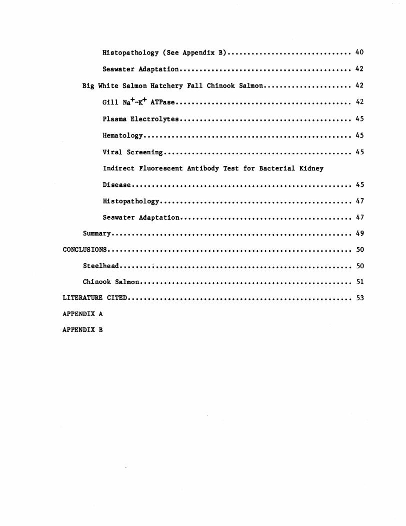
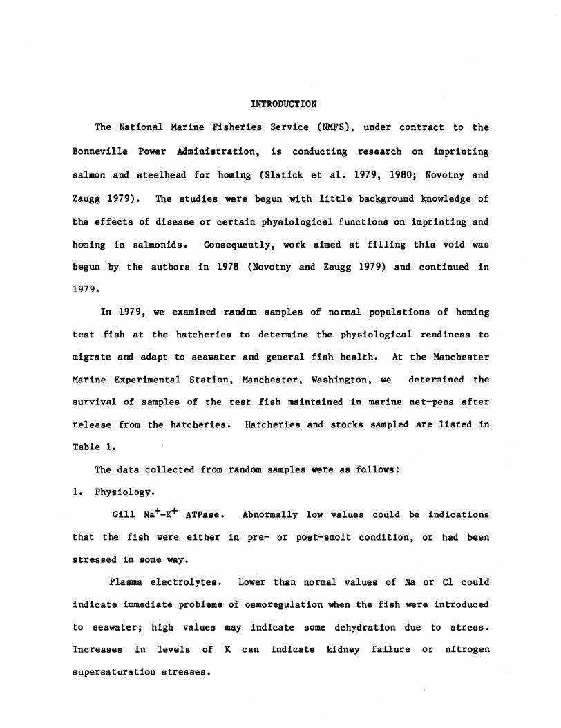
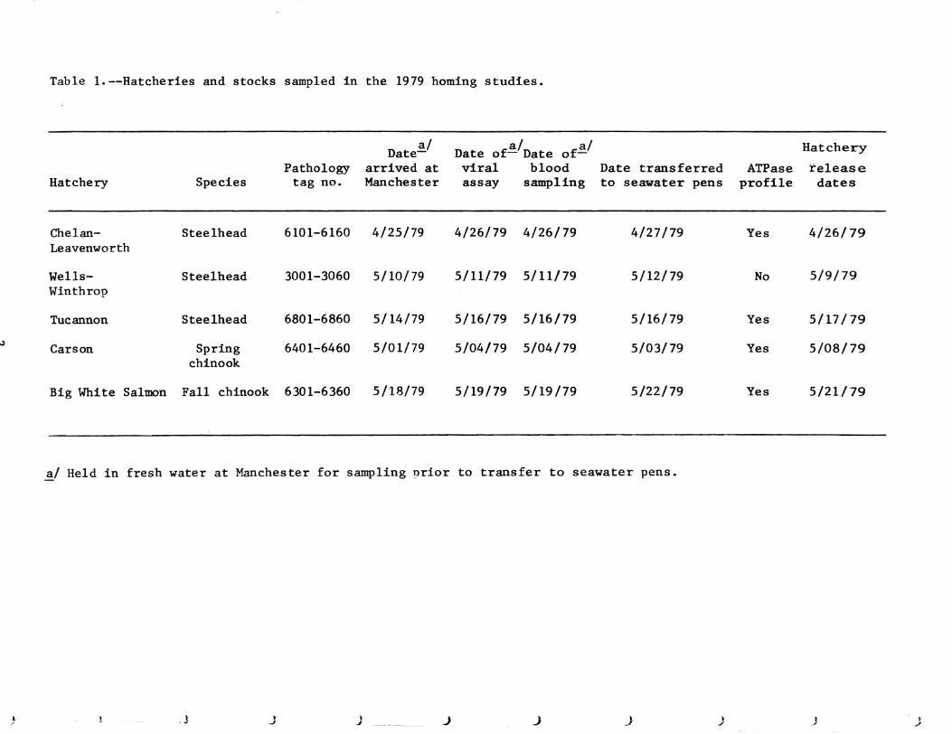