Embed Size (px)
Citation preview

Ranaviral Disease Pathology and Physiology
Debra L. Miller, DVM, PhD
Center for Wildlife Health and Department Biomedical and Diagnostic Sciences, University of
Tennessee, Knoxville, TN
http://scienceblogs.com/tetrapodzoology/wp-content/blogs.dir/471/files/2012/05/i-ef0fe026ef8adf268fbce8dda99e3d45-Uroplatus_fimbriatus_Piotr-Naskrecki_April-2010.jpg
Photo: Blind Pony Hatchery
Photo: N Haislip
Photo: N Haislip
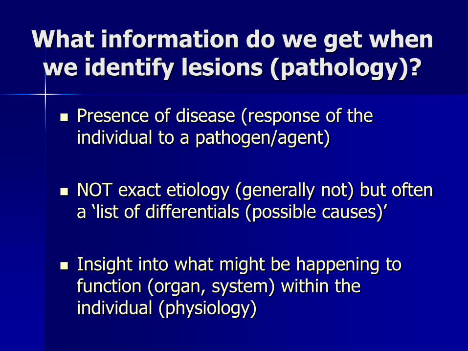
What information do we get when we identify lesions (pathology)?
Presence of disease (response of the individual to a pathogen/agent)
NOT exact etiology (generally not) but often a ‘list of differentials (possible causes)’
Insight into what might be happening to function (organ, system) within the individual (physiology)

Ranavirus
Amphibians: Anurans and Caudates
Reptiles: Turtles, Lizards, Snakes
Fish: Boney fish
3 Classes
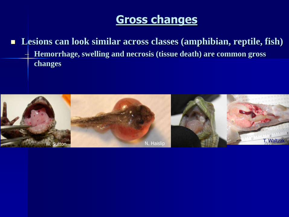
Gross changes
Lesions can look similar across classes (amphibian, reptile, fish)
– Hemorrhage, swelling and necrosis (tissue death) are common gross
changes
W. Sutton N. HaislipT. Waltzak

Amphibians: larvae
Photo: J. ChaneyBoreal Toad
Photo: Nathan HaislipBullfrog
affected
unaffectedPhoto: N. HaislipBullfrog
Photo: N. HaislipWood frog
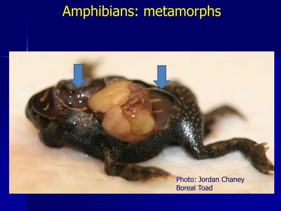
Amphibians: metamorphs
Photo: Jordan ChaneyBoreal Toad
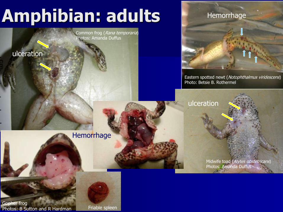
Amphibian: adultsCommon frog (Rana temporaria)Photos: Amanda Duffus
ulceration
Midwife toad (Alytes obstetricans)Photos: Amanda Duffus
ulceration
Eastern spotted newt (Notophthalmus viridescens)Photo: Betsie B. Rothermel
Hemorrhage
Friable spleen
Hemorrhage
Gopher frogPhotos: B Sutton and R Hardman

Is this ranaviral disease?
Cryptobranchus alleganiensis alleganiensisPhoto: Dale McGinnity and Sherri Reinsch

Photo: B Sutton and R Hardman
And what role do ectoparasites (leeches) play?

Bullfrog (~10%; 0% FV3)
Cope’s Gray tree frog (~70% RI; ~40% FV3)
Wood frog (~ 100% for both)
Varies by host (species)-susceptibility & isolate(mortality: RI [ranaculture isolate] vs FV3 = Amphibian isolates)
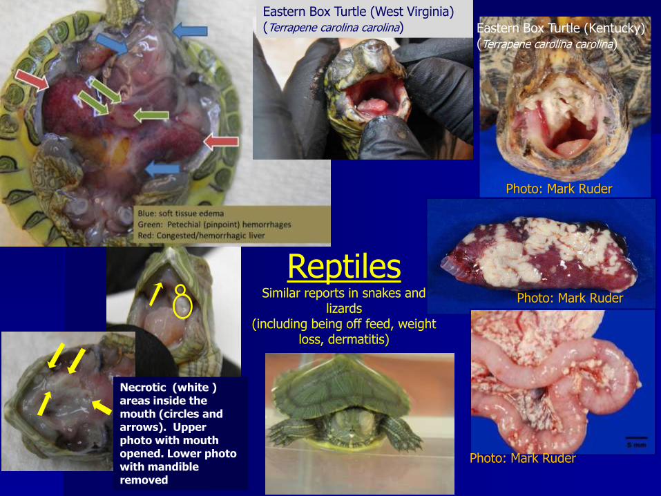
Necrotic (white ) areas inside the mouth (circles and arrows). Upper photo with mouth opened. Lower photo with mandible removed
Photo: Mark Ruder
Photo: Mark Ruder
Photo: Mark Ruder
ReptilesSimilar reports in snakes and
lizards(including being off feed, weight
loss, dermatitis)
Eastern Box Turtle (West Virginia)(Terrapene carolina carolina) Eastern Box Turtle (Kentucky)
(Terrapene carolina carolina)
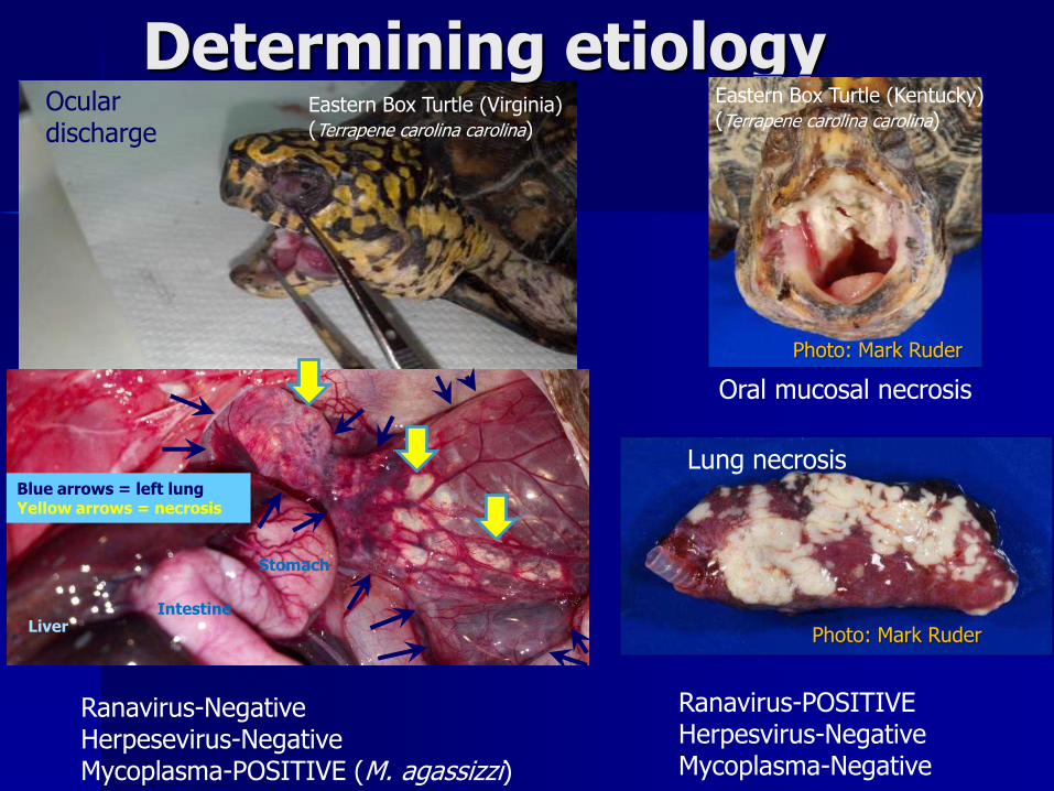
Blue arrows = left lungYellow arrows = necrosis
LiverIntestine
Stomach
Ocular discharge
Ranavirus-NegativeHerpesevirus-NegativeMycoplasma-POSITIVE (M. agassizzi)
Ranavirus-POSITIVEHerpesvirus-NegativeMycoplasma-Negative
Determining etiology
Oral mucosal necrosis
Photo: Mark Ruder
Lung necrosis
Photo: Mark Ruder
Eastern Box Turtle (Virginia)(Terrapene carolina carolina)
Eastern Box Turtle (Kentucky)(Terrapene carolina carolina)
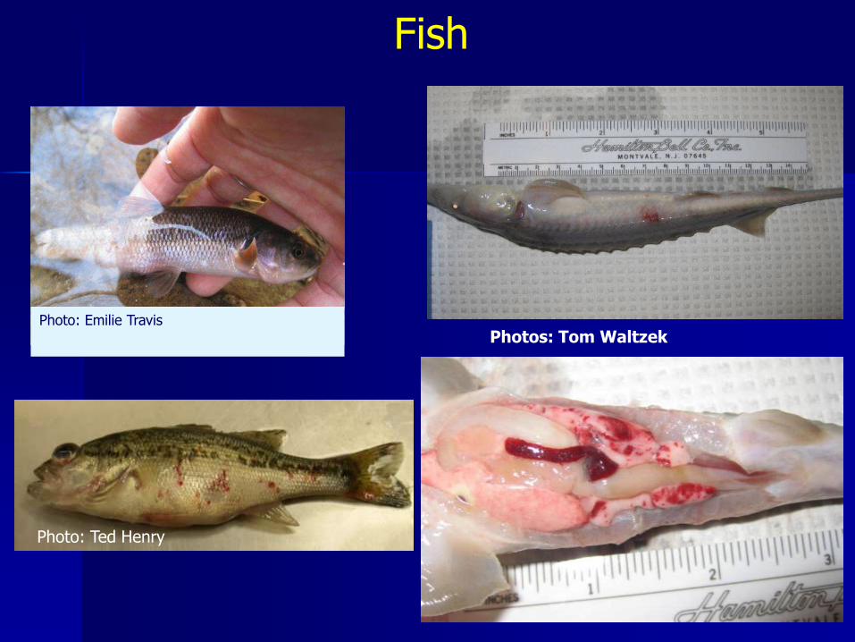
Fish
Photo: Emilie Travis
Photos: Tom Waltzek
Photo: Ted Henry

Histology
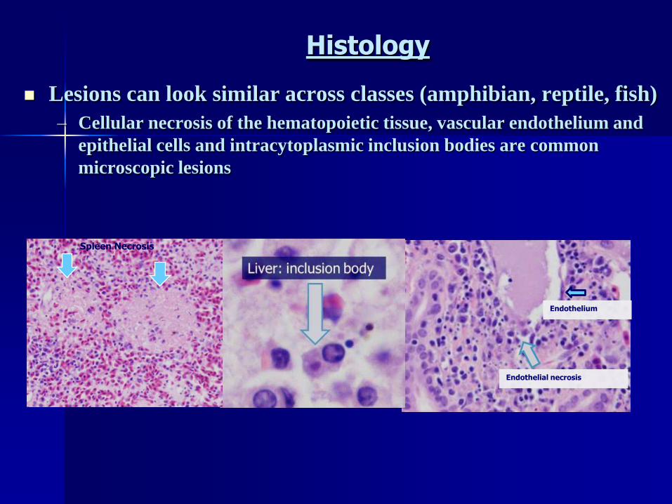
Histology
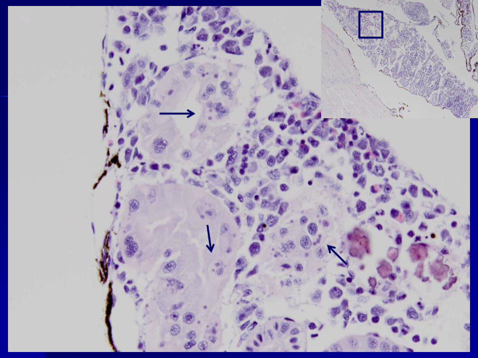
Lesions can look similar across classes (amphibian, reptile, fish)
– Cellular necrosis of the hematopoietic tissue, vascular endothelium and
epithelial cells and intracytoplasmic inclusion bodies are common
microscopic lesions
Endothelium
Endothelial necrosis
Spleen Necrosis
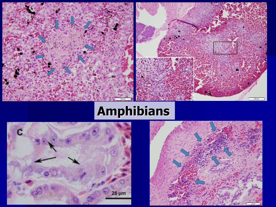
Amphibians


FV3 Box turtle isolate
Pallid sturgeon isolate Ranaculture isolate
Wood Frog Spleen

Reptiles

Pallid isolate; bath exposure

Pallid isolate; bath exposure


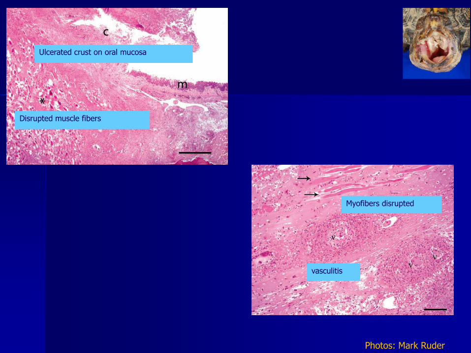
vasculitis
Myofibers disrupted
Ulcerated crust on oral mucosa
Disrupted muscle fibers
Photos: Mark Ruder

Fish
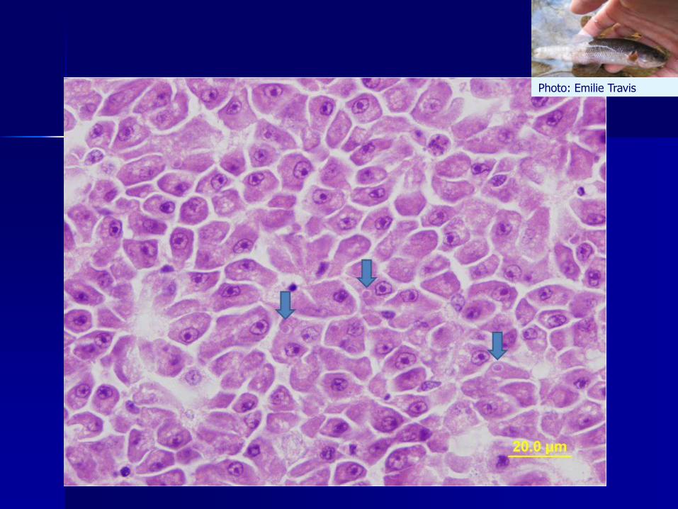
Photo: Emilie Travis
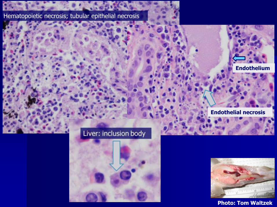
Hematopoietic necrosis; tubular epithelial necrosis
Endothelium
Endothelial necrosis
Photo: Tom Waltzek
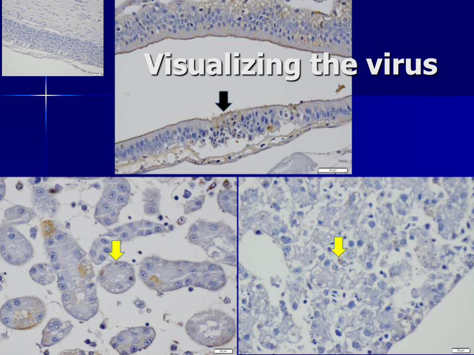
Visualizing the virus

Concurrent Infections
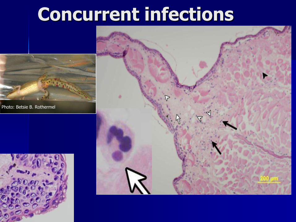
Concurrent infections
Photo: Betsie B. Rothermel
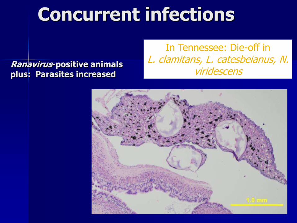
Ranavirus-positive animalsplus: Parasites increased
In Tennessee: Die-off in L. clamitans, L. catesbeianus, N.
viridescens
Concurrent infections
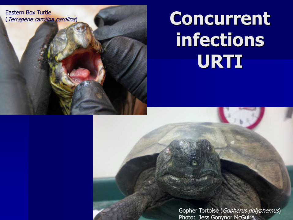
Concurrent infections
URTI
Gopher Tortoise (Gopherus polyphemus)Photo: Jess Gonynor McGuire
Eastern Box Turtle(Terrapene carolina carolina)
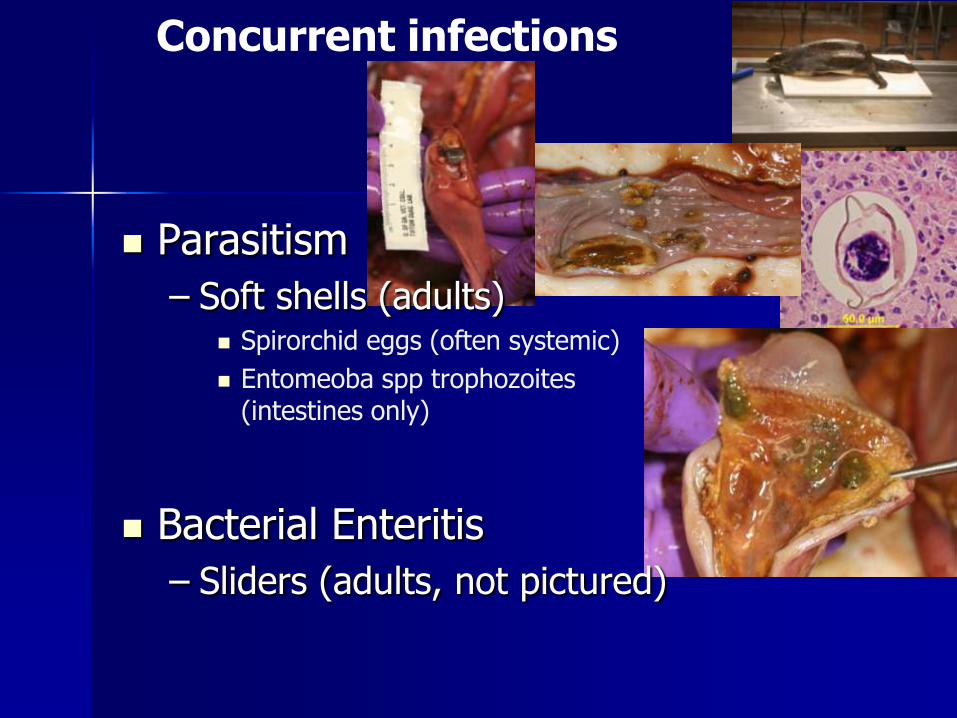
Concurrent infections
Parasitism
– Soft shells (adults) Spirorchid eggs (often systemic)
Entomeoba spp trophozoites(intestines only)
Bacterial Enteritis
– Sliders (adults, not pictured)

Conclusions
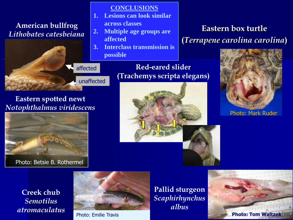
Eastern spotted newtNotophthalmus viridescens
American bullfrogLithobates catesbeiana
Creek chubSemotilus
atromaculatus
affected
unaffected
Eastern box turtle
(Terrapene carolina carolina)
Photo: Mark Ruder
Red-eared slider (Trachemys scripta elegans)
Pallid sturgeonScaphirhynchus
albusPhoto: Tom WaltzekPhoto: Emilie Travis
Photo: Betsie B. Rothermel
CONCLUSIONS
1. Lesions can look similar
across classes
2. Multiple age groups are
affected
3. Interclass transmission is
possible
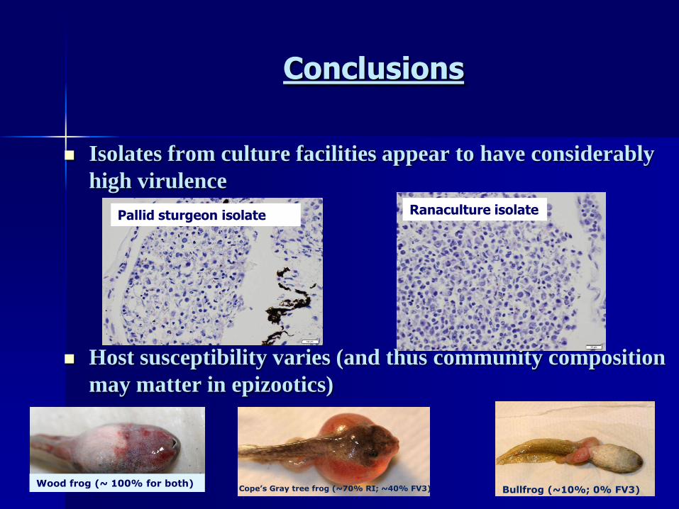
Conclusions
Isolates from culture facilities appear to have considerably
high virulence
Host susceptibility varies (and thus community composition
may matter in epizootics)
Pallid sturgeon isolate Ranaculture isolate
Bullfrog (~10%; 0% FV3)Cope’s Gray tree frog (~70% RI; ~40% FV3)Wood frog (~ 100% for both)
Ranaviral Disease Pathology and Physiology
Current research directives:
– characterizing the pathogenesis of ranaviral disease among virus
isolates and among hosts (including lizards and snakes)
– Elucidating the effects of concurrent pathogens on disease
progression (chytrid and ranavirus; other viruses; mycoplasma)
In situ hybridization and immunohistochemistry are being
used to visualize the virus within the tissues. Optimization
for detecting multiple ranaviruses is being explored.

Acknowledgements
Matt Gray
Tom Waltzek
Bill Sutton
Jordan Chaney
Rachel Marschang
Becky Hardman
Rachel Goodman
Julia Lankton
Sharon Schlosshan and UT histology laboratory
Histology Funding: UT CVM Faculty
Education and Research (FEAR) Fund
UT CVM Center of Excellence

Questions?