Embed Size (px)
Citation preview
http://www.pharmacophorejournal.com 299
Pharmacophore 2015, Vol. 6 (6), 299-305 USA CODEN: PHARM7 ISSN 2229-5402
Pharmacophore
(An International Research Journal)
Available online at http://www.pharmacophorejournal.com/
Original Research Paper
STUDY OF ANTIUROLITHIATIC ACTIVITY OF DIOSPYROS MALABARICA
(DESR) KOSTEL ON RATS
Laxmikant Maruti Purane1* and Suryadevara Vidyadhara
2
1Pharmacology Department, Shivnagar Vidya Prasarak Mandal’s, College of Pharmacy,
Malegaon Bk, Baramati, Pune-413115, Maharashtra, India 2Chebrolu Hanumaiah, Institute of Pharmaceutical Sciences, Chowdavaram,
Guntur-522019, A.P. India
ABSTRACT The purpose of this study was to investigate the antiurolithiatic activity of ethanolic extract of fruits of
Diospyros malabarica (Desr.) Kostel (EEFDMDK) on rats in ethylene glycol (EG) and Ammonium
chloride (AC) induced urolithiasis model. Twenty four male rats ware randomly divided in to four groups
(n = 6). EG 0.75 % (v/v) and AC 2 % (w/v) in drinking water were feed to all groups of rats (Groups II, III,
and IV) except normal control (Group I) rats for 10 days to induce urolithiasis. Group III and IV rats were
treated with EEFDMDK at 250 and 500 mg/ kg oral (P.O.) for 10 days. Group I and Group II (positive
control) rats were administered 6μl distilled water (DW) per 1g of body weight by gavage for 10 days. The
change in body weight of animals was calculated (final weight on 10th
day - initial weight on 1st
day) and
we observed that, weight of rats in positive control group was significantly decreased as compared other
groups of rats. At the end of 10th
day of the experimental period, blood samples were collected and
analyzed for biochemical parameters i.e. serum concentration of urea, creatinine, calcium and phosphorus.
The kidneys were removed and sectioned for histopathological studies. Treatment with the EEFDMDK
restored all the elevated biochemical parameters when compare to positive control group. The
histopathological studies confirmed the induction of urolithiasis as damages in kidney and crystal
deposition was observed in section of kidney from animal treated with EG and AC. This was reduced,
however after treatment with the EEFDMDK. The conclusion of this study was EEFDMDK showed
significant antiurolithiatic activity and possible mechanism underlying this effect is mediated collectively
through diuretic and antioxidant properties.
Keywords: Diospyros malabarica (Desr.) Kostel, antiurolithiatic, Ethylene glycol, Ammonium chloride.
INTRODUCTION
Urinary stone constitute one of the commonest
diseases in our country and pain due to kidney
stone is known as worse than that of labour pain.
Among all the pain, abdominal pain always drags
not only patient’s attention but also the curiosity
of the surgeon. It is estimated that 12% of world
population experience renal stone disease with a
recurrence rate of 70-80% in men and 47-60% in
women.1,2,3
Kidney stone formation or urolithiasis
is complex process that occurs due to imbalance
between promoters and inhibitors in the kidneys.4
In the treatment of kidney stone various synthetic
drugs are available in the market. Even though
surgery is the treatment of choice for urinary
stones, life style changes are important as the
recurrence rate is high as 50% within 5 years after
surgery without medical treatment.5 Herbal drugs
can be easily available and it does not produce
any type of complications like synthetic drugs to
patients. Several plant extract have been used to
Laxmikant Maruti Purane et al. / Pharmacophore 2015, Vol. 6 (6), 299-305
http://www.pharmacophorejournal.com 300
treat kidney stones with promising effect in
prevention and treatment. Diospyros malabarica
(Desr.) Kostel (DMDK), Family - Ebenaceae is a
tree distributed throughout India. The plant used
in the traditional system for various clinical
conditions such as liver diseases, snake bites,
diabetes, diarrhea, cancer, urinary diseases and
renal stone.6,7,8,9
The plant possesses flavonoids,
tannins, terpenoides, sugars and steroids.10
In
recent times, focus on plant research has
increased all over the world and large body
evidence has collected to show immense of
medicinal plants used in various traditional
systems. The present study is focused on the
investigation of antiurolithiatic activity of fruits
of DMDK on Ethylene glycol (EG) and
ammonium chloride (AC) induced urolithiasis in
rats.
MATERIAL AND METHODS
Plant Material
Fruits of DMDK was collected in bulk quantities
from our college campus area and authenticated
by Department of Botany, S.S.M.M. Baramati.
The fruits were shade dried separately at room
temperature and powder was obtained.
Preparation of Extract
The powder of fruits was subjected to successive
soxhlet extraction with solvents of increased
polarity. The ethanolic extract was selected for
the present study. The extract was concentrated
using rotary flash evaporator and stored at room
temperature.
Preliminary Phytochemical Screening
Preliminary Phytochemical screening was carried
out on ethanolic extract of fruits of Diospyros
malabarica (Dser.) Kostal (EEFDMDK) for
detection of phytoconstituents present following
the standard methods described in practical
pharmacognosy book by Dr. C. K. Kokate11
and
K. R. Khandelwal.12
Acute Toxicity
Acute toxicity study of EEFDMDK was
performed on albino mice (20 – 30 g) maintained
under standard conditions. Fixed dose method of
CPCSEA (Committee for the Purpose of Control
and Supervision of Experiments on Animals) was
adopted for toxicity studies13,14
(OECD Guideline
No. 420).
Animals
Healthy, male Wistar rats each weighing between
230 to 275 g were used for the study. The rats
were housed in polypropylene cages and
maintained under standard conditions (12 h light
and dark cycles, at 25±30 C and 35-60%
humidity). They were feed with standard rat feed
and water ad libitum. The study was approved by
the Institutional Animal Ethical Committee of
S.V.P.M’s College of Pharmacy, Malegaon Bk II,
Baramati, registered under CPCSEA, India
(Registration No. 1214/ac/08/CPCSEA).
Ethylene Glycol-Ammonium Chloride Induced
Urolithiasis
Ethylene glycol-Ammonium chloride induced
urolithiasis model was used for the experiments.15
Twenty-four male rats were divided in to four
groups six animal each. The treatment protocol
for 10 days for each group was as follows:
Group I: ad libitum access to regular food and
drinking water and administered 6μl distilled
water (DW) per 1g of body weight by gavage
(normal control).
Group II, III and IV: ad libitum access to regular
food and drinking water containing 0.75% (v/v)
ethylene glycol (EG) and 2% (w/v) ammonium
chloride (AC) in order to promote urolithiasis.
Group III: Rats were administered 250 mg/kg oral
(P.O.) dose of EEFDMDK.
Group IV: Rats were administered 500 mg/kg P.
O. dose of EEFDMDK.
Group II: Rats were administered 6μl DW per 1g
of body weight by gavage (positive control). All
the rats were weighed daily.
Assessment of Antiurolithiatic Activity
At the end of 10th
day of the experimental period,
rats were anaesthetized and blood was collected
from the retro-orbital region, centrifuged at
10,000 X g for 10 min. The serum was estimated
for calcium, urea, creatinine and phosphorus
using the respective diagnostic kits.
Histopathological Studies
Laxmikant Maruti Purane et al. / Pharmacophore 2015, Vol. 6 (6), 299-305
http://www.pharmacophorejournal.com 301
The rats were killed by high dose of ether,
abdomen was opened and the kidneys were
removed. The kidneys were stored in 10% neutral
formalin solution, fixed in bouin liquid, soaked in
paraffin and section were taken using a
microtome. The sections were stained with
hematoxylin (H) and eosin (E) and observed
under a computerized microscope (100X and
400X).
Statistical Analysis
The data were presented as men ± standard error
of mean (SEM) and analyzed using Student’s “t”
test and one-way analysis of variance (ANOVA)
followed by Dunnett’s and P< 0.05 was
considered statically significant. Statistical
Package for social Science (SPSS 20.0) version
software was used for statistical analysis.
RESULT
Acute toxicity study
The EEFDMDK was studied for acute toxicity at
dose of 2000 mg/kg P.O. The extract was found
to be safe and no mortality of the animals
observed. Hence 2500 mg/kg was considered as
LD50 cut off value as per fixed dose method of
CPCSEA. So, the doses selected for the
evaluation of antiurolithiatic activity were
250mg/kg and 500mg/kg P.O.
Antiurolithiatic Activity
The body weight of rats before experiment (initial
weight) i.e. on 1st
day and after experiment (final
weight) i.e. on 10th
day were compared in each
group of rats and we observed that, there was a
significant difference in change in body weight as
shown in table number (no.)1. As shown in table
no. 2 serum urea, creatinine, calcium, phosphorus
level were found to be significantly increased in
rats of positive control group; Whereas treatment
with the EEFDMDK were found to protect the
rats form elevation of serum urea, creatinine,
calcium, phosphorus level . The change in body
weight (final weight on 10th
day - initial weight
on 1st
day) was calculated and we observed that,
weight of rats in positive control group were
significantly decreased as compared other groups
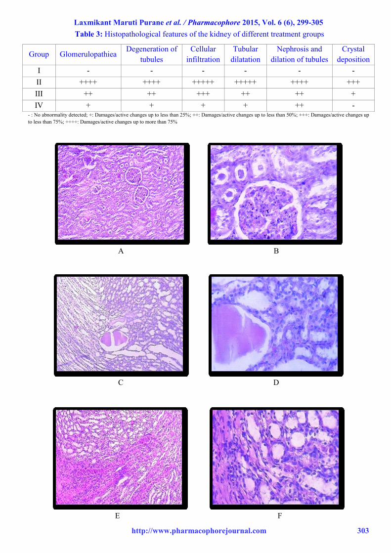
of rats as shown in table no. 2. Similarly, as
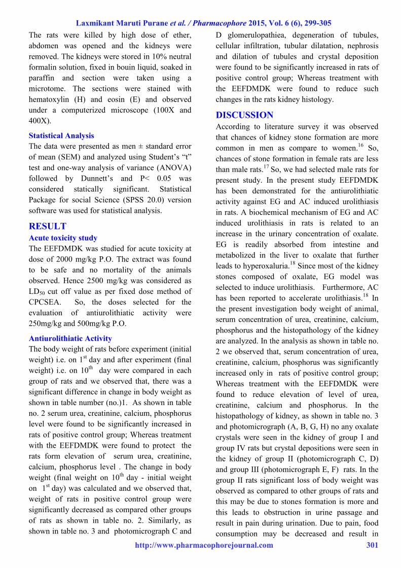
shown in table no. 3 and photomicrograph C and
D glomerulopathiea, degeneration of tubules,
cellular infiltration, tubular dilatation, nephrosis
and dilation of tubules and crystal deposition
were found to be significantly increased in rats of
positive control group; Whereas treatment with
the EEFDMDK were found to reduce such
changes in the rats kidney histology.
DISCUSSION
According to literature survey it was observed
that chances of kidney stone formation are more
common in men as compare to women.16
So,
chances of stone formation in female rats are less
than male rats.17
So, we had selected male rats for
present study. In the present study EEFDMDK
has been demonstrated for the antiurolithiatic
activity against EG and AC induced urolithiasis
in rats. A biochemical mechanism of EG and AC
induced urolithiasis in rats is related to an
increase in the urinary concentration of oxalate.
EG is readily absorbed from intestine and
metabolized in the liver to oxalate that further
leads to hyperoxaluria.18
Since most of the kidney
stones composed of oxalate, EG model was
selected to induce urolithiasis. Furthermore, AC
has been reported to accelerate urolithiasis.18
In
the present investigation body weight of animal,
serum concentration of urea, creatinine, calcium,
phosphorus and the histopathology of the kidney
are analyzed. In the analysis as shown in table no.
2 we observed that, serum concentration of urea,
creatinine, calcium, phosphorus was significantly
increased only in rats of positive control group;
Whereas treatment with the EEFDMDK were
found to reduce elevation of level of urea,
creatinine, calcium and phosphorus. In the
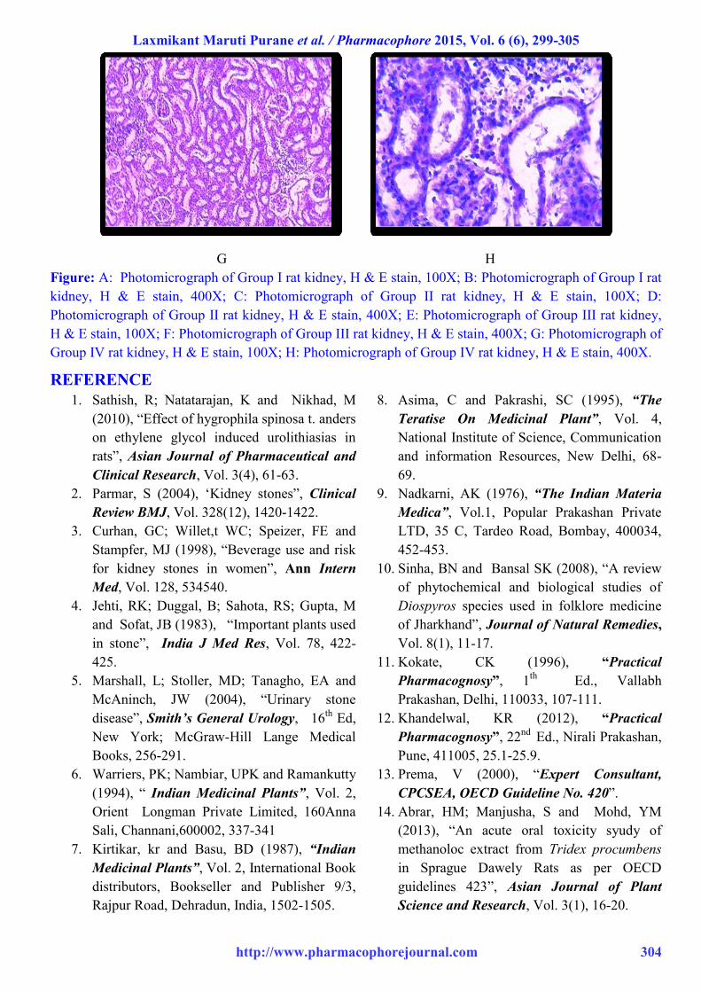
histopathology of kidney, as shown in table no. 3
and photomicrograph (A, B, G, H) no any oxalate
crystals were seen in the kidney of group I and
group IV rats but crystal depositions were seen in
the kidney of group II (photomicrograph C, D)
and group III (photomicrograph E, F) rats. In the
group II rats significant loss of body weight was
observed as compared to other groups of rats and
this may be due to stones formation is more and
this leads to obstruction in urine passage and
result in pain during urination. Due to pain, food
consumption may be decreased and result in
Laxmikant Maruti Purane et al. / Pharmacophore 2015, Vol. 6 (6), 299-305
http://www.pharmacophorejournal.com 302
decrease in body weight of animal. In kidney
histology of positive control rats showed marked
changes and damages but all such histological
changes were significantly reduced in rats treated
with EEFDMDK. In the present study possible
mode of action of EEFDMDK may be due to
diuretic property of DMDK. This property
favours antiurolithiasis by hastening the process
of dissolving or by flushing of the performed
stones or by preventing the new stone formation
in urinary system. The other possible mode of
action of EEFDMDK may be due to its
antioxidant effect of DMDK19
because there is
evidence that hyperoxaluria induced per oxidative
damage to the renal tubular membrane surface
provides a favorable environment for calcium
oxalate crystal attachment and development of
kidney stone.20
In the present investigation
Preliminary Phytochemical studies of EEFDMDK
gave positive test for tannins and flavonoids and
there is evidence that, presence of these
phytoconstituents (specially flavonoids) by virtue
of their antioxidant potential are suggested to
play a important role in antiurolithiatic
effects.21,22
CONCLUSION
From the obtained data it is conclude that
administration of EEFDMDK reduced and
prevented the growth of urinary stones. From the
data we also observed that both low dose and
high dose of EEFDMDK showed significant
antiurolithiatic effect at various stages. The
possible mechanism underlying this effect is
mediated collectively through diuretic and
antioxidant properties and lowering the
concentration of urinary stone forming
constituents. Further work is necessary to isolate
the active constituents responsible for the
antiurolithiatic activity and studies on larger
animal models and on human is warranted to
draw final conclusion.
ACKNOWLEDGMENT
The authors are thankful to the management and
principal of SVPM’s college of pharmacy,
Malegaon BkII, Baramati, India and Chebrolu
Hanumaiah, Institute of Pharmaceutical Sciences,
Chowdavaram, Guntur, 522019, A. P. India for
providing research facilities to carry out the work.
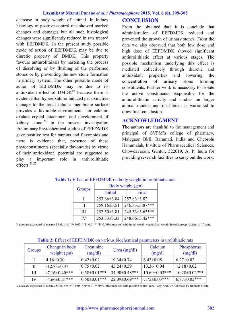
Table 1: Effect of EEFDMDK on body weight in urolithiatic rats
Groups Body weight (gm)
Initial Final
I 253.66±3.84 257.83±3.82
II 259.16±3.51 246.33±3.87***
III 252.50±3.81 245.33±3.63***
IV 253.33±3.33 248.66±3.42***
Values are expressed as mean ± SEM, n=6, *P<0.05, **P<0.01 ***P<0.001compaired with initial weight versus final weight in each group (student’s “t” test).
Table 2: Effect of EEFDMDK on various biochemical parameters in urolithiatic rats
Groups Change in body
weight (gm)
Creatinine
(mg/dl) Urea (mg/dl)
Calcium
(mg/dl
Phosphorus
(mg/dl)
I 4.16±0.30 0.42±0.02 19.34±0.74 6.43±0.05 6.27±0.02
II -12.83±0.47 0.75±0.02 45.24±0.59 13.36±0.04 12.18±0.02
III -7.16±0.40*** 0.58±0.01*** 34.90±0.48*** 10.69±0.03*** 10.28±0.02***
IV -4.66±0.21*** 0.50±0.01*** 22.09±0.69*** 7.72±0.03*** 6.87±0.02***
Values are expressed as mean ± SEM, n=6, *P<0.05, **P<0.01 ***P<0.001compaired with positive control (one –way ANOVA followed by Dunnett’s test).
Laxmikant Maruti Purane et al. / Pharmacophore 2015, Vol. 6 (6), 299-305
http://www.pharmacophorejournal.com 303
Table 3: Histopathological features of the kidney of different treatment groups
- : No abnormality detected; +: Damages/active changes up to less than 25%; ++: Damages/active changes up to less than 50%; +++: Damages/active changes up
to less than 75%; ++++: Damages/active changes up to more than 75%
A B
C D
E F
Group Glomerulopathiea Degeneration of
tubules
Cellular
infiltration
Tubular
dilatation
Nephrosis and
dilation of tubules
Crystal
deposition
I - - - - - -
II ++++ ++++ +++++ +++++ ++++ +++
III ++ ++ +++ ++ ++ +
IV + + + + ++ -
Laxmikant Maruti Purane et al. / Pharmacophore 2015, Vol. 6 (6), 299-305
http://www.pharmacophorejournal.com 304
G H
Figure: A: Photomicrograph of Group I rat kidney, H & E stain, 100X; B: Photomicrograph of Group I rat
kidney, H & E stain, 400X; C: Photomicrograph of Group II rat kidney, H & E stain, 100X; D:
Photomicrograph of Group II rat kidney, H & E stain, 400X; E: Photomicrograph of Group III rat kidney,
H & E stain, 100X; F: Photomicrograph of Group III rat kidney, H & E stain, 400X; G: Photomicrograph of
Group IV rat kidney, H & E stain, 100X; H: Photomicrograph of Group IV rat kidney, H & E stain, 400X.
REFERENCE
1. Sathish, R; Natatarajan, K and Nikhad, M
(2010), “Effect of hygrophila spinosa t. anders
on ethylene glycol induced urolithiasias in
rats”, Asian Journal of Pharmaceutical and
Clinical Research, Vol. 3(4), 61-63.
2. Parmar, S (2004), ‘Kidney stones”, Clinical
Review BMJ, Vol. 328(12), 1420-1422.
3. Curhan, GC; Willet,t WC; Speizer, FE and
Stampfer, MJ (1998), “Beverage use and risk
for kidney stones in women”, Ann Intern
Med, Vol. 128, 534540.
4. Jehti, RK; Duggal, B; Sahota, RS; Gupta, M
and Sofat, JB (1983), “Important plants used
in stone”, India J Med Res, Vol. 78, 422-
425.
5. Marshall, L; Stoller, MD; Tanagho, EA and
McAninch, JW (2004), “Urinary stone
disease”, Smith’s General Urology, 16th
Ed,
New York; McGraw-Hill Lange Medical
Books, 256-291.
6. Warriers, PK; Nambiar, UPK and Ramankutty
(1994), “ Indian Medicinal Plants”, Vol. 2,
Orient Longman Private Limited, 160Anna
Sali, Channani,600002, 337-341
7. Kirtikar, kr and Basu, BD (1987), “Indian
Medicinal Plants”, Vol. 2, International Book
distributors, Bookseller and Publisher 9/3,
Rajpur Road, Dehradun, India, 1502-1505.
8. Asima, C and Pakrashi, SC (1995), “The
Teratise On Medicinal Plant”, Vol. 4,
National Institute of Science, Communication
and information Resources, New Delhi, 68-
69.
9. Nadkarni, AK (1976), “The Indian Materia
Medica”, Vol.1, Popular Prakashan Private
LTD, 35 C, Tardeo Road, Bombay, 400034,
452-453.
10. Sinha, BN and Bansal SK (2008), “A review
of phytochemical and biological studies of
Diospyros species used in folklore medicine
of Jharkhand”, Journal of Natural Remedies,
Vol. 8(1), 11-17.
11. Kokate, CK (1996), “Practical
Pharmacognosy”, 1th
Ed., Vallabh
Prakashan, Delhi, 110033, 107-111.
12. Khandelwal, KR (2012), “Practical
Pharmacognosy”, 22nd
Ed., Nirali Prakashan,
Pune, 411005, 25.1-25.9.
13. Prema, V (2000), “Expert Consultant,
CPCSEA, OECD Guideline No. 420”.
14. Abrar, HM; Manjusha, S and Mohd, YM
(2013), “An acute oral toxicity syudy of
methanoloc extract from Tridex procumbens
in Sprague Dawely Rats as per OECD
guidelines 423”, Asian Journal of Plant
Science and Research, Vol. 3(1), 16-20.
Laxmikant Maruti Purane et al. / Pharmacophore 2015, Vol. 6 (6), 299-305
http://www.pharmacophorejournal.com 305
15. Narumalla, J; Somashekara, SC; Damodaram,
G and Golla, Devasankaraiah (2014), “ Study
of antiurolithiatic activity of Asparagus
racemosus on albino rats”, Indian Journal of
Pharmacology, Vol. 44(5), 576-579.
16. Vahleensieck, EW; Bach, D and Hesse, A
(1982), “Incidence, prevalence and mortality
of urolithiasis in the German federal
republic”, Urologic Research, Vol. 10, 161.
17. Karadi, RV; Gadge, NB; Alagawadi, KR and
Savadi, RV (2006), “Effect of Moringa
oleifera Lam. root-wood on ethylene glycol
induced urolithiasis in rats”, J.
Ethnopharmacol, Vol.105, 306-311.
18. Pawar, AT and Vyawahare, NS (2015),
“Protective effect of methanolic extract of
Abelmoschus sees against calcium oxalate
urolithiasis in rats”, Journal of Chemical and
Pharmaceutical Research, Vol.7 (6), 269-
278.
19. Mondal, SK; Chakraborty, G; Gupta, M and
Mazumder, UK ((2006), “In vitro antioxidant
activity of Dyospyrous malbarica kostal
bark”, Indian J. Exp. Biol., Vol. 44(1), 39-44.
20. Touhami,M; Laroubia, A; Elhabazi, K;
Loubna, F; Zara, A and Chaita, A et al.
(2007), “Lemon juice has protective activity
in rat urolithiasis model”, BMC, Vol. 7, 18.
21. Ragini, V (2014), “Antiurolithiatic activity of
extract of Aerva javanica in rats”, Int.J. Drug
Dev. & Res., Vol. 6(4), 35-45.
22. Naimesh, P; Sachin, Bobade; Pankaj, Gupta
and Brijendra Jain et al.(2008), “Effect of
ethanolic extract of leaves of Cocculus
hirsutus (L) diels on experimentally induced
urolithiasis in rats”, Journal of Natural
Remedies, Vol. 8(1), 24-31.
Correspondence Author:
Laxmikant Maruti Purane
Pharmacology Department, Shivnagar Vidya Prasarak Mandal’s, College of Pharmacy, Malegaon Bk, Baramati, Pune-
413115, Maharashtra, India
Cite This Article: Laxmikant, Maruti Purane and Suryadevara, Vidyadhara (2015), “Study of
antiurolithiatic activity of Diospyros malabarica (desr.) kostel on rats”, Pharmacophore, Vol. 6 (6), 299-
305.