Embed Size (px)
Citation preview

Studies on thrombopoiesis and spleen tyrosine kinase-mediated signaling in platelets
* * *
Untersuchungen der Thrombopoese und der spleen tyrosine kinase-vermittelten Signaltransduktion in
Thrombozyten
Doctoral thesis for a doctoral degree at the Graduate School of Life Sciences, Julius-Maximilians-Universität Würzburg,
Section Biomedicine
submitted by
Judith Martina Maria van Eeuwijk
from Leerdam, the Netherlands
Würzburg, 2016

Submitted on:
Members of the Promotionskomitee:
Chairperson: Prof. Dr. Manfred Gessler
Primary Supervisor: Prof. Dr. Bernhard Nieswandt
Supervisor (Second): Dr. Katrin Heinze
Supervisor (Third): Prof. Dr. Guido Stoll
Date of Public Defense:
Date of Receipt of Certificates:

“One should not pursue goals that are easily achieved. One must develop an instinct for what
one can just barely achieve through one's greatest efforts.”
- Albert Einstein

Table of contents
IV
Table of contents
Summary ............................................................................................................................. VII
Zusammenfassung ............................................................................................................. VIII
Samenvatting ........................................................................................................................ X
1. Introduction ....................................................................................................................... 1
1.1. Hematopoiesis ........................................................................................................... 1
1.1.1. Hematopoietic stem cells and bone marrow niches ............................................. 1
1.1.2. Megakaryopoiesis ............................................................................................... 3
1.1.3. Thrombopoiesis and proplatelet formation ........................................................... 5
1.2. Platelets ..................................................................................................................... 7
1.3. Platelet activation and thrombus formation ................................................................. 8
1.3.1. G protein-coupled receptor signaling in platelets ................................................10
1.3.2. (Hem)immunoreceptor tyrosine-based activation motif signaling in platelets ......12
1.4. Imaging modalities ....................................................................................................18
1.4.1. Multiphoton intravital microscopy .......................................................................19
1.4.2. Light-sheet fluorescence microscopy .................................................................20
2. Aim of the study ...............................................................................................................23
3. Materials and Methods .....................................................................................................24
3.1. Materials ...................................................................................................................24
3.1.1. Reagents and chemicals ....................................................................................24
3.1.2. Materials ............................................................................................................27
3.1.3. Labeling kits and fluorophores ............................................................................27
3.1.4. Antibodies ..........................................................................................................27
3.1.5. Buffers ...............................................................................................................28
3.1.6. Mice ...................................................................................................................32
3.2. Methods ....................................................................................................................32
3.2.1. Mouse genotyping ..............................................................................................32
3.2.2. Blood preparation ...............................................................................................36
3.2.3. Compound preparation .......................................................................................37
3.2.4. Biochemistry ......................................................................................................37
3.2.5. In vitro analyses of platelet function ....................................................................38
3.2.6. In vivo analyses of platelet function ....................................................................40
3.2.7. Megakaryocyte analyses ....................................................................................42
3.2.8. Statistical analyses .............................................................................................44
4. Results ............................................................................................................................45
4.1. Multiphoton intravital microscopic analyses of thrombopoiesis and proplatelet formation ...........................................................................................................................45

Table of contents
V
4.1.1. Establishment of multiphoton intravital microscopy of the murine bone marrow and brain .......................................................................................................................45
4.1.2. The role of Profilin1, TRPM7 and RhoA in thrombopoiesis in vivo ......................50
4.2. Thrombopoiesis is spatially regulated by the bone marrow vasculature ....................53
4.2.1. Megakaryocytes in the bone marrow are largely sessile .....................................53
4.2.2. The majority of megakaryocytes is in direct contact with the vasculature ...........54
4.2.3. Establishment of light-sheet fluorescence microscopy of the murine bone marrow 54
4.2.4. Megakaryocytes are homogenously distributed throughout the bone marrow ....59
4.2.5. Platelet depletion or CXCR4 blockade do not affect megakaryocyte localization 61
4.2.6. Computational modeling implies a vessel-biased megakaryocyte distribution ....61
4.2.7. Light-sheet fluorescence microscopy as a tool to study thrombopoiesis .............63
4.3. Syk deficiency or inhibition protect mice from arterial thrombosis and thrombo-inflammatory brain infarction .............................................................................................65
4.3.1. (hem)ITAM signaling is abolished in Syk-/- platelets ............................................65
4.3.2. Murine Syk is dispensable for integrin-dependent outside-in signaling ...............67
4.3.3. Syk deficiency results in moderately prolonged tail bleeding times and impaired arterial thrombus formation ............................................................................................70
4.3.4. Syk deficiency protects from thrombo-inflammatory brain infarction ...................71
4.3.5. Syk inhibition abolishes (hem)ITAM signaling ....................................................72
4.3.6. Human platelets require Syk for integrin-dependent outside-in signaling ............73
4.3.7. Syk inhibition protects from arterial thrombosis and cerebral infarct progression, without affecting hemostasis .........................................................................................73
4.4. Thrombus formation in vivo can occur independently of Syk kinase function ............76
4.4.1. Sykki mice die perinatally ....................................................................................77
4.4.2. (hem)ITAM signaling is abolished in Sykki platelets ............................................78
4.4.3. Integrin outside-in signaling is unaffected by the kinase exchange .....................79
4.4.4. Zap-70 partially compensates for the loss of Syk in vivo ....................................80
5. Discussion .......................................................................................................................82
5.1. Profilin1, TRPM7 and RhoA are critical regulators of thrombopoiesis ........................83
5.2. Thrombopoiesis is spatially regulated by the bone marrow vasculature ....................85
5.3. Syk inhibition protects mice from arterial thrombosis and thrombo-inflammatory brain infarction ...........................................................................................................................89
5.4. Thrombus formation in vivo can occur independently of Syk kinase function ............92
5.5. Concluding remarks and future prospects .................................................................94
6. References ......................................................................................................................96
7. Appendix ........................................................................................................................ 114
7.1. Abbreviations .......................................................................................................... 114
7.2. Acknowledgements ................................................................................................. 118
7.3. Publications ............................................................................................................ 120

Table of contents
VI
7.3.1. Original articles ................................................................................................ 120
7.3.2. Oral presentations ............................................................................................ 120
7.3.3. Poster presentations ........................................................................................ 121
7.4. Curriculum vitae ...................................................................................................... 122
7.5. Affidavit ................................................................................................................... 123
7.6. Eidesstattliche Erklärung ......................................................................................... 123

Summary
VII
Summary
In mammals, anucleate blood platelets are constantly produced by their giant bone marrow
(BM) progenitors, the megakaryocytes (MKs), which originate from hematopoietic stem cells.
Megakaryopoiesis and thrombopoiesis have been studied intensively, but the exact
mechanisms that control platelet generation from MKs remain poorly understood. Using
multiphoton intravital microscopy (MP-IVM), thrombopoiesis and proplatelet formation were
analyzed in the murine BM in real-time and in vivo, identifying an important role for several
proteins, including Profilin1, TRPM7 and RhoA in thrombopoiesis. Currently, it is thought that
blood cell precursors, such as MKs, migrate from the endosteal niche towards the vascular
niche during maturation. In contrast to this paradigm, it was shown that MKs are
homogeneously distributed within the dense BM blood vessel network, leaving no space for
vessel-distant niches. By combining results from in vivo MP-IVM, in situ light-sheet
fluorescence microscopy (LSFM) of the intact BM as well as computational simulations,
surprisingly slow MK migration, limited intervascular space and a vessel-biased MK pool
were revealed, contradicting the current concept of directed MK migration during
thrombopoiesis.
Platelets play an essential role in hemostasis and thrombosis, but also in the pathogenesis of
ischemic stroke. Ischemic stroke, which is mainly caused by thromboembolic occlusion of
brain arteries, is among the leading causes of death and disability worldwide with limited
treatment options. The platelet collagen receptor glycoprotein (GP) VI is a key player in
arterial thrombosis and a critical determinant of stroke outcome, making its signaling pathway
an attractive target for pharmacological intervention. The spleen tyrosine kinase (Syk) is an
essential signaling mediator downstream of GPVI, but also of other platelet and immune cell
receptors. In this thesis, it was demonstrated that mice lacking Syk specifically in platelets
are protected from arterial thrombus formation and ischemic stroke, but display unaltered
hemostasis. Furthermore, it was shown that mice treated with the novel, selective and orally
bioavailable Syk inhibitor BI1002494 were protected in a model of arterial thrombosis and
had smaller infarct sizes and a significantly better neurological outcome 24 h after transient
middle cerebral artery occlusion (tMCAO), also when BI1002494 was administered
therapeutically, i.e. after ischemia. These results provide direct evidence that
pharmacological Syk inhibition might become a safe therapeutic strategy. The T cell receptor
chain-associated protein kinase of 70 kDA (Zap-70) is also a spleen tyrosine kinase family
member, but has a lower intrinsic activity compared to Syk and is expressed in T cells and
natural killer (NK) cells, but not in platelets. Unexpectedly, arterial thrombus formation in vivo
can occur independently of Syk kinase function as revealed by studies in Sykki mice, which
express Zap-70 under the control of intrinsic Syk promoter elements.

Zusammenfassung
VIII
Zusammenfassung
In Säugetieren werden kernlose Thrombozyten durch ihren riesigen Knochenmark- (KM-)
Vorläuferzellen, die Megakaryozyten (MK), die von hämatopoetischen Stammzellen
stammen, ständig produziert. Megakaryopoese und Thrombopoese wurden schon intensiv
untersucht, aber die genauen Mechanismen, die die Thrombozytenproduktion aus MK
kontrollieren, bleiben weitgehend unverstanden. Mittels Multiphotonen-Intravitalmikroskopie
(MP-IVM) wurden Thrombopoese und Proplättchenbildung im murinen KM in Echtzeit in vivo
untersucht. Dadurch wurde eine wichtige Rolle für die Proteine Profilin1, TRPM7 und RhoA
in der Thrombopoese identifiziert. Derzeit wird angenommen, dass Blutzellvorläufer, wie MK,
während der Reifung von der endostalen Nische in Richtung der Gefäßnische migrieren. Im
Gegensatz zu diesem Paradigma wurde hier gezeigt, dass MK homogen innerhalb des
dichten KM Blutgefäßnetzes verteilt sind, so dass kein Raum für Gefäß-ferne Nischen
besteht. Durch Ergebnisse von in vivo MP-IVM, in situ Licht-Blatt-Fluoreszenzmikroskopie
(LSFM) des intakten KM sowie Computersimulationen wurden eine überraschend langsame
MK-Migration, ein begrenzter intervaskulärer Raum und eine asymmetrische MK-Verteilung
gezeigt, was im Widerspruch zum derzeitig akzeptierten Konzept der gerichteten MK-
Migration während der Thrombopoese steht.
Die Thrombozyten spielen eine wesentliche Rolle nicht nur bei der Hämostase und
Thrombose, sondern auch in der Pathogenese des ischämischen Schlaganfalls. Der
ischämische Schlaganfall, der vor allem durch einen thromboembolischen Verschluss von
Gehirnarterien verursacht wird, ist eine der häufigsten Ursachen für Tod und Behinderung
weltweit und die Behandlungsmöglichkeiten sind sehr eingeschränkt. Der thrombozytäre
Kollagenrezeptor Glykoprotein (GP) VI ist ein wichtiger Faktor in der arteriellen Thrombose
und trägt entscheidend zur Pathogenese des ischämischen Schlaganfalls bei, sodass
dessen Signalweg ein attraktives Ziel für pharmakologische Interventionen darstellen könnte.
Die spleen tyrosine kinase (Syk) ist ein wichtiges Molekül im GPVI-Signalweg, aber auch in
den Signalkaskaden von anderen Thrombozyten- und Immunzellrezeptoren. Es wurde
nachgewiesen, dass Mäuse mit einer thrombozytären Syk-Defizienz, vor arterieller
Thrombusbildung und ischämischem Schlaganfall geschützt sind, aber unveränderte
Hämostase zeigen. Darüber hinaus wurde gezeigt, dass Mäuse, die mit dem neuartigen,
selektiven und oral bioverfügbaren Syk-Inhibitor BI1002494 behandelt wurden, geschützt
sind in einem Modell der arteriellen Thrombose. Auch hatten sie kleinere Infarkte und eine
deutlich bessere neurologische Funktion 24 Stunden nach der transienten Arteria cerebri
media Okklusion (tMCAO), auch wenn BI1002494 therapeutisch, d.h. nach der Ischämie,
verabreicht wurde. Diese Ergebnisse deuten darauf hin, dass die pharmakologische
Hemmung von Syk eine sichere therapeutische Strategie bei Schlaganfall sein könnte. Der

Zusammenfassung
IX
T-Zell Rezeptor -chain-associated protein kinase of 70 kDa (Zap-70) ist auch ein spleen
tyrosine kinase-Familienmitglied, hat aber eine geringere intrinsische Aktivität im Vergleich
zu Syk und wird in T-Zellen und natural killer (NK) Zellen exprimiert, nicht aber in
Thrombozyten. Studien in Sykki Mäusen, die unter der Kontrolle der intrinsischen Syk
Promotorelemente Zap-70 exprimieren, ergaben, dass die arterielle Thrombusbildung in vivo
unabhängig von der Syk-Kinasefunktion stattfinden kann.

Samenvatting
X
Samenvatting
In zoogdieren worden kernloze bloedplaatjes constant door hun grote beenmerg (BM)
voorlopercellen, de megakaryocyten (MK), afkomstig van hematopoietische stamcellen,
geproduceerd. Megakaryopoïese en trombopoïese zijn tot nu toe intensief bestudeerd, maar
de exacte mechanismen die de generatie van bloedplaatjes vanuit MK controleren, blijven
slecht begrepen. Met behulp van multiphoton intravitale microscopie (MP-IVM), zijn in real-
time en in vivo, trombopoïese en proplatelet vorming in het BM van muizen bestudeerd,
waarbij een belangrijke rol voor de eitwitten Profilin1, TRPM7 en RhoA in trombopoïese is
geïdentificeerd. Momenteel wordt aangenomen dat bloedcelvoorlopers, zoals MK, tijdens de
rijping van de endostale niche naar de vasculaire niche migreren. In tegenstelling tot dit
paradigma, is hier aangetoond dat MK homogeen verdeeld zijn binnen het dichte
bloedvatnetwerk van het BM, waarbij geen ruimte meer is voor bloedvat-verwijderde niches.
Door het combineren van de resultaten, verkregen met behulp van in vivo MP-IVM, in situ
licht-blad fluorescentie microscopie (LSFM) van het intacte BM evenals computationele
simulaties, zijn, een verrassend langzame MK migratie, een beperkte intervasculaire ruimte
en een asymmetrische MK verdeling aangetoond, in tegenspraak tot het huidig
geaccepteerde concept van de gerichte MK migratie tijdens de trombopoïese.
Bloedplaatjes spelen een essentiële rol niet alleen in hemostase en trombose, maar ook in
de pathogenese van een ischemische beroerte. Een ischemische beroerte, die vooral
veroorzaakt wordt door een trombo-embolische occlusie van de hersenslagaders, is een van
de belangrijkste oorzaken van overlijden en invaliditeit wereldwijd en de
behandelingsmogelijkheden zijn zeer beperkt. De bloedplaatjes collageen receptor
glycoproteïne (GP) VI is een belangrijke speler in arteriële trombose en een kritische factor
voor de gevolgen van een beroerte, waardoor de signaalweg een aantrekkelijk doelwit voor
farmacologische interventie zou kunnen zijn. De spleen tyrosine kinase (Syk) is een
essentiële signaalmediator in de GPVI-signaalweg, maar ook in signaalwegen van andere
bloedplaatjes- en immuuncelreceptoren. In deze thesis is aangetoond, dat muizen die Syk
specifiek in bloedplaatjes missen, beschermd zijn tegen de vorming van arteriële trombose
en ischemische beroerte, maar ongewijzigde hemostase vertonen. Verder is getoond, dat
muizen, die behandeld waren met de nieuwe, selectieve en oraal biologisch beschikbare Syk
inhibitor BI1002494, beschermd waren in een model van arteriële trombose. Ook hadden ze
kleinere infarcten en een significant beter neurologische functie 24 uur na transiënte
middelste cerebrale arterie occlusie (tMCAO), ook toen BI1002494 therapeutisch, dat wil
zeggen na ischemie, werd toegediend. Deze resultaten leveren direct bewijs dat
farmacologische Syk remming een veilige therapeutische strategie zou kunnen bieden. De T-
cel receptor -chain-associated protein kinase of 70 kDa (Zap-70) is ook een spleen tyrosine

Samenvatting
XI
kinase-familielid, maar heeft een lagere intrinsieke activiteit vergeleken met Syk en wordt tot
expressie gebracht in T-cellen en natural killer (NK) cellen, maar niet in bloedplaatjes.
Studies in Sykki muizen, die Zap-70 onder de controle van intrinsieke Syk
promotorelementen tot expressie brengen, laten zien dat arteriële trombusvorming in vivo
onafhankelijk van Syk kinase functie kan optreden.

Introduction
1
1. Introduction
1.1. Hematopoiesis
Hematopoiesis, the process of blood cell formation, begins during embryogenesis in the yolk
sac and the aorta-gonad mesonephros region, is taken over by different embryonic organs
including the fetal liver and spleen and finally resides in the bone marrow (BM), which
functions as the primary lymphoid organ in the adult human body.1-3 In children, the BM is
mainly located in the long bones, like the femur and tibia, whereas in adults it is mostly
located in flat bones, like the skull, pelvis, sternum and ribs. The BM contains arteries,
carrying oxygen, nutrients and growth factors into the BM and sinusoids, which are
specialized venules forming a network of fenestrated vessels allowing cells to enter and
leave the circulation. A dense network of arterioles and sinusoids can be found near the
endosteum, the interface of bone and BM, which is covered by bone-lining cells such as
osteoblasts and osteoclasts. Blood cells and their precursors are located in the extravascular
spaces between the sinusoids.4,5 Hematopoiesis involves hematopoietic stem cells (HSCs),
which are capable of self-renewal and are precursors of progenitor cells that become
committed to one of the hematopoietic lineages at a later stage.6 Maintenance and lineage
differentiation of the HSC pool in the BM is thought to depend on its specific environment and
either occurs via asymmetrical cell division or environmental asymmetry.7 During
asymmetrical cell division, cellular determinants in the cytoplasm like mRNA and/or proteins
are localized to only one of the two daughter cells leading to two daughter cells with different
cellular fates. Environmental asymmetry depends on the presence of specialized BM niche
microenvironments.7
1.1.1. Hematopoietic stem cells and bone marrow niches
In 1978, Ray Schofield was the first to propose the concept of the stem cell niche for HSCs in
the BM.8 The BM niche or microenvironment is decisive for the development of HSCs and,
depending on the association with adjacent cells and local cytokines in the specific niche or
microenvironment, the proliferation or differentiation of HSCs is stimulated (Figure 1).
Environmental asymmetry occurs after division of the HSC. According to this model, one
daughter cell is exposed to extrinsic signals from the local BM microenvironment promoting
lineage differentiation, while the other daughter cell remains in the self-renewing niche,
thereby balancing HSC self-renewal and differentiation. Over the past years, significant
progress has been made to identify the structure, localization and composition of the HSC
niches/microenvironments in the BM and different niche functions have been proposed, such
as the quiescent-storage niche and the self-renewal niche.7 HSCs and distinct
subpopulations of HSCs or progenitor cells can also occupy specialized niches.9

Introduction
2
Figure 1. Model of bone marrow hematopoietic stem cell niches. Endosteal bone surfaces are lined with specialized (blue) and non-specialized (green) stromal cells. The osteoblasts (SNOs) function as a regulatory component of the niche and maintain quiescence and prevent differentiation of hematopoietic stem cells (HSCs) in the quiescent-storage niche. In response to injury, these quiescent HSCs might be activated and recruited to the vascular niche. The self-renewal niche contains both quiescent and dividing HSCs. The HSCs are able to differentiate into multipotential progenitors (MPPs), either by asymmetrical cell division or environmental asymmetry. These progenitors can give rise to all hematopoietic lineages, including megakaryocytes (MKs). From: Wilson and Trumpp, 20067
BM niches are likely to be located near the bone surfaces (endosteal niche) and/or
associated with sinusoidal endothelium (vascular niche). This concept is, however, heavily
debated and it is now thought that in adult BM, multiple niche cell populations from distinct
BM zones regulate HSCs for long-term maintenance, proliferation, and differentiation. These
zones or niches cooperate to control HSC quiescence and self-renewal and the
differentiation of HSCs into progenitor cells to maintain homeostasis. The endosteal niche
appears to contain mainly dormant HSCs. Kunisaki et al. showed that quiescent HSCs
associate specifically with BM arterioles, which are mainly found in the endosteal BM,
providing an arteriolar niche.10 However, a recent report by Acar et al. reveals that most
HSCs are distant from arterioles and bone surfaces and mainly localize in perisinusoidal
niches throughout the BM.11 In the vascular niche, many proliferating cells, most likely self-
renewing HSCs, are located in close contact to the BM sinusoidal endothelial cells
(BMECs).7 A recent report by Itkin et al. provided strong evidence to support the concept that
BM stem cell maintenance is regulated by distinct blood vessel types with different
permeability properties.12
Osteoblasts are an essential part of the endosteal niche and are limiting for the niche size
and its activity. The osteoblastic cells are a regulatory component of the niche and influence
HSC maturation and function.13 HSCs localize in close proximity to the endosteum and HSC
progenitors can be found distributed throughout the central BM.14,15 On the other hand, it has
been shown that HSCs localize adjacent to sinusoid vessels in close contact to perivascular

Introduction
3
cells. The HSCs attach to the fenestrated endothelium of the BM sinusoids serving as a
niche for adult HSCs.16 The mesenchymal stromal cells (MSCs) are a component of the
perivascular niche and together with the BMECs synthesize cytokines, such as stem cell
factor (SCF) and stromal cell-derived factor 1/CXC-chemokine ligand 12 (SDF1/CXCL12),
and adhesion molecules like E-selectin and vascular cell-adhesion molecule 1 (VCAM1) that
promote HSC maintenance, migration, mobilization and homing.17-20 HSCs are often found in
contact to cells expressing high amounts of SDF1, the so-called CXCL12 abundant reticular
(CAR) cells, which are a key component of HSC niches. These cells surround BMECs, but
can also be found near the endosteum.21,22 SDF1 is a chemokine that is required for HSC
maintenance, migration and retention in the BM during homeostasis and upon injury. Its
expression and secretion is upregulated in situations of increased hematopoietic cell loss, for
instance in response to irradiation, chemotherapy or hypoxia.23,24 SDF1 is the ligand for the
CXC-chemokine receptor 4 (CXCR4) and HSC migration only occurs in response to a SDF1
gradient.25 Mice lacking either SDF1 or CXCR4 show embryonic lethality, indicating an
important role for the SDF1-CXCR4 axis in hematopoiesis.22,26,27
1.1.2. Megakaryopoiesis
Megakaryocytes (MKs) are the largest cells within the BM with an average diameter of 50-
100 m in humans and up to 50 m in mice. They are responsible for the production of blood
platelets. MKs belong to the myeloid cell lineage and mainly reside in the BM, but can also
be found in the spleen and the lungs. MKs are derived from the multipotent HSCs, which
differentiate into the common myeloid progenitor (CMP) or colony-forming unit-granulocyte/
erythrocyte/macrophage/megakaryocyte (CFU-GEMM). These cells develop into erythrocyte/
megakaryocyte progenitors (CFU-erythrocyte/megakaryocyte (CFU-EM) and burst-forming
unit-EM (BFU-EM)), finally leading to the formation of the direct MK precursor (CFU-Meg)
and the differentiation into MKs.28 Before MKs have the capacity to release platelets, they
undergo a complex maturation process, which involves nuclear proliferation. MKs become
polyploid by several cycles of DNA replication without terminal cytokinesis, a process called
endomitosis, which depends on thrombopoietin (Thpo).29 Due to endomitosis, the multilobed
nucleus of a mature MK can contain between 4 to 128 sets of chromosomes (4N-128N).
Concomitantly, maturation of MKs includes cytoplasm expansion by increased protein
synthesis, the synthesis of platelet-specific granules and organelles and the formation of a
demarcation membrane system (DMS), also known as invaginated membrane system
(IMS).30 The DMS derives from the peripheral plasma membrane and the Golgi apparatus
and is an elaborate meshwork of membrane channels containing cisternae and tubules that
serve as a membrane reservoir for platelet biogenesis.31-33 The DMS forms the plasma
membrane of newly generated platelets and this is considered to be a Thpo-driven

Introduction
4
process.34-36 Prior to the onset of proplatelet formation from mature MKs and the release of
discoid platelets into the circulation, substantial MK cytoplasm remodeling and cytoskeletal
rearrangements occur. After proplatelet release, the MK nucleus is extruded and degraded.29
The primary regulator for inducing megakaryopoiesis is the cytokine Thpo.37 Thpo signals
through the myeloproliferative leukemia virus oncogene (c-Mpl) receptor on MKs and
platelets and stimulates the generation of MKs from their HSC progenitors. Hepatocytes are
the major source of Thpo and its production is critically regulated by signaling through the
hepatic Ashwell-Morell receptor, which recognizes and removes aged platelets.38 Thpo
induces transcription factors such as Fli-1, ETV6, AML1 and GATA1/FOG1, leading to
expression of markers like CD42 (GPIb-V-IX complex) or CD41 (GPIIb) that are specific for
the platelet lineage.39,40 During final maturation, under the transcription factor NF-E2, MKs
express the late marker and MK/platelet-specific tubulin isoform 1.28,41 While mice deficient
in c-Mpl or Thpo have severely reduced platelet counts, they still successfully produce
platelets,42-44 indicating that MK maturation and platelet biogenesis can occur independently
of Thpo. Thpo acts in concert with the cytokines interleukin (IL-) 3, IL-6, IL-11, granulocyte
macrophage colony-stimulating factor (GM-CSF), Kit-ligand and the chemokines SDF1,
fibroblast growth factor 4 (FGF4) and platelet factor 4 (PF4) to stimulate MK maturation.28,45
A seminal paper by Avecilla et al. shows that chemokine-mediated interactions of MK
progenitors with BMECs promote Thpo-independent platelet production. While mice lacking
Thpo or its receptor c-Mpl have severely reduced platelet counts, the systemic application of
the chemokines SDF1 and FGF4 can restore thrombopoiesis via a vascular endothelial-
cadherin (VE-cadherin) dependent mechanism.18,39
Figure 2. Current concept of MK maturation and migration. During MK maturation, MKs are thought to migrate from the endosteal niche towards the vascular niche. Stromal cell-derived factor 1 (SDF1) enhances thrombopoiesis by promoting the motility and transendothelial migration of MKs. Once the MKs have reached the vascular niche, proplatelets pass the endothelial barrier, releasing platelets into the bloodstream. Modified from Bluteau et al., 200945
During MK maturation, MKs are thought to migrate from the endosteal niche, which is
enriched in collagen I, towards the vascular niche, enriched in collagen IV, laminin, von
Willebrand factor (vWF), fibronectin and fibrinogen (Figure 2).18,46,47 Avecilla et al. showed

Introduction
5
that FGF4 supports the adhesion of MKs to BMECs, enhancing MK survival and maturation.
Besides its role in HSC maintenance and migration, SDF1 also modulates thrombopoiesis by
promoting the motility and transendothelial migration of MKs.18,48,49 Recently, Niswander et al.
further argued in favor of an important role of SDF1 and its downstream signaling through the
CXCR4 receptor in MK localization and migration, as intravenous administration of SDF1
enhanced the association of MKs with the BMECs and increased circulating platelet
numbers.50 MKs also interact with the extracellular environment during maturation and
migration. To facilitate the penetration of the basement membrane of a sinusoidal vessel or
proplatelet extension, MKs form podosomes that degrade and remodel the extracellular
matrix (ECM) via matrix metalloproteases (MMPs) in the BM.51,52 In addition, MKs also have
regulatory functions. Recently, Bruns et al. and Zhao et al. showed that mature MKs regulate
HSC quiescence by secretion of PF4 and transforming growth factor 1 (TGF-1), both
negatively regulating HSC cell cycle activity. In response to stress, fibroblast growth factor 1
(FGF1) secretion of MKs overcomes the inhibitory signal of TGF-1, thereby stimulating HSC
expansion.53,54
It is known that BMECs support proliferation and differentiation of myeloid and
megakaryocytic progenitors, indicating that megakaryopoiesis might be predominantly taking
place in the vascular niche.18,55,56 However, the exact location of MK maturation, the
functional significance of MK migration towards the vascular niche for thrombopoiesis and
the exact mechanisms of platelet generation from MKs still remain ill-defined.
1.1.3. Thrombopoiesis and proplatelet formation
Platelets are constantly produced by their BM precursors. A mature MK is thought to release
up to 5000 platelets in vivo, and recent developments in intravital microscopy allowed real-
time visualization of proplatelet formation in the BM of living animals.29,57,58 Although the
precise mechanisms still remain elusive, recent in vivo studies provide evidence in favor of
the following model of thrombopoiesis. Upon MK maturation and migration, polyploid MKs
reside in close proximity to the sinusoidal BMECs, where they form distinct transendothelial
pseudopods. These pseudopods or proplatelets consist of a thin cytoplasmic shaft containing
a central microtubule bundle and bulbous platelet-sized swellings containing actin.30,34 The
proplatelets pass the endothelial barrier and are shed into the circulation, where the final
sizing into hundreds of virtually identical platelets from each single MK takes place, most
likely due to shear forces in the blood stream.40,57 The mechanisms that control platelet
generation remain poorly understood, but recent work by Zhang et al. revealed that
endothelial sphingosine-1-phosphate and its receptor S1pR1 on MKs are critical factors
guiding the elongation of proplatelet extensions into the BM sinusoids and inducing platelet
Introduction
6
release into the blood stream.58 During normal physiology, the classical process of Thpo-
driven thrombopoiesis via proplatelet formation is sufficient to maintain platelet count, but in
response to stress, this mechanism might not be efficient enough. Nishimura et al. recently
identified a novel mechanism of platelet release by IL-1 induced MK rupture, leading to
increased platelet counts during acute platelet needs.59
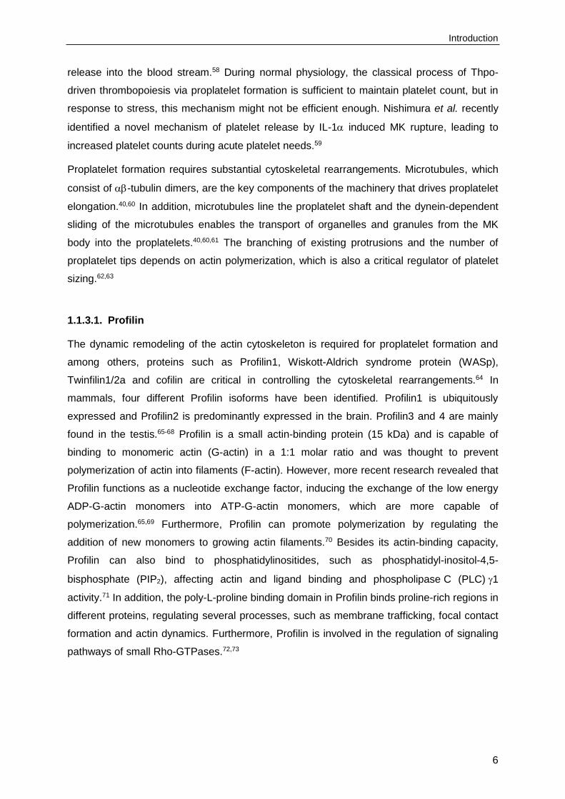
Proplatelet formation requires substantial cytoskeletal rearrangements. Microtubules, which
consist of -tubulin dimers, are the key components of the machinery that drives proplatelet
elongation.40,60 In addition, microtubules line the proplatelet shaft and the dynein-dependent
sliding of the microtubules enables the transport of organelles and granules from the MK
body into the proplatelets.40,60,61 The branching of existing protrusions and the number of
proplatelet tips depends on actin polymerization, which is also a critical regulator of platelet
sizing.62,63
1.1.3.1. Profilin
The dynamic remodeling of the actin cytoskeleton is required for proplatelet formation and
among others, proteins such as Profilin1, Wiskott-Aldrich syndrome protein (WASp),
Twinfilin1/2a and cofilin are critical in controlling the cytoskeletal rearrangements.64 In
mammals, four different Profilin isoforms have been identified. Profilin1 is ubiquitously
expressed and Profilin2 is predominantly expressed in the brain. Profilin3 and 4 are mainly
found in the testis.65-68 Profilin is a small actin-binding protein (15 kDa) and is capable of
binding to monomeric actin (G-actin) in a 1:1 molar ratio and was thought to prevent
polymerization of actin into filaments (F-actin). However, more recent research revealed that
Profilin functions as a nucleotide exchange factor, inducing the exchange of the low energy
ADP-G-actin monomers into ATP-G-actin monomers, which are more capable of
polymerization.65,69 Furthermore, Profilin can promote polymerization by regulating the
addition of new monomers to growing actin filaments.70 Besides its actin-binding capacity,
Profilin can also bind to phosphatidylinositides, such as phosphatidyl-inositol-4,5-
bisphosphate (PIP2), affecting actin and ligand binding and phospholipase C (PLC) 1
activity.71 In addition, the poly-L-proline binding domain in Profilin binds proline-rich regions in
different proteins, regulating several processes, such as membrane trafficking, focal contact
formation and actin dynamics. Furthermore, Profilin is involved in the regulation of signaling
pathways of small Rho-GTPases.72,73
Introduction
7
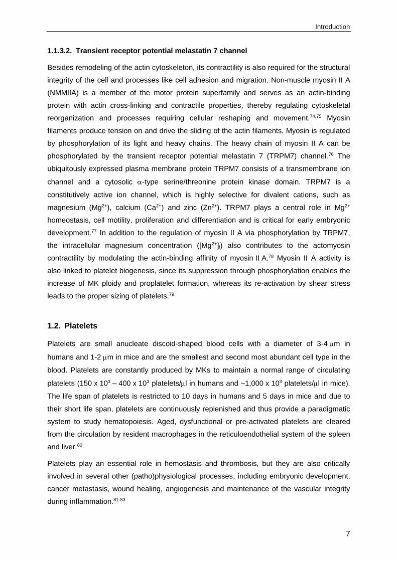
1.1.3.2. Transient receptor potential melastatin 7 channel
Besides remodeling of the actin cytoskeleton, its contractility is also required for the structural
integrity of the cell and processes like cell adhesion and migration. Non-muscle myosin II A
(NMMIIA) is a member of the motor protein superfamily and serves as an actin-binding
protein with actin cross-linking and contractile properties, thereby regulating cytoskeletal
reorganization and processes requiring cellular reshaping and movement.74,75 Myosin
filaments produce tension on and drive the sliding of the actin filaments. Myosin is regulated
by phosphorylation of its light and heavy chains. The heavy chain of myosin II A can be
phosphorylated by the transient receptor potential melastatin 7 (TRPM7) channel.76 The
ubiquitously expressed plasma membrane protein TRPM7 consists of a transmembrane ion
channel and a cytosolic -type serine/threonine protein kinase domain. TRPM7 is a
constitutively active ion channel, which is highly selective for divalent cations, such as
magnesium (Mg2+), calcium (Ca2+) and zinc (Zn2+). TRPM7 plays a central role in Mg2+
homeostasis, cell motility, proliferation and differentiation and is critical for early embryonic
development.77 In addition to the regulation of myosin II A via phosphorylation by TRPM7,
the intracellular magnesium concentration ([Mg2+]i) also contributes to the actomyosin
contractility by modulating the actin-binding affinity of myosin II A.78 Myosin II A activity is
also linked to platelet biogenesis, since its suppression through phosphorylation enables the
increase of MK ploidy and proplatelet formation, whereas its re-activation by shear stress
leads to the proper sizing of platelets.79
1.2. Platelets
Platelets are small anucleate discoid-shaped blood cells with a diameter of 3-4 m in
humans and 1-2 m in mice and are the smallest and second most abundant cell type in the
blood. Platelets are constantly produced by MKs to maintain a normal range of circulating
platelets (150 x 103 – 400 x 103 platelets/l in humans and ~1,000 x 103 platelets/l in mice).
The life span of platelets is restricted to 10 days in humans and 5 days in mice and due to
their short life span, platelets are continuously replenished and thus provide a paradigmatic
system to study hematopoiesis. Aged, dysfunctional or pre-activated platelets are cleared
from the circulation by resident macrophages in the reticuloendothelial system of the spleen
and liver.80
Platelets play an essential role in hemostasis and thrombosis, but they are also critically
involved in several other (patho)physiological processes, including embryonic development,
cancer metastasis, wound healing, angiogenesis and maintenance of the vascular integrity
during inflammation.81-83

Introduction
8
Platelets contain different types of organelles and granules, such as mitochondria, glycogen
stores, peroxisomes, - and dense-granules and lysosomes.28,84 -granules are the most
abundant platelet granules and store platelet adhesion proteins (e.g. fibrinogen, fibronectin,
vWF, P-selectin), coagulation factors, chemokines (e.g. PF4), growth factors and
glycoproteins (e.g. IIb3). Dense granules are smaller, less abundant and contain many
inorganic molecules, such as adenosine diphosphate (ADP), adenosine triphosphate (ATP),
Ca2+, thromboxane A2 (TxA2), polyphosphates, catecholamines and 5-hydroxytryptamine (5-
HT). Lysosomes store enzymes that are required for the degradation of carbohydrates,
proteins and lipids. In addition, platelets contain the open canalicular system (OCS), the
dense tubular system (DTS), an actin-based cytoskeletal network and a peripheral
microtubule coil. The OCS consists of plasma membrane invaginations and serves as a
membrane reservoir, allowing surface area increase following platelet activation and shape
change. The DTS derives from residual endoplasmatic reticulum and is thought to be a Ca2+
reservoir.85-87
1.3. Platelet activation and thrombus formation
Under normal physiological conditions, most of the circulating platelets never come into
contact with components of the ECM. However, at sites of vascular wall damage, for
example upon injury or atherosclerotic plaque rupture, thrombogenic components of the
ECM become exposed. Subendothelial matrix components, including vWF, fibrillar collagens
and laminins, come into contact with the flowing blood, initiating the activation, adhesion and
aggregation of platelets and subsequent thrombus formation. This process is essential to
seal vessel lesion, minimize blood loss and prevent infection. Under pathological conditions,
however, platelet aggregation can lead to uncontrolled thrombus formation, possibly causing
arterial occlusion or embolism, leading to acute ischemic disease states, such as myocardial
infarction or stroke. These pathologies are currently the primary causes of death and
disability in the industrialized world.88 Platelet activation is a tightly regulated multistep
process and can be divided into three major steps: (1) tethering and platelet adhesion to the
exposed ECM; (2) platelet activation and granule release and (3) firm adhesion, platelet
aggregation and thrombus growth (Figure 3).89 Tethering of the platelets occurs under
intermediate or high shear conditions (1,000-10,000 s-1), which are found in arterioles or
stenosed arteries. The initial contact of circulating platelets to exposed components of the
ECM is facilitated by the interaction of the glycoprotein (GP) Ib-V-IX receptor complex with
vWF immobilized on collagen.90-93 This transient and weak interaction leads to rapid
deceleration and ‘rolling’ of the circulating platelets and enables the interaction of the ECM
component collagen with the central activating platelet collagen receptor, GPVI.94-96

Introduction
9
Figure 3. Multistep process of platelet adhesion, activation and thrombus formation. Upon vascular wall injury, tethering of circulating platelets to exposed components of the ECM is facilitated by the GPIb-vWF interaction. This enables the binding of GPVI to collagen, leading to intracellular signaling and the release of second wave mediators, like ADP and TxA2. Exposed TF triggers thrombin generation, which contributes to platelet activation. The cellular signaling events induce the conversion of integrins to a high-affinity state, resulting in firm platelet adhesion and thrombus growth. From: Nieswandt et al., 201197
Cross-linking of GPVI by collagen induces an intracellular signaling cascade, culminating in
an increase in intracellular calcium levels ([Ca2+]i), surface exposure of negatively charged
procoagulant phosphatidylserine (PS), cytoskeletal rearrangements and the mobilization of
- and dense granules, leading to the release of second wave mediators, including ADP and
TxA2.97,98 In addition, exposed tissue factor (TF) triggers thrombin generation (extrinsic
coagulation pathway). This is also induced by the release of polyphosphates from the dense
granules, leading to the activation of factor XII and the intrinsic pathway of coagulation,
culminating in the cleavage of the proenzyme prothrombin (factor II) to thrombin.99 Thrombin
is a potent platelet activator and converts fibrinogen to fibrin. The soluble agonists ADP, TxA2
and thrombin bind to and activate G protein-coupled receptors (GPCRs) (Gq, G12/13 and Gi),
inducing platelet activation and additional recruitment of platelets to the growing thrombus.100
Finally, firm adhesion, platelet aggregation and thrombus growth is mediated by the
conformational change of integrin adhesion receptors. The extra- and intracellular signaling
events induce the conversion of the integrins from a low (inactive) to a high (active) affinity
state (inside-out signaling), allowing the interaction of the activated integrins with their
ligands (outside-in signaling).89,101 The major platelet integrin, IIb3 (GPIIb/IIIa; CD41), is
able to bind to fibrinogen or exposed ECM components, such as collagen-bound vWF,
Introduction
10
fibronectin and vitronectin. Platelet binding to fibrinogen enables bridging of adjacent
platelets, inducing the growth and formation of a stable thrombus. Integrin IIb3 binding to
ECM components leads to firm platelet adhesion. This is further mediated by binding of high-
affinity 1- and 3-integrins to collagen (21), fibronectin (51), laminin (61) and
vitronectin (v3).89,102,103 The activation of platelets also results in the rearrangement of the
actin and tubulin cytoskeleton, leading to the formation of membrane protrusions, such as
filopodia and lamellipodia.104
Platelet activation is mediated by two major signaling pathways (Figure 4): signaling via the
GPCRs or the (hem)immunoreceptor tyrosine-based activation motif (ITAM)-bearing
receptors, which are described in more detail in section 1.3.1 and 1.3.2. Both signaling
pathways culminate in the activation of PLC isoforms, leading to hydrolysis of PIP2 to inositol-
1,4,5-trisphosphate (IP3) and diacyl-glycerol (DAG).105 IP3 binds to the IP3 receptor on the
endoplasmatic reticulum membrane, inducing Ca2+ release from the intracellular stores.
Subsequently, the Ca2+ store content decreases, stimulating the Ca2+ channels in the plasma
membrane to open, a process called store-operated Ca2+ entry (SOCE), which is mediated
by stromal interaction molecule 1 (STIM1) and Ca2+ release-activated calcium channel
protein 1 (Orai1).106 This results in increased [Ca2+]i, leading to integrin activation, platelet
shape change, granule secretion and platelet aggregation.105
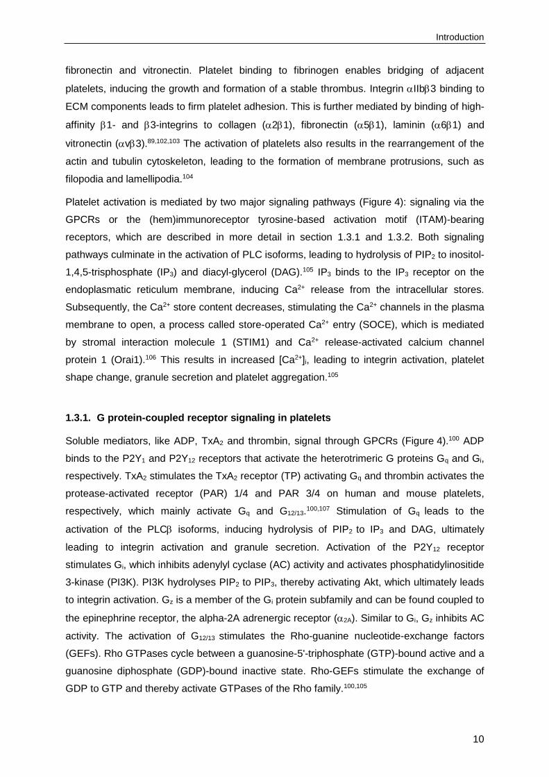
1.3.1. G protein-coupled receptor signaling in platelets
Soluble mediators, like ADP, TxA2 and thrombin, signal through GPCRs (Figure 4).100 ADP
binds to the P2Y1 and P2Y12 receptors that activate the heterotrimeric G proteins Gq and Gi,
respectively. TxA2 stimulates the TxA2 receptor (TP) activating Gq and thrombin activates the
protease-activated receptor (PAR) 1/4 and PAR 3/4 on human and mouse platelets,
respectively, which mainly activate Gq and G12/13.100,107 Stimulation of Gq leads to the
activation of the PLC isoforms, inducing hydrolysis of PIP2 to IP3 and DAG, ultimately
leading to integrin activation and granule secretion. Activation of the P2Y12 receptor
stimulates Gi, which inhibits adenylyl cyclase (AC) activity and activates phosphatidylinositide
3-kinase (PI3K). PI3K hydrolyses PIP2 to PIP3, thereby activating Akt, which ultimately leads
to integrin activation. Gz is a member of the Gi protein subfamily and can be found coupled to
the epinephrine receptor, the alpha-2A adrenergic receptor (2A). Similar to Gi, Gz inhibits AC
activity. The activation of G12/13 stimulates the Rho-guanine nucleotide-exchange factors
(GEFs). Rho GTPases cycle between a guanosine-5'-triphosphate (GTP)-bound active and a
guanosine diphosphate (GDP)-bound inactive state. Rho-GEFs stimulate the exchange of
GDP to GTP and thereby activate GTPases of the Rho family.100,105

Introduction
11
Figure 4. Major signaling pathways in platelets. Soluble agonists, including ADP, TxA2 and thrombin, signal through GPCRs and activate downstream effectors, culminating in the activation of
PLC. Platelet activating receptors, including GPVI, CLEC-2 and active integrins induce PLC2 activation upon ligand binding. The activation of the PLC isoforms finally leads to enhanced integrin activation, platelet shape change, aggregation and secretion. From: Stegner and Nieswandt, 2011105
1.3.1.1. Ras homolog gene family, member A
Small GTPases of the Rho family are known to be critical regulators of cytoskeletal
rearrangements in platelets.108,109 Ras homolog gene family, member A (RhoA) regulates the
actin cytoskeleton in the formation of stress fibers and mediates platelet shape change from
discoid to spherical following activation.110,111 The Rho-GEFs activate RhoA, which binds to
and activates Rho-associated protein kinase (ROCK), leading to ROCK phosphorylation and
inhibition of the MLC phosphatase, resulting in increased phosphorylation of the myosin light
chain (MLC), inducing actin remodeling.110,112 Studies in MK- and platelet-specific RhoA-
deficient mice showed that RhoA signaling is essential for platelet shape change and

Introduction
12
contributes to integrin IIb3 activation, granule release, clot retraction in vitro and arterial
thrombus formation and stability in vivo.111 Although Rho GTPases, such as RhoA are known
to be critical regulators of cytoskeletal rearrangements in platelets, little is known about their
specific roles during platelet biogenesis. The moderate thrombocytopenia observed in RhoA-
deficient mice suggests that RhoA is necessary for platelet production. Recent studies
revealed a minor shift to higher mean ploidy levels and slightly increased numbers of BM
MKs in MK-specific RhoA-deficient mice, but the role of RhoA in platelet biogenesis remains
poorly understood.111,113
1.3.2. (Hem)immunoreceptor tyrosine-based activation motif signaling in platelets
The second major signaling pathway in platelets involves signaling via the (hem)ITAM-
bearing receptors (Figure 5). An ITAM is a highly conserved sequence of four amino acids,
which is repeated twice in the cytoplasmic tail of several hematopoietic immunoglobulin (Ig)
receptors, including Fc receptors (FcR), T and B cell receptors (TCR, BCR) and C-type
lectin-like receptors. The motif is defined by the presence of two YxxL/I motifs separated by
6-12 amino acids (YxxL/Ix(6-12)YxxL/I).114 Human platelets express three ITAM-bearing
receptors: the Fc receptor FcRIIA, GPVI and the hemITAM C-type lectin-like receptor 2
(CLEC-2), whereas mouse platelets only express GPVI and CLEC-2. FcRIIA signals
through an ITAM in its cytoplasmic tail and is critically involved in immune-mediated
thrombocytopenia and thrombosis, but also enhances integrin outside-in signaling via spleen
tyrosine kinase (Syk).81,115 GPVI signals via the non-covalently associated ITAM-bearing
FcR -chain. Upon ligand-induced receptor activation of GPVI, the tyrosine residues in the
ITAM become phosphorylated by Src family kinases (SFKs), such as Fyn and Lyn, inducing
the recruitment and activation of tandem Src homology (SH)2 domain-containing Syk.81,98 In
contrast to classical ITAM signaling, recent studies suggest that Syk itself phosphorylates the
hemITAM within CLEC-2, whereas the SFKs together with Syk are involved in the regulation
of downstream signaling.116 The downstream signaling cascade involves a large number of
adaptor and effector proteins, such as the linker for activated T cells (LAT), the SH2 domain-
containing leukocyte protein of 76 kDa (SLP-76) and growth factor receptor-bound protein 2
(Grb2), ultimately leading to the activation of effector enzymes, such as PI3K and
PLC2.81,117 Subsequently, Ca2+ mobilization, integrin activation and granule secretion are
induced.

Introduction
13
Figure 5. (hem)ITAM signaling in platelets. The Fc receptor FcRIIA is expressed on human, but not on mouse platelets and signals through an ITAM, thereby enhancing integrin outside-in signaling
via Syk. GPVI is non-covalently associated with the ITAM-bearing FcR -chain. Ligand-induced receptor activation initiates phosphorylation of the tyrosine residues in the ITAM by SFKs, leading to the recruitment and activation of Syk. Syk initiates a downstream signaling cascade that involves a large number of adaptor and effector proteins, such as LAT, SLP-76 and Grb2, culminating in the
activation of PLC2. CLEC-2 contains one copy of the YxxL motif (hemITAM), which becomes phosphorylated by Syk. Syk together with the SFKs regulate a downstream signaling cascade similar to that found downstream of GPVI, leading to Ca2+ mobilization, integrin activation and granule secretion. From: Stegner et al., 201481
1.3.2.1. Glycoprotein VI
Platelets express several receptors that are able to interact with collagens, such as 21 and
GPVI. Integrin 21 mainly mediates adhesion, whereas GPVI is the central and
indispensable activating collagen receptor on the platelet surface.94,117 GPVI is a 62 kDa
type I transmembrane protein that belongs to the Ig superfamily of surface receptors and is
exclusively expressed on MKs and platelets.118 The extracellular part of GPVI consists of two
IgG domains bearing the collagen binding site, followed by a mucin-like region containing O-
glycosylation sites, a transmembrane region and a cytoplasmic tail.94,98 The cytoplasmic tail
is short, comprising only 51 amino acids in human and 27 amino acids in mice and contains
a calmodulin binding motif and a proline-rich region, which is recognized by the SH3 domain
of the SFKs Fyn and Lyn.119,120
The transmembrane region of GPVI contains a positively charged arginine that is essential
for the non-covalent association with the FcR -chain. The FcR -chain is required for
initiation of downstream signaling and serves as the signal transducing subunit of the

Introduction
14
GPVI/FcR -chain complex.94 Each FcR -chain contains one ITAM and is expressed as a
covalently linked homodimer. One GPVI molecule associates with one FcR -chain dimer
and only the dimeric form of GPVI binds collagen with a high affinity.117,121 GPVI specifically
recognizes the glycine-proline-hydroxyproline (GPO) repeats within collagen.94 Platelet
activation via GPVI can also be induced by the specific agonists collagen-related peptide
(CRP), which is composed of repeated GPO motifs, and the snake venom toxin convulxin
(CVX), which is capable of clustering four GPVI proteins and therefore a strong agonist.94
Ligand-induced crosslinking of GPVI leads to phosphorylation of the FcR-chain ITAM,
inducing downstream signaling and finally activation of PI3K and PLC2.122 Signaling through
the GPVI/FcR -chain is, among others, negatively regulated by immunoreceptor tyrosine-
based inhibition motif (ITIM) containing receptors, including platelet endothelial cell adhesion
molecule 1 (PECAM-1), Carcinoembryonic antigen-related cell adhesion molecule 1
(CEACAM1) and G6b-B.117
Since GPVI is a MK- and platelet specific receptor, it may represent an attractive potential
target for antithrombotic therapy. Patients with a congenital deficiency of GPVI or
autoantibody-mediated GPVI deficiency only suffered from a mild bleeding disorder, but
platelet aggregation and thrombus formation ex vivo on collagen were impaired.98,123 In
experimental models, the deficiency, blockade or immunodepletion of GPVI provide a strong
protection from arterial thrombosis without or only moderately affecting hemostasis.98,124
Besides its crucial role in occlusive arterial thrombosis, GPVI has been identified as a critical
modulator of several thrombo-inflammatory disease states, including ischemic stroke, loss of
vascular integrity at sites of inflammation, rheumatoid arthritis, atheroprogression and tumor
metastasis.125-130
Different possibilities exist to target GPVI, including the direct blockade of the ligand binding
site on GPVI, immunodepletion or downregulation of the GPVI receptor using monoclonal
antibodies and targeting of the GPVI signaling pathway.98 Receptor blockade can be
achieved by using a F(ab) fragment of an anti-GPVI antibody.131 In vivo administration of
monoclonal anti-GPVI antibodies (JAQ1-3) induces specific and irreversible removal of the
receptor from the surface of circulating platelets, leading to a knockout-like phenotype.132-134
To induce GPVI downregulation, two principal pathways exist in platelets, namely
ectodomain shedding by the metalloproteinases A disintegrin and metalloprotease
(ADAM)10 and ADAM17, generating a soluble GPVI fragment and a transmembrane
remnant, and internalization/degradation of the GPVI receptor.134-137 Both processes require
signaling through the FcR -chain ITAM.136 Ectodomain shedding appears to be the
predominant mechanism of antibody-induced GPVI loss, however, downregulation in vivo
seems to involve another sheddase, since antibody-induced shedding of GPVI still occurs in

Introduction
15
mice lacking ADAM10 and ADAM17.135,137 To reduce GPVI activity, molecules in the
downstream signaling pathway, for example the kinase Syk, could be targeted. Several Syk
inhibitors have been developed and clinically used138-145 and Syk might be a potential
antithrombotic target.
1.3.2.2. C-type lectin-like receptor 2
CLEC-2 plays a crucial role in different (patho)physiological processes, such as thrombosis
and hemostasis, tumor metastasis, maintenance of the integrity of high endothelial venules,
maintenance of the vascular integrity during inflammation, lymph node development and
blood/lymph vessel separation.146-149
CLEC-2 is a 32 kDa type II transmembrane protein, that is highly expressed on MKs and
platelets and, in addition, at lower levels on different immune cells and liver sinusoidal
cells.117,150 CLEC-2 was identified as the receptor for the snake venom toxin rhodocytin
(Rhod) and the glycoprotein podoplanin, the only known physiological ligand of CLEC-2.151,152
Podoplanin is expressed in different tissues, including lymphatic endothelial cells, type 1 lung
alveolar cells, kidney podocytes, lymph node stromal cells and is also expressed on tumor
cells.151,153 However, podoplanin is not expressed on platelets and vascular endothelial cells.
Mice lacking either CLEC-2 or podoplanin, fail to separate blood and lymphatic vessels
during development, leading to embryonic or neonatal lethality associated with blood-filled
lymphatics and severe edema formation.154-157 The interaction of podoplanin with CLEC-2
induces platelet activation and aggregation leading to closing of the blood-lymphatic shunts.
Blood-filled lymphatics can also be found in mice lacking key (hem)ITAM-signaling
molecules, such as Syk, SLP-76 and PLC2, but the exact mechanism in separation of blood
and lymphatic vessels remains poorly understood.147,154,158
CLEC-2 contains a conserved YxxL motif in its cytoplasmic tail, which mediates downstream
signaling. This motif is similar to the ITAM, however, only one copy of the sequence is
present within the protein, therefore it is called a hemITAM.151 CLEC-2 is expressed as a
homodimer on the platelet surface and upon ligand binding, clustering of the receptor brings
together two hemITAM domains, which, upon phosphorylation, serve as a binding site for the
two SH2 domains of Syk.151,159 The signaling pathway downstream of CLEC-2 resembles the
GPVI signaling pathway and involves similar molecules, such as Syk, SFKs, LAT, SLP-76
and PLC2.117,151 However, recent studies suggest a difference in the phosphorylation events
and recruitment of SFKs and Syk. Following GPVI receptor activation, phosphorylation of the
ITAM on the FcR -chain is mediated by the SFKs Fyn and Lyn, leading to the recruitment
and activation of Syk, whereas Syk itself phosphorylates the hemITAM within CLEC-2, while
the SFKs are involved in the regulation of downstream signaling.116

Introduction
16
CLEC-2 also plays an important role in the stabilization of platelet thrombi and studies on the
role of CLEC-2 in thrombosis and hemostasis revealed the receptor as a potential target for
antithrombotic therapy.155,160 Immunodepletion of CLEC-2 from the surface of circulating
platelets or CLEC-2 deficiency in mice led to impaired aggregate formation in vitro and
abolished thrombus formation in vivo, while bleeding times are only moderately
prolonged.155,160 However, the combined deficiency of CLEC-2 and GPVI resulted in a
dramatic hemostatic defect and inflammation-induced hemorrhage, demonstrating a
functional redundancy of these two receptors.128,133 Therefore, the suitability of CLEC-2 as a
safe antithrombotic therapy target needs to be reconsidered. Since podoplanin is not
expressed in platelets and vascular endothelial cells, the mechanism that contributes to
CLEC-2 dependent thrombus stabilization, may involve a yet unidentified CLEC-2 ligand.160
Similar to GPVI, CLEC-2 can be immunodepleted from the surface of platelets by a
monoclonal anti-CLEC-2 antibody (INU1), resulting in a knockout-like phentotype.133,160,161
INU1 induces receptor downregulation and a severe, but transient thrombocytopenia.133,160,161
The INU1-induced receptor downregulation occurs through SFK-dependent receptor
internalization in vitro and in vivo, whereas the thrombocytopenia is Syk-dependent. This
indicates that CLEC-2 downregulation occurs through receptor internalization and this
mechanism can be uncoupled from the associated antibody-induced thrombocytopenia.161
1.3.2.3. Spleen tyrosine kinase
The adaptive recognition of self and foreign antigens is mediated by the classical
immunoreceptors, such as the BCR, TCR and FcR. These receptors or their associated
transmembrane adaptors contain and signal through ITAMs, which become phosphorylated
during receptor activation, primarily by SFKs. The phosphorylation of the tyrosine residues
induces the recruitment of the tandem SH2 domain-containing Syk (BCR and FcR) or the
related protein -chain-associated protein kinase of 70 kDa (Zap-70) (TCR), initiating a
downstream signaling cascade. In addition to the immunoreceptors, Syk is also activated by
GPVI, C-type lectins and integrin receptors.162-165 The downstream signaling of Syk or Zap-70
involves molecules, such as Vav proteins, PLC isoforms, PI3Ks and SLP family members.
These molecules induce activation of other downstream processes, including PKC signaling,
Rho-mediated cytoskeletal rearrangements, reactive oxygen species (ROS) production and
transcriptional regulation.162
Syk plays a crucial role in adaptive immune receptor signaling, but is also involved in other
(patho)physiological processes, including cellular adhesion, innate pathogen recognition,
damage recognition, bone metabolism, platelet activation, vascular development, allergy,
autoimmune diseases and hematological malignancies.162

Introduction
17
Syk is highly expressed in hematopoietic cells, but it is also expressed in other tissues, such
as fibroblasts, epithelial cells, breast tissue, neuronal cells and vascular endothelial cells.166
Syk is a 72 kDa multiple-domain cytoplasmic tyrosine kinase that contains two tandem SH2
domains and a carboxy-terminal tyrosine kinase domain. The two SH2 domains are linked by
interdomain A and the C-terminal SH2 domain and the kinase domain are linked by
interdomain B. Syk-B, an alternatively spliced form of Syk lacks 23 amino acids of
interdomain B, but the generation of this isoform is poorly understood.162,167 In the resting
state, the kinase domain of Syk is inactive or autoinhibited, but upon binding of the SH2
domains to the phosphorylated ITAMs, the domain can be activated. In addition, the tyrosine
residues in interdomain A and B can become phosphorylated, inducing kinase activation.
The dual activation of Syk provides rapid activation by ITAM binding and sustained activation
by autophosphorylation, which could even occur in the absence of binding to phosphorylated
ITAMs. The phosphorylation of the tyrosine residues in Syk allows binding of different
molecules, resulting in downstream signaling or the degradation of Syk.162 Its function can be
negatively regulated by SH2 domain-containing protein tyrosine phosphatase 1 (SHP1) and
the E3 ubiquitin ligase Casitas B-lineage lymphoma (CBL).168,169 Syk deficiency in mice leads
to perinatal lethality and blocked B-cell development, leading to the lack of mature B
cells.170,171 Syk plays an important role in the conversion of pro-B cells into pre-B cells and is
required for the maturation of transitional B cells.170,172 Syk also has a critical role in pre-TCR
signaling, FcRI signaling in mast cells and FcR signaling in neutrophils and
macrophages.173-176
Due to its central role in immunological processes, several Syk inhibitors have been
developed and clinically used, including R112143, PRT-060318145, the prodrug fostamatinib
(R788)138,139 and its active metabolite R406.140 However, these inhibitors have a rather limited
specificity and adverse effects.177 Recently, entospletinib (GS-9973), a more selective and
orally efficacious Syk inhibitor was developed141 and phase II trials are underway to assess
its effectiveness in treating leukemia.178 Syk inhibitors have been used in the treatment of
allergy,143 B cell malignancies,138 heparin-induced thrombocytopenia145 and autoimmune
diseases like rheumatoid arthritis139 and immune thrombocytopenic purpura.144
Several platelet functions rely on Syk signaling, including GPVI and CLEC-2 receptor
signaling. In addition, Syk has been proposed to contribute to outside-in signaling of platelet
integrin IIb3.179-181 Studies using Syk inhibitors or Syk-deficient mice (radiation chimeras)
provide minimal evidence for a role for Syk in thrombosis and hemostasis.145,182,183 Thrombus
formation ex vivo and in vivo was strongly inhibited, without affecting hemostasis, therefore
Syk could be a potential antithrombotic target.

Introduction
18
1.3.2.4. -chain-associated protein 70
As mentioned in section 1.3.2.3, Zap-70 is a downstream mediator of the immunoreceptor
TCR and is mainly expressed in T cells and natural killer (NK) cells.184 Like Syk, Zap-70
contains two tandem SH2 domains and a carboxy-terminal tyrosine kinase domain. The
domains are connected by two linker regions, interdomain A and B. In comparison to Syk,
Zap-70 lacks 11 amino acids in the interdomain B and has a much lower intrinsic kinase
activity.162 It is thought that due to the shorter linker region, the structure of Zap-70 is less
flexible and therefore, it would be more difficult to bridge two hemITAMS or to bind
phosphorylated tyrosine residues with a bigger distance between them.185,186 Following TCR
engagement, the ITAMs present in the cytoplasmic tails of the TCR homodimer and CD3
become phosphorylated by SFKs Lck or Fyn. This leads to the recruitment of the SH2
domain-containing Zap-70 and disassembly of the autoinhibited, inactive conformation,
allowing the kinase domain to adopt an active confirmation.187,188 Subsequently, Lck or
autophosphorylation of the tyrosine residues activate the kinase domain and Zap-70
activates the downstream targets LAT and SLP-76, leading to T cell activation. In addition,
Zap-70 can recruit additional signaling proteins, such as CBL, Vav, Lck and PLC to the
stimulated TCR complex by interaction with the phosphorylated tyrosine residues.184 In
contrast to Syk, Zap-70 is not able to phosphorylate the ITAM residues itself and is therefore
more dependent on SFKs for its activation and the unique ability to enhance the activation of
mitogen-activated protein kinase (MAPK) p38.167,189 Zap-70 deficient humans and mice fail to
produce functional T cells and suffer from severe combined immunodeficiency (SCID).190-192
Zap-70 plays an important role in thymocyte development and pre-TCR signaling, specifically
during thymic selection at the double negative and double positive stage.171,192,193 In addition,
Zap-70 is expressed throughout B cell development and participates in pre-BCR signaling,
and its expression is associated with increased BCR signaling in B-cell chronic lymphocyte
leukemia.184,194 Furthermore, decreased function of Zap-70 is associated with autoimmune
disease.195
1.4. Imaging modalities
Over the last few decades, the processes of megakaryopoiesis and thrombopoiesis have
been studied intensively, but mainly in vitro using cultured MKs or ex vivo using BM explants
and histological sections of the BM, since in vivo investigations are complicated by the
anatomic inaccessibility of the BM. However, to further understand these processes,
visualization of the localization and morphology of MKs as well as the generation of platelets
within the intact BM ex vivo and real-time in vivo would be of great benefit. Recent
developments in imaging methods enable us to observe dynamic cellular events in vivo using

Introduction
19
multiphoton intravital microscopy (MP-IVM) and to observe the intact three-dimensional (3D)
structure of different tissues ex vivo using light-sheet fluorescence microscopy (LSFM).
1.4.1. Multiphoton intravital microscopy
IVM enables the microscopic imaging of a living animal and combining this method with MP
imaging allows us to visualize and analyze the murine BM real-time and in vivo. Mazo et al.
described an IVM technique to study physiologically perfused murine BM microvessels using
fluorescence microscopy and characterized the anatomy of the skull BM microvasculature.196
The flat bones forming the calvaria of the murine skull contain BM. Imaging of the BM in the
frontoparietal skull without surgical manipulation of the bone is achievable, since the thin
layer of bone covering the BM cavities is sufficiently transparent.196 However, light scattering,
due to refractive index mismatches in heterogeneous tissues, has hampered imaging within
living tissues or thick samples using fluorescence microscopy. Light scattering leads to the
degradation of resolution and contrast with increasing tissue depth and high resolution
imaging eventually becomes impossible. Confocal microscopy improves the image quality,
but also has its limitations, since excitation of the sample often leads to photodestruction of
the fluorophore (photobleaching) or photodynamic damage to the specimen (photodamage).
These drawbacks can be evaded by the use of MP microscopy. In 1931, Maria Göppert-
Mayer first predicted the possibility of MP excitation, but it was only after the invention of the
laser, that it was experimentally confirmed.197 A seminal paper by Denk et al. showed the
application of two-photon excitation to laser scanning microscopy and the capability of MP
microscopy for biology.198,199 The key difference between confocal microscopy and MP
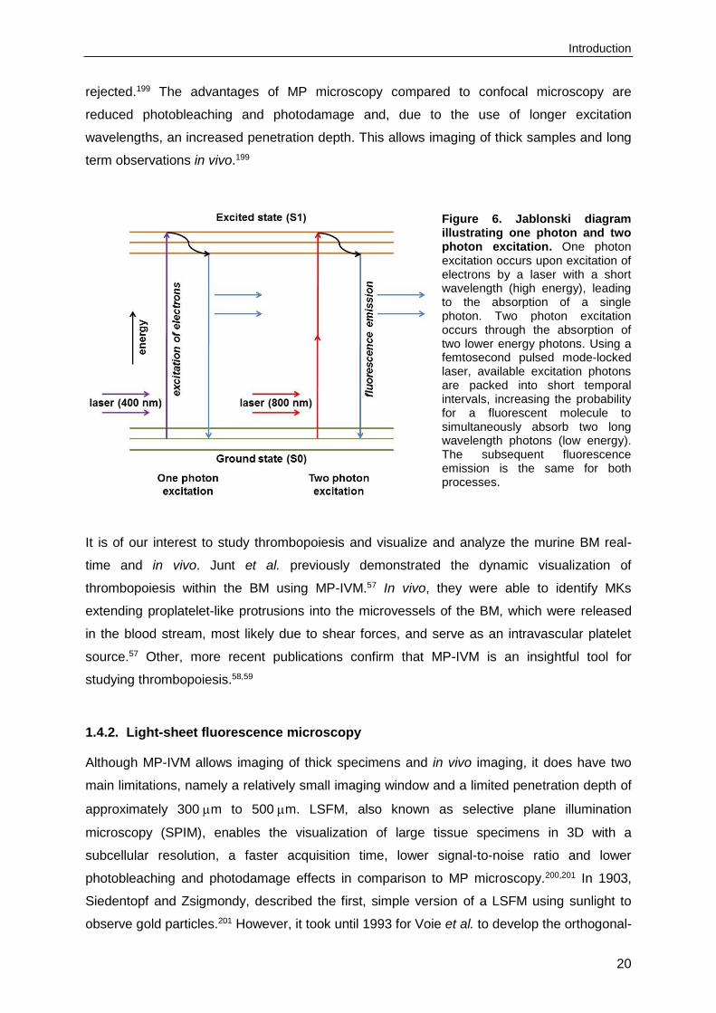
microscopy is the excitation of the fluorescent molecules. In one photon excitation, used in
confocal microscopy, a laser with a short wavelength (high energy) can excite or energize
electrons (Figure 6). An electron of, for example, a fluorescent molecule then absorbs a
single photon of the excitation light. This raises the energy level of the electron to an excited
state (S1). During the excitation period, some of the energy is lost and the remaining energy
is emitted as a photon. The energy level of the electron falls back to the lowest-energy state,
the ground state (S0). The emitted photon carries less energy and has a longer wavelength.
In MP excitation, a femtosecond pulsed mode-locked laser with a longer wavelength is used.
The short pulses of the laser induce the packing of available excitation photons into short
temporal intervals. This increases the probability for a fluorescent molecule to simultaneously
absorb two or more long wavelength photons (low energy), thus combining the energy of two
photons to reach the excited state (S1) (Figure 6).198,199 Subsequently, fluorescence is only
generated in the focal plane, where the light intensity is high, allowing intrinsic optical
sectioning of the specimen. A detector pinhole is not required, since all fluorescence photons
contribute to the signal and fluorescence from off-focus locations does not need to be
Introduction
20
rejected.199 The advantages of MP microscopy compared to confocal microscopy are
reduced photobleaching and photodamage and, due to the use of longer excitation
wavelengths, an increased penetration depth. This allows imaging of thick samples and long
term observations in vivo.199
Figure 6. Jablonski diagram illustrating one photon and two photon excitation. One photon excitation occurs upon excitation of electrons by a laser with a short wavelength (high energy), leading to the absorption of a single photon. Two photon excitation occurs through the absorption of two lower energy photons. Using a femtosecond pulsed mode-locked laser, available excitation photons are packed into short temporal intervals, increasing the probability for a fluorescent molecule to simultaneously absorb two long wavelength photons (low energy). The subsequent fluorescence emission is the same for both processes.
It is of our interest to study thrombopoiesis and visualize and analyze the murine BM real-
time and in vivo. Junt et al. previously demonstrated the dynamic visualization of
thrombopoiesis within the BM using MP-IVM.57 In vivo, they were able to identify MKs
extending proplatelet-like protrusions into the microvessels of the BM, which were released
in the blood stream, most likely due to shear forces, and serve as an intravascular platelet
source.57 Other, more recent publications confirm that MP-IVM is an insightful tool for
studying thrombopoiesis.58,59
1.4.2. Light-sheet fluorescence microscopy
Although MP-IVM allows imaging of thick specimens and in vivo imaging, it does have two
main limitations, namely a relatively small imaging window and a limited penetration depth of
approximately 300 m to 500 m. LSFM, also known as selective plane illumination
microscopy (SPIM), enables the visualization of large tissue specimens in 3D with a
subcellular resolution, a faster acquisition time, lower signal-to-noise ratio and lower
photobleaching and photodamage effects in comparison to MP microscopy.200,201 In 1903,
Siedentopf and Zsigmondy, described the first, simple version of a LSFM using sunlight to
observe gold particles.201 However, it took until 1993 for Voie et al. to develop the orthogonal-

Introduction
21
plane fluorescence optical sectioning (OPFOS) and to image whole fluorophore-stained and
cleared cochleas.202 A seminal paper by Huisken et al. accelerated the development and use
of LSFM. They were able to visualize embryogenesis in Drosophila melanogaster in vivo,
thereby showing that LSFM is a useful method for imaging large, living biological
specimens.203 Dodt et al. further improved image quality by adding dual-sided illumination to
LSFM, making the illumination of large specimens such as the brain more uniform throughout
the specimen.204 LSFM uses a thin sheet of light to illuminate and optically section the
transparent specimen, thereby generating sections that are suitable for 3D reconstruction of
the whole tissue (Figure 7). The focal point of the light sheet is positioned within the
specimen chamber, which contains either a physiological solution or a clearing solution. The
specimen is attached to a glass rod and is placed at the intersection of the focal point of the
light sheet and the focal plane of the detecting objective, where the fluorescence emission is
detected orthogonally to the illumination pathway. The light sheet excites fluorescence in the
specimen, which is collected by the detection optics and projected onto a camera. To obtain
the 3D structure, the sample is moved through the light sheet in small increments, acquiring
a stack of optical sections.200,201
Figure 7. Schematic overview of a custom-built light-sheet fluorescence microscope. A thin sheet of light is generated (arrows), illuminating the sample (red) from one side. The focal plane of the light sheet is positioned within the clearing solution-containing specimen chamber. The specimen is attached to a glass rod and placed at the intersection of the excitation and detection pathway. The fluorescence emission is detected orthogonally to the illumination pathway and projected onto a camera. Image courtesy of Katrin G. Heinze.
The imaging process is mainly conducted ex vivo, since most samples need to undergo a
clearing process to become transparent to allow penetration of the laser light sheet. Clearing
is achieved by immersing the specimen in a solution with the same refractive index as
protein. By matching the refractive indices, light is not scattered and, when no absorption

Introduction
22
occurs, the specimen turns transparent. Tissue clearing methods were already used a
century ago to study the anatomy of the heart and have been developed since, mainly for
soft tissues, such as embryonic tissue and brain.204 In general, tissue is dehydrated and
subsequently immersed in an optical clearing solution, such as glucose, glycerol, benzyl
alcohol benzyl benzoate (BABB or Murray’s clear) or dibenzyl ether (DBE).205,206 However,
dehydration and clearing of tissues expressing fluorescent proteins, such as green
fluorescent protein (GFP), degraded the fluorescent signal, thereby preventing its detection.
Therefore, to preserve fluorescent signals, new clearing agents have recently been
developed, such as 3DISCO, CLARITY and ScaleS.205-207

Aim of the study
23
2. Aim of the study
In order to understand the regulatory mechanisms of hematopoiesis, it is important to
determine the spatiotemporal organization of blood cell precursors within the BM and their
interaction with the BM microenvironment. Therefore, the major aim of this thesis was the
establishment of imaging modalities to visualize these cellular processes in vitro and in vivo.
Megakaryopoiesis and thrombopoiesis have been studied intensively, but the exact
mechanisms that control platelet generation from MKs remain poorly understood. Recent
developments in IVM enable us to study thrombopoiesis and proplatelet formation in the
murine BM real-time and in vivo. The first aim of this thesis was to establish MP-IVM to study
thrombopoiesis and proplatelet formation and to identify the role of Profilin1, TRPM7 and
RhoA in thrombopoiesis in vivo using MP-IVM.
Currently, it is thought that blood cell precursors, such as MKs, migrate from the endosteal
niche towards the vascular niche during maturation. However, the exact location of MK
maturation and the functional significance of MK migration towards the vascular niche for
thrombopoiesis still remain ill-defined. Therefore, the second aim of this thesis was to
visualize the distribution and morphology of MKs in the intact BM using LSFM.
Platelets are constantly produced by their BM precursors. In platelets, Syk is an essential
signaling mediator downstream of immune cell receptors, but also of GPVI and CLEC-2.
However, only limited evidence exists on the role of Syk in hemostasis, arterial thrombosis
and ischemic stroke. Thus, the third aim of this thesis was to identify the pathophysiological
significance of Syk deficiency and to determine if Syk could be a potential anti-thrombotic
target.
Zap-70 has a lower intrinsic activity compared to Syk and is expressed in T cells and NK
cells, but not in platelets. To assess if Syk and Zap70 exhibit interchangeable functions in
vivo, a knock-in mouse was generated, which expresses Zap-70 under the control of intrinsic
Syk promoter elements. The fourth aim of this thesis was to investigate the consequence of
the kinase replacement for platelet function in vitro and in vivo.

Materials and Methods
24
3. Materials and Methods
3.1. Materials
3.1.1. Reagents and chemicals
Acetic acid Roth (Karlsruhe, Germany)
Adenosine diphosphate (ADP) Sigma-Aldrich (Schnelldorf, Germany)
Agarose Roth (Karlsruhe, Germany)
Amersham® Hyperfilm®, ECL GE Healthcare (Little Chalfont, UK)
Ammonium persulphate (APS) Roth (Karlsruhe, Germany)
Apyrase (grade III) Sigma-Aldrich (Schnelldorf, Germany)
Atipamezole Pfizer (Karlsruhe, Germany)
-mercaptoethanol Roth (Karlsruhe, Germany)
Benzyl alcohol Sigma-Aldrich (Schnelldorf, Germany)
Benzyl benzoate Sigma-Aldrich (Schnelldorf, Germany)
Bovine serum albumin (BSA) AppliChem (Darmstadt, Germany)
Bromophenol blue Sigma-Aldrich (Schnelldorf, Germany)
Collagen Horm® suspension + SKF sol. Takeda (Linz, Austria)
Complete protease inhibitor cocktail Roche Diagnostics (Mannheim, Germany)
Convulxin Enzo Life Sciences (Lörrach, Germany)
Cryo-Gel Leica Biosystems (Wetzlar, Germany)
Cyanoacrylate Weicon (Münster, Germany)
Deoxynucleotide triphosphates (dNTP) mix Thermo Fisher (Schwerte, Germany)
4',6-diamidino-2-phenylindole (DAPI)
Fluoromount-G
Southern Biotech (Birmingham, AL, USA)
Dimethyl sulfoxide (DMSO) Sigma-Aldrich (Schnelldorf, Germany)
Disodium hydrogen phosphate (Na2HPO4) Roth (Karlsruhe, Germany)
Dulbecco's Modified Eagle Media (DMEM) Gibco (Karlsruhe, Germany)
Enhanced chemiluminescence (ECL)
detection substrate
MoBiTec (Göttingen, Germany)
Ethanol Sigma-Aldrich (Schnelldorf, Germany)
Ethylenediaminetetraacetic acid (EDTA) AppliChem (Darmstadt, Germany)
Ethylene glycol tetraacetic acid (EGTA) Sigma-Aldrich (Schnelldorf, Germany)
Fat-free dry milk AppliChem (Darmstadt, Germany)
Fentanyl Janssen-Cilag (Neuss, Germany)
Fetal calf serum (FCS) Perbio (Bonn, Germany)
Fibrinogen from human plasma (F3879) Sigma-Aldrich (Schnelldorf, Germany)

Materials and Methods
25
Fibrinogen from human plasma (F4883) Sigma-Aldrich (Schnelldorf, Germany)
Flumazenil AlleMan Pharma (Pfullingen, Germany)
GeneRuler DNA Ladder Mix Thermo Fisher (Schwerte, Germany)
Glucose Roth (Karlsruhe, Germany)
Glycerin Roth (Karlsruhe, Germany)
Glycerol Roth (Karlsruhe, Germany)
Glycine Sigma-Aldrich (Schnelldorf, Germany)
Heparin Ratiopharm (Ulm, Germany)
Hoechst 33258 Biomol (Hamburg, Germany)
4-(2-hydroxyethyl)-1-piperazine-
ethanesulfonic acid (HEPES)
Roth (Karlsruhe, Germany)
IGEPAL® CA-630 Sigma-Aldrich (Schnelldorf, Germany)
Immobilon-P transfer membrane, PVDF Merck Millipore (Darmstadt, Germany)
Indomethacin Sigma-Aldrich (Schnelldorf, Germany)
Iron-III-chloride hexahydrate (FeCl3∙6 H2O) Roth (Karlsruhe, Germany)
Isofluran CP® cp-pharma (Burgdorf, Germany)
Isopropanol Roth (Karlsruhe, Germany)
Magnesium chloride (MgCl2) Roth (Karlsruhe, Germany)
Medetomidine Pfizer (Karlsruhe, Germany)
Methanol Sigma-Aldrich (Schnelldorf, Germany)
Midazolam Roche Diagnostics (Mannheim, Germany)
Midori Green Advanced DNA stain Nippon Genetics Europe (Düren, Germany)
Monopotassium phosphate (KH2PO4) Sigma-Aldrich (Schnelldorf, Germany)
3-(N-morpholino)propanesulfonic acid
(MOPS)
AppliChem (Darmstadt, Germany)
Naloxone AlleMan Pharma (Pfullingen, Germany)
Neomycin Sulfate Bela-Pharm (Vechta, Germany)
NuPAGE® LDS Sample Buffer (4x) Life Technologies (Darmstadt, Germany)
PageRuler Prestained Protein Ladder Thermo Fisher (Schwerte, Germany)
Paraformaldehyde (PFA) Sigma-Aldrich (Schnelldorf, Germany)
Phenol/chloroform/isoamyl alcohol Roth (Karlsruhe, Germany)
Phosphate-buffered saline (PBS) Life Technologies (Darmstadt, Germany)
Plerixafor (AMD3100) Selleckchem, Munich, Germany
Potassium chloride (KCl) Roth (Karlsruhe, Germany)
Propidium iodide (PI) Life Technologies (Darmstadt, Germany)
Prostacyclin (PGI2) Calbiochem (Bad Soden, Germany)
Protease inhibitor cocktail Sigma-Aldrich (Schnelldorf, Germany)
Materials and Methods
26
Proteinase K, recombinant, PCR grade Thermo Fisher (Schwerte, Germany)
Rotiphorese® Gel 30 (37,5:1) acrylamide Roth (Karlsruhe, Germany)
SeeBlue® Plus2 pre-stained standard Life Technologies (Darmstadt, Germany)
Sodium azide (NaN3) Sigma-Aldrich (Schnelldorf, Germany)
Sodium bicarbonate (NaHCO3) Sigma-Aldrich (Schnelldorf, Germany)
Sodium chloride (NaCl) AppliChem (Darmstadt, Germany)
Sodium citrate AppliChem (Darmstadt, Germany)
Sodium dodecyl sulfate (SDS) Sigma-Aldrich (Schnelldorf, Germany)
Sodium fluoride (NaF) Sigma-Aldrich (Schnelldorf, Germany)
Sodium orthovanadate (Na3VO4) Sigma-Aldrich (Schnelldorf, Germany)
SRP4388 (SDF1 alpha) Sigma-Aldrich (Schnelldorf, Germany)
Sucrose Sigma-Aldrich (Schnelldorf, Germany)
10x Taq Buffer (+KCl, -MgCl2) Thermo Fisher (Schwerte, Germany)
Taq DNA polymerase Thermo Fisher (Schwerte, Germany)
N,N,N',N'-Tetramethylethylenediamine
(TEMED)
Roth (Karlsruhe, Germany)
Thrombin from human plasma Roche Diagnostics (Mannheim, Germany)
Thrombin from human plasma Sigma-Aldrich (Schnelldorf, Germany)
Tissue-Tek® O.C.T.™ Compound Sakura Finetek (Staufen, Germany)
2,3,5-triphenyltetrazolium chloride (TTC) Sigma-Aldrich (Schnelldorf, Germany)
Tris(hydroxymethyl)aminomethane (Tris) Roth (Karlsruhe, Germany)
Tween® 20 Roth (Karlsruhe, Germany)
U46619 Enzo Life Sciences (Lörrach, Germany)
Western Lightning® Plus-ECL PerkinElmer (Baesweiler, Germany)
Collagen-related peptide (CRP) was kindly provided by Dr. Steve Watson (University of
Birmingham, UK). Rhodocytin was a generous gift from Dr. Johannes Eble (University
Hospital, Frankfurt, Germany). Primers were obtained from Metabion (Planegg-Martinsried,
Germany). All other non-listed chemicals were obtained from AppliChem (Darmstadt,
Germany), Roth (Karlsruhe, Germany) or Sigma-Aldrich (Schnelldorf, Germany).

Materials and Methods
27
3.1.2. Materials
18G x 1 ½”, 20G x 1 ½”, 26G x 1 ½” needles BD Biosciences (Heidelberg, Germany)
Glass coverslips (24 x 60 mm) Roth (Karlsruhe, Germany)
Glass vials Hartenstein (Würzburg, Germany)
Heparinized capillaries Hartenstein (Würzburg, Germany)
Metal plate skull holder Boer Metaal en Draad (Deil, Netherlands)
Microcapillaries (50 l and 100 l) Hartenstein (Würzburg, Germany)
Perma-Hand silk suture (3-0, 4-0, 7-0) Ethicon (Somerville, NJ, USA)
Syringes (1 ml) Dispomed (Gelnhausen, Germany)
Syringe filters (0.22 m and 0.45 m) Roth (Karlsruhe, Germany)
Well plates (6-well, 12-well and 24-well) Greiner (Frickenhausen, Germany)
3.1.3. Labeling kits and fluorophores
Alexa Fluor 488 protein labeling kit Life Technologies (Darmstadt, Germany)
Alexa Fluor 546 protein labeling kit Life Technologies (Darmstadt, Germany)
Alexa Fluor 594 protein labeling kit Life Technologies (Darmstadt, Germany)
Alexa Fluor 647 protein labeling kit Life Technologies (Darmstadt, Germany)
Alexa Fluor 750 SAIVI™ Rapid Antibody
Labeling Kit
Life Technologies (Darmstadt, Germany)
DyLight™ 488 antibody labeling kit Life Technologies (Darmstadt, Germany)
Fluorescein (FITC) Life Technologies (Darmstadt, Germany)
R-Phycoerythrin (PE) EUROPA (Cambridge, UK)
Tetramethylrhodamine dextran (2 MDa) Life Technologies (Darmstadt, Germany)
3.1.4. Antibodies
3.1.4.1. Commercial primary and secondary antibodies
Goat anti-rabbit IgG Life Technologies (Darmstadt, Germany)
Goat anti-rabbit IgG-HRP Cell Signaling (Danvers, MA, USA)
Goat anti-rat IgG Life Technologies (Darmstadt, Germany)
Hamster anti-CD29 (1-int.)-FITC (HM1-1) BioLegend (San Diego, CA, USA)
Mouse anti--tubulin (B-5-1-2) Sigma-Aldrich (Schnelldorf, Germany)
Platelet depletion antibody (# R300) Emfret Analytics (Eibelstadt, Germany)
Rabbit anti-1-tubulin Sigma-Aldrich (Schnelldorf, Germany)
Rabbit anti-PLC2 Santa Cruz (Dallas, TX, USA)
Rabbit anti-Syk (N-19) Santa Cruz (Dallas, TX, USA)
Rabbit anti-total-Syk (D1I5Q) Cell Signaling (Danvers, MA, USA)
Rabbit anti-Zap-70 (99F2) Cell Signaling (Danvers, MA, USA)

Materials and Methods
28
Rat anti-CD105 (endoglin) (MJ7/18) BioLegend (San Diego, CA, USA)
Rat anti-CD41-FITC (MWReg30) BD Biosciences (Heidelberg, Germany)
3.1.4.2. Monoclonal rat antibodies
Monoclonal antibodies generated and/or modified in our laboratory. Antibodies were
fluorescently labeled using antibody labeling kits.
Antibody Clone Isotype Antigen Reference
p0p3 7A9 IgG2a GPIb 208
p0p4 15E2 IgG2b GPIb 208
p0p5 13G12 IgG1 GPIb 209
p0p/B 57E12 IgG2b GPIb 209
DOM2 89H11 IgG2a GPV 210
p0p6 56F8 IgG2b GPIX 210
JAQ1 98A3 IgG2a GPVI 211
INU1 11E9 IgG1 CLEC-2 160
ULF1 97H1 IgG2a CD9 unpublished
JER1 10B6 IgG1 CD84 212
LEN1 12C6 IgG2b 2 211
LEN/B 23C11 - 21 213
BAR-1 25B11 IgG1 5 214
EDL1 57B10 IgG2a 3 208
MWReg30 5D7 IgG1 IIb3 215
JON2 14A3 IgG2b IIb3 210
JON/A 4H5 IgG2b IIb3 209
WUG 1.9 5C8 IgG1 P-selectin 132
3.1.5. Buffers
If not stated otherwise, all buffers were prepared and diluted using deionized water obtained
from a MilliQ Water Purification System (Millipore, Schwalbach, Germany). pH was adjusted
with HCl or NaOH.
Materials and Methods
29
Blocking buffer
BSA or fat-free dry milk 5%
in washing buffer
Clearing solution (BABB)
Benzyl alcohol 1 part
Benzyl benzoate 2 parts
Decalcification buffer, pH 7.4
EDTA 10%
in PBS
Laemmli SDS sample buffer (4x)
Tris-HCl, pH 6.8 200 mM
Glycerol 40%
Bromophenol blue 0.04%
SDS 8%
-mercaptoethanol (reducing conditions) 20%
Laemmli running buffer for SDS-PAGE
Tris 0.25 M
Glycine 1.92 M
SDS 35 mM
Loading dye solution (6x) for analysis of PCR products
Tris buffer (150 mM) 33%
Glycerin 60%
Bromophenol blue 0.04%
Lysis buffer for DNA isolation
Tris base 100 mM
EDTA 5 mM
NaCl 200 mM
SDS 0.2%
Proteinase K (20 mg/ml) 100 g/ml

Materials and Methods
30
Lysis buffer for platelet lysates, pH 8.0
Tris, pH 7.4 15 mM
NaCl 155 mM
EDTA 1 mM
NaN3 0.005%
IGEPAL CA-630 1%
Protease inhibitor cocktail 1%
MOPS buffer (20x)
MOPS 1 M
Tris base 1 M
SDS 69.3 mM
EDTA 20.5 mM
PBS/EDTA
EDTA 5 mM
in PBS
Paraformaldehyde (PFA), pH 7.2
PFA 4%
in PBS
Phosphate-buffered saline (PBS), pH 7.14
NaCl 137 mM
KCl 2.7 mM
KH2PO4 1.5 mM
Na2HPO4 8 mM
Separating gel buffer (SDS-PAGE), pH 8.8
Tris-HCl 1.5 M
Sodium citrate buffer, pH 7.0
Sodium citrate 0.129 M
Stacking gel buffer (SDS-PAGE), pH 6.8
Tris-HCl 0.5 M
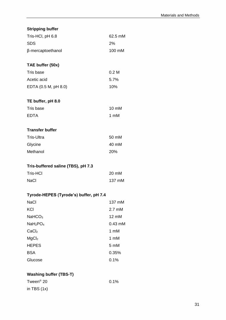
Materials and Methods
31
Stripping buffer
Tris-HCl, pH 6.8 62.5 mM
SDS 2%
β-mercaptoethanol 100 mM
TAE buffer (50x)
Tris base 0.2 M
Acetic acid 5.7%
EDTA (0.5 M, pH 8.0) 10%
TE buffer, pH 8.0
Tris base 10 mM
EDTA 1 mM
Transfer buffer
Tris-Ultra 50 mM
Glycine 40 mM
Methanol 20%
Tris-buffered saline (TBS), pH 7.3
Tris-HCl 20 mM
NaCl 137 mM
Tyrode-HEPES (Tyrode’s) buffer, pH 7.4
NaCl 137 mM
KCl 2.7 mM
NaHCO3 12 mM
NaH2PO4 0.43 mM
CaCl2 1 mM
MgCl2 1 mM
HEPES 5 mM
BSA 0.35%
Glucose 0.1%
Washing buffer (TBS-T)
Tween® 20 0.1%
in TBS (1x)

Materials and Methods
32
3.1.6. Mice
Sykfl/fl,161 RhoAfl/fl,216 Pfn1fl/fl 217 and Trpm7fl/fl 218 mice were intercrossed with mice carrying the
Cre-recombinase under the Pf4 promoter to generate megakaryocyte and platelet-specific
knockout mice.219 SykZap70/Zap70 mice were generated as previously described.220 Flk1-
mCherry221 were kindly provided by Mary E. Dickinson (Houston, Texas). Tie2-Cre/R26-
tdRFP222 were kindly provided by Friedemann Kiefer (Münster, Germany). CD41-YFP223 and
Lys-eGFP224 mice were kindly provided by Thomas Graf (Barcelona, Spain). Thy1-eGFP225
mice were kindly provided by Anna-Lena Sirén (Würzburg, Germany). C57BL/6JRj mice
were obtained from Janvier Labs (Saint-Berthevin, France). All mice used in experiments
were 8 to 12 weeks old and sex-matched, if not stated otherwise. Animal studies were
approved by the district government of Lower Franconia (Bezirksregierung Unterfranken).
3.1.6.1. Generation of fetal liver chimeric and bone marrow chimeric mice
The insertion of Zap-70 cDNA into the first coding exon of Syk resulted in perinatal lethality
of the SykZap70/Zap70 mice and therefore fetal liver chimeric mice and bone marrow chimeric
mice were generated. Recipient C57BL/6JRj mice at 6 weeks of age were lethally irradiated
with 10 Gray. For fetal liver chimeric mice, female mice were time-mated and the livers of
13.5 to 14.5 day old mouse embryos were isolated. For bone marrow chimeric mice, the
femur and tibia of donor mice were prepared and bone marrow was flushed out with a 22G
needle into cold DMEM. A single cell suspension was prepared and cells were counted in a
Neubauer chamber. 4 million cells diluted in 150 l DMEM were intravenously injected into
one recipient mouse. Mice were treated with 2 g/l neomycin in water for the following
6 weeks.
3.2. Methods
3.2.1. Mouse genotyping
3.2.1.1. Mouse genotyping by flow cytometry
CD41-YFP mice were genotyped by flow cytometry. 50 l blood were drawn from the retro-
orbital plexus of anesthetized mice using heparinized capillaries and collected in a tube
containing 300 l heparin in TBS (20 U/ml, pH 7.3). Blood was diluted 1:20 with Ca2+-free
Tyrode’s buffer or 1x PBS and either stained with saturating amounts of anti-IIb-PE
antibodies for 15 minutes (min) at room temperature (RT) to determine surface protein
expression or left unstained to determine the percentage of YFP-positive platelets. The
reaction was stopped by the addition of 500 l 1x PBS and samples were analyzed on a
FACSCalibur (BD Biosciences, Heidelberg, Germany).

Materials and Methods
33
3.2.1.2. Isolation of genomic DNA from mouse tissue
Mouse ear punch samples were lysed in 500 l DNA lysis buffer containing 100 g/ml
proteinase K. The samples were incubated overnight (o/n) at 56 ºC under shaking conditions
(900 rpm, Eppendorf Thermomixer). Phenol/chloroform/isoamyl alcohol mixture (500 l) was
added to each sample and upon vigorous shaking, the samples were centrifuged at
10,000 rpm for 10 min at RT. The upper phase (approximately 440 l) was transferred to
reaction tubes containing 500 l isopropanol and upon vigorous shaking, the samples were
centrifuged at 14,000 rpm for 10 min at 4ºC. The supernatant was discarded and the DNA
pellet was washed with 500 l 70% ethanol and centrifuged again at 14,000 rpm for 10 min
at 4ºC. The DNA pellet was allowed to dry at 37 ºC for 30 min and solubilized in 70 l TE
buffer for 30 min at 37 ºC under stirring conditions. 1-2 l of DNA was used for a polymerase
chain reaction (PCR).
3.2.1.3. Mouse genotyping by PCR
Genotyping was accomplished by PCR using different mixtures of primers and PCR
programs as indicated below.
Detection of the Syk floxed allele by PCR
Primers:
Syk_flox_F 5’ GGT GCC TAC AGG TCT ACA GC 3’
Syk_flox_R 5’ AAC CTG GTA ATT TCA TAA CGC C 3’
Pipetting scheme:
DNA 1 l
Syk_flox_F (1:10 in H2O, stock: 100 pmol/l) 1 l
Syk_flox_R (1:10 in H2O, stock: 100 pmol/l) 1 l dNTPs (10 mM) 1 l 10x Taq buffer (+KCl, -MgCl2) 2.5 l MgCl2 (25 mM) 2.5 l
Taq polymerase (5 U/l) 0.25 l H2O 15.75 l
PCR program:
96ºC 3:00 min 94ºC 0:30 min 56ºC 0:30 min 35 cycles 72ºC 1:00 min 72ºC 10:00 min 4ºC ∞
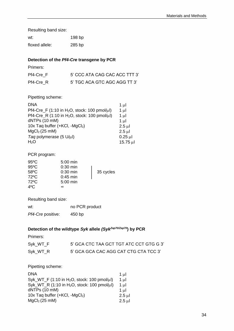
Materials and Methods
34
Resulting band size:
wt: 198 bp
floxed allele: 285 bp
Detection of the Pf4-Cre transgene by PCR
Primers:
Pf4-Cre_F 5’ CCC ATA CAG CAC ACC TTT 3’
Pf4-Cre_R 5’ TGC ACA GTC AGC AGG TT 3’
Pipetting scheme:
DNA 1 l
Pf4-Cre_F (1:10 in H2O, stock: 100 pmol/l) 1 l
Pf4-Cre_R (1:10 in H2O, stock: 100 pmol/l) 1 l dNTPs (10 mM) 1 l 10x Taq buffer (+KCl, -MgCl2) 2.5 l MgCl2 (25 mM) 2.5 l
Taq polymerase (5 U/l) 0.25 l H2O 15.75 l
PCR program:
95ºC 5:00 min 95ºC 0:30 min 58ºC 0:30 min 35 cycles 72ºC 0:45 min 72ºC 5:00 min 4ºC ∞
Resulting band size:
wt: no PCR product
Pf4-Cre positive: 450 bp
Detection of the wildtype Syk allele (SykZap70/Zap70) by PCR
Primers:
Syk_WT_F 5’ GCA CTC TAA GCT TGT ATC CCT GTG G 3’
Syk_WT_R 5’ GCA GCA CAC AGG CAT CTG CTA TCC 3’
Pipetting scheme:
DNA 1 l
Syk_WT_F (1:10 in H2O, stock: 100 pmol/l) 1 l
Syk_WT_R (1:10 in H2O, stock: 100 pmol/l) 1 l dNTPs (10 mM) 1 l 10x Taq buffer (+KCl, -MgCl2) 2.5 l MgCl2 (25 mM) 2.5 l

Materials and Methods
35
Taq polymerase (5 U/l) 0.25 l H2O 15.75 l
PCR program:
95ºC 5:00 min 95ºC 0:30 min 60ºC 0:45 min 30 cycles 72ºC 1:00 min 72ºC 5:00 min 4 ºC ∞
Resulting band size:
wt: 661 bp
Detection of the Zap70 allele (SykZap70/Zap70) by PCR
Primers:
Syk_KI_F 5’ AAA TCG GCA AAA TCC CTT ATA AAT C 3’
Syk_KI_R1 5’ GAA GAA CGA GAT CAG CAG C 3’
Syk_KI_R2 5’ CCT GAT GAG GTT CTC CTT C 3’
Pipetting scheme:
DNA 1 l
Syk_KI_F (1:10 in H2O, stock: 100 pmol/l) 1 l
Syk_KI_R1 (1:10 in H2O, stock: 100 pmol/l) 1 l
Syk_KI_R2 (1:10 in H2O, stock: 100 pmol/l) 1 l dNTPs (10 mM) 1 l 10x Taq buffer (+KCl, -MgCl2) 2.5 l MgCl2 (25 mM) 2.5 l
Taq polymerase (5 U/l) 0.25 l H2O 14.75 l
PCR program:
95ºC 5:00 min 95ºC 0:30 min 60ºC 0:45 min 30 cycles 72ºC 1:00 min 72ºC 5:00 min 4 ºC ∞
Resulting band size:
Zap70: 142 bp
Materials and Methods
36
3.2.1.4. Agarose gel electrophoresis
Agarose (final concentration (f.c.) 1.5%) was dissolved in 1x TAE buffer and boiled in a
microwave. When the temperature of the solution had decreased to approximately 60°C, 5 l
Midori green per 100 l solution were added and the fluid was poured into a tray with a
comb. After solidification of the gel, the tray was positioned in an electrophoresis chamber
containing 1x TAE buffer. 4 l 6x loading dye solution were added to 20 l DNA and the
samples were loaded into the gel slots. For size separation, DNA samples were run for
30 min at 120-160 V and a DNA ladder was used to determine the size of the PCR products.
3.2.2. Blood preparation
3.2.2.1. Plasma samples
Capillaries were anti-coagulated with sodium citrate. 100 l blood were drawn from the retro-
orbital plexus of anesthetized mice and collected in tubes containing 100 l sodium citrate.
Samples were centrifuged at 2800 rpm for 5 min at RT and the supernatant was again
centrifuged at 2800 rpm for 5 min at RT. Platelet-poor plasma (PPP) was spun down at
14,000 rpm for 5 min at RT and supernatants were immediately used for experiments or
frozen at -20 °C.
3.2.2.2. Washed blood
50 l blood were drawn from the retro-orbital plexus of anesthetized mice using heparinized
capillaries and collected in a tube containing 300 l heparin in TBS (20 U/ml, pH 7.3). Ca2+-
free Tyrode’s buffer (650 l) was added to each sample and blood was centrifuged twice at
2800 rpm for 5 min at RT and resuspended in Ca2+-free Tyrode’s buffer. Finally, samples
were resuspended in 750 l Tyrode’s buffer with 2 mM Ca2+.
3.2.2.3. Washed platelets
Mice were bled under isoflurane anesthesia from the retro-orbital plexus with heparin-coated
capillaries and 700 l blood were collected in a reaction tube containing 300 l heparin in
TBS (20 U/ml, pH 7.3). Blood was centrifuged at 800 rpm for 7 min at RT (Eppendorf
Centrifuge 5415 C). Supernatant and buffy coat were transferred into a new tube and again
centrifuged at 800 rpm for 7 min at RT to obtain platelet-rich plasma (PRP). To prepare
washed platelets, PRP was centrifuged at 2800 rpm for 5 min at RT and the platelet pellet
was resuspended in 1 ml Ca2+-free Tyrode’s buffer containing PGI2 (0.1 g/ml) and apyrase
(0.02 U/ml) and incubated for 10 min at 37°C. This washing step was repeated and the

Materials and Methods
37
platelet count was determined using a Sysmex KX-21N automated hematology analyzer
(Sysmex Corporation, Kobe, Japan). After centrifugation, the pellet was resuspended in
Ca2+-free Tyrode’s buffer containing apyrase (0.02 U/ml) to obtain the desired platelet
concentration. Washed platelets were incubated for 30 min at 37°C prior to analysis.
3.2.2.4. Platelet lysates
Washed platelets (1 x 10⁶/l) were lysed in lysis buffer containing 1% IGEPAL CA-360 and
protease inhibitors for 20 min on ice. The samples were then centrifuged at 14,000 rpm for
10 min at 4°C and the supernatant was used for experiments.
3.2.3. Compound preparation
3.2.3.1. BI1002494
For in vitro assays, BI1002494 (C23H24N3O5)226, kindly provided by Boehringer Ingelheim,
was dissolved in DMSO and final DMSO concentration in the assays was less than 0.1%.
The washed platelets were incubated at 37°C with BI1002494 10 min prior to the experiment.
For animal experiments, BI1002494 was dissolved in water and applied by oral gavage
15 hours (h) and 1 h prior to blood taking (ex vivo aggregation experiments) or prior to the in
vivo experiments (100 mg/kg b.i.d.). To assess the therapeutic function of the Syk inhibitor,
BI1002494 was dissolved in water and applied by oral gavage directly after tMCAO (100
mg/kg), i.e. directly upon removal of the filament.
3.2.4. Biochemistry
3.2.4.1. Western blotting
4x Laemmli SDS sample buffer (with -mercaptoethanol for reducing conditions) was added
to platelet lysates and samples were boiled for 5 min at 95°C. Proteins were separated by
sodium dodecyl sulfate polyacrylamide gel electrophoresis (SDS-PAGE) on a 12%
separating gel with a 4% stacking gel. Samples were loaded onto the gel including a
molecular weight marker, run in Laemmli running buffer and transferred onto a polyvinylidene
difluoride (PVDF) membrane for 1 h using a semi-dry transfer system. To prevent unspecific
antibody binding, membranes were blocked in 5% fat-free dry milk or 5% BSA dissolved in
washing buffer for 2 h at RT or o/n at 4°C. Membranes were then incubated with primary
antibodies o/n at 4°C. Membranes were washed with washing buffer 3x 15 min at RT and
incubated with HRP-conjugated secondary antibodies for 1 h at RT. After three washing
steps, membranes were developed using ECL detection substrate with autoradiography
films. To re-probe a membrane, blots were incubated in stripping buffer for 30 min at RT,

Materials and Methods
38
washed thoroughly in washing buffer, blocked and subsequently probed with antibodies.
PLC2 levels were used as loading control.
3.2.5. In vitro analyses of platelet function
3.2.5.1. Determination of blood parameters
To determine blood parameters, 50 l blood were drawn from the retro-orbital plexus of
anesthetized mice using heparinized capillaries and collected into a tube containing 300 l
heparin in TBS (20 U/ml, pH 7.3). Blood cell counts and parameters were determined using a
Sysmex KX-21N automated hematology analyzer (Sysmex Corporation, Kobe, Japan).
3.2.5.2. Determination of platelet count, size and surface protein expression
To measure platelet count, size and surface protein expression, 50 l blood were drawn from
the retro-orbital plexus of anesthetized mice using heparinized capillaries and collected in a
tube containing 300 l heparin in TBS (20 U/ml, pH 7.3). Blood was diluted 1:20 with Ca2+-
free Tyrode’s buffer or 1x PBS and stained with saturating amounts of fluorophore-
conjugated antibodies for 15 min at RT. The reaction was stopped by the addition of 500 l
1x PBS and samples were analyzed on a FACSCalibur (BD Biosciences, Heidelberg,
Germany).
3.2.5.3. Determination of platelet activation responses
To measure platelet activation responses, 50 l washed blood were activated with the
respective agonist (10-fold concentrated) in the presence of saturating amounts of a 1:1
mixture of PE-coupled 4H5 (anti-activated IIb3) and FITC-coupled 5C8 (anti-P-selectin)
antibodies. Samples were incubated for 7 min at 37 ºC and additionally for 7 min at RT. The
reaction was stopped by the addition of 500 l 1x PBS and samples were analyzed on a
FACSCalibur (BD Biosciences, Heidelberg, Germany).
3.2.5.4. Aggregometry
Washed platelets (50 l with 5 x 10⁵ platelets/l) were diluted into 110 l Tyrode’s buffer
containing 2 mM Ca2+ and 100 g/ml human fibrinogen. Alternatively, heparinized PRP was
diluted into Tyrode’s buffer containing 2 mM Ca2+ and used for investigation of aggregation.
Agonists were added at indicated concentrations to the continuously stirring (1000 rpm)
platelet suspension. Light transmission was recorded on a Fibrintimer 4-channel
aggregometer (APACT Laborgeräte und Analysensysteme, DiaSys Deutschland, Flacht,

Materials and Methods
39
Germany) for 10 min and expressed in arbitrary units with buffer representing 100%
transmission and washed platelet suspension or PRP representing 0% transmission. For
platelet activation using thrombin, washed platelets were diluted in fibrinogen-free Tyrode’s
buffer containing 2 mM Ca2+.
3.2.5.5. Platelet adhesion on fibrinogen under static conditions
Glass coverslips (24 x 60 mm) were coated with 100 g/ml human fibrinogen diluted in sterile
1x PBS o/n at 4 °C under humid conditions. Coverslips were blocked with sterile filtered
1% BSA in PBS for 2 h at RT and rinsed with Tyrode’s buffer containing 2 mM Ca2+. Washed
platelets (50 l with 3 x 10⁵ platelets/l for early time points and 30 l with 3 x 10⁵ platelets/l
for late time points) were left unstimulated or were stimulated with thrombin (0.01 U/ml f.c.)
and/or second wave inhibitors (indomethacin (1.4 M f.c.) and apyrase (2 U/ml f.c.)) and
were allowed to spread for the indicated time points at RT. Platelets were fixed with 4% PFA
in PBS for 5 min at RT and visualized with a Zeiss Axiovert 200 inverted microscope (100x
oil objective, Zeiss, Oberkochen, Germany) using differential interference contrast (DIC)
microscopy. Representative images were taken and evaluated according to different platelet
spreading stages using ImageJ software (NIH, Bethesda, MD, USA).
3.2.5.6. Clot retraction
700 l blood were drawn from the retro-orbital plexus of anesthetized mice using non-
heparinized capillaries and collected in a tube containing 70 l sodium citrate. Platelets were
washed and PPP and erythrocytes were collected. The platelet concentration in the
suspension was adjusted to 3 x 10⁵ platelets/l using PPP. 1.5 l of erythrocytes (to contrast
the clot) and 20 mM CaCl2 were added to 250 l of the platelet suspension. To induce clot
retraction, the samples were stimulated with 4 U/ml thrombin. Clot retraction was monitored
at 37°C under non-stirring conditions. Images were taken every 15 min and after 4 h the
residual fluid was measured.
3.2.5.7. Platelet adhesion on collagen under flow conditions
Glass coverslips (24 x 60 mm) were coated with 200 g/ml fibrillar type I collagen (Horm) o/n
at 37 °C and blocked with sterile filtered 1% BSA in PBS for 1 h at RT. They were rinsed with
Tyrode’s buffer containing 2 mM Ca2+ and gently fixed in a transparent flow chamber with a
slit depth of 50 m. Heparinized whole blood (700 l blood in 300 l heparin in TBS) was
diluted with Tyrode’s buffer containing 2 mM Ca2+ (800 l heparinized blood and 400 l
buffer). Platelets were labeled with a DyLight 488 conjugated anti-GPIX antibody derivative

Materials and Methods
40
(0.2 g/ml) for 5 min at 37 °C and blood was filled into a 1 ml syringe, which was connected
to the flow chamber. Perfusion was accomplished using a pulse-free pump under high shear
stress equivalent to a wall shear rate of 1000 s-1 for 4 min. Microscopic phase-contrast
images were recorded in real-time during perfusion. Thereafter, coverslips were washed by a
2 min perfusion with Tyrode’s buffer containing 2 mM Ca2+ at the same shear rate. Phase-
contrast and fluorescence images were obtained from at least seven different collagen-
containing microscopic fields for each sample using a Zeiss Axiovert 200 inverted
microscope (40x objective, Zeiss Oberkochen, Germany) and analyzed offline using Metavue
software (Visitron, Puchheim, Germany).
3.2.6. In vivo analyses of platelet function
3.2.6.1. Platelet depletion
Thrombocytopenia was induced by intravenous injection of rat anti-GPIbα (anti-CD42b)
antibodies (2.0 g/g body weight). Peripheral platelet counts were determined directly before
the experiments using a Sysmex KX-21N automated hematology analyzer (Sysmex
Corporation, Kobe, Japan) or by flow cytometry.
3.2.6.2. Tail bleeding time
Mice were anesthetized by intraperitoneal injection of medetomidine (0.5 g/g), midazolam
(5 g/g) and fentanyl (0.05 g/g) and 1 mm of the tail tip was removed using a scalpel. Tail
bleeding was monitored by gently absorbing blood on filter paper at 20 s intervals without
interfering with the wound site. Bleeding was determined to have ceased when no blood was
observed on the paper. Experiments were stopped manually after 20 min by cauterization to
prevent excessive blood loss and anesthesia was antagonized by intraperitoneal injection of
atipamezole (2.5 g/g), flumazenil (0.05 g/g) and naloxone (1.2 g/g).
3.2.6.3. Thrombus formation in FeCl3-injured mesenteric arterioles
Mice (4 to 5 weeks old, 15–18 g) were anesthetized by intraperitoneal injection of
medetomidine (0.5 g/g), midazolam (5 g/g) and fentanyl (0.05 g/g) and were
intravenously injected with 1.5 g of a DyLight 488-conjugated anti-GPIX antibody derivative
to label platelets in vivo. The mesentery was then gently exteriorized through a midline
abdominal incision. Mesenterial arterioles with a diameter between 35-60 m were visualized
with a Zeiss Axiovert 200 inverted microscope (10x objective, Zeiss, Oberkochen, Germany).
The microscope was equipped with a 100 W HBO fluorescent lamp and a CoolSNAP-EZ

Materials and Methods
41
camera. Injury was induced by topical application of a 3 mm2 filter paper saturated with 20%
FeCl3. Adhesion and aggregation of fluorescently labeled platelets in mesenterial arterioles
was monitored until complete vessel occlusion occurred for at least 1 min or for a maximum
of 40 min. Digital images were recorded and analyzed offline using Metavue software
(Visitron, Puchheim, Germany). Experiments were conducted by Ina Thielmann.
3.2.6.4. Mechanical injury of the abdominal aorta
Mice (8 to 12 weeks old) were anesthetized by intraperitoneal injection of medetomidine
(0.5 g/g), midazolam (5 g/g) and fentanyl (0.05 g/g) and a longitudinal incision was used
to open the abdominal cavity to expose the abdominal aorta. An ultrasonic flow probe
(0.5PSB699; Transonic Systems, New York, USA) was placed around the abdominal aorta
and thrombus formation was induced by a single firm compression with forceps. Blood flow
was monitored until complete blood vessel occlusion occurred for at least 5 min or for a
maximum of 30 min. Experiments were conducted by Ina Thielmann and Sarah Beck.
3.2.6.5. Transient middle cerebral artery occlusion (tMCAO)
Focal cerebral ischemia was induced in 8 to 12 week old mice by a transient middle cerebral
artery occlusion (tMCAO) as previously described.227 Inhalation anesthesia was induced by
2% isoflurane in a 70% N2/30% O2 mixture and a servo-controlled heating device was used
to record and maintain body temperature during the surgical procedure. The duration of the
surgical procedure per animal was kept below 15 min. A silicon rubber–coated 6.0 nylon
monofilament (6021PK10, Doccol, Redlands, CA, USA) was advanced through the right
common carotid artery and the internal carotid artery up to the origin of the middle cerebral
artery (MCA) causing an MCA occlusion. After an occlusion time of 60 min, the filament was
removed allowing reperfusion. Animals were sacrificed 24 h after reperfusion and brains
were checked for intracerebral hemorrhages. The extent of the infarction was quantitatively
assessed on 2,3,5-triphenyltetrazolium chloride (TTC, 2% (w/v) solution) stained brain
sections. Global neurological outcome and motor function were evaluated by the Bederson
score228 and the grip test,229 respectively. Planimetric measurements of infarcted areas
(ImageJ software, NIH, Bethesda, MD, USA) corrected for brain edema and assessment of
functional outcome were performed in a blinded fashion. Experiments were carried out by
Dr. Peter Kraft from the Department of Neurology, University of Würzburg according to the
recommendations in mechanism-driven basic stroke research.230
Materials and Methods
42
3.2.6.6. Multiphoton intravital microscopy of the brain vasculature
Mice (8 to 12 weeks old) were anesthetized by intraperitoneal injection of medetomidine
(0.5 g/g), midazolam (5 g/g) and fentanyl (0.05 g/g). A 1-cm incision was made along the
midline to expose the frontoparietal skull. The external layer of the compact bone and most
of the spongy bone were removed from a small part of the skull using a high-speed microdrill,
creating a thinned skull window. The scalp was sutured and the mouse was left to recover for
at least 24 h. On the day of imaging, the mouse was anesthetized and subsequently placed
on a customized metal stage equipped with a stereotactic holder to immobilize its head.
Images were acquired with a fluorescence microscope equipped with a 20x water objective
with a numerical aperture of 0.95 and a TriM Scope II multiphoton system (LaVision BioTec),
controlled by ImSpector Pro-V380 software (LaVision BioTec). Emission was detected with
HQ535/50-nm and ET605/70-nm filters. A tunable broad-band Ti:Sa laser (Chameleon,
Coherent) was used at 760 nm to capture Alexa Fluor 488 and tetramethylrhodamine dextran
fluorescence. ImageJ software (NIH) was used to generate movies.
3.2.7. Megakaryocyte analyses
3.2.7.1. Immunofluorescence staining of whole femora cryo-sections
Femora and sterna of mice (6 to 12 weeks old) were isolated and fixed in 4% PFA for 2-4 h
at 4°C and subsequently transferred into 10% sucrose/PBS and dehydrated using a graded
sucrose series (incubation in 10% sucrose/PBS, followed by 20% sucrose/PBS and 30%
sucrose/PBS for 24 h at 4°C). Samples were either embedded in Cryo-Gel, frozen and cryo-
sections were produced using a CryoJane tape transfer system (Leica Biosystems, Wetzlar,
Germany) or embedded in Tissue-Tek medium, snapfrozen in liquid nitrogen and cryo-
sections were produced as described previously.231 MKs were stained with anti-CD41-FITC
antibodies, anti-1-tubulin antibodies or anti-GPIX antibody derivatives. CD105 was used as
an endothelial cell marker. Nuclei were stained with Hoechst 33258 or DAPI-containing
mounting medium. Staining with antibodies and secondary reagents was performed for 30
min at RT. Sections were analyzed by confocal microscopy (Leica TCS-SP5, Leica
Microsystems, Wetzlar, Germany) and processed with Leica LAS AF software and ImageJ
software (NIH). MK number and density was measured in every visual field. Three femur
sections from three separate animals were examined and 20 MKs per section were
quantified with regards to area and shortest distance to sinusoids.

Materials and Methods
43
3.2.7.2. Multiphoton intravital microscopy of the bone marrow
Mice (8 to 12 weeks old) were anesthetized by intraperitoneal injection of medetomidine
(0.5 g/g), midazolam (5 g/g) and fentanyl (0.05 g/g). A 1-cm incision was made along the
midline to expose the frontoparietal skull, while carefully avoiding damage to the bone tissue.
The mouse was placed on a customized metal stage equipped with a stereotactic holder to
immobilize its head. BM vasculature was visualized by injection of tetramethylrhodamine
dextran (8 g/g body weight) and anti-CD105 Alexa Fluor 546/Alexa Fluor 594 (0.4 g/g
body weight). Platelets and MKs were antibody stained with anti-GPIX (CD42a) Alexa Fluor
488 (0.6 g/g body weight). Images were acquired with a fluorescence microscope equipped
with a 20x water objective with a numerical aperture of 0.95 and a TriM Scope II multiphoton
system (LaVision BioTec), controlled by ImSpector Pro-V380 software (LaVision BioTec).
Emission was detected with HQ535/50-nm and ET605/70-nm filters. A tunable broad-band
Ti:Sa laser (Chameleon, Coherent) was used at 760 nm to capture Alexa Fluor 488 and
Alexa Fluor 546/Alexa Fluor 594/tetramethylrhodamine dextran fluorescence. ImageJ
software (NIH) was used to generate movies.
3.2.7.3. Light-sheet fluorescence microscopy of the bone marrow
Platelets were antibody stained with an anti-GPIX Alexa Fluor 750 antibody derivative
(0.6 g/g body weight). BM vasculature was stained with an anti-CD105 Alexa Fluor 647
antibody (0.4 g/g body weight). After 25 min, the mice were sacrificed and transcardially
perfused with ice-cold PBS to wash out the blood and ice-cold 4% PFA to fix the tissues.
Sterna and femora were harvested and stored in 4% PFA for 30 min. The samples were
washed in PBS and decalcified in 10% EDTA for 96 h at 4°C on a shaker. The samples were
then washed in PBS, followed by dehydration in a graded methanol series (50%, 70%, 95%,
100% for 30 min each) at RT and stored at 4°C overnight. The methanol was replaced
stepwise by a clearing solution consisting of 1 part benzyl alcohol in 2 parts benzyl benzoate
(BABB). After incubation in the clearing solution for at least 2h at RT, tissue specimens
became optically transparent and were used for LSFM imaging on the following day.
3.2.7.4. Image analysis of the light-sheet fluorescence microscopy stacks
Multicolor LSFM stacks were processed and analyzed by FIJI232 and Bitplane Imaris® 7.7.2
(Bitplane AG, Zurich, Switzerland). Data visualization and analysis was performed in three
major steps: (i) image preprocessing, (ii) segmentation, (iii) data extraction. Step (i) was
performed using either Imaris or FIJI, steps (ii) and (iii) were exclusively performed using
Bitplane Imaris® 7.7.2, particularly the ImarisCell module. Statistical analysis of object
positions, numbers and volumes (MK, vessel, bone marrow), edge-to-edge distance MK to

Materials and Methods
44
vessel and available vessel interspace were performed using Microsoft Excel (ver. 2013,
Microsoft Corporation, Redmond, WA, USA), Origin (ver. 8.6, OriginLab Corporation,
Northampton, MA, USA) and SPSS statistics (ver.23, IBM, Ehningen, Germany). Multi-
photon microscopy images of 37 MKs were corrected for noise and converted to binary
images using FIJI. Cell tracking of binary images was performed with the active contours
module of Icy software (www.icy.bioimageanalysis.org, Institut Pasteur, France). Cell
trajectories were analyzed using Matlab (Mathworks, USA) and msdanalyzer class.233 Image
processing was performed by Oğuzhan Angay and Patrick Schmithausen.
3.2.7.5. Computer simulations
Simulations were performed with a self-written algorithm in Matlab software (Mathworks,
USA) using volume image stacks of mouse sterna acquired by LSFM and processed with
Bitplane Imaris® 7.7.2 software (see above). Thus, the modeled MKs and vasculature
perfectly reflected the spatial physiological situation. For random simulation (SimR), MKs
were randomly placed in the 3D intervascular space. For vessel associated random
simulation (SimVA), 60% of MKs from a pool of 30 different MKs were randomly placed at
the vessels with minimal cross-volume of 1 voxel. Subsequently, the remaining MKs were
placed into the intervascular space without vessel contact. The MK-vessel distances were
calculated by using vessel distance transformation maps. For both types of simulations, the
number and size distribution of MKs in the pool were matched to the experimentally
estimated values. The presented results are mean ± standard deviation (SD) from at least
five independent experiments per group, if not stated otherwise. Computational modeling
was performed by Mari Gorelashvili.
3.2.8. Statistical analyses
Results are shown as mean ± SD from three individual experiments per group, unless
indicated otherwise. Statistical analysis was conducted using the Student’s t-test, apart from
the Fischer’s exact test which was applied to assess variance between non-occluded vessels
in models of arterial thrombosis or in tail bleeding assays and the Mann-Whitney U test which
was used for analysis of Bederson score and grip test after tMCAO. Differences between
more than two groups were analyzed with one-way ANOVA with Dunnett’s T3 as post-hoc
test using SPSS 21. P-values <0.05 were considered statistically significant with: not
significant (ns), p>0.05; *, p<0.05; **, p<0.01; ***, p<0.001.

Results
45
4. Results
4.1. Multiphoton intravital microscopic analyses of thrombopoiesis and
proplatelet formation
Recent developments in imaging methods allow the observation of dynamic cellular events in
vivo using MP-IVM.57,59 Megakaryopoiesis and thrombopoiesis have been studied intensively,
but the exact mechanisms that control platelet generation from MKs remain poorly
understood, mainly due to the inaccessibility of MKs in their natural BM environment.
Therefore, MP-IVM is a helpful tool to study thrombopoiesis and proplatelet formation in vivo.
4.1.1. Establishment of multiphoton intravital microscopy of the murine bone marrow
and brain
The flat bones forming the calvaria of the murine skull contain BM, which is macroscopically
visible. Imaging of the BM in the frontoparietal skull without surgical manipulation of the bone
is achievable, since the thin layer of bone covering the BM cavities is sufficiently
transparent.196 In order to study thrombopoiesis by MP-IVM, an in vivo imaging protocol had
to be established first. Crucial steps in the establishment of the MP-IVM protocol involved the
immobilization of the mouse, thinning of the skull for transcranial imaging, characterization of
fluorescent markers and reporter mice and visualization of dynamic processes using one
laser.
For the in vivo imaging set-up, several methods were tested, including rings from agarose
and plastic to spread the skin of the skull in combination with a stereotactic holder,234 but the
metal plate skull holder provided the best result in terms of stable fixation. The mouse was
placed on a customized plate equipped with a skull holder to immobilize its head, thereby
allowing imaging of the BM without movement artefacts. The skull holder was fixed onto the
mouse’s skull and the region of interest was exposed by a circular hole in the metal plate, the
imaging window (Figure 8).
To visualize the cortical blood vessels and the mouse brain in vivo, a thinned skull cranial
window was used. This transcranial imaging method allows the visualization of fluorescently
labeled blood vessels, neurons, microglia and astrocytes.234 The procedure involves thinning
of the skull by removing the external layer of the compact bone and the spongy bone, leading
to an optically transparent cranium.

Results
46
Figure 8. In vivo imaging set-up to study thrombopoiesis by multiphoton intravital microscopy. For in vivo visualization of the bone marrow (BM) or the brain by multiphoton intravital microscopy (MP-IVM, left panel), the mouse was placed on a customized plate equipped with a skull holder to immobilize its head (middle panel). The skull holder was fixed onto the mouse’s skull and the region of interest was exposed by the imaging window (right panel).
Figure 9. Characterization of fluorescent markers and reporter mice. (A) Staining of the endothelium in the brain of Flk1-mCherry+/- mice (red). (B) Tetramethylrhodamine dextran marks the plasma of the meningeal blood vessels (red). (C) Transcranial imaging of the Tie2-Cre/R26-tdRFP+/-
mice showed a strong staining of the endothelial lining (red), whereas FITC-BSA provided intraluminal staining (green). (D) Macrophages and neutrophil granulocytes (green) are clearly visible in Lys-eGFP+/ - mice. Blood vessels were stained with tetramethylrhodamine dextran (red). (E) Thy1-eGFP+/- mice are used to visualize neurons ex vivo (yellow) in a brain slice or (F) in vivo (green).
Blood vessels were stained with tetramethylrhodamine dextran (red). Scale bar 50 m.

Results
47
Upon mouse preparation, several plasma and endothelial markers were tested to visualize
and characterize the vascular anatomy in the BM and the brain. Tetramethylrhodamine
dextran and FITC-BSA provided an intense intraluminal staining and only minimal
extravasation of the substances was observed (Figure 9B,C,D,F). An anti-endoglin antibody
was used for staining of the endothelium (Figure 12, Video 1). In addition, a number of
reporter mouse lines were analyzed. Reporter mice are transgenic mice, that express
fluorescent proteins under the control of a specific promoter to ensure that only the desired
cell type expresses the fluorescent protein. They can be used for visualizing these specific
cells in vivo.235 The Flk1-mCherry+/- 221 and Tie2-Cre/R26-tdRFP+/- 222 mice showed a strong
staining of the endothelial lining (Figure 9A,C). Besides reporter mice for endothelial cells,
reporters for macrophages and neutrophil/granulocytes (Lys-eGFP+/- 224), neurons (Thy1-
eGFP+/- 225) and MKs and platelets (CD41-YFP 223) were analyzed using MP-IVM
(Figure 9D-F, Figure 11A).
Figure 10. A subset of platelets is YFP-positive in CD41-YFP mice. The percentage of YFP-positive platelets in whole blood was measured by flow cytometry in CD41-YFPwt/ki and CD41-YFPki/ki mice. Each symbol represents one animal. The lines represent the median value.
The CD41-YFP mouse line was developed to allow in vivo imaging of megakaryopoiesis.223
The mice expressing yellow fluorescent protein (YFP) in MKs and platelets specifically were
generated by insertion of the gene encoding YFP (eyfp) by homologous recombination into
embryonic stem cells at the transcriptional start site of the GpIIb locus. To analyze the YFP
expression in platelets, the percentage of YFP-positive platelets in whole blood was
measured by flow cytometry (Figure 10). In both CD41-YFPwt /ki and CD41-YFPki/ki mice, only
a subset of the platelets was YFP-positive, confirming previous data by Zhang et al..223 Large
variations between individual animals were observed and the proportion of YFP-labeled
platelets was gender-dependent. The male CD41-YFPwt/ki mice had a median value of 30%
YFP-positive platelets, whereas the female heterozygous mice only contained 17% YFP-
positive platelets. The lower percentages of YFP-positive platelets in female mice were also
found in the CD41-YFPki/ki mice, although the proportion of YFP-labeled platelets was higher

Results
48
compared to the CD41-YFPwt/ki mice with 48% and 28% in male and female mice,
respectively. The YFP-positive platelets from the CD41-YFPwt/ki mice did not have a reduced
life span (data not shown). In the CD41-YFPki/ki mice, a bleeding phenotype was observed,
probably due to the lack of the IIb3 receptor complex. YFP-positive platelets were also
detected in vivo by MP-IVM of the BM (Figure 11A). Since only a subset of the MKs and
platelets was YFP-labeled, Thpo treatment or platelet depletion were required to enhance
megakaryopoiesis and thereby increase the number of labeled MKs within the BM. To
improve cell labeling without the necessity to use Thpo treatment or platelet depletion, other
staining methods using fluorophore-conjugated antibodies were tested. In vivo labeling
targeting the MK- and platelet-specific vWF receptor subunit CD42a (GPIX), and not CD41
(GPIIb) as anti-CD41 antibodies cause thrombocytopenia210, was used to stain the entire MK
population in the BM. The optimal staining was achieved using Alexa 488-labeled anti-GPIX
antibody derivatives, leading to the visualization of MKs in the BM and single platelets within
the circulation in vivo (Figure 11B,C). In addition, using the Alexa 488-labeled anti-GPIX
antibody derivatives, platelet thrombus formation in the meningeal vessels in vivo could also
be observed (Figure 11D).
Figure 11. MP-IVM of megakaryocytes within the bone marrow and of platelets in the circulation. (A) YFP-positive platelet aggregates (green) in CD41-YFPwt/ki mice were detected in vivo. (B) Megakaryocytes (MK, green) were labeled using Alexa 488-labeled anti-GPIX antibody derivatives. (C) Single platelets (anti-GPIX, green) were visualized in the cortical blood vessels. (D) 3D reconstruction of a thrombus (anti-GPIX, green) in a meningeal vessel. Blood vessels were stained
with tetramethylrhodamine dextran (red). Scale bar 50 m.

Results
49
Since the MP-IVM was equipped with one Ti:Sa laser and dynamic cellular processes were
to be observed, a fixed excitation wavelength needed to be used to excite different
fluorescent molecules at the same time to obtain dual-color fluorescence.236 Several
combinations of fluorescent proteins and reporter mice were tested to achieve optimal
imaging of different cell types while using one excitation wavelength. For the visualization of
MKs/platelets and blood vessels at the same time, a combination of anti-GPIX-Alexa 488
antibody derivatives and tetramethylrhodamine dextran provided the best result. In addition,
to stain the endothelial lining, anti-endoglin-Alexa 594 was used. Consequently, an imaging
method was established to monitor the dynamic process of proplatelet formation in blood
vessels within the BM of the murine skull in real-time and in vivo. MKs extending proplatelet-
like protrusions into the microvessels of the BM, which were sheared off in the blood stream,
were identified in vivo (Figure 12, Video 1).
Figure 12. In vivo proplatelet formation in the murine bone marrow. Proplatelet formation by MKs can be observed by MP-IVM. The MKs/platelets were stained using anti-GPIX-Alexa 488 antibody derivatives (green). The blood vessels were labeled with tetramethylrhodamine dextran (lumen, red) and anti-endoglin-Alexa 594 (endothelium, red). The white dashed lines represent the outline of the
blood vessels. Scale bar 50 m.

Results
50
4.1.2. The role of Profilin1, TRPM7 and RhoA in thrombopoiesis in vivo
Platelets are constantly produced by their BM precursors in vivo and MP-IVM allows real-
time visualization of proplatelet formation in the BM of living animals. However, the
mechanisms that control platelet generation and the release from BM MKs remain poorly
understood. It is known that proplatelet formation requires substantial cytoskeletal
rearrangements. Therefore, the role of three proteins involved in these cytoskeletal
rearrangements, Profilin1, TRPM7 and RhoA, was studied in thrombopoiesis in vivo.
4.1.2.1. Profilin1-deficiency leads to premature platelet release into the bone marrow
Wiskott–Aldrich syndrome (WAS) is a X-linked disease caused by mutations in the WAS
gene and is characterized by immunodeficiency, eczema and microthrombocytopenia.237
Platelets from WAS patients display defective microtubule reorganization.217 Profilin1 is a key
regulator of actin turnover, but also interacts with other proteins, including the WAS protein
(WASp)/WASp-interacting protein (WIP) complex. MK and platelet-specific Profilin1-
deficiency mimics the MK and platelet phenotype of WAS patients.217 MP-IVM of
Pfn1fl/fl, Pf4-cre+/- (Pfn1-/-) mice revealed that the majority of the Pfn1-/- MKs released platelets
prematurely into the BM compartment next to the sinusoids (arrowheads in Figure 13B,
Video 3). The premature platelet release was, however, not caused by defective proplatelet
formation, as proplatelets were observed in both Wt and Pfn1-/- mice (indicated by arrows in
Figure 13A,B, Video 2,3). These results suggest that the microthrombocytopenia in Pfn1-/-
mice is caused by, besides accelerated platelet clearing by macrophages217, the premature
platelet release into the BM, revealing Profilin1 as a key regulator of thrombopoiesis.
Figure 13. Premature platelet release into the bone marrow in Pfn1-/- mice. (A,B) MP-IVM of BM MKs in the murine skull revealed unaltered proplatelet formation into the BM sinusoids (arrows), but also prematurely released platelets in Pfn1-deficient mice (arrowheads). MKs and platelets were stained with anti-GPIX-Alexa 488 (green) and blood vessels were stained with tetramethylrhodamine
dextran (lumen, red) and anti-endoglin-Alexa 594 (endothelium, red). Scale bar 25 m. (Bender, Stritt et al., Nat Commun. 2014)217

Results
51
4.1.2.2. TRPM7-deficiency causes impaired proplatelet formation
Mg2+ plays a pivotal role in platelet function. The TRPM7 channel is a ubiquitously
expressed, constitutively active ion channel, which is highly selective for divalent cations,
such as Mg2+, Ca2+ and Zn2+. TRPM7 plays an essential role in Mg2+ homeostasis, cell
motility, proliferation and differentiation and is critical for early embryonic development.77 A
human genetic variant in TRPM7 caused macrothrombocytopenia.238 This phenotype was
strongly resembled by the Trpm7fl/fl, Pf4-cre+/- (Trpm7-/-) mice, where impaired channel function
of TRPM7 in MKs also resulted in macrothrombocytopenia. Trpm7-/- mice were analyzed by
MP-IVM and decreased proplatelet formation in vivo was observed compared to Wt mice
(Figure 14A, Video 4). Furthermore, MKs in Trpm7-/- mice formed short and bulky proplatelet
protrusions that remained attached to the MK cell body during the observation period
(arrowhead in Figure 14B, Video 5). The impaired proplatelet formation was mainly caused
by cytoskeletal alterations due to increased actomyosin contractility and could be rescued by
either Mg2+ supplementation or chemical inhibition of NMMIIA activity.238 These findings
reveal that the regulation of Mg2+ homeostasis in MKs by TRPM7 plays a critical role in
thrombopoiesis.
Figure 14. Impaired proplatelet formation in the bone marrow of Trpm7-/- mice. (A) Normal proplatelet formation by MKs in the BM of Wt mice was observed by MP-IVM (arrow). (B) MKs in Trpm7-/- mice formed short and bulky proplatelet protrusions in vivo (arrowhead). MKs and platelets were stained with anti-GPIX-Alexa 488 (green) and blood vessels were stained with tetramethylrhodamine dextran (lumen, red) and anti-endoglin-Alexa 594 (endothelium, red). Scale bar
50 m. (Stritt et al., Nat Commun. 2016)238

Results
52
4.1.2.3. RhoA-deficiency induces megakaryocyte mislocalization and transmigration
Small GTPases of the Rho family are known to be critical regulators of cytoskeletal
rearrangements in platelets.108,109 Studies in MK and platelet-specific RhoA-deficient
(RhoAfl/fl, Pf4-cre+/- (RhoA-/-)) mice showed that RhoA signaling is essential for platelet shape
change and the observed macrothrombocytopenia suggests that RhoA is necessary for
platelet production.111 In cryo-sections of the femoral BM of RhoA-/- mice, a mislocalization of
the MKs was observed and on average 30% of the MKs were detected inside the BM
sinusoids. The remaining MKs were mostly found in direct contact with the BM sinusoids
(Dütting et al., unpublished). MP-IVM was used for dynamic visualization of MK localization
and proplatelet formation in the BM of RhoA-/- mice (Figure 15, Video 6,7). MKs in Wt mice
were found near the sinusoids or in close sinusoidal contact and released long proplatelets
into the blood vessel (arrows in Figure 15A, Video 6). In sharp contrast, MKs in RhoA-/- mice
were able to completely transmigrate from the BM into the sinusoidal vascular bed
(arrowheads in Figure 15B, Video 7) or were stably present inside the BM sinusoid for up to
50 min (star in Figure 15B). These observations indicate that RhoA negatively regulates MK
localization towards BM vascular sinusoids and acts as a central regulator of thrombopoiesis
by inhibiting MK transmigration.
Figure 15. Megakaryocyte mislocalization and transmigration in RhoA-/- mice. (A) MP-IVM of BM MKs in the murine skull of Wt mice revealed unaltered proplatelet formation into the BM sinusoids (arrows). (B) MKs in RhoA-/- mice transmigrated into the sinusoidal vascular bed (arrowheads) or adhered to the vessel wall inside the BM sinusoid (star). MKs and platelets were stained with anti-GPIX-Alexa 488 (green) and blood vessels were stained with tetramethylrhodamine dextran (red). The
white dashed lines represent the outline of the blood vessels. Scale bar 25 m.
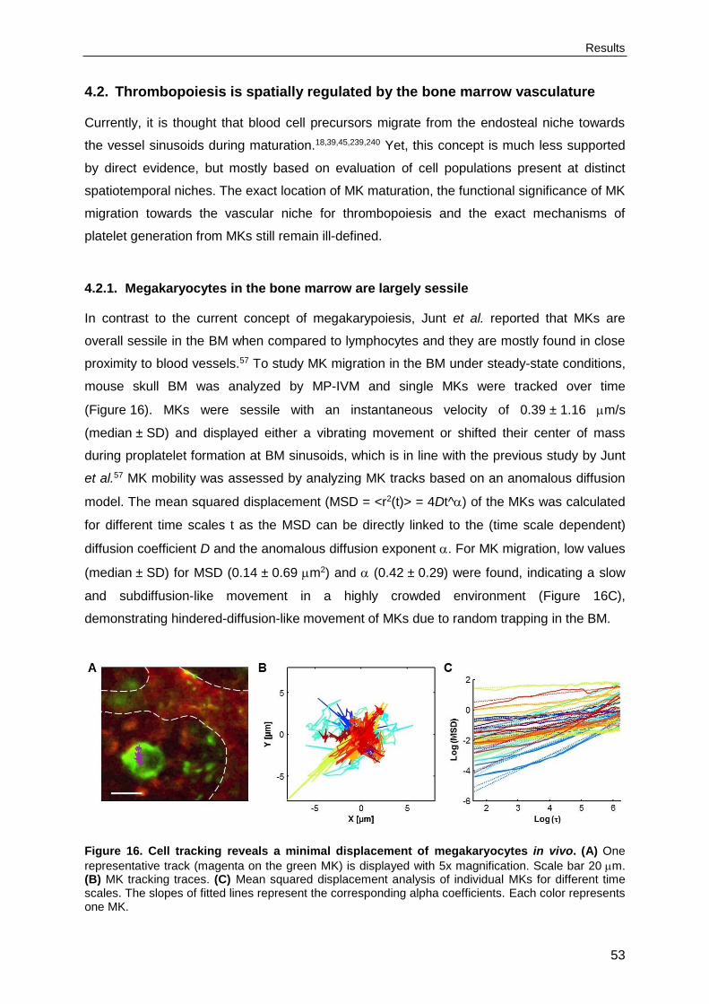
Results
53
4.2. Thrombopoiesis is spatially regulated by the bone marrow vasculature
Currently, it is thought that blood cell precursors migrate from the endosteal niche towards
the vessel sinusoids during maturation.18,39,45,239,240 Yet, this concept is much less supported
by direct evidence, but mostly based on evaluation of cell populations present at distinct
spatiotemporal niches. The exact location of MK maturation, the functional significance of MK
migration towards the vascular niche for thrombopoiesis and the exact mechanisms of
platelet generation from MKs still remain ill-defined.
4.2.1. Megakaryocytes in the bone marrow are largely sessile
In contrast to the current concept of megakarypoiesis, Junt et al. reported that MKs are
overall sessile in the BM when compared to lymphocytes and they are mostly found in close
proximity to blood vessels.57 To study MK migration in the BM under steady-state conditions,
mouse skull BM was analyzed by MP-IVM and single MKs were tracked over time
(Figure 16). MKs were sessile with an instantaneous velocity of 0.39 ± 1.16 m/s
(median ± SD) and displayed either a vibrating movement or shifted their center of mass
during proplatelet formation at BM sinusoids, which is in line with the previous study by Junt
et al.57 MK mobility was assessed by analyzing MK tracks based on an anomalous diffusion
model. The mean squared displacement (MSD = <r2(t)> = 4Dt^) of the MKs was calculated
for different time scales t as the MSD can be directly linked to the (time scale dependent)
diffusion coefficient D and the anomalous diffusion exponent . For MK migration, low values
(median ± SD) for MSD (0.14 ± 0.69 m2) and (0.42 ± 0.29) were found, indicating a slow
and subdiffusion-like movement in a highly crowded environment (Figure 16C),
demonstrating hindered-diffusion-like movement of MKs due to random trapping in the BM.
Figure 16. Cell tracking reveals a minimal displacement of megakaryocytes in vivo. (A) One
representative track (magenta on the green MK) is displayed with 5x magnification. Scale bar 20 m. (B) MK tracking traces. (C) Mean squared displacement analysis of individual MKs for different time scales. The slopes of fitted lines represent the corresponding alpha coefficients. Each color represents one MK.
Results
54
4.2.2. The majority of megakaryocytes is in direct contact with the vasculature
Since MP-IVM only has a limited field of view and most studies on megakarypoiesis and
thrombopoisesis have been performed on BM of the long bones, especially femur, tibia and
sternum, immunohistochemical analyses of cryo-sections231 of murine femora (Figure 17A)
and sterna (Figure 17B) were performed in addition. Under steady-state conditions, a
homogeneous distribution of CD41 (GPIIb) positive cells, a highly expressed early marker for
the MK lineage, was found.41 Surprisingly, MKs were found even in close contact to
endosteal zones (indicated by dark regions surrounding the BM, Figure 17A,B). MK
diameters were comparable in sternum and femur (22.19 0.14 m vs. 22.57 1.44 m)
and in both, the majority of MKs (68.2 6.9% vs. 73.1 5.2%) was found in direct contact
with the vessel, which is in agreement with previous studies.57,58 Complementary cryo-
sections confirmed that more than 95% of all CD41-positive MKs were positive for CD42a
and both groups showed identical MK diameters and percentage of vessel-association. The
MK-specific tubulin isoform 1241 was used as a marker for mature MKs (Figure 17C). As
expected, double-positive MKs were larger than 1-tubulin-negative CD41-positive cells
(23.53 6.23 m vs. 20.19 4.24 m, Figure 17D). Surprisingly, however, this was not
accompanied by altered MK distribution, as the majority of both 1-tubulin negative and
positive cells were in direct contact with the vessel (66.9 4.5 vs. 73.9 4.9%; Figure 17E)
and the vessel distant MKs were distributed similarly throughout the BM (Figure 17F).
4.2.3. Establishment of light-sheet fluorescence microscopy of the murine bone
marrow
MP-IVM revealed that MKs in the BM are largely sessile and the analysis of cryo-sections of
bones showed that the majority of MKs is localized in close contact with the sinusoid. These
methods allow studies on megakaryopoiesis and thrombopoiesis in the BM with sufficient
resolution, however, they only cover a small field of view, have a limited penetration depth
and provide restricted 3D information. Facing the opaque and dense bone tissue,
quantitative imaging of the MKs in their intact 3D environment would be challenging, but of
great interest, since little is known on the architecture of the BM and the overall MK
distribution. Therefore, LSFM of intact, murine bones was established. Crucial steps in the
establishment of the LSFM protocol were cell staining, tissue fixation and dehydration, optical
clearing, visualization of the samples by optical sectioning using LSFM, semi-automated
image processing and quantitative data analysis (Figure 18).

Results
55
Figure 17. Homogeneous distribution of megakaryocytes in the bone marrow. MKs (CD41+, green) in cryo-sections of whole-length femur (A) and sternum (B) showing vessels (CD105+, red) and nuclei (Hoechst33258, blue). Scale bar 1 mm. Right panels display a higher magnification (scale bar
100 m). (C) MKs were counter-stained for 1-tubulin (magenta, indicated with arrowheads - in
contrast to 1- MKs which are indicated with arrows). Scale bar 20 m. These mature MKs were
increased in size (D), but their distribution was similar between 1-tubulin negative and positive cells as depicted by the percentage of vessel-associated MKs (E) and the MK-vessel distance of non-vessel associated MKs (F). Bar graphs represent mean ± SD. **, p<0.01.

Results
56
Figure 18. Establisment of light-sheet fluorescence microscopy of the murine bone marrow. For the 3D visualization of the BM of intact murine bones with a cellular resolution and without cutting artefacts, a protocol was established. This protocol involves cell staining, tissue fixation and dehydration, optical clearing, visualization of the samples by optical sectioning using light-sheet fluorescence microscopy (LSFM), semi-automated image processing and quantitative data analysis.
First, cell labeling protocols for the visualization of MKs and blood vessels had to be
established. Ex vivo and in vivo staining protocols were tested, but to avoid staining artefacts
due to insufficient antibody penetration in situ, in vivo labeling of MKs by anti-GPIX antibody
derivatives was used (Figure 19).
Several fluorescent conjugates were tested, including Alexa 488, Alexa 546, Alexa 647 and
Alexa 750 (Figure 19A,B). The optimal MK staining was achieved using Alexa 750-labeled
anti-GPIX antibody derivatives, since excitation of the near-infrared fluorophore led to a low
autofluorescence signal and a high signal-to-noise ratio (Figure 19C,D). For staining of the
endothelium, Alexa 647-labeled anti-endoglin antibodies were used (Figure 19C,D).
LSFM uses a thin sheet of light to illuminate and optically section the tissue specimen. The
tissue has to be transparent to allow penetration of the laser light sheet. To obtain optically
transparent samples, a clearing protocol for complete and intact bones had to be established
(Figure 20). Only clearing protocols for soft tissues, but not for bones were available and,
therefore, different tissue clearing methods had to be tested.203-205,242 The first step towards
tissue clearing was transcardial perfusion of the mice with ice-cold PBS to wash out the
blood and ice-cold 4% PFA to fix the tissues. The bones were perfusion-fixed to protect the
structure of the tissue. To ensure optimal clearing and reduce scattering effects during the
measurements, adherent tissues, such as muscles and connective tissue had to be
removed. Upon harvesting of the sterna and femora, the bones were decalcified using EDTA
and were dehydrated using either ethanol, methanol (MeOH) or tetrahydrofuran (THF), which

Results
57
was stepwise replaced by a clearing solution (DBE or BABB). After incubation in the clearing
solution, tissue specimens became optically transparent. Dehydration with MeOH followed by
clearing with BABB provided the best result compared to bone clearing using THF and DBE
(Figure 20).
Figure 19. In vivo cell labeling for LSFM. 3D reconstructions of the sternum including MKs, which were stained in vivo by anti-GPXI antibodies labeled with either Alexa 647 (cyan, A,B) or Alexa 750 (green, C,D). The endothelium was labeled with anti-endoglin-Alexa 647 (red, C,D). Autofluorescence of the tissue was shown in red and green (A,B).
Scale bar 100 m.
Initially, a LSFM prototype with 3-color detection was successfully used to map immune
processes in intact tissues at cellular resolution242 and to visualize the BM in intact bones
(Figure 21A). However, during the time of this thesis, the LSFM prototype has been further
developed by implementing 4-color detection, new sCMOS cameras and a filterwheel. As a
result, resolution, contrast and imaging speed of larger images were enhanced, whereas
photobleaching effects were further reduced, improving the image quality dramatically
(Figure 21B).

Results
58
Figure 20. Optical clearing of bone tissue. Femur (A) and sternum (B) before (left panel) and after clearing (middle and right panel) using the combination of either tetrahydrofuran (THF) and dibenzyl ether (DBE) or methanol (MeOH) and benzyl alcohol benzyl benzoate (BABB).
To finally obtain quantitive data including MK number, volume and localization, the multicolor
LSFM stacks were processed and analyzed by FIJI232 and Bitplane Imaris® 7.7.2 (Figure 21,
Video 8). Image processing was performed by Oğuzhan Angay and Patrick Schmithausen.
Data visualization and analysis was performed in three major steps: image preprocessing,
segmentation and data extraction. Image preprocessing involved the removal of noise and
crosstalk, contrast enhancement, the removal of debris and background subtraction
(Figure 21B,C), leading to minimized noise and reduced unspecific signals. Next, MKs and
vessels were identified automatically and segmented based on morphology, fluorescence
intensity and size. Segmentation of MKs, proplatelets and vessels allowed for their
recognition as individual objects (Figure 21D,E,J). Objects were exported to iso-surfaces
(Figure 21D,E,K,L) to allow the generation of distance transformation maps to perform vessel
interspace analyses (Figure 22D) to obtain quantitive data for further statistical analyses.
Since MKs are labeled by antibody staining of the cell surface, the unstained lumen of the
MK had to be filled with artificial “soma” (Figure 21F-I). Furthermore, due to size and
morphological differences of MKs, sequential segmentation was performed using two MK
populations: MKs with a spherical cell diameter ≥ 20 m (green, Figure 21K) and MKs with a
spherical cell diameter of 10 m (purple, Figure 21K).
By combining cell staining, optical tissue clearing, microscope optimization and semi-
automated image processing, a method for the 3D visualization of the BM of intact murine
bones with a cellular resolution and without cutting artefacts was, for the first time,
established (Figure 21L).

Results
59
Figure 21. LSFM image processing and analysis. Imaging of the BM in intact bones (A) was improved by optimization of the microscope design (B). (C) Image preprocessing involved the removal of noise and crosstalk, contrast enhancement and background subtraction. Segmentation of MKs (green and yellow, D,E), proplatelets (yellow, J) and vessels (red, J) allowed for their recognition as individual objects. (F-I) The unstained lumen of the MK had to be filled with artificial “soma”. (K) Sequential segmentation was performed using two MK populations. (L) 3D visualization of the BM of intact murine bones with a cellular resolution showing MKs (green) and blood vessels (red) was
achieved. Scale bar A-C, J-L 50 m. Scale bar D-I 20 m.
4.2.4. Megakaryocytes are homogenously distributed throughout the bone marrow
The femur comprises the epiphysis and diaphysis and stacks were recorded in the diaphysis,
the shaft of the long bone (Figure 20A). The sternum consists of several segments
containing BM and stacks were recorded per BM segment (Figure 20B). Optical sectioning of
the sternum segments revealed a big central vessel in each BM segment. Femora contained
a central vessel along the inter-epiphyseal axis. The sinusoids in femora appeared more
aligned and structurally organized along the bone axis compared to the sinusoids in sterna.
LSFM imaging of the cleared intact femora (Figure 22A) and sterna (Figure 22B)
transversally allowed visualization of MKs with a resolution (<2 m) that was sufficient to
identify proplatelets (Figure 22C). The 3D reconstructions of LSFM data enabled the analysis
of 30-fold more MKs per bone sample compared to conventional cryo-sections and revealed
a surprisingly dense blood vessel network with an average maximal vessel-to-vessel

Results
60
distance of only 43.18 12.72 m (Figure 22D). Given that the mean MK-diameter was
20.36 8.19 m (Figure 22E), the spatial distribution of MKs was highly restricted by the
vasculature. Consequently 79.3 5.1% of all MKs localized adjacent to the blood vessels
(Figure 22G), values which are higher than those obtained with cryo-sections.
Figure 22. Dense blood vessel network and homogeneous distribution of megakaryocytes in reconstructed bone marrow using LSFM. 3D reconstructions of the BM of femur (A) and sternum (B) with MKs (green) and blood vessels (red). (C) These reconstructions enable visualization of proplatelets in intact BM. (D) Reconstruction of sternal BM revealed a dense blood vessel network with limited space for MKs shown by vessel-to-vessel distances and average MK diameter (E). Cyan
dots represent the middle between two adjacent vessels. Scale bar 50 m. (E,F) Comparison of the MK diameter and MK-vessel distance in cryo-sections and LSFM stacks. (G) Most MKs are localized adjacent to sinusoids and (H) MK distribution was homogeneous throughout the whole sternum as shown by the percentage of vessel associated MKs and the MK-vessel distance in five different
sections of the reconstructed BM. Bar graphs represent mean ± SD; grid size 100 m.

Results
61
This is presumably due to the additional structural 3D information, which enables the
detection of vessels surrounding each MK in all possible directions and minimizes the
underestimation of MK diameters (Figure 22E) and MK-to-vessel contacts (Figure 22F) due
to cutting artefacts. Analysis of five equally sized substacks of the sternum did not reveal
differences in any analyzed MK parameter (Figure 22H), confirming the conception of a
homogeneous MK distribution throughout the complete BM of the entire and intact bone.
4.2.5. Platelet depletion or CXCR4 blockade do not affect megakaryocyte localization
To determine the effects of increased platelet consumption on MK distribution, bones were
analyzed upon platelet depletion with anti-CD42b (GPIb) antibodies that cause rapid Fc-
independent platelet clearance (Figure 23A).210 Interestingly, despite the severe
thrombocytopenia in the treated animals (Figure 23B), MK migration and destruction were
not observed and the main MK parameters remained grossly unaltered, with only a mild
elevation in MK volume at day 3 after platelet depletion, resulting in an increased fraction of
vessel-associated MKs (Figure 23C-E). This indicates that even upon high platelet demand,
the MK distribution remains overall unaltered, which stands in clear contrast to the current
concept that immature MKs migrate towards the vessels.
The chemokine receptor CXCR4 has been shown to modulate MK migration in vitro and to
contribute to platelet production, especially in the absence of the Thpo-c-Mpl axis.18,49
Therefore, CXCR4 in wildtype mice was blocked using 5 mg/kg AMD3100 (AMD/Plerixafor),
a CXCR4 antagonist, and the effects on MK localization were determined after 24 h.
Treatment with AMD3100 resulted in elevated HSC counts (Sca-1+, c-kit+, Lin- cells) in the
peripheral blood after 24 h (not shown), confirming previous results.243 However, CXCR4
blockade did not modulate MK localization or volume (Figure 23F-H). Similar results were
obtained in mice receiving the CXCR4 ligand SDF1 (16 g/kg) 24 hours before analysis
(Figure 23F-H), indicating that despite the relevance of the SDF1-CXCR4 for HSC
mobilization,244 it appears to be negligible for MK (re)localization.
4.2.6. Computational modeling implies a vessel-biased megakaryocyte distribution
High MK numbers and their limited capability of (chemotactic) migration led to the hypothesis
that MKs are randomly distributed throughout the BM. To test this hypothesis, computational
modeling was performed to identify possible 3D MK distributions matching the data obtained
by LSFM. Computational modeling was performed by Mari Gorelashvili. For modeling, the
virtual MKs were randomly placed into the intervascular space, with the 3D vasculature
serving as a space-restricting scaffold (Figure 24A).

Results
62
Figure 23. Dysregulated platelet homeostasis does not affect megakaryocyte distribution or
migration within the bone marrow. (A) Platelets were depleted using anti-CD42b (GPIb) antibodies and analysis of sterna was performed on day 1, 3, 5 and 7 after depletion. Day 0 (PBS) served as control. (B) Mice displayed severe, but reversible thrombocytopenia. (C) Increase in vessel associated MKs upon platelet depletion correlates with an increase in MK-volume of vessel associated (grey bars) but not of non-vessel associated MKs (black bars, D). (E) MK numbers remain unaltered. Treatment with AMD3100 (AMD) or SDF1 did not modify MK localization (F), MK volume (G) or MK numbers (F). Bar graphs represent mean ± SD. *, p<0.05; **, p<0.01.
Since LSFM-derived MKs and vessel isosurfaces were exclusively used, the morphology and
size of the modeled MKs and vasculature perfectly reflected the spatial physiological
situation. Furthermore, a clear distinction was made between vessel-associated MKs (light
green) and non-vessel associated MKs (dark green) in the simulation. The first simulation, a
random simulation (SimR) of total MK distribution in the BM, did not reflect the measured
results (actual data). Instead, it predicted significantly larger total MK-vessel distances
(Figure 24B) and a lower percentage of vessel associated MKs, indicating a non-random, but
vessel-biased MK distribution for the LSFM data. The single data stack used for the actual
data to compare to the simulation data, displayed 60% of the MKs in association with the

Results
63
vessels. Therefore, for the next simulation (SimVA), a vast percentage of the MKs (60%)
were vessel-associated and the remaining 40% of the MKs were randomly distributed within
the intervascular space. When only taking the non-vessel associated MKs into account (dark
green in Figure 24), the SimVA, and not the SimR, fully reflects the actual 3D imaging data
(Figure 24B). These results indicate that MK localization is vessel-biased, but the non-vessel
associated MKs are distributed within the BM in a random fashion. The current concept of
megakaryopoiesis involves MK migration and, in that situation, a non-random distribution of
non-vessel associated MKs would be expected. In addition, MK localization did not correlate
with MK volume of non-vessel associated MKs, pointing towards a random distribution of
immature and mature MKs throughout the BM and thereby confirming the absence of MK
migration.
Figure 24. Megakaryocytes display a vessel-biased random distribution. (A) Computational modeling was performed using the 3D vasculature (red) as a space-restricting scaffold. LSFM-derived MK and vessel isosurfaces were used to reflect the spatial physiological situation. Vessel associated MKs are depicted in light green and non-vessel associated MKs in dark green. (B) A random MK distribution (SimR) and a vessel-biased random MK distribution (SimVA) were simulated by placing the virtual MKs randomly into the intervascular space with a vast percentage of vessel-associated MKs in SimVA. The MK-vessel distances were measured and revealed a vessel-biased random MK distribution. ***, p<0.001.
4.2.7. Light-sheet fluorescence microscopy as a tool to study thrombopoiesis
LSFM allows the 3D visualization of intact murine bones, but also the quantitative structural
analysis of BM components. MP-IVM already revealed that the majority of the Pfn1-/- MKs
released platelets prematurely into the BM (Figure 13B, Video 3) and therefore Pfn1-/- mice
were analyzed using LSFM. It was confirmed that MKs of Pfn1-/- mice showed increased
fragmentation and platelets were prematurely released into the BM compartment
(Figure 25A,B). These results demonstrate the potential of LFSM for studying animal models
of human disease.
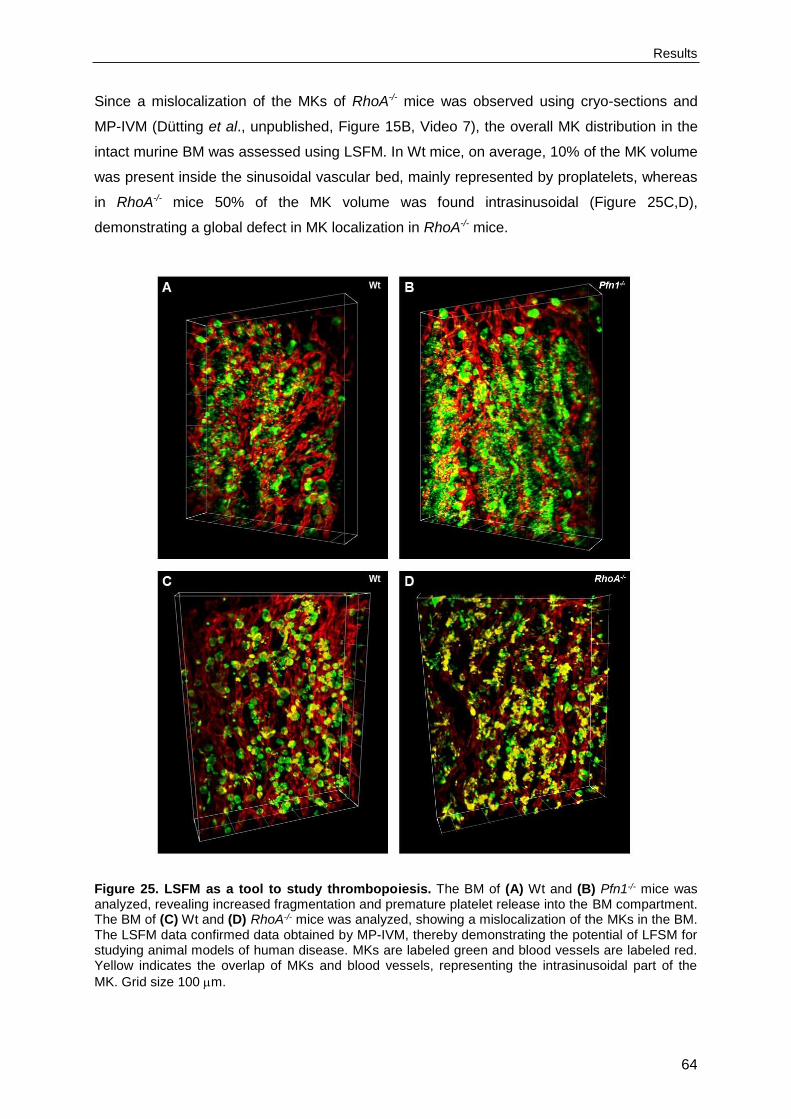
Results
64
Since a mislocalization of the MKs of RhoA-/- mice was observed using cryo-sections and
MP-IVM (Dütting et al., unpublished, Figure 15B, Video 7), the overall MK distribution in the
intact murine BM was assessed using LSFM. In Wt mice, on average, 10% of the MK volume
was present inside the sinusoidal vascular bed, mainly represented by proplatelets, whereas
in RhoA-/- mice 50% of the MK volume was found intrasinusoidal (Figure 25C,D),
demonstrating a global defect in MK localization in RhoA-/- mice.
Figure 25. LSFM as a tool to study thrombopoiesis. The BM of (A) Wt and (B) Pfn1-/- mice was analyzed, revealing increased fragmentation and premature platelet release into the BM compartment. The BM of (C) Wt and (D) RhoA-/- mice was analyzed, showing a mislocalization of the MKs in the BM. The LSFM data confirmed data obtained by MP-IVM, thereby demonstrating the potential of LFSM for studying animal models of human disease. MKs are labeled green and blood vessels are labeled red. Yellow indicates the overlap of MKs and blood vessels, representing the intrasinusoidal part of the
MK. Grid size 100 m.

Results
65
4.3. Syk deficiency or inhibition protect mice from arterial thrombosis and
thrombo-inflammatory brain infarction
Syk is an essential signaling mediator downstream of immune cell receptors, but also of
GPVI and CLEC-2 in platelets.179-181 Due to its central role in immunological processes,
several Syk inhibitors have been developed and clinically used in the treatment of for
example allergy,143 B cell malignancies,138 heparin-induced thrombocytopenia145 and
autoimmune diseases.139,144 However, definite evidence for a role of Syk in hemostasis,
arterial thrombosis and ischemic stroke is limited.
4.3.1. (hem)ITAM signaling is abolished in Syk-/- platelets
To investigate the role of Syk in platelet biology, mice deficient for Syk in platelets and MKs,
Sykfl/fl, Pf4-cre+/- mice, were analyzed (further referred to as Syk-/- mice). Syk-/- mice were
generated by intercrossing mice containing the Syk gene flanked by loxP sites (Sykfl/fl) with
Pf4-cre+/- mice. Sykfl/fl, Pf4-cre-/- mice were used as controls and are further referred to as
wildtype (Wt) mice. Conditional knockout mice were used for the analysis, since constitutive
Syk-deficient mice die perinatally.170 Syk-/- mice were born at normal Mendelian ratios and
the absence of Syk in platelet lysates was confirmed by Western Blot analysis (Figure 26A).
Syk-/- mice displayed normal basal blood parameters, platelet counts and size (Table 1). Flow
cytometric analysis showed unaltered expression levels of prominent platelet surface
receptors in Syk-/- platelets (Table 1). The effect of Syk deficiency on platelet activation was
measured by flow cytometric analyses of agonist-induced activation of the major platelet
integrin IIb3 (JON-A/PE binding) and degranulation-dependent P-selectin surface
exposure (Figure 26B). In response to GPCR agonists, such as ADP, the TxA2 analog
U46619 and thrombin, platelet integrin activation and degranulation were unaltered, whereas
responses to GPVI and CLEC-2 agonists, including CRP, CVX and rhodocytin were
abolished.
In addition, platelet aggregation of Syk-/- platelets (washed platelet and PRP) was analyzed in
response to various agonists (Figure 27A,B). The lack of functional Syk resulted in severely
defective aggregation in response to GPVI (collagen, CRP) or CLEC-2 (rhodocytin) agonists.
However, Syk-/- platelets showed unaltered aggregation upon stimulation with ADP, thrombin
and U46619. To determine the effect of Syk deficiency on platelet adhesion and aggregation
under flow conditions, thrombus formation ex vivo was analyzed by a flow adhesion assay
(Figure 27C).

Results
66
Wt Syk-/-
Platelets (nl-1) 1097 ± 69 1134 ± 136
Mean platelet volume (fl) 5.4 ± 0.1 5.4 ± 0.1
GPIb 365 ± 42 347 ± 45
GPV 314 ± 15 301 ± 14
GPIX 428 ± 40 412 ± 37
CD9 943 ± 55 920 ± 32
CD84 32 ± 2 31 ± 2
GPVI 49 ± 5 55 ± 5
CLEC-2 143 ± 8 138 ± 6
2 53 ± 3 53 ± 2
5 20 ± 2 19 ± 2
1 128 ± 20 132 ± 8
3 177 ± 23 160 ± 20
IIb3 539 ± 29 521 ± 25
White blood cells (nl-1) 6.0 ± 2.1 6.9 ± 2.1
Red blood cells (pl-1) 9.1 ± 0.7 8.6 ± 1.4
Hemoglobin (g dl-1) 14.8 ± 1.6 14.7 ± 2.5
Hematocrit (%) 47.8 ± 4.7 46.9 ± 8.2
Mean red blood cell volume (fl) 54.2 ± 1.6 54.6 ± 1.0
Table 1. Analysis of basal blood parameters and surface expression of glycoproteins on platelets of Wt and Syk-/- mice. Basal blood parameters were analyzed on a Sysmex KX-21N automated hematology analyzer. Diluted whole blood was stained with saturating amounts of fluorophore-labeled antibodies and platelets were analyzed by flow cytometry to determine the expression of prominent platelet surface receptors. Results represent mean fluorescence intensities (MFI) ± SD if not stated otherwise. (van Eeuwijk, Stegner et al., Arterioscler Thromb Vasc Biol. 2016)245
Therefore, heparinized whole blood of Wt and Syk-/- mice was perfused over a collagen-
coated surface at a shear rate of 1000 s-1. Wt platelets adhered to the collagen surface and
recruited additional platelets from the flowing blood, leading to the formation of stable, 3D
thrombi that finally covered approximately 50% of the total surface area. In sharp contrast,
thrombus formation was abolished when perfusing heparinized whole blood from Syk-
deficient mice and only a few, single platelets adhered to the immobilized collagen. These
results confirm that Syk is required for (hem)ITAM signaling downstream of GPVI and CLEC-
2 in platelets.

Results
67
Figure 26. Abolished (hem)ITAM signaling in the absence of Syk. (A) Western blot analysis
demonstrating the absence of Syk in Syk-/- platelets. PLC2 expression was used as loading control.
(B) Flow cytometric analysis of IIb3 integrin activation (JON/A-PE, left panel) and degranulation-dependent P-selectin exposure (right panel) in response to the indicated agonists of Wt (black bars) and Syk-/- (grey bars) platelets. Results represent mean fluorescence intensities (MFI) ± SD. ADP, adenosine diphosphate; U46, U46619; A/U, ADP/U46; Thr, thrombin; CRP, collagen-related peptide; RC, rhodocytin; CVX, convulxin. *, p<0.05; ***, p<0.001. (van Eeuwijk, Stegner et al., Arterioscler Thromb Vasc Biol. 2016)245
4.3.2. Murine Syk is dispensable for integrin-dependent outside-in signaling
Syk has been proposed to contribute to outside-in signaling of integrin IIb3 for platelet
adhesion and cytoskeletal remodeling.179-181 To test this hypothesis, Wt and Syk-/- platelets
were allowed to spread on fibrinogen (Figure 28), a process dependent on outside-in
signaling. The Syk-deficient platelets were treated with indomethacin and apyrase to inhibit
the effect of the second wave mediators TxA2 and ADP. Under these conditions, Syk-/-
platelets spread on fibrinogen to a similar extent and with similar kinetics as Wt platelets
(Figure 28A). Spreading of Wt and Syk-/- platelets with thrombin stimulation was also
comparable (Figure 28B). The integrin IIb3 also plays a critical role in regulating clot
retraction by platelets.152 Upon ligand binding, clot retraction is mediated by the IIb3
integrin and the cytoskeleton to consolidate thrombus formation in vivo. To investigate
whether the loss of Syk alters clot retraction, clot formation was induced in PRP of Wt and
Syk-/- mice (Figure 28C). Clot retraction was monitored over time and started as early as 20
minutes after the start of the experiment. No differences were observed in clot retraction of
Wt and Syk-/- samples and the excess serum levels extruded during clot retraction were
similar between Wt and Syk-/- (95 ± 2.0% versus 94 ± 5.2% of the initial PRP volume).
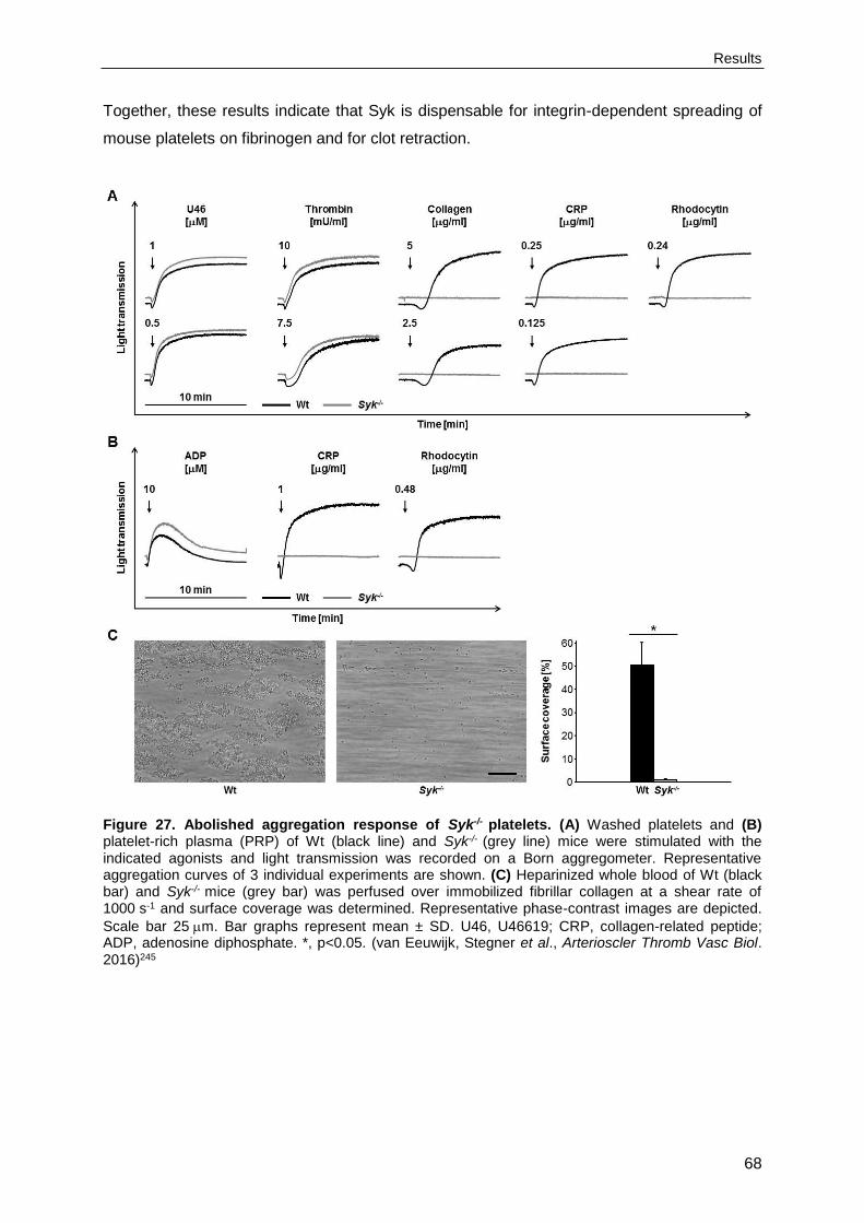
Results
68
Together, these results indicate that Syk is dispensable for integrin-dependent spreading of
mouse platelets on fibrinogen and for clot retraction.
Figure 27. Abolished aggregation response of Syk-/- platelets. (A) Washed platelets and (B) platelet-rich plasma (PRP) of Wt (black line) and Syk-/- (grey line) mice were stimulated with the indicated agonists and light transmission was recorded on a Born aggregometer. Representative aggregation curves of 3 individual experiments are shown. (C) Heparinized whole blood of Wt (black bar) and Syk-/- mice (grey bar) was perfused over immobilized fibrillar collagen at a shear rate of 1000 s-1 and surface coverage was determined. Representative phase-contrast images are depicted.
Scale bar 25 m. Bar graphs represent mean ± SD. U46, U46619; CRP, collagen-related peptide; ADP, adenosine diphosphate. *, p<0.05. (van Eeuwijk, Stegner et al., Arterioscler Thromb Vasc Biol. 2016)245

Results
69
Figure 28. Platelet spreading is unaffected by Syk-deficiency. Washed platelets from Wt and Syk-/- mice were allowed to adhere to immobilized human fibrinogen (100 µg/ml) in the presence of
either (A) the second wave inhibitors indomethacin (1.4 M) and apyrase (1 U/ml) or (B) thrombin (0.01 U/ml). Images were taken at the indicated time points and representative images are shown. Results indicate percentage of phase abundancy with (1), round platelets; (2), only filopodia; (3),
filopodia and lamellipodia; (4), full platelet spreading. Scale bar 5 m. (C) Clot formation was induced in PRP of Wt and Syk-/- mice by 4 U/ml thrombin and clot retraction was monitored over time. Images were taken at the indicated time points and representative images are shown. (van Eeuwijk, Stegner et al., Arterioscler Thromb Vasc Biol. 2016)245

Results
70
4.3.3. Syk deficiency results in moderately prolonged tail bleeding times and impaired
arterial thrombus formation
Since only limited evidence exists on the role of Syk in hemostasis and thrombosis, several
in vivo analyses were performed. To investigate the role of Syk in hemostasis, tail bleeding
times were measured (Figure 29A). Bleeding times after amputation of the tail tip were
moderately prolonged in Syk-/- mice compared to their respective controls. The hemostatic
defect was, however, not as dramatic as upon downregulation of both GPVI and CLEC-2
from the platelet surface by antibody treatment, where no hemostatic function is present.133
Figure 29. Syk-deficiency leads to a mild hemostatic defect, but protects from arterial thrombosis. (A) Tail bleeding times of Wt and Syk-/- mice. Each symbol represents one animal. (B) In an aorta occlusion model, the blood flow of Wt and Syk-/- mice was monitored for 30 min or until complete occlusion occurred. Time to stable vessel occlusion is depicted. Each symbol represents one animal. (C) In a chemical injury model, the blood flow of Wt and Syk-/- mice in the mesenteric arterioles was monitored for 40 min or until complete occlusion occurred. Time to stable vessel occlusion is depicted. Each symbol represents one animal. (D) Images of thrombus formation in the mesenteric arterioles were taken at the indicated time points and representative images are shown. *, p<0.05; **, p<0.01. (van Eeuwijk, Stegner et al., Arterioscler Thromb Vasc Biol. 2016)245
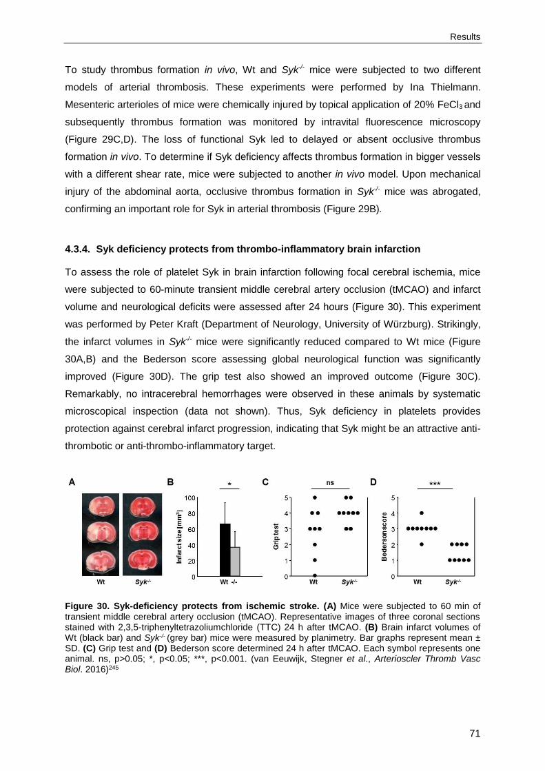
Results
71
To study thrombus formation in vivo, Wt and Syk-/- mice were subjected to two different
models of arterial thrombosis. These experiments were performed by Ina Thielmann.
Mesenteric arterioles of mice were chemically injured by topical application of 20% FeCl3 and
subsequently thrombus formation was monitored by intravital fluorescence microscopy
(Figure 29C,D). The loss of functional Syk led to delayed or absent occlusive thrombus
formation in vivo. To determine if Syk deficiency affects thrombus formation in bigger vessels
with a different shear rate, mice were subjected to another in vivo model. Upon mechanical
injury of the abdominal aorta, occlusive thrombus formation in Syk-/- mice was abrogated,
confirming an important role for Syk in arterial thrombosis (Figure 29B).
4.3.4. Syk deficiency protects from thrombo-inflammatory brain infarction
To assess the role of platelet Syk in brain infarction following focal cerebral ischemia, mice
were subjected to 60-minute transient middle cerebral artery occlusion (tMCAO) and infarct
volume and neurological deficits were assessed after 24 hours (Figure 30). This experiment
was performed by Peter Kraft (Department of Neurology, University of Würzburg). Strikingly,
the infarct volumes in Syk-/- mice were significantly reduced compared to Wt mice (Figure
30A,B) and the Bederson score assessing global neurological function was significantly
improved (Figure 30D). The grip test also showed an improved outcome (Figure 30C).
Remarkably, no intracerebral hemorrhages were observed in these animals by systematic
microscopical inspection (data not shown). Thus, Syk deficiency in platelets provides
protection against cerebral infarct progression, indicating that Syk might be an attractive anti-
thrombotic or anti-thrombo-inflammatory target.
Figure 30. Syk-deficiency protects from ischemic stroke. (A) Mice were subjected to 60 min of transient middle cerebral artery occlusion (tMCAO). Representative images of three coronal sections stained with 2,3,5-triphenyltetrazoliumchloride (TTC) 24 h after tMCAO. (B) Brain infarct volumes of Wt (black bar) and Syk-/- (grey bar) mice were measured by planimetry. Bar graphs represent mean ± SD. (C) Grip test and (D) Bederson score determined 24 h after tMCAO. Each symbol represents one animal. ns, p>0.05; *, p<0.05; ***, p<0.001. (van Eeuwijk, Stegner et al., Arterioscler Thromb Vasc Biol. 2016)245

Results
72
4.3.5. Syk inhibition abolishes (hem)ITAM signaling
Platelet inhibition is commonly used in secondary stroke prevention.246 In acute stroke,
however, revascularization by recombinant tissue plasminogen activator (rt-PA) or
mechanical thrombectomy are the only therapeutic means246,247 with limited efficacy.248
Addition of conventional anti-platelet agents such as acetylsalicylic acid (ASA) led to
bleeding complications.249 In order to improve functional outcome in acute stroke, novel anti-
thrombotic strategies preserving physiological hemostasis are eagerly awaited.
To determine if Syk could be an anti-thrombotic or anti-thrombo-inflammatory target, several
commercially available Syk inhibitors were tested, but, in our laboratory, the anti-platelet
efficacy in vivo was limited (data not shown). Therefore, the novel and highly selective oral
ATP-competitive Syk inhibitor, BI1002494 was used.226 C57BL/6JRj mice were obtained from
Janvier Labs and treated with either the vehicle or the inhibitor.
Figure 31. Pharmacological inhibition of Syk abolishes (hem)ITAM signaling. (A) Dose-response
curves showing flow cytometric analysis of IIb3 integrin activation (JON/A-PE, left panel) and degranulation-dependent P-selectin exposure (right panel) of wildtype platelets in response to the indicated agonists and the indicated concentrations of BI1002494. Results represent percentage of inhibition ± SD compared to vehicle-treated platelets. (B) Platelet-rich plasma (PRP) of vehicle-treated (black line) and BI1002494-treated (grey line) mice was stimulated with the indicated agonists and light transmission was recorded on a Born aggregometer. Representative aggregation curves of 3 individual experiments are shown. CRP, collagen-related peptide; CVX; convulxin; Rhod, rhodocytin; ADP, adenosine diphosphate; U46, U46619. (van Eeuwijk, Stegner et al., Arterioscler Thromb Vasc Biol. 2016)245
Results
73
The effect of Syk inhibition on platelet activation was measured by flow cytometric analyses
of agonist-induced activation of the major platelet integrin IIb3 (JON-A/PE binding) and
degranulation-dependent P-selectin surface exposure (Figure 31A). In response to ADP and
U46619, no effects on platelet integrin activation and degranulation via GPCR signaling were
observed with any of the tested inhibitor concentrations, whereas responses to GPVI and
CLEC-2 agonists, including CRP, CVX and rhodocytin were abolished. In vitro incubation of
Wt platelets with 150 nM BI1002494 abrogated CRP responses, while a concentration of
500 nM of the inhibitor was required to abolish CVX and rhodocytin responses.
Oral administration of BI1002494 (100 mg/kg b.i.d.) in mice resulted in plasma
concentrations greater than 980 nM over a 24-hour time period.226 Subsequent ex vivo
analysis of PRP showed abolished GPVI-induced platelet aggregation, while aggregation
responses to rhodocytin were reduced, but not abolished (Figure 31B). In contrast, PRP from
inhibitor-treated mice showed unaltered aggregation upon stimulation with ADP. These
results show that BI1002494 is a suitable inhibitor for in vivo blockade of platelet (hem)ITAM
signaling.
4.3.6. Human platelets require Syk for integrin-dependent outside-in signaling
To determine the effect of BI1002494 on outside-in signaling of integrin IIb3, thrombin-
stimulated Wt and BI1002494-treated mouse platelets were allowed to spread on fibrinogen
(Figure 32A). Inhibitor-treated platelets spread on a fibrinogen-coated surface to a similar
extent and with similar kinetics as Wt platelets. Interestingly and in contrast to mouse
platelets, outside-in signaling of integrin IIb3 in human platelets was impaired upon
inhibitor treatment (Figure 32B), revealing a requirement of Syk for integrin-dependent
outside-in signaling in human platelets.
4.3.7. Syk inhibition protects from arterial thrombosis and cerebral infarct
progression, without affecting hemostasis
To investigate if Syk inhibition would alter hemostasis, mice were treated with BI1002494
(100 mg/kg b.i.d.) and tail bleeding times were determined (Figure 33A). Bleeding times after
amputation of the tail tip were unaltered in inhibitor-treated mice compared to vehicle-treated
controls. To study thrombus formation in vivo, vehicle-treated and BI1002494-treated mice
were subjected to mechanical injury of the aorta (Figure 33B). The experiment was
performed by Sarah Beck. Inhibitor-treated mice were completely protected from vessel
occlusion compared to their respective controls.

Results
74
Figure 32. Syk inhibition impairs spreading of human platelets. Vehicle-treated (veh) and BI1002494-treated (BI) washed (A) mouse and (B) human platelets were allowed to adhere to immobilized human fibrinogen (100 µg/ml). Mouse platelets were stimulated with thrombin (0.01 U/ml). Images were taken at the indicated time points and representative images are shown. Results indicate percentage of phase abundancy with (1), round platelets; (2), only filopodia; (3), filopodia and lamellipodia; (4), full platelet spreading. Scale bar 5 µm. (van Eeuwijk, Stegner et al., Arterioscler Thromb Vasc Biol. 2016)245
To assess the effect of the Syk inhibitor on ischemic stroke, Wt mice were pretreated with
Bl1002494 and subsequently subjected to 60-minute tMCAO (Figure 34A). The experiment
was performed by Peter Kraft. Similar to Syk-/- mice, these animals displayed markedly
reduced infarct sizes and improved motor function 24 hours after tMCAO without evidence of
major intracerebral bleeding (Figure 34A-C). The neurological function also showed an
improved outcome (Figure 34D). To determine if the Syk inhibitor could be used
therapeutically, i.e. when given after the occurrence of ischemic stroke, mice were treated
with the inhibitor directly after tMCAO (directly upon removal of the filament) (Figure 34E-H).
Strikingly, also under these conditions, inhibitor-treated mice had reduced infarct sizes and a
better motor function as compared to the vehicle-treated controls (Figure 34E-G). A tendency
towards a generally improved neurological outcome was also observed (Figure 34H).
Together, these results indicate that Syk inhibition might be effective in preventing cerebral

Results
75
reperfusion injury and could serve as an anti-thrombotic therapy or anti-inflammatory
therapy.
Figure 33. Pharmacological inhibition of Syk protects from occlusive thrombus formation. (A) Tail bleeding times of vehicle-treated and BI1002494-treated mice. Each symbol represents one animal. (B) In an aorta occlusion model, the blood flow of vehicle-treated and BI1002494-treated mice was monitored for 30 min or until complete occlusion occurred. Time to stable vessel occlusion is depicted. Each symbol represents one animal. ns, p>0.05; ***, p<0.001. (van Eeuwijk, Stegner et al., Arterioscler Thromb Vasc Biol. 2016)245
Figure 34. Pharmacological inhibition of Syk protects from ischemic stroke. (A) Vehicle-treated and prophylactic BI1002494-treated mice were subjected to 60 min of transient middle cerebral artery occlusion (tMCAO). Representative images of three coronal sections stained with TTC 24 h after tMCAO. (B) Brain infarct volumes of vehicle-treated (black bar) and prophylactic BI1002494-treated (grey bar) mice were measured by planimetry. Bar graphs represent mean ± SD. (C) Grip test and (D) Bederson score determined 24 h after tMCAO. Each symbol represents one animal. (E) Vehicle-treated and therapeutic BI1002494-treated mice were subjected to 60 min of transient middle cerebral artery occlusion (tMCAO). Representative images of three coronal sections stained with TTC 24 h after tMCAO. (F) Brain infarct volumes of vehicle-treated (black bar) and therapeutic BI1002494-treated (grey bar) mice were measured by planimetry. Bar graphs represent mean ± SD. (G) Grip test and (H) Bederson score determined 24 h after tMCAO. Each symbol represents one animal. ns, p>0.05; *, p<0.05; **, p<0.01. (van Eeuwijk, Stegner et al., Arterioscler Thromb Vasc Biol. 2016)245

Results
76
4.4. Thrombus formation in vivo can occur independently of Syk kinase
function
Syk and Zap-70 are both spleen tyrosine kinase family members, which phosphorylate key
adapter proteins and PLC isoforms. Both kinases display partly overlapping expression
patterns, but the intrinsic kinase activity of Zap-70 is lower. In contrast to Syk, which is highly
expressed by hematopoietic cells, Zap-70 expression is largely confined to T cells and
natural killer cells. In mouse platelets, Zap-70 is not expressed, but Syk is present in the
(hem)ITAM signaling pathways mediating signal transduction by GPVI and CLEC-2.
Wt Sykki
Platelets (nl-1) 840 ± 181 636 ± 108
Mean platelet volume (fl) 5.3 ± 0.2 5.6 ± 0.5
GPIb 395 ± 17 408 ± 29
GPV 291 ± 11 302 ± 11
GPIX 414 ± 14 433 ± 14
CD9 892 ± 56 891 ± 45
CD84 35 ± 2 33 ± 4
GPVI 53 ± 4 55 ± 3
CLEC-2 135 ± 8 126 ± 11
2 58 ± 5 59 ± 5
5 17 ± 2 16 ± 2
1 132 ± 6 128 ± 9
3 181 ± 19 157 ± 11
IIb3 588 ± 38 540 ± 34
White blood cells (nl-1) 9.6 ± 3.2 8.4 ± 1.7
Red blood cells (pl-1) 8.5 ± 1.0 9.2 ± 1.9
Hemoglobin (g dl-1) 13.5 ± 1.5 15.0 ± 3.1
Hematocrit (%) 44.1 ± 3.4 48.9 ± 13.0
Mean red blood cell volume (fl) 51.9 ± 1.8 52.7 ± 3.2
Table 2. Analysis of basal blood parameters and surface expression of glycoproteins on platelets of Wt and Sykki mice. Basal blood parameters were analyzed on a Sysmex KX-21N automated hematology analyzer. Diluted whole blood was stained with saturating amounts of fluorophore-labeled antibodies and platelets were analyzed by flow cytometry to determine the expression of prominent platelet surface receptors. Results represent mean fluorescence intensities (MFI) ± SD if not stated otherwise.

Results
77
To assess if the two kinases exhibit interchangeable functions in vivo, the group of
Friedemann Kiefer generated mice, which express Zap-70 under the control of intrinsic Syk
promoter elements (SykZap70/Zap70 mice, further referred to as Sykki mice), thereby disrupting
wildtype Syk expression. Syk+/+ mice, further referred to as Wt mice, were used as
controls.220
4.4.1. Sykki mice die perinatally
To determine the consequence of the kinase replacement for platelet activation, Sykki mice
were analyzed. Initially, Sykki mice were born at normal Mendelian ratios and the mice were
vital and fertile. However, at a later stage, upon several rounds of animal mating, Sykki mice
were born at submendelian ratios due to perinatal lethality. Backcrossing of the mice with
either C57BL/6J or 129/Sv mice did not improve the birth ratios. Therefore, fetal liver
chimeric mice and BM chimeric mice were generated and used for analysis.
Figure 35. Abolished (hem)ITAM signaling in Sykki platelets. (A) Western blot analysis
demonstrating the absence of Syk and the presence of Zap-70 in Sykki platelets. -tubulin expression
was used as loading control. (B) Flow cytometric analysis of IIb3 integrin activation (JON/A-PE, left panel) and degranulation-dependent P-selectin exposure (right panel) in response to the indicated agonists of Wt (black bars) and Sykki (grey bars) platelets. Results represent mean fluorescence intensities (MFI) ± SD. ADP, adenosine diphosphate; U46, U46619; A/U, ADP/U46; Thr, thrombin; CRP, collagen-related peptide; RC, rhodocytin; CVX, convulxin. *, p<0.05; **, p<0.01; ***, p<0.001.

Results
78
4.4.2. (hem)ITAM signaling is abolished in Sykki platelets
The presence of Zap-70 and the absence of Syk in platelet lysates were confirmed by
Western Blot analysis (Figure 35A). Sykki mice displayed normal basal blood parameters,
platelet counts and size (Table 2). Flow cytometric analysis showed unaltered expression
levels of prominent platelet surface receptors in Sykki platelets (Table 2). The impact of the
kinase exchange on platelet activation was measured by flow cytometric analyses of agonist-
induced activation of the major platelet integrin IIb3 (JON-A/PE binding) and
degranulation-dependent P-selectin surface exposure (Figure 35B). In response to GPCR
agonists, such as ADP, U46619 and thrombin, platelet integrin activation and degranulation
were unaltered. However, IIb3 activation and degranulation in response to the GPVI and
CLEC-2 agonists CRP, CVX and rhodocytin failed to exceed resting state levels, indicating
an abrogated GPVI and CLEC-2 signaling.
Figure 36. Abolished aggregation response of Sykki platelets. (A) Washed platelets of Wt (black line) and Sykki (grey line) mice were stimulated with the indicated agonists and light transmission was recorded on a Born aggregometer. Representative aggregation curves of 3 individual experiments are shown. (B) Heparinized whole blood of Wt (black bar) and Sykki mice (grey bar) was perfused over immobilized fibrillar collagen at a shear rate of 1000 s-1 and surface coverage was determined.
Representative phase-contrast images are depicted. Scale bar 25 m. Bar graphs represent mean ± SD. CRP, collagen-related peptide. *, p<0.05.
Furthermore, platelet activation in vitro in response to different agonists was analyzed by
aggregometry (Figure 36A). Replacing Syk by Zap-70 resulted in abrogated aggregation in
response to CRP and rhodocytin. However, a delayed and weak residual aggregation of the

Results
79
Sykki platelets was observed in response to collagen. Since platelet binding to collagen is
mediated by integrin 21 besides GPVI, Sykki platelets were pre-treated with the anti-21
antibody LEN/B (10 g/ml) and stimulated with collagen. This resulted in a completely
abolished aggregation response comparable to CRP stimulation of Sykki platelets. Upon
stimulation with thrombin, Sykki platelets showed unaltered aggregation compared to Wt
platelets. To determine the effect of the kinase replacement on platelet adhesion and
aggregation under flow conditions, thrombus formation ex vivo was analyzed by a flow
adhesion assay (Figure 36B). Therefore, heparinized whole blood of Wt and Sykki mice was
perfused over a collagen-coated surface at a shear rate of 1000 s-1. Wt platelets adhered to
the collagen surface and recruited additional platelets from the flowing blood, leading to the
formation of stable, 3D thrombi that finally covered approximately 40% of the total surface
area. In sharp contrast, thrombus formation was abolished when perfusing heparinized whole
blood from Sykki mice and only a few, single platelets adhered to the immobilized collagen.
These results confirm that the Zap-70 kinase is insufficient in sustaining signaling
downstream of (hem)ITAM-coupled receptors in platelets.
4.4.3. Integrin outside-in signaling is unaffected by the kinase exchange
To assess if the kinase exchange affects outside-in signaling of integrin IIb3, thrombin-
stimulated platelets from Wt and Sykki mice were allowed to spread on fibrinogen (Figure 37).
Similar to Syk-deficient platelets, Sykki platelets showed unaltered spreading compared to Wt
platelets. Also, spreading of unstimulated Wt and Sykki platelets was comparable (data not
shown).
Figure 37. The kinase exchange does not affect platelet spreading. Washed platelets from Wt and Sykki mice were allowed to adhere to immobilized human fibrinogen (100 µg/ml) in the presence of thrombin (0.01 U/ml). Images were taken at the indicated time points and representative images are shown. Results indicate percentage of phase abundancy with (1), round platelets; (2), only filopodia;
(3), filopodia and lamellipodia; (4), full platelet spreading. Scale bar 5 m.

Results
80
4.4.4. Zap-70 partially compensates for the loss of Syk in vivo
To determine if Zap-70 could functionally replace Syk in vivo, hemostatic and thrombotic
assays were performed. To investigate the role of Zap-70 in hemostasis, tail bleeding times
were measured (Figure 38A). Bleeding times after amputation of the tail tip were significantly
increased in Sykki mice compared to their respective controls, which is in agreement with the
results obtained in the Syk-deficient mice (see section 4.3.3).
Figure 38. Zap-70 partially compensates for the loss of Syk in vivo. (A) Tail bleeding times of Wt and Sykki mice. Each symbol represents one animal. (B) In an aorta occlusion model, the blood flow of Wt and Sykki mice was monitored for 30 min or until complete occlusion occurred. Time to stable vessel occlusion is depicted. Each symbol represents one animal. (C) In a chemical injury model, the blood flow of Wt and Sykki mice in the mesenteric arterioles was monitored for 40 min or until complete occlusion occurred. Time to stable vessel occlusion is depicted. Each symbol represents one animal. (D) Images of thrombus formation in the mesenteric arterioles were taken at the indicated time points and representative images are shown. ns, p>0.05; **, p<0.01.
To study thrombus formation in vivo, Wt and Sykki mice were subjected to two different
models of arterial thrombosis. The experiments were performed by Ina Thielmann.
Mesenteric arterioles of mice were chemically injured by topical application of 20% FeCl3 and
subsequently thrombus formation was monitored by intravital fluorescence microscopy
(Figure 38C,D). The loss of functional Syk, but the presence of Zap-70 surprisingly led to

Results
81
unaltered occlusive thrombus formation in vivo. To determine if Zap-70 is also able to
compensate for the loss of Syk during thrombus formation in bigger vessels with a different
shear rate, mice were subjected to another in vivo model (Figure 38B). Upon mechanical
injury of the abdominal aorta, occlusive thrombus formation in Sykki mice was again
unaffected, in stark contrast to GPVI-, CLEC-2-, double-deficient or Syk-deficient mice.
These results indicate that Zap-70 partially compensates for the loss of Syk in vivo.

Discussion
82
5. Discussion
Over the past years, significant progress has been made in identifying the regulatory
mechanisms of hematopoiesis, which was also facilitated by the development of new
imaging methods. Blood platelets are produced by MKs, which originate from HSCs. Before
MKs are able to produce platelets, they undergo maturation and during this process, they are
thought to migrate from the endosteal niche towards the vascular niche within the
BM.18,39,45,239,240 However, the concept of MK maturation and migration is mainly based on the
evaluation of cell populations present at distinct spatiotemporal niches. The exact location of
MK maturation and the functional significance of MK migration towards the vascular niche for
thrombopoiesis are unknown. In order to understand the regulatory mechanisms of
megakaryopoiesis, it is important to determine the spatiotemporal organization of MKs within
the BM and their interaction with the BM microenvironment. In addition, the exact
mechanisms of platelet generation from MKs still remain ill-defined. In vivo studies already
showed that sphingosine-1-phosphate and Thpo are critical factors stimulating
thrombopoiesis via proplatelet formation.18,36,58 This process also requires substantial
cytoskeletal rearrangements.40,60-62 Therefore, there is an increasing demand for further
investigation of the contribution of critical regulators to platelet generation, such as proteins
and receptors involved in the control of cytoskeletal dynamics.
Platelets play an essential role in hemostasis, as platelet activation and aggregation at sites
of vascular injury are required to limit blood loss. The platelet activation process, however,
needs to be tightly controlled, since excessive platelet aggregation under pathological
conditions could lead to uncontrolled thrombus formation, causing life-threatening vessel
occlusion and acute ischemic disease states, such as myocardial infarction and stroke.
Therefore, platelet signaling has been studied intensively over the last decades. However,
currently available treatment options are limited and potent anti-platelet agents often induce
an increased risk of bleeding complications, thereby outweighing their therapeutic benefits. In
order to improve functional outcome in acute ischemic disease states, novel anti-thrombotic
strategies preserving physiological hemostasis are eagerly awaited. The (hem)ITAM-bearing
receptors GPVI and CLEC-2 have been identified as critical regulators of platelet activation
and have been proposed as promising new anti-thrombotic targets, due to their role in
thrombosis and thrombo-inflammation.98,117 Syk is an essential signaling mediator
downstream of immune cell receptors, but also of GPVI and CLEC-2.179-181 Since there is an
increasing demand to identify the platelet activation mechanisms governing acute ischemic
disease states, the role of Syk in hemostasis, arterial thrombosis and ischemic stroke was
further investigated.

Discussion
83
5.1. Profilin1, TRPM7 and RhoA are critical regulators of thrombopoiesis
MP-IVM is an insightful tool to study thrombopoiesis and proplatelet formation in vivo and in
the first part of this thesis, the establishment of MP-IVM of the murine BM was described. By
using a metal plate skull holder, the mouse’s head was immobilized, allowing in vivo imaging
of the BM without movement artefacts (Figure 8). Since the thin layer of bone covering the
BM is sufficiently transparent, the BM can be studied without trauma-induced hemorrhage or
inflammation. The same imaging set-up can be used to visualize the blood vessels of the
brain or the brain tissue (Figure 9), although the use of a cranial window is then required.
The procedure of a thinned skull cranial window involves the removal of the external layer of
the compact bone, allowing transcranial imaging.234,250 This minimally invasive method can
be repeated multiple times and permits studying of the brain tissue in the same mice over
short or long time intervals.250,251 Using a thinned skull cranial window, side effects caused by
skull removal are avoided and imaging can take place directly following the procedure. To
minimize signal distortion and further increase penetration depth, an open skull cranial
window can be used, where a part of the skull is replaced by a cover glass.250,252 This
procedure, however, can induce a severe inflammatory response possibly affecting the
neuronal structures in the brain.253 Furthermore, using an open skull cranial window, optimal
imaging can only be achieved after a resting period of several days upon craniotomy.254
For imaging specific cell types or structures in vivo, reporter mice expressing fluorescent
proteins or fluorophore-conjugated proteins/antibodies can be used. A fluorophore-
conjugated anti-endoglin antibody and the Flk1-mCherry+/- and Tie2-Cre/R26-tdRFP+/- mice
provided a strong staining of the endothelial lining and both tetramethylrhodamine dextran
and FITC-BSA were successfully used for intraluminal vessel staining (Figure 9). To detect
macrophages and neutrophil granulocytes, neurons and MKs and platelets, Lys-eGFP+/-,
Thy1-eGFP+/- and CD41-YFP mice, respectively, were used (Figure 9, Figure 11). However,
in CD41-YFP mice only a subset of the platelets was YFP-positive (Figure 10). In addition,
male mice showed a higher proportion of YFP-labeled platelets compared to female mice,
but it is unclear why. One possible explanation could be an increased turnover of the YFP-
expressing platelets, but these platelets did not have a reduced life span (data not shown).
Furthermore, the YFP signal of the platelets is not reduced in aged platelets (data not
shown). Zhang et al. generated the CD41-YFP mouse line and suggest that perhaps, due to
the insert, a repressor complex is recruited, targeting the eyfp gene, leading to the repression
of YFP expression.223 To improve labeling of MKs and platelets for MP-IVM, stainings using
fluorophore-conjugated antibodies were performed. For in vivo labeling, anti-GPIX antibody
derivatives were used as anti-CD41 antibodies induce thrombocytopenia,210 resulting in the
visualization of MKs within the BM and, to the best of our knowledge, first time detection of
single platelets in the circulation (Figure 11). In the BM, the blood vessel lumen was

Discussion
84
visualized using fluorophore-conjugated dextran or BSA. The endothelial cells were stained
with anti-endoglin antibodies. Endoglin (CD105) is a co-receptor for TGF- and is expressed
on vascular endothelial cells and considered to be a suitable marker for sinusoidal vessels. It
can also be found on the surface of several other cell types, such as BM stromal fibroblasts
and macrophages, but it is not present on MKs.255-257 Altogether, the combination of anti-
GPIX antibody derivatives, tetramethylrhodamine dextran and anti-endoglin antibodies,
enabled the observation of the dynamic process of proplatelet formation within the BM
sinusoids of the murine skull in vivo (Figure 12, Video 1), thereby allowing studies on factors
critical for platelet biogenesis.
Over the past years, technical improvements of the MP-IVM, such as the addition of a
motorized stage and more sensitive photomultiplier tubes, enhanced microscope usability
and increased image quality. Furthermore, due to the more sensitive detectors, a lower
illumination intensity is now required. However, since the MP-IVM was only equipped with
one functional Ti:Sa laser, the use of different fluorophore-conjugated antibodies for staining
of different cell types during live imaging was limited. One excitation wavelength could be
used to excite different fluorophores with similar excitation and emission spectra at the same
time, leading to dual-color two-photon fluorescence.236 Yet, the tunable Ti:Sa laser only emits
light in the range from 690 to 1020 nm, suitable for excitation of green, yellow and orange
fluorophores, but not for red fluorescent proteins, such as RFP or mCherry. The microscope
is, in addition, equipped with an optical parametric oscillator (OPO), which converts an input
laser wave with a fixed wavelength into a tunable range of longer wavelengths (1100 to 1600
nm), making the excitation of red and near-infrared fluorophores possible. By making use of
the longer wavelengths provided by the OPO, the penetration depth was further increased
and phototoxicity and photobleaching were reduced.258 The Ti:Sa laser and the OPO can be
used simultaneously, although a fixed wavelength of the Ti:Sa laser is required to be able to
use the OPO, limiting the number of fluorescent dyes which can be excited concomitantly.
With the recent addition of a second laser to our MP-IVM, the Ti:Sa laser can be used at any
wavelength, providing a system where more than two colors with largely different excitation
and emission spectra can be visualized to monitor dynamic cellular interactions in deep
tissue in vivo.
Upon establishment of MP-IVM of the murine BM in our laboratory, the role of Profilin1,
TRPM7 and RhoA was studied in thrombopoiesis in vivo. MP-IVM of Profilin1-deficient mice
showed that the majority of the MKs released platelets prematurely into the BM compartment
next to the sinusoids, but proplatelet formation was still observed (Figure 13, Video 2,3). A
study by Bender et al.217 revealed that Pfn1-/- mice mimic the MK/platelet phenotype
observed in WAS patients and identified Profilin1 as a negative regulator of microtubule
stability and reorganization. It is suggested that WASp modulates Profilin1 function in MKs,

Discussion
85
causing premature platelet release into the BM and accelerated platelet clearing by
macrophages, ultimately leading to microthrombocytopenia in Pfn1-/- mice and WAS patients.
In contrast to the Pfn1-/- mice, Trpm7-/- mice displayed a macrothrombocytopenia and, in vivo,
impaired proplatelet formation was observed (Figure 14, Video 4,5). MKs from TRPM7-
deficient mice formed short and bulky proplatelet protrusions that remained attached to the
MK cell body. A recent report by Stritt et al.238 demonstrated a regulatory role of TRPM7-
mediated Mg2+ influx for NMMIIA activity and cytoskeletal rearrangements during
thrombopoiesis in mice and humans. The cytoskeletal alterations caused by increased
actomyosin contractility due to TRPM7-deficiency resulted in impaired proplatelet formation,
but could be rescued by Mg2+ supplementation or chemical inhibition of NMMIIA activity.238
Like the Trpm7-/- mice, RhoA-deficient mice displayed a macrothrombocytopenia. However,
dynamic visualization of the RhoA-/- MKs within the BM using MP-IVM revealed a
mislocalization of these cells, with them being stably present inside the BM sinusoid
(Figure 15, Video 6,7). Additionally, a high percentage of the MKs in these mice completely
transmigrated from the BM into the sinusoidal vascular bed. Dütting et al. (unpublished)
provided evidence that RhoA negatively regulates MK guidance and/or localization towards
the BM sinusoids and thereby acts as a critical regulator of proplatelet formation by providing
a stop-signal for MK transmigration. Together with Cdc42, RhoA appears to act in a
regulatory circuit downstream of GPIb to control transendothelial platelet generation.
Taken together, the process of platelet biogenesis, thrombopoiesis, is tightly regulated and
Profilin1, TRPM7 and RhoA were identified as central players herein, by regulating
microtubule organization, MK Mg2+ homeostasis and transmigration, respectively.
5.2. Thrombopoiesis is spatially regulated by the bone marrow vasculature
Upon final maturation of the MKs, cytoplasmic extensions, the proplatelets, pass the
endothelial barrier and are shed into the circulation, where the final sizing into hundreds of
virtually identical platelets from each single MK takes place.40,57 To be able to release
platelets, MKs have to reside at the sinusoidal blood vessel. According to the current concept
of megakaryopoiesis, blood cell precursors migrate from the endosteal niche towards the
vessel sinusoids during maturation (Figure 39).18,39,45,239,240 In contrast to this concept, Junt et
al. reported that MKs are overall sessile in the BM and mostly found in close proximity to
blood vessels.57 By tracking single MKs over time in vivo, MK migration was studied and it
was confirmed that MKs were largely sessile, displaying either a vibrating movement or a
shift in their center of mass during thrombopoiesis (Figure 16). Immunohistochemical
analyses of cryo-sections of murine femora and sterna verified the observation by Junt et al.
that the majority of the MKs is in direct contact with the vasculature independent of the

Discussion
86
maturation state of these cells (Figure 17).57 Since cryo-sections and MP-IVM provide only
limited structural and 3D information when studying megakaryopoiesis and thrombopoiesis,
LSFM of the murine BM was established and described in the second part of this thesis
(Figure 18).
Since LSFM enables the visualization of large tissue specimens in 3D with a subcellular
resolution, this technique can be positioned between microscopic imaging methods like
confocal microscopy and macroscopic imaging systems such as magnetic resonance
imaging (MRI). The method is based on optical sectioning and allows studying intact organs
on a single-cell level. Cell staining was achieved by in vivo labeling using fluorophore-
conjugated antibodies (Figure 19). Ex vivo staining would be preferred to exclude additional,
negative effects of the antibodies in vivo. It would also provide the opportunity to analyze
human samples by LSFM. Unfortunately, several ex vivo staining protocols were tested, but
sufficient antibody penetration was not achieved. Multiple factors, including fixation and
permeabilization of the tissue, the antibody concentration and the duration of the incubation
steps, should be taken into account for optimization of the ex vivo staining protocol. In LSFM,
fixed samples possess a high tissue autofluorescence, especially in the green channel, and
to avoid this, red and near-infrared fluorescent conjugates were used for antibody labeling
(Figure 19).
For optical sectioning, tissues need to be transparent and optimal clearing of murine bones
was accomplished by dehydration with MeOH and clearing with BABB (Figure 20). Using this
clearing protocol, only minimal tissue shrinkage was observed and samples were imaged
with a high penetration depth and low scattering effects. However, dehydration and clearing
of tissues expressing fluorescent proteins, such as GFP and YFP, degraded the fluorescent
signal, thereby preventing its detection. The tissue damage is a result of the lipid-solubilizing
components of the clearing solution, specifically the high concentrations of the organic
solvents required for full tissue clearing.206 Therefore, to reduce background fluorescence
and to preserve fluorescent signals by enhancing fluorescence protein stability, new clearing
agents based on aqueous solutions have recently been developed, such as CLARITY and
ScaleS.206,207 These developments provide the opportunity to use reporter mice for LSFM
studies in addition to or instead of immunohistochemical labeling.
To allow an accurate computational 3D reconstruction and subsequent quantitative analysis
of the intact tissue, an image processing protocol was established (Figure 21). The image
analysis involved image preprocessing, including noise removal and contrast enhancement,
segmentation and data extraction. Developments in microscopic imaging techniques, like
LSFM also require improved image analysis software. Current advances include stitching
software to combine several images and deconvolution software for LSFM data sets to
further enhance contrast and resolution, providing more accurate and detailed 3D
Discussion
87
reconstructions. In addition to software improvements, technical enhancements of the LSFM,
such as dual-sided illumination would increase the image quality.
Upon establishment of LSFM of the intact murine BM, sterna and femora of Wt mice were
analyzed. Surprisingly, a homogeneous distribution of MKs within the BM was found (Figure
22). Furthermore, an extremely dense blood vessel network was observed, with, on average,
79% of the MKs localized in close proximity to the BM sinusoids (Figure 22G). To determine
the effect of increased platelet consumption on MK distribution, mice were treated with anti-
CD42b antibodies to induce rapid platelet clearance (Figure 23A-E). Interestingly, the MK
distribution remained largely unaltered. In addition, when blocking the chemokine receptor
CXCR4 using AMD3100 or when stimulating the receptor using SDF1, MK localization was
not affected (Figure 23F-H). The vessel-biased MK distribution was also confirmed by
computational modeling (Figure 24). Altogether, these data contradict the current concept of
MK migration during terminal megakaryopoiesis and therefore argue for a revised model of
megakaryopoiesis, with the majority of MKs residing directly at the sinusoids (Figure 39).
The current concept is primarily based on the seminal paper by Avecilla et al., who studied
the relevance of the chemokine receptor CXCR4 in megakaryopoiesis18, and on in vitro data
of migrating MKs.48,49 As the recruitment of HSCs and other progenitor cells from the
osteoblastic niche is also affected by the CXCR4-SDF1 signaling pathway18,259, inhibition of
CXCR4 could block the recruitment of HSCs. Therefore, it is possible that the study by
Avecilla et al.18, which had relatively long observation periods after SDF1 treatment, was
confounded by effects acting rather indirectly on the spatiotemporal MK distribution.
Moreover, as shown in this thesis, the dense BM vasculature restricts the extraluminal
space, thereby omitting the need for MK migration, as growing MKs will inevitably reach the
vessel at some point with minimal movement. Since the stem cell niche concept for HSCs
was proposed in 1978 by Schofield,8 major progress in identifying the localization and
characterization of the distinct niches in the BM has been made. However, important
questions still remain, concerning the different types of niches in the BM, the cellular
complexity of the niche, the molecular pathways regulating the cellular interactions and the
role of the endosteal niche, vascular niche and the perivascular microenvironments in normal
physiology or under stress conditions. The vessel-biased localization of MKs, as shown by
LSFM, enables a rapid response upon conditions of elevated platelet needs. MK precursor
cells already reside in close proximity to the vasculature, allowing the rapid generation of
new MKs upon elevated platelet demand.

Discussion
88
Figure 39. Revised model of megakaryopoiesis. The current concept of MK maturation involves the migration of MKs from the endosteal niche towards the vascular niche (left panel). Once the MKs have reached the vascular niche, proplatelets pass the endothelial barrier, releasing platelets into the bloodstream. The proposed revised model shows an extremely dense blood vessel network in which the MKs display a homogeneous, but vessel-biased distribution (right panel).
These results support the notion that the endosteal and vascular niches within the BM are
not completely separable and that they should not be viewed as static microenvironments,
but rather as dynamic environments capable of adapting to physiological requirements.18,260
This is in agreement with previous studies indicating perivascular ‘niches’ for HSCs within the
BM.7,16,261 This concept is further supported by recent reports showing that distinct vascular
niches mediate HSC quiescence10 and non-dividing stem cells mainly localize at a
perisinusoidal site.11 In addition, the vessel-biased distribution of MKs might be supported by
MK polarization (for example via ‘directed growth’). This polarization could be triggered by
mechanical cues, such as interactions with matrix proteins or endothelial cells expressing
VCAM118 and VE-cadherin18 or by environmental soluble factors, such as SDF1, FGF4 or
sphingosine-1-phosphate,18,58 thereby affecting platelet production rather than
megakaryopoiesis. However, it could also be possible that two pools of MKs exist, one of
which resides directly at the vessel to readily produce platelets (“on demand”), whereas a
second, more quiescent pool of MKs, resides more distant from the vasculature. A similar
concept has been proposed for HSCs,16,262 which either reside directly at the blood vessels
or at the endosteal niche.7,11,16,261 Previous reports on MK-biased HSCs, where MKs appear
to regulate HSC activity,263 for example via CLEC-2 signaling,264 and the fact that MK-
secreted factors, like TGF-1 and PF4 keep HSCs quiescent,53,54 raise the intriguing
possibility that MKs function as niche cells and regulate the HSCs pool in the BM. In addition,
our data have translational implications. The vessel-biased MK distribution implies that
promoting megakaryopoiesis (for example by differentiation-promoting chemokines) would
be sufficient to rapidly enhance platelet production, as MKs do not need to migrate towards
the vasculature. Besides the MK niche within the BM, MKs can also be found in the spleen
and platelet production has also been described to occur within the capillary beds of the
Discussion
89
lungs.265,266 However, using MP-IVM, transmigration of whole MKs in Wt mice was never
observed and in LSFM analyses, which allow assessment of much higher sample numbers,
MKs were rarely detected within the vasculature (Dütting et al., unpublished). Thus, it
appears unlikely that the migration of BM-derived MKs into different organs would be a
dominant phenomenon. The use of LSFM provided the possibility to examine the BM in a
physiological context. By combining LSFM with other in vitro and in vivo imaging techniques,
it could be possible to identify the key players in the regulation of megakaryopoiesis and
thrombopoiesis and study the different BM niches in more detail.
5.3. Syk inhibition protects mice from arterial thrombosis and thrombo-
inflammatory brain infarction
In the third part of this thesis, the role of Syk in hemostasis, arterial thrombosis and ischemic
stroke was investigated. Syk is an essential signaling mediator downstream of GPVI and
CLEC-2. The role of both (hem)ITAM receptors on platelets has been studied intensively
using either genetically engineered knockout mice or antibody-induced receptor
downregulation. Gp6-/- mice and mice depleted of platelet GPVI by JAQ1 treatment showed
inhibition of platelet aggregation in response to collagen and CRP.98,134,267 In vivo, GPVI
deficiency revealed only moderately increased tail bleeding times, indicating a minor role of
GPVI in hemostasis.98,134 In sharp contrast, a prominent role for GPVI in arterial thrombus
formation exists, as the absence of GPVI on the platelet surface results in a long-term
antithrombotic protection.98,134,268 CLEC-2 deficiency on platelets induced by treatment with
INU1 resulted in abolished aggregation in response to rhodocytin.160 In addition, defective
thrombus formation in vivo and prolonged tail bleeding times were observed in CLEC-2
deficient mice.160 Since both receptors can be easily immunodepleted from the circulating
platelets in vivo and are considered potential targets for effective and safe antithrombotic
therapy, Bender et al. studied the combined loss of GPVI and CLEC-2, to assess possible
redundant functions of the two receptors.133 Interestingly, the combined deficiency of GPVI
and CLEC-2 resulted in a severe hemostatic defect and virtually abolished thrombus
formation,133 indicating partially redundant functions of the receptors in hemostasis and
pathological thrombus formation. It also showed that concomitant lack of GPVI and CLEC-2
possibly may be an effective, but not necessarily safe antithrombotic therapy. Since Syk is
located downstream of the (hem)ITAM receptors, its function in hemostasis and arterial
thrombosis was studied, using a conditional knock-out approach and a newly developed Syk
inhibitor, BI1002494. It was shown that either the absence or the inhibition of Syk led to
abolished (hem)ITAM signaling in platelets (Figure 26, Figure 31), resulting in defective
aggregation responses under stirring conditions (Figure 27, Figure 31) and flow conditions
(Figure 27). These results were not surprising, considering that similar observations were

Discussion
90
previously reported for platelets from GPVI/CLEC-2 double-deficient animals.133 In vivo, a
mild hemostatic defect was observed in the Syk-/- mice and they were protected from arterial
thrombosis (Figure 29). Pharmacological inhibition of Syk protected from occlusive thrombus
formation without affecting hemostasis (Figure 33). Bl1002494 dose-dependently abolished
(hem)ITAM signaling in platelets, wherein CRP-induced platelet activation appeared to be
more sensitive towards inhibition than CVX or rhodocytin-induced platelet activation. CVX
activates platelets through the GPVI/FcR -chain receptor complex and subsequent signaling
is mediated solely via Syk. A report by Asazuma et al. showed that CVX is a significantly
stronger agonist than CRP or collagen, presumably due to greater GPVI cross-linking
abilities.269 Therefore, different clustering capacities and/or variation in receptor binding sites
of the different agonists might contribute to the observed different sensitivities towards
inhibition. Similar agonist and signaling pathway-dependent differences in Bl1002494
efficacy were observed in various immune cells.226 Furthermore, GPVI and CLEC-2 signaling
differ in the sequential order of SFKs and Syk,116 which could explain the different potencies
of BI1002494 for inhibiting GPVI and CLEC-2 signaling, respectively, and thereby the
unaltered tail bleeding times in inhibitor-treated mice compared to the slightly prolonged
bleeding times in Syk-deficient mice. Also, given previous reports that simultaneous targeting
of GPVI and CLEC-2 severely compromised hemostasis,133 whereas in Syk-deficient mice
only a mild hemostatic defect was observed, this could point towards signaling-independent
functions of the two (hem)ITAM receptors in hemostasis. The unaltered bleeding times in
Bl1002494-treated mice are in line with observations made with other Syk inhibitors, such as
fostamatinib149 or PRT060318145,183 and strongly suggest that Syk inhibition might be a safe
anti-thrombotic regimen. However, as with all preclinical studies, differences between animal
studies and the human situation need to be considered.
Besides its function downstream of GPVI and CLEC-2, Syk has been proposed to contribute
to integrin-dependent outside-in signaling. Hence, clot retraction experiments were
performed, showing no differences between Wt and Syk-/- mice (Figure 28). In addition,
platelets were allowed to spread on a fibrinogen coated surface, but spreading of Syk-
deficient platelets was comparable to spreading of Wt platelets (Figure 28). A similar result
was obtained upon treatment of mouse platelets with Bl1002494 (Figure 32).
Interestingly, spreading of Bl1002494-treated human platelets was impaired (Figure 32). One
notable difference between human and mouse platelets is the expression of FcRIIA, which
is present on human, but not on mouse platelets. FcRIIA signals through an ITAM in its
cytoplasmic tail and is critically involved in immune-mediated thrombocytopenia and
thrombosis, but also enhances platelet integrin outside-in signaling via Syk.81 This receptor
expression difference is the most likely explanation for the observed differences between
human and mouse platelet spreading upon inhibitor treatment.

Discussion
91
While these studies indicate that short-term inhibition of Syk is well-tolerated, a longer
treatment should be carefully considered. It is known that mice deficient in proteins involved
in the CLEC-2 signaling pathway, such as SLP-76270 and PLC2,271 exhibit impaired blood-
lymphatic vessel separation during development, resulting in blood-filled lymphatics.
Platelets are important regulators during this developmental separation process, by
interacting with podoplanin expressed on the lymphatic endothelial cells via CLEC-2.149 This
phenotype was also observed in GPVI/CLEC-2 double-deficient animals, although the defect
was more severe compared to the CLEC-2 deficient mice.133 Blood-lymphatic mixing or
blood-filled lymph nodes were not observed in the Syk-deficient mice used for these studies.
However, Syk-deficiency could result in a blood-lymphatic mixing phenotype158,175,270 or
defective integrity of high endothelial venules,148 indicating that chronic Syk inhibition might
have a similar effect in mice and possibly humans.
Since Syk deficiency and inhibition protected mice from arterial thrombosis and because a
possible significance of Syk-mediated signaling in the pathogenesis of ischemic stroke has
not been established to date, the role of Syk in ischemia reperfusion injury was determined.
Ischemic stroke is primarily caused by thromboembolic occlusion of a brain vessel, inducing
ischemia and infarction of the brain parenchyma.272 Ischemia reperfusion injury paradoxically
often occurs upon recanalization of previously occluded cerebral arteries in the setting of
acute stroke. The tMCAO model is a well-established model to study this process and
clinically reflects successful thrombectomy. Although the exact mechanisms by which
platelets contribute to infarction of the brain are still poorly understood, ischemic stroke has
been recognized as a complex disease. Over the past years, central players in reperfusion
injury have been identified, including platelets, T cells,273 blood coagulation,274 and neuronal
calcium homeostasis.275 It has also become evident that interfering with early platelet
adhesion and activation mechanisms involving GPIb, vWF and GPVI decrease infarct
development, whereas blockade of platelet aggregation via IIb3 in the setting of acute
stroke leads to intracranial hemorrhage.97,272 The therapeutic setting, in which mice were
treated with BI1002494 directly upon removal of the filament, would reflect a clinical situation
where a patient undergoes successful thrombectomy with additional treatment with an anti-
platelet agent. In the Syk-deficient mice, the neurological outcome was significantly improved
compared to Wt mice, whereas only a tendency towards a better motor function was
observed (Figure 30). In contrast, the inhibitor-treated mice showed a significantly improved
motor function, both in a prophylactic and in a therapeutic setting (Figure 34). However, a
clear reduction of infarct sizes was observed for both Syk-deficient and inhibitor-treated mice
compared to Wt mice. This was reflected by the improved outcomes in the neurological and
motor function tests, providing direct evidence that pharmacological Syk inhibition might be a
safe therapeutic strategy to limit infarct progression in acute stroke. Nevertheless, these data

Discussion
92
were obtained using mouse models and when translating the results to a clinical situation,
other risk factors, besides dysregulated platelet function, such as atherosclerosis, diabetes,
hypertension and obesity have to be taken into account.
5.4. Thrombus formation in vivo can occur independently of Syk kinase
function
Syk and Zap-70 are both spleen tyrosine kinase family members and display partly
overlapping expression patterns. In the fourth part of this thesis, it was investigated whether
the two kinases exhibit interchangeable functions in vivo. Therefore, Sykki mice, which
express Zap-70 under the control of intrinsic Syk promoter elements were analyzed. For the
analysis, fetal liver and BM chimeric mice were used, as, in contrast to the published report
by Königsberger et al.220, Sykki mice showed perinatal lethality in our laboratory. However,
the results in the initial study were comparable to the data obtained with chimeric animals,
making these mice a suitable tool to study the differential kinase fitness in vivo. The perinatal
lethality is probably caused by the disruption of Syk expression, leading to a defect in the
separation of the lymphatic vasculature from the blood vasculature, resulting in the
appearance of blood-filled lymphatic vessels. A similar phenotype is found in mice deficient
in Syk.158,170,270 This assumption is further supported by the observation that Sykki chimeric
animals develop blood-filled lymphatic vessels. However, these animals also presented with
cloudy and bloody ascites, possibly due to an inflammatory response, indicating a more
severe phenotype due to the absence of Syk/presence of Zap-70 in other cell types besides
platelets.
In vitro analyses of Sykki platelets showed abolished (hem)ITAM signaling in response to
CRP, CVX and rhodocytin (Figure 35), which is comparable to Syk-/- platelets. Interestingly,
platelet aggregation responses were abrogated in response to CRP and rhodocytin, but not
upon stimulation with collagen, where a delayed and weak residual aggregation was
observed (Figure 36). Pre-treatment of Sykki platelets with the anti-21 antibody LEN/B and
stimulation with collagen resulted in a completely abolished aggregation. In addition, Sykki
platelet adhesion to collagen and subsequent aggregate formation under flow conditions was
strongly impaired (Figure 36). The integrin 21 mainly mediates platelet adhesion, whereas
GPVI is the central and indispensable activating collagen receptor on the platelet
surface.94,117 A previous report has shown that collagen-induced aggregation is GPVI- and
21-dependent, whereas platelet adhesion to fibrillar collagen under shear stress is GPVI-,
but not entirely 21-dependent.95 It can be assumed that, upon collagen stimulation of Sykki
platelets, Zap-70 is not able to completely sustain signaling downstream of GPVI, but due to
the adhesion of 21 to collagen, a minor aggregation response is still observed. However,
Discussion
93
under flow conditions, platelets are not able to adhere, resulting in a phenotype comparable
to Gp6-/- and Syk-/- mice.95 As CVX and CRP solely bind to GPVI, the residual aggregation
response of the Sykki platelets is only observed upon collagen stimulation.
Sykki platelets spread normally on a fibrinogen-coated surface (Figure 37). Similar
observations were made for Syk-/- platelets (Figure 28) or murine platelets treated with
BI1002494 (Figure 32A), indicating that either Zap-70 compensated for the loss of Syk in the
integrin-dependent outside-in signaling pathway or, more likely, both Syk and Zap-70 are
dispensable for murine platelet spreading.
To determine whether Zap-70 could functionally replace Syk in vivo, the role of Zap-70 in
hemostasis and arterial thrombosis was analyzed. Tail bleeding times were prolonged in
Sykki mice (Figure 38), but the hemostatic defect was similar to the Syk-/- mice (Figure 29)
and not as severe as in the GPVI/CLEC-2 double-deficient animals.133 Remarkably, a striking
difference between the Syk-/- mice and Sykki mice was observed concerning occlusive
thrombus formation in vivo. Whereas Syk-/- mice were protected from arterial thrombosis
(Figure 29), the presence of Zap-70 led to unaltered occlusive thrombus formation (Figure
38). It might be possible that the minimal (hem)ITAM signaling observed in Sykki platelets,
due to partial signal transduction via Zap-70, is sufficient for arterial thrombus formation in
vivo. This process could be slower than the classical (hem)ITAM signaling pathway and
probably requires the activation of other receptors, such as the GPCRs. These findings
indicate that Zap-70 partially compensates for the loss of Syk in vivo, but not in vitro.
Moreover, hemostatic function seems to be more Syk-dependent than arterial thrombus
formation. In contrast to Syk, Zap-70 has a much lower intrinsic kinase activity162 and is not
able to phosphorylate the ITAM residues in the receptors itself, probably due to
conformational restraints. Consequently, Zap-70 has a greater dependency on SFKs for its
activation and ability to phosphorylate and promote the activation of MAPK p38.167,189 From a
report by Lorenz et al., it is known that INU1-induced CLEC-2 receptor downregulation
occurs through SFK-dependent receptor internalization in vitro and in vivo, whereas the
associated antibody-induced thrombocytopenia is Syk-dependent.161 These results suggest
that either the hemITAM of CLEC-2 can be phosphorylated by SFKs or that INU1 induces
activation of SFKs without hemITAM phosphorylation, thereby contributing to CLEC-2
internalization via a currently unknown mechanism. As Zap-70 strongly depends on SFKs, it
might be possible that in the absence of functional Syk, but in the presence of Zap-70, the
SFKs mediate downstream signaling of the (hem)ITAM receptors, possibly playing a role in
arterial thrombus formation in vivo.

Discussion
94
5.5. Concluding remarks and future prospects
The findings summarized in this thesis shed new light on megakaryopoiesis and
thrombopoiesis. MP-IVM and LSFM were established in our laboratory and the use of both
imaging techniques helped us to better understand the mechanisms underlying the
thrombocytopenia in mice lacking Profilin1, TRPM7 or RhoA. In addition, it was found that
thrombopoiesis is spatially regulated by the BM vasculature and for MKs to produce
platelets, no migration towards the vasculare niche is required, contradicting the current
concept of megakaryopoiesis. Without the recent developments in imaging methods it would
not have been possible to visualize these processes. Although it was shown that the murine
BM contains an extremely dense blood vessel network with the majority of MKs residing
directly at the sinusoids, further investigations are required to determine the exact location of
MK maturation, the cellular interactions within the different BM microenvironments and the
regulatory mechanisms of megakaryopoiesis and thrombopoiesis.
In addition, the results presented here showed that Syk deficiency and inhibition protect mice
from arterial thrombosis and thrombo-inflammatory brain infarction, without affecting
hemostatic function, revealing Syk as a potential anti-thrombotic target. However, the role of
GPVI and CLEC-2 is not restricted to their involvement in thrombosis and primary
hemostasis. GPVI has been identified as a critical modulator of several thrombo-
inflammatory disease states, including ischemic stroke, loss of vascular integrity at sites of
inflammation, rheumatoid arthritis and atheroprogression125-130, whereas CLEC-2 plays a
crucial role in different (patho)physiological processes, such as tumor metastasis,
maintenance of the integrity of high endothelial venules, lymph nodes development and
blood/lymph vessel separation.146-149 It would be interesting to identify the role of Syk in these
processes.
Over the past years, several Syk inhibitors have been developed and have been used in the
treatment of allergy,143 B cell malignancies,138 heparin-induced thrombocytopenia145 and
autoimmune diseases like rheumatoid arthritis139 and immune thrombocytopenic purpura.144
It would be of interest to determine if Syk inhibitors could be used as anti-thrombotic or anti-
thrombo-inflammatory therapy in a clinical setting. Yet, knowing that GPVI and CLEC-2 have
functions beyond thrombosis and hemostasis, the possibility of severe side-effects of
therapeutic agents interfering with the (hem)ITAM signaling pathway, like Syk inhibitors,
should be taken into account. Furthermore, since Syk is present in other cell types besides
platelets, the effect of a Syk inhibitor as anti-platelet agent on these cell types should be
considered, as well as the influence of the use of Syk inhibitors for other diseases, such as
autoimmune diseases or cancer, on platelet function. It has also become clear that the
absence of GPVI decreases infarct development, whereas GPVI/CLEC-2 double deficiency

Discussion
95
leads to severely impaired hemostasis and blockade of platelet aggregation via IIb3 in the
setting of acute stroke leads to intracranial hemorrhage.97,133,272 Further studies are
warranted to identify if in combination with thrombectomy, rt-PA treatment or in the absence
of GPVI or CLEC-2, Syk inhibition still provides a safe anti-thrombo-inflammatory therapy
without inducing intracranial hemorrhage.
The data presented here, surprisingly, also showed that thrombus formation in vivo can
occur independently of Syk kinase function in Sykki mice. Zap-70 is a downstream mediator
of the immunoreceptor TCR and is mainly expressed in T cells and NK cells.184 T cells and
platelet Syk contribute to the pathophysiology of ischemic stroke.245,273 Therefore, it would be
interesting to determine whether Zap-70 plays a role in ischemia reperfusion injury.
Furthermore, additional studies are required to elucidate the exact signaling mechanisms
downstream of GPVI and CLEC-2. Since Zap-70 was able to sustain signaling downstream
of the (hem)ITAM receptors in vivo, it would be important to know the effects of reduced
kinase activity for platelet function, if SFKs play a critical role under these conditions and
whether pharmacological inhibition of Syk has to provide a complete blockade of Syk activity
in order to prevent thrombosis or ischemic stroke.

References
96
6. References
1. Dzierzak E, Medvinsky A. Mouse embryonic hematopoiesis. Trends Genet. 1995;11(9):359-366.
2. Muller AM, Medvinsky A, Strouboulis J, Grosveld F, Dzierzak E. Development of hematopoietic stem cell activity in the mouse embryo. Immunity. 1994;1(4):291-301.
3. Mikkola HK, Orkin SH. The journey of developing hematopoietic stem cells. Development. 2006;133(19):3733-3744.
4. Lichtman MA. The ultrastructure of the hemopoietic environment of the marrow: a review. Exp Hematol. 1981;9(4):391-410.
5. Morrison SJ, Scadden DT. The bone marrow niche for haematopoietic stem cells. Nature. 2014;505(7483):327-334.
6. Weissman IL. Stem cells: units of development, units of regeneration, and units in evolution. Cell. 2000;100(1):157-168.
7. Wilson A, Trumpp A. Bone-marrow haematopoietic-stem-cell niches. Nat Rev Immunol. 2006;6(2):93-106.
8. Schofield R. The relationship between the spleen colony-forming cell and the haemopoietic stem cell. Blood Cells. 1978;4(1-2):7-25.
9. Ding L, Morrison SJ. Haematopoietic stem cells and early lymphoid progenitors occupy distinct bone marrow niches. Nature. 2013;495(7440):231-235.
10. Kunisaki Y, Bruns I, Scheiermann C, et al. Arteriolar niches maintain haematopoietic stem cell quiescence. Nature. 2013;502(7473):637-643.
11. Acar M, Kocherlakota KS, Murphy MM, et al. Deep imaging of bone marrow shows non-dividing stem cells are mainly perisinusoidal. Nature. 2015;526(7571):126-130.
12. Itkin T, Gur-Cohen S, Spencer JA, et al. Distinct bone marrow blood vessels differentially regulate haematopoiesis. Nature. 2016;532(7599):323-328.
13. Lord BI, Testa NG, Hendry JH. The relative spatial distributions of CFUs and CFUc in the normal mouse femur. Blood. 1975;46(1):65-72.
14. Calvi LM, Adams GB, Weibrecht KW, et al. Osteoblastic cells regulate the haematopoietic stem cell niche. Nature. 2003;425(6960):841-846.
15. Zhang J, Niu C, Ye L, et al. Identification of the haematopoietic stem cell niche and control of the niche size. Nature. 2003;425(6960):836-841.
16. Kiel MJ, Yilmaz OH, Iwashita T, Yilmaz OH, Terhorst C, Morrison SJ. SLAM family receptors distinguish hematopoietic stem and progenitor cells and reveal endothelial niches for stem cells. Cell. 2005;121(7):1109-1121.
17. Dexter TM, Allen TD, Lajtha LG. Conditions controlling the proliferation of haemopoietic stem cells in vitro. J Cell Physiol. 1977;91(3):335-344.
References
97
18. Avecilla ST, Hattori K, Heissig B, et al. Chemokine-mediated interaction of hematopoietic progenitors with the bone marrow vascular niche is required for thrombopoiesis. Nat Med. 2004;10(1):64-71.
19. Rafii S, Mohle R, Shapiro F, Frey BM, Moore MA. Regulation of hematopoiesis by microvascular endothelium. Leuk Lymphoma. 1997;27(5-6):375-386.
20. Sipkins DA, Wei X, Wu JW, et al. In vivo imaging of specialized bone marrow endothelial microdomains for tumour engraftment. Nature. 2005;435(7044):969-973.
21. Sugiyama T, Kohara H, Noda M, Nagasawa T. Maintenance of the hematopoietic stem cell pool by CXCL12-CXCR4 chemokine signaling in bone marrow stromal cell niches. Immunity. 2006;25(6):977-988.
22. Nagasawa T, Hirota S, Tachibana K, et al. Defects of B-cell lymphopoiesis and bone-marrow myelopoiesis in mice lacking the CXC chemokine PBSF/SDF-1. Nature. 1996;382(6592):635-638.
23. Ara T, Tokoyoda K, Sugiyama T, Egawa T, Kawabata K, Nagasawa T. Long-term hematopoietic stem cells require stromal cell-derived factor-1 for colonizing bone marrow during ontogeny. Immunity. 2003;19(2):257-267.
24. Ponomaryov T, Peled A, Petit I, et al. Induction of the chemokine stromal-derived factor-1 following DNA damage improves human stem cell function. J Clin Invest. 2000;106(11):1331-1339.
25. Wright DE, Bowman EP, Wagers AJ, Butcher EC, Weissman IL. Hematopoietic stem cells are uniquely selective in their migratory response to chemokines. J Exp Med. 2002;195(9):1145-1154.
26. Lapidot T, Petit I. Current understanding of stem cell mobilization: the roles of chemokines, proteolytic enzymes, adhesion molecules, cytokines, and stromal cells. Exp Hematol. 2002;30(9):973-981.
27. Zou YR, Kottmann AH, Kuroda M, Taniuchi I, Littman DR. Function of the chemokine receptor CXCR4 in haematopoiesis and in cerebellar development. Nature. 1998;393(6685):595-599.
28. Pang L, Weiss MJ, Poncz M. Megakaryocyte biology and related disorders. J Clin Invest. 2005;115(12):3332-3338.
29. Machlus KR, Thon JN, Italiano JE, Jr. Interpreting the developmental dance of the megakaryocyte: a review of the cellular and molecular processes mediating platelet formation. Br J Haematol. 2014;165(2):227-236.
30. Machlus KR, Italiano JE, Jr. The incredible journey: From megakaryocyte development to platelet formation. J Cell Biol. 2013;201(6):785-796.
31. Schulze H, Korpal M, Hurov J, et al. Characterization of the megakaryocyte demarcation membrane system and its role in thrombopoiesis. Blood. 2006;107(10):3868-3875.
32. Eckly A, Heijnen H, Pertuy F, et al. Biogenesis of the demarcation membrane system (DMS) in megakaryocytes. Blood. 2014;123(6):921-930.
33. Mahaut-Smith MP, Thomas D, Higham AB, et al. Properties of the demarcation membrane system in living rat megakaryocytes. Biophys J. 2003;84(4):2646-2654.

References
98
34. Behnke O. An electron microscope study of the megacaryocyte of the rat bone marrow. I. The development of the demarcation membrane system and the platelet surface coat. J Ultrastruct Res. 1968;24(5):412-433.
35. Yamada E. The fine structure of the megakaryocyte in the mouse spleen. Acta Anat (Basel). 1957;29(3):267-290.
36. Zucker-Franklin D, Kaushansky K. Effect of thrombopoietin on the development of megakaryocytes and platelets: an ultrastructural analysis. Blood. 1996;88(5):1632-1638.
37. Kaushansky K, Lok S, Holly RD, et al. Promotion of megakaryocyte progenitor expansion and differentiation by the c-Mpl ligand thrombopoietin. Nature. 1994;369(6481):568-571.
38. Grozovsky R, Begonja AJ, Liu K, et al. The Ashwell-Morell receptor regulates hepatic thrombopoietin production via JAK2-STAT3 signaling. Nat Med. 2015;21(1):47-54.
39. Grozovsky R, Giannini S, Falet H, Hoffmeister KM. Regulating billions of blood platelets: glycans and beyond. Blood. 2015;126(16):1877-1884.
40. Patel SR, Hartwig JH, Italiano JE, Jr. The biogenesis of platelets from megakaryocyte proplatelets. J Clin Invest. 2005;115(12):3348-3354.
41. Chang Y, Bluteau D, debili N, Vainchenker W. From hematopoietic stem cells to platelets. Journal of Thrombosis and Haemostasis. 2007;5(Suppl. 1):318-327.
42. Ng AP, Kauppi M, Metcalf D, et al. Mpl expression on megakaryocytes and platelets is dispensable for thrombopoiesis but essential to prevent myeloproliferation. Proc Natl Acad Sci U S A. 2014;111(16):5884-5889.
43. Gurney AL, de Sauvage FJ. Dissection of c-Mpl and thrombopoietin function: studies of knockout mice and receptor signal transduction. Stem Cells. 1996;14 Suppl 1:116-123.
44. Bunting S, Widmer R, Lipari T, et al. Normal platelets and megakaryocytes are produced in vivo in the absence of thrombopoietin. Blood. 1997;90(9):3423-3429.
45. Bluteau D, Lordier L, Di Stefano A, et al. Regulation of megakaryocyte maturation and platelet formation. J Thromb Haemost. 2009;7 Suppl 1:227-234.
46. Malara A, Gruppi C, Pallotta I, et al. Extracellular matrix structure and nano-mechanics determine megakaryocyte function. Blood. 2011;118(16):4449-4453.
47. Malara A, Currao M, Gruppi C, et al. Megakaryocytes contribute to the bone marrow-matrix environment by expressing fibronectin, type IV collagen, and laminin. Stem Cells. 2014;32(4):926-937.
48. Hamada T, Mohle R, Hesselgesser J, et al. Transendothelial migration of megakaryocytes in response to stromal cell-derived factor 1 (SDF-1) enhances platelet formation. J Exp Med. 1998;188(3):539-548.
49. Dhanjal TS, Pendaries C, Ross EA, et al. A novel role for PECAM-1 in megakaryocytokinesis and recovery of platelet counts in thrombocytopenic mice. Blood. 2007;109(10):4237-4244.

References
99
50. Niswander LM, Fegan KH, Kingsley PD, McGrath KE, Palis J. SDF-1 dynamically mediates megakaryocyte niche occupancy and thrombopoiesis at steady state and following radiation injury. Blood. 2014;124(2):277-286.
51. Schachtner H, Calaminus SD, Sinclair A, et al. Megakaryocytes assemble podosomes that degrade matrix and protrude through basement membrane. Blood. 2013;121(13):2542-2552.
52. Gawden-Bone C, Zhou Z, King E, Prescott A, Watts C, Lucocq J. Dendritic cell podosomes are protrusive and invade the extracellular matrix using metalloproteinase MMP-14. J Cell Sci. 2010;123(Pt 9):1427-1437.
53. Bruns I, Lucas D, Pinho S, et al. Megakaryocytes regulate hematopoietic stem cell quiescence through CXCL4 secretion. Nat Med. 2014;20(11):1315-1320.
54. Zhao M, Perry JM, Marshall H, et al. Megakaryocytes maintain homeostatic quiescence and promote post-injury regeneration of hematopoietic stem cells. Nat Med. 2014;20(11):1321-1326.
55. Rafii S, Shapiro F, Pettengell R, et al. Human bone marrow microvascular endothelial cells support long-term proliferation and differentiation of myeloid and megakaryocytic progenitors. Blood. 1995;86(9):3353-3363.
56. Kopp HG, Avecilla ST, Hooper AT, Rafii S. The bone marrow vascular niche: home of HSC differentiation and mobilization. Physiology (Bethesda). 2005;20:349-356.
57. Junt T, Schulze H, Chen Z, et al. Dynamic visualization of thrombopoiesis within bone marrow. Science. 2007;317(5845):1767-1770.
58. Zhang L, Orban M, Lorenz M, et al. A novel role of sphingosine 1-phosphate receptor S1pr1 in mouse thrombopoiesis. J Exp Med. 2012;209(12):2165-2181.
59. Nishimura S, Nagasaki M, Kunishima S, et al. IL-1alpha induces thrombopoiesis through megakaryocyte rupture in response to acute platelet needs. J Cell Biol. 2015;209(3):453-466.
60. Bender M, Thon JN, Ehrlicher AJ, et al. Microtubule sliding drives proplatelet elongation and is dependent on cytoplasmic dynein. Blood. 2015;125(5):860-868.
61. Richardson JL, Shivdasani RA, Boers C, Hartwig JH, Italiano JE, Jr. Mechanisms of organelle transport and capture along proplatelets during platelet production. Blood. 2005;106(13):4066-4075.
62. Italiano JE, Jr., Lecine P, Shivdasani RA, Hartwig JH. Blood platelets are assembled principally at the ends of proplatelet processes produced by differentiated megakaryocytes. J Cell Biol. 1999;147(6):1299-1312.
63. Bender M, Eckly A, Hartwig JH, et al. ADF/n-cofilin-dependent actin turnover determines platelet formation and sizing. Blood. 2010;116(10):1767-1775.
64. Lee SH, Dominguez R. Regulation of actin cytoskeleton dynamics in cells. Mol Cells. 2010;29(4):311-325.
65. Carlsson L, Nystrom LE, Sundkvist I, Markey F, Lindberg U. Actin polymerizability is influenced by profilin, a low molecular weight protein in non-muscle cells. J Mol Biol. 1977;115(3):465-483.

References
100
66. Honore B, Madsen P, Andersen AH, Leffers H. Cloning and expression of a novel human profilin variant, profilin II. FEBS Lett. 1993;330(2):151-155.
67. Hu E, Chen Z, Fredrickson T, Zhu Y. Molecular cloning and characterization of profilin-3: a novel cytoskeleton-associated gene expressed in rat kidney and testes. Exp Nephrol. 2001;9(4):265-274.
68. Obermann H, Raabe I, Balvers M, Brunswig B, Schulze W, Kirchhoff C. Novel testis-expressed profilin IV associated with acrosome biogenesis and spermatid elongation. Mol Hum Reprod. 2005;11(1):53-64.
69. Goldschmidt-Clermont PJ, Furman MI, Wachsstock D, Safer D, Nachmias VT, Pollard TD. The control of actin nucleotide exchange by thymosin beta 4 and profilin. A potential regulatory mechanism for actin polymerization in cells. Mol Biol Cell. 1992;3(9):1015-1024.
70. Pring M, Weber A, Bubb MR. Profilin-actin complexes directly elongate actin filaments at the barbed end. Biochemistry. 1992;31(6):1827-1836.
71. Goldschmidt-Clermont PJ, Kim JW, Machesky LM, Rhee SG, Pollard TD. Regulation of phospholipase C-gamma 1 by profilin and tyrosine phosphorylation. Science. 1991;251(4998):1231-1233.
72. Witke W. The role of profilin complexes in cell motility and other cellular processes. Trends Cell Biol. 2004;14(8):461-469.
73. Suetsugu S, Miki H, Takenawa T. Distinct roles of profilin in cell morphological changes: microspikes, membrane ruffles, stress fibers, and cytokinesis. FEBS Lett. 1999;457(3):470-474.
74. Murrell M, Oakes PW, Lenz M, Gardel ML. Forcing cells into shape: the mechanics of actomyosin contractility. Nat Rev Mol Cell Biol. 2015;16(8):486-498.
75. Vicente-Manzanares M, Ma X, Adelstein RS, Horwitz AR. Non-muscle myosin II takes centre stage in cell adhesion and migration. Nat Rev Mol Cell Biol. 2009;10(11):778-790.
76. Clark K, Middelbeek J, Lasonder E, et al. TRPM7 regulates myosin IIA filament stability and protein localization by heavy chain phosphorylation. J Mol Biol. 2008;378(4):790-803.
77. Chubanov V, Schafer S, Ferioli S, Gudermann T. Natural and Synthetic Modulators of the TRPM7 Channel. Cells. 2014;3(4):1089-1101.
78. Swenson AM, Trivedi DV, Rauscher AA, et al. Magnesium modulates actin binding and ADP release in myosin motors. J Biol Chem. 2014;289(34):23977-23991.
79. Spinler KR, Shin JW, Lambert MP, Discher DE. Myosin-II repression favors pre/proplatelets but shear activation generates platelets and fails in macrothrombocytopenia. Blood. 2015;125(3):525-533.
80. Grozovsky R, Hoffmeister KM, Falet H. Novel clearance mechanisms of platelets. Curr Opin Hematol. 2010;17(6):585-589.
81. Stegner D, Haining EJ, Nieswandt B. Targeting glycoprotein VI and the immunoreceptor tyrosine-based activation motif signaling pathway. Arterioscler Thromb Vasc Biol. 2014;34(8):1615-1620.

References
101
82. Nurden AT, Nurden P, Sanchez M, Andia I, Anitua E. Platelets and wound healing. Front Biosci. 2008;13:3532-3548.
83. Jenne CN, Kubes P. Platelets in inflammation and infection. Platelets. 2015;26(4):286-292.
84. Thon JN, Italiano JE. Platelets: production, morphology and ultrastructure. Handb Exp Pharmacol. 2012(210):3-22.
85. Flaumenhaft R. Molecular basis of platelet granule secretion. Arterioscler Thromb Vasc Biol. 2003;23(7):1152-1160.
86. White JG. Interaction of membrane systems in blood platelets. Am J Pathol. 1972;66(2):295-312.
87. Harrison P, Cramer EM. Platelet alpha-granules. Blood Rev. 1993;7(1):52-62.
88. Lozano R, Naghavi M, Foreman K, et al. Global and regional mortality from 235 causes of death for 20 age groups in 1990 and 2010: a systematic analysis for the Global Burden of Disease Study 2010. Lancet. 2012;380(9859):2095-2128.
89. Varga-Szabo D, Pleines I, Nieswandt B. Cell adhesion mechanisms in platelets. Arterioscler Thromb Vasc Biol. 2008;28(3):403-412.
90. Ruggeri ZM, Orje JN, Habermann R, Federici AB, Reininger AJ. Activation-independent platelet adhesion and aggregation under elevated shear stress. Blood. 2006;108(6):1903-1910.
91. Maxwell MJ, Westein E, Nesbitt WS, Giuliano S, Dopheide SM, Jackson SP. Identification of a 2-stage platelet aggregation process mediating shear-dependent thrombus formation. Blood. 2007;109(2):566-576.
92. Nesbitt WS, Westein E, Tovar-Lopez FJ, et al. A shear gradient-dependent platelet aggregation mechanism drives thrombus formation. Nat Med. 2009;15(6):665-673.
93. Savage B, Almus-Jacobs F, Ruggeri ZM. Specific synergy of multiple substrate-receptor interactions in platelet thrombus formation under flow. Cell. 1998;94(5):657-666.
94. Nieswandt B, Watson SP. Platelet-collagen interaction: is GPVI the central receptor? Blood. 2003;102(2):449-461.
95. Nieswandt B, Brakebusch C, Bergmeier W, et al. Glycoprotein VI but not alpha2beta1 integrin is essential for platelet interaction with collagen. EMBO J. 2001;20(9):2120-2130.
96. Furie BC, Furie B. Tissue factor pathway vs. collagen pathway for in vivo platelet activation. Blood Cells Mol Dis. 2006;36(2):135-138.
97. Nieswandt B, Pleines I, Bender M. Platelet adhesion and activation mechanisms in arterial thrombosis and ischaemic stroke. J Thromb Haemost. 2011;9 Suppl 1:92-104.
98. Dutting S, Bender M, Nieswandt B. Platelet GPVI: a target for antithrombotic therapy?! Trends Pharmacol Sci. 2012;33(11):583-590.
99. Muller F, Mutch NJ, Schenk WA, et al. Platelet polyphosphates are proinflammatory and procoagulant mediators in vivo. Cell. 2009;139(6):1143-1156.

References
102
100. Offermanns S. Activation of platelet function through G protein-coupled receptors. Circ Res. 2006;99(12):1293-1304.
101. Clemetson KJ. Platelet activation: signal transduction via membrane receptors. Thromb Haemost. 1995;74(1):111-116.
102. Shattil SJ, Newman PJ. Integrins: dynamic scaffolds for adhesion and signaling in platelets. Blood. 2004;104(6):1606-1615.
103. Bergmeier W, Hynes RO. Extracellular matrix proteins in hemostasis and thrombosis. Cold Spring Harb Perspect Biol. 2012;4(2).
104. Fox JE. The platelet cytoskeleton. Thromb Haemost. 1993;70(6):884-893.
105. Stegner D, Nieswandt B. Platelet receptor signaling in thrombus formation. J Mol Med (Berl). 2011;89(2):109-121.
106. Braun A, Vogtle T, Varga-Szabo D, Nieswandt B. STIM and Orai in hemostasis and thrombosis. Front Biosci (Landmark Ed). 2011;16:2144-2160.
107. Coughlin SR. Thrombin signalling and protease-activated receptors. Nature. 2000;407(6801):258-264.
108. Heasman SJ, Ridley AJ. Mammalian Rho GTPases: new insights into their functions from in vivo studies. Nat Rev Mol Cell Biol. 2008;9(9):690-701.
109. Jaffe AB, Hall A. Rho GTPases: biochemistry and biology. Annu Rev Cell Dev Biol. 2005;21:247-269.
110. Klages B, Brandt U, Simon MI, Schultz G, Offermanns S. Activation of G12/G13 results in shape change and Rho/Rho-kinase-mediated myosin light chain phosphorylation in mouse platelets. J Cell Biol. 1999;144(4):745-754.
111. Pleines I, Hagedorn I, Gupta S, et al. Megakaryocyte-specific RhoA deficiency causes macrothrombocytopenia and defective platelet activation in hemostasis and thrombosis. Blood. 2012;119(4):1054-1063.
112. Hart MJ, Jiang X, Kozasa T, et al. Direct stimulation of the guanine nucleotide exchange activity of p115 RhoGEF by Galpha13. Science. 1998;280(5372):2112-2114.
113. Suzuki A, Shin JW, Wang Y, et al. RhoA is essential for maintaining normal megakaryocyte ploidy and platelet generation. PLoS One. 2013;8(7):e69315.
114. Underhill DM, Goodridge HS. The many faces of ITAMs. Trends Immunol. 2007;28(2):66-73.
115. Boylan B, Gao C, Rathore V, Gill JC, Newman DK, Newman PJ. Identification of FcgammaRIIa as the ITAM-bearing receptor mediating alphaIIbbeta3 outside-in integrin signaling in human platelets. Blood. 2008;112(7):2780-2786.
116. Severin S, Pollitt AY, Navarro-Nunez L, et al. Syk-dependent phosphorylation of CLEC-2: a novel mechanism of hem-immunoreceptor tyrosine-based activation motif signaling. J Biol Chem. 2011;286(6):4107-4116.
117. Watson SP, Herbert JM, Pollitt AY. GPVI and CLEC-2 in hemostasis and vascular integrity. J Thromb Haemost. 2010;8(7):1456-1467.

References
103
118. Clemetson JM, Polgar J, Magnenat E, Wells TN, Clemetson KJ. The platelet collagen receptor glycoprotein VI is a member of the immunoglobulin superfamily closely related to FcalphaR and the natural killer receptors. J Biol Chem. 1999;274(41):29019-29024.
119. Suzuki-Inoue K, Tulasne D, Shen Y, et al. Association of Fyn and Lyn with the proline-rich domain of glycoprotein VI regulates intracellular signaling. J Biol Chem. 2002;277(24):21561-21566.
120. Ezumi Y, Shindoh K, Tsuji M, Takayama H. Physical and functional association of the Src family kinases Fyn and Lyn with the collagen receptor glycoprotein VI-Fc receptor gamma chain complex on human platelets. J Exp Med. 1998;188(2):267-276.
121. Miura Y, Takahashi T, Jung SM, Moroi M. Analysis of the interaction of platelet collagen receptor glycoprotein VI (GPVI) with collagen. A dimeric form of GPVI, but not the monomeric form, shows affinity to fibrous collagen. J Biol Chem. 2002;277(48):46197-46204.
122. Watson SP, Auger JM, McCarty OJ, Pearce AC. GPVI and integrin alphaIIb beta3 signaling in platelets. J Thromb Haemost. 2005;3(8):1752-1762.
123. Arthur JF, Dunkley S, Andrews RK. Platelet glycoprotein VI-related clinical defects. Br J Haematol. 2007;139(3):363-372.
124. Vogtle T, Cherpokova D, Bender M, Nieswandt B. Targeting platelet receptors in thrombotic and thrombo-inflammatory disorders. Hamostaseologie. 2015;35(2).
125. Schonberger T, Siegel-Axel D, Bussl R, et al. The immunoadhesin glycoprotein VI-Fc regulates arterial remodelling after mechanical injury in ApoE-/- mice. Cardiovasc Res. 2008;80(1):131-137.
126. Bultmann A, Li Z, Wagner S, et al. Impact of glycoprotein VI and platelet adhesion on atherosclerosis--a possible role of fibronectin. J Mol Cell Cardiol. 2010;49(3):532-542.
127. Kleinschnitz C, Pozgajova M, Pham M, Bendszus M, Nieswandt B, Stoll G. Targeting platelets in acute experimental stroke: impact of glycoprotein Ib, VI, and IIb/IIIa blockade on infarct size, functional outcome, and intracranial bleeding. Circulation. 2007;115(17):2323-2330.
128. Boulaftali Y, Hess PR, Getz TM, et al. Platelet ITAM signaling is critical for vascular integrity in inflammation. J Clin Invest. 2013;123(2):908-916.
129. Boilard E, Nigrovic PA, Larabee K, et al. Platelets amplify inflammation in arthritis via collagen-dependent microparticle production. Science. 2010;327(5965):580-583.
130. Jain S, Russell S, Ware J. Platelet glycoprotein VI facilitates experimental lung metastasis in syngenic mouse models. J Thromb Haemost. 2009;7(10):1713-1717.
131. Ohlmann P, Hechler B, Ravanat C, et al. Ex vivo inhibition of thrombus formation by an anti-glycoprotein VI Fab fragment in non-human primates without modification of glycoprotein VI expression. J Thromb Haemost. 2008;6(6):1003-1011.
132. Schulte V, Rabie T, Prostredna M, Aktas B, Gruner S, Nieswandt B. Targeting of the collagen-binding site on glycoprotein VI is not essential for in vivo depletion of the receptor. Blood. 2003;101(10):3948-3952.

References
104
133. Bender M, May F, Lorenz V, et al. Combined in vivo depletion of glycoprotein VI and C-type lectin-like receptor 2 severely compromises hemostasis and abrogates arterial thrombosis in mice. Arterioscler Thromb Vasc Biol. 2013;33(5):926-934.
134. Nieswandt B, Schulte V, Bergmeier W, et al. Long-term antithrombotic protection by in vivo depletion of platelet glycoprotein VI in mice. J Exp Med. 2001;193(4):459-469.
135. Gardiner EE, Karunakaran D, Shen Y, Arthur JF, Andrews RK, Berndt MC. Controlled shedding of platelet glycoprotein (GP)VI and GPIb-IX-V by ADAM family metalloproteinases. J Thromb Haemost. 2007;5(7):1530-1537.
136. Rabie T, Varga-Szabo D, Bender M, et al. Diverging signaling events control the pathway of GPVI down-regulation in vivo. Blood. 2007;110(2):529-535.
137. Bender M, Hofmann S, Stegner D, et al. Differentially regulated GPVI ectodomain shedding by multiple platelet-expressed proteinases. Blood. 2010;116(17):3347-3355.
138. Friedberg JW, Sharman J, Sweetenham J, et al. Inhibition of Syk with fostamatinib disodium has significant clinical activity in non-Hodgkin lymphoma and chronic lymphocytic leukemia. Blood. 2010;115(13):2578-2585.
139. Weinblatt ME, Kavanaugh A, Burgos-Vargas R, et al. Treatment of rheumatoid arthritis with a Syk kinase inhibitor: a twelve-week, randomized, placebo-controlled trial. Arthritis Rheum. 2008;58(11):3309-3318.
140. Braselmann S, Taylor V, Zhao H, et al. R406, an orally available spleen tyrosine kinase inhibitor blocks fc receptor signaling and reduces immune complex-mediated inflammation. J Pharmacol Exp Ther. 2006;319(3):998-1008.
141. Currie KS, Kropf JE, Lee T, et al. Discovery of GS-9973, a selective and orally efficacious inhibitor of spleen tyrosine kinase. J Med Chem. 2014;57(9):3856-3873.
142. Thoma G, Smith AB, van Eis MJ, et al. Discovery and profiling of a selective and efficacious Syk inhibitor. J Med Chem. 2015;58(4):1950-1963.
143. Meltzer EO, Berkowitz RB, Grossbard EB. An intranasal Syk-kinase inhibitor (R112) improves the symptoms of seasonal allergic rhinitis in a park environment. J Allergy Clin Immunol. 2005;115(4):791-796.
144. Podolanczuk A, Lazarus AH, Crow AR, Grossbard E, Bussel JB. Of mice and men: an open-label pilot study for treatment of immune thrombocytopenic purpura by an inhibitor of Syk. Blood. 2009;113(14):3154-3160.
145. Reilly MP, Sinha U, Andre P, et al. PRT-060318, a novel Syk inhibitor, prevents heparin-induced thrombocytopenia and thrombosis in a transgenic mouse model. Blood. 2011;117(7):2241-2246.
146. Suzuki-Inoue K, Inoue O, Ozaki Y. Novel platelet activation receptor CLEC-2: from discovery to prospects. J Thromb Haemost. 2011;9 Suppl 1:44-55.
147. Boulaftali Y, Hess PR, Kahn ML, Bergmeier W. Platelet immunoreceptor tyrosine-based activation motif (ITAM) signaling and vascular integrity. Circ Res. 2014;114(7):1174-1184.
148. Herzog BH, Fu J, Wilson SJ, et al. Podoplanin maintains high endothelial venule integrity by interacting with platelet CLEC-2. Nature. 2013;502(7469):105-109.
References
105
149. Hess PR, Rawnsley DR, Jakus Z, et al. Platelets mediate lymphovenous hemostasis to maintain blood-lymphatic separation throughout life. J Clin Invest. 2014;124(1):273-284.
150. Gitz E, Pollitt AY, Gitz-Francois JJ, et al. CLEC-2 expression is maintained on activated platelets and on platelet microparticles. Blood. 2014;124(14):2262-2270.
151. Suzuki-Inoue K, Fuller GL, Garcia A, et al. A novel Syk-dependent mechanism of platelet activation by the C-type lectin receptor CLEC-2. Blood. 2006;107(2):542-549.
152. Suzuki-Inoue K, Kato Y, Inoue O, et al. Involvement of the snake toxin receptor CLEC-2, in podoplanin-mediated platelet activation, by cancer cells. J Biol Chem. 2007;282(36):25993-26001.
153. Navarro-Nunez L, Langan SA, Nash GB, Watson SP. The physiological and pathophysiological roles of platelet CLEC-2. Thromb Haemost. 2013;109(6):991-998.
154. Bertozzi CC, Schmaier AA, Mericko P, et al. Platelets regulate lymphatic vascular development through CLEC-2-SLP-76 signaling. Blood. 2010;116(4):661-670.
155. Suzuki-Inoue K, Inoue O, Ding G, et al. Essential in vivo roles of the C-type lectin receptor CLEC-2: embryonic/neonatal lethality of CLEC-2-deficient mice by blood/lymphatic misconnections and impaired thrombus formation of CLEC-2-deficient platelets. J Biol Chem. 2010;285(32):24494-24507.
156. Fu J, Gerhardt H, McDaniel JM, et al. Endothelial cell O-glycan deficiency causes blood/lymphatic misconnections and consequent fatty liver disease in mice. J Clin Invest. 2008;118(11):3725-3737.
157. Uhrin P, Zaujec J, Breuss JM, et al. Novel function for blood platelets and podoplanin in developmental separation of blood and lymphatic circulation. Blood. 2010;115(19):3997-4005.
158. Finney BA, Schweighoffer E, Navarro-Nunez L, et al. CLEC-2 and Syk in the megakaryocytic/platelet lineage are essential for development. Blood. 2012;119(7):1747-1756.
159. Hughes CE, Pollitt AY, Mori J, et al. CLEC-2 activates Syk through dimerization. Blood. 2010;115(14):2947-2955.
160. May F, Hagedorn I, Pleines I, et al. CLEC-2 is an essential platelet-activating receptor in hemostasis and thrombosis. Blood. 2009;114(16):3464-3472.
161. Lorenz V, Stegner D, Stritt S, et al. Targeted downregulation of platelet CLEC-2 occurs through Syk-independent internalization. Blood. 2015.
162. Mocsai A, Ruland J, Tybulewicz VL. The SYK tyrosine kinase: a crucial player in diverse biological functions. Nat Rev Immunol. 2010;10(6):387-402.
163. Poole A, Gibbins JM, Turner M, et al. The Fc receptor gamma-chain and the tyrosine kinase Syk are essential for activation of mouse platelets by collagen. EMBO J. 1997;16(9):2333-2341.
164. Jakus Z, Fodor S, Abram CL, Lowell CA, Mocsai A. Immunoreceptor-like signaling by beta 2 and beta 3 integrins. Trends Cell Biol. 2007;17(10):493-501.

References
106
165. Kerrigan AM, Brown GD. Syk-coupled C-type lectin receptors that mediate cellular activation via single tyrosine based activation motifs. Immunol Rev. 2010;234(1):335-352.
166. Yanagi S, Inatome R, Takano T, Yamamura H. Syk expression and novel function in a wide variety of tissues. Biochem Biophys Res Commun. 2001;288(3):495-498.
167. Turner M, Schweighoffer E, Colucci F, Di Santo JP, Tybulewicz VL. Tyrosine kinase SYK: essential functions for immunoreceptor signalling. Immunol Today. 2000;21(3):148-154.
168. Veillette A, Latour S, Davidson D. Negative regulation of immunoreceptor signaling. Annu Rev Immunol. 2002;20:669-707.
169. Lupher ML, Jr., Rao N, Lill NL, et al. Cbl-mediated negative regulation of the Syk tyrosine kinase. A critical role for Cbl phosphotyrosine-binding domain binding to Syk phosphotyrosine 323. J Biol Chem. 1998;273(52):35273-35281.
170. Turner M, Mee PJ, Costello PS, et al. Perinatal lethality and blocked B-cell development in mice lacking the tyrosine kinase Syk. Nature. 1995;378(6554):298-302.
171. Cheng AM, Rowley B, Pao W, Hayday A, Bolen JB, Pawson T. Syk tyrosine kinase required for mouse viability and B-cell development. Nature. 1995;378(6554):303-306.
172. Henderson RB, Grys K, Vehlow A, et al. A novel Rac-dependent checkpoint in B cell development controls entry into the splenic white pulp and cell survival. J Exp Med. 2010;207(4):837-853.
173. Costello PS, Turner M, Walters AE, et al. Critical role for the tyrosine kinase Syk in signalling through the high affinity IgE receptor of mast cells. Oncogene. 1996;13(12):2595-2605.
174. Crowley MT, Costello PS, Fitzer-Attas CJ, et al. A critical role for Syk in signal transduction and phagocytosis mediated by Fcgamma receptors on macrophages. J Exp Med. 1997;186(7):1027-1039.
175. Kiefer F, Brumell J, Al-Alawi N, et al. The Syk protein tyrosine kinase is essential for Fcgamma receptor signaling in macrophages and neutrophils. Mol Cell Biol. 1998;18(7):4209-4220.
176. Palacios EH, Weiss A. Distinct roles for Syk and ZAP-70 during early thymocyte development. J Exp Med. 2007;204(7):1703-1715.
177. Clemens GR, Schroeder RE, Magness SH, et al. Developmental toxicity associated with receptor tyrosine kinase Ret inhibition in reproductive toxicity testing. Birth Defects Res A Clin Mol Teratol. 2009;85(2):130-136.
178. Sharman J, Hawkins M, Kolibaba K, et al. An open-label phase 2 trial of entospletinib (GS-9973), a selective spleen tyrosine kinase inhibitor, in chronic lymphocytic leukemia. Blood. 2015;125(15):2336-2343.
179. Hughan SC, Hughes CE, McCarty OJ, et al. GPVI potentiation of platelet activation by thrombin and adhesion molecules independent of Src kinases and Syk. Arterioscler Thromb Vasc Biol. 2007;27(2):422-429.

References
107
180. Hughes CE, Finney BA, Koentgen F, Lowe KL, Watson SP. The N-terminal SH2 domain of Syk is required for (hem)ITAM, but not integrin, signaling in mouse platelets. Blood. 2015;125(1):144-154.
181. Obergfell A, Eto K, Mocsai A, et al. Coordinate interactions of Csk, Src, and Syk kinases with [alpha]IIb[beta]3 initiate integrin signaling to the cytoskeleton. J Cell Biol. 2002;157(2):265-275.
182. Law DA, Nannizzi-Alaimo L, Ministri K, et al. Genetic and pharmacological analyses of Syk function in alphaIIbbeta3 signaling in platelets. Blood. 1999;93(8):2645-2652.
183. Andre P, Morooka T, Sim D, et al. Critical role for Syk in responses to vascular injury. Blood. 2011;118(18):5000-5010.
184. Au-Yeung BB, Deindl S, Hsu LY, et al. The structure, regulation, and function of ZAP-70. Immunol Rev. 2009;228(1):41-57.
185. Futterer K, Wong J, Grucza RA, Chan AC, Waksman G. Structural basis for Syk tyrosine kinase ubiquity in signal transduction pathways revealed by the crystal structure of its regulatory SH2 domains bound to a dually phosphorylated ITAM peptide. J Mol Biol. 1998;281(3):523-537.
186. Grucza RA, Futterer K, Chan AC, Waksman G. Thermodynamic study of the binding of the tandem-SH2 domain of the Syk kinase to a dually phosphorylated ITAM peptide: evidence for two conformers. Biochemistry. 1999;38(16):5024-5033.
187. Deindl S, Kadlecek TA, Brdicka T, Cao X, Weiss A, Kuriyan J. Structural basis for the inhibition of tyrosine kinase activity of ZAP-70. Cell. 2007;129(4):735-746.
188. Deindl S, Kadlecek TA, Cao X, Kuriyan J, Weiss A. Stability of an autoinhibitory interface in the structure of the tyrosine kinase ZAP-70 impacts T cell receptor response. Proc Natl Acad Sci U S A. 2009;106(49):20699-20704.
189. Pao LI, Bedzyk WD, Persin C, Cambier JC. Molecular targets of CD45 in B cell antigen receptor signal transduction. J Immunol. 1997;158(3):1116-1124.
190. Chan AC, Kadlecek TA, Elder ME, et al. ZAP-70 deficiency in an autosomal recessive form of severe combined immunodeficiency. Science. 1994;264(5165):1599-1601.
191. Elder ME, Lin D, Clever J, et al. Human severe combined immunodeficiency due to a defect in ZAP-70, a T cell tyrosine kinase. Science. 1994;264(5165):1596-1599.
192. Negishi I, Motoyama N, Nakayama K, et al. Essential role for ZAP-70 in both positive and negative selection of thymocytes. Nature. 1995;376(6539):435-438.
193. Kadlecek TA, van Oers NS, Lefrancois L, et al. Differential requirements for ZAP-70 in TCR signaling and T cell development. J Immunol. 1998;161(9):4688-4694.
194. Schweighoffer E, Vanes L, Mathiot A, Nakamura T, Tybulewicz VL. Unexpected requirement for ZAP-70 in pre-B cell development and allelic exclusion. Immunity. 2003;18(4):523-533.
195. Sakaguchi N, Takahashi T, Hata H, et al. Altered thymic T-cell selection due to a mutation of the ZAP-70 gene causes autoimmune arthritis in mice. Nature. 2003;426(6965):454-460.

References
108
196. Mazo IB, Gutierrez-Ramos JC, Frenette PS, Hynes RO, Wagner DD, von Andrian UH. Hematopoietic progenitor cell rolling in bone marrow microvessels: parallel contributions by endothelial selectins and vascular cell adhesion molecule 1. J Exp Med. 1998;188(3):465-474.
197. Masters BR, So PT. Antecedents of two-photon excitation laser scanning microscopy. Microsc Res Tech. 2004;63(1):3-11.
198. Denk W, Strickler JH, Webb WW. Two-photon laser scanning fluorescence microscopy. Science. 1990;248(4951):73-76.
199. Denk W, Svoboda K. Photon upmanship: why multiphoton imaging is more than a gimmick. Neuron. 1997;18(3):351-357.
200. Huisken J, Stainier DY. Selective plane illumination microscopy techniques in developmental biology. Development. 2009;136(12):1963-1975.
201. Santi PA. Light sheet fluorescence microscopy: a review. J Histochem Cytochem. 2011;59(2):129-138.
202. Voie AH, Burns DH, Spelman FA. Orthogonal-plane fluorescence optical sectioning: three-dimensional imaging of macroscopic biological specimens. J Microsc. 1993;170(Pt 3):229-236.
203. Huisken J, Swoger J, Del Bene F, Wittbrodt J, Stelzer EH. Optical sectioning deep inside live embryos by selective plane illumination microscopy. Science. 2004;305(5686):1007-1009.
204. Dodt H-U, Leischner U, Schierloh A, et al. Ultramicroscopy: three-dimensional visualization of neuronal networks in the whole mouse brain. Nature methods. 2007;4(4):331-336.
205. Erturk A, Becker K, Jahrling N, et al. Three-dimensional imaging of solvent-cleared organs using 3DISCO. Nat Protoc. 2012;7(11):1983-1995.
206. Hama H, Hioki H, Namiki K, et al. ScaleS: an optical clearing palette for biological imaging. Nat Neurosci. 2015;18(10):1518-1529.
207. Chung K, Wallace J, Kim SY, et al. Structural and molecular interrogation of intact biological systems. Nature. 2013;497(7449):332-337.
208. Bergmeier W, Rackebrandt K, Schroder W, Zirngibl H, Nieswandt B. Structural and functional characterization of the mouse von Willebrand factor receptor GPIb-IX with novel monoclonal antibodies. Blood. 2000;95(3):886-893.
209. Bergmeier W, Schulte V, Brockhoff G, Bier U, Zirngibl H, Nieswandt B. Flow cytometric detection of activated mouse integrin alphaIIbbeta3 with a novel monoclonal antibody. Cytometry. 2002;48(2):80-86.
210. Nieswandt B, Bergmeier W, Rackebrandt K, Gessner JE, Zirngibl H. Identification of critical antigen-specific mechanisms in the development of immune thrombocytopenic purpura in mice. Blood. 2000;96(7):2520-2527.
211. Nieswandt B, Bergmeier W, Schulte V, Rackebrandt K, Gessner JE, Zirngibl H. Expression and function of the mouse collagen receptor glycoprotein VI is strictly dependent on its association with the FcRgamma chain. J Biol Chem. 2000;275(31):23998-24002.

References
109
212. Hofmann S, Vogtle T, Bender M, Rose-John S, Nieswandt B. The SLAM family member CD84 is regulated by ADAM10 and calpain in platelets. J Thromb Haemost. 2012;10(12):2581-2592.
213. Shida Y, Rydz N, Stegner D, et al. Analysis of the role of von Willebrand factor, platelet glycoprotein VI-, and alpha2beta1-mediated collagen binding in thrombus formation. Blood. 2014;124(11):1799-1807.
214. Stritt S, Wolf K, Lorenz V, et al. Rap1-GTP-interacting adaptor molecule (RIAM) is dispensable for platelet integrin activation and function in mice. Blood. 2015;125(2):219-222.
215. Nieswandt B, Echtenacher B, Wachs FP, et al. Acute systemic reaction and lung alterations induced by an antiplatelet integrin gpIIb/IIIa antibody in mice. Blood. 1999;94(2):684-693.
216. Jackson B, Peyrollier K, Pedersen E, et al. RhoA is dispensable for skin development, but crucial for contraction and directed migration of keratinocytes. Mol Biol Cell. 2011;22(5):593-605.
217. Bender M, Stritt S, Nurden P, et al. Megakaryocyte-specific Profilin1-deficiency alters microtubule stability and causes a Wiskott-Aldrich syndrome-like platelet defect. Nat Commun. 2014;5:4746.
218. Jin J, Desai BN, Navarro B, Donovan A, Andrews NC, Clapham DE. Deletion of Trpm7 disrupts embryonic development and thymopoiesis without altering Mg2+ homeostasis. Science. 2008;322(5902):756-760.
219. Tiedt R, Schomber T, Hao-Shen H, Skoda RC. Pf4-Cre transgenic mice allow the generation of lineage-restricted gene knockouts for studying megakaryocyte and platelet function in vivo. Blood. 2007;109(4):1503-1506.
220. Konigsberger S, Prodohl J, Stegner D, et al. Altered BCR signalling quality predisposes to autoimmune disease and a pre-diabetic state. EMBO J. 2012;31(15):3363-3374.
221. Larina IV, Shen W, Kelly OG, Hadjantonakis AK, Baron MH, Dickinson ME. A membrane associated mCherry fluorescent reporter line for studying vascular remodeling and cardiac function during murine embryonic development. Anat Rec (Hoboken). 2009;292(3):333-341.
222. Kisanuki YY, Hammer RE, Miyazaki J, Williams SC, Richardson JA, Yanagisawa M. Tie2-Cre transgenic mice: a new model for endothelial cell-lineage analysis in vivo. Dev Biol. 2001;230(2):230-242.
223. Zhang J, Varas F, Stadtfeld M, Heck S, Faust N, Graf T. CD41-YFP mice allow in vivo labeling of megakaryocytic cells and reveal a subset of platelets hyperreactive to thrombin stimulation. Exp Hematol. 2007;35(3):490-499.
224. Faust N, Varas F, Kelly LM, Heck S, Graf T. Insertion of enhanced green fluorescent protein into the lysozyme gene creates mice with green fluorescent granulocytes and macrophages. Blood. 2000;96(2):719-726.
225. Feng G, Mellor RH, Bernstein M, et al. Imaging neuronal subsets in transgenic mice expressing multiple spectral variants of GFP. Neuron. 2000;28(1):41-51.

References
110
226. Lamb DJ, Wollin SL, Schnapp A, et al. BI 1002494, a novel potent and selective oral SYK inhibitor displays differential potency in human basophils and B-cells. J Pharmacol Exp Ther. 2016.
227. Braeuninger S, Kleinschnitz C, Nieswandt B, Stoll G. Focal cerebral ischemia. Methods Mol Biol. 2012;788:29-42.
228. Bederson JB, Pitts LH, Tsuji M, Nishimura MC, Davis RL, Bartkowski H. Rat middle cerebral artery occlusion: evaluation of the model and development of a neurologic examination. Stroke. 1986;17(3):472-476.
229. Moran PM, Higgins LS, Cordell B, Moser PC. Age-related learning deficits in transgenic mice expressing the 751-amino acid isoform of human beta-amyloid precursor protein. Proc Natl Acad Sci U S A. 1995;92(12):5341-5345.
230. Dirnagl U. Bench to bedside: the quest for quality in experimental stroke research. J Cereb Blood Flow Metab. 2006;26(12):1465-1478.
231. Kawamoto T. Use of a new adhesive film for the preparation of multi-purpose fresh-frozen sections from hard tissues, whole-animals, insects and plants. Arch Histol Cytol. 2003;66(2):123-143.
232. Schneider CA, Rasband WS, Eliceiri KW. NIH Image to ImageJ: 25 years of image analysis. Nat Methods. 2012;9(7):671-675.
233. Tarantino N, Tinevez JY, Crowell EF, et al. TNF and IL-1 exhibit distinct ubiquitin requirements for inducing NEMO-IKK supramolecular structures. J Cell Biol. 2014;204(2):231-245.
234. Grutzendler J, Yang G, Pan F, Parkhurst CN, Gan WB. Transcranial two-photon imaging of the living mouse brain. Cold Spring Harb Protoc. 2011;2011(9).
235. Abe T, Fujimori T. Reporter mouse lines for fluorescence imaging. Dev Growth Differ. 2013;55(4):390-405.
236. Heinze KG, Koltermann A, Schwille P. Simultaneous two-photon excitation of distinct labels for dual-color fluorescence crosscorrelation analysis. Proc Natl Acad Sci U S A. 2000;97(19):10377-10382.
237. Thrasher AJ, Burns SO. WASP: a key immunological multitasker. Nat Rev Immunol. 2010;10(3):182-192.
238. Stritt S, Nurden P, Favier R, et al. Defects in TRPM7 channel function deregulate thrombopoiesis through altered cellular Mg(2+) homeostasis and cytoskeletal architecture. Nat Commun. 2016;7:11097.
239. Yin T, Li L. The stem cell niches in bone. J Clin Invest. 2006;116(5):1195-1201.
240. Malara A, Abbonante V, Di Buduo CA, Tozzi L, Currao M, Balduini A. The secret life of a megakaryocyte: emerging roles in bone marrow homeostasis control. Cell Mol Life Sci. 2015;72(8):1517-1536.
241. Shivdasani RA, Rosenblatt MF, Zucker-Franklin D, et al. Transcription factor NF-E2 is required for platelet formation independent of the actions of thrombopoietin/MGDF in megakaryocyte development. Cell. 1995;81(5):695-704.

References
111
242. Brede C, Friedrich M, Jordan-Garrote AL, et al. Mapping immune processes in intact tissues at cellular resolution. Journal of Clinical Investigation. 2012;122(12):4439-4446.
243. Mierzejewska K, Klyachkin YM, Ratajczak J, Abdel-Latif A, Kucia M, Ratajczak MZ. Sphingosine-1-phosphate-mediated mobilization of hematopoietic stem/progenitor cells during intravascular hemolysis requires attenuation of SDF-1-CXCR4 retention signaling in bone marrow. Biomed Res Int. 2013;2013:814549.
244. Ma Q, Jones D, Borghesani PR, et al. Impaired B-lymphopoiesis, myelopoiesis, and derailed cerebellar neuron migration in CXCR4- and SDF-1-deficient mice. Proc Natl Acad Sci U S A. 1998;95(16):9448-9453.
245. van Eeuwijk JM, Stegner D, Lamb DJ, et al. The Novel Oral Syk Inhibitor, Bl1002494, Protects Mice From Arterial Thrombosis and Thromboinflammatory Brain Infarction. Arterioscler Thromb Vasc Biol. 2016;36(6):1247-1253.
246. Jauch EC, Saver JL, Adams HP, Jr., et al. Guidelines for the early management of patients with acute ischemic stroke: a guideline for healthcare professionals from the American Heart Association/American Stroke Association. Stroke. 2013;44(3):870-947.
247. Berkhemer OA, Fransen PS, Beumer D, et al. A randomized trial of intraarterial treatment for acute ischemic stroke. N Engl J Med. 2015;372(1):11-20.
248. Soares BP, Tong E, Hom J, et al. Reperfusion is a more accurate predictor of follow-up infarct volume than recanalization: a proof of concept using CT in acute ischemic stroke patients. Stroke. 2010;41(1):e34-40.
249. Zinkstok SM, Beenen LF, Majoie CB, Marquering HA, de Haan RJ, Roos YB. Early deterioration after thrombolysis plus aspirin in acute stroke: a post hoc analysis of the Antiplatelet Therapy in Combination with Recombinant t-PA Thrombolysis in Ischemic Stroke trial. Stroke. 2014;45(10):3080-3082.
250. Yang G, Pan F, Parkhurst CN, Grutzendler J, Gan WB. Thinned-skull cranial window technique for long-term imaging of the cortex in live mice. Nat Protoc. 2010;5(2):201-208.
251. Grutzendler J, Kasthuri N, Gan WB. Long-term dendritic spine stability in the adult cortex. Nature. 2002;420(6917):812-816.
252. Holtmaat A, Bonhoeffer T, Chow DK, et al. Long-term, high-resolution imaging in the mouse neocortex through a chronic cranial window. Nat Protoc. 2009;4(8):1128-1144.
253. Xu HT, Pan F, Yang G, Gan WB. Choice of cranial window type for in vivo imaging affects dendritic spine turnover in the cortex. Nat Neurosci. 2007;10(5):549-551.
254. Holtmaat AJ, Trachtenberg JT, Wilbrecht L, et al. Transient and persistent dendritic spines in the neocortex in vivo. Neuron. 2005;45(2):279-291.
255. Dallas NA, Samuel S, Xia L, et al. Endoglin (CD105): a marker of tumor vasculature and potential target for therapy. Clin Cancer Res. 2008;14(7):1931-1937.
256. Barry FP, Boynton RE, Haynesworth S, Murphy JM, Zaia J. The monoclonal antibody SH-2, raised against human mesenchymal stem cells, recognizes an epitope on endoglin (CD105). Biochem Biophys Res Commun. 1999;265(1):134-139.

References
112
257. Ponzoni M, Savage DG, Ferreri AJ, et al. Chronic idiopathic myelofibrosis: independent prognostic importance of bone marrow microvascular density evaluated by CD105 (endoglin) immunostaining. Mod Pathol. 2004;17(12):1513-1520.
258. Herz J, Siffrin V, Hauser AE, et al. Expanding two-photon intravital microscopy to the infrared by means of optical parametric oscillator. Biophys J. 2010;98(4):715-723.
259. Kiel MJ, Morrison SJ. Maintaining hematopoietic stem cells in the vascular niche. Immunity. 2006;25(6):862-864.
260. Coskun S, Hirschi KK. Establishment and regulation of the HSC niche: Roles of osteoblastic and vascular compartments. Birth Defects Res C Embryo Today. 2010;90(4):229-242.
261. Wang L, Benedito R, Bixel MG, et al. Identification of a clonally expanding haematopoietic compartment in bone marrow. EMBO J. 2013;32(2):219-230.
262. Can A. Haematopoietic stem cells niches: interrelations between structure and function. Transfus Apher Sci. 2008;38(3):261-268.
263. Sanjuan-Pla A, Macaulay IC, Jensen CT, et al. Platelet-biased stem cells reside at the apex of the haematopoietic stem-cell hierarchy. Nature. 2013;502(7470):232-236.
264. Nakamura-Ishizu A, Takubo K, Kobayashi H, Suzuki-Inoue K, Suda T. CLEC-2 in megakaryocytes is critical for maintenance of hematopoietic stem cells in the bone marrow. J Exp Med. 2015;212(12):2133-2146.
265. Howell WH, Donahue DD. The Production of Blood Platelets in the Lungs. J Exp Med. 1937;65(2):177-203.
266. Weyrich AS, Zimmerman GA. Platelets in lung biology. Annu Rev Physiol. 2013;75:569-591.
267. Kato K, Kanaji T, Russell S, et al. The contribution of glycoprotein VI to stable platelet adhesion and thrombus formation illustrated by targeted gene deletion. Blood. 2003;102(5):1701-1707.
268. Bender M, Hagedorn I, Nieswandt B. Genetic and antibody-induced glycoprotein VI deficiency equally protects mice from mechanically and FeCl(3) -induced thrombosis. J Thromb Haemost. 2011;9(7):1423-1426.
269. Asazuma N, Wilde JI, Berlanga O, et al. Interaction of linker for activation of T cells with multiple adapter proteins in platelets activated by the glycoprotein VI-selective ligand, convulxin. J Biol Chem. 2000;275(43):33427-33434.
270. Abtahian F. Regulation of Blood and Lymphatic Vascular Separation by Signaling Proteins SLP-76 and Syk. Science. 2003;299.
271. Ichise H, Ichise T, Ohtani O, Yoshida N. Phospholipase Cgamma2 is necessary for separation of blood and lymphatic vasculature in mice. Development. 2009;136(2):191-195.
272. Nieswandt B, Kleinschnitz C, Stoll G. Ischaemic stroke: a thrombo-inflammatory disease? J Physiol. 2011;589(17):4115-4123.

References
113
273. Kleinschnitz C, Schwab N, Kraft P, et al. Early detrimental T-cell effects in experimental cerebral ischemia are neither related to adaptive immunity nor thrombus formation. Blood. 2010;115(18):3835-3842.
274. Kleinschnitz C, Stoll G, Bendszus M, et al. Targeting coagulation factor XII provides protection from pathological thrombosis in cerebral ischemia without interfering with hemostasis. J Exp Med. 2006;203(3):513-518.
275. Berna-Erro A, Braun A, Kraft R, et al. STIM2 regulates capacitive Ca2+ entry in neurons and plays a key role in hypoxic neuronal cell death. Sci Signal. 2009;2(93):ra67.

Appendix
114
7. Appendix
7.1. Abbreviations
3D three-dimensional
5-HT 5-hydroxytryptamine
2A alpha-2A adrenergic receptor
AC adenylyl cyclase
ADAM a disintegrin and metalloprotease
ADP adenosine diphosphate
AMD AMD3100
ASA acetylsalicylic acid
ATP adenosine triphosphate
b.i.d. bis in die
BABB benzyl alcohol benzyl benzoate
BCR B cell receptor
BFU-EM burst-forming unit-EM
BI BI1002494
BM bone marrow
BMECs BM sinusoidal endothelial cells
BSA bovine serum albumin
Ca2+ calcium
[Ca2+]i intracellular calcium concentration
CAR CXCL12 abundant reticular
CBL casitas B-lineage lymphoma
CEACAM1 carcinoembryonic antigen-related cell adhesion molecule 1
CFU colony-forming unit
CFU-EM CFU-erythrocyte/megakaryocyte
CFU-GEMM CFU-granulocyte/erythrocyte/macrophage/megakaryocyte
CLEC-2 C-type lectin-like receptor 2
CMP common myeloid progenitor
c-Mpl myeloproliferative leukemia virus oncogene
CRP collagen-related peptide
CVX convulxin
CXCL12 CXC-chemokine ligand 12
CXCR4 CXC-chemokine receptor 4
DAG diacyl-glycerol
DAPI 4',6-diamidino-2-phenylindole
DBE dibenzyl ether
DIC differential interference contrast
DMEM Dulbecco's Modified Eagle Media
DMS demarcation membrane system
DMSO dimethyl sulfoxide
dNTPs deoxynucleotide triphosphates
DTS dense tubular system
ECL enhanced chemiluminescence
ECM extracellular matrix
EDTA ethylenediaminetetraacetic acid

Appendix
115
EGTA ethylene glycol tetraacetic acid
EM erythrocyte/megakaryocyte
EtOH ethanol
f.c. final concentration
FcR Fc receptors
FCS fetal calf serum
FGF fibroblast growth factor
FITC fluorescein
GDP guanosine diphosphate
GEFs guanine nucleotide-exchange factors
GFP green fluorescent protein
GM-CSF granulocyte macrophage colony-stimulating factor
GP glycoprotein
GPCRs G protein-coupled receptors
GPO glycine-proline-hydroxyproline
Grb2 growth factor receptor-bound protein 2
GTP guanosine-5'-triphosphate
h hour
HEPES 4-(2-hydroxyethyl)-1-piperazineethanesulfonic acid
Het heterozygous
HSC hematopoietic stem cell
Ig immunoglobulin
IL interleukin
IMS invaginated membrane system
IP3 inositol-1,4,5-trisphosphate
ITAM immunoreceptor tyrosine-based activation motif
ITIM immunoreceptor tyrosine-based inhibition motif
LAT linker for activated T cells
LSFM light-sheet fluorescence microscopy
MAPK mitogen-activated protein kinase
MeOH methanol
MFI mean fluorescence intensities
Mg2+ magnesium
[Mg2+]i intracellular magnesium concentration
min minutes
MK megakaryocyte
MLC myosin light chain
MMPs matrix metalloproteases
MOPS 3-(N-morpholino)propanesulfonic acid
MP-IVM multiphoton intravital microscopy
MRI magnetic resonance imaging
MSCs mesenchymal stromal cells
MSD mean squared displacement
NK natural killer
NMMIIA non-muscle myosin II A
ns not significant
o/n overnight
OCS open canalicular system
OPFOS orthogonal-plane fluorescence optical sectioning

Appendix
116
OPO optical parametric oscillator
Orai1 Ca2+ release-activated calcium channel protein 1
PAR protease-activated receptor
PBS phosphate-buffered saline
PCR polymerase chain reaction
PE R-Phycoerythrin
PECAM-1 platelet endothelial cell adhesion molecule 1
PF4 platelet factor 4
PFA paraformaldehyde
Pfn1 Profilin1
PGI2 prostacyclin
PI3K phosphatidylinositide 3-kinase
PIP2 phosphatidylinositol-4,5-bisphosphate
PLC phospholipase C
PPP platelet-poor plasma
PRP platelet-rich plasma
PS phosphatidylserine
RhoA Ras homolog gene family, member A
Rhod/RC rhodocytin
ROCK Rho-associated protein kinase
ROS reactive oxygen species
RT room temperature
rt-PA recombinant tissue plasminogen activator
s seconds
SCF stem cell factor
SCID severe combined immunodeficiency
SD standard deviation
SDF1 stromal cell-derived factor 1
SDS-PAGE sodium dodecyl sulfate polyacrylamide gel electrophoresis
SFKs Src family kinases
SH Src homology
SHP1 SH2 domain-containing protein tyrosine phosphatase 1
SimR random simulation
SimVA vessel associated random simulation
SLP-76 SH2 domain-containing leukocyte protein of 76 kDa
SOCE store-operated Ca2+ entry
SPIM selective plane illumination microscopy
STIM1 stromal interaction molecule 1
Syk spleen tyrosine kinase
TBS tris-buffered saline
TCR T cell receptor
TEMED N,N,N',N'-Tetramethylethylenediamine
TF tissue factor
TGF-1 transforming growth factor 1
THF tetrahydrofuran
Thpo thrombopoietin
Thr thrombin
tMCAO transient middle cerebral artery occlusion
TP TxA2 receptor

Appendix
117
TRPM7 transient receptor potential melastatin 7
TTC 2,3,5-triphenyltetrazolium chloride
TxA2 thromboxane A2
U46 U46619
VCAM1 vascular cell-adhesion molecule 1
VE-cadherin vascular endothelial-cadherin
Veh vehicle
vs. versus
vWF von Willebrand factor
WAS Wiskott–Aldrich syndrome
WASp Wiskott-Aldrich syndrome protein
WIP WASp-interacting protein
Wt wildtype
YFP yellow fluorescent protein
Zap-70 -chain-associated protein kinase of 70 kDa
Zn2+ zinc

Appendix
118
7.2. Acknowledgements
The work presented here was accomplished at the Department of Experimental Biomedicine
- Vascular Medicine, University Hospital and Rudolf Virchow Center for Experimental
Biomedicine, University of Würzburg, in the group of Prof. Dr. Bernhard Nieswandt between
January 2011 and June 2016. Some results summarized in this thesis have been published,
as indicated in the respective result sections.
Many persons supported me throughout my PhD studies, whom I would like to thank:
Prof. Dr. Bernhard Nieswandt, for giving me the opportunity to perform my PhD thesis in his
laboratory, for his enthusiasm, constant support, encouragement, trust and valuable scientific
discussions.
Dr. Katrin Heinze, for introducing me to the wonderful world of microscopy, for constant
support, fruitful discussions and for reviewing this thesis.
Prof. Dr. Guido Stoll, for fruitful discussions and for reviewing this thesis.
Dr. David Stegner, for his critical and valuable scientific input, for his commitment to our joint
projects, for inspiration, constant support and friendship.
Prof. Dr. Harald Schulze, for valuable discussions, support and great memories.
Dr. Sebastian Dütting, Dr. Markus Bender and Dr. Irina Pleines, for valuable discussions,
critical input and joint efforts in different projects.
Prof. Dr. Friedemann Kiefer, Dr. Sebastian Königsberger, Prof. Dr. Andreas Beilhack,
Dr. Christian Brede and all other collaborators that have not been mentioned here by name,
for their support, helpful suggestions and valuable contributions.
Dr. Katharina Remer and all animal caretakers in the animal facilities in the RVZ and ZEMM,
for their excellent work.
Dr. Peter Kraft, Dr. Ina Thielmann and Sarah Beck, for their support with tMCAO analyses
and in vivo models of thrombosis.
The BioImaging team, especially Mike Friedrich, Jürgen Pinnecker, Oğuzhan Angay, Mari
Gorelashvili, Patrick Schmithausen and Stephanie Hinze, for great collaboration, support and
for providing technical infrastructure.
The technicians, especially Juliana Goldmann and Stefanie Hartmann, for excellent technical
support.
The secretaries, Elke Hauck and Kerstin Siegmann, for great organizational support.

Appendix
119
The German Excellence Initiative and the Graduate School of Life Sciences, for providing the
fellowship that allowed me to perform my PhD thesis at the University of Würzburg, for the
organization of the transferable skills program and for the coordination of the PhD study
program.
Jan-Joost van Eeuwijk and Oğuzhan Angay, for the cover design.
All current and former members of the Nieswandt lab, for sharing their knowledge, for their
support and for the excellent working atmosphere.
David, Deya, Sarah, Ayesha, Julia, Katja and Inga, for carefully proofreading this thesis.
Viola, Sarah, Deya, Ina, Simon, Michael and Carsten, for wonderful memories, friendship
and for making Würzburg feel like my home away from home.
Claudia, Attie en Valentina, met jullie begon het bloedplaatjesonderzoek in Utrecht.
Leonie, Ingrid, Marleen, Floor, Linda en Christel, bedankt voor de ontspanning, gezelligheid
en vriendschap.
Mijn familie, in het bijzonder Martin, Ria en Elly, voor jullie steun en interesse.
Jan-Joost en Jenny, Esther en Freek, jullie zijn er altijd voor mij geweest. Bedankt voor jullie
interesse en steun.
Papa en mama, bedankt voor jullie luisterend oor en onvoorwaardelijke steun en liefde. Het
is niet altijd makkelijk geweest, maar jullie zijn altijd in mij blijven geloven en daar ben ik jullie
heel dankbaar voor.

Appendix
120
7.3. Publications
7.3.1. Original articles
Stegner, D.*, van Eeuwijk, J.M.M.*, Angay, O., Gorelashvili, M., Pinnecker, J.,
Schmithausen, P., Semeniak, D., Friedrich, M., Brede, C., Beilhack, A., Schulze, H.,
Nieswandt, B., Heinze, K.G. Thrombopoiesis is spatially regulated by the bone marrow
vasculature. Submitted.
van Eeuwijk, J.M.M.*, Stegner, D.*, Lamb, D. J., Kraft, P., Beck, S., Thielmann, I., Kiefer, F.,
Walzog, B., Stoll, G. and Nieswandt, B. The Novel Oral Syk Inhibitor, Bl1002494, Protects
Mice From Arterial Thrombosis and Thromboinflammatory Brain Infarction. Arterioscler
Thromb Vasc Biol. 2016;36(6):1247-1253.
Stritt, S., Nurden, P., Favier, R., Favier, M., Ferioli, S., Gotru, S.K., van Eeuwijk, J.M.M.,
Schulze, H., Nurden, A.T., Lambert, M.P., Turro, E., Burger-Stritt, S., Matsushita, M.,
Mittermeier, L., Ballerini, P., Zierler, S., Laffan, M.A., Chubanov, V., Gudermann, T.,
Nieswandt, B. and Braun, A. Defects in TRPM7 channel function deregulate thrombopoiesis
through altered cellular Mg(2+) homeostasis and cytoskeletal architecture. Nat Commun.
2016;7:11097.
Bender, M., Stritt, S., Nurden, P., van Eeuwijk, J.M.M., Zieger, B., Kentouche, K., Schulze,
H., Morbach, H., Stegner, D., Heinze, K.G., Dutting, S., Gupta, S., Witke, W., Falet, H.,
Fischer, A., Hartwig, J.H. and Nieswandt, B. Megakaryocyte-specific Profilin1-deficiency
alters microtubule stability and causes a Wiskott-Aldrich syndrome-like platelet defect. Nat
Commun. 2014;5:4746.
Konigsberger, S., Prodohl, J., Stegner, D., Weis, V., Andreas, M., Stehling, M., Schumacher,
T., Bohmer, R., Thielmann, I., van Eeuwijk, J.M.M., Nieswandt, B. and Kiefer, F. Altered
BCR signalling quality predisposes to autoimmune disease and a pre-diabetic state. EMBO
J. 2012;31(15):3363-3374.
7.3.2. Oral presentations
The dense blood vessel network in the murine bone marrow spatially limits megakaryocyte
migration. XXVth Congress of the International Society on Thrombosis and Haemostasis
(ISTH), 2015, Toronto, Canada. (ISTH 2015 Young Investigator Award)
Syk deficiency protects mice from occlusive arterial thrombus formation and ischaemic stroke
without inducing intracranial haemorrhage. 2nd European Platelet Network (EUPLAN)
Conference, 2014, Le Bischenberg, France.

Appendix
121
Thrombus formation in vivo can occur independently of Syk kinase function. XXIVth Congress
of the International Society on Thrombosis and Haemostasis (ISTH), 2013, Amsterdam, the
Netherlands.
7.3.3. Poster presentations
Thrombus formation in vivo can occur independently of Syk kinase function. 8th International
Symposium of the Graduate School of Life Science: “SCIENTIFIC CROSSTALK”, 2013,
Würzburg, Germany.
Syk kinase family exchange abrogates ITAM and hemITAM signaling in platelets. 7th
International Symposium of the Graduate School of Life Science: “EPOS”, 2012, Würzburg,
Germany.
Syk kinase family exchange abrogates ITAM and hemITAM signaling in platelets. Joint
Symposium of the Collaborative Research Center (SFB) 688 and the Comprehensive Heart
Failure Center Würzburg, 2012, Würzburg, Germany.
Intravital two-photon imaging of proplatelet formation and of ischemic stroke. 6th International
Symposium of the Graduate School of Life Science: “BIO BANG”, 2011, Würzburg,
Germany.

Appendix
122
7.4. Curriculum vitae
Name: Judith Martina Maria van Eeuwijk
Date of birth: 19.08.1987
Place of birth: Leerdam, the Netherlands
Nationality: Dutch
Postgraduate education:
2011 - present PhD student in the group of Prof. Dr. Bernhard Nieswandt, Department
of Experimental Biomedicine and Rudolf Virchow Center for
Experimental Biomedicine, Julius-Maximilians-Universität Würzburg,
Würzburg, Germany
Education:
2008 - 2010 Biology of Disease, Master of Science (MSc), Utrecht University,
Utrecht, the Netherlands
2005 - 2008 Biomedical Sciences, Bachelor of Science (BSc), Utrecht University,
Utrecht, the Netherlands
1999 - 2005 A levels, Gymnasium Camphusianum, Gorinchem, the Netherlands
1991 - 1999 Primary school, Leerdam, the Netherlands
Würzburg, August 2016 ____________________________________________
Judith Martina Maria van Eeuwijk

Appendix
123
7.5. Affidavit
I hereby declare that my thesis entitled, “Studies on thrombopoiesis and spleen tyrosine
kinase-mediated signaling in platelets”, is the result of my own work. I did not receive any
help or support from commercial consultants. All sources and/or materials applied are listed
and specified in the thesis.
Furthermore, I confirm that this thesis has not yet been submitted as part of another
examination process neither in identical nor in similar form.
Würzburg, August 2016 ____________________________________________
Judith Martina Maria van Eeuwijk
7.6. Eidesstattliche Erklärung
Hiermit erkläre ich an Eides statt, die Dissertation „Untersuchungen der Thrombopoese und
der spleen tyrosine kinase-vermittelten Signaltransduktion in Thrombozyten“ eigenständig,
d.h. insbesondere selbstständig und ohne Hilfe eines kommerziellen Promotionsberaters,
angefertigt und keine anderen als die von mir angegebenen Quellen und Hilfsmittel
verwendet zu haben.
Ich erkläre außerdem, dass die Dissertation weder in gleicher noch in ähnlicher Form bereits
in einem anderen Prüfungsverfahren vorgelegen hat.
Würzburg, August 2016 ____________________________________________
Judith Martina Maria van Eeuwijk